Note: Descriptions are shown in the official language in which they were submitted.
CA 02716508 2010-08-23
WO 2009/103165 PCT/CA2009/000207
PHOTOTHERAPY DEVICE FOR ILLUMINATING THE PERIPHERY OF A
WOUND AND PHOTOTHERAPY SYSTEM INCORPORATING THE SAME
Field of the Invention
The present invention relates generally to therapeutic devices
and in particular, to a phototherapy device for illuminating the periphery of
a
wound and to a phototherapy system incorporating one or more such
phototherapy devices. The present invention also relates to a wound sensing
device and to a method of treating a wound.
Background of the Invention
Wounds have commonly been treated by covering them with
bandages, gauze or other suitable flexible, sterile materials which tend to
block exposure of the wounds to natural light. Unfortunately, contrary to this
common practice, medical research and literature have shown a positive
correlation to the healing process in animal and human tissue repair when
exposed to narrow band light.
Many phototherapy techniques for applying light to an area of a
subject to be treated have been considered. For example, U.S. Patent No.
5,616,140 to Prescott discloses a battery operated, portable laser bandage
having one or many lasers or hyper-red light emitting diodes imbedded therein
to be worn by a patient and applied to a specific treatment area. The
bandage supplies the patient with a preprogrammed laser therapy regimen.
The patient may wear the bandage for up to a week between visits to a
physician. At the end of the prescribed treatment length or at the end of the
week, batteries in the bandage may be changed or recharged and the
physician may re-program the bandage for a different laser therapy regimen, if
desired.
U.S. Patent No. 6,443,978 to Zharov discloses a device for the
physiotherapeutic irradiation of spatially extensive pathologies by light. The
device comprises a matrix of optical radiation sources such as lasers or light
emitting diodes placed on the surface of a substrate having a shape that
generally conforms to the shape of the pathology to be treated. In addition,
the device contains stops and a holder to fix the substrate against the
CA 02716508 2010-08-23
WO 2009/103165 PCT/CA2009/000207
-2-
bioobject. Additional modules are provided to adjust the temperature,
pressure and gas composition over the pathology to be treated.
U.S. Patent No. 7,081128 to Hart et al. discloses a device to be
placed in direct skin contact and surround an injured area to be treated. The
device comprises a therapeutic light source including a multiplicity of light
emitting diodes (LEDs) having wavelengths in the ranges of 350nm to
1000+nm. A neoprene-type or other non-allergenic material is used to set
arrays of LEDs in layers at different spacings from the skin tissue. The
distances of the various arrays of LEDs from the skin tissue vary from contact
or near contact to several millimeters. Each LED array is independently
controlled allowing for optimal modulation of light frequencies and
wavelengths. Technology is integrated allowing for biomedical feedback of
skin tissue temperature and other statistical information. A low voltage,
portable power supply and an analog/digital, input/output connection device
are integrated into the device.
U.S. Patent Application Publication No. 2004/0166146 to
Holloway et al. discloses a phototherapy bandage capable of providing
radiation to a localized area of a patient for accelerating wound healing and
pain relief, providing photodynamic therapy, and for aesthetic applications.
The phototherapy bandage may include a flexible light source that is
continuous across the bandage and that outputs selected light, such as visible
light, near-infrared light or other light. The intensity of the output light
is
substantially constant across the bandage. The phototherapy bandage may
also be flexible and capable of being attached to a patient without
interfering
with the patient's daily routine. The phototherapy bandage may conform to
the curves of the patient and may come in a variety of shapes and sizes.
U.S. Patent Application Publication No. 2006/0173253 to
Ganapathy et al. discloses a fluid blood detection system that is operable in
conjunction with a reduced pressure wound treatment (RPWT) system, as
well as with ancillary therapy and monitoring systems applied concurrently
with the RPWT system. The fluid blood detection system operates by
optically characterizing the content of wound fluids to the extent of
identifying
CA 02716508 2010-08-23
WO 2009/103165 PCT/CA2009/000207
-3-
percentage blood content. This identification relies upon the transmission of
select wavelengths of light across a volume of wound fluid to a photodetector
connected to signal processing instrumentation capable of quantifying the
absorption characteristics of the wound fluid. The photodetector may be
implemented in conjunction with either a fluid flow conduit (i.e. reduced
pressure tubing directing wound fluid away from the wound dressing) or more
directly in association with the materials that comprise the wound dressing
positioned within the wound bed itself. In addition, the fluid blood detection
system is configured to operate in conjunction with blood gas monitoring
systems operating with the RPWT system.
U.S. Patent Application Publication No. 2006/0173514 to Biel et
al. discloses a light emitting treatment device including one or more light
members, which are configured to emit light energy for the purpose of
performing localized photodynamic therapy at a targeted field. The light
members may be disposed in a substantially uniform array and be configured
to emit light energy in a substantially uniform pattern. The light emitting
treatment device has a self-contained energy supply and may be controlled to
deliver one or more various light doses and dose rates at various light
frequencies per treatment. The light emitting treatment device may be made
of a polymeric material configured to conform to a body surface. The light
emitting treatment device may further include a heat dissipating layer such as
a layer of gold or gold alloy, or a layer of adhesive.
U.S. Patent Application Publication No. 2006/0217787 to Olson
et al. discloses a light therapy device comprising a light source for
delivering
light energy to a portion of a patient's body. The light source comprises one
or more light emitters for providing input light. A light coupling means
directs
the input light into a light guide comprising flexible optically transparent
light
guide material. A light extraction means is applied to a surface of the light
guide material. The light extraction means is positioned to provide light
therapy treatment to one or more localized areas of the patient's body. A
control means controls light dosage relative to intensity, wavelength,
modulation frequency, repetition, and timing of treatments.
CA 02716508 2010-08-23
WO 2009/103165 PCT/CA2009/000207
-4-
As will be appreciated, the above-described phototherapy
devices show a variety of techniques to deliver light to the area of the
subject
to be treated. Unfortunately however, these phototherapy devices have been
found to be less than ideal in terms of ability to sense the wound healing
process. Although wound sensing techniques do exist, prior art wound
sensing has revealed some common trends. Much of the work carried out in
wound sensing has focused on biochemical assays and wound progression
metrics, such as wound size and coloration rather than monitoring factors that
contribute directly to wound formation such as wound-site pressure. As is
known, common pressure wounds and wounds due to peripheral vascular
disorder form due to pressure and bony protrudances in the body. Monitoring
patient activity at high risk sites on the body is a difficult task requiring
regular
observation by clinical staff.
Although patient monitoring systems and devices have been
considered, these systems and devices have proven to be unsatisfactory as
they do not take into account the pressure of wound tissue or mobile long-
term monitoring for patients. For example, U.S. Patent No. 6,840,117 to
Hubbard Jr. discloses a patient monitoring system including a replaceable
laminar sensor to be placed on a bed, the sensor including distributed force
sensing elements providing output signals to processing apparatus including a
near-bed processor and a central processor coupled to the near-bed
processor by a wireless communication link. The processing apparatus
applies spatial weighting to the sensor output signals to derive the force
distribution across the sensor, and processes the force distribution over time
to generate patient status information such as patient presence, position,
agitation, seizure activity, respiration, and security. This information can
be
displayed at a central monitoring station, provided to a paging system to
alert
attending medical personnel, and used to update medical databases. The
sensor may be manufactured from layers of olefin film and conductive ink to
form capacitive sensing elements.
U.S. Patent No. 7,276,917 to Deangelis et al. discloses a a
flexible, resilient capacitive sensor suitable for large-scale manufacturing.
CA 02716508 2010-08-23
WO 2009/103165 PCT/CA2009/000207
-5-
The sensor includes a dielectric, an electrically conductive detector and
trace
layer on the first side of the dielectric layer including a detector and
trace, an
electrically conductive reference layer on a second side of the dielectric
layer,
and a capacitance meter electrically connected to the trace and to the
conductive reference layer to detect changes in capacitance. The sensor is
shielded to reduce the effects of outside interference.
U.S. Patent Application Publication No. 2006/0052678 to Drinan
et al. discloses systems and techniques for monitoring hydration. In one
implementation, a method includes measuring an electrical impedance of a
region of a subject to generate an impedance measurement result, and
wirelessly transmitting the data to a remote apparatus. The probe with which
impedance is measured may in the form of a patch adhesively secured to the
subject.
Notwithstanding the above techniques for phototherapy and
patient monitoring, improvements in phototherapy devices and wound sensing
devices are desired. It is therefore an object of the present invention to
provide a novel phototherapy device for illuminating the periphery of a wound
and a phototherapy system incorporating one or more such phototherapy
devices. It is also an object of the present invention to provide a novel
wound
sensing device and method of treating a wound.
Summary of the Invention
Accordingly, in one aspect there is provided a phototherapy
device comprising:
a plurality of radiation emitting sources arranged at spaced
locations along at least a portion of the periphery of a wound to be treated;
and
a controller communicating with and controlling operation of said
radiation emitting sources.
According to another aspect there is provided a phototherapy
system comprising:
at least one computing station; and
CA 02716508 2010-08-23
WO 2009/103165 PCT/CA2009/000207
-6-
one or more phototherapy devices as described above
communicating with said at least one computing station.
According to yet another aspect there is provided a method of
treating a wound comprising irradiating the skin tissue adjacent the periphery
of the wound with light energy at intervals.
According to still yet another aspect there is provided a wound
sensing device comprising:
a plurality of sensors for monitoring at least one wound
parameter to be positioned adjacent a wound; and
a controller communicating with and reading said sensors.
According to still yet another aspect there is provided a
phototherapy bandage comprising:
an upper layer;
a lower layer; and
a plurality of spaced light emitting devices arranged in a ring and
positioned between said upper and lower layers.
According to still yet another aspect there is provided a
phototherapy bandage comprising:
an upper layer;
a lower layer; and
a plurality of spaced sensors arranged in a ring and positioned
between said upper and lower layers.
Brief Description of the Drawings
Embodiments will now be described more fully with reference to
the accompanying drawings in which:
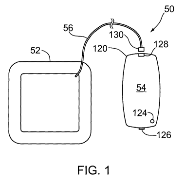
Figure 1 shows a phototherapy device comprising a
phototherapy bandage and a controller connected to the phototherapy
bandage;
Figure 2 is a top plan view of an emitter and sensor assembly
forming part of the phototherapy bandage of Figure 1;
CA 02716508 2010-08-23
WO 2009/103165 PCT/CA2009/000207
-7-
Figure 3 is a side view of the emitter and sensor assembly of
Figure 2;
Figure 4 is an enlarged side view of a portion of the emitter and
sensor assembly of Figure 2;
Figure 5 is a schematic block diagram of the emitter and sensor
assembly of Figure 2;
Figure 6 is a cross-sectional view of the phototherapy bandage
of Figure 1 being applied to a wound to be treated;
Figure 7 is a schematic block diagram of the controller of Figure
1;
Figure 8 is a schematic diagram of a phototherapy system
employing one or more phototherapy devices;
Figure 9 is a data record displayed by the phototherapy system
of Figure 9;
Figure 10 is a top plan view of an alternative emitter and sensor
assembly;
Figure 11 is a perspective view taken from above and from the
side of an alternative phototherapy bandage;
Figure 12 is a perspective view taken from below and from the
side of the phototherapy bandage of Figure 11 being applied to a wound to be
treated;
Figure 13 is a cross-sectional view of the phototherapy bandage
of Figure 12;
Figure 14 is a perspective view taken from below and from the
side of yet another phototherapy bandage;
Figure 15a is a cross-sectional view of a pressure sensor; and
Figure 15b is a cross-sectional view of an alternative pressure
sensor.
Detailed Description of the Embodiments
Turning now to Figure 1, a phototherapy device is shown and is
generally identified by reference numeral 50. As can be seen, phototherapy
CA 02716508 2010-08-23
WO 2009/103165 PCT/CA2009/000207
-8-
device 50 comprises a phototherapy bandage 52 to be applied to a patient
and cover a wound or other pathology to be treated and a controller 54
releasably connected to the phototherapy bandage 52 by a multi-conductor
cable 56. In this embodiment, the phototherapy bandage 52 is designed to
illuminate the periphery of the wound covered by the phototherapy bandage
thereby to promote the healing process without disturbing the dressing
overlying the wound bed. The controller 54 provides the operating power for
the phototherapy bandage 52 and controls the operation of the phototherapy
bandage so that the phototherapy bandage 52 subjects the wound to the
desired phototherapeutic treatment regime. The phototherapy bandage 52
and the controller 54 are portable and lightweight allowing the phototherapy
device 50 to be worn by a patient without affecting the patient's daily
routine.
Further specifics of the phototherapy device 50 will now be described.
Figures 2 to 6 better illustrate the phototherapy bandage 52. As
can be seen, the phototherapy bandage 52 comprises an emitter and sensor
assembly 70 in the shape of a ring that surrounds a simple or complex
dressing 72 sized to overlay the wound bed. The dimension and shape of the
ring is selected so that the emitter and sensor assembly 70 surrounds the
periphery of the wound and is spaced from the edges of the wound by a
distance in the range of from about 1 cm to about 3cm. The emitter and
sensor assembly 70 and the dressing 72 are accommodated in a breathable
pouch 76 thereby to promote airflow through the phototherapy bandage 52.
Pouch 76 comprises a perforated upper layer 78 and a lower adhesive layer
80 to affix the pouch 76 to the patient. The adhesive layer 80 has a cut-out
therein sized to expose the dressing 72 so that the dressing can be brought
into direct contact with the wound bed when the phototherapy bandage 52 is
applied to the patient. The upper and lower layers 78 and 80 are formed of
biologically safe material to inhibit the pouch 76 from adversely affecting
the
wound or surrounding tissue.
The emitter and sensor assembly 70 comprises a plurality of
segments electrically connected in series, with each segment having one of
two (2) shapes. In this embodiment, the emitter and sensor assembly 70
CA 02716508 2010-08-23
WO 2009/103165 PCT/CA2009/000207
-9-
comprises four (4) straight segments 100, three (3) curved segments 102 and
one (1) curved segment 103. Curved segment 103 differs from the curved
segments 102 in that one end of the cable 56 is permanently affixed thereto
thereby to connect electrically the emitter and sensor assembly 70 to the
controller 54.
The straight and curved segments 100, 102 and 103 are
arranged in an alternating pattern thereby to form a generally rectangular
ring.
Aside from shape, the segments are virtually identical. In this embodiment,
each segment 100, 102 and 103 comprises a short, rigid printed circuit board
104. A row of spaced radiation emitting sources 106, in this case four (4)
radiation emitting sources, is surface mounted on each printed circuit board
104 at locations so that when the phototherapy bandage 52 is applied to the
patient, the radiation emitting sources 106 are aimed at and positioned
proximate to the patient's skin tissue. The radiation emitting sources 106 in
this embodiment are red, solid-state, light emitting diodes (LEDs) that emit
visible light having a wavelength in the range of from about 630nm to about
690nm as wound healing is expected to occur primarily in the epidermis and
shallow musculoskeletal regions.
Each segment also comprises a plurality of sensors. In
particular, in this embodiment, a temperature sensor 108a, a photoreceptor
108b having appropriate spectral filtering and a contact sensor 108c are also
surface mounted on the printed circuit board 104. The temperature sensors
108a measure the temperature of the skin tissue at a location proximate the
periphery of the wound. Temperature changes provide an indication as to
whether the wound is receiving sufficient blood flow and microcirculation or
if
blood flow is affected by an infection. The photoreceptors 108b measure light
emitted by the LEDs 106 that has entered the skin tissue surrounding the
wound and has backscattered into the wound bed as a result of cellular
membranes. The amount of backscattered light received by the
photoreceptors 108b provides information concerning the healing stage of the
wound. Pairs of contact sensors 108c are used to measure electrical
impedance across the wound. Measuring electrical impedance provides an
CA 02716508 2010-08-23
WO 2009/103165 PCT/CA2009/000207
-10-
indication of the moisture content in the vicinity of the wound bed allowing
situations where the wound fluid has saturated the dressing 72 and leaked
outside the periphery of the wound bed to be detected so that appropriate
steps can be taken to change the dressing 72.
Flexible, insulated multi-conductor cables 110 interconnect
adjacent segments electrically and mechanically. Use of the flexible cables
110 permits the segments 100, 102 and 103 to take on various angles and to
move relative to one another. In this manner, when the phototherapy
bandage 52 is applied to a patient, each segment can take on an orientation
independent of the other segments. This allows the LEDs 106 to remain
generally coplanar with the tissue surrounding the wound even when the
underlying tissue is flexed by muscular, tendon or fat movement. A
biologically safe, translucent material 112 encapsulates the segments 100,
102 and 103 and the cables 110 to provide the emitter and sensor assembly
70 with a smooth patient contact surface that does not adversely affect the
wound or surrounding tissue.
The controller 54 comprises an outer housing 120 that is
accommodated by a disposable outer sleeve 122 formed of biologically safe
material. The outer sleeve 122 has an adhesive coating covered by a release
layer (not shown) that can be removed to expose the adhesive coating
thereby to allow the controller 54 to be affixed to the patient adjacent the
phototherapy bandage 52. A light emitting diode (LED) 124 and a switch 126
are provided on the housing 120. The LED 124 provides a user with visual
operational feedback. A connector 128 on the housing 120 receives a low
profile connector 130 at the opposite end of the cable 56. The interior of the
housing 120 accommodates a printed circuit board 132 on which the
controller electronics are mounted.
Figure 7 best illustrates the controller electronics. As can be
seen, the controller electronics comprise a microprocessor 140, a wireless
communications transceiver 142 to enable bi-directional communications with
remote devices, a driver 144 that is responsive to the microprocessor 140 to
control operation of the LEDs 106, temperature sensors 108a, photoreceptors
CA 02716508 2010-08-23
WO 2009/103165 PCT/CA2009/000207
-11-
108b and contact sensors 108c, and random access memory (RAM) (not
shown). A power source 146 provides operating power to the microprocessor
140, wireless communications transceiver 142 and driver 144. The power
source 146 comprises one or more chargeable or rechargeable batteries.
The number and type of batteries are selected to enable the controller 54 to
operate the phototherapy bandage 52 for extended periods of time thereby to
ensure that the phototherapy bandage 52 functions over the intended
phototherapeutic treatment regime. If desired, the power source 146 may
comprise other components to supplement the batteries such as for example,
ultra capacitors. In this manner, very high instantaneous output currents may
be realized allowing the controller 54 to operate the LEDs 106 at higher peak
output levels as well as to drive larger rings of segments. Alternatively, the
power source 146 may comprise a transformer and regulator to convert power
from a conventional ac mains supply to the appropriate operating power for
the microprocessor 140, wireless communications transceiver 142 and driver
144.
The RAM stores one or more phototherapy treatment protocol
programs that can be executed by the microprocessor 140 to control the
operation of the phototherapy bandage 52. The phototherapy treatment
protocol program that is being executed by the microprocessor 140
determines the nature, timing and duration of the phototherapeutic treatment
regime to which the wound is subjected. In particular, the phototherapy
treatment protocol program that is being executed determines the intervals at
which power is supplied to the segments by the driver 144 to illuminate the
LEDs 106, the duration the LEDs 106 are powered, the pattern by which the
LEDs 106 are powered and the intensity level at which the LEDs 106 are
operated. The phototherapy treatment protocol program also determines the
intervals at which the outputs of the temperature sensors 108a,
photoreceptors 108b and contact sensors 108c are read by the
microprocessor 140 and stored in the RAM.
The wireless communications transceiver 142 allows the
controller 54 to communicate with remote devices such as for example
CA 02716508 2010-08-23
WO 2009/103165 PCT/CA2009/000207
-12-
personal digital assistants (PDAs), cellular telephones, laptop computers,
tablet PCs or other computers and other processing devices via a wireless
communications link (radio frequency (RF), infrared etc.) using a suitable
wireless protocol such as for example, Zigbee, Bluetooth, WiFi, MICS, ANT
etc. In this manner, the phototherapy treatment protocol programs stored in
the RAM can be updated allowing the phototherapy bandage 52 to operate
according to different phototherapeutic treatment regimes. The read
temperature, light and impedance data stored in the RAM can also be
communicated to a remote computing device allowing the temperature, light
and impedance data to be analyzed and displayed. For example, Figure 8
shows the phototherapy device 50 communicating with a remote computing
station 200 over an Internet connection 202 via a wireless modem 204. The
remote computing station 200 executes a program to analyze the
temperature, light and impedance data received from the controller 54 and
present the results of the analysis graphically. Figure 9 is a data record 210
displayed by remote computing station 200. In this example, the data record
210 comprises a graph of the temperature readings recorded by the
phototherapy device 50 and the average recorded temperature. The data
record also comprises a graph of reflectance readings recorded by the
phototherapy device 50 and the average recorded reflectance. Of course,
other data records presenting different data can be displayed.
As will be appreciated by those of skill in the art, although only
one phototherapy device 50 is shown communicating the remote computing
station 200, in typical situations, the remote computing station 200 collects
data from a significant number of phototherapy devices 50. In this manner,
over time, recorded data from different phototherapy devices and patients can
be used to establish acceptable wound healing profiles. With acceptable
wound healing profiles known, a wound covered by a phototherapy bandage
52 can be assessed simply by examining the recorded temperature, light and
impedance data retrieved from the phototherapy bandage 52. This allows the
wound to be assessed remotely without requiring the phototherapy bandage
52 to be removed from the patient reducing the burden on medical personnel.
CA 02716508 2010-08-23
WO 2009/103165 PCT/CA2009/000207
-13-
Recorded temperature, light and impedance data that deviate from the
acceptable wound healing profiles can be detected and used to generate an
alarm or other indicator.
The phototherapy device 50 is intended to be used in a manner
following standard wound assessment and treatment methods currently
followed by medical personnel. When a patient suffers a wound, assuming
the wound has been cleansed, debrided and/or otherwise treated, a
phototherapy bandage 52 having segments that form a ring large enough to
surround the wound is selected. The selected phototherapy bandage 52 is
then applied to the patient so that the dressing 72 overlies the wound bed
allowing the dressing 72 to absorb exudate fluid. The adhesive layer 80
maintains the phototherapy bandage 52 in position. Of course, additional
adhesive tape may be used to supplement attachment of the phototherapy
bandage 52 to the patient. Once the phototherapy bandage 52 has been
properly affixed to the patient, the connector 130 on the cable 56 is brought
into engagement with the connector 128 on the controller housing 120. The
controller 54 is then turned on by operating the switch 126 and the controller
is placed in the disposable sleeve 122 and affixed to the patient at a
location
proximate the phototherapy bandage 52.
Once turned on, the microprocessor 140 executes the selected
phototherapy treatment protocol program. When the phototherapy treatment
protocol program signifies the start of an LED illumination interval, the
microprocessor 140 signals the driver 144. The driver 144 in response
provides operating power to the emitter and sensor assembly 70 causing the
LEDs 106 of the segments 100, 102 and 103 to illuminate at the desired
intensity level. As the LEDs 106 are oriented towards the skin tissue, the
periphery of the wound is subjected to light having a wavelength designed to
promote wound healing. Thus, the periphery of the wound is subjected to
timed doses of light selected to affect growth factors, microcirculation and
angiogenesis positively as well as to promote the natural healing process.
With the wound subjected to emitted light, the temperature sensors 108a
measure the temperature adjacent the wound. The photoreceptors 108b
CA 02716508 2010-08-23
WO 2009/103165 PCT/CA2009/000207
-14-
measure light backscattered through the wound bed. Pairs of contact sensors
108c at diametric locations along the ring of segments measure the
impedance across the wound bed. The output of the temperature sensors
108a, the output of the photoreceptors 108b and the output of the pairs of
contact sensors 108c are read by the microprocessor 140 at intervals during
execution of the phototherapy treatment protocol program and stored in the
RAM. At the end of the interval, the driver 144 isolates the emitter and
sensor
assembly 70 from the operating power so that the LEDs 106 turn off. During
gaps between LED illumination intervals, the controller electronics are
conditioned to a sleep mode to conserve power. The above process is
performed for each LED illumination interval. The read temperature, light and
impedance data stored in the RAM is transmitted to the remote computing
station 200 at intervals under the control of the microprocessor 140. Of
course, if desired the microprocessor 140 can be programmed so that it only
transmits the read temperature, light and impedance data in response to
requests received from the remote computing station 200.
Although the controller 54 is described as illuminating all of the
LEDs 106 continuously during the LED illumination intervals, if desired, the
LEDs 106 can be turned on and off during the LED illumination intervals
according to a duty cycle. Also, the LEDs 106 of different segments can be
illuminated at different times to reduce peak level power drawn from the
power source 146.
The phototherapy bandage 52 in this embodiment is intended
for single patient use and is disposed of at the conclusion of
phototherapeutic
treatment regime. The controller 54 is however reused.
If desired, the emitter and sensor assembly 70 may comprise
LEDs 106 that operate at different wavelengths. In this case, the
photoreceptors 108b measure the amount of backscattered light at each
frequency allowing changes in wound color to be detected. Knowing the color
of the wound allows the stage (i.e. blood filled (very red), pre-scab (white)
and
hard scab (brown)) of wound healing to be identified.
CA 02716508 2010-08-23
WO 2009/103165 PCT/CA2009/000207
-15-
Although the emitter and sensor assembly 70 is described and
shown as comprising eight (8) segments shaped and arranged to form a
generally rectangular ring, those of skill in the art will appreciate that
other
segment configurations are possible. The number of segments employed is
generally a function of the size of the wound over which the phototherapy
bandage 52 is placed. For smaller wounds, the emitter and sensor assembly
70 may comprise fewer segments. For example, as can be seen in Figure 10,
an emitter and sensor assembly 70 comprising only four (4) curved segments
102 and 103 is shown. For larger wounds, the emitter and sensor assembly
70 may comprise more segments. For most wound situations, it is anticipated
that phototherapy bandages 52 having emitter and sensor assemblies 70
comprising either four (4), six (6) or eight (8) segments will be suitable as
the
segment rings of such phototherapy bandages encompass areas equal to
approximately 4cm2, 8cm2 or 18cm2 respectively. Of course, depending on
the shape of the wound, the number of straight segments and curved
segments that are used may be varied. Also, the segments forming the
emitter and sensor assembly 70 need not be arranged to form an enclosed
ring. For example, the segments can be arranged in a C-shaped
configuration, in a linear strand or other suitable configuration. In such
cases,
as will be appreciated, the segments will extend along only a portion of the
wound periphery.
Although the use of segments interconnected by flexible cables
allows the LEDs 106 to remain generally coplanar with the skin tissue
surrounding the wound even though the LEDs 106 are mounted on rigid
printed circuit boards, alternative phototherapy bandage structures can be
employed. For example, turning now to Figures 11 to 13, another
embodiment of a phototherapy bandage is shown and is generally identified
by reference number 300. In this embodiment, the phototherapy bandage
300 is of a multilayer construction and comprises an upper perforated
breathable layer 302 disposed on one side of an absorbent layer 304 formed
of gauze or other suitable material. The breathable layer 302 has a centrally
located, circular raised portion 306 formed thereon. A cable 308 having a
CA 02716508 2010-08-23
WO 2009/103165 PCT/CA2009/000207
-16-
connector 310 at one end extends through the breathable layer 302. The
connector 310 mates with the connector 128 on the controller housing 120.
A flexible printed circuit board 320 is disposed on the other side
of the absorbent layer 304 and has a circular cut-out 322 therein that is
generally aligned with the raised portion 306. The printed circuit board 320
is
of a polymide and copper multilayer construction. Red LEDs 324 are surface
mounted on the printed circuit board 320 about the periphery of the cut-out
322. A temperature sensor 326, a photoreceptor 328 and contact sensors
329 are also surface mounted on the printed circuit board 320 adjacent the
cut-out 322. The cable 308 is permanently affixed to the printed circuit board
at its other end allowing the controller 54 to control the operation of the
phototherapy bandage 300. An adhesive layer 330 is provided beneath the
printed circuit board 320. The adhesive layer 330 is formed of biologically
safe material and is designed to contact the patient directly thereby to affix
the
phototherapy bandage 300 to the patient. A circular cut-out 332 that is
generally aligned with the raised portion 306 is also provided in the adhesive
layer 330. As will be appreciated, the cut-outs 322 and 332 are dimensioned
so that the wound bed is not contacted by the adhesive layer 330 or the
printed circuit board 320. In this manner, when the phototherapy bandage
300 is applied to a patient to cover a wound, the wound bed is only covered
by the breathable and absorbent layers 302 and 304. If desired separate
dressing material may be provided in the cut-out region to overlie the wound
bed and isolate the absorbent layer 304 from direct contact with the wound
bed.
The phototherapy bandage 300 is responsive to the controller
54 and operates in a manner similar to the phototherapy bandage 52. During
execution of a phototherapy treatment protocol program by the
microprocessor 140, at the start of an LED illumination interval, the
microprocessor 140 conditions the driver 144 to provide an operating voltage
to the LEDs 324 so that the LEDs 324 are illuminated at the desired intensity
levels. The microprocessor 140 also reads the outputs of the temperature
CA 02716508 2010-08-23
WO 2009/103165 PCT/CA2009/000207
-17-
sensor 326, photoreceptor 326 and contact sensors 329 and stores the read
temperature, light and impedance data in the RAM.
Figure 14 shows one side of yet another phototherapy bandage
400. The phototherapy bandage 400 is very similar to phototherapy bandage
300. In this embodiment, the cut-outs formed in the adhesive layer and
printed circuit boards are ovoid rather than circular making the phototherapy
bandage 400 better suited for covering elongate wounds. Although Figures
13 and 14 show circular and ovoid cut outs, those of skill in the art will
appreciate that cutouts having other geometric shapes (oval, crescent, square
etc.) can be provided in the adhesive layer and printed circuit board.
Although the controller 54 is shown as comprising a wireless
communications transceiver 142, if desired the controller may alternatively
comprise a wireless communication receiver such as for example, an infrared
receiver. In this case, the controller 54 is able to receive phototherapy
treatment protocol programs from a remote device such as for example a
personal digital assistant (PDA) or cellular telephone having an IrDA
compatible infrared communications interface but is unable to transmit
temperature, light and impedance data recorded by the temperature sensors,
photoreceptors and contact sensors.
Although the phototherapy bandages are described and shown
as comprising radiation emitting sources in the form of red LEDs 106, 324,
those of skill in the art will appreciate that alternative radiation emitting
sources may be employed. For example, radiation emitting sources that emit
light at other visible wavelengths or at non-visible wavelengths, such as for
example ultraviolet and near infrared wavelengths may be employed. The
type of radiation emitting sources that are employed is selected for their
therapeutic and/or energy properties. Longer wavelengths in the near infrared
can have significant depth of penetration.
Ultraviolet radiation sources may be employed in order to
stimulate a light emission response in nanocrystals. Nanocrystals (also called
quantum dots) give off very narrow band light which is related to the physical
size of the crystal. Wavelengths from violet to the near-infrared are possible
CA 02716508 2010-08-23
WO 2009/103165 PCT/CA2009/000207
-18-
by selecting the appropriate crystal size and positioning them near the
ultraviolet radiation sources. Combining different sized crystals in a matrix
can also provide unique spectral bandwidths of multiple wavelengths all
emitting simultaneously. Alternately, the radiation emitting sources may
comprise a matrix of nanocrystals which are aligned across a larger surface
and sandwiched between two conducting media such that the flow of
electrical current causes electroluminescence of the matrix.
In the embodiments described above, the phototherapy
bandage comprises temperature sensors, photoreceptors and contact
sensors. As will be appreciated by those of skill in the art, the phototherapy
bandage need not include each of these sensors. Rather the phototherapy
bandage may comprise a subset of the sensors or other sensors in addition to
the temperature sensors, photoreceptors and contact sensors. Alternatively,
the phototherapy bandage may comprise different sensors to sense other
parameters indicative of wound healing.
For example, turning now to Figure 15a, a pressure sensor
suitable for use with the phototherapy bandages 300 and 400 described
above is shown and is generally identified by reference numeral 500. As can
be seen, the pressure sensor 500 is partially embedded in foam dressing
material 502 positioned in the cut-outs 322 and 332 and overlying the wound
and comprises a sense electrode 504 surface mounted on one side of a
portion of the printed circuit board 320 that has been extended into the cut-
out
region. The sense electrode 504 is separated from a reference electrode 506
by a portion of the dressing material 502. The dressing material 502
interposed between the sense and reference electrodes 504 and 506
respectively acts as an elastic dielectric. As a result, the sense and
reference
electrodes 504 and 506 respectively, form the plates of a parallel-plate
capacitor. The reference electrode 506 is folded around the sense electrode
504 to shield the sense electrode from external noise and is surface mounted
on the opposite side of the extended portion of the printed circuit board 320.
In this embodiment, the reference electrode 506 is formed of flexible
conductive tape, ribbon, foil etc. that can be easily folded. A membrane 508
CA 02716508 2010-08-23
WO 2009/103165 PCT/CA2009/000207
-19-
isolates the portion of the dressing material in contact with the wound from
the
portion of the dressing material separating the sense and reference
electrodes. The dressing material 502 separating the sense and reference
electrodes has a thickness in the range of from about 1/8" to about 1/".
As will be appreciated, when the dressing material 502 is
subjected to pressure and compresses, the spacing between the sense
electrode 504 and the reference electrode 506 changes resulting in a change
in capacitance of the capacitor occurring. This change in capacitance is read
by the controller 54 allowing the pressure applied to the dressing material
502
and hence, to the wound area to be determined.
Depending on the size of the wound and hence the size of the
dressing material 502 applied on the wound bed, the number of pressure
sensors 500 incorporated into the dressing material may vary.
Figure 15b shows an alternative pressure sensor 520. In this
embodiment, one end of the sense electrode 524 is trapped between two
layers of foam dressing material 522. The other end of the sense electrode
524 undergoes a curve and is surface mounted on the top surface of the
extended portion of the printed circuit board 320. The reference electrode
526 is also surface mounted on the top surface of the extended portion of the
printed circuit board 320 and has a first arm 526a overlying the top layer of
the foam dressing material 522 and a second arm 526b extending beneath
the lower layer of the foam dressing material 522 to yield a layered capacitor
configuration. Similar to the previous embodiment, the reference electrode
526 shields the sense electrode 524 from external noise. As will be
appreciated, the layered capacitor configuration of pressure sensor 520 has
improved sensitivity as compared to that of pressure sensor 500 but requires
greater printed circuit board area.
Although the pressure sensors 500 and 502 have been
described for use with the phototherapy bandages 300 and 400, those of skill
in the art with appreciate that the pressure sensors may be used with the
phototherapy bandage 52. In this case, access for the sense and reference
electrodes to the printed circuit boards of the segments needs to be provided
CA 02716508 2010-08-23
WO 2009/103165 PCT/CA2009/000207
-20-
through the encapsulating material 112. Of course, the pressure sensors may
be used in other bandage configurations where it is desired to measure and/or
monitor the pressure being applied to a wound region.
Although embodiments have been described with reference to
the drawings, those of skill in the art will appreciate that variations and
modifications may be made without departing from the spirit and scope
thereof as defined by the appended claims.