Note: Descriptions are shown in the official language in which they were submitted.
CA 02716713 2010-10-07
WOUND CLOSURE DEVICE
TECHNICAL FIELD
[0002] The present disclosure relates to an implant for providing closure to
wounds and, in
particular, to a wound closure device for repairing and sealing perforations
in tissue, such as
laparoscopic port sites.
DESCRIPTION OF THE RELATED ART
[0003] A variety of surgical procedures, for example, laparoscopic procedures,
are performed
through an access port, during which the access device punctures the tissue to
provide access to
the surgical site.
[0004] A hernia is a protrusion of a tissue, structure, or part of an organ
through injured
muscle tissue or an injured membrane by which the tissue, structure, or organ
is normally
contained. Trocar site herniation is a potential complication of minimally
invasive surgery.
Upon removal of a minimally invasive surgical device or the access port,
tissues may not
properly heal and can present concerns including reherniation. More
specifically, omental and
intestinal herniation has been reported with larger trocar sites (10mm).
CA 02716713 2010-10-07
1
[0005] Currently, wound closure devices, such as sutures, are used to close
various layers of
tissue post-surgery. Suturing a patient after removal of an access device may
be cumbersome,
while accumulating additional costs to the patient such as increased time
spent in the operating
room.
[0006] While conventional methods such as suturing exist, improvements in the
field are
desired.
SUMMARY
[0007] The present disclosure provides wound closure devices, methods for
making same,
and methods for using same. In embodiments, a wound closure device of the
present disclosure
may include an elongate body having a proximal end and a distal end, and a
plug member having
a tissue facing surface coupled to the distal end of the elongate body, the
plug member including
a hydrogel, wherein the elongate body, the plug member, or both, include at
least one reactive
group. The elongate body, the plug member, or both, may be a hydrogel.
[0008] In embodiments, the plug member, the elongate body, or both, may
include at least
one reactive group that bonds to tissue. In embodiments, the elongate body and
the plug member
may be connected by a hinge.
[0009] In embodiments, a wound closure device of the present disclosure may
include an
elongate body having a proximal end and a distal end, and a plug member having
a tissue facing
surface coupled to the distal end of the elongate body, wherein at least the
elongate body
includes a tissue scaffold.
2
CA 02716713 2010-10-07
BRIEF DESCRIPTION OF THE DRAWINGS
[0010] Various embodiments of the wound closure devices are described herein
with
reference to the drawings, in which:
[0011] FIG. I is a perspective cross-sectional view of a wound closure device
in accordance
with one embodiment of the present disclosure;
[00121 FIG. 2 is a cross-sectional view a wound closure device in accordance
with another
embodiment of the present disclosure;
[0013] FIG. 3A is a perspective view of a wound closure device having a
dehydrated
component in accordance with an alternate embodiment of the present
disclosure;
[0014] FIG. 3B is a perspective view of the wound closure device of FIG. 3A
after
rehydration;
[0015] FIG. 4 is a perspective view of a wound closure device in accordance
with another
embodiment of the present disclosure;
[0016] FIG. 5 is a perspective view of a wound closure device in accordance
with yet another
embodiment of the present disclosure;
[0017] FIG. 6 is a side view of a wound closure device in accordance with
another
embodiment of the present disclosure;
[0018] FIG.7 is a side view of a wound closure device in accordance with yet
another
embodiment of the present disclosure;
[0019] FIG. 8 is a side view of a wound closure device in accordance with one
embodiment
of the present disclosure;
[0020] FIG.9 is a cross-sectional view of an alternate embodiment of a wound
closure device
in accordance with the present disclosure;
3
CA 02716713 2010-10-07
[00211 FIG. 10 is a perspective view of a wound closure device in accordance
with one
embodiment of the present disclosure;
[0022] FIG. 11 is a perspective view of a wound closure device in accordance
with another
embodiment of the present disclosure;
[0023] FIG. 12 is a perspective view of a wound closure device in accordance
with yet
another embodiment of the present disclosure;
[0024] FIG. 13A is a side view of a wound closure device in a first, folded
position, in
accordance with an embodiment of the present disclosure;
[0025] FIG. 13B is a side perspective view of the wound closure device of
FIG.13A;
[0026] FIG. 13C is a side view of the wound closure device of FIG. 13A in a
second,
expanded position;
[0027] FIG. 13D is a top view of the wound closure device of FIG. 13C;
[0028] FIG. 14A is a perspective view of a wound closure device in a deployed
position in
accordance with one embodiment of the present disclosure;
[0029] FIG. 14B is a side view of the wound closure device of FIG. 14A in a
folded position;
[0030] FIG. 14C is a side view of the wound closure device of FIG. 14A
illustrated in a
deployed position and the folded position of FIG. 14B is shown in phantom;
[0031] FIG. 15A is a perspective view of a wound closure device in a deployed
position in
accordance with another embodiment of the present disclosure; and
[0032] FIG. 15B is a side view of the wound closure device of FIG. 15A
illustrated in a first,
folded position with the second, deployed position shown in phantom.
4
CA 02716713 2010-10-07
DETAILED DESCRIPTION
[0033] The present wound closure devices facilitate wound closure and may be
used to
deliver biologics and/or therapeutics to improve healing and reduce scarring,
pain, and infection,
as well as to provide mechanical stability at the wound site and prevent port
site herniation. The
wound closure device includes an elongate body for insertion into the
perforated tissue of a
wound to fill and hold the tissue together, and a plug member attached to a
distal end portion of
the elongate body, having a substantially flat tissue facing surface for
positioning against the
internal surface of the tissue to plug or close the wound. In embodiments, the
wound closure
device is inserted through an insertion device, such as a trocar which, when
removed, leaves the
wound closure device behind to close the wound.
[0034] The components of the wound closure device, i.e., the elongate body
and/or plug
member, may be fabricated from any biodegradable material that can be used in
surgical
procedures. The term "biodegradable" as used herein is defined to include both
bioabsorbable
and bioresorbable materials. By biodegradable, it is meant that the materials
decompose, or lose
structural integrity under body conditions (e.g., enzymatic degradation or
hydrolysis) or are .
broken down (physically or chemically) under physiologic conditions in the
body such that the
degradation products are excretable or absorbable by the body. It should be
understood that such
materials include natural, synthetic, bioabsorbable, and/or non-absorbable
materials, as well as
combinations thereof, for forming the components of the wound closure device
of the present
disclosure.
[0035] Representative natural biodegradable polymers include: polysaccharides,
such as
alginate, dextran, chitin, hyaluronic acid, cellulose, collagen, gelatin,
fucans,
glycosaminoglycans, and chemical derivatives thereof (substitutions and/or
additions of chemical
CA 02716713 2010-10-07
groups, for example, alkyl, alkylene, hydroxylations, oxidations, and other
modifications
routinely made by those skilled in the art); proteins, such as albumin,
casein, zein, and silk; and
copolymers and blends thereof, alone or in combination with synthetic
biodegradable polymers.
[0036] Synthetically modified natural polymers include cellulose derivatives,
such as alkyl
celluloses, hydroxyalkyl celluloses, cellulose ethers, cellulose esters,
nitrocelluloses, and
chitosan. Examples of suitable cellulose derivatives include methyl cellulose,
ethyl cellulose,
hydroxypropyl cellulose, hydroxypropyl methyl cellulose, hydroxybutyl methyl
cellulose,
cellulose acetate, cellulose propionate, cellulose acetate butyrate, cellulose
acetate phthalate,
carboxymethyl cellulose, cellulose triacetate, and cellulose sulfate sodium
salt. These may be
collectively referred to herein, in embodiments, as "celluloses."
[0037] Representative synthetic biodegradable polymers include polyhydroxy
acids prepared
from lactone monomers, such as glycolide, lactide, caprolactone (including c-
caprolactone),
valerolactone (including 8-valerolactone), as well as carbonates (e.g.,
trimethylene carbonate,
tetramethylene carbonate, and the like), dioxanones (e.g., 1,4-dioxanone and p-
dioxanone),
1,dioxepanones (e.g., 1,4-dioxepan-2-one and 1,5-dioxepan-2-one), and
combinations thereof.
Polymers formed therefrom include: poly(lactic acid); poly(glycolic acid);
poly(trimethylene
carbonate); poly(dioxanone); poly(hydroxybutyric acid); poly(hydroxyvaleric
acid); poly(lactide-
co-(a-caprolactone-)); poly(glycolide-co-(c-caprolactone)); polycarbonates;
poly(pseudo amino
acids); poly(amino acids); polyhydroxyalkanoates; polyalkylene oxalates;
polyoxaesters;
polyanhydrides; polyortho esters; and copolymers, block copolymers,
homopolymers, blends,
and combinations thereof.
[0038] Other non-limiting examples of biodegradable materials from which the
wound
closure device may be made include: poly(phosphazine), aliphatic polyesters,
polyethylene
6
CA 02716713 2010-10-07
glycols, glycerols, copoly (ether-esters), polyalkylene oxalates, p mides,
poly
(iminocarbonates), polyalkylene oxalates, polyoxaesters, polyphosphazenes, and
copolymers,
block copolymers, homopolymers, blends, and combinations thereof.
[00391 Rapidly bioerodible polymers, such as poly(lactide-co-glycolide)s,
polyanhydrides,
and polyorthoesters, which have carboxylic groups exposed on the external
surface as the surface
of the polymer erodes, may also be used.
[0040] In embodiments, the elongate body, the plug member, or both, or a
coating on the
elongate body, the plug member, or both, may be formed from a hydrogel. The
hydrogel may be
formed of any components within the purview of those skilled in the art. In
some embodiments,
as discussed further below, the hydrogel may be formed of a natural component,
such as
collagen,. gelatin, serum, hyaluronic acid, combinations thereof, and the
like. The natural
component may degrade or otherwise be released at the site of implantation as
any hydrogel
utilized as part of the wound closure device degrades. The term "natural
component" as used
herein includes polymers, compositions of matter, materials, combinations
thereof, and the like,
which can be found in nature or derived from compositions/organisms found in
nature. Natural
components also may include compositions which are found in nature but can be
synthesized by
man, for example, using methods to create natural/synthetic/biologic
recombinant materials, as
well as methods capable of producing proteins with the same sequences as those
found in nature,
and/or methods capable of producing materials with the same structure and
components as
natural materials, such as synthetic hyaluronic acid, which is commercially
available, for
example, from Sigma Aldrich.
[0041] The hydrogels may be formed from a single precursor or multiple
precursors. This
may occur prior to implantation or at the time of implantation. In either
case, the formation of
7
CA 02716713 2010-10-07
the hydrogel may be accomplished by having a precursor that can be activated
at the time of
application to create, in embodiments, a hydrogel. Activation can be through a
variety of
methods including, but not limited to, environmental changes such as pH,
ionicity, pressure,
temperature, etc. In other embodiments, the components for forming a hydrogel
may be
contacted outside the body and then introduced into a patient as an implant,
such as a pre-formed
wound closure device or component thereof.
[0042] Where the hydrogel is formed from multiple precursors, for example two
precursors,
the precursors may be referred to as a first and second hydrogel precursor.
The terms "first
hydrogel precursor" and "second hydrogel precursor" each mean a polymer,
functional polymer,
macromolecule, small molecule, or crosslinker that can take part in a reaction
to form a network
of crosslinked molecules, e.g., a hydrogel.
[0043] In embodiments, the precursor utilized to form the hydrogel may be,
e.g., a monomer
or a macromer. One type of precursor may have a functional group that is an
electrophile or
nucleophile. Electrophiles react with nucleophiles to form covalent bonds.
Covalent crosslinks
or bonds refer to chemical groups formed by reaction of functional groups on
different polymers
that serve to covalently bind the different polymers to each other. In certain
embodiments, a first
set of electrophilic functional groups on a first precursor may react with a
second set of
nucleophilic functional groups on a second precursor. When the precursors are
mixed in an
environment that permits a reaction (e.g., as relating to pH, temperature,
ionicity, and/or solvent),
the functional groups react with each other to form covalent bonds. The
precursors become
crosslinked when at least some of the precursors can react with more than one
other precursor.
For instance, a precursor with two functional groups of a first type may be
reacted with a
8
CA 02716713 2010-10-07
crosslinking precursor that has at least three functional groups of a second
type capable of
reacting with the first type of functional groups.
[0044] The term "functional group" as used herein refers to groups capable of
reacting with
each other to form a bond. In embodiments, such groups may be electrophilic or
nucleophilic.
Electrophilic functional groups include, for example, N-hydroxysuccinimides,
sulfosuccinimides, carbonyldiimidazole, sulfonyl chloride, aryl halides,
sulfosuccinimidyl esters,
N-hydroxysuccinimidyl esters, succinimidyl esters, epoxides, aldehydes,
maleimides,
imidoesters and the like. In embodiments, the electrophilic functional group
is a succinimidyl
ester.
[0045] The first and second hydrogel precursors may have biologically inert
and water
soluble cores. More specifically, the electrophilic hydrogel precursors may
have biologically
inert and water soluble cores, as well as non-water soluble cores. When the
core is a polymeric
region that is water soluble, suitable polymers that may be used include:
polyethers, for example,
polyalkylene oxides such as polyethylene glycol("PEG"), polyethylene oxide
("PEO"),
polyethylene oxide-co-polypropylene oxide ("PPO"), co-polyethylene oxide block
or random
copolymers, and polyvinyl alcohol ("PVA"); poly(vinyl pyrrolidinone) ("PVP");
poly(amino
acids); poly(saccharides), such as dextran, chitosan, alginates,
carboxymethylcellulose, oxidized
cellulose, hydroxyethylcellulose, hydroxymethylcellulose, and hyaluronic acid;
and proteins,
such as albumin, collagen, casein, and gelatin. Other suitable hydrogels may
include
components such as methacrylic acid, acrylamides, methyl methacrylate,
hydroxyethyl
methacrylate, combinations thereof, and the like. In embodiments, combinations
of the
foregoing polymers and components may be utilized.
9
CA 02716713 2010-10-07
[0046] The polyethers, and more particularly poly(oxyalkylenes) or
polyethylene glycol, may
be utilized in some embodiments. When the core is small in molecular nature,
any of a variety of
hydrophilic functionalities can be used to make the first and second hydrogel
precursors water
soluble. For example, functional groups like hydroxyl, amine, sulfonate and
carboxylate, which
are water soluble, may be used to make the precursor water soluble. For
example, the n-
hydroxysuccinimide ("NHS") ester of subaric acid is insoluble in water, but by
adding a
sulfonate group to the succinimide ring, the NHS ester of subaric acid may be
made water
soluble, without affecting its reactivity towards amine groups. In
embodiments, the precursor
having electrophilic functional groups may be a PEG ester.
[0047] As noted above, each of the first and second hydrogel precursors may be
multifunctional, meaning that they may include two or more electrophilic or
nucleophilic
functional groups, such that, for example, a nucleophilic functional group on
the first hydrogel
precursor may react with an electrophilic functional group on the second
hydrogel precursor to
form a covalent bond. At least one of the first or second hydrogel precursors
includes more than
two functional groups, so that, as a result of electrophilic-nucleophilic
reactions, the precursors
combine to form cross-linked polymeric products, in embodiments, hydrogels.
[0048] A macromolecule having the electrophilic functional group may be multi-
armed. For
example, the macromolecule may be a multi-armed PEG having four, six, eight,
or more arms
extending from a core. The core may be the same or different from the
macromolecule forming
the arms. For example, the core may be PEG and the multiple arms may also be
PEG. In
embodiments, the core may be a natural polymer.
[0049] The molecular weight (MW) of the electrophilic crosslinker may be from
about 2,000
g/mol to about 100,000 g/mol; in embodiments from about 10,000 mol to about
40,000 mol.
CA 02716713 2010-10-07
Multi-arm precursors may have a molecular weight that varies depending on the
number of arms.
For example, an arm having a 1000 g/mol of PEG has enough CH2CH2O groups to
total at least
1000 mol. The combined molecular weight of an individual arm may be from about
250 g/mol
to about 5,000 g/mol; in embodiments from about 1,000 mol to about 3,000 mol;
in
embodiments from about 1,250 g/mol to about 2,500 g/mol. In embodiments, the
electrophilic
crosslinker may be a multi-arm PEG functionalized with multiple NHS groups
having, for
example, four, six or eight arms and a molecular weight from about 5,000 g/mol
to about 25,000
g/mol. Other examples of suitable precursors are described in U.S. Patent Nos.
6,152,943;
6,165,201; 6,179,862; 6,514,534; 6,566,406; 6,605,294; 6,673,093; 6,703,047;
6,818,018;
7,009,034; and 7,347,850, the entire disclosures of each of which are
incorporated herein by
reference.
[0050] The electrophilic precursor may be a cross-linker that provides an
electrophilic
functional group capable of bonding with nucleophiles on another component,
such as, in certain
embodiments, a natural component containing primary amines. The natural
component may be
endogenous (to the patient, i.e., collagen) to which the electrophilic
crosslinker is applied.
[0051] In embodiments, one of the precursors may be a nucleophilic precursor
possessing
nucleophilic groups. Nucleophilic groups which may be present include, for
example, -NH2,
-SH, -OH, -PH2, and -CO-NH-NH2. Any monomer, macromer, polymer, or core
described
above as suitable for use in forming the electrophilic precursor may be
functionalized with
nucelophilic groups to form a nucleophilic precursor. In other embodiments, a
natural
component possessing nucleophilic groups, such as those listed above, may be
utilized as the
nucleophilic precursor.
11
CA 02716713 2010-10-07
[0052] The natural component may be, for example, collagen, gelatin, blood
(including serum,
which may be whole serum or extracts therefrom), hyaluronic acid, proteins,
albumin, other
serum proteins, serum concentrates, platelet rich plasma (prp), combinations
thereof, and the
like. Additional suitable natural components which may be utilized or added to
another natural
component include, for example, stem cells, DNA, RNA, enzymes, growth factors,
peptides,
polypeptides, antibodies, other nitrogenous natural molecules, combinations
thereof, and the like.
Other natural components may include derivatives of the foregoing, for
example, modified
polysaccharides such as hyaluronic acid or dextran,which may be naturally
derived, synthetic, or
biologically derived. For example, in some embodiments, the natural component
may be
aminated hyaluronic acid.
[0053] In embodiments, any of the above natural components may be
synthetically prepared,
e.g., synthetic hyaluronic acid, which may be purchased from Sigma Aldrich,
for example.
Similarly, in embodiments the natural component could be a natural or
synthetic long chain
aminated polymer.
[0054] The natural component may provide cellular building blocks or cellular
nutrients to the
tissue that it contacts in situ. For example, serum contains proteins,
glucose, clotting factors,
mineral ions, and hormones which may be useful in the formation or
regeneration of tissue.
[0055] In embodiments, the natural component includes whole serum. In some
embodiments,
the natural component is autologous, i.e., collagen, serum, blood, and the
like.
[0056] In embodiments, a multifunctional nucleophilic polymer, such as a
natural component
having multiple amine groups, may be used as a first hydrogel precursor and a
multifunctional
electrophilic polymer, such as a multi-arm PEG functionalized with multiple
NHS groups, i.e., a
PEG ester, may be used as a second hydrogel precursor. In embodiments, the
precursors may be
12
CA 02716713 2010-10-07
in solution(s), which may be combined to permit formation of the hydrogel. Any
solutions
utilized as part of the in situ forming material system should not contain
harmful or toxic
solvents. In embodiments, the precursor(s) may be substantially soluble in a
solvent such as
water to allow application in a physiologically-compatible solution, such as
buffered isotonic
saline.
[0057] In some embodiments, a pre-formed hydrogel may be formed from a
combination of
collagen and gelatin as the natural component, with a multi-functional PEG
utilized as a
crosslinker. In embodiments, the collagen and gelatin may be placed in
solution, utilizing a
suitable solvent. To this solution, hyaluronic acid may be added along with a
high pH buffer.
Such a buffer may have a pH from about 8 to about 12, in embodiments from
about 8.2 to about
9. Examples of such buffers include, but are not limited to, borate buffers,
and the like.
[0058] In a second solution, an electrophilic crosslinker, in embodiments, a
multi-arm PEG
functionalized with electrophilic groups such as n-hydroxysuccinimide, may be
prepared in a
buffer such as Hanks Balanced Salt Solution, Dulbecco's Modified Eagle's
Medium, Phosphate
Buffered Saline, water, phosphate buffer, combinations thereof, and the like.
The electrophilic
crosslinker, in embodiments, a multi-arm PEG functionalized with n-
hydroxysuccinimide
groups, may be present in a solution including the above buffer at a
concentration from about
0.02 grams/mL to about 0.5 grams/mL, in embodiments, from about 0.05 grams/mL
to about 0.3
grams/mL.
[0059] The two components may be combined, wherein the electrophilic groups on
the multi-
arm PEG crosslink the amine nucleophilic components of the collagen and/or
gelatin. The ratio
of natural component to electrophilic component may be from about .01:1 to
about 100:1, in
embodiments, from about 1:1 to about 10:1.
13
CA 02716713 2010-10-07
[00601 The nucleophilic component, in certain embodiments, the natural
components, e.g.,
collagen, gelatin, and/or hyaluronic acid, may together be present at a
concentration of at least
about 1.5 percent by weight of the hydrogel, in embodiments, from about 1.5
percent by weight
to about 20 percent by weight of the hydrogel, in other embodiments, from
about 2 percent by
weight to about 10 percent by weight of the hydrogel. In certain embodiments,
collagen may be
present from about 0.5 percent to about 7 percent by weight of the hydrogel,
in further
embodiments, from about 1 percent to about 4 percent by weight of the
hydrogel. In another
embodiment, gelatin may be present from about 1 percent to about 20 percent by
weight of the
hydrogel, in further embodiments, from about 2 percent to about 10 percent by
weight of the
hydrogel. In yet another embodiment, hyaluronic acid and collagen combined as
the natural
component(s) may be present from about 0.5 percent to about 8 percent by
weight of the
hydrogel, in further embodiments, from about 1 percent to about 5 percent by
weight of the
hydrogel. It is also envisioned that the hyaluronic acid may not be present as
a "structural"
component, but as more of a bioactive agent. For example, hyaluronic acid may
be present in
solution/gel in concentrations as low as 0.001 percent by weight of the
solution/gel and have
biologic activity.
[00611 The electrophilic crosslinker may be present in amounts of from about
0.5 percent by
weight to about 20 percent by weight of the hydrogel, in embodiments, from
about 1.5 percent
by weight to about 15 percent by weight of the hydrogel.
[00621 The hydrogels may be formed either through covalent, ionic or
hydrophobic bonds.
Physical (non-covalent) crosslinks may result from complexation, hydrogen
bonding,
desolvation, Van der Waals interactions, ionic bonding, combinations thereof,
and the like, and
may be initiated by mixing two precursors that are physically separated until
combined in situ, or
14
CA 02716713 2010-10-07
as a consequence of a prevalent condition or change in the physiological
environment, including
temperature, pressure, pH, ionic strength, combinations thereof, and the like.
Thus, the hydrogel
may be sensitive to these environmental conditions/changes. Chemical
(covalent) crosslinking
may be accomplished by any of a number of mechanisms, including: free radical
polymerization,
condensation polymerization, anionic or cationic polymerization, step growth
polymerization,
electrophile-nucleophile reactions, combinations thereof, and the like.
[00631 In some embodiments, hydrogel systems may include biocompatible multi-
precursor
systems that spontaneously crosslink when the precursors are mixed, but
wherein the two or
more precursors are individually stable for the duration of the deposition
process. In other
embodiments, hydrogels may be formed from a single precursor that crosslinks
with endogenous
materials and/or tissues.
[00641 The crosslinking density of the resulting hydrogel may be controlled by
the overall
molecular weight of the crosslinker and natural component and the number of
functional groups
available per molecule. A lower molecular weight between crosslinks, such as
600 daltons (Da),
will give much higher crosslinking density as compared to a higher molecular
weight, such as
10,000 Da. Elastic gels may be obtained with higher molecular weight natural
components with
molecular weights of more than 3000 Da. It should be noted that I Dalton
equals 1 g/mol and
the terms may be used interchangeable when describing molecular weight
throughout the
disclosure.
[00651 The crosslinking density may also be controlled by the overall percent
solids of the
crosslinker and natural component solutions. Increasing the percent solids
increases the
probability that an electrophilic group will combine with a nucleophilic group
prior to
inactivation by hydrolysis. Yet another method to control crosslink density is
by adjusting the
CA 02716713 2010-10-07
stoichiometry of nucleophilic groups to electrophilic groups. A one to one
ratio may lead to the
highest crosslink density, however, other ratios of reactive functional groups
(e.g.,
electrophile:nucleophile) are envisioned to suit a desired formulation.
[0066] The hydrogel thus produced may be bioabsorbable. For example, hydrogels
of the
present disclosure may be absorbed from about one day to about 18 months or
longer.
Absorbable polymers materials include both natural and synthetic polymers, as
well as
combinations thereof
[0067] In embodiments, one or more precursors having biodegradable linkages
present in
between functional groups may be included to make the hydrogel biodegradable
or absorbable.
In some embodiments, these linkages may be, for example, esters, which may be
hydrolytically
degraded. The use of such linkages is in contrast to protein linkages that may
be degraded by
proteolytic action. A biodegradable linkage optionally also may form part of a
water soluble
core of one or more of the precursors. Alternatively, or in addition,
functional groups of
precursors may be chosen such that the product of the reaction between them
results in a
biodegradable linkage. For each approach, biodegradable linkages may be chosen
such that the
resulting biodegradable biocompatible crosslinked polymer degrades or is
absorbed in a desired
period of time. Generally, biodegradable linkages may be selected that degrade
the hydrogel
under physiological conditions into non-toxic or low toxicity products.
[0068] Biodegradable gels utilized in the present disclosure may degrade due
to hydrolysis or
enzymatic degradation of the biodegradable region, whether part of the natural
component or
introduced into a synthetic electrophilic crosslinker. The degradation of gels
containing
synthetic peptide sequences will depend on the specific enzyme and its
concentration. In some
cases, a specific enzyme may be added during the crosslinking reaction to
accelerate the
16
CA 02716713 2010-10-07
degradation process. In the absence of any degradable enzymes, the crosslinked
polymer may
degrade solely by hydrolysis of the biodegradable segment. In embodiments in
which
polyglycolate is used as the biodegradable segment, the crosslinked polymer
may degrade in
from about 1 day to about 30 days depending on the crosslinking density of the
network.
Similarly, in embodiments in which a polycaprolactone-based crosslinked
network is used,
degradation may occur over a period of time from about 1 month to about 8
months. The
degradation time generally varies according to the type of degradable segment
used, in the
following order: polyglycolate<polylactate<polytrimethylene
carbonate<polycaprolactone.
Thus, it is possible to construct a hydrogel with a desired degradation
profile, from a few days to
months, using a different degradable segments.
[0069] Where utilized, the hydrophobicity generated by biodegradable blocks
such as
oligohydroxy acid blocks or the hydrophobicity of PPO blocks in PLURONICTM or
TETRONICTM polymers utilized to form the electrophilic precursor may be
helpful in dissolving
small organic drug molecules. Other properties which will be affected by
incorporation of
biodegradable or hydrophobic blocks include: water absorption, mechanical
properties and
thermosensitivity.
[0070] In other embodiments, the precursors utilized to form the hydrogel may
be non-
degradable, i.e., they may include any of the macromers, polymers, or cores
described above as
suitable for use in forming the electrophilic precursor, but possess no ester
or other similar
degradable linkage. The non-biodegradable linkages may be created through the
reaction of an
N-hydroxysuccinimidyl carbonate. In one embodiment, the reaction of a multi-
arm polyol with a
N, N'-dihydroxysuccinimidyl carbonate creates an N-hydroxysuccinimidyl
carbonate. The N-
hydroxysuccinimidyl carbonate can then be further reacted with a high
molecular weight
17
CA 02716713 2010-10-07
polyamine, such as collagen, aminated hyaluronic acid, gelatin, or dextran, to
create the pre-
formed hydrogel. High molecular weight polyamines may provide longer implant
stability as
compared to lower molecular weight polyamines. High molecular weight
polyamines may
comprise molecular weights from about 15,000g/mol to about 250,000g/mol, in
certain
embodiments, from about 75,000g/mol to about 150,000 g/mol. It should be
understood that
when a non-biodegradable linkage is used, the implant is still biodegradable
through use of a
biodegradable first hydrogel precursor, such as collagen. For example, the
collagen may be
enzymatically degraded, breaking down the hydrogel, which is then
subsequentlly eroded.
[0071] Synthetic materials that are readily sterilized and avoid the dangers
of disease
transmission involved in the use of natural materials may also be used.
Indeed, certain
polymerizable hydrogels made using synthetic precursors are within the purview
of those skilled
in the art, e.g., as used in commercially available products such as FOCALSEAL
(Genzyme,
Inc.), COSEAL (Angiotech Pharmaceuticals), and DURASEAL (Confluent Surgical,
Inc).
Other known hydrogels include, for example, those disclosed in U.S. Patent
Nos. 6,656,200;
5,874,500; 5,543,441; 5,514,379; 5,410,016; 5,162,430; 5,324,775; 5,752,974;
and 5,550,187.
[0072] As noted above, in embodiments, a multi-arm PEG, sometimes referred to
herein as a
PEG star, may be included to form a hydrogel utilized in forming at least a
portion of a wound
closure device of the present disclosure. A PEG star may be functionalized so
that its arms
include biofunctional groups such as amino acids, peptides, antibodies,
enzymes, drugs, or other
moieties in its cores, its arms, or at the ends of its arms. The biofunctional
groups may also be
incorporated into the backbone of the PEG, or attached to a reactive group
contained within the
PEG backbone. The binding can be covalent or non-covalent, including
electrostatic, thiol
18
CA 02716713 2010-10-07
mediated, peptide mediated, or using known reactive chemistries, for example,
biotin with
avidin.
[0073] Amino acids incorporated into a PEG star may be natural or synthetic,
and can be used
singly or as part of a peptide. Sequences may be utilized for cellular
adhesion, cell
differentiation, combinations thereof, and the like, and may be useful for
binding other biological
molecules, such as growth factors, drugs, cytokines, DNA, antibodies, enzymes,
combinations
thereof, and the like. Such amino acids may be released upon enzymatic
degradation of the PEG
star.
[0074] These PEG stars may also include functional groups as described above
to permit their
incorporation into a hydrogel. The PEG star may be utilized as the
electrophilic crosslinker or,
in embodiments, be utilized as a separate component in addition to the
electrophilic crosslinker
described above. In embodiments, the PEG stars may include electrophilic
groups that bind to
nucleophilic groups. As noted above, the nucleophilic groups may be part of a
natural
component utilized to form a hydrogel of the present disclosure.
[0075] In some embodiments a biofunctional group may be included in a PEG star
by way of
a degradable linkage, including an ester linkages formed by the reaction of
PEG carboxylic acids
or activated PEG carboxylic acids with alcohol groups on a biofunctional
group. In this case, the
ester groups may hydrolyze under physiological conditions to release the
biofunctional group.
[0076] The elongate body and/or plug member, and/or a coating on a portion
thereof, may
thus be a hydrogel formed from one precursor (as by free radical
polymerization), two
precursors, or made with three or more precursors, with one or more of the
precursors
participating in crosslinking to form the elongate body and/or plug member, or
participating to
form a coating or layer over the elongate body and/or plug member.
19
CA 02716713 2010-10-07
[0077] The elongate body and the plug member can take the form of foams,
fibers, filaments,
meshes, woven and non-woven webs, porous substrates, compresses, pads,
powders, flakes,
particles, and combinations thereof as described in the embodiments detailed
below. Suitable
techniques for forming the components of the wound closure device are within
the purview of
those skilled in the art and include lyophilization, weaving, solvent
evaporation, molding, and
the like.
[0078] In embodiments, one or both of the elongate body and plug member of the
wound
closure device of the present disclosure may be in the form of a mesh.
Techniques for forming a
mesh are within the purview of those skilled in the art and include, for
example, casting,
molding, needle-punching, hooking, weaving, rolling, pressing, bundling,
braiding, spinning,
piling, knitting, felting, drawing, splicing, cabling, extruding, and/or
combinations thereof. In
some embodiments, the mesh may form at least the elongate body and/or plug
member. In some
embodiments, which will be later described, the mesh may further include
reactive groups as
described herein. In embodiments, the mesh may be bioabsorbable or non-
bioabsorbable.
[0079] Where the mesh forms a layer on both the elongate body and the plug
member, the
mesh itself may act as a living hinge, pivotably connecting the elongate body
to the plug
member. Filaments utilized to produce the strands of a mesh may have a
diameter of from
about I um to about 2 mm, in embodiments, from about 100 um to about 1 mm.
100801 The mesh thus produced may have a thickness of from about 0.2 mm to
about 5 mm,
in embodiments, from about 1 mm to about 3 mm. The strands may be spaced apart
to form
pores of from about 100 microns to about 2000 microns in diameter, in
embodiments, from about
200 microns to about 1500 microns, in other embodiments, from about 750
microns to about
1250 microns in diameter. Examples of various meshes include those disclosed
in U.S. Patent
CA 02716713 2010-10-07
Nos. 6,596,002; 6,408,656; 7,021,086; 6,971,252; 6,695,855; 6,451,032;
6,443,964; 6,478,727;
6,391,060; and U.S. Patent Application Publication No. 2007/0032805, the
entire disclosures of
each of which are incorporated by reference herein.
[0081] Filaments of the mesh maybe monofilament or multi-filament. Where multi-
filament
constructs are utilized, they may be plaited, braided, weaved, twisted, and
the like, or laid
parallel to form a unit for further construction into a fabric, textile,
patch, mesh, and the like.
The distribution of the filaments or strands may be random or oriented.
[0082] The mesh may include natural or synthetic, bioabsorbable or non-
bioabsorbable
materials including those listed herein. Suitable meshes include a collagen
composite mesh such
as PARIETEXTM (Tyco Healthcare Group LP, d/b/a Covidien, North Haven, CT) may
be used.
PARIETEXTM Composite mesh is a 3-dimensional polyester weave with a resorbable
collagen
film bonded on one side.
[0083] In embodiments, the mesh component may be a substantially flat sheet.
In other
embodiments, the mesh component may be cylindrical in shape. Cylindrical mesh
components
maybe formed by rolling a flat sheet of mesh to form a hollow cylinder.
[0084] In embodiments, where the elongate body is formed of a mesh, the mesh
may act as a
tissue scaffold, thereby providing a means for tissue integration/ingrowth.
Tissue scaffolds also
are capable of providing cells with growth and development components. Thus,
where the
hydrogel of the present disclosure is utilized as a tissue scaffold, it may
assist in native tissue
regrowth by providing the surrounding tissue with needed nutrients and
bioactive agents. In
some embodiments, as discussed herein, the hydrogel itself may include a
natural component,
such as collagen, gelatin, hyaluronic acid, combinations thereof, and the
like, and thus the natural
21
CA 02716713 2010-10-07
component may be released or otherwise degrade at the site of implantation as
the tissue scaffold
degrades.
[0085] A hydrogel utilized to form the elongate body, the plug member, or
both, may also
function as a tissue scaffold.
[0086] The elongate body and plug member of the wound closure device provide
wound
closure by any of a variety of chemical and/or physical means. The elongate
body and/or plug
member may include reactive groups on its surface to bind to tissue, or a pre-
treated moiety may
be applied to the tissue surface that will bond with the device upon
implantation. The reactive
groups may be applied to the wound closure device utilizing a variety of means
including, but
not limited to, spray coating, dip coating, melt pressing, extrusion or co-
extrusion, etc. The
reactive groups may be in the form of solids, liquids, powders or
particulates.
[0087] In embodiments, a polymer possessing at least one reactive group is
capable of
immobilizing the components of the wound closure device to tissue. In other
embodiments, the
polymer may possess multiple reactive groups. For example, a first reactive
group can be used
to chemically bond the polymer with the elongate body and/or the plug member
and a second
reactive group can be used to chemically bond the wound closure device to
tissue; the reactive
polymer thus forms a bridge between the elongate body and/or plug member and
tissue.
Chemical bonding refers to all types of chemical bonding including covalent
bonding, cross-
linking, ionic bonding, and the like.
[0088] In some embodiments, any polymer used to make a component of the wound
closure
device in accordance with the present disclosure may be functionalized with
one or more reactive
groups. The polymer may be any suitable biodegradable or non-degradable
polymer as
described above.
22
CA 02716713 2010-10-07
[0089] The elongate body and/or plug member may include at least one reactive
group for
crosslinking the device to the surrounding tissue when placed in situ. As
noted above, the
resulting reactive device may have single or multi-reactive functionality, or
may include a mix of
small or oligomeric molecules with reactive moieties capable of covalently
bonding with tissue.
[0090] In embodiments the reactive device may include crosslinkers, adhesives,
sealants,
couplers, and the like that are functionalized with at least one free reactive
group capable of
linking the same to tissue. Additionally, reactive groups may include free
functional groups
from a precursor utilized to form a hydrogel component of a wound closure
device of the present
disclosure, as well as any coating thereon.
[0091] More specifically, reactive groups include, but are not limited to,
isocyanates, N-
hydroxy succinimide ("NHS"), cyanoacrylates, aldehydes (e.g., formaldehydes,
glutaraldehydes,
glyceraldehydes, and dialdehydes), genipin, combinations thereof, as well as
other compounds
possessing chemistries having some affinity for other components of the
composition, tissue, or
both. The reactive device may also include any natural or synthetic
crosslinkers, including, but
not limited to, aldehydes, such as those listed above; lysines, such as
trilysine, tetralysine, and/or
polylysines; diimides; diisocyanates; cyanamide; carbodiimides; dimethyl
adipimidate; starches;
and combinations thereof. The reactive components may be monofunctional,
difunctional, or
multi-functional monomers, dimers, small molecules, or oligomers formed prior
to or during
implantation.
[0092] It is contemplated that a plurality of different reactive groups may be
present and that
they may be terminally located, or alternatively located along the length of
the polymer chain. In
embodiments, the polymer has from about 2 to about 50 reactive groups.
23
CA 02716713 2010-10-07
[0093] In embodiments, the elongate body and/or plug member may include dried
components, in embodiments, precursors and/or reactive components as described
herein,
optionally in particle form. These dry materials may be activated by the
presence of aqueous
physiological fluids. For example, the precursors and/or reactive components
may be applied in
a dry form, such as particulate matter or in a solid or semi-solid state, such
as a film or foam. In
embodiments, at least one of the first or second hydrogel precursors may be
provided as a film
on a wound closure device of the present disclosure. In some embodiments,
these dried
precursors may be applied to, or embedded within, a mesh utilized as a
component or a portion
of a component of a wound closure device of the present disclosure. In
embodiments, a first
portion of the wound closure device of the present disclosure having a first
hydrogel precursor
applied thereto is spatially separated from a second portion of the wound
closure device having a
second hydrogel precursor applied thereto. Having the first and second
hydrogel precursors
spatially separated from each other prevents them from reacting with each
other until the wound
closure device is placed at the site of implantation and exposed to the
physiological fluids of a
patient. In embodiments, this spatial separation of the precursors may occur
on the plug
member, the elongate body, or both. In other embodiments, this spatial
separation may occur for
any porous substrate, for example, a mesh, hydrogel, film, foam, combinations
thereof, and the
like, which may be applied as an outer layer to the elongate body, the plug
member, or both.
[0094] Alternatively, the first hydrogel precursor(s) and/or reactive
components may be
applied as a coating to the wound closure device of the present disclosure
using any suitable
method known to those skilled in the art, including, but not limited to,
spraying, dipping,
brushing, submersion, vapor deposition, co-extrusion, capillary wicking, film
casting, molding,
24
CA 02716713 2010-10-07
solvent evaporation, and by any other physical contact between the device and
the polymer,
combinations thereof, and the like.
[0095] Where the coating includes dried components, in embodiments, dry
precursors,
optionally in particle form, upon introduction into a wound, body fluids may
provide the
necessary moisture to initiate reaction of the precursors and/or reactive
components with each
other and/or tissue.
[0096] Alternatively, the coating may be applied to the device prior to
implantation, for
example, soaking the medical device in the operating room, prior to
implantation. In
embodiments, the reactive solution may be contacted with the device by
flooding the device with
the reactive solution so that an intricate network is formed around the device
and/or through the
device or portions thereof, optionally bonding with the device. The free
reactive groups may
then bond to tissue, thereby affixing the device to tissue. For example, in
embodiments, a
reactive solution may be supplied in a conduit to be used in concert with a
specialized injectable
package material containing a device. The reactive solution may be injected
into the device
package any time prior to surgical use. The reactive solution, which may be
water soluble or
dispersible, may saturate and swell the device in preparation for use. A
bioactive agent,
described in greater detail below, may also be added either to the reactive
solution or directly
into the device package at the time of use. Examples of such packaging include
those disclosed
in U.S. Patent Publication No. 2007/0170080, the entire disclosure of which is
incorporated by
reference herein.
10097] In embodiments, the first hydrogel precursor(s) and/or reactive
components may be
incorporated into the wound closure device of the present disclosure prior to
forming the wound
closure device. In embodiments, the first hydrogel precursor(s) and/or
reactive components may
CA 02716713 2010-10-07
be applied to the wound closure device in solution followed by evaporation or
lyophilization of
the solvent. In embodiments, the first hydrogel precursor(s) and/or reactive
components may be
applied to the wound closure device as a coating on at least one surface of
the wound closure
device, or as as a film present on at least one surface of the wound closure
device.
[0098] The second hydrogel precursor likewise may be applied as a coating to
the wound
closure device using any suitable method within the purview of those skilled
in the art including,
but not limited to, spraying, brushing, dipping, pouring, laminating, etc. In
embodiments, the
second hydrogel precursor may be applied as a coating on the wound closure
device in any
concentration, dimension, and configuration. The coating may form a non-porous
layer or a
porous layer. In embodiments, the second hydrogel precursor may be applied to
the wound
closure device as a coating on at least one surface thereof, or in other
embodiments, as a film,
which may be laminated onto at least one surface thereof.
[0099] In embodiments where either the first or second hydrogel precursor
forms a non-
porous layer, i.e., a film, the thickness of the non-porous layer may be
sufficient to allow for only
portions of the first hydrogel precursor to react with the second hydrogel
precursor before the
wound closure device seals a wound. In such embodiments, the remaining
unreacted hydrogel
precursers may act as a barrier layer between the wound and the surrounding
tissue to prevent the
formation of adhesions. In forming the hydrogel wound closure device, the
precursors may also
impart upon the physiological fluids certain properties, such as anti-
adhesion. The physiological
fluid hydrogel may also act as a barrier layer between the wound and the
surrounding tissue to
prevent the formation of adhesions. In embodiments, the wound closure device
may further
contain non-reactive materials that are known to reduce or prevent adhesions,
such as hyaluronic
26
CA 02716713 2010-10-07
acid, PEG and the like. In such embodiments, the non-reactive materials may
prevent the
formation of adhesions after the first and second hydrogel precursors
interact.
[001001 Upon introduction into a wound, body fluids may provide the necessary
moisture to
initiate reaction of the precursors and/or reactive components with each other
and/or tissue. In
embodiments, this reaction may also result in an uptake of fluids, resulting
in a volumetric
expansion of the elongate body, the plug member, or both.
[00101] Once the components of the wound closure device have reacted, the
shape of the
device may vary depending upon the condition to be treated. Due to the
variability of patient
morphology and anatomy, the device may be of any suitable size. In
embodiments, the elongate
body of the wound closure device may have a length from about 10 mm to about
150 mm and
the plug member may have a width from about 5 mm to about 36 mm, in
embodiments, the
elongate body may have a length from about 30 mm to about 80 mm and the plug
member may
have a width from about 10 mm to about 15 mm, and in other embodiments, the
elongate body
may have a length from at least 10 mm and the plug member may have a width
from about at
least 5 mm. In one particular embodiment, the elongate body may have a width
of about 39 mm
and a length of about 50 mm.
[00102] The wound closure device in accordance with the present disclosure may
also be
prepared from a polymer having at least one functional group known to have
click reactivity,
capable of reacting via click chemistry. Click chemistry refers to a
collection of reactive groups
having a high chemical potential energy capable of producing highly selective,
high yield
reactions. Examples of click chemistry which may be utilized with a device of
the present
disclosure include those disclosed in U.S. Patent Application Serial No.
12/368,415, the entire
disclosure of which is hereby incorporated by reference herein.
27
CA 02716713 2010-10-07
[00103] The reactive groups react to form extremely reliable molecular
connections in most
solvents, including physiologic fluids, and often do not interfere with other
reagents and
reactions. Examples of click chemistry reactions include Huisgen
cycloaddition, Diels-Alder
reactions, thiol-alkene reactions, and maleimide-thiol reactions. Once
fabricated into a desired
shape, the wound closure device will have a plurality of functional groups
known to have click
reactivity at the surface thereof.
[00104] Huisgen cycloaddition is the reaction of a dipolarophile with a 1,3-
dipolar compound
that leads to 5-membered (hetero)cycles. Examples of dipolarophiles are
alkenes and alkynes
and molecules that possess related heteroatom functional groups (such as
carbonyls and nitriles).
1,3-dipolar compounds contain one or more heteroatoms and can be described as
having at least
one mesomeric structure that represents a charged dipole. They include nitril
oxides, azides, and
diazoalkanes. Metal catalyzed click chemistry is an extremely efficient
variant of the Huisgen
1,3-dipolar cycloaddition reaction between alkyl-aryly-sulfonyl azides, C-N
triple bonds, and C-
C triple bonds. The results of these reactions are 1,2 oxazoles, 1,2,3
triazoles, or tetrazoles. For
example, 1,2,3 triazoles are formed by a copper catalyzed Huisgen reaction
between alkynes and
alkly/aryl azides. Metal catalyzed Huisgen reactions proceed at ambient
temperature, are not
sensitive to solvents, and are highly tolerant of functional groups. Non-metal
Huisgen reactions
(also referred to as strain promoted cycloaddition) involve use of a
substituted cyclooctyne,
which possesses ring strain and electron-withdrawing substituents, such as
fluorine, that together
promote a [3+ 2] dipolar cycloaddition with azides. These reactions may be
well-suited for use
herein due to low toxicity as compared to the metal catalyzed reactions.
Examples include
difluorinated cyclooctynes (DIFO) and azacyclooctynes, such as 6,7-
dimethoxyazacyclooct-4-
28
CA 02716713 2010-10-07
yne (DIMAC). Reaction of the alkynes and azides is very specific and
essentially inert against
the chemical environment of biological tissues.
[00105] The Diels-Alder reaction combines a diene (a molecule with two
alternating double
bonds) and a dienophile (an alkene) to make rings and bicyclic compounds.
[00106] The thiol-alkene (thiol-ene) reaction is a hydrothiolation, i.e.,
addition of RS-H across
a C=C bond. The thiol-ene reaction proceeds via a free-radical chain
mechanism. Initiation
occurs by radical formation upon UV excitation of a photoinitiator or the
thiol itself. Thiol-ene
systems form ground state charge transfer complexes and therefore
photopolymerize even in the
absence of initiators in reasonable polymerization times. However, the
addition of UV light
increases the speed at which the reaction proceeds. The wavelength of the
light can be
modulated as needed, depending upon the size and nature of the constituents
attached to the thiol
or alkene.
[00107] Thus, suitable reactive members that maybe applied to the polymer
include, for
example, an amine, sulfate, thiol, hydroxyl, azide, alkyne, alkene, carboxyl
groups, aldehyde
groups, sulfone groups, vinylsulfone groups, isocyanate groups, acid anhydride
groups, epoxide
groups, aziridine groups, episulfide groups, and groups such as -CO2N(COCH2)2,
-
CO2N(000H2)2, -CO2H, -CHO, -CHOCH2, -N=C=O, -SO2CH=CH2, -N(COCH)2, and -S-S-
(C5H4)N.
[00108] The polymer can be provided with click reactive groups using any
variety of suitable
chemical processes. For example, the monomers from which the core is made can
be
functionalized so that the reactive groups appear along the length of the
core. In such
embodiments, monomers can be initially functionalized with a group such as a
halogen to
provide a reactive site at which the desired first click reactive group can be
attached after
29
CA 02716713 2010-10-07
polymerization. Thus, for example, a cyclic lactone (e.g., glycolide, lactide,
caprolactone, etc.)
can be halogenated and then polymerized using known techniques for ring
opening
polymerization. Once polymerized, the halogenated sites along the resulting
polyester chain can
be functionalized with the first reactive group. For example, the halogenated
polyester can be
reacted with sodium azide to provide azide groups along the polymer chain or
with propargyl
alcohol to provide alkyne groups along the polymer chain. In another example,
a propargyl
group may be introduced into a cyclic carbonate monomer to form 5-methyl-5-
propargyloxycarbonyl-l,3-dioxan-2-one (MPC) which is polymerizable with
lactide to form-
p(LA-co-MPC). Alternatively, the polymer or copolymer backbone may be
halogenated. Once
halogenated, the backbone can be functionalized with a click reactive
functionality by reacting it
with a hydroxyacid followed by reaction with sodium azide. The halogen may
also be converted
directly to the alkyne by reacting it with an alcoholic alkyne such as
propargyl alcohol.
[00109] Those skilled in the art reading this disclosure will readily envision
chemical reactions
for activating other materials to render them suitable for use as precursors
in the presently
described wound closure devices.
[00110] In embodiments, polymers possessing reactive groups utilized to forma
portion of a
wound closure device, or a coating thereon, may be in solution. Suitable
solvents for use in
forming such a solution include, but are not limited to, saline, water,
alcohol, acetone, and
combinations thereof.
[00111] Methods for forming such solutions are within the purview of those
skilled in the art
and include, but are not limited to, mixing, blending, sonication, heating,
combinations thereof,
and the like.
CA 02716713 2010-10-07
[00112] Alternatively, the composition of the present disclosure maybe
immobilized to the
implant through mechanical interactions, such as wicking into pores or
capillary action. For
example, with woven or knitted implants, such as grafts or meshes, a solution
including the
composition of the present disclosure may be physically entrapped in pores or
between fibers.
The implant may be further dried at a specified temperature and humidity
level, removing
residual solvent and leaving behind a reactive coating, creating a reactive
implant.
[00113] In embodiments in which a polymer possessing reactive groups is
applied to a
component of the wound closure device and utilized to adhere the device to
tissue, the polymer
possessing a reactive group may be applied to a device utilizing any method
within the purview
of those skilled in the art. For example, the implant maybe combined with a
composition having
at least one free reactive group capable of chemically bonding with living
tissue. Chemical
bonding with living tissue will immobilize the device to the tissue and reduce
the need to utilize
other mechanical or physical attachment devices, such as staples, tacks,
sutures, and the like to
attach the device. The amount of time for the reactive composition to bind to
tissue may vary
from about 3 seconds to about 20 minutes, in embodiments, from about 10
seconds to about 5
minutes. The amount of time may vary depending upon the concentration of the
reactive
composition, the use of additives, and the like.
[00114] In other embodiments, the composition may crosslink with itself. For
example, the
reactive groups on a polymer utilized to form a portion of the wound closure
device or any
coating thereon may self-react around the device, forming an intricate network
around and
throughout the device, thereby encompassing the device, or portions thereof,
without chemically
bonding to the device, while maintaining free reactive groups for reacting
with tissue.
31
CA 02716713 2010-10-07
[00115] In some embodiments, a first reactive group in the composition can be
used to
chemically bond to the device and a second reactive group in the composition
can be used to
chemically bond the device to tissue. Thus, the composition has more than two
reactive groups.
More than one reactive group may be free for reacting with tissue; in
embodiments, from about 1
reactive group to about 8 reactive groups may be free for reacting with
tissue. For example, the
reactive composition may react with functional groups in tissue, such as
primary amino groups,
secondary amino groups, hydroxyl groups, combinations thereof, and the like.
In embodiments,
the reactive groups may cross-link with collagen in tissue thereby fixing the
implant in place. In
another example, the reactive component may be reactive to a proteinaceous
implant. The
chemical reaction between the reactive groups and the device may bind the
composition to the
device while leaving some reactive groups unreacted for future chemical
reactions with a tissue
surface in situ.
[00116] The reactive composition maybe immobilized to a device prior to
placement in a
patient or, alternatively, may be contacted with the device in situ, thereby
anchoring the device to
tissue. The device may be supplied as a commercially available implant, such
as a mesh, or may
be assembled prior to use. As noted above, in embodiments the substrate itself
may be made of
the reactive precursors. In other embodiments the reactive precursors may form
a coating on the
implant. The entire surface area, or just a portion of the surface, may have a
reactive coating
thereon for reacting with tissue. The reactive coating, as noted above, may be
applied as a
solution. The device may be packaged with the solution, or the solution may be
applied to the
device prior to application to tissue. In embodiments, the solution may be
sprayed, coated,
dipped, solvent evaporated, or swabbed onto the device.
32
CA 02716713 2010-10-07
[00117] Alternatively, adhesion of the elongate body or plug member to the
tissue may also be
provided by mechanical means, including for example, micro-texture (gecko
feet) or barbs. In
an embodiment, a knit fabric or mesh may include spiked naps which protrude
perpendicularly
with respect to the mesh to penetrate and fasten to the device. Examples of
such fabrics and
textiles include those disclosed in U.S. Patent No. 7,331,199, the entire
disclosure of which is
incorporated by reference herein.
[00118] Turning now to the figures, embodiments of the wound closure device of
the present
disclosure are provided. In the description that follows, the term "proximal"
as used herein,
means the portion of the device which is nearer to the user, while the term
"distal" refers to the
portion of the device which is further away from the user. The term "tissue"
as defined herein
means various skin layers, muscles, tendons, ligaments, nerves, fat, fascia,
bone, and different
organs.
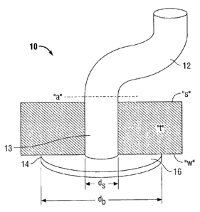
[00119] Referring now to FIG. 1, an embodiment of a wound closure device 10
according to
the present disclosure is shown. The wound closure device 10 includes an
elongate body 12
coupled to a plug member 14. The elongate body 12 is substantially
perpendicular to a tissue
facing surface 16 of plug member 14. In some embodiments, the elongate body 12
may be
integral with the plug member 14, while in other embodiments, the elongate
body 12 may be
attached or otherwise connected to the plug member 14.
[00120] The elongate body or stem 12 is adapted to fill or seal the
perforation in the tissue "t"
and/or bind the perforated tissue together. Accordingly, elongate body 12 may
be any shape that
fits into the wound. As illustrated in the current embodiment, the elongate
body 12 is cylindrical
in shape, and elliptical is cross-sectional geometry, but the shape and cross-
sectional geometry
may also be rectangular, flat, or other shapes within the purview of those
skilled in the art and as
33
CA 02716713 2010-10-07
shown in embodiments disclosed hereafter. For example, as illustrated in FIG.
2, wound closure
device 20 is accordion-shaped to allow the elongate body 22 to grow or shrink
in length
depending on the thickness of tissue "t." Thus, referring again to FIG. 1, the
elongate body 12
may be a predefined length which is substantially about the length or depth of
the tissue to be
sealed, or the elongate body 12 may be made longer to allow for variability in
the patient wall
thickness. For example, excess length of the elongate body 12 may be trimmed
at surface "s" of
tissue "t" as indicated by dashed line "a" in FIG. 1.
[00121] Plug member or base 14 is adapted to provide closure to the wound by
sealing the
perforation in the tissue at the inner wall "w" of the tissue "t." The plug
member 14 has a tissue
facing surface 16 coupled to a distal end 13 of elongate body 12. Plug member
14 may be any
shape having a substantially flat, tissue facing surface for abutting the
inner wall "w" of the
tissue "t," such as a mushroom shape, among others, as envisioned by those
skilled in the art.
Tissue facing surface 16 defines a diameter "db" which is larger than the
diameter "d," of the
elongate body 12 which is attached thereto for adhering to the inner wall "w"
surrounding the
perforated tissue "t."
[00122] In embodiments, the wound closure device 10 maybe a hydrogel or
include a
hydrogel on at least a portion thereof. For example, the hydrogel could be
composed of serum
proteins (nucleophilic) crosslinked with succinimidyl ester reactive PEG
(electrophilic) to
provide the desired adhesion to the tissue and tissue growth.
[00123] Upon reacting with amine-containing tissues, the reactive device
should fixate to
tissue within a useful time range. In alternate embodiments, the reactive
groups may be
chemically "shielded" or "blocked" in aid of slowing the reaction with tissue,
or the reactive
groups may simply have slow reaction kinetics.
34
CA 02716713 2010-10-07
[00124] The amount of time necessary for the reactive component of the
composition of the
present disclosure to bind the implant to tissue may vary from about 3 seconds
to about 20
minutes, in embodiments, about 10 seconds to about 5 minutes.
[00125] At least a portion of the wound closure device may include a-polymer
foam, as
illustrated in FIGS. 3A and 3B. Drying a polymer (such as a hydrogel) to
create a foam before
placement into tissue may ease the insertion of the device therein and/or may
provide control of
the size and fit of the device within the tissue. The foam may be created
through use of
techniques such as lyophilization, particulate leaching, compression molding
and others within
the purview of those skilled in the art. Various techniques can yield pores of
different size and
distribution. Varying the pore size and distribution may allow more rapid
ingress of water and
other aqueous fluids into the foam. Foams may be open-cell or closed-cell
foams. It is also
possible to affect the rate at which a foam rehydrates in a physiological
environment, such as
encountered upon implantation in tissue. For example, incorporating a blowing
agent during the
formation of the foam may lead to more rapid re-hydration due to the enhanced
surface area
available for the water to diffuse into the foam structure. The hydration of
the foam enables the
device to become anchored in place to prevent migration and hold the tissue
together.
[00126] FIG. 3A illustrates a wound closure device 30 having a pre-hydrated
foam elongate
body 32. Upon placement of the wound closure device 30 into perforation "p" of
tissue "t," the
elongate body 32 may rapidly rehydrate by irrigating the elongate body 32 with
a fluid, such as
saline, and/or through contact with the bodily fluids in the physiologic
environment. As
illustrated in FIG. 313, the elongate body 32 swells to fill the perforation
"p" in the tissue "t."
The foam may rehydrate rapidly, in some embodiments, within a few seconds of
being placed in
a moist tissue environment, or may rehydrate at a slower rate over the course
of a few hours.
CA 02716713 2010-10-07
During the hydration process, the foam may expand volumetrically, e.g., in
one, two, or three
dimensions, to several times its original size, thereby lodging the wound
closure device within
the tissue and sealing against leakage of fluids through the tissue.
[001271 In other embodiments, the wound closure device may include a
substantially
dehydrated hydrogel, which may, in embodiments, include a foam. The hydrogel
component of
a device of the present disclosure may swell and/or expand in an amount of
from about 5% to
about 100% of its original volume, in embodiments, from about 20% to about 80%
of its original
volume. In embodiments, the swelling of the hydrogel may substantially seal at
least one tissue
plane.
[00128] In embodiments, the wound closure device may have an aperture or
channel running
through a portion thereof to enable volumetric expansion and facilitate
hydration of the device.
As illustrated in FIG. 4, an aperture 47 is longitudinally disposed within the
elongate body 42,
extending from the proximal end 41 into the distal end 43. The aperture 47
allows for moisture
to reach parts of the elongate body 42, as well as parts of the plug member
44.
[00129] Turning now to FIG. 5, a wound closure device 50 may combine a
hydrogel with a
textile, such as a mesh, to facilitate wound healing. In embodiments, a mesh
59 may be disposed
on the tissue facing surface 56 of plug member 54 to aid in tissue adhesion
and ingrowth. For
example, mesh 59 may be encapsulated or coated with a hydrogel, such as a
serum-based
hydrogel as described above, and placed on the biodegradable polymer plug
member 54, or the
mesh 59 maybe disposed on at least one surface of the hydrogel plug member 54,
as illustrated
in FIG. 5. Moreover, mesh 59 may be self-tacking, such as including spiked
naps or barbs, to aid
the hydrogel in tissue adhesion. In some embodiments, a self-tacking mesh may
be utilized
without a hydrogel or other adhesive component, which will be later described.
36
CA 02716713 2010-10-07
[00130] The elongate body 52 may also be formed from a hydrogel or maybe
composed of a
polymer which is subsequently coated with a hydrogel. It is contemplated that
a mesh may also
be combined with the elongate body 52 to provide additional tissue adhesion
and ingrowth. The
elongate body 52 may be provided in a variety of forms to hold the perforated
tissue together.
For example, as illustrated in FIG. 6, the elongate body of the wound closure
device may include
sutures 62 which extend from plug member 64 and may be passed through the
perforated tissue
to hold the tissue together. In embodiments, the sutures may be coated with a
polymer
possessing at least one reactive group to aid in tissue adhesion. In some
embodiments, the
sutures may be barbed or have barb-like projections, extending generally
outward from the
suture body, which assist in tissue retention.
[00131] FIGS. 7 and 8 illustrate wound closure devices 70 and 80,
respectively, including an
elongate body 72, 82 formed from a hydrogel and a plug member 74, 84
fabricated from a mesh.
The plug member 74, 84 may be any of the textile and fabric materials as
described herein and
may include a coating composition including any of the functional precursor(s)
also as described
herein. As illustrated in FIG. 8, the elongate body 82 may include a grooved
exterior for
increased surface area and tissue integration.
[00132] Referring now to FIG. 9, the plug member 94 of the wound closure
device 90 maybe
constructed to include more than one layer, such as a laminate. The tissue
facing surface 96 of
the plug member 94 may be fabricated from a material having adhesive
properties, such as a
polymer having reactive groups or a mesh as described above, and the distal
surface 95 of the
plug member 94 may include a material having anti-adhesive properties, such as
a coating of
hyaluronic acid or PEG, to prevent adhesion of the device to internal organs.
In embodiments,
the plug member 94 may be fabricated from a composite material, such as a
PARIETEX TM
37
CA 02716713 2010-10-07
composite mesh, having a porous layer on the tissue facing surface 96 to
effect adhesion of the
tissue with the plug member 94, and a non-porous layer on the distal surface
95 to prevent
adhesion of the plug member 94 with other tissue or organs surrounding the
perforated tissue. In
other embodiments, the tissue facing surface may include a mesh modified with
biodegradable
linkers and reactive end groups to bind a second layer, such as a
biodegradable collagen film (not
shown) on the distal surface of the plug member 94. The distal surface,
including the collagen
film, may be non-porous to prevent adhesions. Alternatively, the distal
surface, including the
collagen film, may adhere to the internal organs, and the linkers binding the
collagen film to the
mesh may degrade in a short period of time thereby separating the-two layers
and preventing
adhesions. In embodiments, both the mesh and the collagen film may be designed
to degrade
over a longer period of time.
[00133] Methods for forming composite meshes are within the purview of those
skilled in the
art. Multiple layers may be adhered utilizing adhesives, crosslinking of
reactive groups on
multiple layers, heat molding, co-extrusion, solvent casting, melt pressing,
combinations thereof,
and the like.
100134] FIG. 10 illustrates an embodiment of a wound closure device 100
including a
composite plug member 104 including a mesh on the tissue facing surface 106
and an anti-
adhesive distal surface 105. The elongate body 102 includes a mesh which maybe
utilized alone
or in combination with a coating having reactive groups as described herein.
In some
embodiments, a wound closure device 110 and/or 120 may be solely formed from a
mesh, either
alone or in combination with a reactive polymer as illustrated in FIGS. 11 and
12. As depicted in
FIGS. 11 and 12, wound closure devices 110 and 120, respectively, may include
plug members
38
CA 02716713 2010-10-07
114 and 124, having tissue facing surfaces 116 and 126, and elongate bodies
112 and 122, all
formed of mesh.
[00135] FIGS. 13A-13D illustrate a wound closure device 130 including an
elongate body 132
and a plug member 134 which are pivotably connected, it being understood that
other
embodiments described herein may also be pivotably connected. FIGS. 13A and
13B illustrate
the wound closure device 130 in a first, collapsed or folded position and
FIGS. 13C-13D
illustrate the wound closure device 130 in a second, deployed position. The
elongate body 132
and the plug member 134 of the wound closure device 130 are coupled via a
hinged connection
131. The distal end 133 of the elongate body 132 is hingedly connected to
tissue facing surface
136 of the plug member 134 so that the plug member 134 may pivot with respect
to the elongate
body 132 from the folded position to the deployed position. In certain
embodiments, the
elongate body 132 and plug member 134 may be hingedly coupled with any of a
variety of
biodegradable fasteners as envisioned by those skilled in the art. The
fasteners may be formed
from any of the biodegradable polymers described above, which may be adapted
and configured
to have high strength to withstand the stresses of pivoting from the folded to
deployed positions
and maintain the integrity of the wound closure device upon implantation. The
fastener can
slowly degrade so that the fastener is replaced with new tissue over time.
Alternatively, hinge
131 may be a living hinge, such as a thin flexible web of a polymer, formed at
the intersection of
the elongate body 132 with the plug member 134. In other embodiments, a hinge
may be formed
through welding the plug member and the elongate body together.
[00136] The elongate body 132 and plug member 134 may transform from the
folded position
(for insertion) as depicted in FIG. 13A to a deployed state as depicted in
FIG. l3C for
positioning and placement within tissue. In embodiments, plug member 134 may
be normally
39
CA 02716713 2010-10-07
biased toward the folded position such that the plug member 134 is
longitudinally aligned with
the elongate body 132. Consequently, as the wound closure device 130 is placed
within tissue
and pulled in the direction of arrow "p" depicted in FIG. 13C, the plug member
134 pivots away
from the elongate body 132 to be substantially perpendicular to the elongate
body 132, the plug
member 134 effectively acting as a flange to prevent pullout of the wound
closure device 130
from tissue. Alternatively, wound closure devices may be positioned through
use of an insertion
device, which will be later described.
[00137] In the collapsed or folded position, as illustrated in FIG. 13A, the
slim profile is
preferentially used for insertion into tissue. Moreover, as shown in FIG. 13B,
the plug member
134 includes rounded edges to provide an atraumatic tip for insertion. In the
deployed state, as
illustrated in FIGS. 13C and 13D, the width "w" of the plug member 134 may be
based on the
size of the incision, and the length "1" of the elongate body 132 may be made
any length, such as
longer than needed so as to be cut to length after insertion into tissue.
[00138] In some embodiments, the elongate body 132 may be fabricated from a
material that
encourages surrounding tissue of the wound to chemically bond thereto and to
encourage cell
growth and tissue proliferation. Moreover, the tissue facing surface 136 of
the plug member 134
may include other polymer materials to encourage tissue integration. The
distal surface 135 of
the plug member 134 may include an anti-adhesive coating to prevent tissue
adhesion.
[00139] Another embodiment of a wound closure device which is pivotably
connected for
insertion into a wound is shown in FIGS. 14A-14C. The wound closure device 140
includes an
elongate body 142 and a plug member 144 formed from a pair of substantially
identically shaped
sections 144a and 144b. The elongate body 142 and the sections 144a and 144b
of the plug
CA 02716713 2010-10-07
member 144 are coupled via one or more hinges 141. Sections 144a and 144b of
the plug
member 144 are pivotably mounted on hinges 141 to move on a common pivot axis.
[001401 Shaped sections 144a and 144b are illustrated as generally triangular
in geometry,
although other geometries are envisioned, such as rectangular. Shaped sections
144a, 144b, each
include an abutment surface 143 a, and 143b, respectively (FIG. 14B). For
insertion, abutment
surfaces 143a, 143b are positioned generally parallel the elongate body 142.
Once inserted and
positioned, the abutment surfaces are approximated so as to dispose sections
144a and 144b
generally perpendicular to the elongate body 142. The abutment surfaces 143a,
143b provide
control as to how angled the shaped sections reside with respect to the
elongate body 142. For
example, if the abutment surfaces were angled greater than or less than 90
with respect to the
elongate body (as opposed to generally perpendicular in FIG. 14B), the shaped
sections 144a and
144b would similarly be disposed at an angle greater than or less than 90
with respect to the
elongate body.
1001411 Prior to placement within tissue, sections 144a and 144b of the plug
member maybe
folded up in the direction of arrow "b" depicted in FIG. 14B. As the wound
closure device 140
is placed within tissue and pulled in the direction of arrow "p" depicted in
FIG. 14C, sections
144a and 144b of the plug member 144 pivots away from the elongate body 142
(shown in
phantom) to be substantially perpendicular to the elongate body 142, the plug
member 144
effectively acting as a flange to prevent pullout of the wound closure device
140 from tissue. As
illustrated, sections 144a and 144b are triangular in shape to prevent the
plug member 144 from
deploying beyond about 90 degrees from the elongate member 142 to prevent the
inadvertent
removal of the wound closure device 140 from the tissue. It is envisioned that
sections 144a and
144b of the plug member 144 need not be substantially similar in shape.
41
CA 02716713 2010-10-07
[001421 In embodiments, the elongate body 142 may be a pre-formed hydrogel
having an
absorbable mesh layer. In embodiments, the pre-formed hydrogel maybe formed
from an 8 arm,
15 kDa PEG first precursor and a second precursor, such as collagen, gelatin,
or other aminated
biodegradable polymer, such as polysaccharides like aminated dextran or
hyaluronic acid. In
embodiments, the plug member 144 may be a pre-formed hydrogel which may or may
not have
an anti-adhesive coating on the distal end 145 thereof and a degradable or non-
degradable mesh
attached to the tissue facing surface 146.
[00143] A pre-formed hydrogel enables quick insertion and delivery of the
wound closure
device as there is no waiting period for the device to form in situ. Moreover,
a pre-formed
component avoids the possibility of components reacting with tissue other than
the target wound
and avoids spilling of material into the body cavity or elsewhere, such as on
a skin surface.
Methods for making pre-formed hydrogel include simultaneously spraying the
first precursor and
the second precursor into a mold of a desired geometry.
[001441 In embodiments, the elongate body and/or the plug member may include
unreacted
hydrogel which is embedded in the mesh or on which the mesh is attached, which
can be
solubilized and reacted within the tissue, thereby gelling within the mesh
structure and binding
the tissue thereto. This allows the mesh to bind to the interior wall of the
tissue and prevents
other components from working their way into the wound.
[00145] Alternatively, the wound closure device including a mesh may be first
inserted into a
wound and subsequently a hydrogel may be injected with a static mixer into the
wound to fill the
void and encase the mesh. In embodiments, the plug member may include a pre-
formed
hydrogel and the elongate body may be unreacted so that a hydrogel can be
injected into the
wound to hold the tissue and mesh in place.
42
CA 02716713 2010-10-07
[00146] Turning now to FIGS. 15A-15B, another embodiment of a wound closure
device
which includes an elongate body and plug member including a pair of shaped
sections is shown.
FIG. 15A illustrates wound closure device 150 in a deployed position having an
elongate body
152 and a plug member 154 formed from a pair of substantially identically
shaped sections 154a
and 154b. Sections 154a and 154b of the plug member 154 are pivotably mounted
on
independent hinges 151 a and 15 lb as illustrated in FIG. 15B. Sections 154a
and 154b are shown
pivoting away from the elongate body 152 to the deployed position of FIG. 15A
(in phantom).
Elongate body 152 includes a stop member 152a at distal end 153 to prevent
sections 154a and
154b of plug member 154 from over extending beyond angle a, which is about 90
degrees.
[00147] Wound closure devices of the present disclosure may be inserted into a
passageway of
a cannula or other portal access device having a sleeve extending through the
tissue wall into the
cavity of the patient. The wound closure device is moved through the
passageway of the sleeve
until the plug member exits the sleeve into the cavity. The plug member may be
positioned so
that the tissue facing surface abuts the wound and the sleeve is removed
leaving the elongate
body disposed within the perforated tissue. Accordingly, the wound closure
device must be
sufficiently pliable to be placed within the access device, yet be resilient
enough to support the
tissue and seal the wound. Alternatively, the wound closure device may include
mechanical
means for ease of insertion and placement of the device.
[00148] In embodiments, additional methods of securing a wound closure device
of the present
disclosure to tissue may be utilized. For example, bandages, films, gauzes,
tapes, felts,
combinations thereof, and the like, may be combined therewith or applied over
a wound closure
device of the present disclosure, as well as tissue surrounding the device.
Similarly, additional
43
CA 02716713 2010-10-07
adhesives may be applied thereto; sutures may be utilized to affix the wound
closure device to
tissue, combinations thereof, and the like.
[00149] Bioactive agents may be added to the wound closure device to provide
specific
biological or therapeutic properties thereto. Any product which may enhance
tissue repair, limit
the risk of sepsis, and modulate the mechanical properties of the wound
closure device may be
added during the preparation of the device or may be coated on the device or
into the pores of a
mesh attached thereto.
[00150] Moreover, the wound closure device may also be used for delivery of
one or more
bioactive agents. The bioactive agents may be incorporated into the wound
closure device
during formation of the device, such as by free suspension, liposomal
delivery, microspheres,
etc., or by coating a surface of the wound closure device, or portion thereof,
such as by polymer
coating, dry coating, freeze drying, applying to a mesh surface, ionically,
covalently, or affinity
binding to functionalize the degradable components of the wound closure
device. Thus, in some
embodiments, at least one bioactive agent may be combined with a component of
the wound
closure device, i.e., the elongate body and/or plug member, during formation
to provide release
of the bioactive agent during degradation of the wound closure device. As the
wound closure
device degrades or hydrolyzes in situ, the bioactive agents are released. In
other embodiments,
bioactive agents may be coated onto a surface or a portion of a surface of the
elongate body or
plug member of the wound closure device for quick release of the bioactive
agent.
[00151] A bioactive agent as used herein is used in the broadest sense and
includes any
substance or mixture of substances that have clinical use. Consequently,
bioactive agents may or
may not have pharmacological activity per se, e.g., a dye. Alternatively a
bioactive agent could
be any agent that provides a therapeutic or prophylactic effect; a compound
that affects or
44
CA 02716713 2010-10-07
participates in tissue growth, cell growth, and/or cell differentiation; an
anti-adhesive compound;
a compound that may be able to invoke a biological action such as an immune
response; or could
play any other role in one or more biological processes. A variety of
bioactive agents may be
incorporated into the mesh.
[001521 Examples of classes of bioactive agents, which maybe utilized in
accordance with the
present disclosure include, for example, anti-adhesives, antimicrobials,
analgesics, antipyretics,
anesthetics, antiepileptics, antihistamines, anti-inflammatories,
cardiovascular drugs, diagnostic
agents, sympathomimetics, cholinomimetics, antimuscarinics, antispasmodics,
hormones, growth
factors, muscle relaxants, adrenergic neuron blockers, antineoplastics,
immunogenic agents,
immunosuppressants, gastrointestinal drugs, diuretics, steroids, lipids,
lipopolysaccharides,
polysaccharides, platelet activating drugs, clotting factors and enzymes. It
is also intended that
combinations of bioactive agents may be used.
[001531 Other bioactive agents, which may be included as a bioactive agent
include: local
anesthetics; non-steroidal antifertility agents; parasympathomimetic agents;
psychotherapeutic
agents; tranquilizers; decongestants; sedative hypnotics; steroids;
sulfonamides;
sympathomimetic agents; vaccines; vitamins; antimalarials; anti-migraine
agents; anti-parkinson
agents, such as L-dopa; anti-spasmodics; anticholinergic agents (e.g.,
oxybutynin); antitussives;
bronchodilators; cardiovascular agents, such as coronary vasodilators and
nitroglycerin;
alkaloids; analgesics; narcotics, such as codeine, dihydrocodeinone,
meperidine, morphine and
the like; non-narcotics, such as salicylates, aspirin, acetaminophen, d-
propoxyphene and the like;
opioid receptor antagonists, such as naltrexone and naloxone; anti-cancer
agents; anti-
convulsants; anti-emetics; antihistamines; anti-inflammatory agents, such as
hormonal agents,
hydrocortisone, prednisolone, prednisone, non-hormonal agents, allopurinol,
indomethacin,
CA 02716713 2010-10-07
phenylbutazone and the like; prostaglandin and cytotoxic drugs;
chemotherapeutics; estrogens;
antibacterials; antibiotics; anti-fungals; anti-virals; anticoagulants;
anticonvulsants;
antidepressants; antihistamines; and immunological agents.
[00154] Other examples of suitable bioactive agents, which maybe included in
the wound
closure device include, for example, viruses and cells; peptides, polypeptides
and proteins, as
well as analogs, muteins, and active fragments thereof; immunoglobulins;
antibodies; cytokines
(e.g., lymphokines, monokines, chemokines); blood clotting factors;
hemopoietic factors;
interleukins (IL-2, IL-3, IL-4, IL-6); interferons (13-IFN, a-IFN and 'y-IFN);
erythropoietin;
nucleases; tumor necrosis factor; colony stimulating factors (e.g., GCSF, GM-
CSF, MCSF);
insulin; anti-tumor agents and tumor suppressors; blood proteins, such as
fibrin, thrombin,
fibrinogen, synthetic thrombin, synthetic fibrin, synthetic fibrinogen;
gonadotropins (e.g., FSH,
LH, CG, etc.); hormones and hormone analogs (e.g., growth hormone); vaccines
(e.g., tumoral,
bacterial and viral antigens); somatostatin; antigens; blood coagulation
factors; growth factors
(e.g., nerve growth factor, insulin-like growth factor); bone morphogenic-
proteins; TGF-B;
protein inhibitors; protein antagonists; protein agonists; nucleic acids, such
as antisense
molecules, DNA, RNA, RNAi; oligonucleotides; polynucleotides; and ribozymes.
[001551 In embodiments, the polymers forming the wound closure device, such as
precursors
and/or hydrogels formed from the precursors, may contain visualization agents
to improve their
visibility during surgical procedures. Visualization agents may be selected
from a variety of
non-toxic colored substances, such as dyes, suitable for use in implantable
medical devices.
Suitable dyes are within the purview of those skilled in the art and may
include, for example, a
dye for visualizing a thickness of the hydrogel as it is formed in situ, e.g.,
as described in U.S.
Patent No. 7,009,034. In some embodiments, a suitable dye may include, for
example, FD&C
46
CA 02716713 2010-10-07
Blue #1, FD&C Blue #2, FD&C Blue #3, FD&C Blue #6, D&C Green #6, methylene
blue,
indocyanine green, other colored dyes, and combinations thereof. It is
envisioned that additional
visualization agents may be used such as fluorescent compounds (e.g.,
flurescein or eosin), x-ray
contrast agents (e.g., iodinated compounds), ultrasonic contrast agents, and
MRI contrast agents
(e. g., Gadolinium containing compounds).
[00156] The visualization agent may be present in any precursor component
solution. The
colored substance may or may not become incorporated into the resulting
hydrogel. In
embodiments, however, the visualization agent does not have a functional group
capable of
reacting with the precursor(s).
[00157] In embodiments, the bioactive agent may be encapsulated by polymers
utilized to form
the wound closure device. For example, the polymer may form microspheres
around the
bioactive agent.
[00158] Suitable bioactive agents may be combined with the wound plug either
prior to or
during the manufacturing process. Bioactive agents may be admixed or combined
with polymers
to yield a plug with bioactive properties. In other embodiments, the bioactive
agent may be
combined with the present disclosure for example, in the form of a coating,
after the plug has
been shaped. It is envisioned that the bioactive agent may be applied to the
present disclosure in
any suitable form of matter, e.g., films, powders, liquids, gels and the like.
[00159] It should be understood that various combinations of elongate bodies
and plug
members may be used to fabricate the wound closure device according to the
present disclosure.
For example, any of the elongate bodies of the embodiments described above may
be combined
with any of the plug members also described above, dependent upon the type of
wound to be
treated and the properties desired from the wound closure device.
47
CA 02716713 2010-10-07
[001601 While several embodiments of the disclosure have been described, it is
not intended
that the disclosure be limited thereto, as it is intended that the disclosure
be as broad in scope as
the art will allow and that the specification be read likewise. Therefore, the
above description
should not be construed as limiting, but merely as exemplifications of
embodiments of the
present disclosure. Various modifications and variations of the wound closure
device, as well as.
methods of forming the elongate body and plug member of the wound closure
device and
attaching the components together, will be apparent to those skilled in the
art from the foregoing
detailed description. Such modifications and variations are intended to come
within the scope
and spirit of the claims appended hereto.
48