Note: Descriptions are shown in the official language in which they were submitted.
CA 02716826 2016-07-19
DRUG SELECTION FOR BREAST CANCER THERAPY USING
ANTIBODY-BASED ARRAYS
BACKGROUND OF THE INVENTION
[0002] The process of signal transduction in cells is responsible for a
variety of biological
functions including cell division and death, metabolism, immune cell
activation,
neurotransmission, and sensory perception to name but a few. Accordingly,
derangements in
normal signal transduction in cells can lead to a number of disease states
such as diabetes, heart
disease, autoimmunity, and cancer.
[0003] One well characterized signal transduction pathway is the MAP kinase
pathway,
which is responsible for transducing the signal from epidermal growth factor
(EGF) to the
promotion of cell proliferation in cells (see, Figure 1). EGF binds to a
transmembrane receptor-
linked tyrosine kinase, the epidermal growth factor receptor (EGER), which is
activated by the
binding of EGF. The binding of EGF to EGFR activates the tyrosine kinase
activity of the
cytoplasmic domain of the receptor. One consequence of this kinase activation
is the
autophosphorylation of EGFR on tyrosine residues. The phosphorylated tyrosine
residues on
the activated EGFR provide a docking site for the binding of SH2 domain
containing adaptor
proteins such as GRB2. In its function as an adaptor, GRB2 further binds to a
guanine
nucleotide exchange factor. SOS, by way of an S113 domain on GRB2. The
formation of the
complex of EGFR-GRB2-SOS leads to SOS activation of a guanine nucleotide
exchange factor
that promotes the removal of GDP from Ras. Upon removal of GDP, Ras binds GIP
and
becomes activated.
[0004] Following activation, Ras binds to and activates the protein kinase
activity of RAF
kinase, a serine/threonine-specific protein kinase. What follows is the
activation of a protein
kinase cascade that leads to cell proliferation. In outline, RAF kinase then
phosphorylates
1
CA 02716826 2010-08-24
WO 2009/108637
PCT/US2009/035013
and activates MEK, another serine/threonine kinase. Activated MEK
phosphorylates and
activates mitogen-activated protein kinase (MAPK). Among the targets for
further
phosphorylation by MAPK are 40S ribosomal protein S6 kinase (RSK). The
phosphorylation
of RSK by MAPK results in activation of RSK, which in turn phosphorylates
ribosomal
protein S6. Another known target of MAPK is the proto-oncogene, c-Myc, a gene
important
for cell proliferation, which is mutated in a variety of cancers. MAPK also
phosphorylates
and activates another protein kinase, MNK, which in turn phosphorylates the
transcription
factor, CREB. Indirectly, MAPK also regulates the transcription of the Fos
gene, which
encodes yet another transcription factor involved in cell proliferation. By
altering the levels
and activities of such transcription factors, MAPK transduces the original
extracellular signal
from EGF into altered transcription of genes that are important for cell cycle
progression.
[0005] Given the central role that signal transduction pathways play in cell
growth, it is not
surprising that many cancers arise as a result of mutations and other
alterations in signal
transduction components that result in aberrant activation of cell
proliferation pathways. For
example, overexpression or hyperactivity of EGFR has been associated with a
number of
cancers, including glioblastoma multiforme, colon cancer, and lung cancer.
This has
prompted the development of anticancer therapeutics directed against EGFR,
including
gefitinib and erlotinib for lung cancer, and cetuximab for colon cancer.
[0006] Cetuximab is an example of a monoclonal antibody inhibitor, which binds
to the
extracellular ligand-binding domain of EGFR, thus preventing the binding of
ligands which
activate the EGFR tyrosine kinase. In contrast, gefitinib and erlotinib are
small molecules
which inhibit the intracellularly-located EGFR tyrosine kinase. In the absence
of kinase
activity, EGFR is unable to undergo autophosphorylation at tyrosine residues,
which is a
prerequisite for binding of downstream adaptor proteins, such as GRB2. By
halting the
signaling cascade in cells that rely on this pathway for growth, tumor
proliferation and
migration is diminished.
[0007] Additionally, other studies have shown that about 70% of human
melanomas and a
smaller fraction of other tumors have a point mutation (V599E) in the Raf gene
which leads
to persistent activation of the MAPK pathway (see, e.g., Davies et at.,
Nature, 417:949-954
(2002)). Such results suggest that mutations in particular signal transduction
pathways may
be characteristic of particular types of tumors and that such specific,
altered signal
transduction pathways may be a promising target for chemotherapeutic
intervention.
2
CA 02716826 2010-08-24
WO 2009/108637
PCT/US2009/035013
[0008] Given that different cancer treatments, particularly cancer
chemotherapy, may
function either directly or indirectly by means of either blocking or
activating cellular signal
transduction pathways that are involved in cell proliferation or death,
respectively, the
activity of a given signal transduction pathway in a particular form of cancer
may serve as a
good indicator of the efficacy of various cancer treatments. Accordingly, in
addition to
fulfilling other needs, the present invention provides a method for evaluating
the
effectiveness of potential anticancer therapies for an individual patient. As
such, the present
invention provides methods for assisting a physician in selecting a suitable
cancer therapy at
the right dose and at the right time for every patient.
BRIEF SUMMARY OF THE INVENTION
[0009] The present invention provides compositions and methods for detecting
the
activation states of components of signal transduction pathways in tumor cells
(e.g.,
circulating cells of a breast tumor). Information on the activation states of
components of
signal transduction pathways derived from practice of the present invention
can be used for
cancer diagnosis, prognosis, and in the design of cancer treatments.
[0010] In one aspect, the present invention provides a method for selecting a
suitable
anticancer drug for the treatment of a breast tumor, the method comprising:
(a) isolating cells of a breast tumor after administration of an anticancer
drug, or
prior to incubation with an anticancer drug;
(b) lysing the isolated cells to produce a cellular extract;
(c) detecting an activation state of one or more analytes in the cellular
extract using
an assay comprising a plurality of dilution series of capture antibodies
specific
for the one or more analytes, wherein the capture antibodies are restrained on
a
solid support; and
(d) determining whether the anticancer drug is suitable or unsuitable for the
treatment of the breast tumor by comparing the activation state detected for
the
one or more analytes with a reference activation profile generated in the
absence
of the anticancer drug.
[0011] In a preferred embodiment, the method for selecting a suitable
anticancer drug for
the treatment of a breast tumor comprises:
(a) isolating cells of a breast tumor after administration of an anticancer
drug, or
prior to incubation with an anticancer drug;
3
CA 02716826 2010-08-24
WO 2009/108637
PCT/US2009/035013
(b) lysing the isolated cells to produce a cellular extract;
(c) detecting an activation state of one or more analytes in the cellular
extract using
an assay comprising a plurality of dilution series of capture antibodies
specific
for the one or more analytes, wherein the capture antibodies are restrained on
a
solid support;
(d) comparing the activation state detected for the one or more analytes with
a
reference activation profile generated in the absence of the anticancer drug;
and
(e) indicating that the anticancer drug is suitable for the treatment of the
breast
tumor when the activation state detected for the one or more analytes is
substantially decreased compared to the reference activation profile.
[0012] In some embodiments, the methods of the present invention may be useful
to aid or
assist in the selection of a suitable anticancer drug for the treatment of a
breast tumor. In
other embodiments, the methods of the present invention may be useful for
improving the
selection of a suitable anticancer drug for the treatment of a breast tumor.
[0013] In another aspect, the present invention provides a method for
identifying the
response of a breast tumor to treatment with an anticancer drug, the method
comprising:
(a) isolating cells of a breast tumor after administration of an anticancer
drug, or
prior to incubation with an anticancer drug;
(b) lysing the isolated cells to produce a cellular extract;
(c) detecting an activation state of one or more analytes in the cellular
extract using
an assay comprising a plurality of dilution series of capture antibodies
specific
for the one or more analytes, wherein the capture antibodies are restrained on
a
solid support; and
(d) identifying the breast tumor as responsive or non-responsive to treatment
with
the anticancer drug by comparing the activation state detected for the one or
more analytes with a reference activation profile generated in the absence of
the
anticancer drug.
[0014] In a preferred embodiment, the method for identifying the response of a
breast
tumor to treatment with an anticancer drug comprises:
(a) isolating cells of a breast tumor after administration of an anticancer
drug, or
prior to incubation with an anticancer drug;
(b) lysing the isolated cells to produce a cellular extract;
4
CA 02716826 2010-08-24
WO 2009/108637
PCT/US2009/035013
(c) detecting an activation state of one or more analytes in the cellular
extract using
an assay comprising a plurality of dilution series of capture antibodies
specific
for the one or more analytes, wherein the capture antibodies are restrained on
a
solid support;
(d) comparing the activation state detected for the one or more analytes with
a
reference activation profile generated in the absence of the anticancer drug;
and
(e) indicating that the breast tumor is responsive to treatment with the
anticancer
drug when the activation state detected for the one or more analytes is
substantially decreased compared to the reference activation profile.
[0015] In some embodiments, the methods of the present invention may be useful
to aid or
assist in the identification of a breast tumor's response to treatment with an
anticancer drug.
In other embodiments, the methods of the present invention may be useful for
improving the
identification of a breast tumor's response to treatment with an anticancer
drug.
[0016] In yet another aspect, the present invention provides a method for
predicting the
response of a subject having a breast tumor to treatment with an anticancer
drug, the method
comprising:
(a) isolating cells of a breast tumor after administration of an anticancer
drug, or
prior to incubation with an anticancer drug;
(b) lysing the isolated cells to produce a cellular extract;
(c) detecting an activation state of one or more analytes in the cellular
extract using
an assay comprising a plurality of dilution series of capture antibodies
specific
for the one or more analytes, wherein the capture antibodies are restrained on
a
solid support; and
(d) predicting the likelihood that the subject will respond to treatment with
the
anticancer drug by comparing the activation state detected for the one or more
analytes with a reference activation profile generated in the absence of the
anticancer drug.
[0017] In a preferred embodiment, the method for predicting the response of a
subject
having a breast tumor to treatment with an anticancer drug comprises:
(a) isolating cells of a breast tumor after administration of an anticancer
drug, or
prior to incubation with an anticancer drug;
(b) lysing the isolated cells to produce a cellular extract;
5
CA 02716826 2010-08-24
WO 2009/108637
PCT/US2009/035013
(c) detecting an activation state of one or more analytes in the cellular
extract using
an assay comprising a plurality of dilution series of capture antibodies
specific
for the one or more analytes, wherein the capture antibodies are restrained on
a
solid support;
(d) comparing the activation state detected for the one or more analytes with
a
reference activation profile generated in the absence of the anticancer drug;
and
(e) indicating that the subject will likely respond to treatment with the
anticancer
drug when the activation state detected for the one or more analytes is
substantially decreased compared to the reference activation profile.
[0018] In some embodiments, the methods of the present invention may be useful
to aid or
assist in the prediction of a subject's likelihood of responding to treatment
with an anticancer
drug. In other embodiments, the methods of the present invention may be useful
for
improving the prediction of a subject's likelihood of responding to treatment
with an
anticancer drug.
[0019] In a further aspect, the present invention provides an array having
superior dynamic
range comprising a plurality of dilution series of capture antibodies
restrained on a solid
support, wherein the capture antibodies in each dilution series are specific
for one or more
analytes corresponding to a component of a signal transduction pathway or
other protein
(e.g., nuclear hormone receptor) in a cellular extract. The addressable arrays
described herein
are particularly useful for determining the expression and/or activation state
of signal
transduction molecules and other proteins involved in breast cancer.
[0020] In an additional aspect, the present invention provides a method for
detecting the
presence (or absence) of a truncated receptor, the method comprising:
(a) incubating a cellular extract with a plurality of beads specific for an
extracellular
domain (ECD) binding region of a full-length receptor;
(b) removing the plurality of beads from the cellular extract, thereby
removing the
full-length receptor to form a cellular extract devoid of the full-length
receptor;
(c) incubating the cellular extract devoid of the full-length receptor with a
plurality
of capture antibodies, wherein the plurality of capture antibodies is specific
for
an intracellular domain (ICD) binding region of a truncated receptor and
wherein the plurality of capture antibodies is restrained on a solid support
to
form a plurality of captured truncated receptors;
6
CA 02716826 2010-08-24
WO 2009/108637
PCT/US2009/035013
(d) incubating the plurality of captured truncated receptors with detection
antibodies
specific for the corresponding truncated receptors to form a plurality of
detectable captured truncated receptors;
(e) incubating the plurality of detectable captured truncated receptors with
first and
second members of a signal amplification pair to generate an amplified signal;
and
(f) detecting an amplified signal generated from the first and second members
of the
signal amplification pair.
[0021] In a related aspect, the present invention provides a method for
detecting the
presence (or absence) of a truncated receptor, the method comprising:
(a) incubating a cellular extract with a plurality of beads specific for an
extracellular
domain (ECD) binding region of a full-length receptor;
(b) removing the plurality of beads from the cellular extract, thereby
removing the
full-length receptor to form a cellular extract devoid of the full-length
receptor;
(c) incubating the cellular extract devoid of the full-length receptor with a
plurality
of capture antibodies, wherein the plurality of capture antibodies is specific
for
an intracellular domain (ICD) binding region of the truncated receptor and
wherein the plurality of capture antibodies is restrained on a solid support
to
form a plurality of captured truncated receptors;
(d) incubating the plurality of captured truncated receptors with detection
antibodies
comprising a plurality of activation state-independent antibodies and a
plurality
of activation state-dependent antibodies specific for the corresponding
truncated
receptors to form a plurality of detectable captured truncated receptors,
wherein the activation state-independent antibodies are labeled with a
facilitating
moiety, the activation state-dependent antibodies are labeled with a first
member
of a signal amplification pair, and the facilitating moiety generates an
oxidizing
agent which channels to and reacts with the first member of the signal
amplification pair;
(e) incubating the plurality of detectable captured truncated receptors with a
second
member of the signal amplification pair to generate an amplified signal; and
(f) detecting the amplified signal generated from the first and second members
of
the signal amplification pair.
7
CA 02716826 2010-08-24
WO 2009/108637
PCT/US2009/035013
[0022] Other objects, features, and advantages of the present invention will
be apparent to
one of skill in the art from the following detailed description and figures.
BRIEF DESCRIPTION OF THE DRAWINGS
[0023] Figure 1 shows an example of a signal transduction pathway involved in
cell
proliferation that may be used in the practice of the invention. Depicted are
components of
the EGFR/MAPK/ERK pathway that is used by cells to convert a mitogenic signal
into cell
proliferation.
[0024] Figure 2 shows one embodiment of the present invention in which the
proximity
assays described herein detected phosphorylated EGFR (pEGFR) and
phosphorylated HER-2
(pHER-2) with single cell sensitivity.
[0025] Figure 3 shows that the proximity assays described herein resulted in
highly
specific assays for the detection of HER-2 at the single cell level only in
cells expressing
HER-2.
[0026] Figure 4 shows schematically the application of the addressable arrays
of the
invention for drug selection throughout the course of cancer treatment.
[0027] Figure 5 shows a schematic example of an addressable array comprising
dilutions
of antibodies to components of a receptor tyrosine kinase pathway, such as
those in the
EGFR/MAPK/ERK pathway. Antibodies are plated in triplicate in four different
dilutions on
the addressable array.
[0028] Figure 6 shows a schematic example of an addressable array comprising
dilutions
of antibodies to components of signal transduction pathways activated in tumor
angiogenesis.
Antibodies are plated in triplicate in four different dilutions on the
addressable array.
[0029] Figure 7 shows a schematic example of an alternative addressable array
comprising
dilutions of antibodies to components of signal transduction pathways
activated in tumor
angiogenesis. Antibodies are plated in triplicate in four different dilutions
on the addressable
array.
[0030] Figure 8 shows a schematic example of an addressable array comprising
dilutions
of antibodies to components of a receptor tyrosine kinase pathway and signal
transduction
pathways activated in tumor angiogenesis. Antibodies are plated in triplicate
in four different
dilutions on the addressable array.
8
CA 02716826 2010-08-24
WO 2009/108637
PCT/US2009/035013
[0031] Figure 9 shows a schematic example of an alternative addressable array
comprising
dilutions of antibodies to components of a receptor tyrosine kinase pathway
and signal
transduction pathways activated in tumor angiogenesis. Antibodies may be
plated in
triplicate in a dilution series on the addressable array.
[0032] Figure 10 shows the relative phosphorylation levels of EGFR for 5
breast cancer
and 6 normal samples. Data is also shown in Table 40.
[0033] Figure 11 shows the relative phosphorylation levels of HER-2 for 5
breast cancer
and 6 normal samples. Data is also shown in Table 41.
[0034] Figure 12 shows images of CTC staining on the Veridex CellSearchTM
System for 5
breast cancer patients. Cell lines controls are A431 (positive for EGFR) and
SKBr3 (positive
for HER-2).
[0035] Figure 13 shows that full-length HER-2 (ErbB2) can be removed from a
patient
sample using antibodies which bind to the extracellular domain of ErbB2
attached to a
polystyrene bead or a polymeric dextran.
[0036] Figure 14 shows one embodiment of the present invention for detecting
truncated
receptors such as p95ErbB2. SA = streptavidin; HRP = horseradish peroxidase;
TSA =
tyramide signal amplification.
[0037] Figure 15 shows that pretreatment with beads coated with an antibody
directed to
the extracellular domain (ECD) of ErbB2 (HER-2) almost completely removed the
full-
length ErbB2 signal without affecting the ErbB2 intracellular domain (ICD)
signal.
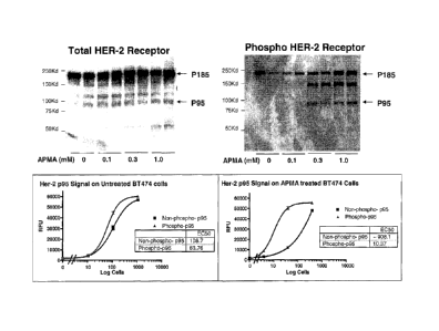
[0038] Figure 16 shows that APMA ((4-aminophenyl)mercuric acetate) treatment
increased p95ErbB2 phosphorylation in BT-474 cells.
[0039] Figure 17 shows that heregulin increased p95ErbB2 phosphorylation in
T47D cells.
[0040] Figure 18 shows multiple points in which the methods of the present
invention may
be used to influence clinical practice with respect to selecting the
appropriate breast cancer
therapy for a particular patient.
[0041] Figure 19 shows one embodiment of the assay format of the present
invention,
which relies on the co-localization of two additional detector antibodies
linked with enzymes
for subsequent channeling events per each target protein bound.
9
CA 02716826 2010-08-24
WO 2009/108637
PCT/US2009/035013
[0042] Figure 20 shows single cell sensitivity for pHER-1 and pHER-2 assays.
[0043] Figure 21 shows ErbB expression/activation with EGF or HRG 0 treatment
in
various cell lines.
[0044] Figure 22 shows the T47D ErbB RTK profile with EGF or HRG 0
stimulation.
[0045] Figure 23 shows an exemplary embodiment of an ErbB pathway array.
DETAILED DESCRIPTION OF THE INVENTION
I. Introduction
[0046] As described above, the activation of signal transduction pathways that
are involved
in cell proliferation and the deactivation of pathways that are involved in
cell death are non-
limiting examples of molecular features that characterize many different types
of cancer. In
many cases, the activity of particular signal transduction pathways, and
components thereof,
may serve as molecular signatures for a given type of cancer. Such activated
components
may further provide useful targets for therapeutic intervention. Accordingly,
knowledge of
the activity level of a particular signal transduction system within a cancer
cell prior to,
during, and after treatment provides a physician with highly relevant
information that may be
used to select an appropriate course of treatment to adopt. Furthermore, the
continued
monitoring of signal transduction pathways that are active in cancer cells as
treatment
progresses can provide the physician with additional information on the
efficacy of treatment,
prompting the physician to either continue a particular course of treatment or
to switch to
another line of treatment, when, for example, cancer cells have become
resistant to treatment
through further aberrations that activate either the same or another signal
transduction
pathway.
[0047] Accordingly, the present invention provides methods and compositions
for detecting
the expression and activation states of a plurality of deregulated signal
transduction
molecules in tumor tissue or extratumoral cells such as rare circulating cells
of a solid tumor
in a specific, multiplex, high-throughput assay. The invention also provides
methods and
compositions for the selection of appropriate therapy (single drugs or
combinations of drugs)
to down-regulate or shut down a deregulated signaling pathway. Thus, the
invention may be
used to facilitate the design of personalized therapies for cancer patients.
[0048] The ability to detect and identify tumor cells in the circulation
through the
determination of the activity of signal transduction pathways at the level of
single cells is an
CA 02716826 2010-08-24
WO 2009/108637
PCT/US2009/035013
important advantage of the present invention. Tumor cells are often found in
the blood of
patients with various early stages of cancer as "micrometastases"
(disseminated tumor cells)
and are also found in metastatic cancers. The number of tumor cells in blood
will depend on
the stage and type of tumor. While biopsies are typically obtained on primary
tumors, most
metastatic tumors are not biopsied, making molecular analysis of such tumor
samples very
difficult. During tumor metastasis, the most aggressive tumor cells leave the
primary tumor
and travel through the blood and lymphatic system to reach a distant location.
Thus,
circulating tumor cells from blood represent the most aggressive and
homogenous population
of tumor cells. However, the number of metastatic tumor cells in blood is
frequently very
low, varying from one to several thousand cells per milliliter of blood. The
ability to isolate
and assay signal transduction pathways in such rare cells and to apply this
information toward
more effective cancer treatments is one object of the present invention.
[0049] In some embodiments, the multiplex, high-throughput immunoassays of the
present
invention can detect the activation state of one or more signal transduction
molecules in
circulating cells of a solid tumor at the single cell level. In fact, signal
transduction
molecules such as EGFR can be detected with a sensitivity of about 100
zeptomoles and a
linear dynamic range of from about 100 zeptomoles to about 100 femtomoles. As
such,
single-cell detection of the activation state of multiple signal transducers
in rare circulating
cells facilitates cancer prognosis and diagnosis as well as the design of
personalized, targeted
therapies.
[0050] Rare circulating cells include circulating cells of a solid tumor that
have either
metastasized or micrometastasized from a solid tumor. Circulating tumor cells,
cancer stem
cells, and cells that are migrating to a tumor (e.g., due to chemoattraction)
such as circulating
endothelial progenitor cells, circulating endothelial cells, circulating pro-
angiogenic myeloid
cells, and circulating dendritic cells are some examples of circulating cells
associated with a
solid tumor.
[0051] Signal transduction molecules of interest are typically extracted
shortly after the
circulating cells are isolated to preserve their in situ activation state,
preferably within about
24, 6, or 1 hr, and more preferably within about 30, 15, or 5 minutes. The
isolated cells may
also be incubated with one or more growth factors, usually at nanomolar to
micromolar
concentrations, for about 1-30 minutes to resuscitate or stimulate activation
of the signal
transduction molecules (see, e.g., Irish et at., Cell, 118:217-228 (2004)).
11
CA 02716826 2010-08-24
WO 2009/108637
PCT/US2009/035013
[0052] As explained in greater detail herein, to evaluate potential anticancer
therapies for
an individual patient, the isolated cells can be incubated with one or more
anticancer drugs at
varying doses. Growth factor stimulation can then be performed for a few
minutes (e.g.,
about 1-5 minutes) or for several hours (e.g., about 1-6 hours). The
differential activation of
signaling pathways with and without anticancer drugs can aid in the selection
of a suitable
cancer therapy at the proper dose for each individual patent. Circulating
cells can also be
isolated from a patient sample during anticancer drug treatment and stimulated
with one or
more growth factors to determine whether a change in therapy should be
implemented. As
such, the methods of the present invention advantageously assist the clinician
in providing the
right anticancer drug at the right dose at the right time for every patient.
[0053] With regard to breast cancer, current testing options are
unsatisfactory because
treatment of both primary and metastatic tumors in a breast cancer patient is
based on a one-
time diagnosis from a biopsy sample taken during an early stage of the
disease. In particular,
therapeutic intervention for both the early and metastatic stages of breast
cancer is based
solely on the initial diagnosis from the biopsy sample taken during an early
stage of the
disease because of the impracticality of obtaining a biopsy sample from a
metastatic cancer
patient. However, breast tumors are evolving as a function of time and
treatment such that
temporal monitoring of breast tumors is critical for optimal management of
breast cancer
patients. For example, a change in the activation state of one or more of the
ErbB (HER)
family of receptor tyrosine kinases may affect therapy selection at
recurrence. Indeed,
discordance in HER-2 status between primary and metastatic cancer is common
because up
to 37% of all breast cancer patients change from a HER-2-negative primary
tumor to HER-2-
positive metastatic cancer. In addition, patients may have de novo resistance
or develop
acquired resistance to hormonal therapy due to HER-1/2 activation. In some
instances,
patients may have de novo resistance or develop acquired resistance to ErbB-
targeted
therapies due to the presence of tumor cells expressing p95HER-2. As a result,
there is an
unmet clinical need for assays to assist the clinician in prescribing the
appropriate cancer
therapy at the appropriate time because current technology lacks sensitivity
and specificity,
cannot be used to monitor patients on therapy, and do not utilize pathway
profiling to guide
individualized treatment decisions.
[0054] In contrast to currently available breast cancer testing options, the
methods of the
present invention enable the monitoring of breast cancer patients through all
stages of the
disease by providing a "real-time biopsy" of solid breast tumors using samples
such as
12
CA 02716826 2010-08-24
WO 2009/108637
PCT/US2009/035013
circulating tumor cells (CTCs) from blood and/or fine needle aspirates (FNAs).
As a non-
limiting example, the breast cancer assays described herein can be used in the
initial
diagnosis of breast cancer in a patient at an early stage of the disease.
Selection of a suitable
cancer therapy is guided by profiling the activation states of specific
signaling pathways with
and without anticancer drugs using the single detection and proximity dual
detection assays
described herein. Advantageously, the methods of the present invention can
also be used to
monitor the progression or regression of the disease because therapeutic
intervention may be
based on samples taken at any stage of the disease and analyzed using the
single detection
and proximity dual detection assays described herein. As such, selection of
suitable cancer
therapies for the early and metastatic stages of breast cancer is guided by
real-time diagnosis
and an analysis of the activation status of specific signaling pathway
molecules.
[0055] The methods of the present invention are beneficially tailored to
address key issues
in cancer management and provide a higher standard of care for breast cancer
patients
because they (1) provide increased sensitivity (e.g., single cell detection
can be achieved for
detecting total and phosphorylated signal transduction molecules such as EGFR
and HER-2),
(2) provide increased specificity (e.g., three-antibody proximity assays
enhance specificity
for detecting phosphorylated signal transduction molecules), (3) enable
pathway profiling
(e.g., activation status of specific signal transduction molecules can be
detected in CTCs or
FNA from patients), and (4) eliminate any issues with obtaining patient
samples (e.g., assays
can be performed on a few tumor cells). Although any sample may be used in the
novel
assays described herein, CTCs are particularly useful because they represent
the most
aggressive tumor cells, every tumor is known to shed CTCs, they can be the
only source of
residual tumors or hard-to-access metastatic tumors, and they are found in
blood. As such,
the methods of the present invention enable the serial sampling of breast
tumor tissues,
resulting in valuable information on changes occurring in tumor cells as a
function of time
and therapy and providing clinicians with a means to monitor rapidly evolving
cancer
pathway signatures.
[0056] In sum, the methods of the present invention advantageously provide
accurate
selection and monitoring of cancer patients (e.g., breast cancer patients)
most likely to benefit
from targeted therapy by performing pathway profiling on easily accessible
tumor cells using
multiplexed, antibody-based single detection or proximity assays.
13
CA 02716826 2010-08-24
WO 2009/108637 PCT/US2009/035013
II. Definitions
[0057] As used herein, the following terms have the meanings ascribed to them
unless
specified otherwise.
[0058] The term "cancer" is intended to include any member of a class of
diseases
characterized by the uncontrolled growth of aberrant cells. The term includes
all known
cancers and neoplastic conditions, whether characterized as malignant, benign,
soft tissue, or
solid, and cancers of all stages and grades including pre- and post-metastatic
cancers.
Examples of different types of cancer include, but are not limited to, breast
cancer; lung
cancer (e.g., non-small cell lung cancer); digestive and gastrointestinal
cancers such as
colorectal cancer, gastrointestinal stromal tumors, gastrointestinal carcinoid
tumors, colon
cancer, rectal cancer, anal cancer, bile duct cancer, small intestine cancer,
and stomach
(gastric) cancer; esophageal cancer; gallbladder cancer; liver cancer;
pancreatic cancer;
appendix cancer; ovarian cancer; renal cancer (e.g., renal cell carcinoma);
cancer of the
central nervous system; skin cancer; lymphomas; choriocarcinomas; head and
neck cancers;
osteogenic sarcomas; and blood cancers. As used herein, a "tumor" comprises
one or more
cancerous cells. In one preferred embodiment, the breast tumor is derived from
a subject
with an invasive or in situ form of ductal carcinoma or lobular carcinoma. In
another
preferred embodiment, the breast tumor is derived from a subject with
recurrent or metastatic
breast cancer.
[0059] The term "analyte" includes any molecule of interest, typically a
macromolecule
such as a polypeptide, whose presence, amount, and/or identity is determined.
In certain
instances, the analyte is a cellular component of circulating cells of a solid
tumor, preferably
a signal transduction molecule.
[0060] As used herein, the term "dilution series" is intended to include a
series of
descending concentrations of a particular sample (e.g., cell lysate) or
reagent (e.g., antibody).
A dilution series is typically produced by a process of mixing a measured
amount of a
starting concentration of a sample or reagent with a diluent (e.g., dilution
buffer) to create a
lower concentration of the sample or reagent, and repeating the process enough
times to
obtain the desired number of serial dilutions. The sample or reagent can be
serially diluted at
least 2, 3, 4, 5, 6, 7, 8, 9, 10, 15, 20, 25, 30, 35, 40, 45, 50, 100, 500, or
1000-fold to produce
a dilution series comprising at least 2, 3, 4, 5, 6, 7, 8, 9, 10, 11, 12, 13,
14, 15, 16, 17, 18, 19,
20, 25, 30, 35, 40, 45, or 50 descending concentrations of the sample or
reagent. For
14
CA 02716826 2010-08-24
WO 2009/108637
PCT/US2009/035013
example, a dilution series comprising a 2-fold serial dilution of a capture
antibody reagent at
a 1 mg/ml starting concentration can be produced by mixing an amount of the
starting
concentration of capture antibody with an equal amount of a dilution buffer to
create a 0.5
mg/ml concentration of the capture antibody, and repeating the process to
obtain capture
antibody concentrations of 0.25 mg/ml, 0.125 mg/ml, 0.0625 mg/ml, 0.0325
mg/ml, etc.
[0061] The term "superior dynamic range" as used herein refers to the ability
of an assay to
detect a specific analyte in as few as one cell or in as many as thousands of
cells. For
example, the immunoassays described herein possess superior dynamic range
because they
advantageously detect a particular signal transduction molecule of interest in
about 1-10,000
cells (e.g., about 1, 5, 10, 25, 50, 75, 100, 250, 500, 750, 1000, 2500, 5000,
7500, or 10,000
cells) using a dilution series of capture antibody concentrations.
[0062] The term "signal transduction molecule" or "signal transducer" includes
proteins
and other molecules that carry out the process by which a cell converts an
extracellular signal
or stimulus into a response, typically involving ordered sequences of
biochemical reactions
inside the cell. Examples of signal transduction molecules include, but are
not limited to,
receptor tyrosine kinases such as EGFR (e.g., EGFR/HER-1/ErbBl, HER-
2/Neu/ErbB2,
HER-3/ErbB3, HER-4/ErbB4), VEGFR-1/FLT-1, VEGFR-2/FLK-1/KDR, VEGFR-3/FLT-4,
FLT-3/FLK-2, PDGFR (e.g., PDGFRA, PDGFRB), c-KIT/SCFR, INSR (insulin
receptor),
IGF-IR, IGF-IIR, IRR (insulin receptor-related receptor), CSF-1R, FGFR 1-4,
HGFR 1-2,
CCK4, TRK A-C, MET, RON, EPHA 1-8, EPHB 1-6, AXL, MER, TYR03, TIE 1-2, TEK,
RYK, DDR 1-2, RET, c-ROS, V-cadherin, LTK (leukocyte tyrosine kinase), ALK
(anaplastic
lymphoma kinase), ROR 1-2, MUSK, AATYK 1-3, RTK 106, and truncated forms of
the
receptor tyrosine kinases such as p95ErbB2; non-receptor tyrosine kinases such
as BCR-
ABL, Src, Frk, Btk, Csk, Abl, Zap70, Fes/Fps, Fak, Jak, Ack, and LIMK;
tyrosine kinase
signaling cascade components such as Akt, MAPK/ERK, MEK, RAF, PLA2, MEKK,
JNKK,
INK, p38, Shc (p66), PI3K, Ras (e.g., K-Ras, N-Ras, H-Ras), Rho, Racl, Cdc42,
PLC, PKC,
p70 S6 kinase, p53, cyclin D1, STAT1, STAT3, PIP2, PIP3, PDK, mTOR, BAD, p21,
p2'7,
ROCK, IP3, TSP-1, NOS, PTEN, RSK 1-3, JNK, c-Jun, Rb, CREB, Ki67, and
paxillin;
nuclear hormone receptors such as estrogen receptor (ER), progesterone
receptor (PR),
androgen receptor, glucocorticoid receptor, mineralocorticoid receptor,
vitamin A receptor,
vitamin D receptor, retinoid receptor, thyroid hormone receptor, and orphan
receptors;
nuclear receptor coactivators and repressors such as amplified in breast
cancer-1 (AIB1) and
nuclear receptor corepressor 1 (NCOR), respectively; and combinations thereof
CA 02716826 2010-08-24
WO 2009/108637
PCT/US2009/035013
[0063] As used herein, the term "circulating cells" comprises extratumoral
cells that have
either metastasized or micrometastasized from a solid tumor. Examples of
circulating cells
include, but are not limited to, circulating tumor cells, cancer stem cells,
and/or cells that are
migrating to the tumor (e.g., circulating endothelial progenitor cells,
circulating endothelial
cells, circulating pro-angiogenic myeloid cells, circulating dendritic cells,
etc.).
[0064] The term "sample" as used herein includes any biological specimen
obtained from a
patient. Samples include, without limitation, whole blood, plasma, serum, red
blood cells,
white blood cells (e.g., peripheral blood mononuclear cells), ductal lavage
fluid, nipple
aspirate, lymph (e.g., disseminated tumor cells of the lymph node), bone
marrow aspirate,
saliva, urine, stool (i.e., feces), sputum, bronchial lavage fluid, tears,
fine needle aspirate
(e.g., harvested by random periareolar fine needle aspiration), any other
bodily fluid, a tissue
sample (e.g., tumor tissue) such as a biopsy of a tumor (e.g., needle biopsy)
or a lymph node
(e.g., sentinel lymph node biopsy), and cellular extracts thereof In some
embodiments, the
sample is whole blood or a fractional component thereof such as plasma, serum,
or a cell
pellet. In preferred embodiments, the sample is obtained by isolating
circulating cells of a
solid tumor from whole blood or a cellular fraction thereof using any
technique known in the
art. In other embodiments, the sample is a formalin fixed paraffin embedded
(FFPE) tumor
tissue sample, e.g., from a solid tumor of the breast.
[0065] A "biopsy" refers to the process of removing a tissue sample for
diagnostic or
prognostic evaluation, and to the tissue specimen itself. Any biopsy technique
known in the
art can be applied to the methods and compositions of the present invention.
The biopsy
technique applied will generally depend on the tissue type to be evaluated and
the size and
type of the tumor (i.e., solid or suspended (i.e., blood or ascites)), among
other factors.
Representative biopsy techniques include excisional biopsy, incisional biopsy,
needle biopsy
(e.g., core needle biopsy, fine-needle aspiration biopsy, etc.), surgical
biopsy, and bone
marrow biopsy. Biopsy techniques are discussed, for example, in Harrison's
Principles of
Internal Medicine, Kasper, et al., eds., 16th ed., 2005, Chapter 70, and
throughout Part V.
One skilled in the art will appreciate that biopsy techniques can be performed
to identify
cancerous and/or precancerous cells in a given tissue sample.
[0066] The term "subject" or "patient" or "individual" typically includes
humans, but can
also include other animals such as, e.g., other primates, rodents, canines,
felines, equines,
ovines, porcines, and the like.
16
CA 02716826 2010-08-24
WO 2009/108637
PCT/US2009/035013
[0067] An "array" or "microarray" comprises a distinct set and/or dilution
series of capture
antibodies immobilized or restrained on a solid support such as, for example,
glass (e.g., a
glass slide), plastic, chips, pins, filters, beads (e.g., magnetic beads,
polystyrene beads, etc.),
paper, membrane (e.g., nylon, nitrocellulose, polyvinylidene fluoride (PVDF),
etc.), fiber
bundles, or any other suitable substrate. The capture antibodies are generally
immobilized or
restrained on the solid support via covalent or noncovalent interactions
(e.g., ionic bonds,
hydrophobic interactions, hydrogen bonds, Van der Waals forces, dipole-dipole
bonds). In
certain instances, the capture antibodies comprise capture tags which interact
with capture
agents bound to the solid support. The arrays used in the assays of the
present invention
typically comprise a plurality of different capture antibodies and/or capture
antibody
concentrations that are coupled to the surface of a solid support in different
known/addressable locations.
[0068] The term "capture antibody" is intended to include an immobilized
antibody which
is specific for (i.e., binds, is bound by, or forms a complex with) one or
more analytes of
interest in a sample such as a cellular extract of circulating cells of a
solid tumor. In
preferred embodiments, the capture antibody is restrained on a solid support
in an array.
Suitable capture antibodies for immobilizing any of a variety of signal
transduction molecules
on a solid support are available from Upstate (Temecula, CA), Biosource
(Camarillo, CA),
Cell Signaling Technologies (Danvers, MA), R&D Systems (Minneapolis, MN), Lab
Vision
(Fremont, CA), Santa Cruz Biotechnology (Santa Cruz, CA), Sigma (St. Louis,
MO), and BD
Biosciences (San Jose, CA).
[0069] The term "detection antibody" as used herein includes an antibody
comprising a
detectable label which is specific for (i.e., binds, is bound by, or forms a
complex with) one
or more analytes of interest in a sample. The term also encompasses an
antibody which is
specific for one or more analytes of interest, wherein the antibody can be
bound by another
species that comprises a detectable label. Examples of detectable labels
include, but are not
limited to, biotin/streptavidin labels, nucleic acid (e.g., oligonucleotide)
labels, chemically
reactive labels, fluorescent labels, enzyme labels, radioactive labels, and
combinations
thereof Suitable detection antibodies for detecting the activation state
and/or total amount of
any of a variety of signal transduction molecules are available from Upstate
(Temecula, CA),
Biosource (Camarillo, CA), Cell Signaling Technologies (Danvers, MA), R&D
Systems
(Minneapolis, MN), Lab Vision (Fremont, CA), Santa Cruz Biotechnology (Santa
Cruz, CA),
Sigma (St. Louis, MO), and BD Biosciences (San Jose, CA). As a non-limiting
example,
17
CA 02716826 2010-08-24
WO 2009/108637
PCT/US2009/035013
phospho-specific antibodies against various phosphorylated forms of signal
transduction
molecules such as EGFR, c-KIT, c-Src, FLK-1, PDGFRA, PDGFRB, Akt, MAPK, PTEN,
Raf, and MEK are available from Santa Cruz Biotechnology.
[0070] The term "activation state-dependent antibody" includes a detection
antibody which
is specific for (i.e., binds, is bound by, or forms a complex with) a
particular activation state
of one or more analytes of interest in a sample. In preferred embodiments, the
activation
state-dependent antibody detects the phosphorylation, ubiquitination, and/or
complexation
state of one or more analytes such as one or more signal transduction
molecules. In some
embodiments, the phosphorylation of members of the EGFR family of receptor
tyrosine
kinases and/or the formation of heterodimeric complexes between EGFR family
members is
detected using activation state-dependent antibodies. Non-limiting examples of
activation
states (listed in parentheses) that are suitable for detection with activation
state-dependent
antibodies include: EGFR (EGFRvIII, phosphorylated (p-) EGFR, EGFR:Shc,
ubiquitinated
(u-) EGFR, p-EGFRvIII); ErbB2 (p95 :truncated (Tr)-ErbB2, p-ErbB2, p95:Tr-p-
ErbB2,
HER-2:Shc, ErbB2:PI3K, ErbB2:EGFR, ErbB2:ErbB3, ErbB2:ErbB4); ErbB3 (p-ErbB3,
ErbB3:PI3K, p-ErbB3:PI3K, ErbB3:Shc); ErbB4 (p-ErbB4, ErbB4:Shc); ER (p-ER
(S118,
S167); IGF-1R (p-IGF-1R, IGF-1R:IRS, IRS:PI3K, p-IRS, IGF-1R:PI3K); INSR (p-
INSR);
KIT (p-KIT); FLT3 (p-FLT3); HGFRI (p-HGFRI); HGFR2 (p-HGFR2); RET (p-RET);
PDGFRa (p-PDGFRa); PDGFRP (p-PDGFRP); VEGFRI (p-VEGFRI, VEGFRI:PLCg,
VEGFR1:Src); VEGFR2 (p-VEGFR2, VEGFR2:PLCy, VEGFR2:Src, VEGFR2:heparin
sulphate, VEGFR2:VE-cadherin); VEGFR3 (p-VEGFR3); FGFR1 (p-FGFR1); FGFR2 (p-
FGFR2); FGFR3 (p-FGFR3); FGFR4 (p-FGFR4); Tiel (p-Tiel); Tie2 (p-Tie2); EphA
(p-
EphA); EphB (p-EphB); NFKB and/or IKB (p-IK (S32), p-NFKB (S536), p-P65:IKBa);
Akt
(p-Akt (T308, S473)); PTEN (p-PTEN); Bad (p-Bad (S112, S136), Bad:14-3-3);
mTor (p-
mTor (S2448)); p7056K (p-p70S6K (T229, T389)); Mek (p-Mek (S217, S221)); Erk
(p-Erk
(T202, Y204)); Rsk-1 (p-Rsk-1 (T357, S363)); Jnk (p-Jnk (T183, Y185)); P38 (p-
P38 (T180,
Y182)); Stat3 (p-Stat-3 (Y705, S727)); Fak (p-Fak (Y576)); Rb (p-Rb (S249,
T252, S780));
Ki67; p53 (p-p53 (S392, S20)); CREB (p-CREB (S133)); c-Jun (p-c-Jun (S63));
cSrc (p-cSrc
(Y416)); and paxillin (p-paxillin (Y118)).
[0071] The term "activation state-independent antibody" includes a detection
antibody
which is specific for (i.e., binds, is bound by, or forms a complex with) one
or more analytes
of interest in a sample irrespective of their activation state. For example,
the activation state-
18
CA 02716826 2010-08-24
WO 2009/108637
PCT/US2009/035013
independent antibody can detect both phosphorylated and unphosphorylated forms
of one or
more analytes such as one or more signal transduction molecules.
[0072] The term "nucleic acid" or "polynucleotide" includes
deoxyribonucleotides or
ribonucleotides and polymers thereof in either single- or double-stranded form
such as, for
example, DNA and RNA. Nucleic acids include nucleic acids containing known
nucleotide
analogs or modified backbone residues or linkages, which are synthetic,
naturally occurring,
and non-naturally occurring, and which have similar binding properties as the
reference
nucleic acid. Examples of such analogs include, without limitation,
phosphorothioates,
phosphoramidates, methyl phosphonates, chiral-methyl phosphonates, 2'-0-methyl
ribonucleotides, and peptide-nucleic acids (PNAs). Unless specifically
limited, the term
encompasses nucleic acids containing known analogues of natural nucleotides
that have
similar binding properties as the reference nucleic acid. Unless otherwise
indicated, a
particular nucleic acid sequence also implicitly encompasses conservatively
modified
variants thereof and complementary sequences as well as the sequence
explicitly indicated.
[0073] The term "oligonucleotide" refers to a single-stranded oligomer or
polymer of RNA,
DNA, RNA/DNA hybrid, and/or a mimetic thereof In certain instances,
oligonucleotides are
composed of naturally-occurring (i.e., unmodified) nucleobases, sugars, and
internucleoside
(backbone) linkages. In certain other instances, oligonucleotides comprise
modified
nucleobases, sugars, and/or internucleoside linkages.
[0074] As used herein, the term "mismatch motif" or "mismatch region" refers
to a portion
of an oligonucleotide that does not have 100% complementarity to its
complementary
sequence. An oligonucleotide may have at least one, two, three, four, five,
six, or more
mismatch regions. The mismatch regions may be contiguous or may be separated
by 1, 2, 3,
4, 5, 6, 7, 8, 9, 10, 11, 12, or more nucleotides. The mismatch motifs or
regions may
comprise a single nucleotide or may comprise two, three, four, five, or more
nucleotides.
[0075] The phrase "stringent hybridization conditions" refers to conditions
under which an
oligonucleotide will hybridize to its complementary sequence, but to no other
sequences.
Stringent conditions are sequence-dependent and will be different in different
circumstances.
Longer sequences hybridize specifically at higher temperatures. An extensive
guide to the
hybridization of nucleic acids is found in Tijssen, Techniques in Biochemistry
and Molecular
Biology--Hybridization with Nucleic Probes, "Overview of principles of
hybridization and
the strategy of nucleic acid assays" (1993). Generally, stringent conditions
are selected to be
19
CA 02716826 2010-08-24
WO 2009/108637
PCT/US2009/035013
about 5-10 C lower than the thermal melting point (Tm) for the specific
sequence at a defined
ionic strength pH. The Tm is the temperature (under defined ionic strength,
pH, and nucleic
concentration) at which 50% of the probes complementary to the target
hybridize to the target
sequence at equilibrium (as the target sequences are present in excess, at Tm,
50% of the
probes are occupied at equilibrium). Stringent conditions may also be achieved
with the
addition of destabilizing agents such as formamide. For selective or specific
hybridization, a
positive signal is at least two times background, preferably 10 times
background
hybridization.
[0076] The terms "substantially identical" or "substantial identity," in the
context of two or
more nucleic acids, refer to two or more sequences or subsequences that are
the same or have
a specified percentage of nucleotides that are the same (i.e., at least about
60%, preferably at
least about 65%, 70%, 75%, 80%, 85%, 90%, or 95% identity over a specified
region) when
compared and aligned for maximum correspondence over a comparison window or
designated region as measured using a sequence comparison algorithm or by
manual
alignment and visual inspection. This definition, when the context indicates,
also refers
analogously to the complement of a sequence. Preferably, the substantial
identity exists over
a region that is at least about 5, 10, 15, 20, 25, 30, 35, 40, 45, 50, 75, or
100 nucleotides in
length.
[0077] The term "incubating" is used synonymously with "contacting" and
"exposing" and
does not imply any specific time or temperature requirements unless otherwise
indicated.
III. Description of the Embodiments
[0078] In one embodiment, the present invention provides methods for detecting
the
expression and activation states of a plurality of deregulated signal
transducers in tumor cells
derived from tumor tissue or circulating cells of a solid tumor in a specific,
multiplex, high-
throughput assay. The invention also provides methods and compositions for the
selection of
appropriate therapies to down-regulate or shut down one or more deregulated
signaling
pathways. Thus, embodiments of the invention may be used to facilitate the
design of
personalized therapies based on the particular molecular signature provided by
the collection
of activated signal transduction proteins in a given patient's tumor.
[0079] Circulating cells of a solid tumor include cells that have either
metastasized or
micrometastasized from a solid tumor, including cancer stem cells or cells
that are migrating
to the tumor (e.g., due to chemoattraction), such as endothelial progenitor
cells, circulating
CA 02716826 2010-08-24
WO 2009/108637
PCT/US2009/035013
endothelial cells, pericytes, circulating pro-angiogenic myeloid cells,
dendritic cells, etc.
Patient samples containing the circulating cells can be obtained from any
accessible
biological fluid (e.g., whole blood, serum, plasma, sputum, bronchial lavage
fluid, urine,
nipple aspirate, lymph, saliva, fine needle aspirate, etc.). In certain
instances, the whole
blood sample is separated into a plasma or serum fraction and a cellular
fraction (i.e., cell
pellet). The cellular fraction typically contains red blood cells, white blood
cells, and/or
circulating cells of a solid tumor such as circulating tumor cells (CTCs),
circulating
endothelial cells (CECs), circulating endothelial progenitor cells (CEPCs),
cancer stem cells
(CSCs), disseminated tumor cells of the lymph node, and combinations thereof.
The plasma
or serum fraction usually contains, inter alia, nucleic acids (e.g., DNA, RNA)
and proteins
that are released by circulating cells of a solid tumor.
[0080] The circulating cells are typically isolated from a patient sample
using one or more
separation methods including, for example, immunomagnetic separation (see,
e.g., Racila et
at., Proc. Natl. Acad. Sci. USA, 95:4589-4594 (1998); Bilkenroth et at., Int.
J. Cancer,
92:577-582 (2001)), the CellTracks System by Immunicon (Huntingdon Valley,
PA),
microfluidic separation (see, e.g., Mohamed et at., IEEE Trans. Nanobiosci.,
3:251-256
(2004); Lin et al., Abstract No. 5147, 97th AACR Annual Meeting, Washington,
D.C.
(2006)), FACS (see, e.g., Mancuso et at., Blood, 97:3658-3661 (2001)), density
gradient
centrifugation (see, e.g., Baker et al., Clin. Cancer Res., 13:4865-4871
(2003)), and depletion
methods (see, e.g., Meye et at., Int. J. Oncol., 21:521-530 (2002)).
[0081] To preserve the in situ activation states, the signal transducers are
advantageously
extracted shortly after the cells are isolated, preferably within 96, 72, 48,
24, 6, or 1 hr, more
preferably within 30, 15, or 5 minutes. The isolated cells may also be
advantageously
incubated with growth factors usually at nanomolar to micromolar
concentrations for about 1-
30 minutes to resuscitate or stimulate signal transducer activation (see,
e.g., Irish et at., Cell,
118:217-228 (2004)). Stimulatory growth factors include epidermal growth
factor (EGF),
heregulin (HRG), TGF-a, PIGF, angiopoietin (Ang), NRG1, PGF, TNF-aõ VEGF,
PDGF,
IGF, FGF, HGF, cytokines, and the like. To evaluate potential anticancer
therapies for an
individual patient, prior to growth factor stimulation, the isolated cells can
be incubated with
one or more anticancer drugs of varying doses. Growth factor stimulation can
be performed
for a few minutes or hours (e.g., 1-5 minutes to 1-6 hours). The differential
activation of
signaling pathways with and without anticancer drugs aids in the selection of
a suitable
cancer therapy at the proper dose for each individual patent. After isolation,
anticancer agent
21
CA 02716826 2010-08-24
WO 2009/108637
PCT/US2009/035013
treatment, and/or growth factor stimulation, the cells are lysed to extract
the signal
transducers using any technique known in the art. Preferably, the cell lysis
is initiated
between about 1-360 minutes after growth factor stimulation, and more
preferably at two
different time intervals: (1) at about 1-5 minutes after growth factor
stimulation; and (2)
between about 30-180 minutes after growth factor stimulation. Alternatively,
the lysate can
be stored at -80 C until use.
[0082] In some embodiments, the anticancer drug comprises an agent that
interferes with
the function of activated signal transduction pathway components in cancer
cells. Non-
limiting examples of such agents include those listed below in Table 1.
Table 1
EGFR (ErbB1) (A) HER-2 (ErbB2) (C) HER-3 (ErbB3) (E) HER-4
(ErbB4) target
Cetuximab Trastuzumab Antibody (U3)
Panitumumab (Herceptin )
Matuzumab Pertuzumab (DNA)
Nimotuzumab BMS-599626*
ErbB1 vaccine
*Heterodimerization HER-1/2;
Phase 1
EGFR (ErbB1) (B) HER-2 (ErbB2) (D) ErbB1/2 (F) ErbB1/2/4 (G)
Erlotinib CP-724714 (Pfizer) Lapatinib (Tykerb )
Canertinib*
Gefitinib HKI-272* ARRY-334543
EKB 569* HKI-357 (Preclinical) JNJ-
26483327
CL-387-785** BIBW 2992** JNJ-26483327
*Wyeth, Irreversible, I/II
*(Wyeth, Irreversible, ll NSCLC, Breast
CRC) ** Boehringer *Pfizer,
Irreversible, ll
**(Wyeth, Irreversible, Ingelheim, Irreversible,
NSCLC, Breast
Preclinical) I/II Prostate, Ovarian,
Breast
Raf (H) SRC (H) Mek: (I) NFkB-IkB (I)
Sorafenib AZ PD-325901 (II: NSCLC)
PLX4032 (Plexxikon) AZD6244 - Array/Az
XL518 Exelisis/DNA
VEGFR2 and
mTor (J) PI3K (J) VEGFR1/2/3:
VEGFR1 (K)
Rad 001 : Everolimus* PX-866* Avastin (DNA) AZD 2171
(NSCLC,
CRC)
Temsirolimus** HuMV833* AMG-706 (+
PDGFR)
AP-23573*** VEGF-Trap**
*Everolimus (Novartis, *P110alpha specific inhibition;
* (PDL) anti-VEGFa
combination with ProIX Pharma; Preclinical **Regeneron/Aventis
22
CA 02716826 2010-08-24
WO 2009/108637 PCT/US2009/035013
Gefetinib/Erlotinib; I/II: NSCLC (Receptor mimic)
NSCLC, Glioblastoma) (Phase 2)
**Temsirolimus (Wyeth,
combination with
Gefetinib/Erlotinib; I/II:
NSCLC, Glioblastoma)
***AP-23573 (Ariad, I/II
: Endometrial)
VEGFR2 target (L) EPH A-D
DC101* CDP-791 (UCB) Bay-579352 (+ PDGFR)
IMC-IC11** CP-547632* ABT-869*
IMC1121B Fully
AG13736** BMS-540215 (+FGFR1)
humanized
CDP-791*** E-7080 (Eisai) KRN-951
Pazopanib**** CHIR-258*** BBIW
OSI-930 (+ cKit, PDGFR)
*ImoIone (Phase 2/3?)
**Chimeric IgG1 against *OSI, PFIZER: (+ ErbB1 +
VEGFR2
PDGFR) (NSCLC, Ovarian
***Cel!tech, pegalated
Phase 2)
*(+CSF1R, Erk, Flt-3,
di-Fab antibody against **Pfizer: VEGFR1,2 and
PDGFR)
R2
PDGFRbeta) (RCC II)
**** GSK, Multiple
***(VEGFR1,2
myeloma, ovarian,RCC
FGFR3,PDGFR)
Phase 3 enrollment
completed, sarcoma II)
VEGFR 2/ErbB1/2 VEGFR2/1/3, Flt-3,
VEGFR2/3/Raf/PDGFR/cKit/F
TIE 1/2 cFMS,
PDGFR/cKit
(ErbB1)/cMet/FGFR It-3 (N)
(
(M) 0)
ZD6474* Sorafenib* PTK787 (Not
cFMS,
FLT-3)
XL647** Sunitinib
AEE 788*** XL-999
SU-6668 (Pfizer)
GSK
AZ (AZD2171)
BMS
Novartis (AEE-788)
Amgen
Others
*(vandetanib) (Phase
III: thyroid, NSCLC)
**(Exelixis; Also
EPHB2): (Patient *(RCC, HOC, NSCLC(III),
resistant to Erlotinib; Melanoma(III))
Asian patients) (Phase
2)
***(Novartis, Phase1/2)
PDGFR target (P) Abl target: (Q) FTL 3 RET
Tandutinib Imatinib
Nilotinib Dasatinib
23
CA 02716826 2010-08-24
WO 2009/108637
PCT/US2009/035013
Nilotinib
AT-9283
AZD-0530
Bosutinib
Kit target (R) HGFR1/2 FGFR1-4 IGF-1R Target
(S)
AMG-706 Chiron Merck
XL-880 Pfizer
XL-999 Novartis
HSP90 inhibitors: Anti-Mitotic Drugs: Other targets:
IPI-504* Docetaxel* HDAC inhibitors
17-AAG** Paclitaxel' BCL2
Vinblastine, Vincristine, Chemotherapeutics
Vinare!bine' (breakdown)
Proteosome inhibitors
*(Microtubule stabilizer;
Adjuvant and advanced
Breast cancer; NSCLC,
*(Infinity Pharma,
Androgen independent
Mutant ErbB1, I/II
Prostate cancer)
multiple myeloma,
'(Microtubule stabilizer;
GIST)
Adjuvant and advanced
'(Kosan, I/II solid
Breast cancer; NSCLC,
tumors)
Ovarian cancer, AIDS related
Kaposi sarcoma)
'*(Microtubule De-stabilizers)
[0083] In another embodiment, the present invention provides an addressable
array having
superior dynamic range comprising a plurality of dilution series of capture
antibodies
restrained on a solid support, in which the capture antibodies in each
dilution series are
specific for one or more analytes corresponding to a component of a signal
transduction
pathway and other target proteins. In various aspects, this embodiment
includes arrays that
comprise components of signal transduction pathways characteristic of
particular tumors,
e.g., signal transduction pathways active in breast cancer cells. Thus, the
invention may be
advantageously practiced wherein each signal transduction molecule or other
protein of
interest with a potential expression or activation defect causing cancer is
represented on a
single array or chip. In some aspects, the components of a given signal
transduction pathway
active in a particular tumor cell are arrayed in a linear sequence that
corresponds to the
sequence in which information is relayed through a signal transduction pathway
within a cell.
Examples of such arrays are shown in Figures 5-9. The capture antibodies
specific for one or
more components of a given signal transduction pathway active in a particular
tumor cell can
also be printed in a randomized fashion to minimize any surface-related
artifacts.
24
CA 02716826 2010-08-24
WO 2009/108637 PCT/US2009/035013
[0084] Non-limiting examples of signal transduction pathways that may be
interrogated
using the present invention include those shown in Table 2.
Table 2
ErbB1 ErbB1 ErbB1
Pathway 1 ErbB1 ErbBl-PI3K PTEN
Phospho Shc ubiquitin
ErbB 'VII
ErbB 'VIII ErbB 'VIII ErbB 'VIII
Pathway 2 ErbB1 ErbBlVIII I PTEN
Phospho Shc ubiquitin
PI3K
ErbB2:
ErbB2
Pathway 3 ErbB2 ErbB2 Phospho HER-2 Shc PI3K PTEN
ubiquitin
Complex
P95Truncated ERBB2:
P95Truncated ErbB2Phosph ErbB2
P95ErbB2:P
Pathway 4 ErbB2 ERBB2 HER-2 Shc PI3K
ErbB2 ubiquitin
I3K
o
Phospho Complex
ErbB3:PI3K ErbB3 PI3K
Pathway 5 ErbB3 ErbB3 Phospho ErbB3:Shc
Complex Phospho
Pathway 6 ErbB4 ErbB4 Phospho ErbB4:Shc
IGF-1R
Pathway 7 IGF -1R IGF-1RPhospho IGF-1R:IRS IRS: PI3 K
Phospho IRS
:PI3K
Pathway 8 INSR INSRPhospho
Pathway 9 KIT KIT Phospho
Pathway 10 FLT3 FLT3Phospho
HGFR 1
Pathway 11 HGFR 1
Phospho
HGFR 2
Pathway 12 HGFR 2
Phospho
Pathway 13 RET RET Phospho
PDGFR PDGFR alpha
Pathway 14
alpha Phospho
PDGFR PDGFR beta
Pathway 15 beta Phospho
VEGFR 1 VEGFR 1: VEGFR 1:
Pathway 16 VEGFR 1
Phospho PLCycomplex Src
VEGFR- VEGFR-
VEGFR 2: VEGFR 2:
VEGFR 2 2/heparin 2, VE-
Pathway 17 VEGFR 2 PLCy Src
Phospho sulphate cadherin
complex
complex complex
VEGFR 3
Pathway 18 VEGFR 3
Phospho
FGFR 1
Pathway 19 FGFR 1
Phospho
FGFR 2
Pathway 20 FGFR 2
Phospho
FGFR 3
Pathway 21 FGFR 3
Phospho
FGFR 4
Pathway 22 FGFR 4
Phospho
Pathway 23 TIE 1 TIE 1 Phospho
TIE 2
Pathway 24 TIE 2
Phospho
EPHA
Pathway 25 EPHA
Phospho
EPHB
Pathway 26 EPHB
Phospho
Total P65
NFkB- phospho-11B Total NFKB
IkBa
Pathway 27 IkB (S32) Phospho
Phospho
complex Total IkB NFKB(S536)
P65 IkBa
Other ER
Pathway 28 ER Phospho ER ER-AIB1
complexes
PR
Pathway 29 PR Phospho Pr
complexes
Pathway 30 Hpeate haoyg
Wnt
Pathway 31 pathway
CA 02716826 2010-08-24
WO 2009/108637
PCT/US2009/035013
Pathway 32 Pathwayawchay
Total Stat3
Total Mek Phospho Stat-
Total Rsk-1 Total Fak
Total cSrc
Phospho Total Erk 3 (Y705) Phospho
Total Ras
Phospho Rsk- Phospho
Phospho
Pathway 33 Mek Phospho Erk (S727) Bad (S112)
Phospho
1 Fak
cSrc(Y416)
(S217/S2 (T202/Y204) Total Stat 1
Bad (total) Ras
(T357/5363) (Y576)
21) Phospho
Statl (Y701)
Total
Akt Total
p7056K
GSK3beta
(Total) Phospho Bad Bad:14-3-3 mTor
Phospho Akt Phospho Bad Phospho
Total
Pathway 34 Phospho (S112) complex Phospho
(T308) (S136) p7056K
(Phospho
Akt Bad (total) ml or
(T229)
Ser 9)
(T473) (S2448)
(T389)
Total Jnk
Total Rb Total p53 phospho- Total c-
Total
Phospho
Total P38 Phospho Rb Phospho p53 CREB(S133
Jun Paxillin
Jnk
Pathway 35 (T183/Y1 Phospho P38 (5249/T252) (S392) phospho-
Phospho
(T180/Y182) Phospho Rb Phospho p53 Total
c-Jun; Paxillin
85)
(S780) (S20) CREB (S63) (Y118)
Cleaved
Pathway 36 Ki67 Caspase 3,8,9 TOP02
others
Pathway 37 TGFbeta
[0085] In certain embodiments, the anticancer drug comprises an anti-signaling
agent (i.e.,
a cytostatic drug) such as a monoclonal antibody or a tyrosine kinase
inhibitor; an anti-
proliferative agent; a chemotherapeutic agent (i.e., a cytotoxic drug); a
hormonal therapeutic
agent; a radiotherapeutic agent; a vaccine; and/or any other compound with the
ability to
reduce or abrogate the uncontrolled growth of aberrant cells such as cancerous
cells. In some
embodiments, the isolated circulating cells are treated with one or more anti-
signaling agents,
anti-proliferative agents, and/or hormonal therapeutic agents in combination
with at least one
chemotherapeutic agent.
[0086] Examples of anti-signaling agents suitable for use in the present
invention include,
without limitation, monoclonal antibodies such as trastuzumab (Herceptinc)),
alemtuzumab
(Compote), bevacizumab (Avastinc)), cetuximab (Erbitux8), gemtuzumab
(Mylotarg8),
panitumumab (VectibixTm), rituximab (Rituxanc)), and tositumomab (BDOCARc));
tyrosine
kinase inhibitors such as gefitinib (Iressac)), sunitinib (Sutent8), erlotinib
(Tarcevac)),
lapatinib (GW-572016; Tykerb8), canertinib (CI 1033), semaxinib (SU5416),
vatalanib
(PTK787/ZK222584), sorafenib (BAY 43-9006; Nexavarc)), imatinib mesylate
(Gleevecc)),
leflunomide (SU101), and vandetanib (ZACTIMATm; ZD6474); and combinations
thereof
[0087] Exemplary anti-proliferative agents include mTOR inhibitors such as
sirolimus
(rapamycin), temsirolimus (CCI-779), and everolimus (RAD001); Akt inhibitors
such as
1L6-hydroxymethyl-chiro-inosito1-2-(R)-2-0-methy1-3-0-octadecyl-sn-
glycerocarbonate, 9-
methoxy-2-methylellipticinium acetate, 1,3-dihydro-1-(1-((4-(6-pheny1-1H-
imidazo[4,5-
26
CA 02716826 2010-08-24
WO 2009/108637
PCT/US2009/035013
g]quinoxalin-7-yl)phenyl)methyl)-4-piperidiny1)-2H-benzimidazol-2-one, 10-(4'-
(N-
diethylamino)buty1)-2-chlorophenoxazine, 3-formylchromone thiosemicarbazone
(Cu(II)C12
complex), API-2, a 15-mer peptide derived from amino acids 10-24 of the proto-
oncogene
TCL1 (Hiromura et at., J. Biol. Chem., 279:53407-53418 (2004), KP372-1, and
the
compounds described in Kozikowski et at., J. Am. Chem. Soc., 125:1144-1145
(2003) and
Kau et at., Cancer Cell, 4:463-476 (2003); and combinations thereof
[0088] Non-limiting examples of chemotherapeutic agents include platinum-based
drugs
(e.g., oxaliplatin, cisplatin, carboplatin, spiroplatin, iproplatin,
satraplatin, etc.), alkylating
agents (e.g., cyclophosphamide, ifosfamide, chlorambucil, busulfan, melphalan,
mechlorethamine, uramustine, thiotepa, nitrosoureas, etc.), anti-metabolites
(e.g., 5-
fluorouracil, azathioprine, 6-mercaptopurine, methotrexate, leucovorin,
capecitabine,
cytarabine, floxuridine, fludarabine, gemcitabine (Gemzarc)), pemetrexed
(ALIMTAc)),
raltitrexed, etc.), plant alkaloids (e.g., vincristine, vinblastine,
vinorelbine, vindesine,
podophyllotoxin, paclitaxel (Taxor), docetaxel (Taxoterec)), etc.),
topoisomerase inhibitors
(e.g., irinotecan, topotecan, amsacrine, etoposide (VP16), etoposide
phosphate, teniposide,
etc.), antitumor antibiotics (e.g., doxorubicin, adriamycin, daunorubicin,
epirubicin,
actinomycin, bleomycin, mitomycin, mitoxantrone, plicamycin, etc.),
pharmaceutically
acceptable salts thereof, stereoisomers thereof, derivatives thereof, analogs
thereof, and
combinations thereof
[0089] Examples of hormonal therapeutic agents include, without limitation,
aromatase
inhibitors (e.g., aminoglutethimide, anastrozole (Arimidexc)), letrozole
(Femarac)), vorozole,
exemestane (Aromasinc)), 4-androstene-3,6,17-trione (6-0X0), 1,4,6-
androstatrien-3,17-
dione (ATD), formestane (Lentaronc)), etc.), selective estrogen receptor
modulators (e.g.,
bazedoxifene, clomifene, fulvestrant, lasofoxifene, raloxifene, tamoxifen,
toremifene, etc.),
steroids (e.g., dexamethasone), fmasteride, and gonadotropin-releasing hormone
agonists
(GnRH) such as goserelin, pharmaceutically acceptable salts thereof,
stereoisomers thereof,
derivatives thereof, analogs thereof, and combinations thereof.
[0090] Non-limiting examples of cancer vaccines useful in the present
invention include
ANYARA from Active Biotech, DCVax-LB from Northwest Biotherapeutics, EP-2101
from
IDM Pharma, GV1001 from Pharmexa, 10-2055 from Idera Pharmaceuticals, INGN 225
from Introgen Therapeutics and Stimuvax from Biomira/Merck.
27
CA 02716826 2010-08-24
WO 2009/108637
PCT/US2009/035013
[0091] Examples of radiotherapeutic agents include, but are not limited to,
radionuclides
such as 47se, 64cu, 67cu, 89sr, 86y5 87y5 90y5 105Rh, 111Ag, "'In,
117msn, 149pm, 153sm, 166H05
1 =
177Lu, 186Re, 188Re, 211At, and 22 Bi, optionally conjugated to antibodies
directed against
tumor antigens.
[0092] In some embodiments, each dilution series of capture antibodies
comprises a series
of descending capture antibody concentrations. In certain instances, the
capture antibodies
are serially diluted at least 2-fold (e.g., 2, 5, 10, 20, 50, 100, 500, or
1000-fold) to produce a
dilution series comprising a set number (e.g., 2, 3, 4, 5, 6, 7, 8, 9, 10, 15,
20, 25, or more) of
descending capture antibody concentrations which are spotted onto the array.
Preferably, at
least 2, 3, 4, 5, or 6 replicates of each capture antibody dilution are
spotted onto the array.
[0093] In other embodiments, the solid support comprises glass (e.g., a glass
slide), plastic,
chips, pins, filters, beads, paper, membrane (e.g., nylon, nitrocellulose,
polyvinylidene
fluoride (PVDF), etc.), fiber bundles, or any other suitable substrate. In a
preferred
embodiment, the capture antibodies are restrained (e.g., via covalent or
noncovalent
interactions) on glass slides coated with a nitrocellulose polymer such as,
for example,
FAST Slides, which are commercially available from Whatman Inc. (Florham
Park, NJ).
[0094] In some embodiments, the cellular extract comprises an extract of
circulating cells
of a solid tumor. The circulating cells are typically isolated from a patient
sample using one
or more separation methods known in the art including, for example,
immunomagnetic
separation, the CellTracks System, microfluidic separation, FACS, density
gradient
centrifugation, and depletion methods.
[0095] In other embodiments, the patient sample comprises a bodily fluid
sample such as,
for example, a whole blood, serum, plasma, ductal lavage fluid, nipple
aspirate, lymph, bone
marrow aspirate, urine, saliva, and/or fine needle aspirate sample. In certain
instances, the
whole blood sample is separated into a plasma or serum fraction and a cellular
fraction (i.e.,
cell pellet). The cellular fraction typically contains red blood cells, white
blood cells, and/or
circulating cells of a solid tumor such as CTCs, CECs, CEPCs, disseminated
tumor cells of
the lymph node, and/or CSCs. The plasma or serum fraction usually contains,
inter alia,
nucleic acids (e.g., DNA, RNA) and proteins that are released by circulating
cells of a solid
tumor.
[0096] In some instances, the isolated circulating cells can be stimulated in
vitro with one
or more growth factors before, during, and/or after incubation with one or
more anticancer
28
CA 02716826 2010-08-24
WO 2009/108637
PCT/US2009/035013
drugs of interest. Stimulatory growth factors are described above. In other
instances, the
isolated circulating cells can be lysed, e.g., following growth factor
stimulation and/or
anticancer drug treatment, to produce the cellular extract (e.g., cell lysate)
using any
technique known in the art. Preferably, the cell lysis is initiated between
about 1-360 minutes
after growth factor stimulation, and more preferably at two different time
intervals: (1) at
about 1-5 minutes after growth factor stimulation; and (2) between about 30-
180 minutes
after growth factor stimulation. Alternatively, the cell lysate can be stored
at -80 C until use.
[0097] In preferred embodiments, the expression and/or activation states of a
plurality of
signal transduction molecules in tumor cells such as circulating cells of a
solid tumor are
detected using a single detection or proximity dual detection assay as
described below.
[0098] Accordingly, in one aspect, the present invention provides a method for
selecting a
suitable anticancer drug for the treatment of a breast tumor, the method
comprising:
(a) isolating cells of a breast tumor after administration of an anticancer
drug, or
prior to incubation with an anticancer drug;
(b) lysing the isolated cells to produce a cellular extract;
(c) detecting an activation state of one or more analytes in the cellular
extract using
an assay comprising a plurality of dilution series of capture antibodies
specific
for the one or more analytes, wherein the capture antibodies are restrained on
a
solid support; and
(d) determining whether the anticancer drug is suitable or unsuitable for the
treatment of the breast tumor by comparing the activation state detected for
the
one or more analytes with a reference activation profile generated in the
absence
of the anticancer drug.
[0099] In certain instances, the methods of the present invention may further
comprise
sending or reporting the results of step (d) to a clinician, e.g., an
oncologist or a general
practitioner. In certain other instances, the methods of the present invention
may further
comprise recording or storing the results of step (d) in a computer database
or other suitable
machine or device for storing information, e.g., at a laboratory.
[0100] In a preferred embodiment, the method for selecting a suitable
anticancer drug for
the treatment of a breast tumor comprises:
(a) isolating cells of a breast tumor after administration of an anticancer
drug, or
prior to incubation with an anticancer drug;
29
CA 02716826 2010-08-24
WO 2009/108637
PCT/US2009/035013
(b) lysing the isolated cells to produce a cellular extract;
(c) detecting an activation state of one or more analytes in the cellular
extract using
an assay comprising a plurality of dilution series of capture antibodies
specific
for the one or more analytes, wherein the capture antibodies are restrained on
a
solid support;
(d) comparing the activation state detected for the one or more analytes with
a
reference activation profile generated in the absence of the anticancer drug;
and
(e) indicating that the anticancer drug is suitable for the treatment of the
breast
tumor when the activation state detected for the one or more analytes is
substantially decreased compared to the reference activation profile.
[0101] In certain instances, the preferred embodiment may further comprise,
i.e., as step
(f), or alternatively comprise, i.e., as step (e), the step of indicating that
the anticancer drug is
unsuitable for the treatment of the breast tumor when the activation state
detected for the one
or more analytes is not substantially decreased compared to the reference
activation profile.
[0102] In certain other instances, the preferred embodiment may further
comprise sending
or reporting the results of step (e) to a clinician, e.g., an oncologist or a
general practitioner.
In yet other instances, the preferred embodiment may further comprise
recording or storing
the results of step (e) in a computer database or other suitable machine or
device for storing
information, e.g., at a laboratory.
[0103] In some embodiments, the activation state of an analyte such as a
signal
transduction molecule is considered to be "substantially decreased" in the
presence of an
anticancer drug when it is at least about 50%, 55%, 60%, 65%, 70%, 75%, 80%,
85%, 90%,
or 95% less activated than in the absence of the anticancer drug. In other
embodiments, the
activation state of an analyte such as a signal transduction molecule is
considered to be
"substantially decreased" in the presence of an anticancer drug (1) when there
is a change
from high or strong activation of the analyte without the anticancer drug to
medium, weak,
low, or very weak activation of the analyte with the anticancer drug, or (2)
when there is a
change from medium activation of the analyte without the anticancer drug to
weak, low, or
very weak activation of the analyte with the anticancer drug.
[0104] In some embodiments, the methods of the present invention may further
comprise
the step of obtaining a sample from a subject having a breast tumor from which
cells of a
breast tumor are isolated. The sample may be obtained from a breast cancer
subject either
CA 02716826 2010-08-24
WO 2009/108637
PCT/US2009/035013
before anticancer drug treatment (e.g., prior to incubation with an anticancer
drug) or after
administration of an anticancer drug (e.g., at any time throughout the course
of cancer
treatment). Suitable samples include, but are not limited to, whole blood,
serum, plasma,
ductal lavage fluid, nipple aspirate, lymph, bone marrow aspirate, urine,
saliva, fine needle
aspirate (FNA), and combinations thereof. In one preferred embodiment, the
sample is a
whole blood or FNA sample. In this embodiment, circulating cells of a breast
tumor may be
isolated from the whole blood sample or breast cancer cells may be isolated
from the FNA
sample. If isolated cells are obtained from a subject who has not received
treatment with an
anticancer drug, the isolated cells may be incubated in vitro under suitable
conditions with
one or a cocktail of anticancer drugs which target one or more of the analytes
to be detected
in step (c).
[0105] Circulating cells of a breast tumor may be isolated from a sample by
any technique
known in the art, e.g., by immunomagnetic separation, the CellTracks System,
microfluidic
separation, FACS, density gradient centrifugation, and depletion methods (see,
Example 1).
Examples of circulating cells that may be isolated from a sample include,
without limitation,
circulating tumor cells, circulating endothelial cells, circulating
endothelial progenitor cells,
cancer stem cells, disseminated tumor cells, and combinations thereof Isolated
cells such as
circulating cells may be lysed to thereby transform the isolated cells into a
cellular extract by
any technique known in the art (see, Example 1).
[0106] In one embodiment, the breast tumor is derived from a subject with
ductal
carcinoma or lobular carcinoma. Examples of ductal carcinomas include, but are
not limited
to, invasive ductal carcinoma and ductal carcinoma in situ. Non-limiting
examples of lobular
carcinomas include invasive lobular carcinoma or lobular carcinoma in situ.
[0107] In certain instances, the cells of a breast tumor are isolated from
tumor tissue. The
tumor tissue may be, e.g., primary tumor tissue or metastatic tumor tissue. In
a preferred
embodiment, the cells are isolated from tumor tissue as a fine needle aspirate
(FNA) sample.
[0108] In some embodiments, the isolated cells are stimulated in vitro with
growth factors
as described herein. In other embodiments, the anticancer drug may comprise
one or more of
the therapeutic agents described herein, including but not limited to
monoclonal antibodies,
tyrosine kinase inhibitors, chemotherapeutic agents, hormonal therapeutic
agents,
radiotherapeutic agents, and vaccines.
31
CA 02716826 2010-08-24
WO 2009/108637
PCT/US2009/035013
[0109] In preferred embodiments, the one or more analytes present in the
cellular extract
comprise a plurality of signal transduction molecules. Examples of signal
transduction
molecules include, without limitation, receptor tyrosine kinases, non-receptor
tyrosine
kinases, tyrosine kinase signaling cascade components, nuclear hormone
receptors, nuclear
receptor coactivators, nuclear receptor repressors, and combinations thereof
In certain
instances, the plurality of signal transduction molecules is selected from the
group consisting
of EGFR (ErbB1), HER-2 (ErbB2), p95ErbB2, HER-3 (ErbB3), HER-4 (ErbB4), Raf,
SRC,
Mek, NFkB-IkB, mTor, PI3K, VEGF, VEGFR-1, VEGFR-2, VEGFR-3, Eph-a, Eph-b, Eph-
c, Eph-d, cMet, FGFR, cKit, Flt-3, Tie-1, Tie-2, Flt-3, cFMS, PDGFRA, PDGFRB,
Abl, FTL
3, RET, Kit, HGFR, FGFR1, FGFR2, FGFR3, FGFR4, IGF-1R, ER, PR, NCOR, AIB1, and
combinations thereof Preferably, the plurality of signal transduction
molecules is selected
from the group consisting of ErbBl, ErbB2, p95ErbB2, ErbB3, ErbB4, VEGFR-1,
VEGFR-
2, VEGFR-3, ER, PR, and combinations thereof
[0110] In some embodiments, the activation state detected for the one or more
analytes
present in the cellular extract may be, e.g., a phosphorylation state, a
ubiquitination state, a
complexation state, or combinations thereof In other embodiments, the solid
support may
comprise, e.g., glass, plastic, chips, pins, filters, beads, paper, membrane,
fiber bundles, and
combinations thereof. In yet other embodiments, the capture antibodies are
restrained on the
solid support in an addressable array.
[0111] In certain embodiments, the assay in step (c) comprises:
(i) incubating (e.g., contacting) the cellular extract with the
plurality of dilution
series of capture antibodies to form a plurality of captured analytes (e.g.,
to
transform the analytes present in the cellular extract into complexes of
captured
analytes comprising the analytes and capture antibodies);
(ii) incubating (e.g., contacting) the plurality of captured analytes with
activation
state-dependent antibodies specific for the corresponding analytes to form a
plurality of detectable captured analytes (e.g., to transform the complexes of
captured analytes into complexes of detectable captured analytes comprising
the captured analytes and activation state-dependent antibodies);
(iii) incubating (e.g., contacting) the plurality of detectable captured
analytes with
first and second members of a signal amplification pair to generate an
amplified
signal; and
32
CA 02716826 2010-08-24
WO 2009/108637
PCT/US2009/035013
(iv) detecting the amplified signal generated from the first and second
members of
the signal amplification pair.
[0112] In some instances, the activation state-dependent antibodies comprise a
first
member of a binding pair (e.g., biotin). In other instances, the first member
of the signal
amplification pair (e.g., a peroxidase such as HRP) comprises a second member
of the
binding pair (e.g., streptavidin). In certain instances, the second member of
the signal
amplification pair may be, for example, a tyramide reagent (e.g., biotin-
tyramide).
Preferably, the amplified signal is generated by peroxidase oxidization of
biotin-tyramide to
produce an activated tyramide (e.g., to transform the biotin-tyramide into an
activated
tyramide). The activated tyramide may be directly detected or indirectly
detected, e.g., upon
the addition of a signal-detecting reagent. Non-limiting examples of signal-
detecting
reagents include streptavidin-labeled fluorophores and combinations of
streptavidin-labeled
peroxidases and chromogenic reagents such as, e.g., 3,3',5,5'-
tetramethylbenzidine (TMB).
[0113] In certain other embodiments, the assay in step (c) comprises:
(i) incubating (e.g., contacting) the cellular extract with the plurality of
dilution
series of capture antibodies to form a plurality of captured analytes (e.g.,
to
transform the analytes present in the cellular extract into complexes of
captured
analytes comprising the analytes and capture antibodies);
(ii) incubating (e.g., contacting) the plurality of captured analytes with
detection
antibodies comprising a plurality of activation state-independent antibodies
and
a plurality of activation state-dependent antibodies specific for the
corresponding analytes to form a plurality of detectable captured analytes
(e.g.,
to transform the complexes of captured analytes into complexes of detectable
captured analytes comprising the captured analytes and detection antibodies),
wherein the activation state-independent antibodies are labeled with a
facilitating
moiety, the activation state-dependent antibodies are labeled with a first
member of a signal amplification pair, and the facilitating moiety generates
an
oxidizing agent which channels to and reacts with the first member of the
signal amplification pair;
(iii) incubating (e.g., contacting) the plurality of detectable captured
analytes with a
second member of the signal amplification pair to generate an amplified
signal;
and
33
CA 02716826 2010-08-24
WO 2009/108637
PCT/US2009/035013
(iv) detecting the amplified signal generated from the first and second
members of
the signal amplification pair.
[0114] The activation state-independent antibodies may be directly labeled
with the
facilitating moiety or indirectly labeled with the facilitating moiety, e.g.,
via hybridization
between an oligonucleotide conjugated to the activation state-independent
antibodies and a
complementary oligonucleotide conjugated to the facilitating moiety.
Similarly, the
activation state-dependent antibodies may be directly labeled with the first
member of the
signal amplification pair or indirectly labeled with the first member of the
signal
amplification pair, e.g., via binding between a first member of a binding pair
conjugated to
the activation state-dependent antibodies and a second member of the binding
pair conjugated
to the first member of the signal amplification pair. In certain instances,
the first member of
the binding pair is biotin and the second member of the binding pair is an
avidin such as
streptavidin or neutravidin.
[0115] In some embodiments, the facilitating moiety may be, for example,
glucose oxidase.
In certain instances, the glucose oxidase and the activation state-independent
antibodies can
be conjugated to a sulfhydryl-activated dextran molecule as described in,
e.g., Examples 16
and 17. The sulfhydryl-activated dextran molecule typically has a molecular
weight of about
500kDa (e.g., about 250, 300, 350, 400, 450, 500, 550, 600, 650, 700, or
750kDa). In other
embodiments, the oxidizing agent may be, for example, hydrogen peroxide
(H202). In yet
other embodiments, the first member of the signal amplification pair may be,
for example, a
peroxidase such as horseradish peroxidase (HRP). In further embodiments, the
second
member of the signal amplification pair may be, for example, a tyramide
reagent (e.g., biotin-
tyramide). Preferably, the amplified signal is generated by peroxidase
oxidization of biotin-
tyramide to produce an activated tyramide (e.g., to transform the biotin-
tyramide into an
activated tyramide). The activated tyramide may be directly detected or
indirectly detected,
e.g., upon the addition of a signal-detecting reagent. Non-limiting examples
of signal-
detecting reagents include streptavidin-labeled fluorophores and combinations
of
streptavidin-labeled peroxidases and chromogenic reagents such as, e.g.,
3,3',5,5'-
tetramethylbenzidine (TMB).
[0116] In certain instances, the horseradish peroxidase and the activation
state-dependent
antibodies can be conjugated to a sulfhydryl-activated dextran molecule. The
sulfhydryl-
34
CA 02716826 2010-08-24
WO 2009/108637
PCT/US2009/035013
activated dextran molecule typically has a molecular weight of about 70kDa
(e.g., about 40,
45, 50, 55, 60, 65, 70, 75, 80, 85, 90, 95, or 100kDa).
[0117] In some embodiments, the methods of the present invention may be useful
to aid or
assist in the selection of a suitable anticancer drug for the treatment of a
breast tumor. In
other embodiments, the methods of the present invention may be useful for
improving the
selection of a suitable anticancer drug for the treatment of a breast tumor.
[0118] In another aspect, the present invention provides a method for
identifying the
response of a breast tumor to treatment with an anticancer drug, the method
comprising:
(a) isolating cells of a breast tumor after administration of an anticancer
drug, or
prior to incubation with an anticancer drug;
(b) lysing the isolated cells to produce a cellular extract;
(c) detecting an activation state of one or more analytes in the cellular
extract using
an assay comprising a plurality of dilution series of capture antibodies
specific
for the one or more analytes, wherein the capture antibodies are restrained on
a
solid support; and
(d) identifying the breast tumor as responsive or non-responsive to treatment
with
the anticancer drug by comparing the activation state detected for the one or
more analytes with a reference activation profile generated in the absence of
the
anticancer drug.
[0119] In certain instances, the methods of the present invention may further
comprise
sending or reporting the results of step (d) to a clinician, e.g., an
oncologist or a general
practitioner. In certain other instances, the methods of the present invention
may further
comprise recording or storing the results of step (d) in a computer database
or other suitable
machine or device for storing information, e.g., at a laboratory.
[0120] In a preferred embodiment, the method for identifying the response of a
breast
tumor to treatment with an anticancer drug comprises:
(a) isolating cells of a breast tumor after administration of an anticancer
drug, or
prior to incubation with an anticancer drug;
(b) lysing the isolated cells to produce a cellular extract;
(c) detecting an activation state of one or more analytes in the cellular
extract using
an assay comprising a plurality of dilution series of capture antibodies
specific
CA 02716826 2010-08-24
WO 2009/108637
PCT/US2009/035013
for the one or more analytes, wherein the capture antibodies are restrained on
a
solid support;
(d) comparing the activation state detected for the one or more analytes with
a
reference activation profile generated in the absence of the anticancer drug;
and
(e) indicating that the breast tumor is responsive to treatment with the
anticancer
drug when the activation state detected for the one or more analytes is
substantially decreased compared to the reference activation profile.
[0121] In certain instances, the preferred embodiment may further comprise,
i.e., as step
(f), or alternatively comprise, i.e., as step (e), the step of indicating that
the breast tumor is
non-responsive to treatment with the anticancer drug when the activation state
detected for
the one or more analytes is not substantially decreased compared to the
reference activation
profile.
[0122] In certain other instances, the preferred embodiment may further
comprise sending
or reporting the results of step (e) to a clinician, e.g., an oncologist or a
general practitioner.
In yet other instances, the preferred embodiment may further comprise
recording or storing
the results of step (e) in a computer database or other suitable machine or
device for storing
information, e.g., at a laboratory.
[0123] The activation state of an analyte (e.g., a signal transduction
molecule) may be
"substantially decreased" in the presence of an anticancer drug as described
above. In some
embodiments, the methods described herein may further comprise the step of
obtaining a
sample from a subject having a breast tumor from which breast cancer cells are
isolated. The
sample may be obtained from a breast cancer subject either before anticancer
drug treatment
(e.g., prior to incubation with an anticancer drug) or after administration of
an anticancer
drug (e.g., at any time throughout the course of cancer treatment). Suitable
samples include,
but are not limited to, whole blood, serum, plasma, ductal lavage fluid,
nipple aspirate,
lymph, bone marrow aspirate, urine, saliva, fine needle aspirate (FNA), and
combinations
thereof In one preferred embodiment, the sample is a whole blood or FNA
sample. In this
embodiment, circulating cells of a breast tumor may be isolated from the whole
blood sample
or breast cancer cells may be isolated from the FNA sample. If isolated cells
are obtained
from a subject who has not received treatment with an anticancer drug, the
isolated cells may
be incubated in vitro under suitable conditions with one or a cocktail of
anticancer drugs
which target one or more of the analytes to be detected in step (c).
36
CA 02716826 2010-08-24
WO 2009/108637
PCT/US2009/035013
[0124] Circulating cells of a breast tumor may be isolated from a sample by
any technique
known in the art, e.g., by immunomagnetic separation, the CellTracks System,
microfluidic
separation, FACS, density gradient centrifugation, and depletion methods (see,
Example 1).
Examples of circulating cells that may be isolated from a sample include,
without limitation,
circulating tumor cells, circulating endothelial cells, circulating
endothelial progenitor cells,
cancer stem cells, disseminated tumor cells, and combinations thereof Isolated
cells such as
circulating cells may be lysed to thereby transform the isolated cells into a
cellular extract by
any technique known in the art (see, Example 1).
[0125] In some embodiments, the breast tumor is derived from a subject with
ductal
carcinoma or lobular carcinoma. Examples of ductal carcinomas include, but are
not limited
to, invasive ductal carcinoma and ductal carcinoma in situ. Non-limiting
examples of lobular
carcinomas include invasive lobular carcinoma or lobular carcinoma in situ.
[0126] In certain instances, the cells of a breast tumor are isolated from
tumor tissue. The
tumor tissue may be, e.g., primary tumor tissue or metastatic tumor tissue. In
a preferred
embodiment, the cells are isolated from tumor tissue as a fine needle aspirate
(FNA) sample.
[0127] In some embodiments, the isolated cells are stimulated in vitro with
growth factors
as described herein. In other embodiments, the anticancer drug may comprise
one or more of
the therapeutic agents described herein, including but not limited to
monoclonal antibodies,
tyrosine kinase inhibitors, chemotherapeutic agents, hormonal therapeutic
agents,
radiotherapeutic agents, and vaccines.
[0128] In preferred embodiments, the one or more analytes present in the
cellular extract
comprise a plurality of signal transduction molecules. Examples of signal
transduction
molecules include, without limitation, receptor tyrosine kinases, non-receptor
tyrosine
kinases, tyrosine kinase signaling cascade components, nuclear hormone
receptors, nuclear
receptor coactivators, nuclear receptor repressors, and combinations thereof
In certain
instances, the plurality of signal transduction molecules is selected from the
group consisting
of EGFR (ErbB1), HER-2 (ErbB2), p95ErbB2, HER-3 (ErbB3), HER-4 (ErbB4), Raf,
SRC,
Mek, NFkB-IkB, mTor, PI3K, VEGF, VEGFR-1, VEGFR-2, VEGFR-3, Eph-a, Eph-b, Eph-
c, Eph-d, cMet, FGFR, cKit, Flt-3, Tie-1, Tie-2, Flt-3, cFMS, PDGFRA, PDGFRB,
Abl, FTL
3, RET, Kit, HGFR, FGFR1, FGFR2, FGFR3, FGFR4, IGF-1R, ER, PR, NCOR, AIB1, and
combinations thereof Preferably, the plurality of signal transduction
molecules is selected
37
CA 02716826 2010-08-24
WO 2009/108637
PCT/US2009/035013
from the group consisting of ErbBl, ErbB2, p95ErbB2, ErbB3, ErbB4, VEGFR-1,
VEGFR-
2, VEGFR-3, ER, PR, and combinations thereof
[0129] In some embodiments, the activation state detected for the one or more
analytes
present in the cellular extract may be, e.g., a phosphorylation state, a
ubiquitination state, a
complexation state, or combinations thereof In other embodiments, the solid
support may
comprise, e.g., glass, plastic, chips, pins, filters, beads, paper, membrane,
fiber bundles, and
combinations thereof. In yet other embodiments, the capture antibodies are
restrained on the
solid support in an addressable array.
[0130] In certain embodiments, the assay in step (c) comprises:
(i) incubating (e.g., contacting) the cellular extract with the plurality of
dilution
series of capture antibodies to form a plurality of captured analytes (e.g.,
to
transform the analytes present in the cellular extract into complexes of
captured
analytes comprising the analytes and capture antibodies);
(ii) incubating (e.g., contacting) the plurality of captured analytes with
activation
state-dependent antibodies specific for the corresponding analytes to form a
plurality of detectable captured analytes (e.g., to transform the complexes of
captured analytes into complexes of detectable captured analytes comprising
the captured analytes and activation state-dependent antibodies);
(iii) incubating (e.g., contacting) the plurality of detectable captured
analytes with
first and second members of a signal amplification pair to generate an
amplified
signal; and
(iv) detecting the amplified signal generated from the first and second
members of
the signal amplification pair.
[0131] In some instances, the activation state-dependent antibodies comprise a
first
member of a binding pair (e.g., biotin). In other instances, the first member
of the signal
amplification pair (e.g., a peroxidase such as HRP) comprises a second member
of the
binding pair (e.g., streptavidin). In certain instances, the second member of
the signal
amplification pair may be, for example, a tyramide reagent (e.g., biotin-
tyramide).
Preferably, the amplified signal is generated by peroxidase oxidization of
biotin-tyramide to
produce an activated tyramide (e.g., to transform the biotin-tyramide into an
activated
tyramide). The activated tyramide may be directly detected or indirectly
detected, e.g., upon
the addition of a signal-detecting reagent. Non-limiting examples of signal-
detecting
38
CA 02716826 2010-08-24
WO 2009/108637
PCT/US2009/035013
reagents include streptavidin-labeled fluorophores and combinations of
streptavidin-labeled
peroxidases and chromogenic reagents such as, e.g., 3,3',5,5'-
tetramethylbenzidine (TMB).
[0132] In certain other embodiments, the assay in step (c) comprises:
(i) incubating (e.g., contacting) the cellular extract with the plurality
of dilution
series of capture antibodies to form a plurality of captured analytes (e.g.,
to
transform the analytes present in the cellular extract into complexes of
captured
analytes comprising the analytes and capture antibodies);
(ii) incubating (e.g., contacting) the plurality of captured analytes with
detection
antibodies comprising a plurality of activation state-independent antibodies
and
a plurality of activation state-dependent antibodies specific for the
corresponding analytes to form a plurality of detectable captured analytes
(e.g.,
to transform the complexes of captured analytes into complexes of detectable
captured analytes comprising the captured analytes and detection antibodies),
wherein the activation state-independent antibodies are labeled with a
facilitating
moiety, the activation state-dependent antibodies are labeled with a first
member of a signal amplification pair, and the facilitating moiety generates
an
oxidizing agent which channels to and reacts with the first member of the
signal amplification pair;
(iii) incubating (e.g., contacting) the plurality of detectable captured
analytes with a
second member of the signal amplification pair to generate an amplified
signal;
and
(iv) detecting the amplified signal generated from the first and second
members of
the signal amplification pair.
[0133] The activation state-independent antibodies may be directly labeled
with the
facilitating moiety or indirectly labeled with the facilitating moiety, e.g.,
via hybridization
between an oligonucleotide conjugated to the activation state-independent
antibodies and a
complementary oligonucleotide conjugated to the facilitating moiety.
Similarly, the
activation state-dependent antibodies may be directly labeled with the first
member of the
signal amplification pair or indirectly labeled with the first member of the
signal
amplification pair, e.g., via binding between a first member of a binding pair
conjugated to
the activation state-dependent antibodies and a second member of the binding
pair conjugated
to the first member of the signal amplification pair. In certain instances,
the first member of
39
CA 02716826 2010-08-24
WO 2009/108637
PCT/US2009/035013
the binding pair is biotin and the second member of the binding pair is an
avidin such as
streptavidin or neutravidin.
[0134] In some embodiments, the facilitating moiety may be, for example,
glucose oxidase.
In certain instances, the glucose oxidase and the activation state-independent
antibodies can
be conjugated to a sulfhydryl-activated dextran molecule as described in,
e.g., Examples 16
and 17. The sulfhydryl-activated dextran molecule typically has a molecular
weight of about
500kDa (e.g., about 250, 300, 350, 400, 450, 500, 550, 600, 650, 700, or
750kDa). In other
embodiments, the oxidizing agent may be, for example, hydrogen peroxide
(H202). In yet
other embodiments, the first member of the signal amplification pair may be,
for example, a
peroxidase such as horseradish peroxidase (HRP). In further embodiments, the
second
member of the signal amplification pair may be, for example, a tyramide
reagent (e.g., biotin-
tyramide). Preferably, the amplified signal is generated by peroxidase
oxidization of biotin-
tyramide to produce an activated tyramide (e.g., to transform the biotin-
tyramide into an
activated tyramide). The activated tyramide may be directly detected or
indirectly detected,
e.g., upon the addition of a signal-detecting reagent. Non-limiting examples
of signal-
detecting reagents include streptavidin-labeled fluorophores and combinations
of
streptavidin-labeled peroxidases and chromogenic reagents such as, e.g.,
3,3',5,5'-
tetramethylbenzidine (TMB).
[0135] In certain instances, the horseradish peroxidase and the activation
state-dependent
antibodies can be conjugated to a sulfhydryl-activated dextran molecule. The
sulfhydryl-
activated dextran molecule typically has a molecular weight of about 70kDa
(e.g., about 40,
45, 50, 55, 60, 65, 70, 75, 80, 85, 90, 95, or 100kDa).
[0136] In some embodiments, the methods of the present invention may be useful
to aid or
assist in the identification of a breast tumor's response to treatment with an
anticancer drug.
In other embodiments, the methods of the present invention may be useful for
improving the
identification of a breast tumor's response to treatment with an anticancer
drug.
[0137] In yet another aspect, the present invention provides a method for
predicting the
response of a subject having a breast tumor to treatment with an anticancer
drug, the method
comprising:
(a) isolating cells of a breast tumor after administration of an anticancer
drug, or
prior to incubation with an anticancer drug;
(b) lysing the isolated cells to produce a cellular extract;
CA 02716826 2010-08-24
WO 2009/108637
PCT/US2009/035013
(c) detecting an activation state of one or more analytes in the cellular
extract using
an assay comprising a plurality of dilution series of capture antibodies
specific
for the one or more analytes, wherein the capture antibodies are restrained on
a
solid support; and
(d) predicting the likelihood that the subject will respond to treatment with
the
anticancer drug by comparing the activation state detected for the one or more
analytes with a reference activation profile generated in the absence of the
anticancer drug.
[0138] In certain instances, the methods of the present invention may further
comprise
sending or reporting the results of step (d) to a clinician, e.g., an
oncologist or a general
practitioner. In certain other instances, the methods of the present invention
may further
comprise recording or storing the results of step (d) in a computer database
or other suitable
machine or device for storing information, e.g., at a laboratory.
[0139] In a preferred embodiment, the method for predicting the response of a
subject
having a breast tumor to treatment with an anticancer drug comprises:
(a) isolating cells of a breast tumor after administration of an anticancer
drug, or
prior to incubation with an anticancer drug;
(b) lysing the isolated cells to produce a cellular extract;
(c) detecting an activation state of one or more analytes in the cellular
extract using
an assay comprising a plurality of dilution series of capture antibodies
specific
for the one or more analytes, wherein the capture antibodies are restrained on
a
solid support;
(d) comparing the activation state detected for the one or more analytes with
a
reference activation profile generated in the absence of the anticancer drug;
and
(e) indicating that the subject will likely respond to treatment with the
anticancer
drug when the activation state detected for the one or more analytes is
substantially decreased compared to the reference activation profile.
[0140] In certain instances, the preferred embodiment may further comprise,
i.e., as step
(f), or alternatively comprise, i.e., as step (e), the step of indicating that
the subject will not
likely respond (e.g., have an unlikely chance or low probability of
responding) to treatment
with the anticancer drug when the activation state detected for the one or
more analytes is not
substantially decreased compared to the reference activation profile.
41
CA 02716826 2010-08-24
WO 2009/108637
PCT/US2009/035013
[0141] In certain other instances, the preferred embodiment may further
comprise sending
or reporting the results of step (e) to a clinician, e.g., an oncologist or a
general practitioner.
In yet other instances, the preferred embodiment may further comprise
recording or storing
the results of step (e) in a computer database or other suitable machine or
device for storing
information, e.g., at a laboratory.
[0142] The activation state of an analyte (e.g., a signal transduction
molecule) may be
"substantially decreased" in the presence of an anticancer drug as described
above. In some
embodiments, the methods described herein may further comprise the step of
obtaining a
sample from a subject having a breast tumor from which breast cancer cells are
isolated. The
sample may be obtained from a breast cancer subject either before anticancer
drug treatment
(e.g., prior to incubation with an anticancer drug) or after administration of
an anticancer
drug (e.g., at any time throughout the course of cancer treatment). Suitable
samples include,
but are not limited to, whole blood, serum, plasma, ductal lavage fluid,
nipple aspirate,
lymph, bone marrow aspirate, urine, saliva, fine needle aspirate (FNA), and
combinations
thereof In one preferred embodiment, the sample is a whole blood or FNA
sample. In this
embodiment, circulating cells of a breast tumor may be isolated from the whole
blood sample
or breast cancer cells may be isolated from the FNA sample. If isolated cells
are obtained
from a subject who has not received treatment with an anticancer drug, the
isolated cells may
be incubated in vitro under suitable conditions with one or a cocktail of
anticancer drugs
which target one or more of the analytes to be detected in step (c).
[0143] Circulating cells of a breast tumor may be isolated from a sample by
any technique
known in the art, e.g., by immunomagnetic separation, the CellTracks System,
microfluidic
separation, FACS, density gradient centrifugation, and depletion methods (see,
Example 1).
Examples of circulating cells that may be isolated from a sample include,
without limitation,
circulating tumor cells, circulating endothelial cells, circulating
endothelial progenitor cells,
cancer stem cells, disseminated tumor cells, and combinations thereof Isolated
cells such as
circulating cells may be lysed to thereby transform the isolated cells into a
cellular extract by
any technique known in the art (see, Example 1).
[0144] In some embodiments, the breast tumor is derived from a subject with
ductal
carcinoma or lobular carcinoma. Examples of ductal carcinomas include, but are
not limited
to, invasive ductal carcinoma and ductal carcinoma in situ. Non-limiting
examples of lobular
carcinomas include invasive lobular carcinoma or lobular carcinoma in situ.
42
CA 02716826 2010-08-24
WO 2009/108637
PCT/US2009/035013
[0145] In certain instances, the cells of a breast tumor are isolated from
tumor tissue. The
tumor tissue may be, e.g., primary tumor tissue or metastatic tumor tissue. In
a preferred
embodiment, the cells are isolated from tumor tissue as a fine needle aspirate
(FNA) sample.
[0146] In some embodiments, the isolated cells are stimulated in vitro with
growth factors
as described herein. In other embodiments, the anticancer drug may comprise
one or more of
the therapeutic agents described herein, including but not limited to
monoclonal antibodies,
tyrosine kinase inhibitors, chemotherapeutic agents, hormonal therapeutic
agents,
radiotherapeutic agents, and vaccines.
[0147] In preferred embodiments, the one or more analytes present in the
cellular extract
comprise a plurality of signal transduction molecules. Examples of signal
transduction
molecules include, without limitation, receptor tyrosine kinases, non-receptor
tyrosine
kinases, tyrosine kinase signaling cascade components, nuclear hormone
receptors, nuclear
receptor coactivators, nuclear receptor repressors, and combinations thereof
In certain
instances, the plurality of signal transduction molecules is selected from the
group consisting
of EGFR (ErbB1), HER-2 (ErbB2), p95ErbB2, HER-3 (ErbB3), HER-4 (ErbB4), Raf,
SRC,
Mek, NFkB-IkB, mTor, PI3K, VEGF, VEGFR-1, VEGFR-2, VEGFR-3, Eph-a, Eph-b, Eph-
c, Eph-d, cMet, FGFR, cKit, Flt-3, Tie-1, Tie-2, Flt-3, cFMS, PDGFRA, PDGFRB,
Abl, FTL
3, RET, Kit, HGFR, FGFR1, FGFR2, FGFR3, FGFR4, IGF-1R, ER, PR, NCOR, AIB1, and
combinations thereof Preferably, the plurality of signal transduction
molecules is selected
from the group consisting of ErbBl, ErbB2, p95ErbB2, ErbB3, ErbB4, VEGFR-1,
VEGFR-
2, VEGFR-3, ER, PR, and combinations thereof
[0148] In some embodiments, the activation state detected for the one or more
analytes
present in the cellular extract may be, e.g., a phosphorylation state, a
ubiquitination state, a
complexation state, or combinations thereof In other embodiments, the solid
support may
comprise, e.g., glass, plastic, chips, pins, filters, beads, paper, membrane,
fiber bundles, and
combinations thereof. In yet other embodiments, the capture antibodies are
restrained on the
solid support in an addressable array.
[0149] In certain embodiments, the assay in step (c) comprises:
(i) incubating (e.g., contacting) the cellular extract with the
plurality of dilution
series of capture antibodies to form a plurality of captured analytes (e.g.,
to
transform the analytes present in the cellular extract into complexes of
captured
analytes comprising the analytes and capture antibodies);
43
CA 02716826 2010-08-24
WO 2009/108637
PCT/US2009/035013
(ii) incubating (e.g., contacting) the plurality of captured analytes with
activation
state-dependent antibodies specific for the corresponding analytes to form a
plurality of detectable captured analytes (e.g., to transform the complexes of
captured analytes into complexes of detectable captured analytes comprising
the captured analytes and activation state-dependent antibodies);
(iii) incubating (e.g., contacting) the plurality of detectable captured
analytes with
first and second members of a signal amplification pair to generate an
amplified
signal; and
(iv) detecting the amplified signal generated from the first and second
members of
the signal amplification pair.
[0150] In some instances, the activation state-dependent antibodies comprise a
first
member of a binding pair (e.g., biotin). In other instances, the first member
of the signal
amplification pair (e.g., a peroxidase such as HRP) comprises a second member
of the
binding pair (e.g., streptavidin). In certain instances, the second member of
the signal
amplification pair may be, for example, a tyramide reagent (e.g., biotin-
tyramide).
Preferably, the amplified signal is generated by peroxidase oxidization of
biotin-tyramide to
produce an activated tyramide (e.g., to transform the biotin-tyramide into an
activated
tyramide). The activated tyramide may be directly detected or indirectly
detected, e.g., upon
the addition of a signal-detecting reagent. Non-limiting examples of signal-
detecting
reagents include streptavidin-labeled fluorophores and combinations of
streptavidin-labeled
peroxidases and chromogenic reagents such as, e.g., 3,3',5,5'-
tetramethylbenzidine (TMB).
[0151] In certain other embodiments, the assay in step (c) comprises:
(i) incubating (e.g., contacting) the cellular extract with the plurality
of dilution
series of capture antibodies to form a plurality of captured analytes (e.g.,
to
transform the analytes present in the cellular extract into complexes of
captured
analytes comprising the analytes and capture antibodies);
(ii) incubating (e.g., contacting) the plurality of captured analytes with
detection
antibodies comprising a plurality of activation state-independent antibodies
and
a plurality of activation state-dependent antibodies specific for the
corresponding analytes to form a plurality of detectable captured analytes
(e.g.,
to transform the complexes of captured analytes into complexes of detectable
captured analytes comprising the captured analytes and detection antibodies),
44
CA 02716826 2010-08-24
WO 2009/108637
PCT/US2009/035013
wherein the activation state-independent antibodies are labeled with a
facilitating
moiety, the activation state-dependent antibodies are labeled with a first
member of a signal amplification pair, and the facilitating moiety generates
an
oxidizing agent which channels to and reacts with the first member of the
signal amplification pair;
(iii) incubating (e.g., contacting) the plurality of detectable captured
analytes with a
second member of the signal amplification pair to generate an amplified
signal;
and
(iv) detecting the amplified signal generated from the first and second
members of
the signal amplification pair.
[0152] The activation state-independent antibodies may be directly labeled
with the
facilitating moiety or indirectly labeled with the facilitating moiety, e.g.,
via hybridization
between an oligonucleotide conjugated to the activation state-independent
antibodies and a
complementary oligonucleotide conjugated to the facilitating moiety.
Similarly, the
activation state-dependent antibodies may be directly labeled with the first
member of the
signal amplification pair or indirectly labeled with the first member of the
signal
amplification pair, e.g., via binding between a first member of a binding pair
conjugated to
the activation state-dependent antibodies and a second member of the binding
pair conjugated
to the first member of the signal amplification pair. In certain instances,
the first member of
the binding pair is biotin and the second member of the binding pair is an
avidin such as
streptavidin or neutravidin.
[0153] In some embodiments, the facilitating moiety may be, for example,
glucose oxidase.
In certain instances, the glucose oxidase and the activation state-independent
antibodies can
be conjugated to a sulfhydryl-activated dextran molecule as described in,
e.g., Examples 16
and 17. The sulfhydryl-activated dextran molecule typically has a molecular
weight of about
500kDa (e.g., about 250, 300, 350, 400, 450, 500, 550, 600, 650, 700, or
750kDa). In other
embodiments, the oxidizing agent may be, for example, hydrogen peroxide
(H202). In yet
other embodiments, the first member of the signal amplification pair may be,
for example, a
peroxidase such as horseradish peroxidase (HRP). In further embodiments, the
second
member of the signal amplification pair may be, for example, a tyramide
reagent (e.g., biotin-
tyramide). Preferably, the amplified signal is generated by peroxidase
oxidization of biotin-
tyramide to produce an activated tyramide (e.g., to transform the biotin-
tyramide into an
activated tyramide). The activated tyramide may be directly detected or
indirectly detected,
CA 02716826 2010-08-24
WO 2009/108637
PCT/US2009/035013
e.g., upon the addition of a signal-detecting reagent. Non-limiting examples
of signal-
detecting reagents include streptavidin-labeled fluorophores and combinations
of
streptavidin-labeled peroxidases and chromogenic reagents such as, e.g.,
3,3',5,5'-
tetramethylbenzidine (TMB).
[0154] In certain instances, the horseradish peroxidase and the activation
state-dependent
antibodies can be conjugated to a sulfhydryl-activated dextran molecule. The
sulfhydryl-
activated dextran molecule typically has a molecular weight of about 70kDa
(e.g., about 40,
45, 50, 55, 60, 65, 70, 75, 80, 85, 90, 95, or 100kDa).
[0155] In some embodiments, the methods of the present invention may be useful
to aid or
assist in the prediction of a subject's likelihood of responding to treatment
with an anticancer
drug. In other embodiments, the methods of the present invention may be useful
for
improving the prediction of a subject's likelihood of responding to treatment
with an
anticancer drug.
[0156] In a further aspect, the present invention provides an array having
superior dynamic
range comprising a plurality of dilution series of capture antibodies
restrained on a solid
support, wherein the capture antibodies in each dilution series are specific
for one or more
analytes corresponding to a component of a signal transduction pathway or
other protein
(e.g., nuclear hormone receptor) in a cellular extract.
[0157] In certain embodiments, the signal transduction pathway may be involved
in cell
proliferation. In such embodiments, the capture antibodies may comprise, for
example, one
or more members selected from the group consisting of antibodies reactive with
IGF1R,
cMET, ErbB1, ErbB2, p95ErbB2, ErbB3, ErbB4, Shc, PI3K, Erk, Rsk, Akt, p70S6K,
ER,
PR, NCOR, and AIBl. In certain other embodiments, the signal transduction
pathway may
be involved in tumor angiogenesis. In such embodiments, the capture antibodies
may
comprise, for example, one or more members selected from the group consisting
of
antibodies reactive with Shc, PI3K, Erk, Rsk, Akt, p70S6K, VEGFR-1, VEGFR-2,
Tie 2, V-
Cadherin-R2 complex, PDGFRA, and PDGFRB. As such, the addressable arrays
described
herein are particularly useful for determining the expression and/or
activation state of signal
transduction molecules and other proteins involved in breast cancer.
[0158] In an additional aspect, the present invention provides a method for
detecting the
presence (or absence) of a truncated receptor, the method comprising:
46
CA 02716826 2010-08-24
WO 2009/108637
PCT/US2009/035013
(a) incubating (e.g., contacting) a cellular extract with a plurality of beads
specific
for an extracellular domain (ECD) binding region of a full-length receptor;
(b) removing the plurality of beads from the cellular extract, thereby
removing the
full-length receptor to form a cellular extract devoid of the full-length
receptor
(e.g., to transform the cellular extract into a cellular extract devoid of a
specific
full-length receptor or family of full-length receptors);
(c) incubating (e.g., contacting) the cellular extract devoid of the full-
length receptor
with a plurality of capture antibodies, wherein the plurality of capture
antibodies
is specific for an intracellular domain (ICD) binding region of a truncated
receptor and wherein the plurality of capture antibodies is restrained on a
solid
support to form a plurality of captured truncated receptors (e.g., to
transform the
truncated receptor present in a full-length receptor-depleted cellular extract
into
complexes of truncated receptors and capture antibodies);
(d) incubating (e.g., contacting) the plurality of captured truncated
receptors with
detection antibodies specific for the corresponding truncated receptors to
form a
plurality of detectable captured truncated receptors (e.g., to transform the
complexes of captured truncated receptors into complexes of detectable
captured
truncated receptors comprising the captured truncated receptors and activation
state-dependent antibodies);
(e) incubating (e.g., contacting) the plurality of detectable captured
truncated
receptors with first and second members of a signal amplification pair to
generate an amplified signal; and
(f) detecting an amplified signal generated from the first and second members
of the
signal amplification pair.
[0159] The truncated receptor is typically a fragment of the full-length
receptor and shares
an an intracellular domain (ICD) binding region with the full-length receptor.
In certain
embodiments, the full-length receptor comprises an extracellular domain (ECD)
binding
region, a transmembrane domain, and an intracellular domain (ICD) binding
region. Without
being bound to any particular theory, the truncated receptor may arise through
the proteolytic
processing of the ECD of the full-length receptor or by alternative initiation
of translation
from methionine residues that are located before, within, or after the
transmembrane domain,
e.g., to create a truncated receptor with a shortened ECD or a truncated
receptor comprising a
membrane-associated or cytosolic ICD fragment.
47
CA 02716826 2010-08-24
WO 2009/108637
PCT/US2009/035013
[0160] In certain preferred embodiments, the truncated receptor is p95ErbB2
and the
corresponding full-length receptor is ErbB2 (HER-2). However, one skilled in
the art will
appreciate that the methods described herein for detecting truncated proteins
can be applied
to a number of different proteins including, but not limited to, the EGFR V111
mutant
(implicated in glioblastoma, colorectal cancer, etc.), other truncated
receptor tyrosine kinases,
caspases, and the like. Example 12 provides an exemplary embodiment of the
assay methods
of the present invention for detecting truncated receptors such as p95ErbB2 in
rare circulating
cells using a multiplex, high-throughput, single detection microarray ELISA
having superior
dynamic range.
[0161] In some embodiments, the plurality of beads specific for an ECD binding
region
comprises a streptavidin-biotin pair, wherein the streptavidin is attached to
the bead and the
biotin is attached to an antibody. In certain instances, the antibody is
specific for the ECD
binding region of the full-length receptor. In other embodiments, the cellular
extract is
produced by lysing circulating cells of a solid tumor such as, for example, a
breast tumor.
The circulating cells may be isolated from a sample by any technique described
herein, e.g.,
by immunomagnetic separation. Suitable samples include, but are not limited
to, whole
blood, serum, plasma, ductal lavage fluid, nipple aspirate, lymph, bone marrow
aspirate,
urine, saliva, fine needle aspirate, and combinations thereof. In a preferred
embodiment, the
sample is whole blood. Alternatively, the cellular extract is produced by
lysing cells isolated
from tumor tissue such as, for example, breast tumor tissue. The tumor tissue
may be, e.g.,
primary tumor tissue or metastatic tumor tissue. In a preferred embodiment,
the cells are
isolated from tumor tissue as a fine needle aspirate (FNA) sample.
[0162] In some embodiments, the isolated cells are stimulated in vitro with
growth factors
as described herein. In other embodiments, the isolated cells are incubated
with an anticancer
drug prior to growth factor stimulation. Suitable anticancer drugs include one
or more of the
therapeutic agents described herein, such as, for example, monoclonal
antibodies, tyrosine
kinase inhibitors, chemotherapeutic agents, hormonal therapeutic agents,
radiotherapeutic
agents, and vaccines.
[0163] In certain embodiments, an activation state of the plurality of
detectable captured
truncated receptors is interrogated. The activation state to be interrogated
may be, e.g., a
phosphorylation state, a ubiquitination state, a complexation state, or
combinations thereof
In certain other embodiments, the solid support to which the plurality of
captured antibodies
48
CA 02716826 2010-08-24
WO 2009/108637
PCT/US2009/035013
is restrained may comprise, e.g., glass, plastic, chips, pins, filters, beads,
paper, membrane,
fiber bundles, and combinations thereof. In further embodiments, the plurality
of capture
antibodies is restrained on the solid support in an addressable array.
[0164] In some instances, the detection antibodies comprise a first member of
a binding
pair (e.g., biotin). In other instances, the first member of the signal
amplification pair (e.g., a
peroxidase such as HRP) comprises a second member of the binding pair (e.g.,
streptavidin).
In certain instances, the second member of the signal amplification pair may
be, for example,
a tyramide reagent (e.g., biotin-tyramide). Preferably, the amplified signal
is generated by
peroxidase oxidization of biotin-tyramide to produce an activated tyramide
(e.g., to transform
the biotin-tyramide into an activated tyramide). The activated tyramide may be
directly
detected or indirectly detected, e.g., upon the addition of a signal-detecting
reagent. Non-
limiting examples of signal-detecting reagents include streptavidin-labeled
fluorophores and
combinations of streptavidin-labeled peroxidases and chromogenic reagents such
as, e.g.,
3,3',5,5'-tetramethylbenzidine (TMB).
[0165] In a related aspect, the present invention provides a method for
detecting the
presence (or absence) of a truncated receptor, the method comprising:
(a) incubating (e.g., contacting) a cellular extract with a plurality of beads
specific
for an extracellular domain (ECD) binding region of a full-length receptor;
(b) removing the plurality of beads from the cellular extract, thereby
removing the
full-length receptor to form a cellular extract devoid of the full-length
receptor
(e.g., to transform the cellular extract into a cellular extract devoid of a
specific
full-length receptor or family of full-length receptors);
(c) incubating (e.g., contacting) the cellular extract devoid of the full-
length receptor
with a plurality of capture antibodies, wherein the plurality of capture
antibodies
is specific for an intracellular domain (ICD) binding region of the truncated
receptor and wherein the plurality of capture antibodies is restrained on a
solid
support to form a plurality of captured truncated receptors (e.g., to
transform the
truncated receptor present in a full-length receptor-depleted cellular extract
into
complexes of truncated receptors and capture antibodies);
(d) incubating (e.g., contacting) the plurality of captured truncated
receptors with
detection antibodies comprising a plurality of activation state-independent
antibodies and a plurality of activation state-dependent antibodies specific
for
the corresponding truncated receptors to form a plurality of detectable
captured
49
CA 02716826 2010-08-24
WO 2009/108637
PCT/US2009/035013
truncated receptors (e.g., to transform the complexes of captured truncated
receptors into complexes of detectable captured truncated receptors comprising
the captured truncated receptors and detection antibodies),
wherein the activation state-independent antibodies are labeled with a
facilitating
moiety, the activation state-dependent antibodies are labeled with a first
member
of a signal amplification pair, and the facilitating moiety generates an
oxidizing
agent which channels to and reacts with the first member of the signal
amplification pair;
(e) incubating (e.g., contacting) the plurality of detectable captured
truncated
receptors with a second member of the signal amplification pair to generate an
amplified signal; and
(f) detecting the amplified signal generated from the first and second members
of
the signal amplification pair.
[0166] The truncated receptor is typically a fragment of the full-length
receptor and shares
an an intracellular domain (ICD) binding region with the full-length receptor.
In certain
embodiments, the full-length receptor comprises an extracellular domain (ECD)
binding
region, a transmembrane domain, and an intracellular domain (ICD) binding
region. Without
being bound to any particular theory, the truncated receptor may arise through
the proteolytic
processing of the ECD of the full-length receptor or by alternative initiation
of translation
from methionine residues that are located before, within, or after the
transmembrane domain,
e.g., to create a truncated receptor with a shortened ECD or a truncated
receptor comprising a
membrane-associated or cytosolic ICD fragment.
[0167] In certain preferred embodiments, the truncated receptor is p95ErbB2
and the
corresponding full-length receptor is ErbB2 (HER-2). However, one skilled in
the art will
appreciate that the methods described herein for detecting truncated proteins
can be applied
to a number of different proteins including, but not limited to, the EGFR V111
mutant
(implicated in glioblastoma, colorectal cancer, etc.), other truncated
receptor tyrosine kinases,
caspases, and the like. Example 12 provides an exemplary embodiment of the
assay methods
of the present invention for detecting truncated receptors such as p95ErbB2 in
rare circulating
cells using a multiplex, high-throughput, proximity dual detection microarray
ELISA having
superior dynamic range.
CA 02716826 2010-08-24
WO 2009/108637
PCT/US2009/035013
[0168] In some embodiments, the plurality of beads specific for an ECD binding
region
comprises a streptavidin-biotin pair, wherein the streptavidin is attached to
the bead and the
biotin is attached to an antibody. In certain instances, the antibody is
specific for the ECD
binding region of the full-length receptor. In other embodiments, the cellular
extract is
produced by lysing circulating cells of a solid tumor such as, for example, a
breast tumor.
The circulating cells may be isolated from a sample by any technique described
herein, e.g.,
by immunomagnetic separation. Suitable samples include, but are not limited
to, whole
blood, serum, plasma, ductal lavage fluid, nipple aspirate, lymph, bone marrow
aspirate,
urine, saliva, fine needle aspirate, and combinations thereof. In a preferred
embodiment, the
sample is whole blood. Alternatively, the cellular extract is produced by
lysing cells isolated
from tumor tissue such as, for example, breast tumor tissue. The tumor tissue
may be, e.g.,
primary tumor tissue or metastatic tumor tissue. In a preferred embodiment,
the cells are
isolated from tumor tissue as a fine needle aspirate (FNA) sample.
[0169] In some embodiments, the isolated cells are stimulated in vitro with
growth factors
as described herein. In other embodiments, the isolated cells are incubated
with an anticancer
drug prior to growth factor stimulation. Suitable anticancer drugs include one
or more of the
therapeutic agents described herein, such as, for example, monoclonal
antibodies, tyrosine
kinase inhibitors, chemotherapeutic agents, hormonal therapeutic agents,
radiotherapeutic
agents, and vaccines.
[0170] In certain embodiments, an activation state of the plurality of
detectable captured
truncated receptors is interrogated. The activation state to be interrogated
may be, e.g., a
phosphorylation state, a ubiquitination state, a complexation state, or
combinations thereof
In certain other embodiments, the solid support to which the plurality of
captured antibodies
is restrained may comprise, e.g., glass, plastic, chips, pins, filters, beads,
paper, membrane,
fiber bundles, and combinations thereof. In further embodiments, the plurality
of capture
antibodies is restrained on the solid support in an addressable array.
[0171] The activation state-independent antibodies may be directly labeled
with the
facilitating moiety or indirectly labeled with the facilitating moiety, e.g.,
via hybridization
between an oligonucleotide conjugated to the activation state-independent
antibodies and a
complementary oligonucleotide conjugated to the facilitating moiety.
Similarly, the
activation state-dependent antibodies may be directly labeled with the first
member of the
signal amplification pair or indirectly labeled with the first member of the
signal
51
CA 02716826 2010-08-24
WO 2009/108637
PCT/US2009/035013
amplification pair, e.g., via binding between a first member of a binding pair
conjugated to
the activation state-dependent antibodies and a second member of the binding
pair conjugated
to the first member of the signal amplification pair. In certain instances,
the first member of
the binding pair is biotin and the second member of the binding pair is an
avidin such as
streptavidin or neutravidin.
[0172] In some embodiments, the facilitating moiety may be, for example,
glucose oxidase.
In certain instances, the glucose oxidase and the activation state-independent
antibodies can
be conjugated to a sulfhydryl-activated dextran molecule as described in,
e.g., Examples 16
and 17. The sulfhydryl-activated dextran molecule typically has a molecular
weight of about
500kDa (e.g., about 250, 300, 350, 400, 450, 500, 550, 600, 650, 700, or
750kDa). In other
embodiments, the oxidizing agent may be, for example, hydrogen peroxide
(H202). In yet
other embodiments, the first member of the signal amplification pair may be,
for example, a
peroxidase such as horseradish peroxidase (HRP). In further embodiments, the
second
member of the signal amplification pair may be, for example, a tyramide
reagent (e.g., biotin-
tyramide). Preferably, the amplified signal is generated by peroxidase
oxidization of biotin-
tyramide to produce an activated tyramide (e.g., to transform the biotin-
tyramide into an
activated tyramide). The activated tyramide may be directly detected or
indirectly detected,
e.g., upon the addition of a signal-detecting reagent. Non-limiting examples
of signal-
detecting reagents include streptavidin-labeled fluorophores and combinations
of
streptavidin-labeled peroxidases and chromogenic reagents such as, e.g.,
3,3',5,5'-
tetramethylbenzidine (TMB).
[0173] In certain instances, the horseradish peroxidase and the activation
state-dependent
antibodies can be conjugated to a sulfhydryl-activated dextran molecule. The
sulfhydryl-
activated dextran molecule typically has a molecular weight of about 70kDa
(e.g., about 40,
45, 50, 55, 60, 65, 70, 75, 80, 85, 90, 95, or 100kDa).
[0174] In some embodiments, the assay methods of the present invention for
detecting the
presence (or absence or level) of a truncated receptor such as p95ErbB2 may be
useful to aid
or assist in cancer diagnosis, prognosis, or in the design of cancer
treatments, e.g., by aiding
or assiting in (i) the selection of a suitable anticancer drug for the
treatment of a breast tumor,
(ii) the identification of a breast tumor's response to treatment with an
anticancer drug, or (iii)
the prediction of a subject's likelihood of responding to treatment with an
anticancer drug.
52
CA 02716826 2010-08-24
WO 2009/108637
PCT/US2009/035013
[0175] In other embodiments, the assay methods of the present invention for
detecting the
presence (or absence or level) of a truncated receptor such as p95ErbB2 may be
useful for
improving cancer diagnosis, prognosis, or the design of cancer treatments,
e.g., by improving
(i) the selection of a suitable anticancer drug for the treatment of a breast
tumor, (ii) the
identification of a breast tumor's response to treatment with an anticancer
drug, or (iii) the
prediction of a subject's likelihood of responding to treatment with an
anticancer drug.
IV. Breast Cancer
[0176] Breast cancer is the fifth most common cause of cancer death worldwide,
after lung
cancer, stomach cancer, liver cancer, and colon cancer. In 2005, breast cancer
caused
502,000 deaths worldwide. Among women worldwide, breast cancer is the most
common
cause of cancer death.
[0177] In the United States, breast cancer is the third most common cause of
cancer death,
after lung cancer and colon cancer. In 2007, breast cancer caused over 40,000
deaths in the
U.S. Among women in the U.S., breast cancer is the most common cancer and the
second-
most common cause of cancer death. In fact, women in the U.S. have a 1 in 8
lifetime chance
of developing invasive breast cancer and a 1 in 33 chance of breast cancer
causing their
death.
[0178] The number of cases of breast cancer worldwide has significantly
increased since
the 1970s, a phenomenon partly blamed on modern lifestyles in the Western
world. Because
the breast is composed of identical tissues in males and females, breast
cancer also occurs in
males, though it is less common.
Classification
[0179] Breast cancers can be described using four different classification
schemes, each
based on the following criteria:
1. Pathology. The pathologist can categorize each tumor based on its
histological
appearance and other criteria. The most common pathologic types of breast
cancer are invasive ductal carcinoma and invasive lobular carcinoma.
2. Grade of tumor. The histological grade can be determined by the
pathologist
under a microscope. A well-differentiated (low grade) tumor resembles normal
tissue. A poorly differentiated (high grade) tumor is composed of disorganized
53
CA 02716826 2010-08-24
WO 2009/108637
PCT/US2009/035013
cells and does not look like normal tissue. Moderately differentiated
(intermediate
grade) tumors are somewhere in between.
3. Protein and gene expression status. Breast cancers can be tested for
expression
and/or activation of signal transduction molecules such as, for example, the
estrogen receptor (ER), progesterone receptor (PR), and Her2/Neu/ErbB2. As
described herein, the profile of expression of a given tumor aids in the
prediction
of its prognosis and assists the oncologist in selecting the most appropriate
treatment.
4. Stage of the tumor. Breast cancers can be staged according to the TNM
classification system:
a. Tumor. Five values (Tis, Ti, T2, T3, or T4) depending on the presence or
absence of invasive cancer, the dimensions of the invasive cancer, and the
presence or absence of invasion outside of the breast (e.g., to the skin of
the
breast or to the muscle or ribcage underneath).
b. Lymph Node. Four values (NO, Ni, N2, or N3) depending on the number,
size, and location of metastatic deposits in lymph nodes.
c. Metastases. Two values (MO or M1) depending on the presence or absence of
metastases other than lymph nodes (so-called distant metastases, e.g., to
bone,
brain, lung, etc.).
Patholoz,
[0180] With respect to pathology, the World Health Organization's
classification of breast
tumors sets forth the following histological types:
1. Invasive breast carcinomas such as invasive ductal carcinoma (e.g., basal-
like
carcinoma, mixed type carcinoma, pleomorphic carcinoma, carcinoma with
osteoclastic giant cells, carcinoma with choriocarcinomatous features,
carcinoma
with melanotic features), invasive lobular carcinoma, tubular carcinoma,
invasive
cribriform carcinoma, medullary carcinoma, mucinous carcinoma and other
tumours with abundant mucin (e.g., mucinous carcinoma, cystadenocarcinoma
and columnar cell mucinous carcinoma, signet ring cell carcinoma),
neuroendocrine tumours (e.g., solid neuroendocrine carcinoma (carcinoid of the
54
CA 02716826 2010-08-24
WO 2009/108637
PCT/US2009/035013
breast), atypical carcinoid tumour, small cell/oat cell carcinoma, large cell
neuroendocrine carcinoma), invasive papillary carcinoma, invasive
micropapillary
carcinoma, apocrine carcinoma, metaplastic carcinomas (e.g., mixed
epithelial/mesenchymal metaplastic carcinomas or pure epithelial metaplastic
carciomas such as squamous cell carcinoma, adenocarcinoma with spindle cell
metaplasia, adenosquamous carcinoma, and mucoepidermoid carcinoma), lipid-
rich carcinoma, secretory carcinoma, oncocytic carcinoma, adenoid cystic
carcinoma, acinic cell carcinoma, glycogen-rich clear cell carcinoma,
sebaceous
carcinoma, inflammatory carcinoma, and bilateral breast carcinoma;
2. Precursor lesions such as lobular neoplasia (e.g., lobular carcinoma in
situ),
intraductal proliferative lesions (e.g., usual ductal hyperplasia, flat
epithelial
hyperplasia, atypical ductal hyperplasia, ductal carcinoma in situ),
microinvasive
carcinoma, and intraductal papillary neoplasms (e.g., central papilloma,
peripheral
papilloma, atypical papilloma, intraductal papillary carcinoma, intracystic
papillary carcinoma, benign epithelial lesions);
3. Benign epithelial lesions such as adenosis, including variants (e.g.,
sclerosing
adenosis, apocrine adenosis, blunt duct adenosis, microglandular adenosis,
adenomyoepithelial adenosis), radial scar/complex sclerosing lesion, and
adenomas (e.g., tubular adenoma, lactating adenoma, apocrine adenoma,
pleomorphic adenoma, ductal adenoma);
4. Myoepithelial lesions such as myoepitheliosis, adenomyoepithelial adenosis,
adenomyoepithelioma, and malignant myoepithelioma;
5. Mesenchymal tumors such as sarcoma, haemangioma, angiomatosis,
haemangiopericytoma, pseudoangiomatous stromal hyperplasia,
myofibroblastoma, flbromatosis (agressive), inflammatory myofibroblastic
tumour, lipoma (e.g., angiolipoma), granular cell tumor, neurofibroma,
schwannoma, angiosarcoma, liposarcoma, rhabdomyosarcoma, osteosarcoma,
leiomyoma, and leiomysarcoma;
6. Fibroepithelial tumors such as flbroadenoma, phyllodes tumor (e.g., benign,
borderline, malignant), low grade periductal stromal sarcoma, and mammary
hamartoma;
CA 02716826 2010-08-24
WO 2009/108637
PCT/US2009/035013
7. Tumors of the nipple such as nipple adenoma, syringomatous adenoma, and
Paget's disease of the nipple;
8. Malignant lymphoma;
9. Metastatic tumors; and
10. Tumors of the male breast such as gynecomastia and in situ or invasive
carcinoma.
[0181] Ductal carcinoma is the most common type of breast cancer in women and
refers to
the development of cancer cells within the milk ducts of the breast. It comes
in two forms:
Invasive ductal carcinoma (IDC), an invasive, malignant neoplasm; and ductal
carcinoma in
situ (DCIS), a noninvasive neoplasm. IDC is the most common form of invasive
breast
cancer. It accounts for about 80% of all types of breast cancer. On a
mammography, it is
usually visualized as a mass with fine spikes radiating from the edges. On
physical
examination, this lump usually feels much harder or firmer than benign breast
lesions. On
microscopic examination, the cancerous cells invade and replace the
surrounding normal
tissues. DCIS is the most common type of noninvasive breast cancer in women.
As
screening mammography has become more widespread, DCIS has become one of the
most
commonly diagnosed breast conditions. It is often referred to as "stage zero"
breast cancer.
DCIS is usually discovered through a mammogram as very small specks of calcium
known as
microcalcifications. However, not all microcalcifications indicate the
presence of DCIS,
which must be confirmed by biopsy. DCIS may be multifocal, and treatment is
aimed at
excising all of the abnormal duct elements, leaving clear margins. After
excision treatment
often includes local radiation therapy. With appropriate treatment, DCIS is
unlikely to
develop into invasive cancer. Surgical excision with radiation lowers the risk
that the DCIS
will recur or that invasive breast cancer will develop.
[0182] Invasive lobular carcinoma (ILC) is a type of breast cancer that starts
in a lobule and
spreads to surrounding breast tissue. If not treated at an earlystage, ILC can
move into other
parts of the body, such as the uterus or ovaries. ILC is the second most
common type of
invasive breast cancer, accounting for about 10-15% of all breast cancer
cases. ILC is
characterized by a general thickening of an area of the breast, usually the
section above the
nipple and toward the arm. ILC is less likely to appear on a mammogram. When
it does
appear, it may show as a mass with fine spikes radiating from the edges or
appear as an
asymmetry compared to the other breast.
56
CA 02716826 2010-08-24
WO 2009/108637
PCT/US2009/035013
Therapies
[0183] A number of alterations in key signal transduction components have been
demonstrated in breast cancer. These include: EGFR mutations that result in
activation;
activation of other receptor tyrosine kinases such as cMet; EGFR activation
with HER-2 and
HER-3 activation or HER-2 amplifcation; EGFR activation with PI3K mutation;
EGFR
activation with PTEN deletion; and EGFR activation with Ras mutation. Various
alterations
in different components of signal transduction pathways have been targeted by
various forms
of chemotherapy.
[0184] At the same time, the formation of new blood vessels to tumor cells, a
process
termed angiogenesis, can be targeted. VEGF is an endothelial cell survival
factor which is
essential for formation of new blood vessels. Accordingly, one approach to the
modulation of
VEGF-mediated angiogenesis is to use antibodies directed against the VEGF
protein itself or
VEGFR. Bevacizumab, a recombinant humanized monoclonal antibody to VEGF, acts
synergistically with chemotherapy and has been shown to improve survival in
patients with
colorectal, breast, and lung cancers.
[0185] All endocrine therapies are designed to block estrogen receptor (ER)
function in a
unique way. For example, selective estrogen receptor modulators (SERMs) such
as
tamoxifen bind ER and partially block its activity. Ovarian ablation,
luteinizing hormone-
releasing hormone agonists, and aromatase inhibitors such as anastrozole
(Arimidex8),
letrozole (Femarac)), and exemestane (Aromasin ) reduce the level of estrogen
and inhibit
ligand-induced activation of ER. The ideal SERM should function as an anti-
estrogen in the
breast and uterus and a partial estrogen agonist in the skeletal,
cardiovascular, and central
nervous systems, as well as the gastrointestinal tract and vagina.
[0186] Steroidal anti-estrogens such as fulvestrant bind ER more tightly,
hence completely
blocking its function and inducing receptor degradation.
[0187] Tamoxifen, a selective estrogen receptor (ER) modulator, is the most
widely used
drug for the treatment of ER-positive breast cancer. Adjuvant therapy studies
of tamoxifen
show a 40% to 50% reduction in the odds of recurrence and mortality. Tamoxifen
also
provides temporary remission in 30% to 50% of patients with metastatic
disease, and it is also
effective in the prevention of breast cancer.
57
CA 02716826 2010-08-24
WO 2009/108637
PCT/US2009/035013
[0188] Aromatase inhibitors are becoming the standard of care in the treatment
of
postmenopausal women with breast cancer, while tamoxifen remains the standard
in
premenopausal women. Although aromatase inhibitors may be slightly more
effective than
tamoxifen, it remains an important drug because of its documented benefits in
sequence with
these agents for adjuvant therapy, and because it will continue to have a role
in metastatic
disease.
Resistance
[0189] De novo (no response to initial therapy; primary resistance) and
acquired resistance
(disease relapse or progression after showing an initial response to therapy;
secondary
resistance) to tamoxifen are major problems. As a result, understanding tumor
biology and
the mechanisms of resistance may provide significant clinical implications.
[0190] ER/PR biology: ER and PR are nuclear hormone receptors which function
as
transcription factors in the nucleus when they are bound to ligand(s). ER and
PR have
similar structures and contain a DNA binding domain, a dimerization domain, a
hormone
binding domain, and several transcription activating domains. A greater
reduction in risk for
recurrence was noted for patients with ER positive, PR positive tumors
compared with those
with ER positive, PR negative tumors.
[0191] ER function: Hormone binding to ER activates the protein through
phosphorylation, dissociates chaperone proteins such as heat-shock protein 90,
and alters its
conformation. Hormone bound ("activated") ER then dimerizes with another
receptor, and
the dimer binds to estrogen response elements (specific DNA sequences) present
in the
promoter of estrogen-responsive genes. Promoter-bound ER dimers form a complex
with co-
regulatory proteins such as amplified in breast cancer 1 (AIBlor SRC3) that
coordinately act
to influence the transcription of estrogen responsive genes. Typically, co-
activators bind ER
when the receptor is bound by estrogen, while co-repressors bind when ER is
bound by
tamoxifen. AIB1 is over-expressed in 65% of breast cancers and the
corresponding gene is
amplified in 5%. High levels of AIB1 may contribute to SERM resistance by
enhancing
estrogen agonist activity (e.g., treat with aromatase inhibitors). ER dimers
also form
complexes with co-repressor proteins such as NCOR to downregulate gene
expression of
certain genes (e.g., HOXB13).
[0192] Several kinases in the growth factor signaling networks can also
activate ER in a
process termed ligand-independent activation. Under certain conditions such as
high ErbB
58
CA 02716826 2010-08-24
WO 2009/108637
PCT/US2009/035013
family activity (e.g., high HER-2 or HER-1 activity), ER bound to tamoxifen
complexes with
AIB1, resulting in increased estrogen agonist activity of tamoxifen (e.g.,
treat with fulvestrant
or aromatase inhibitors along with kinase inhibitors).
[0193] This non-nuclear ER action or membrane-initiated steroid signaling
(MISS) occurs
within minutes of the addition of estrogen. SERMs such as tamoxifen may also
activate
membrane ER. These receptors have been found in bone, neural, uterine, fat,
and endothelial
cells. Mechanisms by which estrogen activates membrane ER function are
beginning to be
clarified. Direct interactions between ER with a variety of membrane-signaling
molecules
such as the insulin-like growth factor 1 receptor, the p85 regulatory subunit
of PI3K, Src, and
Shc, a protein which may directly couple ER to a variety of growth factor
tyrosine kinase
receptors, have been observed. Activation of these pathways by estrogen sends
powerful cell
survival and cell proliferative signals via activation of Akt and MAPK. In
addition, these
kinases can phosphorylate ER and its coregulators to augment nuclear ER
signaling.
Phosphorylation of these proteins can also increase the estrogen agonist-like
activity of
tamoxifen and other SERMs.
[0194] The pure anti-estrogen fulvestrant does not activate membrane ER in
this way;
however, SERMs such as tamoxifen do activate membrane ER in a manner similar
to
estrogen. The membrane effects of ER, like its nuclear activity, may be cell,
receptor-
subtype, and ligand-specific, and it may also be influenced by the growth
factor signaling
milieu being much more prominent, for instance, in breast cancers
overexpressing ErbB1 or
HER-2. Stimulation of the MISS activity of ER by tamoxifen and other SERMs
may, in part,
explain the resistance to these agents sometimes observed in HER-2-
overexpressing tumors.
[0195] In addition to ER functions associated with the nucleus and plasma
membrane
(membrane-initiated steroid signaling; MISS), ER conjugates with other pathway
molecules
to facilitate subsequent tumor progression. This molecular cross-talk can best
be treated with
aromatase inhibitors and not SERMs.
[0196] ER has at least two major functions. It serves as a transcription
factor for estrogen-
regulated genes and a co-activator for other transcription factors in the
nucleus. It also
functions in the cytoplasm and in the plasma membrane to activate growth
factor signaling.
In some breast tumors, particularly those with highly active growth factor
signaling pathways
such as HER-2 amplification, a vicious cycle is established in which estrogen
activates
growth factor signaling, and the growth factor signaling pathway further
activates ER.
59
CA 02716826 2010-08-24
WO 2009/108637
PCT/US2009/035013
Estrogen in such tumors would be expected to be a dominant factor by
activating multiple
pathways important in tumor progression. This molecular crosstalk has
important
implications for the treatment of breast cancer. As an example, estrogen-
deprivation therapy
with aromatase inhibitors should be more effective than SERMs in HER-2
amplified tumors
by shutting off both the nuclear-initiated steroid signaling and MISS
activities of ER.
Metastatic Disease
[0197] Two-thirds or more of breast tumors are dependent on estrogen for
growth. A
number of estrogen-blocking agents may be used for treatment of metastatic
breast cancer.
The treatment response to these agents is unpredictable, however, and
approximately one-
third of patients with metastatic breast cancer with receptors for estrogen or
progesterone
have no benefit from hormonal therapy. Nearly all patients with metastatic
breast cancer will
eventually become resistant to hormonal therapy despite the fact that the
hormone receptors
are still present.
[0198] Therapy selection is determined based on activation of signaling
pathways or a
better understanding of tumor biology. The present invention advantageously
provides an
assay methodology along with a diagnostic/prognostic chip to help oncologists
decide the
best treatment for individual patients.
V. Construction of Antibody Arrays
[0199] In certain aspects, the activation state of a plurality of signal
transduction molecules
in a cellular extract of tumor cells such as circulating cells of a solid
tumor is detected using
an antibody-based array comprising a dilution series of capture antibodies
restrained on a
solid support. The arrays typically comprise a plurality of different capture
antibodies at a
range of capture antibody concentrations that are coupled to the surface of
the solid support
in different addressable locations.
[0200] The solid support can comprise any suitable substrate for immobilizing
proteins.
Examples of solid supports include, but are not limited to, glass (e.g., a
glass slide), plastic,
chips, pins, filters, beads, paper, membranes, fiber bundles, gels, metal,
ceramics, and the
like. Membranes such nylon (BiotransTM, ICN Biomedicals, Inc. (Costa Mesa,
CA); Zeta-
Probe , Bio-Rad Laboratories (Hercules, CA)), nitrocellulose (Protran ,
Whatman Inc.
(Florham Park, NJ)), and PVDF (ImmobilonTM, Millipore Corp. (Billerica, MA))
are suitable
for use as solid supports in the arrays of the present invention. Preferably,
the capture
CA 02716826 2010-08-24
WO 2009/108637
PCT/US2009/035013
antibodies are restrained on glass slides coated with a nitrocellulose
polymer, e.g., FAST
Slides, which are commercially available from Whatman Inc. (Florham Park, NJ).
[0201] Particular aspects of the solid support which are desirable include the
ability to bind
large amounts of capture antibodies and the ability to bind capture antibodies
with minimal
denaturation. Another suitable aspect is that the solid support displays
minimal "wicking"
when antibody solutions containing capture antibodies are applied to the
support. A solid
support with minimal wicking allows small aliquots of capture antibody
solution applied to
the support to result in small, defined spots of immobilized capture antibody.
[0202] The capture antibodies are typically directly or indirectly (e.g., via
capture tags)
restrained on the solid support via covalent or noncovalent interactions
(e.g., ionic bonds,
hydrophobic interactions, hydrogen bonds, Van der Waals forces, dipole-dipole
bonds). In
some embodiments, the capture antibodies are covalently attached to the solid
support using a
homobifunctional or heterobifunctional crosslinker using standard crosslinking
methods and
conditions. Suitable crosslinkers are commercially available from vendors such
as, e.g.,
Pierce Biotechnology (Rockford, IL).
[0203] Methods for generating arrays suitable for use in the present invention
include, but
are not limited to, any technique used to construct protein or nucleic acid
arrays. In some
embodiments, the capture antibodies are spotted onto an array using a
microspotter, which
are typically robotic printers equipped with split pins, blunt pins, or ink
jet printing. Suitable
robotic systems for printing the antibody arrays described herein include the
PixSys 5000
robot (Cartesian Technologies; Irvine, CA) with ChipMaker2 split pins
(TeleChem
International; Sunnyvale, CA) as well as other robotic printers available from
BioRobics
(Woburn, MA) and Packard Instrument Co. (Meriden, CT). Preferably, at least 2,
3, 4, 5, or 6
replicates of each capture antibody dilution are spotted onto the array.
[0204] Another method for generating arrays suitable for use in the present
invention
comprises dispensing a known volume of a capture antibody dilution at each
selected array
position by contacting a capillary dispenser onto a solid support under
conditions effective to
draw a defined volume of liquid onto the support, wherein this process is
repeated using
selected capture antibody dilutions at each selected array position to create
a complete array.
The method may be practiced in forming a plurality of such arrays, where the
solution-
depositing step is applied to a selected position on each of a plurality of
solid supports at each
61
CA 02716826 2010-08-24
WO 2009/108637
PCT/US2009/035013
repeat cycle. A further description of such a method can be found, e.g., in
U.S. Patent No.
5,807,522.
[0205] In certain instances, devices for printing on paper can be used to
generate the
antibody arrays. For example, the desired capture antibody dilution can be
loaded into the
printhead of a desktop jet printer and printed onto a suitable solid support
(see, e.g., Silzel et
at., Clin. Chem., 44:2036-2043 (1998)).
[0206] In some embodiments, the array generated on the solid support has a
density of at
least about 5 spots/cm2, and preferably at least about 10, 20, 30, 40, 50, 60,
70, 80, 90, 100,
110, 120, 130, 140, 150, 160, 170, 180, 190, 200, 210, 220, 230, 250, 275,
300, 325, 350,
375, 400, 425, 450, 475, 500, 550, 600, 650, 700, 750, 800, 850, 900, 950,
1000, 2000, 3000,
4000, 5000, 6000, 7000, 8000 or 9000, or 10,000 spots/cm2.
[0207] In certain instances, the spots on the solid support each represents a
different
capture antibody. In certain other instances, multiple spots on the solid
support represent the
same capture antibody, e.g., as a dilution series comprising a series of
descending capture
antibody concentrations.
[0208] Additional examples of methods for preparing and constructing antibody
arrays on
solid supports are described in U.S. Patent Nos. 6,197,599, 6,777,239,
6,780,582, 6,897,073,
7,179,638, and 7,192,720; U.S. Patent Publication Nos. 20060115810,
20060263837,
20060292680, and 20070054326; and Varnum et at., Methods Mot. Biol., 264:161-
172
(2004).
[0209] Methods for scanning antibody arrays are known in the art and include,
without
limitation, any technique used to scan protein or nucleic acid arrays.
Microarray scanners
suitable for use in the present invention are available from PerkinElmer
(Boston, MA),
Agilent Technologies (Palo Alto, CA), Applied Precision (Issaquah, WA), GSI
Lumonics
Inc. (Billerica, MA), and Axon Instruments (Union City, CA). As a non-limiting
example, a
GSI ScanArray3000 for fluorescence detection can be used with ImaGene software
for
quantitation.
VI. Single Detection Assays
[0210] In some embodiments, the assay for detecting the activation state of a
particular
analyte (e.g., signal transduction molecule) of interest in a cellular extract
of tumor cells such
as circulating cells of a solid tumor is a multiplex, high-throughput two-
antibody assay
62
CA 02716826 2010-08-24
WO 2009/108637
PCT/US2009/035013
having superior dynamic range. As a non-limiting example, the two antibodies
used in the
assay can comprise: (1) a capture antibody specific for the analyte; and (2) a
detection
antibody specific for an activated form of the analyte (i.e., activation state-
dependent
antibody). The activation state-dependent antibody is capable of detecting,
for example, the
phosphorylation, ubiquitination, and/or complexation state of the analyte.
Alternatively, the
detection antibody comprises an activation state-independent antibody, which
detects the
total amount of the analyte in the cellular extract. The activation state-
independent antibody
is generally capable of detecting both the activated and non-activated forms
of the analyte.
[0211] In a preferred embodiment, the two-antibody assay comprises:
(i) incubating the cellular extract with a plurality of dilution series of
capture
antibodies to form a plurality of captured analytes;
(ii) incubating the plurality of captured analytes with activation state-
dependent
antibodies specific for the corresponding analytes to form a plurality of
detectable captured analytes;
(iii) incubating the plurality of detectable captured analytes with first and
second
members of a signal amplification pair to generate an amplified signal; and
(iv) detecting the amplified signal generated from the first and second
members of
the signal amplification pair.
[0212] The two-antibody assays described herein are typically antibody-based
arrays which
comprise a plurality of different capture antibodies at a range of capture
antibody
concentrations that are coupled to the surface of a solid support in different
addressable
locations. Examples of suitable solid supports for use in the present
invention are described
above.
[0213] The capture antibodies and detection antibodies are preferably selected
to minimize
competition between them with respect to analyte binding (i.e., both capture
and detection
antibodies can simultaneously bind their corresponding signal transduction
molecules).
[0214] In one embodiment, the detection antibodies comprise a first member of
a binding
pair (e.g., biotin) and the first member of the signal amplification pair
comprises a second
member of the binding pair (e.g., streptavidin). The binding pair members can
be coupled
directly or indirectly to the detection antibodies or to the first member of
the signal
amplification pair using methods well-known in the art. In certain instances,
the first member
of the signal amplification pair is a peroxidase (e.g., horseradish peroxidase
(HRP), catalase,
63
CA 02716826 2010-08-24
WO 2009/108637
PCT/US2009/035013
chloroperoxidase, cytochrome c peroxidase, eosinophil peroxidase, glutathione
peroxidase,
lactoperoxidase, myeloperoxidase, thyroid peroxidase, deiodinase, etc.), and
the second
member of the signal amplification pair is a tyramide reagent (e.g., biotin-
tyramide). In these
instances, the amplified signal is generated by peroxidase oxidization of the
tyramide reagent
to produce an activated tyramide in the presence of hydrogen peroxide (H202).
[0215] The activated tyramide is either directly detected or detected upon the
addition of a
signal-detecting reagent such as, for example, a streptavidin-labeled
fluorophore or a
combination of a streptavidin-labeled peroxidase and a chromogenic reagent.
Examples of
fluorophores suitable for use in the present invention include, but are not
limited to, an Alexa
Fluor dye (e.g., Alexa Fluor 555), fluorescein, fluorescein isothiocyanate
(FITC), Oregon
GreenTM; rhodamine, Texas red, tetrarhodamine isothiocynate (TRITC), a CyDyeTM
fluor
(e.g., Cy2, Cy3, Cy5), and the like. The streptavidin label can be coupled
directly or
indirectly to the fluorophore or peroxidase using methods well-known in the
art. Non-
limiting examples of chromogenic reagents suitable for use in the present
invention include
3,3',5,5'-tetramethylbenzidine (TMB), 3,3'-diaminobenzidine (DAB), 2,2'-azino-
bis(3-
ethylbenzothiazoline-6-sulfonic acid) (ABTS), 4-chloro-1-napthol (4CN), and/or
porphyrinogen.
[0216] An exemplary protocol for performing the two-antibody assays described
herein is
provided in Example 3.
[0217] In another embodiment, the present invention provides kits for
performing the two-
antibody assays described above comprising: (a) a dilution series of a
plurality of capture
antibodies restrained on a solid support; and (b) a plurality of detection
antibodies (e.g.,
activation state-independent antibodies and/or activation state-dependent
antibodies). In
some instances, the kits can further contain instructions for methods of using
the kit to detect
the activation states of a plurality of signal transduction molecules of
circulating cells of a
solid tumor. The kits may also contain any of the additional reagents
described above with
respect to performing the specific methods of the present invention such as,
for example, first
and second members of the signal amplification pair, tyramide signal
amplification reagents,
wash buffers, etc.
[0218] In another embodiment of a two-antibody approach, the present invention
provides
a method for detecting the presence of a truncated receptor, the method
comprising:
64
CA 02716826 2010-08-24
WO 2009/108637
PCT/US2009/035013
(a) incubating a cellular extract with a plurality of beads specific for an
extracellular
domain (ECD) binding region, wherein the ECD binding region is specific for a
full-length receptor;
(b) removing the plurality of beads from the cellular extract, thereby
removing the
full length receptors to form a cellular extract devoid of the full length
receptors;
(c) incubating said cellular extract devoid of said full length receptors with
a
plurality of capture antibodies, wherein said plurality of capture antibodies
are
specific for an intracellular domain (ICD) binding region of said truncated
receptor and wherein said plurality of captured antibodies are restrained on a
solid support to form a plurality of captured truncated receptors;
(d) incubating the plurality of captured truncated receptors with detection
antibodies
specific for the corresponding truncated receptors to form a plurality of
detectable captured truncated receptors;
(e) incubating the plurality of detectable captured truncated receptors with
first and
second members of a signal amplification pair to generate an amplified signal;
and
(f) detecting an amplified signal generated from the first and second members
of the
signal amplification pair.
[0219] In certain embodiments, the truncated receptor is p95 ErbB2 and the
full-length
receptor is ErbB2 (HER-2). In certain other aspects, the plurality of beads
specific for an
extracellular domain (ECD) binding region comprise a streptavidin-biotin pair,
wherein the
biotin is attached to the bead and the biotin is attached to an antibody
(e.g., wherein the
antibody is specific for the ECD binding region of the full-length receptor).
[0220] Figure 14A shows that beads coated with an antibody directed to the
extracellular
domain (ECD) of a receptor of interest binds the full-length receptor (e.g.,
ErbB2), but not
the truncated receptor (e.g., p95) to remove any full-length receptor from the
assay. Figure
14B shows that the truncated receptor (e.g., p95), once bound to a capture
antibody, may then
be detected by a detection antibody that is specific for the intracellular
domain (ICD) of the
full-length receptor (e.g., ErbB2). The detection antibody may be directly
conjugated to
horseradish peroxidase (HRP). Tyramide signal amplification (TSA) may then be
performed
to generate a signal to be detected. The activation state of the p95 can be
interrogated to
determine, for example, its phosphorylation state, ubiquitination state,
and/or complexation
state.
CA 02716826 2010-08-24
WO 2009/108637
PCT/US2009/035013
VII. Proximity Dual Detection Assays
[0221] In some embodiments, the assay for detecting the activation state of a
particular
analyte (e.g., signal transduction molecule) of interest in a cellular extract
of tumor cells such
as circulating cells of a solid tumor is a multiplex, high-throughput
proximity (i.e., three-
antibody) assay having superior dynamic range. As a non-limiting example, the
three
antibodies used in the proximity assay can comprise: (1) a capture antibody
specific for the
analyte; (2) a detection antibody specific for an activated form of the
analyte (i.e., activation
state-dependent antibody); and (3) a detection antibody which detects the
total amount of the
analyte (i.e., activation state-independent antibody). The activation state-
dependent antibody
is capable of detecting, for example, the phosphorylation, ubiquitination,
and/or complexation
state of the analyte. The activation state-dependent antibody is generally
capable of detecting
both the activated and non-activated forms of the analyte.
[0222] In a preferred embodiment, the proximity assay comprises:
(i) incubating the cellular extract with a plurality of dilution series of
capture
antibodies to form a plurality of captured analytes;
(ii) incubating the plurality of captured analytes with detection antibodies
comprising a plurality of activation state-independent antibodies and a
plurality
of activation state-dependent antibodies specific for the corresponding
analytes
to form a plurality of detectable captured analytes,
wherein the activation state-independent antibodies are labeled with a
facilitating
moiety, the activation state-dependent antibodies are labeled with a first
member of a signal amplification pair, and the facilitating moiety generates
an
oxidizing agent which channels to and reacts with the first member of the
signal amplification pair;
(iii) incubating the plurality of detectable captured analytes with a second
member
of the signal amplification pair to generate an amplified signal; and
(iv) detecting the amplified signal generated from the first and second
members of
the signal amplification pair.
[0223] Alternatively, the activation state-dependent antibodies can be labeled
with a
facilitating moiety and the activation state-independent antibodies can be
labeled with a first
member of a signal amplification pair.
66
CA 02716826 2010-08-24
WO 2009/108637
PCT/US2009/035013
[0224] The proximity assays described herein are typically antibody-based
arrays which
comprise a plurality of different capture antibodies at a range of capture
antibody
concentrations that are coupled to the surface of a solid support in different
addressable
locations. Examples of suitable solid supports for use in the present
invention are described
above.
[0225] The capture antibodies, activation state-independent antibodies, and
activation state-
dependent antibodies are preferably selected to minimize competition between
them with
respect to analyte binding (i.e., all antibodies can simultaneously bind their
corresponding
signal transduction molecules).
[0226] In some embodiments, the activation state-independent antibodies
further comprise
a detectable moiety. In such instances, the amount of the detectable moiety is
correlative to
the amount of one or more of the analytes in the cellular extract. Examples of
detectable
moieties include, but are not limited to, fluorescent labels, chemically
reactive labels, enzyme
labels, radioactive labels, and the like. Preferably, the detectable moiety is
a fluorophore
such as an Alexa Fluor dye (e.g., Alexa Fluor 647), fluorescein, fluorescein
isothiocyanate
(FITC), Oregon GreenTM; rhodamine, Texas red, tetrarhodamine isothiocynate
(TRITC), a
CyDyeTM fluor (e.g., Cy2, Cy3, Cy5), and the like. The detectable moiety can
be coupled
directly or indirectly to the activation state-independent antibodies using
methods well-
known in the art.
[0227] In certain instances, the activation state-independent antibodies are
directly labeled
with the facilitating moiety. The facilitating moiety can be coupled to the
activation state-
independent antibodies using methods well-known in the art. A suitable
facilitating moiety
for use in the present invention includes any molecule capable of generating
an oxidizing
agent which channels to (i.e., is directed to) and reacts with (i.e., binds,
is bound by, or forms
a complex with) another molecule in proximity (i.e., spatially near or close)
to the facilitating
moiety. Examples of facilitating moieties include, without limitation, enzymes
such as
glucose oxidase or any other enzyme that catalyzes an oxidation/reduction
reaction involving
molecular oxygen (02) as the electron acceptor, and photosensitizers such as
methylene blue,
rose bengal, porphyrins, squarate dyes, phthalocyanines, and the like. Non-
limiting examples
of oxidizing agents include hydrogen peroxide (H202), a singlet oxygen, and
any other
compound that transfers oxygen atoms or gains electrons in an
oxidation/reduction reaction.
Preferably, in the presence of a suitable substrate (e.g., glucose, light,
etc.), the facilitating
67
CA 02716826 2010-08-24
WO 2009/108637
PCT/US2009/035013
moiety (e.g., glucose oxidase, photosensitizer, etc.) generates an oxidizing
agent (e.g.,
hydrogen peroxide (H202), single oxygen, etc.) which channels to and reacts
with the first
member of the signal amplification pair (e.g., horseradish peroxidase (HRP),
hapten protected
by a protecting group, an enzyme inactivated by thioether linkage to an enzyme
inhibitor,
etc.) when the two moieties are in proximity to each other.
[0228] In certain other instances, the activation state-independent antibodies
are indirectly
labeled with the facilitating moiety via hybridization between an
oligonucleotide linker
conjugated to the activation state-independent antibodies and a complementary
oligonucleotide linker conjugated to the facilitating moiety. The
oligonucleotide linkers can
be coupled to the facilitating moiety or to the activation state-independent
antibodies using
methods well-known in the art. In some embodiments, the oligonucleotide linker
conjugated
to the facilitating moiety has 100% complementarity to the oligonucleotide
linker conjugated
to the activation state-independent antibodies. In other embodiments, the
oligonucleotide
linker pair comprises at least one, two, three, four, five, six, or more
mismatch regions, e.g.,
upon hybridization under stringent hybridization conditions. One skilled in
the art will
appreciate that activation state-independent antibodies specific for different
analytes can
either be conjugated to the same oligonucleotide linker or to different
oligonucleotide linkers.
[0229] The length of the oligonucleotide linkers that are conjugated to the
facilitating
moiety or to the activation state-independent antibodies can vary. In general,
the linker
sequence can be at least about 5, 10, 15, 20, 25, 30, 35, 40, 45, 50, 75, or
100 nucleotides in
length. Typically, random nucleic acid sequences are generated for coupling.
As a non-
limiting example, a library of oligonucleotide linkers can be designed to have
three distinct
contiguous domains: a spacer domain; signature domain; and conjugation domain.
Preferably, the oligonucleotide linkers are designed for efficient coupling
without destroying
the function of the facilitating moiety or activation state-independent
antibodies to which they
are conjugated.
[0230] The oligonucleotide linker sequences can be designed to prevent or
minimize any
secondary structure formation under a variety of assay conditions. Melting
temperatures are
typically carefully monitored for each segment within the linker to allow
their participation in
the overall assay procedures. Generally, the range of melting temperatures of
the segment of
the linker sequence is between 1-10 C. Computer algorithms (e.g., OLIGO 6.0)
for
determining the melting temperature, secondary structure, and hairpin
structure under defined
68
CA 02716826 2010-08-24
WO 2009/108637
PCT/US2009/035013
ionic concentrations can be used to analyze each of the three different
domains within each
linker. The overall combined sequences can also be analyzed for their
structural
characterization and their comparability to other conjugated oligonucleotide
linker sequences,
e.g., whether they will hybridize under stringent hybridization conditions to
a complementary
oligonucleotide linker.
[0231] The spacer region of the oligonucleotide linker provides adequate
separation of the
conjugation domain from the oligonucleotide crosslinking site. The conjugation
domain
functions to link molecules labeled with a complementary oligonucleotide
linker sequence to
the conjugation domain via nucleic acid hybridization. The nucleic acid-
mediated
hybridization can be performed either before or after antibody-analyte (i.e.,
antigen) complex
formation, providing a more flexible assay format. Unlike many direct antibody
conjugation
methods, linking relatively small oligonucleotides to antibodies or other
molecules has
minimal impact on the specific affinity of antibodies towards their target
analyte or on the
function of the conjugated molecules.
[0232] In some embodiments, the signature sequence domain of the
oligonucleotide linker
can be used in complex multiplexed protein assays. Multiple antibodies can be
conjugated
with oligonucleotide linkers with different signature sequences. In multiplex
immunoassays,
reporter oligonucleotide sequences labeled with appropriate probes can be used
to detect
cross-reactivity between antibodies and their antigens in the multiplex assay
format.
[0233] Oligonucleotide linkers can be conjugated to antibodies or other
molecules using
several different methods. For example, oligonucleotide linkers can be
synthesized with a
thiol group on either the 5' or 3' end. The thiol group can be deprotected
using reducing
agents (e.g., TCEP-HC1) and the resulting linkers can be purified by using a
desalting spin
column. The resulting deprotected oligonucleotide linkers can be conjugated to
the primary
amines of antibodies or other types of proteins using heterobifunctional cross
linkers such as
SMCC. Alternatively, 5'-phosphate groups on oligonucleotides can be treated
with water-
soluble carbodiimide EDC to form phosphate esters and subsequently coupled to
amine-
containing molecules. In certain instances, the diol on the 3'-ribose residue
can be oxidized
to aldehyde groups and then conjugated to the amine groups of antibodies or
other types of
proteins using reductive amination. In certain other instances, the
oligonucleotide linker can
be synthesized with a biotin modification on either the 3' or 5' end and
conjugated to
streptavidin-labeled molecules.
69
CA 02716826 2010-08-24
WO 2009/108637
PCT/US2009/035013
[0234] Oligonucleotide linkers can be synthesized using any of a variety of
techniques
known in the art, such as those described in Usman et at., J. Am. Chem. Soc.,
109:7845
(1987); Scaringe et at., Nucl. Acids Res., 18:5433 (1990); Wincott et
al.,Nucl. Acids Res.,
23:2677-2684 (1995); and Wincott et at., Methods Mot. Rio., 74:59 (1997). In
general, the
synthesis of oligonucleotides makes use of common nucleic acid protecting and
coupling
groups, such as dimethoxytrityl at the 5'-end and phosphoramidites at the 3'-
end. Suitable
reagents for oligonucleotide synthesis, methods for nucleic acid deprotection,
and methods
for nucleic acid purification are known to those of skill in the art.
[0235] In certain instances, the activation state-dependent antibodies are
directly labeled
with the first member of the signal amplification pair. The signal
amplification pair member
can be coupled to the activation state-dependent antibodies using methods well-
known in the
art. In certain other instances, the activation state-dependent antibodies are
indirectly labeled
with the first member of the signal amplification pair via binding between a
first member of a
binding pair conjugated to the activation state-dependent antibodies and a
second member of
the binding pair conjugated to the first member of the signal amplification
pair. The binding
pair members (e.g., biotinistreptavidin) can be coupled to the signal
amplification pair
member or to the activation state-dependent antibodies using methods well-
known in the art.
Examples of signal amplification pair members include, but are not limited to,
peroxidases
such horseradish peroxidase (HRP), catalase, chloroperoxidase, cytochrome c
peroxidase,
eosinophil peroxidase, glutathione peroxidase, lactoperoxidase,
myeloperoxidase, thyroid
peroxidase, deiodinase, and the like. Other examples of signal amplification
pair members
include haptens protected by a protecting group and enzymes inactivated by
thioether linkage
to an enzyme inhibitor.
[0236] In one example of proximity channeling, the facilitating moiety is
glucose oxidase
(GO) and the first member of the signal amplification pair is horseradish
peroxidase (HRP).
When the GO is contacted with a substrate such as glucose, it generates an
oxidizing agent
(i.e., hydrogen peroxide (H202)). If the HRP is within channeling proximity to
the GO, the
H202 generated by the GO is channeled to and complexes with the HRP to form an
HRP-
H202 complex, which, in the presence of the second member of the signal
amplification pair
(e.g., a chemiluminescent substrate such as luminol or isoluminol or a
fluorogenic substrate
such as tyramide (e.g., biotin-tyramide), homovanillic acid, or 4-
hydroxyphenyl acetic acid),
generates an amplified signal. Methods of using GO and HRP in a proximity
assay are
described in, e.g., Langry et at., U.S. Dept. of Energy Report No. UCRL-ID-
136797 (1999).
CA 02716826 2010-08-24
WO 2009/108637
PCT/US2009/035013
When biotin-tyramide is used as the second member of the signal amplification
pair, the
HRP-H202 complex oxidizes the tyramide to generate a reactive tyramide radical
that
covalently binds nearby nucleophilic residues. The activated tyramide is
either directly
detected or detected upon the addition of a signal-detecting reagent such as,
for example, a
streptavidin-labeled fluorophore or a combination of a streptavidin-labeled
peroxidase and a
chromogenic reagent. Examples of fluorophores suitable for use in the present
invention
include, but are not limited to, an Alexa Fluor dye (e.g., Alexa Fluor 555),
fluorescein,
fluorescein isothiocyanate (FITC), Oregon GreenTM; rhodamine, Texas red,
tetrarhodamine
isothiocynate (TRITC), a CyDyeTM fluor (e.g., Cy2, Cy3, Cy5), and the like.
The
streptavidin label can be coupled directly or indirectly to the fluorophore or
peroxidase using
methods well-known in the art. Non-limiting examples of chromogenic reagents
suitable for
use in the present invention include 3,3',5,5'-tetramethylbenzidine (TMB),
3,3'-
diaminobenzidine (DAB), 2,2'-azino-bis(3-ethylbenzothiazoline-6-sulfonic acid)
(ABTS), 4-
chloro-1-napthol (4CN), and/or porphyrinogen.
[0237] In another example of proximity channeling, the facilitating moiety is
a
photosensitizer and the first member of the signal amplification pair is a
large molecule
labeled with multiple haptens that are protected with protecting groups that
prevent binding
of the haptens to a specific binding partner (e.g., ligand, antibody, etc.).
For example, the
signal amplification pair member can be a dextran molecule labeled with
protected biotin,
coumarin, and/or fluorescein molecules. Suitable protecting groups include,
but are not
limited to, phenoxy-, analino-, olefin-, thioether-, and selenoether-
protecting groups.
Additional photosensitizers and protected hapten molecules suitable for use in
the proximity
assays of the present invention are described in U.S. Patent No. 5,807,675.
When the
photosensitizer is excited with light, it generates an oxidizing agent (i.e.,
singlet oxygen). If
the hapten molecules are within channeling proximity to the photosensitizer,
the singlet
oxygen generated by the photosensitizer is channeled to and reacts with
thioethers on the
protecting groups of the haptens to yield carbonyl groups (ketones or
aldehydes) and
sulphinic acid, releasing the protecting groups from the haptens. The
unprotected haptens are
then available to specifically bind to the second member of the signal
amplification pair (e.g.,
a specific binding partner that can generate a detectable signal). For
example, when the
hapten is biotin, the specific binding partner can be an enzyme-labeled
streptavidin.
Exemplary enzymes include alkaline phosphatase,13-galactosidase, HRP, etc.
After washing
to remove unbound reagents, the detectable signal can be generated by adding a
detectable
71
CA 02716826 2010-08-24
WO 2009/108637
PCT/US2009/035013
(e.g., fluorescent, chemiluminescent, chromogenic, etc.) substrate of the
enzyme and detected
using suitable methods and instrumentation known in the art. Alternatively,
the detectable
signal can be amplified using tyramide signal amplification and the activated
tyramide either
directly detected or detected upon the addition of a signal-detecting reagent
as described
above.
[0238] In yet another example of proximity channeling, the facilitating moiety
is a
photosensitizer and the first member of the signal amplification pair is an
enzyme-inhibitor
complex. The enzyme and inhibitor (e.g., phosphonic acid-labeled dextran) are
linked
together by a cleavable linker (e.g., thioether). When the photosensitizer is
excited with light,
it generates an oxidizing agent (i.e., singlet oxygen). If the enzyme-
inhibitor complex is
within channeling proximity to the photosensitizer, the singlet oxygen
generated by the
photosensitizer is channeled to and reacts with the cleavable linker,
releasing the inhibitor
from the enzyme, thereby activating the enzyme. An enzyme substrate is added
to generate a
detectable signal, or alternatively, an amplification reagent is added to
generate an amplified
signal.
[0239] In a further example of proximity channeling, the facilitating moiety
is HRP, the
first member of the signal amplification pair is a protected hapten or an
enzyme-inhibitor
complex as described above, and the protecting groups comprise p-alkoxy
phenol. The
addition of phenylenediamine and H202 generates a reactive phenylene diimine
which
channels to the protected hapten or the enzyme-inhibitor complex and reacts
with p-alkoxy
phenol protecting groups to yield exposed haptens or a reactive enzyme. The
amplified
signal is generated and detected as described above (see, e.g., U.S. Patent
Nos. 5,532,138 and
5,445,944).
[0240] An exemplary protocol for performing the proximity assays described
herein is
provided in Example 4.
[0241] In another embodiment, the present invention provides kits for
performing the
proximity assays described above comprising: (a) a dilution series of a
plurality of capture
antibodies restrained on a solid support; and (b) a plurality of detection
antibodies (e.g.,
activation state-independent antibodies and activation state-dependent
antibodies). In some
instances, the kits can further contain instructions for methods of using the
kit to detect the
activation states of a plurality of signal transduction molecules of
circulating cells of a solid
tumor. The kits may also contain any of the additional reagents described
above with respect
72
CA 02716826 2010-08-24
WO 2009/108637
PCT/US2009/035013
to performing the specific methods of the present invention such as, for
example, first and
second members of the signal amplification pair, tyramide signal amplification
reagents,
substrates for the facilitating moiety, wash buffers, etc.
VIII. Production of Antibodies
[0242] The generation and selection of antibodies not already commercially
available for
analyzing the activation states of signal transduction molecules in tumor
cells such as rare
circulating cells in accordance with the present invention can be accomplished
several ways.
For example, one way is to express and/or purify a polypeptide of interest
(i.e., antigen) using
protein expression and purification methods known in the art, while another
way is to
synthesize the polypeptide of interest using solid phase peptide synthesis
methods known in
the art. See, e.g., Guide to Protein Purification, Murray P. Deutcher, ed.,
Meth. Enzymol.,
Vol. 182 (1990); Solid Phase Peptide Synthesis, Greg B. Fields, ed., Meth.
Enzymol., Vol.
289 (1997); Kiso et al., Chem. Pharm. Bull., 38:1192-99 (1990); Mostafavi et
al., Biomed.
Pept. Proteins Nucleic Acids, 1:255-60, (1995); and Fujiwara et al., Chem.
Pharm. Bull.,
44:1326-31 (1996). The purified or synthesized polypeptide can then be
injected, for
example, into mice or rabbits, to generate polyclonal or monoclonal
antibodies. One skilled
in the art will recognize that many procedures are available for the
production of antibodies,
for example, as described in Antibodies, A Laboratory Manual, Harlow and Lane,
Eds., Cold
Spring Harbor Laboratory, Cold Spring Harbor, N.Y. (1988). One skilled in the
art will also
appreciate that binding fragments or Fab fragments which mimic (e.g., retain
the functional
binding regions of) antibodies can also be prepared from genetic information
by various
procedures. See, e.g., Antibody Engineering: A Practical Approach, Borrebaeck,
Ed.,
Oxford University Press, Oxford (1995); and Huse et al., J. Immunol., 149:3914-
3920 (1992).
[0243] In addition, numerous publications have reported the use of phage
display
technology to produce and screen libraries of polypeptides for binding to a
selected target
antigen (see, e.g, Cwirla et al., Proc. Natl. Acad. Sci. USA, 87:6378-6382
(1990); Devlin et
al., Science, 249:404-406 (1990); Scott et al., Science, 249:386-388 (1990);
and Ladner et al.,
U.S. Patent No. 5,571,698). A basic concept of phage display methods is the
establishment
of a physical association between a polypeptide encoded by the phage DNA and a
target
antigen. This physical association is provided by the phage particle, which
displays a
polypeptide as part of a capsid enclosing the phage genome which encodes the
polypeptide.
The establishment of a physical association between polypeptides and their
genetic material
73
CA 02716826 2010-08-24
WO 2009/108637
PCT/US2009/035013
allows simultaneous mass screening of very large numbers of phage bearing
different
polypeptides. Phage displaying a polypeptide with affinity to a target antigen
bind to the
target antigen and these phage are enriched by affinity screening to the
target antigen. The
identity of polypeptides displayed from these phage can be determined from
their respective
genomes. Using these methods, a polypeptide identified as having a binding
affinity for a
desired target antigen can then be synthesized in bulk by conventional means
(see, e.g., U.S.
Patent No. 6,057,098).
[0244] The antibodies that are generated by these methods can then be selected
by first
screening for affinity and specificity with the purified polypeptide antigen
of interest and, if
required, comparing the results to the affinity and specificity of the
antibodies with other
polypeptide antigens that are desired to be excluded from binding. The
screening procedure
can involve immobilization of the purified polypeptide antigens in separate
wells of
microtiter plates. The solution containing a potential antibody or group of
antibodies is then
placed into the respective microtiter wells and incubated for about 30 minutes
to 2 hours.
The microtiter wells are then washed and a labeled secondary antibody (e.g.,
an anti-mouse
antibody conjugated to alkaline phosphatase if the raised antibodies are mouse
antibodies) is
added to the wells and incubated for about 30 minutes and then washed.
Substrate is added to
the wells and a color reaction will appear where antibody to the immobilized
polypeptide
antigen is present.
[0245] The antibodies so identified can then be further analyzed for affinity
and specificity.
In the development of immunoassays for a target protein, the purified target
protein acts as a
standard with which to judge the sensitivity and specificity of the
immunoassay using the
antibodies that have been selected. Because the binding affinity of various
antibodies may
differ, e.g., certain antibody combinations may interfere with one another
sterically, assay
performance of an antibody may be a more important measure than absolute
affinity and
specificity of that antibody.
[0246] Those skilled in the art will recognize that many approaches can be
taken in
producing antibodies or binding fragments and screening and selecting for
affinity and
specificity for the various polypeptides of interest, but these approaches do
not change the
scope of the present invention.
74
CA 02716826 2010-08-24
WO 2009/108637
PCT/US2009/035013
A. Polyclonal Antibodies
[0247] Polyclonal antibodies are preferably raised in animals by multiple
subcutaneous (sc)
or intraperitoneal (ip) injections of a polypeptide of interest and an
adjuvant. It may be useful
to conjugate the polypeptide of interest to a protein carrier that is
immunogenic in the species
to be immunized, such as, e.g., keyhole limpet hemocyanin, serum albumin,
bovine
thyroglobulin, or soybean trypsin inhibitor using a bifunctional or
derivatizing agent. Non-
limiting examples of bifunctional or derivatizing agents include
maleimidobenzoyl
sulfosuccinimide ester (conjugation through cysteine residues), N-
hydroxysuccinimide
(conjugation through lysine residues), glutaraldehyde, succinic anhydride,
SOC12, and
RiN=C=NR, wherein R and R1 are different alkyl groups.
[0248] Animals are immunized against the polypeptide of interest or an
immunogenic
conjugate or derivative thereof by combining, e.g., 100 j.tg (for rabbits) or
5 j.tg (for mice) of
the antigen or conjugate with 3 volumes of Freund's complete adjuvant and
injecting the
solution intradermally at multiple sites. One month later, the animals are
boosted with about
1/5 to 1/10 the original amount of polypeptide or conjugate in Freund's
incomplete adjuvant
by subcutaneous injection at multiple sites. Seven to fourteen days later, the
animals are bled
and the serum is assayed for antibody titer. Animals are typically boosted
until the titer
plateaus. Preferably, the animal is boosted with the conjugate of the same
polypeptide, but
conjugation to a different immunogenic protein and/or through a different
cross-linking
reagent may be used. Conjugates can also be made in recombinant cell culture
as fusion
proteins. In certain instances, aggregating agents such as alum can be used to
enhance the
immune response.
B. Monoclonal Antibodies
[0249] Monoclonal antibodies are generally obtained from a population of
substantially
homogeneous antibodies, i.e., the individual antibodies comprising the
population are
identical except for possible naturally-occurring mutations that may be
present in minor
amounts. Thus, the modifier "monoclonal" indicates the character of the
antibody as not
being a mixture of discrete antibodies. For example, monoclonal antibodies can
be made
using the hybridoma method described by Kohler et at., Nature, 256:495 (1975)
or by any
recombinant DNA method known in the art (see, e.g., U.S. Patent No.
4,816,567).
[0250] In the hybridoma method, a mouse or other appropriate host animal
(e.g., hamster)
is immunized as described above to elicit lymphocytes that produce or are
capable of
CA 02716826 2010-08-24
WO 2009/108637
PCT/US2009/035013
producing antibodies which specifically bind to the polypeptide of interest
used for
immunization. Alternatively, lymphocytes are immunized in vitro. The immunized
lymphocytes are then fused with myeloma cells using a suitable fusing agent,
such as
polyethylene glycol, to form hybridoma cells (see, e.g., Goding, Monoclonal
Antibodies:
Principles and Practice, Academic Press, pp. 59-103 (1986)). The hybridoma
cells thus
prepared are seeded and grown in a suitable culture medium that preferably
contains one or
more substances which inhibit the growth or survival of the unfused, parental
myeloma cells.
For example, if the parental myeloma cells lack the enzyme hypoxanthine
guanine
phosphoribosyl transferase (HGPRT), the culture medium for the hybridoma cells
will
typically include hypoxanthine, aminopterin, and thymidine (HAT medium), which
prevent
the growth of HGPRT-deficient cells.
[0251] Preferred myeloma cells are those that fuse efficiently, support stable
high-level
production of antibody by the selected antibody-producing cells, and/or are
sensitive to a
medium such as HAT medium. Examples of such preferred myeloma cell lines for
the
production of human monoclonal antibodies include, but are not limited to,
murine myeloma
lines such as those derived from MOPC-21 and MPC-11 mouse tumors (available
from the
Salk Institute Cell Distribution Center; San Diego, CA), SP-2 or X63-Ag8-653
cells
(available from the American Type Culture Collection; Rockville, MD), and
human myeloma
or mouse-human heteromyeloma cell lines (see, e.g., Kozbor, J. Immunol.,
133:3001 (1984);
and Brodeur et al., Monoclonal Antibody Production Techniques and
Applications, Marcel
Dekker, Inc., New York, pp. 51-63 (1987)).
[0252] The culture medium in which hybridoma cells are growing can be assayed
for the
production of monoclonal antibodies directed against the polypeptide of
interest. Preferably,
the binding specificity of monoclonal antibodies produced by hybridoma cells
is determined
by immunoprecipitation or by an in vitro binding assay, such as a
radioimmunoassay (RIA)
or an enzyme-linked immunoabsorbent assay (ELISA). The binding affinity of
monoclonal
antibodies can be determined using, e.g., the Scatchard analysis of Munson et
al., Anal.
Biochem., 107:220 (1980).
[0253] After hybridoma cells are identified that produce antibodies of the
desired
specificity, affinity, and/or activity, the clones may be subcloned by
limiting dilution
procedures and grown by standard methods (see, e.g., Goding, Monoclonal
Antibodies:
Principles and Practice, Academic Press, pp. 59-103 (1986)). Suitable culture
media for this
76
CA 02716826 2010-08-24
WO 2009/108637
PCT/US2009/035013
purpose include, for example, D-MEM or RPMI-1640 medium. In addition, the
hybridoma
cells may be grown in vivo as ascites tumors in an animal. The monoclonal
antibodies
secreted by the subclones can be separated from the culture medium, ascites
fluid, or serum
by conventional antibody purification procedures such as, for example, protein
A-Sepharose,
hydroxylapatite chromatography, gel electrophoresis, dialysis, or affinity
chromatography.
[0254] DNA encoding the monoclonal antibodies can be readily isolated and
sequenced
using conventional procedures (e.g., by using oligonucleotide probes that are
capable of
binding specifically to genes encoding the heavy and light chains of murine
antibodies). The
hybridoma cells serve as a preferred source of such DNA. Once isolated, the
DNA may be
placed into expression vectors, which are then transfected into host cells
such as E. coli cells,
simian COS cells, Chinese Hamster Ovary (CHO) cells, or myeloma cells that do
not
otherwise produce antibody, to induce the synthesis of monoclonal antibodies
in the
recombinant host cells. See, e.g., Skerra et at., Curr. Opin. Immunol., 5:256-
262 (1993); and
Pluckthun, Immunol Rev., 130:151-188 (1992). The DNA can also be modified, for
example,
by substituting the coding sequence for human heavy chain and light chain
constant domains
in place of the homologous murine sequences (see, e.g., U.S. Patent No.
4,816,567; and
Morrison et at., Proc. Natl. Acad. Sci. USA, 81:6851(1984)), or by covalently
joining to the
immunoglobulin coding sequence all or part of the coding sequence for a non-
immunoglobulin polypeptide.
[0255] In a further embodiment, monoclonal antibodies or antibody fragments
can be
isolated from antibody phage libraries generated using the techniques
described in, for
example, McCafferty et at., Nature, 348:552-554 (1990); Clackson et at.,
Nature, 352:624-
628 (1991); and Marks et at., J. Mot. Biol., 222:581-597 (1991). The
production of high
affinity (nM range) human monoclonal antibodies by chain shuffling is
described in Marks et
at., BioTechnology, 10:779-783 (1992). The use of combinatorial infection and
in vivo
recombination as a strategy for constructing very large phage libraries is
described in
Waterhouse et al., Nuc. Acids Res., 21:2265-2266 (1993). Thus, these
techniques are viable
alternatives to traditional monoclonal antibody hybridoma methods for the
generation of
monoclonal antibodies.
C. Humanized Antibodies
[0256] Methods for humanizing non-human antibodies are known in the art.
Preferably, a
humanized antibody has one or more amino acid residues introduced into it from
a source
77
CA 02716826 2010-08-24
WO 2009/108637
PCT/US2009/035013
which is non-human. These non-human amino acid residues are often referred to
as "import"
residues, which are typically taken from an "import" variable domain.
Humanization can be
essentially performed by substituting the hypervariable region sequences of a
non-human
antibody for the corresponding sequences of a human antibody. See, e.g., Jones
et at.,
Nature, 321:522-525 (1986); Riechmann et al., Nature, 332:323-327 (1988); and
Verhoeyen
et at., Science, 239:1534-1536 (1988). Accordingly, such "humanized"
antibodies are
chimeric antibodies (see, e.g.,U U.S. Patent No. 4,816,567), wherein
substantially less than an
intact human variable domain has been substituted by the corresponding
sequence from a
non-human species. In practice, humanized antibodies are typically human
antibodies in
which some hypervariable region residues and possibly some framework region
(FR)
residues are substituted by residues from analogous sites of rodent
antibodies.
[0257] The choice of human variable domains, both light and heavy, to be used
in making
the humanized antibodies described herein is an important consideration for
reducing
antigenicity. According to the so-called "best-fit" method, the sequence of
the variable
domain of a rodent antibody is screened against the entire library of known
human variable-
domain sequences. The human sequence which is closest to that of the rodent is
then
accepted as the human FR for the humanized antibody (see, e.g., Sims et at.,
J. Immunol.,
151:2296 (1993); and Chothia et at., J. Mot. Biol., 196:901 (1987)). Another
method uses a
particular FR derived from the consensus sequence of all human antibodies of a
particular
subgroup of light or heavy chains. The same FR may be used for several
different humanized
antibodies (see, e.g., Carter et at., Proc. Natl. Acad. Sci. USA, 89:4285
(1992); and Presta et
al., J. Immunol., 151:2623 (1993)).
[0258] It is also important that antibodies be humanized with retention of
high affinity for
the antigen and other favorable biological properties. To achieve this goal,
humanized
antibodies can be prepared by a process of analysis of the parental sequences
and various
conceptual humanized products using three-dimensional models of the parental
and
humanized sequences. Three-dimensional immunoglobulin models are commonly
available
and are familiar to those skilled in the art. Computer programs are available
which illustrate
and display probable three-dimensional conformational structures of selected
candidate
immunoglobulin sequences. Inspection of these displays permits analysis of the
likely role of
the residues in the functioning of the candidate immunoglobulin sequence,
i.e., the analysis of
residues that influence the ability of the candidate immunoglobulin to bind
its antigen. In this
way, FR residues can be selected and combined from the recipient and import
sequences so
78
CA 02716826 2010-08-24
WO 2009/108637
PCT/US2009/035013
that the desired antibody characteristic, such as increased affinity for the
target antigen(s), is
achieved. In general, the hypervariable region residues are directly and
specifically involved
in influencing antigen binding.
[0259] Various forms of humanized antibodies are contemplated in accordance
with the
present invention. For example, the humanized antibody can be an antibody
fragment, such
as a Fab fragment. Alternatively, the humanized antibody can be an intact
antibody, such as
an intact IgA, IgG, or IgM antibody.
D. Human Antibodies
[0260] As an alternative to humanization, human antibodies can be generated.
In some
embodiments, transgenic animals (e.g., mice) can be produced that are capable,
upon
immunization, of producing a full repertoire of human antibodies in the
absence of
endogenous immunoglobulin production. For example, it has been described that
the
homozygous deletion of the antibody heavy-chain joining region (JH) gene in
chimeric and
germ-line mutant mice results in complete inhibition of endogenous antibody
production.
Transfer of the human germ-line immunoglobulin gene array in such germ-line
mutant mice
will result in the production of human antibodies upon antigen challenge. See,
e.g.,
Jakobovits et al., Proc. Natl. Acad. Sci. USA, 90:2551 (1993); Jakobovits et
al., Nature,
362:255-258 (1993); Bruggermann et at., Year in Immun., 7:33 (1993); and U.S.
Patent Nos.
5,591,669, 5,589,369, and 5,545,807.
[0261] Alternatively, phage display technology (see, e.g., McCafferty et at.,
Nature,
348:552-553 (1990)) can be used to produce human antibodies and antibody
fragments in
vitro, using immunoglobulin variable (V) domain gene repertoires from
unimmunized
donors. According to this technique, antibody V domain genes are cloned in-
frame into
either a major or minor coat protein gene of a filamentous bacteriophage, such
as M13 or fd,
and displayed as functional antibody fragments on the surface of the phage
particle. Because
the filamentous particle contains a single-stranded DNA copy of the phage
genome,
selections based on the functional properties of the antibody also result in
selection of the
gene encoding the antibody exhibiting those properties. Thus, the phage mimics
some of the
properties of the B cell. Phage display can be performed in a variety of
formats as described
in, e.g., Johnson et at., Curr. Opin. Struct. Biol., 3:564-571 (1993). Several
sources of V-
gene segments can be used for phage display. See, e.g., Clackson et at.,
Nature, 352:624-628
(1991). A repertoire of V genes from unimmunized human donors can be
constructed and
79
CA 02716826 2010-08-24
WO 2009/108637
PCT/US2009/035013
antibodies to a diverse array of antigens (including self-antigens) can be
isolated essentially
following the techniques described in Marks et at., J. Mot. Biol., 222:581-597
(1991);
Griffith et at., EMBO J., 12:725-734 (1993); and U.S. Patent Nos. 5,565,332
and 5,573,905.
[0262] In certain instances, human antibodies can be generated by in vitro
activated B cells
as described in, e.g., U.S. Patent Nos. 5,567,610 and 5,229,275.
E. Antibody Fragments
[0263] Various techniques have been developed for the production of antibody
fragments.
Traditionally, these fragments were derived via proteolytic digestion of
intact antibodies (see,
e.g., Morimoto et at., J. Biochem. Biophys. Meth., 24:107-117 (1992); and
Brennan et at.,
Science, 229:81 (1985)). However, these fragments can now be produced directly
using
recombinant host cells. For example, the antibody fragments can be isolated
from the
antibody phage libraries discussed above. Alternatively, Fab'-SH fragments can
be directly
recovered from E. coli cells and chemically coupled to form F(ab')2 fragments
(see, e.g.,
Carter et at., BioTechnology, 10:163-167 (1992)). According to another
approach, F(ab')2
fragments can be isolated directly from recombinant host cell culture. Other
techniques for
the production of antibody fragments will be apparent to those skilled in the
art. In other
embodiments, the antibody of choice is a single chain Fv fragment (scFv). See,
e.g., PCT
Publication No. WO 93/16185; and U.S. Patent Nos. 5,571,894 and 5,587,458. The
antibody
fragment may also be a linear antibody as described, e.g., in U.S. Patent No.
5,641,870. Such
linear antibody fragments may be monospecific or bispecific.
F. Bispecific Antibodies
[0264] Bispecific antibodies are antibodies that have binding specificities
for at least two
different epitopes. Exemplary bispecific antibodies may bind to two different
epitopes of the
same polypeptide of interest. Other bispecific antibodies may combine a
binding site for the
polypeptide of interest with binding site(s) for one or more additional
antigens. Bispecific
antibodies can be prepared as full-length antibodies or antibody fragments
(e.g., F(ab')2
bispecific antibodies).
[0265] Methods for making bispecific antibodies are known in the art.
Traditional
production of full-length bispecific antibodies is based on the co-expression
of two
immunoglobulin heavy chain-light chain pairs, where the two chains have
different
specificities (see, e.g., Millstein et at., Nature, 305:537-539 (1983)).
Because of the random
CA 02716826 2010-08-24
WO 2009/108637
PCT/US2009/035013
assortment of immunoglobulin heavy and light chains, these hybridomas
(quadromas)
produce a potential mixture of 10 different antibody molecules, of which only
one has the
correct bispecific structure. Purification of the correct molecule is usually
performed by
affinity chromatography. Similar procedures are disclosed in PCT Publication
No. WO
93/08829 and Traunecker et al., EMBO J., 10:3655-3659 (1991).
[0266] According to a different approach, antibody variable domains with the
desired
binding specificities (antibody-antigen combining sites) are fused to
immunoglobulin
constant domain sequences. The fusion preferably is with an immunoglobulin
heavy chain
constant domain, comprising at least part of the hinge, CH2, and CH3 regions.
It is preferred
to have the first heavy chain constant region (CH1) containing the site
necessary for light
chain binding present in at least one of the fusions. DNA encoding the
immunoglobulin
heavy chain fusions and, if desired, the immunoglobulin light chain, are
inserted into separate
expression vectors, and are co-transfected into a suitable host organism. This
provides for
great flexibility in adjusting the mutual proportions of the three polypeptide
fragments in
embodiments when unequal ratios of the three polypeptide chains used in the
construction
provide the optimum yields. It is, however, possible to insert the coding
sequences for two or
all three polypeptide chains into one expression vector when the expression of
at least two
polypeptide chains in equal ratios results in high yields or when the ratios
are of no particular
significance.
[0267] In a preferred embodiment of this approach, the bispecific antibodies
are composed
of a hybrid immunoglobulin heavy chain with a first binding specificity in one
arm, and a
hybrid immunoglobulin heavy chain-light chain pair (providing a second binding
specificity)
in the other arm. This asymmetric structure facilitates the separation of the
desired bispecific
compound from unwanted immunoglobulin chain combinations, as the presence of
an
immunoglobulin light chain in only one half of the bispecific molecule
provides for a facile
way of separation. See, e.g., PCT Publication No. WO 94/04690 and Suresh et
at., Meth.
Enzymol., 121:210 (1986).
[0268] According to another approach described in U.S. Patent No. 5,731,168,
the interface
between a pair of antibody molecules can be engineered to maximize the
percentage of
heterodimers which are recovered from recombinant cell culture. The preferred
interface
comprises at least a part of the CH3 domain of an antibody constant domain. In
this method,
one or more small amino acid side-chains from the interface of the first
antibody molecule
81
CA 02716826 2010-08-24
WO 2009/108637
PCT/US2009/035013
are replaced with larger side chains (e.g., tyrosine or tryptophan).
Compensatory "cavities"
of identical or similar size to the large side-chain(s) are created on the
interface of the second
antibody molecule by replacing large amino acid side-chains with smaller ones
(e.g., alanine
or threonine). This provides a mechanism for increasing the yield of the
heterodimer over
other unwanted end-products such as homodimers.
[0269] Bispecific antibodies include cross-linked or "heteroconjugate"
antibodies. For
example, one of the antibodies in the heteroconjugate can be coupled to
avidin, the other to
biotin. Heteroconjugate antibodies can be made using any convenient cross-
linking method.
Suitable cross-linking agents and techniques are well-known in the art, and
are disclosed in,
e.g., U.S. Patent No. 4,676,980.
[0270] Suitable techniques for generating bispecific antibodies from antibody
fragments are
also known in the art. For example, bispecific antibodies can be prepared
using chemical
linkage. In certain instances, bispecific antibodies can be generated by a
procedure in which
intact antibodies are proteolytically cleaved to generate F(ab')2 fragments
(see, e.g., Brennan
et at., Science, 229:81 (1985)). These fragments are reduced in the presence
of the dithiol
complexing agent sodium arsenite to stabilize vicinal dithiols and prevent
intermolecular
disulfide formation. The Fab' fragments generated are then converted to
thionitrobenzoate
(TNB) derivatives. One of the Fab'-TNB derivatives is then reconverted to the
Fab'-thiol by
reduction with mercaptoethylamine and is mixed with an equimolar amount of the
other
Fab'-TNB derivative to form the bispecific antibody.
[0271] In some embodiments, Fab'-SH fragments can be directly recovered from
E. coli
and chemically coupled to form bispecific antibodies. For example, a fully
humanized
bispecific antibody F(ab')2 molecule can be produced by the methods described
in Shalaby et
at., J. Exp. Med.,175: 217-225 (1992). Each Fab' fragment was separately
secreted from E.
co/i and subjected to directed chemical coupling in vitro to form the
bispecific antibody.
[0272] Various techniques for making and isolating bispecific antibody
fragments directly
from recombinant cell culture have also been described. For example,
bispecific antibodies
have been produced using leucine zippers. See, e.g., Kostelny et at., J.
Immunol., 148:1547-
1553 (1992). The leucine zipper peptides from the Fos and Jun proteins were
linked to the
Fab' portions of two different antibodies by gene fusion. The antibody
homodimers were
reduced at the hinge region to form monomers and then re-oxidized to form the
antibody
heterodimers. This method can also be utilized for the production of antibody
homodimers.
82
CA 02716826 2010-08-24
WO 2009/108637
PCT/US2009/035013
The "diabody" technology described by Hollinger et at., Proc. Natl. Acad. Sci.
USA,
90:6444-6448 (1993) has provided an alternative mechanism for making
bispecific antibody
fragments. The fragments comprise a heavy chain variable domain (VH) connected
to a light
chain variable domain (VL) by a linker which is too short to allow pairing
between the two
domains on the same chain. Accordingly, the VH and VL domains of one fragment
are
forced to pair with the complementary VL and VH domains of another fragment,
thereby
forming two antigen binding sites. Another strategy for making bispecific
antibody
fragments by the use of single-chain Fv (sFv) dimers is described in Gruber et
at., J.
Immunol., 152:5368 (1994).
[0273] Antibodies with more than two valencies are also contemplated. For
example,
trispecific antibodies can be prepared. See, e.g., Tutt et at., J. Immunol.,
147:60 (1991).
G. Antibody Purification
[0274] When using recombinant techniques, antibodies can be produced inside an
isolated
host cell, in the periplasmic space of a host cell, or directly secreted from
a host cell into the
medium. If the antibody is produced intracellularly, the particulate debris is
first removed,
for example, by centrifugation or ultrafiltration. Carter et at.,
BioTech.,10:163-167 (1992)
describes a procedure for isolating antibodies which are secreted into the
periplasmic space of
E. coli. Briefly, cell paste is thawed in the presence of sodium acetate (pH
3.5), EDTA, and
phenylmethylsulfonylfluoride (PMSF) for about 30 min. Cell debris can be
removed by
centrifugation. Where the antibody is secreted into the medium, supernatants
from such
expression systems are generally concentrated using a commercially available
protein
concentration filter, for example, an Amicon or Millipore Pellicon
ultrafiltration unit. A
protease inhibitor such as PMSF may be included in any of the foregoing steps
to inhibit
proteolysis and antibiotics may be included to prevent the growth of
adventitious
contaminants.
[0275] The antibody composition prepared from cells can be purified using, for
example,
hydroxylapatite chromatography, gel electrophoresis, dialysis, and affinity
chromatography.
The suitability of protein A as an affinity ligand depends on the species and
isotype of any
immunoglobulin Fc domain that is present in the antibody. Protein A can be
used to purify
antibodies that are based on human yl, y2, or y4 heavy chains (see, e.g.,
Lindmark et at., J.
Immunol. Meth., 62:1-13 (1983)). Protein G is recommended for all mouse
isotypes and for
human y3 (see, e.g., Guss et al., EMBO J., 5:1567-1575 (1986)). The matrix to
which the
83
CA 02716826 2010-08-24
WO 2009/108637
PCT/US2009/035013
affinity ligand is attached is most often agarose, but other matrices are
available.
Mechanically stable matrices such as controlled pore glass or
poly(styrenedivinyl)benzene
allow for faster flow rates and shorter processing times than can be achieved
with agarose.
Where the antibody comprises a CH3 domain, the Bakerbond ABXTM resin (J. T.
Baker;
Phillipsburg, N.J.) is useful for purification. Other techniques for protein
purification such as
fractionation on an ion-exchange column, ethanol precipitation, reverse phase
HPLC,
chromatography on silica, chromatography on heparin SEPHAROSETM,
chromatography on
an anion or cation exchange resin (such as a polyaspartic acid column),
chromatofocusing,
SDS-PAGE, and ammonium sulfate precipitation are also available depending on
the
antibody to be recovered.
[0276] Following any preliminary purification step(s), the mixture comprising
the antibody
of interest and contaminants may be subjected to low pH hydrophobic
interaction
chromatography using an elution buffer at a pH between about 2.5-4.5,
preferably performed
at low salt concentrations (e.g., from about 0-0.25 M salt).
[0277] One of skill in the art will appreciate that any binding molecule
having a function
similar to an antibody, e.g., a binding molecule or binding partner which is
specific for one or
more analytes of interest in a sample, can also be used in the methods and
compositions of
the present invention. Examples of suitable antibody-like molecules include,
but are not
limited to, domain antibodies, unibodies, nanobodies, shark antigen reactive
proteins,
avimers, adnectins, anticalms, affinity ligands, phylomers, aptamers,
affibodies, trinectins,
and the like.
IX. Methods of Administration
[0278] According to the methods of the present invention, the anticancer drugs
described
herein are administered to a subject by any convenient means known in the art.
The methods
of the present invention can be used to select a suitable anticancer drug or
combination of
anticancer drugs for the treatment of a tumor (e.g., breast tumor) in a
subject. The methods
of the invention can also be used to identify the response of a tumor (e.g.,
breast tumor) in a
subject to treatment with an anticancer drug or combination of anticancer
drugs. In addition,
the methods of the invention can be used to predict the response of a subject
having a tumor
(e.g., breast tumor) to treatment with an anticancer drug or combination of
anticancer drugs.
One skilled in the art will appreciate that the anticancer drugs described
herein can be
84
CA 02716826 2010-08-24
WO 2009/108637
PCT/US2009/035013
administered alone or as part of a combined therapeutic approach with
conventional
chemotherapy, radiotherapy, hormonal therapy, immunotherapy, and/or surgery.
[0279] In certain embodiments, the anticancer drug comprises an anti-signaling
agent (i.e.,
a cytostatic drug) such as a monoclonal antibody or a tyrosine kinase
inhibitor; an anti-
proliferative agent; a chemotherapeutic agent (i.e., a cytotoxic drug); a
hormonal therapeutic
agent; a radiotherapeutic agent; a vaccine; and/or any other compound with the
ability to
reduce or abrogate the uncontrolled growth of aberrant cells such as cancerous
cells. In some
embodiments, the subject is treated with one or more anti-signaling agents,
anti-proliferative
agents, and/or hormonal therapeutic agents in combination with at least one
chemotherapeutic
agent. Exemplary monoclonal antibodies, tyrosine kinase inhibitors, anti-
proliferative agents,
chemotherapeutic agents, hormonal therapeutic agents, radiotherapeutic agents,
and vaccines
are described above.
[0280] In some embodiments, the anticancer drugs described herein can be co-
administered
with conventional immunotherapeutic agents including, but not limited to,
immunostimulants
(e.g., Bacillus Calmette-Guerin (BCG), levamisole, interleukin-2, alpha-
interferon, etc.),
immunotoxins (e.g., anti-CD33 monoclonal antibody-calicheamicin conjugate,
anti-CD22
monoclonal antibody-pseudomonas exotoxin conjugate, etc.), and
radioimmunotherapy (e.g.,
anti-CD20 monoclonal antibody conjugated to "In, 90,-
Y or 1311, etc.).
[0281] Anticancer drugs can be administered with a suitable pharmaceutical
excipient as
necessary and can be carried out via any of the accepted modes of
administration. Thus,
administration can be, for example, oral, buccal, sublingual, gingival,
palatal, intravenous,
topical, subcutaneous, transcutaneous, transdermal, intramuscular, intra-
joint, parenteral,
intra-arteriole, intradermal, intraventricular, intracranial, intraperitoneal,
intravesical,
intrathecal, intralesional, intranasal, rectal, vaginal, or by inhalation. By
"co-administer" it is
meant that an anticancer drug is administered at the same time, just prior to,
or just after the
administration of a second drug (e.g., another anticancer drug, a drug useful
for reducing the
side-effects associated with anticancer drug therapy, a radiotherapeutic
agent, a hormonal
therapeutic agent, an immunotherapeutic agent, etc.).
[0282] A therapeutically effective amount of an anticancer drug may be
administered
repeatedly, e.g., at least 2, 3, 4, 5, 6, 7, 8, or more times, or the dose may
be administered by
continuous infusion. The dose may take the form of solid, semi-solid,
lyophilized powder, or
liquid dosage forms, such as, for example, tablets, pills, pellets, capsules,
powders, solutions,
CA 02716826 2010-08-24
WO 2009/108637
PCT/US2009/035013
suspensions, emulsions, suppositories, retention enemas, creams, ointments,
lotions, gels,
aerosols, foams, or the like, preferably in unit dosage forms suitable for
simple administration
of precise dosages.
[0283] As used herein, the term "unit dosage form" refers to physically
discrete units
suitable as unitary dosages for human subjects and other mammals, each unit
containing a
predetermined quantity of an anticancer drug calculated to produce the desired
onset,
tolerability, and/or therapeutic effects, in association with a suitable
pharmaceutical excipient
(e.g., an ampoule). In addition, more concentrated dosage forms may be
prepared, from
which the more dilute unit dosage forms may then be produced. The more
concentrated
dosage forms thus will contain substantially more than, e.g., at least 1, 2,
3, 4, 5, 6, 7, 8, 9, 10,
or more times the amount of the anticancer drug.
[0284] Methods for preparing such dosage forms are known to those skilled in
the art (see,
e.g., REMINGTON'S PHARMACEUTICAL SCIENCES, 18TH ED., Mack Publishing Co.,
Easton, PA
(1990)). The dosage forms typically include a conventional pharmaceutical
carrier or
excipient and may additionally include other medicinal agents, carriers,
adjuvants, diluents,
tissue permeation enhancers, solubilizers, and the like. Appropriate
excipients can be tailored
to the particular dosage form and route of administration by methods well
known in the art
(see, e.g., REMINGTON'S PHARMACEUTICAL SCIENCES, supra).
[0285] Examples of suitable excipients include, but are not limited to,
lactose, dextrose,
sucrose, sorbitol, mannitol, starches, gum acacia, calcium phosphate,
alginates, tragacanth,
gelatin, calcium silicate, microcrystalline cellulose, polyvinylpyrrolidone,
cellulose, water,
saline, syrup, methylcellulose, ethylcellulose, hydroxypropylmethylcellulose,
and polyacrylic
acids such as Carbopols, e.g., Carbopol 941, Carbopol 980, Carbopol 981, etc.
The dosage
forms can additionally include lubricating agents such as talc, magnesium
stearate, and
mineral oil; wetting agents; emulsifying agents; suspending agents; preserving
agents such as
methyl-, ethyl-, and propyl-hydroxy-benzoates (i.e., the parabens); pH
adjusting agents such
as inorganic and organic acids and bases; sweetening agents; and flavoring
agents. The
dosage forms may also comprise biodegradable polymer beads, dextran, and
cyclodextrin
inclusion complexes.
[0286] For oral administration, the therapeutically effective dose can be in
the form of
tablets, capsules, emulsions, suspensions, solutions, syrups, sprays,
lozenges, powders, and
sustained-release formulations. Suitable excipients for oral administration
include
86
CA 02716826 2010-08-24
WO 2009/108637
PCT/US2009/035013
pharmaceutical grades of mannitol, lactose, starch, magnesium stearate, sodium
saccharine,
talcum, cellulose, glucose, gelatin, sucrose, magnesium carbonate, and the
like.
[0287] In some embodiments, the therapeutically effective dose takes the form
of a pill,
tablet, or capsule, and thus, the dosage form can contain, along with an
anticancer drug, any
of the following: a diluent such as lactose, sucrose, dicalcium phosphate, and
the like; a
disintegrant such as starch or derivatives thereof; a lubricant such as
magnesium stearate and
the like; and a binder such a starch, gum acacia, polyvinylpyrrolidone,
gelatin, cellulose and
derivatives thereof An anticancer drug can also be formulated into a
suppository disposed,
for example, in a polyethylene glycol (PEG) carrier.
[0288] Liquid dosage forms can be prepared by dissolving or dispersing an
anticancer drug
and optionally one or more pharmaceutically acceptable adjuvants in a carrier
such as, for
example, aqueous saline (e.g., 0.9% w/v sodium chloride), aqueous dextrose,
glycerol,
ethanol, and the like, to form a solution or suspension, e.g., for oral,
topical, or intravenous
administration. An anticancer drug can also be formulated into a retention
enema.
[0289] For topical administration, the therapeutically effective dose can be
in the form of
emulsions, lotions, gels, foams, creams, jellies, solutions, suspensions,
ointments, and
transdermal patches. For administration by inhalation, an anticancer drug can
be delivered as
a dry powder or in liquid form via a nebulizer. For parenteral administration,
the
therapeutically effective dose can be in the form of sterile injectable
solutions and sterile
packaged powders. Preferably, injectable solutions are formulated at a pH of
from about 4.5
to about 7.5.
[0290] The therapeutically effective dose can also be provided in a
lyophilized form. Such
dosage forms may include a buffer, e.g., bicarbonate, for reconstitution prior
to
administration, or the buffer may be included in the lyophilized dosage form
for
reconstitution with, e.g., water. The lyophilized dosage form may further
comprise a suitable
vasoconstrictor, e.g., epinephrine. The lyophilized dosage form can be
provided in a syringe,
optionally packaged in combination with the buffer for reconstitution, such
that the
reconstituted dosage form can be immediately administered to a subject.
[0291] A subject can also be monitored at periodic time intervals to assess
the efficacy of a
certain therapeutic regimen. For example, the activation states of certain
signal transduction
molecules may change based on the therapeutic effect of treatment with one or
more of the
anticancer drugs described herein. The subject can be monitored to assess
response and
87
CA 02716826 2010-08-24
WO 2009/108637
PCT/US2009/035013
understand the effects of certain drugs or treatments in an individualized
approach.
Additionally, subjects who initially respond to a specific anticancer drug or
combination of
anticancer drugs may become refractory to the drug or drug combination,
indicating that
these subjects have developed acquired drug resistance. These subjects can be
discontinued
on their current therapy and an alternative treatment prescribed in accordance
with the
methods of the present invention.
[0292] In certain aspects, the methods described herein can be used in
conjunction with
panels of gene expression markers that predict the likelihood of breast cancer
prognosis
and/or recurrence in various populations of women with for example, node-
negative disease.
These gene panels can be useful for identifying women who are unlikely to
experience
recurrence and, thus, unlikely to benefit from adjuvant chemotherapy. The
expression panels
can be used to identify women who can safely avoid adjuvant chemotherapy,
without
negatively affecting disease-free and overall survival outcomes. Suitable
systems include,
but are not limited to, Oncotype DXTM, which is a 21-gene panel from Genomic
Health, Inc.;
MammaPrint, which is a 70-gene panel from Agendia; and a 76-gene panel from
Veridex.
[0293] In addition, in certain other aspects, the methods described herein can
be used in
conjunction with panels of gene expression markers that identify the original
tumors for
cancers of unknown primary (CUP). These gene panels can be useful in
identifying women
with metastatic cancer who would benefit from therapy consistent with that
given to women
diagnosed initially with breast cancer. Suitable systems include, but are not
limited to, the
Aviara CancerTYPE ID assay, an RT-PCR-based expression assay that measures 92
genes to
identify the primary site of origin for 39 tumor types; and the Pathwork
Tissue of Origin
Test, which measures the expression of more than 1600 genes on a microarray
and compares
a tumor's gene expression "signature" against those of 15 known tissue types."
X. Examples
[0294] The following examples are offered to illustrate, but not to limit, the
claimed
invention.
Example 1. Isolation, Stimulation, and Lysis of Circulating Cells.
[0295] Circulating cells of a solid tumor comprise cells that have either
metastasized or
micrometastasized from a solid tumor and include circulating tumor cells
(CTCs), cancer
stem cells (CSCs), and/or cells that are migrating to the tumor (e.g.,
circulating endothelial
88
CA 02716826 2010-08-24
WO 2009/108637
PCT/US2009/035013
progenitor cells (CEPCs), circulating endothelial cells (CECs), circulating
pro-angiogenic
myeloid cells, circulating dendritic cells, etc.). Patient samples containing
circulating cells
can be obtained from any accessible biological fluid (e.g., whole blood,
serum, plasma, ductal
lavage fluid, nipple aspirate, lymph, urine, saliva, fine needle aspirate,
etc.). The circulating
cells can be isolated from a patient sample using one or more separation
methods such as, for
example, immunomagnetic separation (see, e.g., Racila et at., Proc. Natl.
Acad. Sci. USA,
95:4589-4594 (1998); Bilkenroth et at., Int. J. Cancer, 92:577-582 (2001)),
the CellTrackTm
System by Immunicon (Huntingdon Valley, PA), microfluidic separation (see,
e.g., Mohamed
et at., IEEE Trans. Nanobiosci., 3:251-256 (2004); Lin et at., Abstract No.
5147, 97th AACR
Annual Meeting, Washington, D.C. (2006)), FACS (see, e.g., Mancuso et at.,
Blood,
97:3658-3661 (2001)), density gradient centrifugation (see, e.g., Baker et
at., Clin. Cancer
Res., 13:4865-4871 (2003)), and depletion methods (see, e.g., Meye et at.,
Int. J. Oncol.,
21:521-530 (2002)).
Manual isolation of CTCs:
[0296] Immunomagnetic separation of CTCs - manual isolation followed by an
activation
assay:
1) Magnetic beads (Dynal M450; Dynal AS; Oslo, Norway) that have been
previously conjugated to an anti-EpCAM monoclonal antibody (Kordia Life
Sciences; Leiden, The Netherlands) are used. Alternatively, polyclonal
antibodies or mixtures of monoclonal antibodies can be used.
2) Just prior to use, the pre-coated Dynabeads are washed once in an equal
volume
of PBS with BSA at 0.01%.
3) 25 1 of the pre-coated Dynabeads are added to 1 ml of the sample.
4) The mixture is incubated for 20 minutes at 2-8 C with gentle tilting and
rotation.
5) The tube is placed in the magnetic separator (MPL-1 magnet) for 2 minutes.
6) The supernatant is discarded and the bead-bound cells are washed three
times by
resuspending in PBS with BSA at 0.01% followed by magnetic separation.
7) The sample is resuspended in 100 1 of stimulation buffer.
[0297] Sample preparation:
1) Peripheral blood from human subjects is drawn in a siliconized tube
containing
1 mg/ml EDTA. The first 3-5 ml is discarded to avoid contamination with
epithelial cells released from the punctured vein.
2) 1 ml of whole blood is diluted 1:3 with 0.9% NaC1 prior to use.
89
CA 02716826 2010-08-24
WO 2009/108637
PCT/US2009/035013
[0298] Control preparation:
1) Cell line controls are made by spiking human cancer cell lines into HL-60
cells.
2) Cell line controls are made by spiking human cancer cell lines into whole
blood
from healthy donors.
Manual isolation of CECs and CEPCs:
[0299] As a non-limiting example, viable CECs and CEPCs can be isolated using
the
immunomagnetic isolation/enrichment technique described in Beerepoot et at.,
Ann.
Oncology, 15:139-145 (2004). Briefly, peripheral blood is incubated with
magnetic beads
(Dynal M450 IgGi) that have been previously conjugated to an anti-CD146
monoclonal
antibody (Kordia Life Sciences). This antibody recognizes all lineages of
endothelial cells,
but not hematopoetic or epithelial cells, in peripheral blood (George et at.,
J. Immunol. Meth.,
139:65-75 (1991)). Negative selection of hematopoetic and epithelial cells can
be used prior
to the positive selection with magnetic beads conjugated to appropriate
antibodies (e.g.,
Dynal-CD45 beads for depleting leukocytes, Dynal-CD14 beads for depleting
monocytes,
Dynal-EpCAM for depleting epithelial cells (Invitrogen; Carlsbad, CA)). In
this example,
only positive selection is used.
[0300] Immunomagnetic separation of CECs and CEPCs - manual isolation followed
by an
activation assay:
1) Magnetic beads (Dynal M450) that have been previously conjugated to an anti-
CD146 monoclonal antibody (Kordia Life Sciences) are used.
2) Just prior to use, the pre-coated Dynabeads are washed once in an equal
volume
of PBS with BSA at 0.01%.
3) 25 1 pre-coated Dynabeads are added to 1 ml of the sample.
4) The mixture is incubated for 20 minutes at 2-8 C with gentle tilting and
rotation.
5) The tube is placed in the magnetic separator (MPL-1 magnet) for 2 minutes.
6) The supernatant is discarded and the bead-bound cells are washed three
times by
resuspending in PBS with BSA at 0.01% followed by magnetic separation.
7) The sample is resuspended in 100 1 of stimulation buffer.
[0301] Sample preparation:
1) Peripheral blood from human subjects is drawn in a siliconized tube
containing
1 mg/ml EDTA. The first 3-5 ml is discarded to avoid contamination with
endothelial cells released from the punctured vein.
CA 02716826 2010-08-24
WO 2009/108637
PCT/US2009/035013
2) 1 ml of whole blood is diluted 1:3 with 0.9% NaC1 prior to use.
[0302] Control preparation:
1) Cell line controls are made by spiking human umbilical vein endothelial
cells
(HUVEC) into HL-60 cells.
2) Cell line controls are made by spiking human umbilical vein endothelial
cells
(HUVEC) into whole blood donated by healthy individuals.
Manual isolation of CEPCs (without CECs):
[0303] CEPCs are a circulating subtype of bone marrow-derived progenitor cells
that have
the capacity of differentiating into mature endothelial cells in response to
various angiogenic
growth factors. CEPCs may be isolated by selection with antibodies recognizing
the surface
marker CD34. CD133 is a surface marker that differentiates immature
endothelial progenitor
cells (EPCs) or primitive hematopoetic stem cells (HSCs) from CEPCs. Various
isolation
procedures of CEPCs from different sources have been described using adherence
culture or
magnetic microbeads. In this example, a protocol modified from that described
in Asahara et
at., Science, 275:964-967 (1997) is used.
[0304] Immunomagnetic separation of CEPCs - manual isolation followed by an
activation
assay:
1) Magnetic beads (Dynal M450 CD34) are used. These beads are coated with a
monoclonal antibody specific for the CD34 surface antigen.
2) Just prior to use, the pre-coated Dynabeads are washed once in an equal
volume
of PBS with BSA at 0.01%.
3) 25 1 pre-coated Dynabeads are added to 1 ml of the sample.
4) The mixture is incubated for 20 minutes at 2-8 C with gentle tilting and
rotation.
5) The tube is placed in the magnetic separator (MPL-1 magnet) for 2 minutes.
6) The supernatant is discarded and the bead-bound cells are washed three
times by
resuspending in PBS with BSA at 0.01% followed by magnetic separation.
7) The sample is resuspended in 100 1 of stimulation buffer.
[0305] Sample preparation:
1) Peripheral blood from human subjects is drawn in a siliconized tube
containing
1 mg/ml EDTA. The first 3-5 ml is discarded to avoid contamination with
endothelial cells released from the punctured vein.
2) 10 ml of blood is diluted 1:1 with a balanced salt solution.
91
CA 02716826 2010-08-24
WO 2009/108637
PCT/US2009/035013
3) 4 ml of diluted blood is layered onto 3 ml of Ficoll-Paque in 10 ml tubes.
4) Tubes are spun at 400 x g for 30-40 min at 18-20 C.
5) The upper layer containing plasma and platelets is drawn off using a
sterile
Pasteur pipette, leaving the layer of mononuclear cells undisturbed at the
interface.
6) The mononuclear cells are transferred to a sterile centrifuge tube using a
sterile
pipette.
7) 6 ml of balanced salt solution is added and the cells are gently
resuspended.
8) The mixture is centrifuged at 60-100 x g for 10 min at 18-20 C.
9) The supernatant is removed and the mononuclear cells from each tube are
resuspended in 1 ml PBS.
Cell isolation of CTCs, CECs, and CEPCs usinz the Veridex system:
[0306] Veridex, LLC (Warren, NJ) has commercialized the CellSearch system,
which
consists of the CellTracks AutoPrep System, the CellSearchTM Epithelial Cell
Kit, and the
CellTracks Analyzer. The CellTracks AutoPrep System is a semi-automated
sample
preparation system (Kagan et at., J. Clin. Ligand Assay, 25:104-110(2002)).
The
CellSearchTM Epithelial Cell Kit consists of: ferrofluids coated with anti-
EpCAM antibodies
specific for epithelial cells; phycoerythrin-conjugated antibodies to
cytokeratins 8, 18, and
19; an anti-CD45 antibody conjugated to allophycocyanin; DAPI dye; and buffers
for
washing, permeabilizing, and resuspending the cells. The protocol used in this
example is
also described in Allard et at., Clin. Cancer Res., 10:6897-6904 (2004). The
entire Veridex
system can be used for CTC enumeration or, by removing the sample manually
after isolation
with the CellTracks AutoPrep System, can provide a method of isolation prior
to analysis
for pathway activation. The number of CTCs can be informative for algorithm
development.
[0307] Veridex system - CTC enrichment followed by enumeration:
1) 7.5 ml of blood are mixed with 6 ml of buffer, centrifuged at 800 x g for
10
minutes, and then placed on the CellTracks AutoPrep System.
2) After the instrument aspirates the supernatant, the instrument adds the
ferro fluids.
3) The instrument performs the incubation and subsequent magnetic separation
step.
4) Unbound cells and the remaining plasma are aspirated.
92
CA 02716826 2010-08-24
WO 2009/108637
PCT/US2009/035013
5) Staining reagents are added in conjunction with the permeabilization buffer
for
fluorescence staining.
6) After incubation by the system, the cells are again separated magnetically
and
resuspended in the MagNest cell presentation device.
7) The MagNest cell presentation device is then placed on the CellTracks
Analyzer, a four-color semi-automated fluorescence microscope.
8) Images are captured that meet the Veridex defined criteria and are shown
via a
web-based browser for final manual selection.
9) Results of cell enumeration are expressed as the number of cells per 7.5 ml
of
blood.
[0308] Veridex system - CTC enrichment followed by an activation assay:
1) 7.5 ml of blood are mixed with 6 ml of buffer, centrifuged at 800 x g for
10
minutes, and then placed on the CellTracks AutoPrep System.
2) After the instrument aspirates the supernatant, the instrument adds the
ferrofluids.
3) The instrument performs the incubation and subsequent magnetic separation
step.
4) Unbound cells and the remaining plasma are aspirated.
5) The sample is resuspended in 100 1 of stimulation buffer.
[0309] Veridex system - CEC and CEPC enrichment followed by an activation
assay:
1) Veridex offers a CellSearchTM Endothelial Cell Kit utilizing capture with
an anti-
CD146 antibody. The CellSearchTM Endothelial Cell Kit is used in conjunction
with the CellTracks AutoPrep System for blood sample preparation and the
CellTracks Analyzer to count and characterize CECs and CEPCs from whole
blood. The protocol is the same as for the CellSearchTM Epithelial Cell Kit.
[0310] Sample preparation:
1) Enumeration: Peripheral blood from human subjects is drawn in the CellSave
Preservative Tube according to manufacturer's instructions. The first 3-5 ml
is
discarded to avoid contamination with epithelial or endothelial cells released
from the punctured vein.
2) Pathway analysis: Peripheral blood from human subjects is drawn in a
siliconized tube containing 1 mg/ml EDTA. The first 3-5 ml is discarded to
93
CA 02716826 2010-08-24
WO 2009/108637
PCT/US2009/035013
avoid contamination with epithelial or endothelial cells released from the
punctured vein.
Manual isolation of CSCs:
[0311] Evidence is building that tumors contain a small population of putative
cancer stem
cells with unique self-renewal and survival mechanisms (see, e.g., Sells,
Crit. Rev. Oncol.
Hematol., 51:1-28 (2004); Reya et al., Nature, 414:105-111 (2001); Dontu et
al., Trends
Endocrinol. Metal., 15:193-197 (2004); and Dick, Nature, 423:231-233 (2003)).
Cancer
stem cells (CSCs) may exist in a quiescent state for a long time, making them
resistant to
chemotherapeutic drugs which target dividing cells. This cancer-initiating
population can be
characterized for activation of self-renewal and survival pathways subject to
targeted therapy
for selective removal. Isolation procedures of CSCs have been described using
adherence
culture or magnetic microbeads. In this example, a protocol modified from that
described in
Cote et al., Clin. Can. Res., 12:5615 (2006) is used.
[0312] Immunomagnetic CSC isolation - manual isolation followed by an
activation assay:
1) Magnetic beads (Dynal AS; Oslo, Norway) are used. These beads are coated
with a monoclonal antibody specific for either the CD34 or CD133 surface
antigen.
2) Just prior to use, the pre-coated Dynabeads are washed once in an equal
volume
of PBS with BSA at 0.01%.
3) 1-107 pre-coated Dynabeads are added to 3 ml of the sample.
4) The mixture is incubated for 60 minutes at 2-8 C with gentle tilting and
rotating.
5) The mixture is divided into 1 ml portions and each tube is placed in the
magnetic separator (MPL-1 magnet) for at least 6 minutes.
6) The supernatant is discarded and the bead-bound cells are washed three
times by
resuspending in PBS with BSA at 0.01% followed by magnetic separation.
7) The sample is resuspended in 100 1 of stimulation buffer.
[0313] Sample preparation:
1) Bone marrow specimens are obtained from early breast cancer patients
following patient informed consent.
2) Processing the bone marrow aspirates is performed as described in Bauer et
al.,
Clin. Can. Res., 6:3552-3559 (2000)). The mononuclear cell fraction containing
any disseminated tumor cells is enriched by Ficoll-Hypaque density gradient
94
CA 02716826 2010-08-24
WO 2009/108637
PCT/US2009/035013
centrifugation using a Beckman GS-6 centrifuge at 4000 x g for 35 minutes and
washed twice with PBS.
Cell stimulation and lvsis of isolated CTCs:
[0314] Cell stimulation:
1) Growth factors TGF-a (100 nM), Hrg (100 nM), and/or IGF (100 nM) are added
to the cells and incubated at 37 C for 5 minutes.
[0315] Cell stimulation with drug treatment:
1) Sample is incubated with Herceptin, Lapatinib, Tarceva, and/or Rapamycin
analogs at therapeutically effective concentrations for 30 min. at 37 C.
2) Cells are then stimulated by adding factors TGF-a (100 nM), Hrg (100 nM),
and/or IGF (100 nM) and incubated at 37 C for 5 minutes.
[0316] Cell stimulation with drug treatment (feedback loop):
1) Sample is incubated with Herceptin, Lapatinib, Tarceva, and/or Rapamycin
analogs at therapeutically effective concentrations for 30 min. at 37 C.
2) Cells are then stimulated by TGF-a (100 nM), Hrg (100 nM), and/or IGF (100
nM) and incubated at 37 C for 120 minutes.
[0317] Stimulated CTCs are lysed using the following protocol:
1) Fresh lysis buffer is freshly prepared by mixing the reagents set forth in
Table 3.
2) After the final wash, cells are resuspended on ice in 100 ill of chilled
buffer.
3) Incubation is performed on ice for 30 minutes.
4) The mixture is spun in a micro fuge at maximum speed for 10 minutes to
separate the beads from the lysate.
5) The lysate is transferred to a new tube for assay or storage at -80 C.
Table 3
Lysis Buffer Recipe (10 ml)
Reagents Stock conc. Final conc. Volume
10% Triton X-100 10 1 1.00
1M Tris, pH 7.5 1 0.05 0.05
1M NaF 1 0.05 0.05
5M NaC1 5 0.1 0.20
2M B-glycerolphosphate 1 0.05 0.50
0.1M Na3VO4 0.1 0.001 0.10
1 mg/ml pepstatin 1 0.10
Complete mini protease 1 tablet
0.5M EDTA 0.5 0.005 0.10
Total (ml) 3.00
CA 02716826 2010-08-24
WO 2009/108637
PCT/US2009/035013
Water (ml) 7.00
Cell stimulation and lvsis of isolated CECs and/or CEPCs:
[0318] VEGF is thought to promote survival by activating antiapoptotic
pathways in both
CEPCs (Larrivee et at., J. Biol. Chem., 278:22006-22013 (2003)) and mature
CECs, which
have been sloughed off the vessel wall (Solovey et at., Blood, 93:3824-3830
(1999)). VEGF
may also stimulate the proliferation of CEPCs or mature CECs, although mature
CECs seem
to have only a limited proliferative capacity compared with CEPCs (Lin et at.,
J. Clin.
Invest., 105:71-77 (2000)). For these reasons, CECs and/or CEPCs are activated
by
incubation with VEGF family growth factors prior to lysis.
[0319] Cell stimulation:
1) The growth factors VEGF, FGF, PDGF, PIGF, and/or Ang, each at 100 nM, are
added to the cells and incubated at 37 C for 5 minutes.
[0320] Cell stimulation with drug treatment:
1) Sample is incubated with Avastin, Nexavar, Sutent, and/or Rapamycin analogs
at therapeutically effective concentrations for 30 min. at 37 C.
2) Cells are then stimulated by adding factors VEGF, FGF, PDGF, PIGF, and/or
Ang, each at 100 nM, and incubated at 37 C for 5 minutes.
[0321] Cell stimulation with drug treatment (feedback loop):
1) Sample is incubated with Avastin, Nexavar, Sutent, and/or Rapamycin analogs
at therapeutically effective concentrations for 30 min. at 37 C.
2) Cells are then stimulated by adding VEGF, FGF, PDGF, PIGF, and/or Ang,
each at 100 nM, and incubated at 37 C for 120 minutes.
[0322] Isolated CECs and/or CEPC cells are lysed using the following protocol:
1) Fresh lysis buffer is freshly prepared by mixing the reagents set forth in
Table 3.
2) After the final wash, cells are resuspended on ice in 100 1 of chilled
buffer.
3) Incubation is performed on ice for 30 minutes.
4) The mixture is spun in a micro fuge at maximum speed for 10 minutes to
separate the beads from the lysate.
5) The lysate is transferred to a new tube for assay or storage at -80 C.
Cell stimulation and lvsis of isolated CSCs:
[0323] Stimulated cells:
96
CA 02716826 2010-08-24
WO 2009/108637
PCT/US2009/035013
1) Growth factors TGF-a (100nM), Hrg (100 nM), and/or IGF (100 nM) are added
to the cells and incubated at 37 C for 5 minutes.
[0324] Stimulated cells with drug treatment:
1) Sample is incubated with Herceptin, Lapatinib, Tarceva, and/or Rapamycin
analogs at therapeutically effective concentrations for 30 min. at 37 C.
2) Cells are then stimulated by adding factors TGF-a (100 nM), Hrg (100 nM),
and/or IGF (100 nM) and incubated at 37 C for 5 minutes.
[0325] Stimulated cells with drug treatment (feedback loop):
1) Sample is incubated with Herceptin, Lapatinib, Tarceva, and/or Rapamycin
analogs at therapeutically effective concentrations for 30 min. at 37 C.
2) Cells are then stimulated by adding factors TGF-a (100 nM), Hrg (100 nM),
and/or IGF (100 nM) and incubated at 37 C for 120 minutes.
[0326] Isolated CSC cells are lysed using the following protocol:
1) Fresh lysis buffer is freshly prepared by mixing the reagents set forth in
Table 3.
2) After the final wash, cells are re-suspended on ice in 100 1 of chilled
buffer.
3) Incubation is performed on ice for 30 minutes.
4) The mixture is spun in a micro fuge at maximum speed for 10 minutes to
separate the beads from the lysate.
5) The lysate is transferred to a new tube for assay or storage at -80 C.
Example 2. Preparation of Tumor Cell Extracts from Tissue, Biopsy, or Primary
Cultures.
[0327] This example illustrates methods for isolating, stimulating, and lysing
cells from
tumor tissue or biopsy specimens. This example also illustrates methods for
initiating,
stimulating, and lysing primary cultures of tumor cells isolated from tissue,
biopsy, or whole
blood. Additional methods for isolating and culturing tumor cells from
biological specimens
for screening chemotherapeutic agents are described, e.g., in U.S. Patent Nos.
5,728,541;
6,416,967; 6,887,680; 6,900,027; 6,933,129; and 7,112,415; and in U.S. Patent
Publication
Nos. 20040023375 and 20050202411. The cellular extracts prepared in accordance
with this
example can be used in the single detection or proximity assays described
herein.
Isolation of tumor cells from primary or metastatic tissues:
[0328] Cell isolation and culture:
97
CA 02716826 2010-08-24
WO 2009/108637
PCT/US2009/035013
1) Approximately 5-100 mg non-necrotic, non-contaminated tumor tissue are
harvested surgically and placed into 100 ml bottle containing sterile cell
culture
media (e.g., RMPI-1640 with 10% FBS and antibiotics).
2) Samples can be stored or shipped at room temperature within 72 hours of
extraction.
3) Samples are rinsed three times in cell culture media.
4) The tissue is minced into small pieces with a scalpel and then
disaggregated into
a cell suspension by passing through a fine wire mesh.
5) Alternatively, minced tissue is treated with a cocktail containing 0.25%
Collagenase II and 0.001% DNase diluted in serum-free cell culture media
containing antibiotics. Incubation is for 15-20 min with gentle agitation.
Enzymes are removed after treatment by washing 3 times with cell culture
media.
6) Cell concentration is adjusted to 106/m1 and cells are seeded into 6-well
plates
and allowed to settle overnight. The following day, the cells are trypsinized
and
re-seeded into microtiter plates for stimulation with ligands and/or
inhibition
with targeted drugs.
Cell stimulation and lysis of cells from disaggregated tumors:
[0329] Cell stimulation:
1) Growth factors TGF-a (100 nM), Hrg (100 nM), and/or IGF (100 nM) are added
to the cells and incubated at 37 C for 5 minutes.
[0330] Cell stimulation with drug treatment:
1) Sample is incubated with Herceptin, Lapatinib, Tarceva, and/or Rapamycin
analogs at therapeutically effective concentrations for 30 min. at 37 C.
2) Cells are then stimulated by adding factors TGF-a (100 nM), Hrg (100 nM),
and/or IGF (100 nM) and incubated at 37 C for 5 minutes.
[0331] Cell stimulation with drug treatment (feedback loop):
1) Sample is incubated with Herceptin, Lapatinib, Tarceva, and/or Rapamycin
analogs at therapeutically effective concentrations for 30 min. at 37 C.
2) Cells are then stimulated by TGF-a (100 nM), Hrg (100 nM), and/or IGF (100
nM) and incubated at 37 C for 120 minutes.
[0332] Stimulated cells are lysed using the following protocol:
98
CA 02716826 2010-08-24
WO 2009/108637
PCT/US2009/035013
1) Fresh lysis buffer is freshly prepared by mixing the reagents set forth in
Table 3
above.
2) After the final wash, cells are resuspended on ice in 100 1 of chilled
buffer.
3) Incubation is performed on ice for 30 minutes.
4) The mixture is spun in a micro fuge at maximum speed for 10 minutes to
separate the beads from the lysate.
5) The lysate is transferred to a new tube for assay or storage at -80 C.
Isolation of tumor cells from biopsy specimens:
[0333] Cell isolation and culture:
1) Core biopsies are extracted surgically (2 cores for 14 gauge needles, 3
cores for
16 gauge needles, and 4 cores for 18 gauge needles, with 1-2 biopsies for
vacuum-assisted biopsies) and placed into a 10 ml sterile vial containing cell
culture media as for tumor specimens.
2) Samples can be stored or shipped at room temperature within 72 hours of
extraction.
3) Cellular material from core biopsies is disaggregated into a cell
suspension by
passing through a fine wire mesh.
4) Alternatively, biopsies may be treated with a cocktail containing 0.25%
Collagenase II and 0.001% DNase diluted in cell culture media containing
antibiotics. Incubation is for 15-20 min with gentle agitation. Enzymes are
removed after treatment by washing 3 times with cell culture media.
5) Cell concentration is adjusted to 106/m1 and cells are seeded into 6-well
plates
and allowed to settle overnight. The following day, the cells are trypsinized
and
re-seeded into microtiter plates for stimulation with ligands and/or
inhibition
with targeted drugs.
Cell stimulation and lysis of cells from biopsies:
[0334] Cell stimulation:
1) Growth factors TGF-a (100 nM), Hrg (100 nM), and/or IGF (100 nM) are added
to the cells and incubated at 37 C for 5 minutes.
[0335] Cell stimulation with drug treatment:
1) Sample is incubated with Herceptin, Lapatinib, Tarceva, and/or Rapamycin
analogs at therapeutically effective concentrations for 30 min. at 37 C.
99
CA 02716826 2010-08-24
WO 2009/108637
PCT/US2009/035013
2) Cells are then stimulated by adding factors TGF-a (100 nM), Hrg (100 nM),
and/or IGF (100 nM) and incubated at 37 C for 5 minutes.
[0336] Cell stimulation with drug treatment (feedback loop):
1) Sample is incubated with Herceptin, Lapatinib, Tarceva, and/or Rapamycin
analogs at therapeutically effective concentrations for 30 min. at 37 C.
2) Cells are then stimulated by TGF-a (100 nM), Hrg (100 nM), and/or IGF (100
nM) and incubated at 37 C for 120 minutes.
[0337] Stimulated cells are lysed using the following protocol:
1) Fresh lysis buffer is freshly prepared by mixing the reagents set forth in
Table 3
above.
2) After the final wash, cells are resuspended on ice in 100 1 of chilled
buffer.
3) Incubation is performed on ice for 30 minutes.
4) The mixture is spun in a micro fuge at maximum speed for 10 minutes to
separate the beads from the lysate.
5) The lysate is transferred to a new tube for assay or storage at -80 C.
Initiation of primary cultures from tumor cells isolated from tissue, biopsy,
or whole blood:
[0338] Cell culture:
1) Tumor cells isolated from tissue, biopsy, or whole blood as described above
are
cultured in small sterile flasks (e.g., T-25), Petri dishes (e.g., 10 mm), or
plates
(e.g., 24-well plates) depending on the number of isolated tumor cells.
2) Incubation is done in cell culture media (e.g., RMPI-1640 with 2% FBS and
antibiotics) in a humidified 37 C incubation supplemented with 5% CO2. Over
time, cells form a monolayer on the bottom of the vessel and begin to divide.
When the cells are close to confluence, they are trypsinized and re-seeded
into
microtiter plates for stimulation with ligands and/or inhibition with targeted
drugs.
Cell stimulation and lvsis of primary cultures from tumor cells isolated from
tissue, biopsy,
or whole blood:
[0339] Cell stimulation:
1) Growth factors TGF-a (100 nM), Hrg (100 nM), and/or IGF (100 nM) are added
to the cells and incubated at 37 C for 5 minutes.
[0340] Cell stimulation with drug treatment:
100
CA 02716826 2010-08-24
WO 2009/108637
PCT/US2009/035013
1) Sample is incubated with Herceptin, Lapatinib, Tarceva, and/or Rapamycin
analogs at therapeutically effective concentrations for 30 min. at 37 C.
2) Cells are then stimulated by adding factors TGF-a (100 nM), Hrg (100 nM),
and/or IGF (100 nM) and incubated at 37 C for 5 minutes.
[0341] Cell stimulation with drug treatment (feedback loop):
1) Sample is incubated with Herceptin, Lapatinib, Tarceva, and/or Rapamycin
analogs at therapeutically effective concentrations for 30 min. at 37 C.
2) Cells are then stimulated by TGF-a (100 nM), Hrg (100 nM), and/or IGF (100
nM) and incubated at 37 C for 120 minutes.
[0342] Stimulated cells are lysed using the following protocol:
1) Fresh lysis buffer is freshly prepared by mixing the reagents set forth in
Table 3
above.
2) After the final wash, cells are resuspended on ice in 100 1 of chilled
buffer.
3) Incubation is performed on ice for 30 minutes.
4) The mixture is spun in a micro fuge at maximum speed for 10 minutes to
separate the beads from the lysate.
5) The lysate is transferred to a new tube for assay or storage at -80 C.
Example 3. Single Detection Microarray ELISA with Tyramide Signal
Amplification.
[0343] This example illustrates a multiplex, high-throughput, single detection
microarray
ELISA having superior dynamic range that is suitable for analyzing the
activation states of
signal transduction molecules in rare circulating cells:
1) Capture antibody was printed on a 16-pad FAST slide (Whatman Inc.; Florham
Park, NJ) with a 2-fold serial dilution.
2) After drying overnight, the slide was blocked with Whatman blocking buffer.
3) 80 1 of cell lysate was added onto each pad with a 10-fold serial
dilution. The
slide was incubated for two hours at room temperature.
4) After six washes with TBS-Tween, 80 1 of biotin-labeled detection antibody
(e.g., a monoclonal antibody recognizing p-EGFR or a monoclonal antibody
recognizing EGFR regardless of activation state) was incubated for two hours
at
room temperature.
101
CA 02716826 2010-08-24
WO 2009/108637
PCT/US2009/035013
5) After six washes, streptavidin-labeled horseradish peroxidase (SA-HRP) was
added and incubated for 1 hour to allow the SA-HRP to bind to the biotin-
labeled detection antibody.
6) For signal amplification, 80 1 of biotin-tyramide at 5 g/ml was added and
reacted for 15 minutes. The slide was washed six times with TBS-Tween, twice
with 20% DMSO/TBS-Tween, and once with TBS.
7) 80 1 of SA-Alexa 555 was added and incubated for 30 minutes. The slide was
then washed twice, dried for 5 minutes, and scanned on a microarray scanner
(Perkin-Elmer, Inc.; Waltham, MA).
Example 4. Proximity Dual Detection Microarray ELISA with Tyramide Signal
Amplification.
[0344] This example illustrates a multiplex, high-throughput, proximity dual
detection
microarray ELISA having superior dynamic range that is suitable for analyzing
the activation
states of signal transduction molecules in rare circulating cells:
1) Capture antibody was printed on a 16-pad FAST slide (Whatman Inc.) with a
serial dilution ranging from I mg/ml to 0.004 mg/ml.
2) After drying overnight, the slide was blocked with Whatman blocking buffer.
3) 80 1 of A431 cell lysate was added onto each pad with a 10-fold serial
dilution.
The slide was incubated for two hours at room temperature.
4) After six washes with TBS-Tween, 80 1 of detection antibodies for the
proximity assay diluted in TBS-Tween/2% BSA/1% FBS was added to the
slides. The detection antibodies used were: (1) an anti-EGFR monoclonal
antibody that was directly conjugated to glucose oxidase (GO); and (2) a
monoclonal antibody recognizing phosphorylated EGFR that was directly
conjugated to horseradish peroxidase (HRP). The incubation was for 2 hours at
room temperature.
5) Alternatively, the detection step utilized a biotin-conjugate of the
monoclonal
antibody recognizing phosphorylated EGFR. In these instances, after six
washes an additional sequential step of incubation with streptavidin-HRP for 1
hour was included.
6) Alternatively, the detection step utilized an oligonucleotide-mediated
glucose
oxidase (GO) conjugate of the anti-EGFR antibody. Either the directly
102
CA 02716826 2010-08-24
WO 2009/108637
PCT/US2009/035013
conjugated or the biotin-steptavidin (SA) linked conjugate of HRP to the
phosphorylated EGFR antibody was used.
7) For signal amplification, 80 1 of biotin-tyramide at 5 g/ml was added and
reacted for 15 min. The slide was washed six times with TBS-Tween, twice
with 20% DMSO/TBS-Tween, and once with TBS.
8) 80 1 of SA-Alexa 555 was added and incubated for 30 min. The slide was
then
washed twice, dried for 5 minutes, and scanned on a microarray scanner
(Perkin-Elmer, Inc.).
[0345] Figure 2 illustrates one embodiment of the present invention in which
the proximity
assays described herein detected phosphorylated EGFR (pEGFR) and
phosphorylated HER-2
(pHER-2) with single cell sensitivity. Figure 3 shows that the proximity
assays described
herein resulted in highly specific assays for the detection of HER-2 at the
single cell level
only in cells expressing HER-2.
Example 5. Generation of Activation Profiles for Drug Selection.
[0346] The methods and compositions of the present invention can be applied
for drug
selection for cancer treatment. A typical protocol entails the generation of
two profiles, a
reference activation profile and a test activation profile, which are then
compared to
determine the efficacy of a particular drug treatment regimen (see, Figure 4).
Reference Activation Profile
[0347] To derive a reference activation profile, a blood sample is obtained
from a patient
having a specific type of cancer (e.g., breast tumor) prior to anticancer drug
treatment. Rare
circulating cells derived from the cancerous tumor are isolated from the blood
sample using,
e.g., immunomagnetic separation techniques as described in greater detail
herein. The
isolated circulating cells can be stimulated in vitro with one or more growth
factors. The
stimulated cells are then lysed to produce a cellular extract. The cellular
extract is applied to
an addressable array containing a dilution series of a panel of capture
antibodies specific for
signal transduction molecules whose activation states may be altered in the
patient's type of
cancer. Single detection or proximity assays are performed using the
appropriate detection
antibodies (e.g., activation state-independent antibodies and/or activation
state-dependent
antibodies) to determine the activation state of each signal transduction
molecule of interest.
The "Pathway Selection" table shown in Table 2 is particularly useful for
selecting which
103
CA 02716826 2010-08-24
WO 2009/108637
PCT/US2009/035013
activation states to detect based upon the patient's type of cancer. For
example, one patient
may have a type of cancer that displays the activation states of the EGFR
pathway set forth in
"Pathway 1" of Table 2. Alternatively, another patient may have another type
of cancer that
displays the activation states of the EGFR pathway set forth in "Pathway 2" of
Table 2. A
reference activation profile is thus generated providing the activation states
of signal
transduction molecules in the patient's cancer in the absence of any
anticancer drugs.
Test Activation Profile
[0348] To obtain a test activation profile, a second blood sample is obtained
from the
patient having the specific type of cancer (e.g., breast tumor) either prior
to anticancer drug
treatment or after administration of an anticancer drug (e.g., at any time
throughout the
course of cancer treatment). Rare circulating cells derived from the cancerous
tumor are
isolated from the blood sample. If isolated cells are obtained from a patient
who has not
received treatment with an anticancer drug, the isolated cells are incubated
with anticancer
drugs which target one or more of the activated signal transduction molecules
determined
from the reference activation profile described above. The "Drug Selection"
table (Table 1)
is particularly useful for selecting appropriate anticancer drugs that are
either approved or in
clinical trials which inhibit specific activated target signal transduction
molecules. For
example, if it is determined from the reference activation profile that EGFR
is activated, then
the cells can be incubated with one or more of the drugs listed in column "A"
or "B" of Table
1. The isolated cells can then be stimulated in vitro with one or more growth
factors. The
isolated cells are then lysed to produce a cellular extract. The cellular
extract is applied to the
addressable array and proximity assays are performed to determine the
activation state of
each signal transduction molecule of interest. A test activation profile for
the patient is thus
generated providing the activation states of signal transduction molecules in
the patient's
cancer in the presence of specific anticancer drugs.
Druz selection
[0349] The anticancer drugs are determined to be suitable or unsuitable for
treatment of the
patient's cancer by comparing the test activation profile to the reference
activation profile.
For example, if drug treatment causes most or all of the signal transduction
molecules to be
substantially less activated than in the absence of the drugs, e.g., a change
from strong
activation without the drugs to weak or very weak activation with the drugs,
then the
treatment is determined to be suitable for the patient's cancer. In such
instances, treatment is
104
CA 02716826 2010-08-24
WO 2009/108637
PCT/US2009/035013
either initiated with the suitable anticancer drug in a patient who has not
received drug
therapy or subsequent treatment is continued with the suitable anticancer drug
in a patient
already receiving the drug. However, if the drug treatment is deemed
unsuitable for
treatment of the patient's cancer, different drugs are selected and used to
generate a new test
activation profile, which is then compared to the reference activation
profile. In such
instances, treatment is either initiated with a suitable anticancer drug in a
patient who has not
received drug therapy or subsequent treatment is changed to a suitable
anticancer drug in a
patient currently receiving the unsuitable drug.
Example 6. Addressable Arrays for Analysis of Activated Receptor Tyrosine
Kinases.
[0350] Figure 5 illustrates an exemplary addressable receptor tyrosine kinase
array of the
invention. As discussed herein, receptor tyrosine kinases are key components
of many signal
transduction pathways involved in cell proliferation. For example, the ErbB
family of
receptor tyrosine kinases has four family members and plays an important role
in
fundamental cell processes like cell proliferation, differentiation, and
survival. This family of
receptor tyrosine kinases has been reported to be overexpressed in a number of
different
cancers and is associated with worse clinical outcome. On growth factor
binding,
ErbB 1/EGFR, ErbB3/HER-3, and ErbB4/HER-4 homo- and hetero-dimerize to
activate a
number of different signaling pathways. ErbB2/HER-2 does not bind to a growth
factor and
is the preferred hetero-dimerization partner for all three family members.
ErbB2 can also
homo-dimerize when overexpressed and activate signaling pathways. Homo- or
hetero-
dimerization of ErbB family results in trans-phosphorylation. Auto- or trans-
phosphorylation
relieves the inhibitory conformation of receptor tyrosine kinases, enabling
full kinase
activation and at the same time creates binding sites for numerous SH2-
containing signaling
molecules, such as Src, Shc SHP-1, SHEP-1, and PI3K. Adapter proteins or
signaling
proteins like Shc, Grb2, or PI3K are recruited to the phosphorylated
receptors.
Phosphorylation of the adapter proteins results in activation of the MAPK and
Akt pathways.
MAPK pathway activation can be evaluated by determining the phosphorylation
status of Erk
and Rsk, while PI3K pathway activation can be evaluated by determining the
phosphorylation status of Akt and p70S6K.
[0351] Thus, the addressable array shown in Figure 5 allows one to not only
determine the
expression of the ErbB family of receptor tyrosine kinases, but also their
activation status.
Both MAPK and PI3K/Akt pathway activation can also be studied on the
addressable chip.
105
CA 02716826 2010-08-24
WO 2009/108637
PCT/US2009/035013
In addition, the expression and/or activation status of nuclear hormone
receptors such as ER
(estrogen receptor) and PR (progesterone receptor), and other proteins such as
NCOR
(nuclear receptor corepressor), AIB1 (amplified in breast cancer-1), IGF-IR,
cMET, Ki67,
and TOPO II can be studied on the addressable chip. Another feature of the
chip is the
presence of internal controls to determine the tumor or tumor-associated cell
(CEC's, CEP's,
pericytes, etc.) content and non-specific IgG to determine any non-specific
binding.
Example 7. Addressable Arrays for Analysis of Signal Transduction Pathways in
Angiogenesis.
[0352] Figures 6 and 7 illustrate the configuration of addressable arrays for
determining the
activation state of signal transduction components involved in angiogenesis.
As described
herein, tumor angiogenesis is critical for the growth of many solid tumors.
Among the key
signal transduction molecules arrayed include members of the VEGFR, FGFR, and
TIE
family of receptor tyrosine kinases, which are expressed predominantly on
endothelial cells.
PDGFR is typically expressed on pericytes. The expression and activation
status of these
receptors is critical in determining the predominant mechanism of angiogenesis
in individual
tumor specimens. Growth factors like VEGF and PIGF bind to VEGFR-1 and VEGFR-2
and
initiate homo- and hetero-dimerization. Dimerization is followed by
phosphorylation of these
receptors, which in turn is followed by activation of the MAPK and PI3K/Akt
signaling
pathways. FGFR, TIE, and PDGFR receptors are also activated in a similar
manner. Auto-
or trans-phosphorylation relieves the inhibitory conformation of receptor
tyrosine kinases,
enabling full kinase activation and at the same time creates binding sites for
numerous SH2-
containing signaling molecules, such as Src, Shc, SHP-1, V-cadherin, SHEP-1,
and PI3K.
Adapter proteins or signaling proteins like Shc, Grb2, or PI3K are recruited
to the
phosphorylated receptors. Phosphorylation of the adapter proteins results in
activation of the
MAPK and Akt pathways. MAPK pathway activation can be evaluated by determining
the
phosphorylation status of Erk and Rsk, while PI3K pathway activation can be
evaluated by
determining the phosphorylation status of Akt and p70S6K.
[0353] Thus, addressable angiogenesis chips, such as those shown in Figures 6
and 7, allow
one to not only determine the expression of all the signal transduction
components involved
in angiogenesis in a patient sample, but also their activation status. Both
MAPK and
PI3K/Akt pathway activation can also be studied on the addressable chip. The
chip has
106
CA 02716826 2010-08-24
WO 2009/108637
PCT/US2009/035013
internal controls to determine the tumor or tumor-associated cell (CEC's,
CEP's, pericytes,
etc.) content and non-specific IgG to determine any non-specific binding.
[0354] Figures 8 and 9 show combined addressable arrays of the invention for
determining
the expression and/or activation state of the ErbB family of receptor tyrosine
kinases as well
as signal transduction components involved in angiogenesis. In addition, the
expression
and/or activation status of nuclear hormone receptors such as ER (estrogen
receptor) and PR
(progesterone receptor), and other proteins such as NCOR (nuclear receptor
corepressor),
AIB1 (amplified in breast cancer-1), IGF-IR, cMET, Ki67, and TOPO II can be
studied on
these combined addressable chips. Another feature of these chips is the
presence of internal
controls to determine the tumor or tumor-associated cell (CEC's, CEP's,
pericytes, etc.)
content and non-specific IgG to determine any non-specific binding.
Example 8. Selection of Patients for Treatment of Breast Cancer.
[0355] A major challenge of cancer treatment is the selection of therapeutic
regimens that
maximize efficacy and minimize toxicity for a given patient. A related
challenge lies in the
attempt to provide accurate diagnostic, prognostic, and predictive
information.
[0356] At present, tumors are generally classified under the tumor-node-
metastasis (TNM)
system. This system uses the size of the tumor, the presence or absence of
tumor in regional
lymph nodes, and the presence or absence of distant metastases to assign a
stage to the tumor
according to guidelines published in the AJCC Cancer Staging Manual
(Lippincott, 5th ed.,
pp.171-180 (1997)). The assigned stage is used as a basis for selection of
appropriate therapy
and for prognostic purposes. In addition to the TNM parameters, morphologic
appearance is
used to further classify tumors into tumor types and thereby aid in selection
of appropriate
therapy. However, this approach has serious limitations. For example, tumors
with similar
histopathological appearance can exhibit significant variability in terms of
clinical course and
response to therapy. In addition, some tumors are rapidly progressive while
others are not.
Furthermore, some tumors respond readily to hormonal therapy or chemotherapy
while others
are resistant.
[0357] Assays for cell surface markers, e.g., using immunohistochemistry, have
provided
means for categorizing certain tumor types into subclasses. For example, one
factor
considered in prognosis and treatment decisions for breast cancer is the
presence or absence
of the estrogen receptor (ER) in tumor samples. ER-positive breast cancers
typically respond
much more readily to hormonal therapies such as tamoxifen, which acts as an
anti-estrogen in
107
CA 02716826 2010-08-24
WO 2009/108637
PCT/US2009/035013
breast tissue, than ER-negative tumors. Though useful, these analyses only in
part predict the
clinical behavior of breast tumors. There is phenotypic diversity present in
cancers that
current diagnostic tools fail to detect. As a consequence, there is still much
controversy over
how to stratify patients amongst potential treatments in order to optimize
outcome (e.g., for
breast cancer, see "NIH Consensus Development Conference Statement: Adjuvant
Therapy
for Breast Cancer, Nov. 1-3, 2000", J. Nat. Cancer Inst. Monographs, 30:5-15
(2001); and Di
Leo et at., Int. J. Clin. Oncol., 7:245-253 (2002)).
[0358] The present invention encompasses the realization that signaling
pathways can be
used to provide new insights into the biological etiology of cancer and
disease progression.
The present invention further provides methods for treatment of cancers with
various
activated signaling pathways using a personalized therapeutic regimen.
[0359] Three different molecular markers are currently used to define four
different
subclasses of breast cancer with major therapeutic implications. The three
markers are ER,
PR, and HER-2/ErbB2. The four major subclasses are as follows:
1. ER+/PR+/ErbB2-
2. ER+/ErbB2+
3. ER-/ErbB2+
4. ER-/PR-/ErbB2-
[0360] One current theory divides breast cancer into five molecular subtypes:
luminal A;
luminal B; basal-like; HER-2/neu-positive; and normal breast-like (see, e.g.,
Carey et at.,
JAMA, 295:2492-2502 (2006); Fan et at., N. Engl. J. Med., 355:560-569 (2006);
Hannemann
et at., British J Cancer, 95:1334-1341 (2006); Potemski et at., Oncology,
69:478-485
(2005)). Much of what is known to date about these subtypes is directly
related to those
characteristics that are already well understood, such as hormone receptor and
HER-2/neu
status.
[0361] Estrogen plays an important role in breast cancer pathogenesis, and
selective
interference of the estrogen/ER-mediated signaling cascade is the most
effective means of
treating ER-positive breast cancer patients. ER regulates growth and
differentiation in both
normal and malignant breast cells. Expression of functional ER and/or
progesterone
receptors (PR) is essential for a tumor to be responsive to antihormonal
therapies ("hormone-
responsive"), and multiple studies have demonstrated that expression of ER is
strongly
predictive for response to antihormonal therapies, although its expression is
only weakly
108
CA 02716826 2010-08-24
WO 2009/108637
PCT/US2009/035013
prognostic. ER does not act alone to stimulate tumor growth; rather, a complex
interaction
network operates to ensure the viability of cancer cells. Understanding this
network provides
scientific rationale for the selection of targeted therapies.
[0362] The exemplary patient profiles shown below in Tables 4-22 illustrate
how an
analysis of the pathways active in circulating tumor cells (CTCs) from blood
or cancer cells
obtained from a needle biopsy can be used to help physicians decide upon an
effective course
of treatment for a breast tumor, e.g., neoadjuvant treatment prior to surgery
to reduce the size
of the breast tumor or treatment in patients with locally recurrent or
metastatic breast cancer.
In brief, the activation levels of different components of the ErbB and
nuclear hormone
receptor pathways in CTCs or biopsy-derived cancer cells can be determined in
the presence
or absence of different combinations of test therapeutic agents.
Table 4
Patient 4001: (ER+/PR+/ErbB2-)
Receptor Expression Activation Treatment with
(Level of Phosphorylation) Tamoxifen
ER High Medium
ER (Ser 118) High Weak
ER (Ser 167) High Weak
ER:AIB1 Medium Weak
Complex
ER:N-CoR Weak High
Complex
Progesterone High Medium
Receptor
IGF-1R Low Weak Weak
ErbB1 Low Weak Weak
ErbB2 Low Weak Weak
ErbB3 Low Weak Weak
ErbB4 Low Weak Weak
PTEN Medium Medium Medium
Shc Weak Weak
PI3K Weak Weak
Erk Medium Weak
Rsk Medium Weak
Akt Medium Weak
P70S6K Medium Weak
Ki67 Medium Weak
TOPO II Medium Medium
[0363] Patient (premenopausal woman and node negative) was biopsied or CTCs
were
isolated from blood. Analysis of her tumor cells revealed high expression and
activation of
ER/PR. The patient was treated with tamoxifen. The patient responded and on re-
biopsy had
the protein profile as shown above. Thus, a patient with the above protein
profile responds to
109
CA 02716826 2010-08-24
WO 2009/108637
PCT/US2009/035013
tamoxifen. ER is thought to recruit the corepressor protein NCOR in the
presence of
antagonists such as tamoxifen, and this recruitment is thought to be essential
for full
antagonist activity.
Table 5
Patient 4002: (ER+/PR-/ErbB2-)
Receptor Expression Activation
Treatment with
(Level of Phosphorylation)
Tamoxifen +
Chemotherapy
ER High
Medium
ER (Ser 118) High Weak
ER (Ser 167) High Weak
ER:AIB1 Medium Weak
Complex
ER:N-CoR Weak High
Complex
Progesterone Low Weak
Receptor
IGF-1R Low Weak Weak
ErbB1 Low Weak Weak
ErbB2 Low Weak Weak
ErbB3 Low Weak Weak
ErbB4 Low Weak Weak
Shc Weak Weak
PI3K Weak Weak
Erk Medium Weak
Rsk Medium Weak
Akt Medium Weak
P70S6K Medium Weak
Ki67 High Weak
TOPO ll High High
[0364] Patient (premenopausal woman and node negative) was biopsied or CTCs
were
isolated from blood. Analysis of her tumor cells revealed high expression and
activation of
ER and high Ki67 expression. The patient was treated with tamoxifen +
chemotherapy. The
patient responded and on re-biopsy had the protein profile as shown above.
Thus, a patient
with the above protein profile responds to tamoxifen + chemotherapy. ER is
thought to
recruit the corepressor protein NCOR in the presence of antagonists such as
tamoxifen, and
this recruitment is thought to be essential for full antagonist activity.
Table 6
Patient 4003: (ER+/PR+/ErbB2-)
Receptor Expression Activation
Treatment with
(Level of Phosphorylation)
Tamoxifen +
Chemotherapy
ER High
Medium
ER (Ser 118) High Weak
110
CA 02716826 2010-08-24
WO 2009/108637 PCT/US2009/035013
Receptor Expression Activation Treatment with
(Level of Phosphorylation) Tamoxifen +
Chemotherapy
ER (Ser 167) High Weak
ER:AIB1 Medium Weak
Complex
ER:N-CoR Weak High
Complex
Progesterone High Weak
Receptor
IGF-1R Low Weak Weak
ErbB1 Low Weak Weak
ErbB2 Low Weak Weak
ErbB3 Low Weak Weak
ErbB4 Low Weak Weak
Shc Weak Weak
PI3K Weak Weak
Erk Medium Weak
Rsk Medium Weak
Akt Medium Weak
P70S6K Medium Weak
Ki67 High Weak
TOPO II High High
[0365] Patient (premenopausal woman and node positive) was biopsied or CTCs
were
isolated from blood. Analysis of her tumor cells revealed high expression and
activation of
ER/PR. The patient was treated with tamoxifen + chemotherapy. The patient
responded and
on re-biopsy had the protein profile as shown above. Thus, a patient with the
above protein
profile responds to tamoxifen + chemotherapy. ER is thought to recruit the
corepressor
protein NCOR in the presence of antagonists such as tamoxifen, and this
recruitment is
thought to be essential for full antagonist activity.
Table 7
Patient 4004: (ER+/PR+/ErbB2-)
Receptor Expression Activation Treatment with
(Level of Phosphorylation) Aromatase
Inhibitor
ER High Medium
ER (Ser 118) High Weak
ER (Ser 167) High Weak
ER:AIB1 Medium Weak
Complex
ER:N-CoR Weak Medium
Complex
Progesterone High Medium
Receptor
IGF-1R Low Weak Weak
ErbB1 Low Medium Weak
ErbB2 Low Weak Weak
ErbB3 Low Weak Weak
ErbB4 Low Weak Weak
Shc Weak Weak
111
CA 02716826 2010-08-24
WO 2009/108637
PCT/US2009/035013
Receptor Expression Activation
Treatment with
(Level of Phosphorylation) Aromatase
Inhibitor
PI3K Weak Weak
Erk Medium Weak
Rsk Medium Weak
Akt Medium Weak
P70S6K Medium Weak
Ki67 Medium Weak
TOPO II Low
[0366] Patient (pre- or postmenopausal woman) was biopsied or CTCs were
isolated from
blood. Analysis of the tumor cells revealed high expression and activation of
ER/PR along
with some activation of ErbB1 via MISS (ER activation in cytoplasm with
resultant cross-
talk activation of ErbB1). The patient was treated with an aromatase inhibitor
to shut down
all ER-related activity. The patient responded and on re-biopsy had the
protein profile as
shown above. Thus, a patient with the above protein profile responds to
aromatase inhibitors.
Table 8
Patient 4005: (ER+/PR+/ErbB2-)
Receptor Expression Activation
Treatment with
(Level of Phosphorylation) Aromatase
Inhibitor
+ Chemotherapy
ER High Medium
ER (Ser 118) High Weak
ER (Ser 167) High Weak
ER:AIB1 Medium Weak
Complex
ER:N-CoR Weak Medium
Complex
Progesterone High Medium
Receptor
IGF-1R Low Weak Weak
ErbB1 Low Medium Weak
ErbB2 Low Weak Weak
ErbB3 Low Weak Weak
ErbB4 Low Weak Weak
Shc Weak Weak
PI3K Weak Weak
Erk Medium Weak
Rsk Medium Weak
Akt Medium Weak
P70S6K Medium Weak
Ki67 High Weak
TOPO II High
[0367] Patient (pre- or postmenopausal woman) was biopsied or CTCs were
isolated from
blood. Analysis of the tumor cells revealed high expression and activation of
ER/PR along
with some activation of ErbB1 via MISS (ER activation in cytoplasm with
resultant cross-
talk activation of ErbB1). The patient was treated with an aromatase inhibitor
+
112
CA 02716826 2010-08-24
WO 2009/108637
PCT/US2009/035013
chemotherapy to shut down all ER-related activity. The patient responded and
on re-biopsy
had the protein profile as shown above. Thus, a patient with the above protein
profile
responds to aromatase inhibitor + chemotherapy.
Table 9
Patient 4006: (ER+/PR-/ErbB2-)
Receptor Expression Activation Treatment with
(Level of Phosphorylation)
Aromatase Inhibitor OR
Tamoxifen +
Chemotherapy
ER High Medium
ER (Ser 118) High Weak
ER (Ser 167) High Weak
ER:AIB1 Medium Weak
Complex
ER:N-CoR Weak Medium
Complex
Progesterone Low Low
Receptor
IGF-1R Low Weak Weak
ErbB1 Low Medium Weak
ErbB2 Low Weak Weak
ErbB3 Low Weak Weak
ErbB4 Low Weak Weak
Shc Weak Weak
PI3K Weak Weak
Erk Medium Weak
Rsk Medium Weak
Akt Medium Weak
P70S6K Medium Weak
Ki67 High Weak
[0368] Patient (pre- or postmenopausal woman) was biopsied or CTCs were
isolated from
blood. Analysis of the tumor cells revealed high expression and activation of
ER (PR
negative tumors) along with some activation of ErbB1 via MISS (ER activation
in cytoplasm
with resultant cross-talk activation of ErbB1). The patient was treated with
an aromatase
inhibitor + chemotherapy to shut down all ER-related activity. The patient
responded and on
re-biopsy had the protein profile as shown above. Thus, a patient with the
above protein
profile responds to aromatase inhibitor + chemotherapy.
[0369] Table 10 provides an example of a patient with locally recurrent or
metastatic breast
cancer who has relapsed on endocrine therapy and/or chemotherapy. At least 3
weeks had
elapsed since the patient received adjuvant chemotherapy and hormonal therapy
for the
locally recurrent or metastatic breast cancer.
113
CA 02716826 2010-08-24
WO 2009/108637
PCT/US2009/035013
Table 10
Patient 4007: (ER+/PR+/ErbB2-)
Receptor Expression Activation Treatment with
(Level of Phosphorylation) Tamoxifen or
Aromatase Inhibitor +
Taxane + Avastin
ER High Medium
ER (Ser 118) High Weak
ER (Ser 167) High Weak
ER:AIB1 Medium Weak
Complex
ER:N-CoR Weak Medium
Complex
Progesterone High Medium
Receptor
IGF-1R Low Weak Weak
ErbB 1 Low Medium Weak
ErbB2 Low Weak Weak
ErbB3 Low Weak Weak
ErbB4 Low Weak Weak
Shc Weak Weak
PI3K Weak Weak
Erk Medium Weak
Rsk Medium Weak
Akt Medium Weak
P70S6K Medium Weak
Ki67 High Weak
Endothelial cells:
Receptor Expression Activation Activation
(Level of hosphorylation) with Avastin
VEGFR2 Medium Strong Weak
VEGFR1 Medium Strong Weak
Tie 2 Low Weak Weak
V-Cadherin-R2 Null Medium Weak
complex
Shc Strong Weak
PI3K Strong Weak
Erk Strong Weak
Rsk Strong Weak
Akt Strong Weak
P70S6K Strong Weak
[0370] Patient (pre- or postmenopausal woman) with locally recurrent or
metastatic breast
cancer was biopsied or CTCs were isolated from blood. Analysis of her tumor
cells and
endothelial cells revealed high expression and activation of ER/PR along with
some
activation of ErbB1 via MISS (ER activation in cytoplasm with resultant cross-
talk activation
of ErbB1) as well as VEGFR2 activation. The patient was treated with an
aromatase
inhibitor + chemotherapy + Avastin to shut down all ER- and VEGFR2-related
activity.
114
CA 02716826 2010-08-24
WO 2009/108637
PCT/US2009/035013
The patient responded and on re-biopsy had the protein profile as shown above.
Thus, a
patient with the above protein profile responds to the combination of an
aromatase inhibitor +
chemotherapy + Avastin .
Table 11
Patient 4008: (High AIB1; ER+/PR+/ErbB2-)
Receptor Expression Activation Treatment
(Level of Phosphorylation) with
Fulvestrant
ER High Low
ER (Ser 118) High Weak
ER (Ser 167) High Weak
ER:AIB1 V. High Weak
Complex
ER:N-CoR Weak Weak
Complex
Progesterone High Low
Receptor
IGF-1R Low Weak Weak
ErbB1 Low Weak Weak
ErbB2 Low Weak Weak
ErbB3 Low Weak Weak
ErbB4 Low Weak Weak
Shc Weak Weak
PI3K Weak Weak
Erk Medium Weak
Rsk Medium Weak
Akt Medium Weak
P70S6K Medium Weak
Ki67 High Weak
[0371] Patient (pre- or postmenopausal woman) was biopsied or CTCs were
isolated from
blood. Analysis of the tumor cells revealed high expression and activation of
ER/PR along
with very high expression of the ER:AIB1 complex. The patient was treated with
fulvestrant
(Faslodex ) to degrade ER. The patient responded and on re-biopsy had the
protein profile as
shown above. Thus, a patient with the above protein profile responds to
fulvestrant.
Table 12
Patient 4009: (ER+/PR-/ErbB2-)
Receptor Expression Activation Treatment with
(Level of Phosphorylation) Aromatase
Inhibitor
+ Chemotherapy
ER High Medium
ER (Ser 118) High Weak
ER (Ser 167) High Weak
ER:AIB1 Medium Weak
Complex
ER:N-CoR Weak Medium
Complex
115
CA 02716826 2010-08-24
WO 2009/108637 PCT/US2009/035013
Receptor Expression Activation Treatment with
(Level of Phosphorylation) Aromatase
Inhibitor
+ Chemotherapy
Progesterone Low Low
Receptor
IGF-1R Low Weak Weak
ErbB1 Low Medium Weak
ErbB2 Low Weak Weak
ErbB3 Low Weak Weak
ErbB4 Low Weak Weak
Shc Weak Weak
PI3K Weak Weak
Erk Medium Weak
Rsk Medium Weak
Akt Medium Weak
P70S6K Medium Weak
Ki67 High Weak
[0372] Patient (pre- or postmenopausal woman) was biopsied or CTCs were
isolated from
blood. Analysis of her tumor cells revealed high expression and activation of
ER. PR was
expressed at very low levels. The patient was treated with an aromatase
inhibitor +
chemotherapy to shut down all ER-related activity. The patient responded and
on re-biopsy
had the protein profile as shown above. Thus, a patient with the above protein
profile
responds to aromatase inhibitor + chemotherapy.
Table 13
Patient 4010: (ER+/PR-/ErbB1+)
Receptor Expression Activation (+/-GF) Treatment with Al
+
(Level of Phosphorylation)
Lapatinib or Erbitux
ER High Medium
ER (Ser 118) High Weak
ER (Ser 167) High Weak
ER:AIB1 Medium Weak
Complex
ER:N-CoR Weak Medium
Complex
Progesterone Low Low
Receptor
IGF-1R Low Weak Weak
ErbB1 Medium Medium Weak
ErbB2 Low Weak Weak
ErbB3 Low Weak Weak
ErbB4 Low Weak Weak
Shc Medium Weak
PI3K Weak Weak
Erk Medium Weak
Rsk Medium Weak
Akt Weak Weak
P70S6K Weak Weak
Ki67 High Weak
116
CA 02716826 2010-08-24
WO 2009/108637
PCT/US2009/035013
[0373] Patient (pre- or postmenopausal woman) was biopsied or CTCs were
isolated from
blood. Analysis of her tumor cells revealed high expression and activation of
ER. PR was
expressed at very low levels. ErbB1 was activated. The patient was treated
with an
aromatase inhibitor + lapatinib or Erbitux to shut down all ER/ErbBl-related
activity. The
patient responded and on re-biopsy had the protein profile as shown above.
Thus, a patient
with the above protein profile responds to the combination of an aromatase
inhibitor +
lapatinib or Erbitux .
Table 14
Patient 4011: (ER+/PR-/ErbB1+)
Receptor Expression Activation (+/-GF)
Treatment with Al +
(Level of Phosphorylation)
Lapatinib or Erbitux
ER High Medium
ER (Ser 118) High Weak
ER (Ser 167) High Weak
ER:AIB1
Complex Medium Weak
ER:N-CoR
Complex Weak Medium
Progesterone
Receptor Low Low
IGF-1R Low Weak Weak
ErbB1 High High Weak
ErbB2 Low Weak Weak
ErbB3 Low Weak Weak
ErbB4 Low Weak Weak
Shc High Weak
PI3K Weak Weak
Erk High Weak
Rsk High Weak
Akt Weak Weak
P70S6K Weak Weak
Ki67 High Weak
[0374] Patient (pre- or postmenopausal woman) was biopsied or CTCs were
isolated from
blood. Analysis of the tumor cells revealed high expression and activation of
ER. PR was
expressed at very low levels. ErbB1 was activated. The patient was treated
with an
aromatase inhibitor + lapatinib or Erbitux to shut down all ER/ErbBl-related
activity. The
patient responded and on re-biopsy had the protein profile as shown above.
Thus, a patient
with the above protein profile responds to the combination of an aromatase
inhibitor +
lapatinib or Erbitux .
117
CA 02716826 2010-08-24
WO 2009/108637
PCT/US2009/035013
Table 15
Patient 4012: (ER+/PR-/ErbB1+/ErbB2+)
Receptor Expression Activation (+/-GF) Treatment with
(Level of Phosphorylation) Al + Lapatinib
ER High Medium
ER (Ser 118) High Weak
ER (Ser 167) High Weak
ER:AIB1 Medium Weak
Complex
ER:N-CoR Weak Medium
Complex
Progesterone Low Low
Receptor
IGF-1R Low Weak Weak
ErbB1 Medium Medium Weak
ErbB2 Medium Medium Weak
ErbB3 Low Weak Weak
ErbB4 Low Weak Weak
Shc Medium Weak
PI3K Medium Weak
Erk Medium Weak
Rsk Medium Weak
Akt Medium Weak
P70S6K Medium Weak
Ki67 High Weak
[0375] Patient (pre- or postmenopausal woman) was biopsied or CTCs were
isolated from
blood. Analysis of her tumor cells revealed high expression and activation of
ER. PR was
expressed at very low levels. ErbB1 and ErbB2 were activated. The patient was
treated with
an aromatase inhibitor and lapatinib to shut down all ER/ErbBl/ErbB2-related
activity. The
patient responded and on re-biopsy had the protein profile as shown above.
Thus, a patient
with the above protein profile responds to aromatase inhibitor + lapatinib.
Table 16
Patient 4013: (ER+/PR-/ErbB1+/ErbB2+/ErbB3+)
Receptor Expression Activation (+/-GF) Treatment with
(Level of Phosphorylation) Al + Lapatinib
ER High Medium
ER (Ser 118) High Weak
ER (Ser 167) High Weak
ER:AIB1 Medium Weak
Complex
ER:N-CoR Weak Medium
Complex
Progesterone Low Low
Receptor
IGF-1R Low Weak Weak
ErbB1 Medium Medium Weak
ErbB2 Medium Medium Weak
ErbB3 Low Medium Weak
118
CA 02716826 2010-08-24
WO 2009/108637
PCT/US2009/035013
Receptor Expression Activation (+/-GF)
Treatment with
(Level of Phosphorylation) Al
+ Lapatinib
ErbB4 Low Weak Weak
Shc Medium Weak
PI3K Medium Weak
Erk Medium Weak
Rsk Medium Weak
Akt Medium Weak
P70S6K Medium Weak
Ki67 High Weak
[0376] Patient (pre- or postmenopausal woman) was biopsied or CTCs were
isolated from
blood. Analysis of the tumor cells revealed high expression and activation of
ER. PR was
expressed at very low levels. ErbBl, ErbB2, and ErbB3 were activated. The
patient was
treated with an aromatase inhibitor + lapatinib to shut down all
ER/ErbBl/ErbB2/ErbB3-
related activity. The patient responded and on re-biopsy had the protein
profile as shown
above. Thus, a patient with the above protein profile responds to therapy with
an aromatase
inhibitor + lapatinib.
[0377] Table 17 provides an example of a patient with locally recurrent or
metastatic breast
cancer who has relapsed on anti-angiogenic therapy. At least 3 weeks had
elapsed since the
patient received adjuvant chemotherapy and hormonal therapy for the locally
recurrent or
metastatic breast cancer.
Table 17
Patient 4014: (ER+/PR-/ErbB1+/ErbB2+/ErbB3+)
Receptor Expression Activation (+/-GF)
Treatment with Al +
(Level of Phosphorylation)
Lapatinib + Avastin
ER High Medium
ER (Ser 118) High Weak
ER (Ser 167) High Weak
ER:AIB1 Medium Weak
Complex
ER:N-CoR Weak Medium
Complex
Progesterone Low Low
Receptor
IGF-1R Low Weak Weak
ErbB1 Medium Medium Weak
ErbB2 Medium Medium Weak
ErbB3 Low Medium Weak
ErbB4 Low Weak Weak
Shc Medium Weak
PI3K Medium Weak
Erk Medium Weak
Rsk Medium Weak
Akt Medium Weak
P70S6K Medium Weak
Ki67 High Weak
119
CA 02716826 2010-08-24
WO 2009/108637
PCT/US2009/035013
Endothelial cells:
Receptor Expression Activation Activation
(Level of Phosphorylation) with Avastin
VEGFR2 Medium Strong Weak
VEGFR1 Medium Strong Weak
Tie 2 Low Weak Weak
V-Cadherin-R2 Null Medium Weak
complex
Shc Strong Weak
PI3K Strong Weak
Erk Strong Weak
Rsk Strong Weak
Akt Strong Weak
P70S6K Strong Weak
[0378] Patient (pre- or postmenopausal woman) with locally recurrent or
metastatic breast
cancer was biopsied or CTCs were isolated from blood. Analysis of the tumor
cells and
endothelial cells revealed high expression and activation of ER along with
VEGFR2
activation. PR was expressed at very low levels. ErbBl, ErbB2, and ErbB3 were
activated.
The patient was treated with an aromatase inhibitor + lapatinib + Avastin to
shut down all
ER + ErbB1, ErbB2, ErbB3, and VEGFR2-related activity. The patient responded
and on re-
biopsy had the protein profile as shown above. Thus, a patient with the above
protein profile
responds to the combination of an aromatase inhibitor + lapatinib + Avastin .
Table 18
Patient 4015: (ER+/PR-/ErbB2-/IGF-1R+)
Receptor Expression Activation (+/-GF)
Treatment with Al +
(Level of Phosphorylation) Anti-IGF-1R Ab
ER High Medium
ER (Ser 118) High Weak
ER (Ser 167) High Weak
ER:AIB1 Medium Weak
Complex
ER:N-CoR Weak Medium
Complex
Progesterone Low Low
Receptor
IGF-1R High/Medium High Weak
ErbB1 Low Low Weak
ErbB2 Low Low Weak
ErbB3 Low Weak Weak
ErbB4 Low Weak Weak
Shc Medium Weak
PI3K Medium Weak
Erk Medium Weak
Rsk Medium Weak
Akt Medium Weak
P70S6K Medium Weak
120
CA 02716826 2010-08-24
WO 2009/108637
PCT/US2009/035013
Receptor Expression Activation (+/-GF)
Treatment with Al +
(Level of Phosphorylation) Anti-IGF-1R Ab
Ki67 High Weak
[0379] Patient (pre- or postmenopausal woman) was biopsied or CTCs were
isolated from
blood. Analysis of the tumor cells revealed high expression and activation of
ER. PR was
expressed at very low levels. IGF-1R was activated. The patient was treated
with an
aromatase inhibitor + anti-IGF-1R antibodies to shut down all ER/IGF-1R-
related activity.
The patient responded and on re-biopsy had the protein profile as shown above.
Thus, a
patient with the above protein profile responds to aromatase inhibitor + anti-
IGF-1R
antibodies.
Table 19
Patient 4016: (ER+/PR+/ErbB2+)
Receptor Expression Activation (+/-GF)
Treatment with Al +
(Level of Phosphorylation)
Herceptin + Avastin
ER High Medium
ER (Ser 118) High Weak
ER (Ser 167) High Weak
ER:AIB1 Medium Weak
Complex
ER:N-CoR Weak Medium
Complex
Progesterone High Low
Receptor
IGF-1R Low Weak Weak
ErbB1 Medium Weak Weak
ErbB2 High High Weak
P95 ErbB2 Low Low Low
ErbB3 Low Weak Weak
ErbB4 Low Weak Weak
Shc Medium Weak
PI3K Medium Weak
Erk Medium Weak
Rsk Medium Weak
Akt Medium Weak
P70S6K Medium Weak
Ki67 High Weak
Endothelial cells:
Receptor Expression Activation Activation
(Level of Phosphorylation) with Avastin
VEGFR2 Medium Strong Weak
VEGFR1 Medium Strong Weak
Tie 2 Low Weak Weak
V-Cadherin-R2 Null Medium Weak
complex
Shc Strong Weak
PI3K Strong Weak
Erk Strong Weak
121
CA 02716826 2010-08-24
WO 2009/108637
PCT/US2009/035013
Receptor Expression Activation Activation
(Level of Phosphorylation) with Avastin
Rsk Strong Weak
Akt Strong Weak
P70S6K Strong Weak
[0380] Patient (pre- or postmenopausal woman) with locally recurrent or
metastatic breast
cancer was biopsied or CTCs were isolated from blood. Analysis of the tumor
cells and
endothelial cells revealed high expression and activation of ER, PR, and ErbB2
along with
VEGFR2 activation. The patient was treated with an aromatase inhibitor +
Herceptin +
Avastin to shut down all ER, ErbB2, and VEGFR2-related activity. The patient
responded
and on re-biopsy had the protein profile as shown above. Thus, a patient with
the above
protein profile responds to combination therapy with an aromatase inhibitor +
Herceptin +
Avastin .
Table 20
Patient 4017: (ER+/PR+/ErbB2+)
Receptor Expression Activation (+/-GF)
Treatment with Al +
(Level of Phosphorylation) Lapatinib +
Avastin
ER High Medium
ER (Ser 118) High Weak
ER (Ser 167) High Weak
ER:AIB1 Medium Weak
Complex
ER:N-CoR Weak Medium
Complex
Progesterone High Low
Receptor
IGF-1R Low Weak Weak
ErbB1 Medium Medium Weak
ErbB2 High High Weak
P95 ErbB2 Medium High Weak
ErbB3 Low Medium Weak
ErbB4 Low Weak Weak
Shc Medium Weak
PI3K High Weak
Erk High Weak
Rsk High Weak
Akt High Weak
P70S6K High Weak
Ki67 High Weak
Endothelial cells:
Receptor Expression Activation Activation
(Level of Phosphorylation) with Avastin
VEGFR2 Medium Strong Weak
VEGFR1 Medium Strong Weak
Tie 2 Low Weak Weak
122
CA 02716826 2010-08-24
WO 2009/108637
PCT/US2009/035013
Receptor Expression Activation Activation
(Level of Phosphorylation) with Avastin
V-Cadherin- Null Medium Weak
-R2 complex
Shc Strong Weak
PI3K Strong Weak
Erk Strong Weak
Rsk Strong Weak
Akt Strong Weak
P70S6K Strong Weak
[0381] Patient (pre- or postmenopausal woman) with locally recurrent or
metastatic breast
cancer was biopsied or CTCs were isolated from blood. Analysis of the tumor
cells and
endothelial cells revealed high expression and activation of ER, ErbB2, and
p95ErbB2, along
with VEGFR2 activation. The patient was treated with an aromatase inhibitor +
lapatinib +
Avastin to shut down all ER, ErbB1, ErbB2, ErbB3, p95ErbB2, and VEGFR2-
related
activity. The patient responded and on re-biopsy had the protein profile as
shown above.
Thus, a patient with the above protein profile responds to combination therapy
with an
aromatase inhibitor + lapatinib + Avastin .
Table 21
Patient 4018: (ER+/PR-/ErbB2+)
Receptor Expression Activation (+/-GF) Treatment
with
(Level of Phosphorylation)
Al + Herceptin +
Taxanes + Avastin
ER High Medium
ER (Ser 118) High Weak
ER (Ser 167) High Weak
ER:AIB1 Medium Weak
Complex
ER:N-CoR Weak Medium
Complex
Progesterone Low Low
Receptor
IGF-1R Low Weak Weak
ErbB1 Medium Weak Weak
ErbB2 High High Weak
P95 ErbB2 Low Low Low
ErbB3 Low Weak Weak
ErbB4 Low Weak Weak
Shc Medium Weak
PI3K Medium Weak
Erk Medium Weak
Rsk Medium Weak
Akt Medium Weak
P70S6K Medium Weak
Ki67 High Weak
123
CA 02716826 2010-08-24
WO 2009/108637
PCT/US2009/035013
Endothelial cells:
Receptor Expression Activation Activation
(Level of Phosphorylation) with Avastin
VEGFR2 Medium Strong Weak
VEGFR1 Medium Strong Weak
Tie 2 Low Weak Weak
V-Cadherin-R2 Null Medium Weak
complex
Shc Strong Weak
PI3K Strong Weak
Erk Strong Weak
Rsk Strong Weak
Akt Strong Weak
P70S6K Strong Weak
[0382] Patient (pre- or postmenopausal woman) with locally recurrent or
metastatic breast
cancer was biopsied or CTCs were isolated from blood. Analysis of the tumor
cells and
endothelial cells revealed high expression and activation of ER and ErbB2,
along with
VEGFR2 activation. PR levels were low. The patient was treated with an
aromatase
inhibitor + Herceptin + taxanes + Avastin to shut down all ER, ErbB2, and
VEGFR2-
related activity. The patient responded and on re-biopsy had the protein
profile as shown
above. Thus, a patient with the above protein profile responds to the
combination of an
aromatase inhibitor + Herceptin + Avastin + chemotherapy.
Table 22
Patient 4019: (ER+/PR-/ErbB2+)
Receptor Expression Activation (+/-GF)
Treatment with Al +
(Level of Phosphorylation)
Lapatinib + Avastin +
Chemotherapy
ER High Medium
ER (Ser 118) High Weak
ER (Ser 167) High Weak
ER:AIB1 Medium Weak
Complex
ER:N-CoR Weak Medium
Complex
Progesterone Low Low
Receptor
IGF-1R Low Weak Weak
ErbB1 Medium Medium Weak
ErbB2 High High Weak
P95 ErbB2 Medium High Weak
ErbB3 Low Medium Weak
ErbB4 Low Weak Weak
Shc Medium Weak
PI3K High Weak
Erk High Weak
Rsk High Weak
Akt High Weak
124
CA 02716826 2010-08-24
WO 2009/108637
PCT/US2009/035013
Receptor Expression Activation (+/-GF) Treatment with
Al +
(Level of Phosphorylation)
Lapatinib + Avastin +
Chemotherapy
P70S6K High Weak
Ki67 High Weak
Endothelial cells:
Receptor Expression Activation Activation
(Level of Phosphorylation) with Avastin
VEGFR2 Medium Strong Weak
VEGFR1 Medium Strong Weak
Tie 2 Low Weak Weak
V-Cadherin- Null Medium Weak
-R2 complex
Shc Strong Weak
PI3K Strong Weak
Erk Strong Weak
Rsk Strong Weak
Akt Strong Weak
P70S6K Strong Weak
[0383] Patient (pre- or postmenopausal woman) with locally recurrent or
metastatic breast
cancer was biopsied or CTCs were isolated from blood. Analysis of the tumor
cells and
endothelial cells revealed high expression and activation of ER, ErbB2, and
p95ErbB2, along
with VEGFR2 activation. PR level was low. The patient was treated with an
aromatase
inhibitor + lapatinib + Avastin + chemotherapy to shut down all ER, ErbB1,
ErbB2, ErbB3,
p95ErbB2, and VEGFR2-related activity. The patient responded and on re-biopsy
had the
protein profile as shown above. Thus, a patient with the above protein profile
responds to
combination therapy with an aromatase inhibitor + lapatinib + Avastin +
chemotherapy.
[0384] Accordingly, in certain aspects, the present invention allows the
intelligent selection
of activation markers that will best predict survival. The most appropriate
activation markers
may vary between different drugs, and can be used as a guide to select between
anticancer
drug monotherapy versus combination therapy with a cocktail of anticancer
drugs to provide
personalized, targeted therapies.
Example 9. Selection of Patients for Treatment of Her2-Positive Breast Cancer.
[0385] In the United States, there are approximately 200,000 cases of breast
cancer each
year. HER-2/ErbB2, a 185 kDa membrane receptor tyrosine kinase, is found in
18% to 20%
of breast cancers and has been associated with an increased rate of relapse
and death. ErbB2
is now an established predictive marker of benefit from therapies such as
trastuzumab
(Herceptinc)).
125
CA 02716826 2010-08-24
WO 2009/108637
PCT/US2009/035013
[0386] During the last decade, major progress in the treatment of ErbB2+
breast cancer was
achieved. Herceptin has changed the natural history of the disease in the
metastatic and
adjuvant settings. Lapatinib, now commercially available as Tykerb
(GlaxoSmithKline), is
an important addition and the first of what will likely be many agents that
will be available in
the post-Herceptin setting.
[0387] Unfortunately, resistance to Herceptin develops in many cases, and in
almost all
cases in the metastatic setting. Both de novo and acquired resistance to
Herceptin have also
been observed.
[0388] Possible modes of resistance to Herceptin are:
= Altered target expression (change in ErbB2 status)
= Signaling by alternate pathways (IGF-1R)
= Preferential dimerization with other receptors (ErbB1 or ErbB3)
= Sub-optimal drug delivery (CNS metastatic disease among women with ErbB2
breast cancer appears to be particularly common. The incidence of CNS
metastatic disease in patients with ErbB2+ metastatic breast cancer can be as
high as one-third of patients with ErbB2+ metastatic disease).
= PTEN alteration
= PI3K mutations
= P95ErbB2 expression or ErbB2 truncation
= Overexpression or amplification of cMET
[0389] Markers for selection/efficacy of therapy:
= 5-FU/capcitibine: Thymidylate synthetase (TS) expression
= Dihydropyrimidine dehydrogenase (DPD) expression
= HDACs decrease in TS expression
= Taxanes: ErbB2
= Anthracyclines: TOP 02 overexpression
= ErbB2 Positive: (5-FU or taxanes or anthracyclines)
[0390] Multiple chemotherapy regimens have been tested in combination with
Herceptin .
The preferred combination is treatment with paclitaxel, docetaxel, a taxane
plus a platinum
salt, in the metastatic setting. All targeted therapies can also be used in
combination.
[0391] The exemplary patient profiles shown below in Tables 23-25 illustrate
how an
analysis of the pathways active in circulating tumor cells (CTCs) from blood
or cancer cells
126
CA 02716826 2010-08-24
WO 2009/108637
PCT/US2009/035013
obtained from a biopsy can be used to help physicians select patients who may
be responsive
to trastuzumab (Herceptin ) and therefore benefit from such therapy for the
treatment of a
breast tumor. The exemplary patient profiles shown below in Tables 26-31
illustrate how an
analysis of the pathways active in CTCs from blood or cancer cells obtained
from a biopsy
can be used to help physicians select a suitable therapy for patients after
Herceptin relapse
resulting from either de novo or acquired resistance. In brief, the activation
levels of different
components of signal transduction pathways such as the ErbB receptor pathways
in CTCs or
biopsy-derived cancer cells can be determined in the presence or absence of
different
combinations of test therapeutic agents.
Table 23
Patient 5001: (ErbB2+)
Receptor Expression Activation (+/-GF) Treatment with
(Level of Phosphorylation)
Herceptin + Avastin
+ Taxanes (Optional)
IGF-1R Low Weak Weak
ErbB1 Medium Weak Weak
ErbB2 High High Weak
P95 ErbB2 Low Weak Weak
ErbB3 Low Medium Weak
ErbB4 Low Weak Weak
Shc Medium Weak
PI3K Medium Weak
Erk Medium Weak
Rsk Medium Weak
Akt Medium Weak
P70S6K Medium Weak
Ki67 High Weak
TOP02 Low Weak
Endothelial Cells:
Receptor Expression Activation (+/-GF) Activation with
(Level of Phosphorylation) Avastin
VEGFR2 Medium Strong Weak
VEGFR1 Medium Medium Weak
Tie 2 Low Weak Weak
V-Cadherin-R2
complex Null Medium Weak
Shc Strong Weak
PI3K Strong Weak
Erk Strong Weak
Rsk Strong Weak
Akt Strong Weak
P70S6K Strong Weak
127
CA 02716826 2010-08-24
WO 2009/108637
PCT/US2009/035013
[0392] Patient (pre- or postmenopausal woman) with locally recurrent or
metastatic breast
cancer was biopsied or CTCs were isolated from blood. Analysis of her tumor
cells and
endothelial cells revealed high expression and activation of ErbB2, along with
VEGFR2
activation. The patient was treated with Herceptin + taxane + Avastin to
shut down all
ErbB2 and VEGFR2-related activity. The patient responded and on re-biopsy had
the protein
profile as shown above. Thus, a patient with the above protein profile
responds to Herceptin
+ Avastin + chemotherapy.
Table 24
Patient 5002: (ErbB2+)
Receptor Expression Activation (+/-GF) Treatment with
(Level of Phosphorylation) Herceptin +
Avastin
+ FEC: [fluorouracil,
epirubicin (anthracyclin),
and cyclophosphamide]
IGF-1R Low Weak Weak
ErbB1 Medium Weak Weak
ErbB2 High High Weak
P95 ErbB2 Low Weak Weak
ErbB3 Low Medium Weak
ErbB4 Low Weak Weak
Shc Medium Weak
PI3K Medium Weak
Erk Medium Weak
Rsk Medium Weak
Akt Medium Weak
P70S6K Medium Weak
Ki67 High Weak
TOP02 High High
Endothelial Cells:
Receptor Expression Activation Activation with
(Level of Phosphorylation) Avastin
VEGFR2 Medium Strong Weak
VEGFR1 Medium Strong Weak
Tie 2 Low Weak Weak
V-Cadherin-R2
complex Null Medium Weak
Shc Strong Weak
PI3K Strong Weak
Erk Strong Weak
Rsk Strong Weak
Akt Strong Weak
P70S6K Strong Weak
[0393] Patient (pre- or postmenopausal woman) with locally recurrent or
metastatic breast
cancer was biopsied or CTCs were isolated from blood. Analysis of her tumor
cells and
128
CA 02716826 2010-08-24
WO 2009/108637
PCT/US2009/035013
endothelial cells revealed high expression and activation of ErbB2 and TOP02,
along with
VEGFR2 activation. The patient was treated with Herceptin + anthracyclin +
chemotherapy
+ Avastin to shut down all ErbB2, VEGFR2, and TOP02-related activity. The
patient
responded and on re-biopsy had the protein profile as shown above. Thus, a
patient with the
above protein profile responds to combination therapy with Herceptin +
anthracyclin +
chemotherapy + Avastin .
Table 25
Patient 5003: (ErbB2+)
Receptor Expression Activation (+/-GF)
Treatment with
(Level of Phosphorylation)
Herceptin + Sorafinib or
Sunitinib or AZD2171
+ Taxanes (Optional)
IGF-1R Low Weak Weak
ErbB1 Medium Weak Weak
ErbB2 High High Weak
P95 ErbB2 Low Weak Weak
ErbB3 Low Medium Weak
ErbB4 Low Weak Weak
Shc Medium Weak
PI3K Medium Weak
Erk Medium Weak
Rsk Medium Weak
Akt Medium Weak
P70S6K Medium Weak
Ki67 High Weak
TOP02 Low Weak
Endothelial Cells and Pericytes:
Receptor Expression
Activation Activation with Activation with
(Level of Phosphorylation)
AZD2171Avast' .
n
or Sorafinib
or Sunitinib
VEGFR2 Medium Strong Weak
Weak
VEGFR1 Medium Strong Weak Medium
Tie 2 Low Weak Weak
Weak
V-Cadherin-
R2 complex Null Medium Weak
Weak
PDGFRa Medium High Weak
High
PDGFRb Medium High Weak
High
Shc Strong Weak
Weak
PI3K Strong Weak
Weak
Erk Strong Weak
Weak
Rsk Strong Weak
Weak
Akt Strong Weak
Weak
P70S6K Strong Weak
Weak
[0394] Patient (pre- or postmenopausal woman) with locally recurrent or
metastatic breast
cancer was biopsied or CTCs were isolated from blood. Analysis of her tumor
cells and
129
CA 02716826 2010-08-24
WO 2009/108637
PCT/US2009/035013
endothelial cells revealed high expression and activation of ErbB2 and PDGFR,
along with
VEGFR2 activation. The patient was treated with Herceptin + sorafinib +
Avastin to shut
down all ErbB2, PDGFR, and VEGFR2-related activity. Because PDGFR is
overexpressed
and activated in Avastinc)-resistant patients, information such as that
presented above
indicates that AZD2171 or sorafinib, which inhibits PDGFR as well as VEGFR,
may be
agents of choice to treat such tumors. The patient responded and on re-biopsy
had the protein
profile as shown above. Thus, a patient with the above protein profile
responds to Herceptin
+ sorafinib + chemotherapy.
Table 26
Patient 5004: (ErbB2+)
Receptor Expression Activation (+/-GF)
Treatment with
(Level of Phosphorylation)
Lapatinib + Taxanes +
Sorafinib or Sunitinib or
AZD2171
IGF-1R Low Weak Weak
ErbB1 High High Weak
ErbB2 High High Weak
P95 ErbB2 Low Weak Weak
ErbB3 Medium Medium Weak
ErbB4 Low Weak Weak
Shc Medium Weak
PI3K Medium Weak
Erk Medium Weak
Rsk Medium Weak
Akt Medium Weak
P70S6K Medium Weak
Ki67 High Weak
TOP02 Low Weak
Endothelial Cells and Pericytes:
Receptor Expression
Activation Activation with Activation with
(Level of Phosphorylation) AZD2171 or
Avastin
Sorafinib or
Sunitinib
VEGFR2 Medium Strong Weak
Weak
VEGFR1 Medium Strong Weak
Medium
Tie 2 Low Weak Weak
Weak
V-Cadherin-
R2 complex Null Medium Weak
Weak
PDGFRa Medium High Weak
High
PDGFRb Medium High Weak
High
Shc Strong Weak
Weak
PI3K Strong Weak
Weak
Erk Strong Weak
Weak
Rsk Strong Weak
Weak
Akt Strong Weak
Weak
P70S6K Strong Weak
Weak
130
CA 02716826 2010-08-24
WO 2009/108637
PCT/US2009/035013
[0395] Patient (pre- or postmenopausal woman) with locally recurrent or
metastatic breast
cancer was biopsied or CTCs were isolated from blood. Analysis of her tumor
cells and
endothelial cells revealed high expression and activation of ErbBl, ErbB2, and
PDGFR,
along with VEGFR2 activation. The patient was treated with lapatinib +
sorafinib to shut
down all ErbBl, ErbB2, PDGFR, and VEGFR2-related activity. Because ErbB1 and
PDGFR
is overexpressed and activated in Herceptin and Avastinc)-resistant patients,
information
such as that presented above indicates that AZD2171 or sorafinib, which
inhibits PDGFR as
well as VEGFR, might be agents of choice to treat such tumors. Lapatinib was
used instead
of Herceptin since lapatinib inhibits both ErbB1 and ErbB2. The patient
responded and on
re-biopsy had the protein profile as shown above. Thus, a patient with the
above protein
profile responds to lapatinib + sorafinib + chemotherapy.
Table 27
Patient 5005: (ErbB2+)
Receptor Expression Activation (+/-GF) Treatment with
(Level of Phosphorylation)
Herceptin + Taxanes +
Sorafinib or Sunitinib or
AZD2171 + Anti-IGF-1R Ab
IGF-1R High High Weak
ErbB1 Medium Medium Weak
ErbB2 High High Weak
P95 ErbB2 Low Weak Weak
ErbB3 Low Medium Weak
ErbB4 Low Weak Weak
Shc Medium Weak
PI3K Medium Weak
Erk Medium Weak
Rsk Medium Weak
Akt Medium Weak
P70S6K Medium Weak
Ki67 High Weak
TOP02 Low Weak
Endothelial Cells and Pericytes:
Receptor Expression Activation Activation
with Activation with
(Level of Phosphorylation) AZD2171 Avastin
Sorafinib
or Sunitinib
VEGFR2 Medium Strong Weak
Weak
VEGFR1 Medium Strong Weak
Medium
Tie 2 Low Weak Weak
Weak
V-Cadherin-
R2 complex Null Medium Weak
Weak
PDGFRa Medium High Weak
High
PDGFRb Medium High Weak
High
Shc Strong Weak
Weak
131
CA 02716826 2010-08-24
WO 2009/108637
PCT/US2009/035013
Receptor Expression Activation
Activation with Activation with
(Level of Phosphorylation) AZD2171 Avastin
Sorafinib
or Sunitinib
PI3K Strong Weak Weak
Erk Strong Weak Weak
Rsk Strong Weak Weak
Akt Strong Weak Weak
P70S6K Strong Weak Weak
[0396] Patient (pre- or postmenopausal woman) with locally recurrent or
metastatic breast
cancer was biopsied or CTCs were isolated from blood. Analysis of her tumor
cells and
endothelial cells revealed high expression and activation of IGF-1R, ErbB2,
and PDGFR,
along with VEGFR2 activation. The patient was treated with Herceptin +
sorafinib +
Avastin + IGF-1R antibody (Ab) to shut down all IGF-1R, ErbB2, PDGFR, and
VEGFR2-
related activity. Because IGF-1R and PDGFR is overexpressed and activated in
Herceptinc)-
and Avastinc)-resistant patients, information such as that presented above
indicates that
AZD2171 or sorafinib, which inhibits PDGFR as well as VEGFR, may be agents of
choice to
treat such tumors. IGF-1R Ab along with Herceptin was used instead of
Herceptin alone to
inhibit both IGF-1R and ErbB2. The patient responded and on re-biopsy had the
protein
profile as shown above. Thus, a patient with the above protein profile
responds to Herceptin
+ IGF-1R Ab + sorafinib + chemotherapy.
Table 28
Patient 5006: (ErbB2+/PTEN deletion)
Receptor Expression Activation (+/-GF) Treatment with
(Level of Phosphorylation) Lapatinib + Taxanes
+
Sorafinib or Sunitinib or
AZD2171 + Rapamycin
IGF-1R Low Low Weak
ErbB1 Medium Medium Medium
ErbB2 High High Weak
P95 ErbB2 Medium High Weak
ErbB3 Low Medium Medium
ErbB4 Low Weak Weak
PTEN Low Low Low
Shc Medium Weak
PI3K Medium Medium
Erk Medium Weak
Rsk Medium Weak
Akt Medium High
P70S6K Medium Weak
Ki67 High Weak
TOP02 Low Weak
132
CA 02716826 2010-08-24
WO 2009/108637
PCT/US2009/035013
Endothelial Cells and Pericytes:
Receptor Expression
Activation Activation with Activation with
(Level of Phosphorylation) AZD2171 or
Avastin
Sorafinib
or Sunitinib
VEGFR2 Medium Strong Weak Weak
VEGFR1 Medium Strong Weak
Medium
Tie 2 Low Weak Weak Weak
V-Cadherin-
R2 complex Null Medium Weak Weak
PDGFRa Medium High Weak High
PDGFRb Medium High Weak High
Shc Strong Weak Weak
PI3K Strong Weak Weak
Erk Strong Weak Weak
Rsk Strong Weak Weak
Akt Strong Weak Weak
P70S6K Strong Weak Weak
[0397] Patient (pre- or postmenopausal woman) with locally recurrent or
metastatic breast
cancer was biopsied or CTCs were isolated from blood. Analysis of her tumor
cells and
endothelial cells revealed high expression and activation of p95 ErbB2, ErbB2,
and PDGFR,
along with VEGFR2 activation. The patient also has a PTEN deletion. The
patient was
treated with lapatinib + sorafinib to shut down all ErbB2, PDGFR, and VEGFR2-
related
activity. Because p95 ErbB2 and PDGFR is overexpressed and activated in
Herceptin - and
Avastinc)-resistant patients, information such as that presented above
indicates that AZD2171
or sorafinib, which inhibits PDGFR as well as VEGFR, may be agents of choice
to treat such
tumors. Lapatinib was used instead of Herceptin to inhibit both p95 ErbB2 and
ErbB2 and
mTor inhibitor was used to shut down downstream signaling activity. The
patient responded
and on re-biopsy had the protein profile as shown above. Thus, a patient with
the above
protein profile responds to lapatinib + sorafinib + rapamycin + chemotherapy.
Table 29
Patient 5007: (ErbB2+/p95 ErbB2+)
Receptor Expression Activation (+/-GF) Treatment with
(Level of Phosphorylation)
Lapatinib + Taxanes +
Sorafinib or Sunitinib
or AZD2171
IGF-1R Low Low Weak
ErbB1 Medium Medium Weak
ErbB2 High High Weak
P95 ErbB2 High High Weak
ErbB3 Low Medium Weak
ErbB4 Low Weak Weak
Shc Medium Weak
133
CA 02716826 2010-08-24
WO 2009/108637
PCT/US2009/035013
Receptor Expression Activation (+/-GF) Treatment with
(Level of Phosphorylation)
Lapatinib + Taxanes +
Sorafinib or Sunitinib
or AZD2171
PI3K Medium Weak
Erk Medium Weak
Rsk Medium Weak
Akt High Weak
P70S6K Medium Weak
Ki67 High Weak
TOP02 Low Weak
Endothelial Cells and Pericytes:
Receptor Expression Activation Activation
with Activation with
(Level of Phosphorylation) AZD2171 or
Avastin
Sorafinib
or Sunitinib
VEGFR2 Medium Strong Weak
Weak
VEGFR1 Medium Strong Weak
Medium
Tie 2 Low Weak Weak
Weak
V-Cadherin-
R2 complex Null Medium Weak
Weak
PDGFRa Medium High Weak
High
PDGFRb Medium High Weak
High
Shc Strong Weak
Weak
PI3K Strong Weak
Weak
Erk Strong Weak
Weak
Rsk Strong Weak
Weak
Akt Strong Weak
Weak
P70S6K Strong Weak
Weak
[0398] Patient (pre- or postmenopausal woman) with locally recurrent or
metastatic breast
cancer was biopsied or CTCs were isolated from blood. Analysis of her tumor
cells and
endothelial cells revealed high expression and activation of p95 ErbB2, ErbB2,
and PDGFR,
along with VEGFR2 activation. The patient was treated with lapatinib +
sorafinib to shut
down all ErbB2, PDGFR, and VEGFR2-related activity. Because p95 ErbB2 and
PDGFR is
overexpressed and activated in Herceptin and Avastinc)-resistant patients,
information such
as that presented above indicates that AZD2171 or sorafinib, which inhibits
PDGFR as well
as VEGFR, may be agents of choice to treat such tumors. Lapatinib was used
instead of
Herceptin to inhibit both p95 ErbB2 and ErbB2. The patient responded and on
re-biopsy
had the protein profile as shown above. Thus, a patient with the above protein
profile
responds to the combination of lapatinib + sorafinib + chemotherapy.
134
CA 02716826 2010-08-24
WO 2009/108637
PCT/US2009/035013
Table 30
Patient 5008: (ErbB2+)
Receptor Expression Activation (+/-GF) Treatment with
(Level of Phosphorylation) Lapatinib + Taxanes +
Sorafinib or Sunitinib
or AZD2171
IGF-1R Low Low Weak
ErbB1 Medium Medium Weak
ErbB2 High High Weak
P95 ErbB2 Low Weak Weak
ErbB3 Low Medium Weak
ErbB4 Low Weak Weak
Shc Medium Weak
PI3K Medium Weak
Erk Medium Weak
Rsk Medium Weak
Akt Medium Weak
P70S6K Medium Weak
Ki67 High Weak
TOP02 Low Weak
Endothelial Cells and Pericytes:
Receptor Expression Activation
Activation with Activation with
(Level of Phosphorylation)
AZD2171 orAvasti .
n
Sorafinib
or Sunitinib
VEGFR2 Medium Strong Weak Weak
VEGFR1 Medium Strong Weak Medium
Tie 2 Low Weak Weak Weak
V-Cadherin-
R2 complex Null Medium Weak Weak
PDGFRa Medium High Weak
High
PDGFRb Medium High Weak
High
Shc Strong Weak Weak
PI3K Strong Weak Weak
Erk Strong Weak Weak
Rsk Strong Weak Weak
Akt Strong Weak Weak
P70S6K Strong Weak Weak
[0399] Patient (pre- or postmenopausal woman) with metastatic breast cancer
was biopsied
or CTCs were isolated from blood. Analysis of her tumor cells and endothelial
cells revealed
high expression and activation of ErbB2 and PDGFR, along with VEGFR2
activation. The
patient had brain metastases. The patient was treated with lapatinib +
sorafinib to shut down
all ErbB2, PDGFR, and VEGFR2-related activity. Because PDGFR is overexpressed
and
activated in Avastinc)-resistant patients, information such as that presented
above indicates
that AZD2171 or sorafinib, which inhibits PDGFR as well as VEGFR, may be
agents of
choice to treat such tumors. Lapatinib was used instead of Herceptin as the
patient had
135
CA 02716826 2010-08-24
WO 2009/108637
PCT/US2009/035013
brain metastases. The patient responded and on re-biopsy had the protein
profile as shown
above. Thus, a patient with the above protein profile responds to lapatinib +
sorafinib +
chemotherapy.
Table 31
Patient 5009: (ErbB2+)
Receptor Expression Activation (+/-GF) Treatment with
(Level of Phosphorylation) Herceptin + Taxanes
+
Avastin + Lapatinib
IGF-1R Low Weak Weak
ErbB1 Medium Medium Weak
ErbB2 High High Weak
P95 ErbB2 Medium Medium Weak
ErbB3 Low Medium Weak
ErbB4 Low Weak Weak
Shc Medium Weak
PI3K Medium Weak
Erk Medium Weak
Rsk Medium Weak
Akt Medium Weak
P70S6K Medium Weak
Ki67 High Weak
TOP02 Low Weak
Endothelial Cells and Pericytes:
Receptor Expression Activation
Activation with Activation with
(Level of Phosphorylation) AZD2171 orAvasti .
n
Sorafinib
or Sunitinib
VEGFR2 Medium Strong Weak Weak
VEGFR1 Medium Strong Weak Weak
Tie 2 Low Weak Weak Weak
V-Cadherin-
R2 complex Null Medium Weak Weak
PDGFRa Medium Low Weak Weak
PDGFRb Medium Low Weak Weak
Shc Strong Weak Weak
PI3K Strong Weak Weak
Erk Strong Weak Weak
Rsk Strong Weak Weak
Akt Strong Weak Weak
P70S6K Strong Weak Weak
[0400] Patient (pre- or postmenopausal woman) with locally recurrent or
metastatic breast
cancer was biopsied or CTCs were isolated from blood. Analysis of her tumor
cells and
endothelial cells revealed high expression and activation of ErbB1, ErbB2,
ErbB3, and
PDGFR, along with VEGFR2 activation. The patient was treated with Herceptin +
lapatinib
+ sorafinib to shut down all ErbB1, ErbB2, ErbB3, PDGFR, and VEGFR2-related
activity.
136
CA 02716826 2010-08-24
WO 2009/108637
PCT/US2009/035013
Because ErbB1 and PDGFR is overexpressed and activated in Herceptin and
Avastin -
resistant patients, information such as that presented above indicates that
AZD2171 or
sorafinib, which inhibits PDGFR as well as VEGFR, may be agents of choice to
treat such
tumors. Lapatinib was used with Herceptin to inhibit both ErbB1, ErbB2, and
ErbB3. The
patient responded and on re-biopsy had the protein profile as shown above.
Thus, a patient
with the above protein profile responds to the combination of lapatinib +
Herceptin +
sorafinib + chemotherapy.
Example 10. Selection of Patients for Treatment of ER-, PR-, and ErbB2-
Negative
Breast Cancer.
[0401] Approximately 15-20% of women with breast cancer have the triple
negative type
of cancer. Patients with "triple receptor negative breast cancer" have a
complete absence of
hormone receptors ER, PR, and HER-2/ErbB2, with an aggressive clinical course
and a
paucity of treatment options. The only therapeutic option is chemotherapy and
in this respect
the choice of cytostatic agents is limited. The standard treatment for triple
negative breast
cancer is typically a combination of chemotherapy, surgery, and/or radiation
therapy. When
treated with standard therapy, women with triple negative breast cancer have a
worse long-
term outcome compared to women with non-triple negative breast cancer. Triple
negative
breast cancers cells usually have ErbB1 expressed on their cell surface. Women
with ErbB1-
positive breast cancer have worse long-term outcome compared to women whose
tumors do
not express ErbBl. As such, there is a need in the art for methods of
profiling, selecting, and
predicting treatment options for triple negative breast cancer patients.
Table 32
Patient 6001: (Triple negative with low ErbB1)
Receptor Expression Activation (+/-GF) Treatment with
(Level of Phosphorylation) Taxanes + Avastin
ER Low
PR Low
IGF-1R Low Weak Weak
ErbB1 Low Weak Weak
ErbB2 Low Weak Weak
P95 ErbB2 Low Weak Weak
ErbB3 Low Weak Weak
ErbB4 Low Weak Weak
Shc Medium Weak
PI3K Medium Weak
Erk Medium Weak
Rsk Medium Weak
Akt Medium Weak
137
CA 02716826 2010-08-24
WO 2009/108637
PCT/US2009/035013
Receptor Expression Activation (+/-GF) Treatment with
(Level of Phosphorylation)
Taxanes + Avastin
P70S6K Medium Weak
Ki67 High Weak
TOP02 Low Weak
Endothelial Cells and Pericytes:
Receptor Expression Activation Activation
with Activation with
(Level of Phosphorylation) AZD2171 or Avastin
Sorafinib
or Sunitinib
VEGFR2 Medium Strong Weak
Weak
VEGFR1 Medium Strong Weak
Weak
Tie 2 Low Weak Weak
Weak
V-Cadherin-
R2 complex Null Medium Weak
Weak
PDGFRa Medium Low Weak
Weak
PDGFRb Medium Low Weak
Weak
Shc Strong Weak
Weak
PI3K Strong Weak
Weak
Erk Strong Weak
Weak
Rsk Strong Weak
Weak
Akt Strong Weak
Weak
P70S6K Strong Weak
Weak
[0402] Patient (pre- or postmenopausal women) with locally recurrent or
metastatic breast
cancer was biopsied or CTCs were isolated from blood. Analysis of her tumor
cells and
endothelial cells revealed low expression and no activation of ER, ErbB2, and
p95 ErbB2,
with only VEGFR2 activation. The patient was treated with Taxanes + Avastin
to shut
down VEGFR2-related activity. The patient responded and on re-biopsy had the
protein
profile as shown above. Thus, a patient with the above protein profile
responds to Avastin +
chemotherapy.
Table 33
Patient 6002: (Triple negative)
Receptor Expression Activation (+/-GF) Treatment with
(Level of Phosphorylation) Tarceva + Taxanes +
Avastin
ER Low
PR Low
IGF-1R Low Weak Weak
ErbB1 Medium Medium Weak
ErbB2 Low Weak Weak
P95 ErbB2 Low Weak Weak
ErbB3 Low Weak Weak
ErbB4 Low Weak Weak
Shc Medium Weak
PI3K Medium Weak
Erk Medium Weak
138
CA 02716826 2010-08-24
WO 2009/108637
PCT/US2009/035013
Receptor Expression Activation (+/-GF) Treatment with
(Level of Phosphorylation) Tarceva + Taxanes +
Avastin
Rsk Medium Weak
Akt Medium Weak
P70S6K Medium Weak
Ki67 High Weak
TOP02 Low Weak
Endothelial Cells and Pericytes:
Receptor Expression
Activation Activation with Activation with
(Level of Phosphorylation) AZD2171 or Avastin
Sorafinib
or Sunitinib
VEGFR2 Medium Strong Weak Weak
VEGFR1 Medium Strong Weak Weak
Tie 2 Low Weak Weak Weak
V-Cadherin-
R2 complex Null Medium Weak Weak
PDGFRa Medium Low Weak Weak
PDGFRb Medium Low Weak Weak
Shc Strong Weak Weak
PI3K Strong Weak Weak
Erk Strong Weak Weak
Rsk Strong Weak Weak
Akt Strong Weak Weak
P70S6K Strong Weak Weak
[0403] Patient (pre- or postmenopausal woman) with locally recurrent or
metastatic breast
cancer was biopsied or CTCs were isolated from blood. Analysis of her tumor
cells and
endothelial cells revealed medium expression and activation of ErbB1 and
VEGFR2
activation. The patient was treated with Tarceva + Avastin to shut down all
ErbB1 and
VEGFR2-related activity. The patient responded and on re-biopsy had the
protein profile as
shown above. Thus, a patient with the above protein profile responds to
Tarceva + Avastin
+ chemotherapy.
Table 34
Patient 6003: (Triple negative)
Receptor Expression Activation (+/-GF) Treatment with
(Level of Phosphorylation) Pan Her Inhibitor
+ Taxanes + Avastin
ER Low
PR Low
IGF-1R Low Weak Weak
ErbB1 Medium Medium Weak
ErbB2 Low Medium Weak
P95 ErbB2 Nil Weak Weak
ErbB3 Low Medium Weak
ErbB4 Low Weak Weak
139
CA 02716826 2010-08-24
WO 2009/108637
PCT/US2009/035013
Receptor Expression Activation (+/-GF) Treatment
with
(Level of Phosphorylation) Pan Her Inhibitor
+ Taxanes + Avastin
PTEN Medium
Shc Medium Weak
PI3K Medium Weak
Erk Medium Weak
Rsk Medium Weak
Akt Medium Weak
P70S6K Medium Weak
Ki67 High Weak
TOP02 Low Weak
Endothelial Cells and Pericytes:
Receptor Expression Activation
Activation with Activation with
(Level of Phosphorylation) AZD2171 or Avastin
Sorafinib
or Sunitinib
VEGFR2 Medium Strong Weak Weak
VEGFR1 Medium Strong Weak Weak
Tie 2 Low Weak Weak Weak
V-Cadherin-
R2 complex Null Medium Weak Weak
PDGFRa Medium Low Weak Weak
PDGFRb Medium Low Weak Weak
Shc Strong Weak Weak
PI3K Strong Weak Weak
Erk Strong Weak Weak
Rsk Strong Weak Weak
Akt Strong Weak Weak
P70S6K Strong Weak Weak
[0404] Patient (pre- or postmenopausal woman) with locally recurrent or
metastatic breast
cancer was biopsied or CTCs were isolated from blood. Analysis of her tumor
cells and
endothelial cells revealed medium expression and activation of ErbB1, ErbB2,
and ErbB3,
along with VEGFR2 activation. The patient was treated with a Pan Her inhibitor
+ Avastin
to shut down all ErbB1, ErbB2, ErbB3, and VEGFR2-related activity. The patient
responded
and on re-biopsy had the protein profile as shown above. Thus, a patient with
the above
protein profile responds to the combination of a Pan Her inhibitor + Avastin
+
chemotherapy. Examples of Pan Her inhibitors include, but are not limited to,
BMS-599626
and CI-1033.
Table 35
Patient 6004: (Triple negative)
Receptor Expression Activation (+/-GF) Treatment
with
(Level of Phosphorylation) Pan Her inhibitor +
Taxanes
+ Avastin + Rapamycin
140
CA 02716826 2010-08-24
WO 2009/108637 PCT/US2009/035013
Receptor Expression Activation (+/-GF)
Treatment with
(Level of Phosphorylation) Pan Her inhibitor +
Taxanes
+ Avastin + Rapamycin
ER Low
PR Low
IGF-1R Low Weak Weak
ErbB1 Medium Medium Weak
ErbB2 Low Medium Weak
P95 ErbB2 Nil Nil Nil
ErbB3 Low Medium Weak
ErbB4 Low Weak Weak
PTEN Nil
Shc Medium Weak
PI3K Medium Weak
Erk Medium Weak
Rsk Medium Weak
Akt Medium Weak
P70S6K Medium Weak
Ki67 High Weak
TOP02 Low Weak
Endothelial Cells and Pericytes:
Receptor Expression Activation
Activation with Activation with
(Level of Phosphorylation) AZD2171 orAvasti .
n
Sorafinib
or Sunitinib
VEGFR2 Medium Strong Weak Weak
VEGFR1 Medium Strong Weak Weak
Tie 2 Low Weak Weak Weak
V-Cadherin-
R2 complex Null Medium Weak Weak
PDGFRa Medium Low Weak Weak
PDGFRb Medium Low Weak Weak
Shc Strong Weak Weak
PI3K Strong Weak Weak
Erk Strong Weak Weak
Rsk Strong Weak Weak
Akt Strong Weak Weak
P70S6K Strong Weak Weak
[0405] Patient (pre- or postmenopausal woman) with locally recurrent or
metastatic breast
cancer was biopsied or CTCs were isolated from blood. Analysis of her tumor
cells and
endothelial cells revealed medium expression and activation of ErbB1, ErbB2,
and ErbB3,
along with VEGFR2 activation. PTEN was deleted. The patient was treated with a
Pan Her
inhibitor + Avastin + mTOR inhibitor to shut down all ErbB1, ErbB2, ErbB3,
and
VEGFR2-related activity. The patient responded and on re-biopsy had the
protein profile as
shown above. Thus, a patient with the above protein profile responds to a Pan
Her inhibitor +
Avastin + rapamycin + chemotherapy.
141
CA 02716826 2010-08-24
WO 2009/108637
PCT/US2009/035013
Example 11. Monitoring Breast Cancer Patients for EGFR and/or HER-2 Activation
to
Guide Treatment Selection.
[0406] Five breast cancer patients on therapy were examined for circulating
tumor cell
(CTC) number, EGFR expression on CTCs by staining, and EGFR and HER-2 (ErbB2)
phosphorylation using the proximity assays described herein. Patient
demographics, cancer
history, and current medications are shown in Tables 36, 37, and 38,
respectively. The results
of tests on the primary tumor for estrogen receptor (ER), progesterone
receptor (PR), and
HER-2 are provided in Table 39. Tables 40 and 41 show the numbers of CTCs
detected in
each sample and the relative phosphorylation levels for EGFR and HER-2.
Relative
phosphorylation levels were calculated using the mean of 4 buffer controls.
The
phosphorylation information is also plotted in Figures 10 and 11. Figure 12
shows images of
CTC staining for EGFR, cytokeratin (CK), and cytokeratin with DAPI. Cell line
controls
were SKBr3 and A431, which are positive for HER-2 and EGFR expression,
respectively.
Whole blood from 6 normal individuals was processed using the same protocol as
controls.
None of the normal samples showed EGFR or HER-2 phosphorylation above
background.
Table 36
Demographics of the 5 breast cancer patients in the study.
Patient Number Date of Birth Gender Race/Ethnicity
01-003 01/APR/1951 Female Hispanic/Latino
01-006 15/OCT/1929 Female Asian
01-014 29/SEP/1966 Female Hispanic/Latino
01-019 08/JUN/1954 Female Asian
02-017 03/OCT/1954 Female Caucasian
Table 37
Cancer history of the 5 breast cancer patients in the study.
Patient Cancer Stage Site of Metastasis Check
if Type of Treatment
Number Type Ongoing
01-003 BREAST 30 LYMPH NODES Checked
CHEMOTHERAPY
LEFT CHEST
WALL
01-006 BREAST 4 LYMPH NODES Checked
CHEMOTHERAPY
AND LIVER
01-014 BREAST 4 LYMPH NODES Checked
CHEMOTHERAPY
142
CA 02716826 2010-08-24
WO 2009/108637
PCT/US2009/035013
01-019 BREAST 4 BONE LIVER Checked CHEMOTHERAPY
02-017 BREAST 3 LYMPH NODES Not checked RADIATION
CHEMOTHERAPY
Table 38
Current medications for the 5 breast cancer patients in the study.
Patient Drug name Diagnosis associated with the Dose
Number treatment
01-003 BENADRYL PRE-CHEMO MEDICATION 25 MG Q 28 DAYS
01-003 DECADRON PRE-CHEMO MEDICATION 20 MG Q 28 DAYS
01-003 HERCEPTIN BREAST CANCER CHEMO 8 MG Q 28 DAYS
01-003 TAGAMET PRE-CHEMO FOR BREAST CANCER 300 MG Q 28
DAYS
01-003 TAXOTERE BREAST CANCER CHEMO 40 MG Q 28 DAYS
01-003 TYLENOL PAIN 1 GR Q 28 DAYS
01-003 ZOFRAN PRE-CHEMO FOR BREAST CANCER 32 MG Q 28
DAYS
01-006 DECADRON PRE-MED FOR CHEMO 20 MG Q 2 WEEKS
01-006 GEMZAR CHEMO FOR BREAST CANCER 1000 MG Q 2 WEEK
01-006 HERCEPTIN CHEMO FOR BREAST CANCER 100 MG Q WEEKS
01-006 KYTRIL PRE-MED FOR CHEMO 1 MG Q 2 WEEKS
01-006 TYLENOL PRE MED 1 GRAM Q 2 WEEKS
01-014 CARBOPLATIN CHEMO FOR BREAST CANCER 650 MG Q 21 DAYS
01-014 DECADRON PRE-MED FOR CHEMO 20 MG Q 21 DAYS
01-014 HERCEPTIN CHEMO FOR BREAST CANCER 270 MG Q 21 DAYS
01-014 ROCEPHIN ANTIBIOTIC FOR FEVER 1000 MG PRN
01-014 TAXOTERE CHEMO FOR BREAST CANCER 100 MG Q 21 DAYS
01-014 ZOFRAN PRE-MED FOR CHEMO 32 MG Q 21 DAYS
01-019 AREDIA CHEMO FOR BREAST CANCER 90 MG Q 21 DAYS
01-019 BENADRYL PRE-MED FOR CHEMO 25 MG Q 21 DAYS
01-019 CARBOPLATIN CHEMO FOR BREAST CANCER 580 MG Q 21 DAYS
01-019 DECADRON PRE-MED FOR CHEMO 20 MG Q 21 DAYS
01-019 KYTRIL PRE-MED FOR CHEMO 1 MG Q 21 DAYS
01-019 MORPHINE SULFATE PRE-CHEMO 2 MG
PRN
01-019 TAGAMET PRE-MED FOR CHEMO 300 MG Q 21 DAYS
01-019 TAXOTERE CHEMO FOR BREAST CANCER 110 MG Q 21 DAYS
01-019 ZOCOR HYPERCHOLESTEROLEMIA 10 MG ONE QD
02-017 ADRIAMYCIN BREAST CANCER 79 MG QD 21 DAY
02-017 BENADRYL ITCHING 25 MG Q 21 DAYS
02-017 CYTOXEN BREAST CANCER 790 MG Q 21 DAY
02-017 DECADRON NAUSEA 20 MG Q 21 DAYS
02-017 VICODIN CANCER PAIN 325 MG TID PRN
02-017 ZOFRAN NAUSEA & VOMITING 32 MG Q 21 DAYS
143
CA 02716826 2010-08-24
WO 2009/108637 PCT/US2009/035013
Table 39
Diagnostic test results on ER, PR, and HER-2 for the 5 breast cancer patients
in the study.
Patient Number Is the subject Is the
subject Is the subject
ER positive? PR positive? HER2 positive?
01-003 No No Yes
01-006 No No Unknown
01-014 No No Yes
01-019 Yes No No
02-017 No No No
Table 40
Numbers of CTCs (per 7.5 ml) and relative EGFR phosphorylation levels for 5
breast cancer
and 6 normal samples.
Breast cancer Normal
Serial No. Patient ID CTC Relative Patient ID CTC Relative
EGFR Level EGFR Level
1 01-014 3 0.7 02-007 0 1.14
2 01-003 1 1.26 01-013 0 1.31
3 01-019 4 0.88 01-011 0 1.2
4 01-006 1 3.27 01-015 1 0.68
5 02-017 3 2.44 02-012 0 1.32
6 02-013 0 0.68
Table 41
Numbers of CTCs (per 7.5 ml) and relative HER-2 phosphorylation levels for 5
breast cancer
and 6 normal samples.
Breast cancer Normal
Serial No. Patient ID CTC I Relative Patient ID CTC Relative
HER-2 Level HER-2 Level
1 01-014 3 0.66 02-007 0 1.13
2 01-003 1 1 01-013 0 1.15
3 01-019 4 0.94 01-011 0 1.31
4
01-006 1 2.52 01-015 1 0.76
5 02-017 3 2.14 02-012 0 1.07
6 02-013 0 0.59
[0407] Patient 01-019 tested positively for ER and negatively for PR and HER-2
overexpression in the primary tumor. This patient was not given Herceptin and
was being
treated with Taxotere + carboplatin at the time of the blood draw. Four CTCs
were
144
CA 02716826 2010-08-24
WO 2009/108637
PCT/US2009/035013
identified. None of these cells stained positively for EGFR expression by the
Veridex
CellSearchTM System. After stimulation of the isolated CTCs with ligand, there
was no
detectable phosphorylation of either EGFR or HER-2 in the proximity assays.
These data
inform the physician that the patient's tumor cells continue not to be driven
by EGFR/HER-2
pathways, so there is no reason to change the current therapy.
[0408] There was no HER-2 test reported for patient 01-006, but she was
presumably HER-
2 positive since she was given Herceptin therapy. The patient was negative
for both ER and
PR. Patient 01-006 had 1 CTC that was positive for EGFR expression by
staining. There
was significant activation of both EGFR and HER-2 detected. In spite of
therapy that
included Herceptin , neither the EGFR and nor the HER-2 pathways were shut
down.
Activation may be resulting from formation of heterodimers between EFGR and
HER-2,
permitting evasion of Herceptin inhibition. These data inform the physician
that the therapy
needs to be changed. Therapies that include agents targeting both EGFR and HER-
2, such as
lapatinib, Herceptin + ZACTIMATm, Herceptin + Erbitux , Herceptin + Iressa
, or
Herceptin + Tarceva , would be indicated.
[0409] Patient 02-017 tested negatively for ER, PR, and HER-2 overexpression
in the
primary tumor. The patient had previously been treated with adriamycin +
cytoxen, but at the
time of blood collection was not on cancer therapy. The sample contained 3
CTCs, all of
which stained positively for EGFR expression. There was significant activation
of both
EGFR and HER-2 detected in the proximity assays. Although the primary tumor of
this
patient was negative for HER-2 overexpression, the EGFR/HER-2 pathways were
active.
Treatment with Herceptin , either alone or in combination with
chemotherapeutics, or
chemotherapeutics alone would not have been an adequate therapy for this
patient. These
data inform the physician that therapy including agents that target both EGFR
and HER-2,
= o
such as lapatinib, Herceptin + ZACTIMATm, Herceptin + Erbitux , Herceptin +
Iressa ,
or Herceptin + Tarceva , are indicated.
[0410] The primary tumors of patients 01-003 and 01-014 were reported in the
patient
histories as positive for HER-2 overexpression. Both patients were negative
for ER and PR.
Patient 01-003 was being treated with Herceptin and Taxotere , and patient 01-
014 was
being treated with Herceptin , carboplatin and Taxotere . Patient 01-003 had 1
CTC which
was negative for EGFR expression by staining. There was no phosphorylation of
either
EGFR or HER-2 detected. These data inform the physician that the HER-2 driven
pathway
145
CA 02716826 2010-08-24
WO 2009/108637
PCT/US2009/035013
detected originally in the primary tumor is no longer active. Given that the
percentage of
primary tumor actually staining with HER-2 antibody in a positively scored
primary tumor is
frequently ¨10%, it is not unexpected that CTCs associated with recurrence may
not be
overexpressing HER-2. The EGFR pathway is not active. There is no reason to
treat this
patient with targeted therapies directed against either EGFR or HER-2. Patient
01-014 had 3
CTCs, all of which stained positively for EGFR expression. There was no
phosphorylation of
either EGFR or HER-2 detected for this patient. Despite the fact that CTCs
showed EGFR
expression, the EGFR pathways were not active. Lower levels of EGFR, in the
absence of
HER-2, may not be high enough to activate the cancer cells. Again, there is no
reason to treat
this patient with targeted therapies directed against either EGFR or HER-2.
[0411] Table 42 shows a summary of the diagnostic information for each patient
and the
recommendations for therapy.
Table 42
Summary of diagnostic information on the 5 breast cancer patients in the study
with resultant
therapy indications.
Patient # ER status PR status HER-2 CTC # CTC pEGFR pHER-
2 Therapy indicated
status EGFR (chemotherapy may be
added in combination)
01-019 Positive Negative Negative 4 Negative
Negative Negative hormonal therapy
01-006 Negative Negative Unknown 1 Positive Positive
Positive lapatinib,
Herceptin + ZACTI MATM,
Herceptin + Erbitux ,
Herceptin + Iressa ,
Herceptin + Tarceva
02-017 Negative Negative Negative 3 Positive
Positive Positive lapatinib,
Herceptin + ZACTI MATM,
Herceptin + Erbitux ,
Herceptin + Iressa ,
Herceptin + Tarceva
01-003 Negative Negative Positive 1 Negative
Negative Negative (no EGFR +/or HER-2
inhibitors)
01-014 Negative Negative Positive 3 Positive
Negative Negative (no EGFR +/or HER-2
inhibitors)
Example 12. A Novel Assay to Quantitate p95ErbB2 and Other Truncated Receptor
Tyrosine Kinases or Proteins from Clinical Samples.
[0412] HER-2, also known as ErbB2, is one of four members (HER-1, HER-2, HER-
3, and
HER-4) of the epidermal growth factor receptor or HER family. All HER
receptors share a
similar structure: an extracellular ligand-binding domain; a short hydrophobic
146
CA 02716826 2010-08-24
WO 2009/108637
PCT/US2009/035013
transmembrane region; and a cytoplasmic tyrosine kinase domain. Hetero- or
homodimerization of HER receptors, induced by ligand binding or receptor
overexpression,
leads to the activation of the receptor kinase and to the subsequent
phosphorylation of several
tyrosine residues. In turn, these phosphorylated tyrosine residues, located
within the carboxyl
terminus of the receptors, recruit mediator molecules and activate signaling
pathways leading
to modification of cell growth, differentiation, and survival. ErbB2 is
overexpressed/amplified in approximately 15% to 25% of human breast cancers,
and its
overexpression/amplification is associated with an aggressive phenotype.
[0413] Trastuzumab (Herceptinc), a recombinant humanized monoclonal antibody
that
binds with high affinity to the extracellular domain of ErbB2, provides
substantial clinical
benefits in patients with ErbB2-overexpressing or ErbB2-gene-amplified
advanced breast
cancer and improves survival when it is combined with chemotherapy. In
addition,
Herceptin has been recently shown to improve relapse-free survival and
overall survival in
patients with ErbB2-overexpressing early breast cancer.
[0414] However, 70% to 80% of patients with ErbB2-overexpressing breast cancer
do not
respond to Herceptin when given as single agent therapy due to either primary
or acquired
resistance. There are several potential mechanisms for Herceptin resistance,
which include:
inactivation or loss of phosphatase and tensin homolog deleted on chromosome
10 (PTEN);
activation of other tyrosine kinase receptors, including the insulin-like
growth factor receptor
(IGF-1R); and accumulation of truncated forms of the ErbB2 receptor that lack
the amino
terminal extracellular Herceptinc-binding domain.
[0415] The truncated ErbB2 polypeptides containing only cytosolic carboxyl
terminal
fragments, collectively known as p95ErbB2 or C-terminal fragments, are
frequently found in
ErbB2-expressing breast cancer cell lines and tumors. In fact, these fragments
are the
predominant ErbB2 forms in some tumors. These fragments arise through the
proteolytic
processing of the extracellular domain of full-length ErbB2 or by alternative
initiation of
translation from two methionine residues (amino acids 611 or 687) that are
located before and
after the transmembrane domain, respectively.
[0416] The biological function of p95ErbB2 has not been fully characterized,
although
overexpression of p95ErbB2 has been shown to lead to growth of tumor
xenografts in nude
mice. The p95ErbB2 protein has kinase activity, and this activity is required
for tumor
growth. The fact that the truncated receptor p95ErbB2 has kinase activity in
the absence of
147
CA 02716826 2010-08-24
WO 2009/108637
PCT/US2009/035013
the Herceptinc)-binding extracellular domain suggests that p95ErbB2-expressing
tumors may
be resistant to Herceptin but sensitive to the inhibitory effects of pan-HER
inhibitors such
as, for example, lapatinib (a low molecular weight tyrosine kinase inhibitor
of ErbB2 that has
activity in patients with ErbB2-expressing tumors resistant to Herceptinc)).
Early clinical data
indicates that 8 out of 9 patients expressing p95ErbB2 are resistant to
Herceptin . It has also
been recently demonstrated that acquired resistance to pan-HER tyrosine kinase
inhibitors
and Herceptin occurs through either a feedback mechanism leading to
overexpression of
ErbB3 or possible truncation of ErbB2, leading to formation of p95ErbB2.
[0417] Since Herceptin inhibits ErbB2 truncation, a combination of Herceptin
with pan-
HER tyrosine kinase inhibitors may be ideal to treat patients with acquired
resistance to
Herceptin and/or pan-HER tyrosine kinase inhibitors.
[0418] With respect to current methods for the detection of p95ErbB2, the
presence of
p95ErbB2 in human breast tumors can be detected by Western blot analysis.
However, this
technique requires a large amount of fresh-frozen tumor tissue, a serious
limitation because
such tissue is rarely available from clinical samples. Immunoflourescence-
based p95ErbB2
detection assays can be performed on routine formalin-fixed paraffin-embedded
tissue
sections from clinical samples. This technique builds from the observation
that p95ErbB2,
but not full-length ErbB2, is localized both to the cytoplasm and the cell
membrane. The
method uses an anti-ErbB2 antibody which targets the intracellular domain and
relies on
differential cytoplasmic staining. However, immunofluorescence-based methods
have
limited sensitivity (-10,000 receptors per cell) and cannot detect the low
levels of p95ErbB2
which drive tumor proliferation. In addition, the functional p95ErbB2
polypeptide is located
on the cell membrane and not in the cytoplasm. Furthermore, it is difficult to
differentiate
between internalized ErbB2 in the cytoplasm and p95ErbB2 in the cytoplasm.
[0419] The novel, ultra-sensitive, and highly specific assay method described
herein
overcomes the limitations of current methods for the detection of p95ErbB2 and
can be used
on a wide variety of clinical samples such as fine needle aspirates, core
biopsies, and
circulating tumor cells (CTCs) obtained from blood. In addition to measuring
p95ErbB2, the
method of the invention is capable of detecting the activation of all four
members of the ErbB
family along with PTEN and IGF-1R from minute amounts of biological material.
148
CA 02716826 2010-08-24
WO 2009/108637
PCT/US2009/035013
Exemplary Methods
[0420] Figure 13 shows that full-length ErbB2 can be removed from clinical
samples using
antibodies which bind to the extracellular domain of ErbB2 attached to a
polystyrene bead or
a polymeric dextran. The assay takes advantage of the rapid solution phase
binding kinetics,
the selective extraction of full-length protein by receptor antibodies bound
to the beads, and
the inability of bead-bound protein to bind to a planar array. Alternatively,
magnetically
charged beads can be used to retain full-length ErbB2 behind and only
truncated p95ErbB2
would be applied to a microarray.
[0421] Assay method "A" below illustrates the detection of p95ErbB2 using a
high
sensitivity and specificity proximity assay. Assay method "B" below
illustrates the detection
of p95ErbB2 using a single antibody. These methods for detecting truncated
proteins can be
applied to a number of different proteins including, but not limited to,
p95ErbB2, the EGFR
V111 mutant (implicated in glioblastoma, colorectal cancer, etc.), other
truncated receptor
tyrosine kinases, caspases, and the like.
A. Proximity dual detection of truncated receptors using microarray ELISA with
tyramide signal amplification.
[0422] This example illustrates a multiplex, high-throughput, proximity dual
detection
microarray ELISA having superior dynamic range that is suitable for detecting
truncated
receptors such as p95ErbB2 in rare circulating cells:
1) Capture antibodies are printed on a 16-pad FAST slide (Whatman Inc.) with a
serial dilution ranging from 1 mg/ml to 0.004 mg/ml.
2) After drying overnight, the slide is blocked with Whatman blocking buffer.
3) 80 1 of BT474 cell lysate with or without anti-ErbB2 (extracellular)
antibody
coated beads is added onto each pad with a 10-fold serial dilution. The slide
is
incubated for two hours at room temperature.
4) After six washes with TBS-Tween, 80 1 of detection antibodies for the
proximity assay diluted in TBS-Tween/2% BSA/1% FBS is added to the slides.
The detection antibodies used are: (1) an anti-ErbB2 antibody specific for the
intracellular domain of ErbB2 that is directly conjugated to glucose oxidase
(GO); and (2) a monoclonal antibody recognizing phosphorylated ErbB2 that is
directly conjugated to horseradish peroxidase (HRP). The incubation is for 2
hours at room temperature.
149
CA 02716826 2010-08-24
WO 2009/108637
PCT/US2009/035013
5) For signal amplification, 80 1 of biotin-tyramide at 5 g/ml is added and
reacted for 15 minutes along with 50 mM glucose. The slide is washed six
times with TBS-Tween, twice with 20% DMSO/TBS-Tween, and once with
TBS. 80 1 of SA-Alexa 555 is added and incubated for 30 min. The slide is
then washed twice, dried for 5 minutes, and scanned on a microarray scanner
(Perkin-Elmer, Inc.).
6) As a non-limiting example, slide 1 can report on total ErbB2 activation
while
slide 2 can report on truncated ErbB2 activation. Based on the amount of
activated or total truncated ErbB2 in the sample, appropriate therapy can be
selected.
B. Detection of truncated receptors using microarray ELISA with tyramide
signal
amplification.
[0423] This example illustrates a multiplex, high-throughput, single detection
microarray
ELISA having superior dynamic range that is suitable for detecting truncated
receptors such
as p95ErbB2 in rare circulating cells:
1) Capture antibodies are printed on a 16-pad FAST slide (Whatman Inc.) with a
serial dilution ranging from 1 mg/ml to 0.004 mg/ml.
2) After drying overnight, the slide is blocked with Whatman blocking buffer.
3) 80 1 of BT474 cell lysate with or without anti-ErbB2 (extracellular)
antibody
coated beads is added onto each pad with a 10-fold serial dilution. The slide
is
incubated for two hours at room temperature.
4) After six washes with TBS-Tween, 80 1 of detection antibodies for the
assay
diluted in TBS-Tween/2% BSA/1% FBS is added to the slides. The detection
antibody used is an anti-ErbB2 antibody specific for the intracellular domain
of
ErbB2 that is directly conjugated to HRP. The incubation is for 2 hours at
room
temperature.
5) For signal amplification, 80 1 of biotin-tyramide at 5 g/ml is added and
reacted for 15 minutes along with 1 mM hydrogen peroxide. The slide is
washed six times with TBS-Tween, twice with 20% DMSO/TBS-Tween, and
once with TBS.
150
CA 02716826 2010-08-24
WO 2009/108637
PCT/US2009/035013
6) 80 1 of SA-Alexa 555 is added and incubated for 30 min. The slide is then
washed twice, dried for 5 minutes, and scanned on a microarray scanner
(Perkin-Elmer, Inc.).
7) As a non-limiting example, slide 1 can report on total ErbB2 activation
while
slide 2 can report on truncated ErbB2 activation. Based on the amount of
activated or total truncated ErbB2 in the sample, appropriate therapy can be
selected.
[0424] One embodiment of the present invention for detecting a truncated
receptor such as
p95ErbB2 is shown in Figure 14. Figure 14A shows that beads coated with an
antibody
directed to the extracellular domain (ECD) of a receptor of interest binds the
full-length
receptor but not the truncated receptor to remove any full-length receptor
from the assay.
Figure 14B shows that the truncated receptor, once bound to a capture
antibody, may then be
detected by a detection antibody that is specific for the intracellular domain
(ICD) of the full-
length receptor. The detection antibody may be directly conjugated to
horseradish peroxidase
(HRP). Tyramide signal amplification (TSA) may then be performed to generate a
signal to
be detected.
[0425] With regard to p95ErbB2, Figure 15 shows that pretreatment with beads
coated with
an antibody directed to the extracellular domain (ECD) of ErbB2 (HER-2) almost
completely
removed the full-length ErbB2 signal without affecting the ErbB2 intracellular
domain (ICD)
signal. The decrease of the full-length ErbB2 signal was dependent on the
concentration of
HER-2 ECD antibody-coupled beads that was used in the assay as increasing the
amount of
antibody-coupled beads from 4 g/ml to 12 g/ml decreased the full-length
ErbB2 signal
from 9.59% to 2.84%.
[0426] Figures 16 and 17 confirm that p95ErbB2 was specifically detected using
the assay
methods described above. As shown in Figure 16, APMA ((4-aminophenyl)mercuric
acetate)
treatment increased p95ErbB2 phosphorylation in BT-474 cells. Figure 17 shows
that
heregulin increased p95ErbB2 phosphorylation in T47D cells.
[0427] Accordingly, the methods described above for detecting truncated
proteins such as
p95ErbB2 in a patient sample provide at least the following advantages over
methods that are
currently available:
1) Higher sensitivity, providing the ability to detect truncated receptors
from single
cells.
151
CA 02716826 2010-08-24
WO 2009/108637
PCT/US2009/035013
2) Higher specificity.
3) The ability to report on an entire pathway using multiplexed microarrays
instead
of a single protein.
4) Scalability.
Example 13. Selection of Patients for Treatment Having Stage I or Stage II
Lymph-
Node-Negative Invasive Breast Cancer After Identification of Risk of
Recurrence by a
Gene Expression Panel.
[0428] Panels of gene expression markers have been developed that predict the
likelihood
of breast cancer prognosis and/or recurrence in various populations of women
with, for
example, node-negative disease. These gene panels can be useful for
identifying women who
are unlikely to experience recurrence and thus are unlikely to benefit from
adjuvant
chemotherapy. The expression panels can be used to identify women who can
safely avoid
adjuvant chemotherapy, without negatively affecting disease-free and overall
survival
outcomes. Suitable systems include, but are not limited to, Oncotype DXTM,
which is a 21-
gene panel from Genomic Health, Inc. (Redwood City, CA); MammaPrint , which is
a 70-
gene panel from Agendia (Amsterdam, Netherlands); and a 76-gene panel from
Veridex
(Warren, NJ). These panels can be used in conjunction with the analysis of
pathway
activation to determine the need to include chemotherapy with the appropriate
targeted
therapies selected using the methods described in previous Examples.
[0429] The following protocol provides an exemplary embodiment of the present
invention
wherein gene expression profiling is used in conjunction with activation state
profiling to
select the appropriate targeted therapy or combination of targeted therapies
for the treatment
of breast cancer:
1) A tumor sample with a minimum thickness of 3 mm and a maximum thickness
of 5 mm is collected using a biopsy punch. The biopsy is placed directed into
the sample tube containing RNARetainTM preservative. The tube is shipped
immediately to Agendia for testing using the MammaPrint assay.
2) The test report from Agendia assigns the patient to either a "good"
signature/low-risk group or a "poor" signature/high-risk group. If the patient
is
in the low-risk group, she can safely avoid adjuvant chemotherapy without
negatively affecting disease-free and overall survival.
152
CA 02716826 2010-08-24
WO 2009/108637
PCT/US2009/035013
3) The MammaPrint assay is applicable for patients who are either ER positive
or
ER negative. Once the ER and ErbB2 status is determined, the patient is
assigned to one of the subclasses of breast cancer described in Example 8. The
four major subclasses are as follows:
1. ER+/PR+/ErbB2-
2. ER+/ErbB2+
3. ER-/ErbB2+
4. ER-/PR-/ErbB2
4) Tumor cells (e.g., CTCs) are isolated from blood and prepared for analysis
as
described in Example 1. Alternatively, a portion of the biopsy can be used to
prepare a tumor cell extract as described in Example 2. The cell preparations
are assayed as described in either Example 3 or Example 4. The activation
profile is evaluated in a similar manner as described in Example 8 (Tables 4-
22),
Example 9 (Tables 23-31), and Example 10 (Tables 32-35). The appropriate
targeted therapy or combination of targeted therapies is selected. If the
patient is
in the low-risk group, no chemotherapy is added. If the patient is in the high-
risk group, chemotherapy selected by the physician based on clinical
information is added to the targeted therapies.
Example 14. Selection of Patients for Treatment After Determination of Primary
Tissue of Origin by a Gene Expression Panel.
[0430] Approximately 3% to 5% of all metastatic tumors are classified into the
category of
cancer of unknown primary (CUP). Correct diagnosis of the tissue of origin is
important in
treatment decisions because current therapies are based largely on anatomical
site. Gene
expression panels can be useful in identifying women with metastatic cancer
who would
benefit from therapy consistent with that given to women diagnosed initially
with breast
cancer. Suitable systems include, but are not limited to, the Rosetta Genomics
CUP assay,
which classifies cancers and tissues of origin through the analysis of the
expression patterns
of microRNAs (see, e.g., PCT Publication No. WO 08/117278); the Aviara DX
(Carlsbad,
CA) CancerTYPE ID TM assay, an RT-PCR-based expression assay that measures 92
genes to
identify the primary site of origin for 39 tumor types; and the PathworkTM
Tissue of Origin
Test (Sunnyvale, CA), which measures the expression of more than 1600 genes on
a
microarray and compares a tumor's gene expression "signature" against those of
15 known
tissue types. Once the patient has been identified with breast as the tissue
of primary cancer,
153
CA 02716826 2010-08-24
WO 2009/108637
PCT/US2009/035013
pathway activation profiles can be used to select the appropriate targeted
therapies to include
in the treatment schedule.
[0431] The following protocol provides an exemplary embodiment of the present
invention
wherein gene expression profiling is used in conjunction with activation state
profiling to
select the appropriate targeted therapy or combination of targeted therapies
for the treatment
of breast cancer:
1) Two or more glass slides with 7 lam thick sections of a tissue removed,
either
surgically or by fine needle biopsy, from a metastatic tumor are obtained from
the patient. These cells are fixed in formalin and embedded in paraffin
(FFPE).
One additional H&E stained slide of the same tumor is stained with H&E.
2) A pathologist reviews the H&E slide and indicates the area to be collected
for
the CancerTYPE IDTM assay. The slides are sent to Aviara DX for analysis.
3) The test report from Aviara DX indicates the top 5 most probable sites of
origin
as determined from a k-nearest neighbor analysis and a prediction is derived.
If
the prediction for the patient is for breast as the tumor of unknown origin,
the
patient's tumor cells can be assessed for pathway activation.
4) Tumor cells (e.g., CTCs) are isolated from blood and prepared for analysis
as
described in Example 1. Alternatively, a fine needle biopsy can be used to
prepare a tumor cell extract as described in Example 2. The cell preparations
are assayed as described in either Example 3 or Example 4. The activation
profile is evaluated in a similar manner as described in Example 8 (Tables 4-
22),
Example 9 (Tables 23-31), and Example 10 (Tables 32-35). The appropriate
targeted therapy or combination of targeted therapies is selected.
Example 15. Novel Set of Breast Cancer Tests Using Proximity Assays.
Backzround:
[0432] In 2008, an estimated 182,460 new cases of invasive breast cancer will
be identified
among women in the U.S. Approximately 20% of women with breast cancer have an
overexpression of HER-2 at the time of diagnosis. HER-2-overexpressed (HER-2-
positive)
breast cancers are associated with more aggressive forms of cancer and
therefore result in
poorer survival rates and higher recurrence rates. HER-2-positive patients are
often treated
with the monoclonal antibody drug trastazumab (Herceptinc)). However,
Herceptin is an
expensive and potentially cardio-toxic treatment; therefore, accurate
identification of
154
CA 02716826 2010-08-24
WO 2009/108637
PCT/US2009/035013
candidates is imperative to optimize clinical outcomes. Herceptin works by
blocking HER-
2 and therefore reduces tumor cell growth. Lapatinib (Tykerb ) is a small
molecule kinase
inhibitor often used for patients who have failed Herceptin therapy.
Current HER-2 Testinz Options:
[0433] HER-2 status is typically assessed by either or both: (1) receptor
protein testing
using an immunohistochemistry assay (IHC); or (2) gene amplification using a
fluorescence
in situ hybridization (FISH) test technique. However, there is widespread
recognition that
current testing methods lack accuracy and can be highly variable from lab to
lab and can vary
according to differences in specimen handling before it is received in the
lab. In fact,
available evidence suggests that about 20% of current HER-2 testing may be
inaccurate
(ASCO/CAP Guidelines for HER-2 Testing in Breast Cancer, J.Clin. Oncology
(2007)).
Proximity Assay-Based Breast Cancer Tests:
[0434] The novel set of breast cancer tests described in this example takes
advantage of the
multiplex, high-throughput proximity (i.e., three-antibody) assays described
herein. Such
diagnostic testing will be particularly useful in determining the expression
and activation of
HER-2 in circulating tumor cells (CTCs) or fine needle aspirate (FNA)
collected from
patients with breast cancer, and will aid in therapy selection for breast
cancer patients.
[0435] The following protocol describes the standard format used for all the
tests set forth
in this example:
1. Collect blood sample:
a. Collect two 7.5mL tubes of blood.
b. Use the Veridex Epithelial Cell Adhesion Molecule (EpCAM) magnetic
beads with binding proteins specific to epithelial cells to separate
circulating
tumor cells (CTCs) from other blood components.
c. Wash the sample.
2. Activate one sample (only live cells will be activated) and lyse those
cells.
3. Lyse the other sample's cells.
4. (This is the step that is variable between each of the tests) Use of
proximity
microarray assays to detect two proteins (e.g., signal transduction proteins)
and
cytokeratin in the activated sample to quantify total activated proteins and
cytokeratin:
155
CA 02716826 2010-08-24
WO 2009/108637
PCT/US2009/035013
a. Three monoclonal antibodies, specific to the two proteins and cytokeratin
(CK), are fixed to the microarray.
b. The sample is poured over the microarray chip, allowing for the analytes to
bind to the monoclonal antibodies specific for them.
c. A mixture of six additional monoclonal antibodies is poured over the
microarray chip (two monoclonal antibodies per analyte). In order for
fluorescence to occur at the site of an analyte, all three specific monoclonal
antibodies (the one fixed antibody and the two poured antibodies) must bind
to that analyte.
5. Protein microarray analysis of the two proteins in the inactivated sample
to
quantify total protein.
6. Each activated protein result is calibrated with the inactivated sample.
This will
yield quantitative protein results with a "+" or "¨" result for each of the
three
analytes.
[0436] Product A [Herceptinc]: This test is a baseline ErbB activation assay
to detect
HER-2 activation/phosphorylation. This test is also an enhanced ErbB
activation assay for
use in Stage III and IV patients to determine HER-2 discordance. Proximity
microarray
assays are performed to detect ErbBl/HER-1, ErbB2/HER-2, and cytokeratin in
the activated
sample to quantify total activated proteins and cytokeratin. Monoclonal
antibodies against
ErbBl/HER-1 and ErbB2/HER-2 are used, while a pancytokeratin antibody for
epithelial
cells is used. The quantitative signal describes levels of HER-2 and HER-1
protein activation
and expression. The relative activation score is derived from a ratio of
phosphorylation to
expression. The test quantifies: HER-2 activation (phosphorylation); HER-2
expression;
HER-1 activation (phosphorylation); HER-1 expression; and pancytokeratin (for
cell number
normalization). The test has high sensitivity since HER-2 activation is
detected in single
circulating tumor cells. The test also has a specificity of? 99% as measured
by cross-
reactivity of less than 1% with other markers. Reproducibility (intra assay):
CV = 5-15%.
Reproducibility (inter assay): CV = 10-20%. The test report provides the
following: (1)
HER-2 and HER-1 phosphorylation, reported as a quantitative amount and as
positive or
negative; (2) HER-2 and HER-1 expression, reported as a quantitative amount
and as
positive or negative; and (3) cytokeratin quantitation. If HER-1 or HER-2
levels are normal
but cytokeratin levels are high, this would diagnose breast cancer but not
indicate the use of
HER-1 or HER-2 targeted therapies.
156
CA 02716826 2010-08-24
WO 2009/108637
PCT/US2009/035013
[0437] Product B [Tykerb Rule-in Test]: This test is an enhanced ErbB
activation assay
that includes detection of p95HER-2 and EGFR. In particular, this is a rule-in
test for
lapatinib (Tykerb ) therapy for HER-2-positive patients failing treatment with
Herceptin .
This test aids in the selection and monitoring of Herceptin patients, and in
switching patients
from Herceptin to Tykerb therapy. Proximity microarray assays are performed
to detect
ErbBl/HER-1, p95HER-2, and cytokeratin in the activated sample to quantify
total activated
proteins and cytokeratin. Monoclonal antibodies against ErbBl/HER-1 and p95HER-
2 are
used, while a pancytokeratin antibody for epithelial cells is used. The test
quantifies: p95-
HER-2 activation (phosphorylation); p95-HER-2 expression; HER-1 activation
(phosphorylation); HER-1 expression; and pancytokeratin (for cell number
normalization).
This test may be validated in a clinical trial of metastatic patients with p95-
HER-2 disease
who were previously assigned as HER-2-positive based on IHC or FISH and who
failed
Herceptin . Patients who demonstrate activation of p95-HER-2 and HER-1 based
on the
proximity assay can be treated with Tykerb . The trial will show a benefit in
overall survival
among p95-HER-2-positive patients treated with Tykerb . Because p95HER-2 lacks
the
Herceptinc-binding extracellular domain of HER-2, high p95HER-2 levels would
rule out
Herceptin and indicate the use of Tykerb instead. However, if HER-1 or HER-2
levels are
normal but cytokeratin levels are high, this would diagnose breast cancer but
not indicate the
use of HER-1 or HER-2 targeted therapies.
[0438] Figure 18 illustrates multiple points in which the methods of the
present invention
may be used to influence clinical practice with respect to selecting the
appropriate breast
cancer therapy for a particular patient.
Example 16. Preparation of Sulfhydryl-activated Dextran.
[0439] This example describes a protocol to incorporate free sulfhydryl groups
into a
dextran molecule. As illustrated in Example 17, the sulfhydryl-modified
dextran molecules
may be used to prepare conjugates with an antibody and glucose oxidase (GO)
for use in the
single detection and proximity assays described herein. In some embodiments, a
sulfhydryl-
activated 500kDa dextran molecule may be conjugated to an antibody and GO such
that the
ratio of antibody:GO:dextran is 2:12:1. The conjugation of a sulfhydryl-
activated 500kDa
dextran molecule to the antibody and GO advantageously enhances sensitivity of
the assay
about 10-fold. In other embodiments, a sulfhydryl-activated 70kDa dextran
molecule may be
conjugated to an antibody and horseradish peroxidase (HRP).
157
CA 02716826 2010-08-24
WO 2009/108637
PCT/US2009/035013
[0440] Definitions and Acronyms:
1. Dextran = a glucose polymer
2. Hydrochloric Acid = HC1
3. N-(3-Dimethylaminopropyl)N'-ethylcarbodiimide Hydrochloride = EDC
4. Phosphate Buffered Saline = PBS
5. Sodium Hydroxide = NaOH
6. 2-Morpholinoethanesulfonic Acid = MES
7. Ethylenediaminetetraacetic Acid = EDTA
[0441] Instruments and Equipment:
1. Water Bath (Fisher, Isotemp 210)
2. ELISA Plate Reader (Molecular Devices, SpectraMAX1900)
3. ELISA Plate Washer (Nunc, Nunc-Immuno Wash 8)
4. Vortex Mixer (Fisher, Vortex Mixer)
5. Lyophilizer (Virtis, Freezemobile 12)
6. Centrifuge (Beckman, GS-6R)
7. Magnetic Stirrer (Corning, PC-410D)
8. Equipment to generate NANOpure Water (Barnstead, NANOpure Dlamond)
9. Dialysis Cassette (Pierce, 66380)
[0442] Reagents, Chemicals, and Supplies:
1. 500kDa Dextran (Fisher, BP1580-100)
2. Bromoacetic Acid (Sigma, 259357)
3. Sodium Hydroxide (Fisher, S318-500)
4. Isopropanol (Fisherr, A451-4)
5. 12N Hydrochloric Acid (Fisher, A144-500)
6. Cysteamine (Sigma, M9768)
7. N-(3-Dimethylaminopropyl)N'-ethylcarbodiimide Hydrochloride (Pierce,
22980)
8. Phosphate Buffered Saline (Cellgro, 21-040-CV)
9. 0.5M Ethylenediaminetetraacetic Acid solution (GIBCO, 15575-038)
10. 2-Morpholinoethanesulfonic Acid (Fluka, 69892)
11. 10X Phosphate Buffered Saline (Fisher, BP399-500)
[0443] Buffers and Solutions:
158
CA 02716826 2010-08-24
WO 2009/108637
PCT/US2009/035013
1. 2.9M NaOH: Dissolve 5.8 g sodium hydroxide in 50 mL Nanopure water.
2. 50mM MES buffer solution: Dissolve 5.33g 2-Morpholinoethanesulfonic Acid
in 500 mL NANOpure water and adjust the pH to 4.5 using 12N HCL.
3. Dialysis Buffer: Add 10 mL 0.5M EDTA solution and100 mL of 10X PBS to
890 mL Nanopure water to make final concentration of 5mM EDTA in PBS.
[0444] Procedure:
Incorporation of Carboxyl Groups to Dextran:
1. To one gram of 500kDa dextran (2 gmol) dissolved in 8.5 mL of 2.9M NaOH in
a 50 ml, polypropylene screw capped test tube is added 850 mg of bromoacetic
acid. After thorough mixing by vortexing, the tube is incubated in a 50 C
water
bath overnight.
2. After incubation, isopropanol is added to the reaction mixture to a final
concentration of 70% (vol/vol) to precipitate the carboxylated dextran. After
mixing with a vortex mixer, the solution is spun at 3000 rpm in a Beckman
centrifuge at room temperature for 15 min and the supernatant discarded.
3. The precipitate is re-dissolved in 10 mL Nanopure water and the
precipitation
with isopropanol repeated two to three more times until no precipitation with
added isopropanol is obtained. The clear 70% isopropanol solution is then
adjusted to pH 4 with 12N HC1 with vortexing to re-generate the precipitate
and
the mixture is again spun at 3000 rpm in the Beckman centrifuge for 15 min to
precipitate the carboxylated dextran and the supernatant discarded.
4. To remove the residual bromoacetic acid, the precipitate is re-dissolved in
10
mL Nanopure water and the pH of the solution adjusted to 4 with HCL.
Isopropanol is then added to 70% by volume to precipitate the carboxylated
dextran. After mixing in a vortex mixer, the solution is spun at 3000 rpm in a
Beckman centrifuge for 15 min and the supernatant discarded. This washing
process was repeated for a total of three times.
5. After the last washing, the precipitate is dissolved again in 10 mL
Nanopure
water and the solution lyophilized to dryness in the lyophilizer to remove the
HC1.
6. The lyophilized carboxylated dextran is stored at -70 C.
Conversion of the Carboxyl Groups on the Carboxylated Dextran to Sulfhydryl
Groups:
159
CA 02716826 2010-08-24
WO 2009/108637
PCT/US2009/035013
1. Dissolve 10 mg of the lyophilized carboxylated dextran in 0.5
mL of 50mM
MES buffer, pH 4.5 in a 2mL brown glass tube.
2. To the carboxylated dextran solution is added 1.42 mg of EDC
and the mixture
stirred at 40C for 30 min.
3. Ten milligram of cysteamine is then added to the mixture and the resulting
solution stirred for one more hour at 4 C.
4. After stirring, the mixture is transferred to a 0.5-3.0mL dialysis cassette
with a
molecular weight cut-off of 10,000 and dialyzed against 500 mL of PBS, pH
7.4, at 4 C overnight.
5. The dialysis buffer is then changed to 5mM EDTA/PBS and dialyzed for
another two hours. The dialysis process is repeated one more time.
6. The total number of sulfhydryl groups incorporated into each 500kDa dextran
molecule is determined by Ellman's Assay.
7. Fifty microliter aliquots of the sulhydryl-incorporated dextran solution
are
prepared in lmL Eppendorf tubes and the aliquots lyophilized. The lyophilized
aliquots are kept at -70 C for storage.
Example 17. Preparation of a HER-2 Antibody-Glucose Oxidase-Dextran Conjugate.
[0445] This example describes a procedure to conjugate an extracellular domain-
directed
HER-2 antibody and glucose oxidase (GO) to a sulfhydryl-activated 500kDa
dextran
molecule. The HER-2 antibody-GO-dextran conjugate may be used in the single
detection
and proximity assays described herein.
[0446] Definitions and Acronyms:
1. Succinimidy1-4-(N-Maleimidomethyl)cyclohexane-1-carboxyl-(6-
amidocaproate) = LC-SMCC
2. Dimethyl Sulfoxide = DMSO
3. Sodium Hydroxide = NaOH
4. Concentrated Hydrochloric Acid = HC1
5. Phosphate Buffer Saline = PBS
6. Ethylenediaminetetraacetic Acid = EDTA
7. 2-(Ethylmercuriomercapto)benzoic Acid Sodium Salt = Thimerosal
8. HPLC = High Performance Liquid Chromatography
[0447] Instruments and Equipment:
160
CA 02716826 2010-08-24
WO 2009/108637
PCT/US2009/035013
1. HPLC System (Agilent Technologies; Series 1100)
2. Size Exclusion Chromatography Column (Phenomenex; BioSep-SEC-S 3000)
3. Spectrophotometer (Hitachi; U-200)
4. Centrifuge (Beckman; GS-6R)
5. Magnetic Stirrer (Corning; PC-410D)
6. ELISA Plate Reader (Molecular Devices; Spectra MAX 190)
7. Vortex Mixer (Fisher Scientific; 02-215-365.)
8. Desalting Column (Pierce, 43230)
9. Centricon YM-10 Apparatus (Millipore, 4205)
10. 1 mL Pipette (Rainin, L-1000)
11. 200 iut Pipette (Rainin, L-200)
12. 20 iut Pipette (Rainin, L-20)
13. 2 iut Pipette (Rainin, L-2)
14. Multichannel Pipette (Rainin, L8-200)
[0448] Reagents, Chemicals, and Supplies:
1. Mouse Anti-human HER-2 Monoclonal Antibody at 1 mg/mL in PBS (Lab
Vision; MS-301-PABX)
2. Dialized Glucose Oxidase (Prometheus)
3. Sulfhydryl-activated Dextran (Prometheus)
4. LC-SMCC = Succinimidy1-4-(N-Maleimidomethyl)cyclohexane-1-carboxyl-(6-
amidocaproate) (Fisher; 22362)
5. DMSO = Dimethyl Sulfoxide (Sigma; D2650)
6. Bovine Serum Albumin (Sigma; A3294)
7. Sodium Hydroxide (Fisher, S318)
8. Concentrated Hydrochloric Acid (Fisher, A144-500)
9. PBS = Phosphate Buffer Saline (Cellgro, 21-040-CV)
10. 0.5M Ethylenediaminetetraacetic Acid (EDTA) Solution (Invitrogen; 1758)
11. Thimerosal (Sigma, T8784)
[0449] Buffers and Solutions:
1. Degassed 5mM EDTA/PBS Buffer, pH 7.2: Add 2 mL 0.5M EDTA solution to
200 mL PBS and then bubble argon gas into the resulting solution for 5 min to
remove all the other gases in the solution.
161
CA 02716826 2010-08-24
WO 2009/108637
PCT/US2009/035013
2. 10% BSA/PBS Solution: Dissolve 100 mg BSA in 10 mL PBS and filter the
solution through a 0.2 gm filter. The solution is kept in the -20 C freezer.
3. 10% Thimerosal/PBS Solution: Dissolve 100 mg Thimerosal in 10 mL PBS and
filter the solution through a 0.2 gm filter. The solution is kept in the -20 C
freezer.
4. 0.1M PB (Phosphate Buffer), pH 6.8.
[0450] Procedure:
Preparation of the LC-SMCC Solution for Immediate Usage in the Activation
Reaction:
1. Take out a bottle of LC-SMCC from the -20 C freezer and let it warm up to
room temperature.
2. Weigh out between 1-2 mg of LC-SMCC in a 1.5mL Eppendorf tube and add
the proper volume of DMSO to make a 4.5 mg/mL (10mM LC-SMCC) solution.
Store the remaining LC-SMCC back in the freezer.
LC-SMCC Activation of the HER-2 Antibody:
1. Add 2.3 gL of the 10mM LC-SMCC solution to 0.5 mL of the HER-2 antibody
solution, which contains 500 gg of the antibody, and vortex immediately to
start
the reaction. After vortexing, keep the mixture at room temperature to
continue
the reaction for 30 min.
2. Meanwhile pre-equilibrate a desalting column by washing it with 50 mL
degassed 5mM EDTA/PBS buffer.
3. After activation of the HER-2 antibody with LC-SMCC is completed, the
activation mixture is loaded onto the desalting column and the column eluted
with degassed 5mM EDTA/PBS buffer at room temperature. The eluted
solution is collected at 0.5 mL fractions and monitored by UV absorbance at
280nm with a spectrophotometer.
4. The fractions containing the activated antibody based on the UV absorbance
are
pooled and kept on ice for the next reaction.
LC-SMCC Activation of Glucose Oxidase:
1. Take 0.16mL of dialyzed glucose oxidase, which contains 4 mg of
the enzyme,
and adjust its volume to 0.5 mL with the degassed 5mM EDTA/PBS buffer.
162
CA 02716826 2010-08-24
WO 2009/108637
PCT/US2009/035013
2. Add 12.4 iut of the 10mM LC-SMCC solution to the 0.5 mL glucose oxidase
solution and vortex immediately to start the reaction. After vortexing, keep
the
mixture at room temperature to continue the reaction for 30 min.
3. Meanwhile pre-equilibrate a desalting column by washing it with 50 mL
degassed 5mM EDTA/PBS buffer.
4. After activation of the glucose oxidase with LC-SMCC is completed, the
activation mixture is loaded onto the desalting column and the column eluted
with degassed 5mM EDTA/PBS buffer at room temperature. The eluted
solution is collected at 0.5 mL fractions and monitored by UV absorbance at
280nm with a spectrophotometer.
5. The fractions containing the activated glucose oxidase based on the UV
absorbance are pooled and kept on ice for the next reaction.
Conjugation of the Activated HER-2 Antibody and the Activated Glucose Oxidase
to
Sulfhydryl-Activated Dextran:
1 To a lyophilized 1 mg aliquot of sulfhydryl-activated dextran is add 50 iut
of
Nanopure water to make a solution of 20 mg/mL of sulfhydryl-activated
dextran.
2 To the pooled activated antibody solution is added a volume of
the combined
activated glucose oxidase solution that corresponds to 3 mg of glucose
oxidase,
followed by 34.3 iut of the sulfhydryl-modified dextran solution to give an
approximate molar ratio of antibody:glucose oxidase:dextran as 2:12:1. After
vortexing, the mixture is kept at 4 C overnight.
3 The excess sulfhydryl groups remaining on the modified dextran
are blocked by
the addition of 56.4 iut of a 1 mg/mL N-Ethylmaleimide in degassed 0.5mM
EDTA/PBS buffer and the blocking reaction continued for 3 hours at 4 C.
Purification of the HER-2 Antibody-Glucose Oxidase-Dextran Conjugate:
1. After the blocking reaction, the HER-2 antibody-glucose oxidase-dextran
conjugate solution is concentrated to ¨300 iut in a Centricon Apparatus
equipped with a 10,000 molecular weight cut-off YM-10 membrane.
2. The concentrated solution is transferred to a 1.5mL Eppendorf tube and the
tube
spun at 16,000g for 3 min to remove the small amount of precipitate.
163
CA 02716826 2010-08-24
WO 2009/108637
PCT/US2009/035013
3. The supernatant is transferred to a HPLC sample vial and the volume of
the
solution adjusted to 320 iut with the degassed 5mM EDTA/PBS buffer for
purification by HPLC.
4. One hundred microliter of the conjugated solution is injected onto a
BioSep-SE-
S 300 size exclusion column in an Agilent HPLC System and the conjugated
protein separated by elution with 0.1M PB, pH 6.8 at a flow-rate of 0.5 mL/min
for 40 min and the eluted solution monitored by UV absorbance at 280 nm.
5. The first UV absorption peak from the elution fractions is pooled and kept
on
ice.
6. The remaining 200 iut of the conjugated solution are also purified likewise
and
all of the first UV absorption peaks from the three HPLC runs are pooled
together. The pooled conjugated solution is adjusted to 0.1% BSA with the 10%
BSA/PBS solution and 0.02% Thimerosal with the 10% Thimerosal/PBS
solution for long term storage at -70 C.
7. The glucose oxidase enzymatic activity present in the HER-2 antibody-
glucose
oxidase-dextran conjugate is determined by a glucose oxidase functional assay.
8. The antibody activity present in the HER-2 antibody-glucose oxidase-dextran
conjugate is determined by a competition ELISA assay.
Example 18. Novel Multiplexed Assay to Detect Activation of ErbB Family
Receptor
Tyrosine Kinases in a Circulating Tumor Cell.
Abstract:
[0451] The expression/activation profiling of kinases and other signal
transduction pathway
molecules on a serial sampling of tumor tissues provides valuable information
on changes
occurring in tumor cells as a function of time and therapies. This temporal
profiling of tumor
progression enables clinicians to monitor rapidly evolving cancer signatures
in each patient.
This example illustrates a novel and robust assay to detect the level of
expression and the
degree of phosphorylation of the ErbB family of receptor tyrosine kinases
(RTKs) and
demonstrates the advantages of using such a therapy-guiding diagnostic system
with single
cell level sensitivity. The assay generally relies on samples such as fine
needle aspirates
(FNAs) and blood and achieves high sensitivity and specificity for
interrogating the limited
amount of cancer cells obtained from such samples.
Introduction:
164
CA 02716826 2010-08-24
WO 2009/108637
PCT/US2009/035013
[0452] Cancer onset and progression can be associated with abnormally
regulated
expression and activation of receptors and other components of the signal
transduction
pathway. The abnormal activation of HER-1 and HER-2 has been linked to various
types of
cancer progression. Methods for profiling HER-1 and HER-2 phosphorylation
patterns may
provide valuable insight into the overall disease pathogenesis, and therefore
lead to a better
therapy selection by identifying relevant disease causing molecules. The assay
described
herein is based on (1) a multiplexed protein microarray platform combined with
(2) a triple-
antibody-enzyme channeling signal amplification process. The microarray
platform offers
the expandability needed to accommodate multiple markers as well as the
scalability required
for commercial deployment. The unique and novel design of the assay described
herein is
provided by the triple-antibody enzyme approach that confers ultra-high
sensitivity while
preserving specificity. In embodiments where the assay is used to detect and
quantify those
targets that are phosphorylated, and therefore activated, the assay may be
performed as
follows:
1. The selected target is captured by target-specific antibodies printed in
serial
dilutions on a microarray surface. Figure 19 illustrates one embodiment of the
assay of the present invention, which relies on the co-localization of two
additional detector antibodies linked with enzymes for subsequent channeling
events per each target protein bound.
2. In the embodiment shown in Figure 19, the immuno-complex formed by the
initial target binding by capture antibodies and the secondary binding of
glucose
oxidase (G0)-conjugated antibodies that recognize an alternate epitope on the
captured target molecules produces H202 in the presence of a GO substrate such
as glucose. GO is one of the fastest enzymes known with a turnover number
(TON) of 105/min.
3. In the embodiment shown in Figure 19, the target-specific local
influx of H202
is then utilized by phospho-peptide-specific antibodies conjugated to
horseradish peroxidase (HRP, with a TON of 104/min.) that bind to the
phosphorylated site on the captured targets, thereby amplifying the target-
specific signal. Specificity for the detection of phosphorylated targets is
greatly
increased through the collaborative immuno-detection and amplification process
165
CA 02716826 2010-08-24
WO 2009/108637
PCT/US2009/035013
given the requirement for simultaneous binding of three different types of
antibodies.
[0453] The detection and quantification of as few as approximately 2-3 x 10⁴
phosphorylation events is routinely achieved by the assay described herein,
bringing its
detection to a single cell level. This collaborative immunoassay configuration
can be further
applied to investigate protein interactions and activation states.
Methods:
[0454] Tissue Culture: SKBR3, MDA-MB-468, T47D, and BT474 cell lines were
obtained from ATCC. Cells were grown in the following growth media in 100 mm
tissue
culture plates at 37 C in 5% CO2: SKBR3 - MacCoy's 5A medium with 10% FBS; MDA-
MB-468 - DMEM, 10% FBS; BT474 - DMEM, 10% FBS; T47D - RPMI 1640, 10% FBS,
0.2 U/ml bovine insulin. Cells were harvested at 70-80% confluency with gentle
detachment
process (trypsin treatment + subsequent inactivation) and were subsequently
counted and
washed with 1X PBS. Cell stimulation was performed with 100 M EGF or 20 M
heregulin
13 or both in serum-free growth media for 5 min. Subsequently, stimulated
cells were washed
with 1X PBS and then were lysed and kept on ice for 30 min.
[0455] Slide Printing: Capture antibodies were diluted in lx PBS with
detergent. A
contact microarray printer (Genetix) was utilized to print on 16 pad
nitrocellulose FAST
slides (Whatman). The spot diameter was approximately 175 m and printed slides
were kept
in a desiccated chamber at 4 C.
[0456] Multiplexed Proximity Assay: Slides were incubated with blocking buffer
for lhr
and then washed 3X with TBST buffer. Cell lysates were then added onto each
pad for
overnight incubation at room temperature (RT). Upon the completion of the
primary binding,
lysates were aspirated and then each pad was washed several times with TBST.
Then,
secondary detector antibodies (conjugated with GO or HRP) were added to each
pad for 2 hr
at RT. Unbound secondary detector antibodies were removed by washing with
TBST, and
signal amplification buffer containing glucose and biotinylated tyramide was
added to each
pad for 15 min. After removing excess biotinylated tyramide, Alexa-647
conjugated
strepavidin was added for the signal detection.
[0457] Data Analysis: Quantitation was performed using Perkin Elmer ScanArray
Express software and the data obtained was corrected for local and global
background
166
CA 02716826 2010-08-24
WO 2009/108637
PCT/US2009/035013
intensities. A GenePixArray List (GAL) file was used to provide descriptive
name and
identifier information and was incorporated into the image analysis output
file. Signals from
triplicate spots were averaged and data were normalized to correct for pad to
pad variability.
A nonlinear regression model was used for the quantitation of cell-equivalent
amount for
corresponding relative fluorescence unit (RFU) values. The data were fit to a
five parameter
Hill equation to generate the standard curves. Each curve was validated
against known
controls. A known number of cells was predicted for the appropriate dilution,
corresponding
to the curve with the highest slope at the intensity of the unknown sample.
[0458] Western Blot: After roughly equal numbers of cell lysates for each cell
line were
obtained, they were aliquoted into single use vials. The protein concentration
was
determined using a bicinchoninic acid (BCA) protein assay. Samples were
prepared with
sample buffer containing 13-mercaptoethanol, and after boiling for 5 min. and
cooling to RT,
the samples were loaded onto a NuPage 4-12% gel alongside a protein ladder.
Upon
completion of electrophoresis, the separated proteins in the gel were
transferred to a
nitrocellulose membrane. The membrane was washed, blocked with 5% milk blotto
and
incubated first with primary and then with secondary antibodies before the
detection process
using NBT/BCIP.
Results:
[0459] Sensitivity: The activation and expression of HER-1 and HER-2 at a
sensitivity
level of a single cell were detected in multiple cell lines (MDA-MB-468, A431,
BT-474, and
SKBr-3 cell lines). These cell lines express approximately 1 x 10⁶ total RTKs
on their cell
membrane per cell, although only subsets of the total RTKs get phosphorylated
and such
phosphorylation is required for pathway activation. The SKBR-3 cells have
spontaneous
HER-2 activation due to its amplification and therefore they provide a
positive control
reference. MDA-MB-468 cells need to be stimulated with EGF (TGF-a) to induce
HER-1
phosphorylation and their signature before and after stimulation can be used
as negative and
positive controls. MDA-MB-468 has marginal HER-1 activation before
stimulation, while
both cell lines peak at approximately 2-5% of their RTKs activated ( ¨0.5 to 1
x 105
phosphorylation events per cell). Figure 20 shows that the assay format
described herein
enables detection of less than 105 activation events with single cell
sensitivity.
[0460] Specificity: Analytical specificity of the collaborative immunoassay
format
described herein was >99.99% based on a comparative study performed on
multiple cell lines
167
CA 02716826 2010-08-24
WO 2009/108637
PCT/US2009/035013
with various RTK levels. Cell lines used and their dominant ErbB expression
and the RTK
activation upon EGF or HRG f3 stimulation is shown in the Western blot in
Figure 21. The
RTK activation profile for T47D cells, which express some level of ErbB2 and
ErbB3 but an
extremely low amount of ErbB1, is shown in Figure 22. The number of cells
required to
detect EC20 (12000 RFU) pHER-1 or pHER-2 were used to calculate per-cell RTK
activation (RFU/cell) as shown in Table 43. When MB468 cells expressing an
extremely low
amount of HER-2 were used, having ¨1000 cells per reaction pad was not
sufficient to
achieve EC20, and this type of low or non-detectable signal in other cell
lines was indicated
as "ND" in Table 43. MDA MB 468 cells have ¨4000RFU/cell level of pHER-1 when
stimulated with EGF, yet do not show any detectable pHER-2. This collaborative
immunoassay format ensures ultra-specificity while maintaining its sub-single
cell level (104
to 105) molecular level assay sensitivity.
Table 43
Relative Level of ErbB
Per Cell-RTK Activation (RFU/cell)
Expression
:::::.......................
.==
EGF HRG
ss.
:
hR1
_r__ .
F ErbB2 ErbB3
pHER1 pHER2 pHER1 pHER2
MDA MB 468 10 2 4000 ND 4000 ND
BT 474 10 3 96 1200 ND 1200
T47D 2 4 60 80 ND 165
SKBR3 3 10 2 360 1334 105 353
Conclusion:
[0461] This example illustrates a novel assay capable of specifically
detecting the state of
phosphorylation of ErbB family receptor members with sensitivity that enables
its use with
rare circulating tumor cells (CTCs). By identifying HER-1 and HER-2 activation
in CTCs,
this assay platform can provide guidance, not only for initial selection of
targeted
therapeutics, but also in subsequent monitoring for therapy progression. The
expression/activation profiling of kinases and other signal transduction
pathway molecules
(shown in Figure 23) on a serial sampling of CTCs will provide valuable
information on
changes occurring in tumor cells as a function of time and therapies. This
therapy-guiding
diagnostic approach can enter at various stages of the disease management, as
shown in
Figure 18. The temporal and spatial profiling of tumor progression provided by
the assay
format described herein will enable clinicians to monitor rapidly evolving
cancer signatures
168
CA 02716826 2010-08-24
WO 2009/108637
PCT/US2009/035013
in each patient. Because of its unparallel sensitivity and specificity, the
assay format
described in this example can be applied to detect phosphorylation events in
ErbB family
receptor members present in rare CTCs. As such, this method can provide
guidance, not only
for the initial selection of targeted therapeutics, but also in subsequent
monitoring for therapy
progression.
[0462] In sum, the multiplexed proximity-based collaborative-immunoassay
platform
described herein provides valuable clinical information on limited samples
with ultra-
sensitivity and specificity to assist oncologists in maintaining or adjusting
their disease
treatment options for each patient according to a "personal" cancer profile
shift.
Example 19. Method to Detect Activation of ErbB Family Receptor Tyrosine
Kinases.
[0463] The application presents technology capable of specifically detecting
phosphorylation events in ErbB family receptor tyrosine kinases (RTKs) at a
single-cell level
sensitivity. In certain aspects, this multiplexed protein microarray platform
utilizes the
formation of a unique "triple-antibody-enzyme-channeling" immuno-complex. In
one
embodiment, this complex requires co-localization of two detector-antibodies
conjugated
with corresponding channeling-enzymes once target proteins are bound by the
capture
antibodies. The channeling events between two detector enzymes in proximity,
glucose
oxidase (GO, conjugated to anti-RTK antibodies) and horseradish peroxidase
(HRP,
conjugated to anti-phosphorylated sites in RTKs) enabled the profiling of the
RTK with
extreme sensitivity. This principle was applied to two breast cancer model
systems with a
limited number of target cells: cancer cells found in a patient's whole blood
(circulating
tumor cells, CTCs) and in a fine needle aspirate (FNA) sample.
[0464] Here we report the successful detection of activation (phosphorylation)
of HER1
and HER2 (pHER1 and pHER2) in a CTC model system, at a sensitivity level of a
single cell
for the MDA-MB 468 and SKBr-3 cell lines. The analytical specificity of the
"proximity-
immunoassay" format was >99.99% based on comparative studies performed on
multiple cell
lines with various RTK levels. In addition, also presented herein are
xenograft models for
different types of breast cancer using cell lines with varying degrees of ErbB-
RTK expression
(MDA-MB-231, MDA-MB-468, and MDA-MB-435) to demonstrate its potential
application
with FNA (and metastatic FNA) samples. While it was possible to detect
moderate levels of
pHER2 and pHER1 in MD-MB-231 xenograft-FNA and significant pHER1 in FNA
obtained
from MDA-MB-468 xenografts, no HER1 or HER2 activation was detected in FNA
obtained
169
CA 02716826 2010-08-24
WO 2009/108637
PCT/US2009/035013
from MDA-MB-435 xenografts. These findings from the xenograft-FNA model system
are
concordant with the driver cell-line profile, demonstrating that this method
can be used to
detect activation of ErbB receptors in any type of sample obtained from
minimally invasive
procedures (e.g., from CTCs to FNAs).
[0465] This assay's ability to monitor activation status of the targeted-RTKs
with a limited
amount of sample is extremely useful as the success of the targeted therapies
relies on the
drug's ability to switch off (or dephosphorylate) targeted-RTKs. Furthermore,
this principle
can be applied to investigate other signal transduction pathway molecules for
better therapy
selection and effective disease monitoring among available breast cancer
treatment options.
As the disease profile often shifts in recurrent breast cancer, this unique
assay format can be
utilized to provide valuable clinical information on limited samples obtained
from an
"evolving disease" to help oncologists adjust their disease treatment options
for each patient
according to a "personal" cancer profile shift.
Example 20. Method to Detect Activation of Receptor Tyrosine Kinases in
Circulating
Tumor Cells Using a Proximity-Mediated Microarray Immunoassay.
[0466] Background: The abnormal activation of HER1 and HER2 has been linked to
various types of cancer progression, and the changes in their expression
status between
primary tumor and circulating tumor cells (CTCs) have been reported to occur
at a significant
frequency. Methods for detecting HER1 and HER2 phosphorylation in serially
collected
CTCs may provide valuable insight into the overall disease profile shift, and
therefore lead to
better therapy selections/adjustments.
[0467] Methods: A triple-antibody-enzyme-channeling multiplexed protein
microarray
platform has been developed to detect the phosphorylation of target molecules.
This
multiplexed protein microarray platform utilizes a unique immuno-complex
formation via co-
localization of two detector enzyme-conjugated-antibodies once target proteins
are captured
on the microarray surface. The channeling events between the two detector
enzymes in
proximity enables the profiling of the receptor tyrosine kinases (RTKs) with a
single-cell
level sensitivity. Specificity for the detection of phosphorylated targets is
greatly increased
given the requirement for simultaneous binding of three antibodies. In order
to validate the
method on clinical samples, assays were performed on activated CTCs from 75
cancer
patients on various therapy regimens.
170
CA 02716826 2010-08-24
WO 2009/108637
PCT/US2009/035013
[0468] Results: We identified 6 patients (8%) with activated HER1, 6 patients
(8%) with
activated HER2, and 14 (18.5%) patients with dual RTK activation in their
CTCs. 25 normal
samples showed no detectable HER1/HER2 activation. We also observed
discrepancies
between HER2 activation status between CTCs and their corresponding primary
HER2-IHC
status among breast cancer patients. CTCs with activated HER2 were found in 6
patients out
of 16 (38%) HER2-negative primary breast cancers. In addition, 2 out of 5
(40%) HER2-
positive patients had CTCs with no apparent HER2 activation.
[0469] Conclusion: The multiplexed proximity-mediated platform advantageously
provides single-cell level sensitivity for detecting the activation of RTKs in
a limited amount
of sample. As such, CTCs found in metastatic stage cancers can be obtained and
profiled to
provide valuable information to impact clinical practice.
Example 21. Method to Detect Activation of Receptor Tyrosine Kinases on
Metastatic
Lesions Using a Proximity-Mediated Microarray Immunoassay.
[0470] Background: The changes in tumor receptor expression status between
primary
site and metastatic lesions are known to occur at a significant frequency (-15
to 20%). As a
result, methods for profiling receptor tyrosine kinase (RTK) activation
patterns on metastatic
tumors may provide valuable insight into the shifting disease pathogenesis.
[0471] Methods: A novel technology capable of specifically detecting
phosphorylation
events in ErbB family RTKs has been developed. This multiplexed protein
microarray
platform utilizes the formation of a unique immuno-complex requiring the co-
localization of
two detector enzyme-conjugated-antibodies once target proteins are captured on
the
microarray-surface. The channeling events between the two detector enzymes
(e.g., glucose
oxidase and horseradish peroxidase) in proximity enables the profiling of RTKs
with extreme
sensitivity. In fact, the analytical specificity is greatly enhanced given the
requirement for
simultaneous binding of three different antibodies. We used 29 frozen breast
cancer tissues
(stage II to IV) as a model system for metastatic fine needle aspirate (mFNA)
RTK profiling.
[0472] Results: The tumor tissue samples collected using G23 gauge needles
were lysed in
100 pl lysis buffer, and the soluble samples (containing ¨100 to 200 [tg
protein) were
analyzed for RTK activation status. Out of 29 FNA samples, 27% (8/29) showed
highly
activated HER2, and 2 samples (6%) showed an intermediate level of activated
HER2. One
of the samples with intermediate HER2 activation also showed an intermediate
level of
HER1 activation. Two of the 8 HER2 activated samples also showed a significant
level of
171
CA 02716826 2016-07-19
HERI activation, Among the 19 activated HER2-negative samples, 3 showed a
moderate
level of I IER1 activation.
[0473] Conclusion: The multiplexed proximity-mediated platform advantageously
provides single-cell level sensitivity of target phosphorylation events for
detecting the
activation of RTKs in a limited amount of mFNA tissue sample. The ability to
profile tumors
at different metastatic sites therefore provides valuable information on their
differential
metastatic potentials. As such, minimally-invasive single-passage mFNA samples
may be
utilized to tailor therapy options as the disease profile changes.
104741 The scope of the claims should not be limited by particular embodiments
set forth
herein, but should be construed in a manner consistent with the specification
as a whole.
172