Note: Descriptions are shown in the official language in which they were submitted.
CA 02716991 2010-08-27
WO 2008/106605 V PCT/US2008/055332
METHOD FOR STIMULATING RETINAL RESPONSE USING
PHOTOACTIVE DEVICES
Field of Invention
The present invention generally relates to use of devices to stimulate retinal
response within an eye and reduce or prevent degradation of retinal response
in
eyes, and more particularly, the invention relates to use of quantum dot
devices to
induce electrical stimulation of the retina.
Background of The Invention
Many people suffer from various forms of retinal damage, such as retinitis
pigmentosa, retinal detachment, diabetic retinopathy, and macular
degeneration,
which can lead to diminished sight and blindness. And, as the age of the
general
population increases, the number of people suffering from diminished sight due
to
these causes increases.
Several devices have been developed to attempt to restore vision loss due to
retinal damage. For example, silicon-chip based photovoltaic devices, which
are
attached to a portion of a retina, have been developed to stimulate rods and
cones
within the retina. Although such devices may provide some stimulation, the
devices suffer from several drawbacks. In particular, the devices are
relatively
large (e.g., on the order of square millimeters). As a result, when placed on
a
retina, the devices block significant portions of light that would otherwise
reach
rods and cones located behind the devices. Another problem associated with
these
devices is that they are placed on a surface of the retina, which is delicate;
thus, the
retina surface may tear or otherwise become damaged when the devices are
attached to the retina.
Other, silicon-chip based devices, which are implanted subretinally have
also been developed to attempt to improve vision in those suffering from
retinal
damage. Mild improvement of electrical response to light has been observed
using
these devices. However, several problems have also been observed.
Specifically,
because the devices are relatively large, once the devices are attached to the
retina,
1
CA 02716991 2010-08-27
WO 2008/106605 PCT/US2008/055332
oxygen is blocked from reaching cells adjacent to or proximate the devices. In
addition, implantation of the devices is thought to further damage the retinal
tissue.
Accordingly, improved devices and methods for increasing electrical
stimulation of photoreceptors and/or other portions of a retina within an eye
are
desired.
Summary of The Invention
The present invention provides an improved method for stimulating
electrical activity in an eye. More particularly, the invention provides a
technique
for implanting small, nanometer-sized photoactive devices to stimulate
electrical
activity within an eye and mitigate degradation of electrical response in
damaged
retinas.
While the ways in which the present invention addresses the disadvantages
of the prior art will be discussed in greater detail below, in general, the
present
invention provides a method for measurably increasing electrical response of
an
eye to light using non-obtrusive devices, while preserving the neural network.
In accordance with one exemplary embodiment of the invention, a method
for stimulating an electrical response of a retina includes injecting nano-
scale,
light-sensitive devices within a vitreous portion of an eye.
In accordance with another embodiment of the invention, a method for
stimulating electrical activity of a retina includes injecting a plurality of
photoactive devices in a sub-retinal portion of the eye.
In accordance with various embodiments of the invention, the photoactive
devices include a quantum dot or nanocrystal. In accordance with various
aspects
of the exemplary embodiments, the quantum dot fluoresces in the presence of
light.
In accordance with additional aspects, the quantum dot changes potential upon
application of light of certain wavelengths. In accordance with further
aspects, a
plurality of quantum dots, which produce a change in potential in response to
different wavelengths, are used to stimulate electrical activity within an
eye. Using
quantum dots offers several advantages over prior-art techniques, because the
2
CA 02716991 2010-08-27
WO 2008/106605' PCT/US2008/055332
quantum dot devices are much smaller (on the order of nanometers) than
traditional chip-based devices used to stimulate retinal electrical response
to light.
In accordance with further embodiments of the invention, the photoactive
devices are coated with a biocompatible material. In accordance with exemplary
aspects of these embodiments, the biocompatible material is a bio-targeted
material, configured to adhere to native retinal cells (e.g., ganglia, bipolar
cells, or
photoreceptor cells) and maintain a close interaction with these cells for an
extended period of time.
Brief Description of The Figures
The exemplary embodiments of the present invention will be described in
connection with the appended drawing figures in which like numerals denote
like
elements and:
Fig. 1 illustrates an eye and exemplary injection points for photoactive
devices, in accordance with various embodiments of the present invention;
Fig. 2 illustrates a portion of a retina with injected photoactive material in
greater detail, in accordance with various embodiments of the invention;
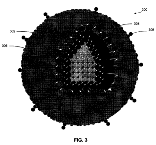
Fig. 3 illustrates an exemplary quantum dot suitable for use in accordance
with various embodiments of the invention;
Figs. 4-7 illustrate electroretinogram (ERG) measurements in control
groups and rats injected with photoactive devices in accordance with the
invention;
Fig. 8 illustrates nuclei count in ganglion cell layers. inner nuclear layers,
and photoreceptor nuclei for control sham, and active groups;
Fig. 9 illustrates Morris Water Maze Test results for control and active
groups;
Fig. 10 illustrates Recovery Round, Maximal dark-adapted ERG results for
active, sham, and control groups;
3
CA 02716991 2010-08-27
WO 2008/106605' PCT/US2008/055332
Fig. 11 illustrates Morris Water Maze Test results for control and active
groups; and
Fig. 12 illustrates photomicrographs of a human retina.
Skilled artisans will appreciate that elements in the figures are illustrated
for simplicity and clarity and have not necessarily been drawn to scale. The
dimensions of some of the elements in the figures may be exaggerated relative
to
other elements to help to improve understanding of embodiments of the present
invention.
Detailed Description Of Exemplary Embodiments
The present invention provides an improved method for stimulating an
electrical response in an eye and mitigating degradation of electrical
response to
light of an eye having a damaged retina. The method of the present invention
may
be used with a retina that is damaged due to retinitis pigmentosa, diabetic
retinopathy, macular degeneration, retinal detachment, or other retinal trauma
and
may be implemented on any animal, having an eye with the general properties
described herein.
Fig. 1 illustrates a mammal eye 100, which includes an optic nerve 102, a
lens 104, a cornea 106, an iris 108, zonules 110, a retina 112, and a vitreous
114.
In accordance with various embodiments of the invention, photoactive material
116 is injected into eye 100, e.g., using a hypodermic needle, such that the
photoactive material is dispersed within vitreous 114 and proximate retina
112. In
accordance with alternative embodiments of the invention, photoactive material
116 is injected subretinally.
Fig. 2 illustrates a portion of retina 112 in greater detail, illustrating
possible injection sites and resting sites for photoactive material 116. The
retina
includes Internal limiting member 201, nerve-fiber layer 203, ganglion-cell
layer
205, inner plexiform layer 207, inner nuclear layer 209, outer plexiform layer
211,
4
CA 02716991 2010-08-27
WO 2008/106605' PCT/US2008/055332
outer nuclear layer 213, inner segments 215, outer segments 208, Bruch's
membrane 206, RPE 219, and Choriocapillaris 221.
As noted above, photoactive material 116 may be placed on a surface 202
of retina 112 or in a subretinal area 204, such as a space located between a
Bruch's
membrane 206 and outer segments 208. Photoactive material 116 may be placed
directly in such locations, or, as described in more detail below, the
material may
be coated with a bio-targeted material, which adheres to particular cells,
such as
ganglia or bipolar cells or photoreceptors 223.
In accordance with various embodiments of the invention, photoactive
material 116 includes a quantum dot. A quantum dot is a semiconductor
nanostructure that confines motion of conduction band electrons, valence band
holes, or excitons (pairs of conduction band electrons and valence band holes)
in
three spatial directions. The confinement can be due to electrostatic
potentials
(generated by external electrodes, doping, strain, impurities), due to the
presence of
an interface between different semiconductor materials (e.g. in the case of
self-
assembled quantum dots), due to the presence of the semiconductor surface
(e.g. in
the case of a semiconductor nanocrystal), or any combination thereof.
Dimensions
of quantum dots are typically on the order of about 1 to about 100 nanometers,
and
typically about 10 to about 50 nanometers for self-assembled quantum dots.
The quantum dots fluoresce, emit an electrical potential or current, or a
combination thereof, when exposed to light. The electrical potential is
thought to
stimulate rods and cones or other portions of retina 112. The color of
fluorescence
and properties of the electrical potential general depend on the shape, size,
and
materials used to form the quantum dot.
Quantum dots for use with the present invention may be formed using a
variety of techniques. For example, the quantum dots may be formed by creating
a
region of a first material having a first bandgap surrounded by a second
material of
a second bandgap, wherein the second badgap is larger than the first bandgap.
For
example, a quantum dot may include a cadmium selenide (CdSe) core surrounded
by a zinc selenide (ZnS) shell.
5
CA 02716991 2010-08-27
WO 2008/106605' PCT/US2008/055332
Alternatively, self-assembled quantum dots nucleate spontaneously under
certain conditions during molecular beam epitaxy (MBE) and metallorganic vapor
phase epitaxy (MOVPE), when a material is grown on a substrate to which it is
not
lattice matched. The resulting strain between the grown layer and the
substrate
produces coherently strained islands on top of a two-dimensional "wetting-
layer."
The islands can be subsequently surrounded by a shell to form the quantum dot.
Individual quantum dots can also be created from two-dimensional electron
or hole gases present in remotely doped quantum wells or semiconductor
heterostructures. In this case, a surface is coated with a thin layer of
photoresist. A
lateral pattern is then defined in the resist by electron beam lithography.
This
pattern can then be transferred to the electron or hole gas by etching, or by
depositing metal electrodes (lift-off process) that allow the application of
external
voltages between the electron gas and the electrodes.
Quantum dots may also be formed in quantum well structures due to
monolayer fluctuations in the well's thickness.
Fig. 3 illustrates a quantum dot 300 suitable for use as photoactive material
116. Quantum dot 300 includes an inner semiconductor 302 core formed of, for
example, indium/gallium/phosphide, silicon, gallium arsenide, cadmium
telluride,
copper indium gallium selenide, indium gallium nitride, or organic materials
such
as polymer-fullerene heterojunctions (e.g., P3HT + C50), organic nanocrystal
solar
cells (e.g., cadmium selenide or cadmium telluride), dye sensitized cells
(e.g., dye
and titanium oxide or nobelium oxide), or a tandem cell (e.g., copper-
phthalocyanin + C60); a shell 304, formed of, for example, zinc selenide or
other
suitable material; a coating 306, formed of, for example, PEG lipids or other
suitable material; and bio-functional material 308, formed of, for example,
biotin
or other suitable proteins.
As noted above, in accordance with various embodiments of the invention,
a plurality of quantum dots exhibiting a plurality of fluorescence wavelengths
or
dots responsive to light of varying wavelengths are employed to stimulate
photoreceptors based on incident light of multiple wavelengths. For example, a
combination of nanoparticles responsive to red, blue, and green incident light
may
6
CA 02716991 2010-08-27
WO 2008/106605' PCT/US2008/055332
be employed. Various other combinations of nanoparticles/quantum dots are also
within the scope of the invention.
Use of photoactive nanoparticles such as quantum dots is advantageous
because it allows for less invasive methods of implanting the devices, which
in
turn minimizes trauma and scaring of the retina. In addition, because the
particles
are so small, the particles block relatively little light from photoreceptors
210
(illustrated in Fig. 2). Further, the quantum dots can be injected into a
wider field
of vision, compared to larger devices.
Figs. 4-7 illustrate electroretinograms (ERG) for Royal College of
Surgeons (RCS) rats with retinal degeneration, injected in vitreous 114 with
about
5 L of quantum dots 300 in saline, for a sham group, and for a control group.
Intravitreal injections: 0.5 l injected 1mm posterior to limbus; subretinal
injection: 0.1 l injected under direct visualization subretinally.
Fig. 4 illustrates maximal dark-adapted ERG, which elicits both rod and
cone photoreceptor response, in RCS rats. The control group (n=4) has had no
intervention, the sham group (n=4) has received intraocular injections of
saline,
and the QD-540 group (n=6) has received intraocular injections of quantum dots
with a biotin coating. Fig. 4 illustrates an increase in the electrical
activity of the
active implant eyes in weeks 3 through 7, compared to the sham and control
groups, which progressively decline.
Fig. 5 illustrates photopic light-adapted ERG results, which elicit
predominantly cone photoreceptor responses, in the RCS rats. The control group
(n=4) has had no interventions, the sham group (n=4) has received intraocular
injections of saline, and the QD-540 group (n=6) has received intraocular
injections of quantum dots with a biotin coating. Fig. 5 demonstrates a
general
trend for increasing electrical activity in the active implant group, compared
with a
tendency for decline in the sham and control groups over time.
Fig. 6 illustrates ERG recordings week 3 after injection. Line 602 indicates
the ERG of an RCS rat with intraocular QD-540, compared with recordings from a
representative sham surgery eye, illustrated by line 604. Fig. 7 illustrates
7
CA 02716991 2010-08-27
WO 2008/106605' PCT/US2008/055332
representative ERG recordings at week 7. Line 702 indicates the ERG of an RCS
rat with intraocular QD-540, compared with recordings from a representative
sham
surgery eye, represented by line 704. As illustrated, although the overall ERG
amplitudes for both sham and injected eyes have decreased, the eye with the
active
implants has maintained a relatively normal ERG, whereas the sham eye
recording
is essentially flat.
Fig. 8 illustrates nuclei count following a 2 month post-implantation ERG
recording. The RCS rats then were euthanized and the eyes enucleated and the
retina embedded in a plastic medium, then cut to 0.5 micron thickness and
stained
with toludine blue. Using image analysis software, the number of nuclei
present in
the ganglion cell layer, inner nuclear layer, and the photoreceptor nuclear
layer
(outer nuclear layer) were measured on five sections each 100 microns in
length.
There were three animals in the active implant group, 2 in the sham surgery
group,
and 1 in the control group.
Fig. 8 shows no appreciable difference between the groups in the number of
cells present in the ganglion cell layer, a trend for increased cells for both
the
active implant and sham surgery groups in the inner nuclear layer, and a
marked
increase in the photoreceptor nuclei in the active implant group. The
photoreceptors are the basis of the electrophysiologic network of signals
which
produce the sensation of vision. Increased numbers of cells in this layer in
the
active implant group indicates a protective effect of the active implant on
these
cells. This is consistent with Figs. 4-7, which depict a preservation of the
electrical
functioning of the retina in the active implant groups. The intraocular
quantum
dots appear to preserve both the function and the anatomy of the retina in
this
model of progressive blindness.
Fig. 9 illustrates results of the Moms Water Maze Test results for three
groups. Each group, consisting of one control animal and one active implant
animal, was tested in a water maze. The Morris Water Maze Test is a functional
test to determine whether or not the animal can see light. The test consists
of a
water escape pool (1.4m diameter, 0.6m deep, water at 20 deg Celsius). Around
the edge of the pool are six lights. The escape platform, a small pedestal
8
CA 02716991 2010-08-27
WO 2008/106605' PCT/US2008/055332
approximately 12 cm in diameter, is randomly placed adjacent to one light,
which
is then illuminated as the rat is placed in the water. The subject then has 60
seconds to swim towards the light and climb up onto the pedestal. If the
subject
does not find the pedestal within 60 seconds, the animal is removed from the
pool.
Each animal is tested a total of ten times.
In the group of animals 8 weeks post-implantation, the active implant
animal was able to escape an average of 30% faster than the control animal. In
the
5 week post-implantation group, the active implant group escaped an average
13%
more rapidly than the control group and in the 4 week post-implantation group,
the
active implant had escape times 15% faster than the control.
The results indicate that the animals receiving the active implant were
consistently able to navigate the maze more rapidly than the control animals.
The
maze is specifically designed to eliminate any tactile or olfactory cues, and
the
animal must rely entirely upon sight to successfully exit.
Fig. 10 illustrates Recovery Round, Maximal dark-adapted ERG in RCS
rats. This test elicits both rod and cone photoreceptor response. Fig. 10
illustrates
results from experiments involved in the intraocular injection of quantum dots
to
reverse blindness. The RCS rats were monitored with electroretinograms every
other week until the recordings became essentially flat, indicating a loss of
retinal
functioning. The control group (n=2) has had no intervention, the sham group
(n=2) has received intraocular injections of saline, while Groups Active
Implant
593 (n-2) and Active Implant 614 (n=2) have received intraocular injections of
quantum dots with an amino acid coating. 593 and 614 refer to the wavelength
of
light to which each quantum dot exhibits a maximum response. Recordings were
taken the day of surgery, 2 weeks post-implantation and 4 weeks post-
implantation.
The graph illustrates that both the control and sham surgery groups exhibit
no gain in the electrical functioning of the retina at any point post-
operatively. In
contrast, both active implant groups had a substantial increase in the
electrical
activity of the retina post-implantation. The Active Implant 593 group had a 2-
fold
increase in the amplitude of the waveform response to light, and the Active
9
CA 02716991 2010-08-27
WO 2008/106605' PCT/US2008/055332
Implant group 614 had a 2.5-fold increase in the amplitude of the waveform
response to light.
Fig. 11 illustrates Morris Water Maze Test, Recovery Round results for
three groups, each group consisting of one representative animal, tested in a
water
maze. The test consists of a water escape pool (1.4m diameter, 0.6m deep,
water at
20 deg Celsius). Around the edge of the pool are six lights. The escape
platform,
a small pedestal approximately 12 cm in diameter, is randomly placed adjacent
to
one light, which is then illuminated as the rat is placed in the water. The
subject
then has 60 seconds to swim towards the light and climb up onto the pedestal.
If
the subject does not find the pedestal within 60 seconds, the animal is
removed
from the pool. Each animal is tested a total of ten times.
The graph indicates that the control group averaged 60 seconds, indicating
that the maze was never successfully completed. The Active Implant 593 group
averaged 50 seconds, 17% quicker escape time than control. The Active Implant
614 group averaged 27.6 seconds, nearly twice as fast as the control group,
indicating a higher level of visual functioning.
The results indicate that the animals receiving the active implant were
consistently able to navigate the maze more rapidly than the control animals.
The
maze is specifically designed to eliminate any tactile or olfactory cues, and
the
animal must rely entirely upon sight to successfully exit.
Fig. 12 illustrates photomicrograph of a human retina (A), and quantum
dots adherent to human retinal photoreceptors (B). A whole human eye was
obtained from the Rocky Mountain Lions Eye Bank and examined grossly and
beneath an operating microscope and found to be free of any structural
abnormalities. Next, 0.05 ml of a biotin linked quantum dot with an absorption
wavelength near 528nm and an excitation wavelength of 547 nm was injected into
the subretinal space. After histological processing, the biotin linked quantum
dots
were visible by fluorescent light microscopy. The quantum dots could be seen
adherent to the native photoreceptors (arrow), as well as in unbound
aggregates in
the subretinal space (arrowhead). This demonstrates that biotin linked quantum
dots bind to human retinal photoreceptors when injected into an eye bank
CA 02716991 2010-08-27
WO 2008/106605' PCT/US2008/055332
specimen. This has practical implications in the area of neural prosthetics
and
neural protection for targeted delivery of drugs, molecules, and electric
current to
photoreceptors in disease states.
The present invention has been described above with reference to
exemplary embodiments. Those skilled in the art having read this disclosure
will
recognize that changes and modifications may be made to the embodiments
without departing from the scope of the invention. These and other changes or
modifications are intended to be included within the scope of the present
invention.
11