Note: Descriptions are shown in the official language in which they were submitted.
CA 02716998 2010-09-02
WO 2009/111459 PCT/US2009/035857
System and Method for Performing a Modular Revision Hip Prosthesis
Cross-Reference to Related Applications
[0001] This application claims the benefit of U.S. Provisional Application No.
61/033,213, filed March 3, 2008. The disclosure of the application is
incorporated by
reference in its entirety.
Field of the Invention
[0002] This invention relates generally to a system and methods for implanting
a
revision hip prosthetic and, more particularly, to a system and method for
implanting a
modular revision hip prosthetic.
Summary of the Invention
[0003] It is in view of the above problems that the present invention was
developed.
[0004] An embodiment of a revision hip implant comprises a stem, a sleeve, and
a
neck. The stem has a body portion, a proximal intramedullary canal portion and
a distal
intramedullary canal portion. The proximal intramedullary canal portion and
the distal
intramedullary canal portion are generally aligned along a long axis of the
stem. The body
portion is proximally oriented relative to the proximal intramedullary canal
portion and
further has a surface extending generally radially out from the long axis of
the stem. The
proximal intramedullary canal portion has a distally extending taper surface.
The sleeve is
configured to be received over the distal intramedullary canal portion of the
stem and further
has an inner surface configured to mate with the distally extending taper
surface of the
proximal intramedullary canal portion. The neck is configured to be received
in the body
portion of the stem. The body portion of the stem has a neck receiving surface
configured to
mate with a neck taper portion of the neck. The neck taper portion is oriented
at an angle
relative to the long axis.
CA 02716998 2010-09-02
WO 2009/111459 PCT/US2009/035857
[0005] According to another embodiment of the invention, the distal
intramedullary portion of the stem further comprises flutes extending along
the length of the
stem.
[0006] In yet another embodiment of the revision hip implant, the sleeve
further
comprises a conical portion and a cylindrical portion. The conical portion is
oriented
proximal to the stem body and the cylindrical portion extends distally away
from the conical
portion.
[0007] Alternatively, the surface extending generally radially out from the
long
axis of the stem on the body portion of the stem may be configured to rest
upon a bone cut.
[0008] In another embodiment, the sleeve is configured to asymmetrically
extend
away from the long axis of the stem.
[0009] According to another embodiment, the neck is received on the stem along
a
first axis. A head is received on the neck along a second axis, the first and
second axes being
parallel to each other.
[0010] Further features, aspects, and advantages of the present invention, as
well as
the structure and operation of various embodiments of the present invention,
are described in
detail below with reference to the accompanying drawings.
Brief Description of the Drawings
[0011] The accompanying drawings, which are incorporated in and form a part of
the
specification, illustrate embodiments of the present invention and together
with the
description, serve to explain the principles of the invention. In the
drawings:
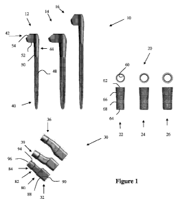
[0012] Figure 1 is a view of an embodiment of a revision hip implant set
including a
group of stems, a group of sleeves, and a group of necks;
[0013] Figure 2 is an exploded view of an embodiment of a hip implant
according to
an aspect of the invention;
-2-
CA 02716998 2010-09-02
WO 2009/111459 PCT/US2009/035857
[0014] Figure 3 is a view of an embodiment of a revision hip implant according
to an
aspect of the invention;
[0015] Figure 4 is a cutaway view of a femur with distal preparation
instrumentation
according to an aspect of the invention;
[0016] Figure 5 is a cutaway view of the femur with trialing instrumentation
according
to an aspect of the invention;
[0017] Figure 6 is a cutaway view of the femur with additional trialing
instrumentation
according to an aspect of the invention;
[0018] Figure 7 is a cutaway view of the femur with proximal preparation
instrumentation according to an aspect of the invention;
[0019] Figure 8 is a cutaway view of the femur with portions of a hip implant
and
implant instrumentation; and
[0020] Figures 9 through 11 are cutaway views of the femur with portions of a
hip
implant and hip instrumentation according to an aspect of the invention.
Detailed Description of the Embodiments
[0021] Referring to the accompanying drawings in which like reference numbers
indicate like elements, Figure 1 illustrates a view of an embodiment of a
revision hip implant
set including a group of stems 10, a group of sleeves 20, and a group of necks
30. The set
includes a three piece modular implant system that may allow a surgeon to
address the
individual patient needs. The set may be used in revision hip arthroplasty
where other
treatments or devices have failed in rehabilitating hips damaged as a result
of trauma or
noninflamatory degenerative joint disease (NIDJD) or any of its composite
diagnoses of
osteoarthritis, avascular necrosis, traumatic arthritis, slipped capital
epiphysis, fused hip,
fracture of the pelvis and diastrophic variant.
-3-
CA 02716998 2010-09-02
WO 2009/111459 PCT/US2009/035857
[0022] Modular hip components may also be indicated for inflammatory
degenerative joint disease including rheumatoid arthritis, arthritis secondary
to a variety of
diseases and anomalies and congenital dysplasia. Other indications include
old, remote
osteomyelitis with an extended drainage-free period, in which case the patient
should be
warned of an above normal danger of infection postoperatively. Such a modular
revision
system may also be used as treatments of non-union, femoral neck fracture and
trochanteric
fractures of the proximal femur with head involvement that are unmanageable
using other
techniques, endoprosthesis, femoral osteotomy or Girdlestone resection,
fracture-dislocation
of the hip, and correction of deformity. The three piece implant system may
provide distal
fixation and secondary proximal support while allowing the surgeon intra-
operative
flexibility to achieve the best patient fit. Better fixation and fit may
provide better implant
stability and outcomes.
[0023] Preoperative planning for a revision total hip arthroplasty should
require a
standard set of radiographs, which includes an antero-posterior (A-P)
radiograph of the
pelvis and a lateral radiograph of the affected hip. Depending on the length
of the existing
femoral component several additional radiographs may be necessary.
Specifically, the A-P
and lateral radiographs should include the entire femoral component. On
occasion a full-
length A-P radiograph of the entire femur may be necessary. As part of the
preoperative
work-up, the surgeon may consider other imaging modalities such as bone scans
and
computerized tomography (CT). However, these are not typically necessary for
preoperative
templating. Determine the appropriate classification for the femoral revision,
for example
the Paprosky Revision Classification. This will aid in determining the
appropriate position
and size of the revision stem you will need. As with primary THA preop
planning,
establishing proper leg length requires assessment of a number of clinical and
radiographic
parameters. Establishing the proper reference lines requires using a
horizontal line between
-4-
CA 02716998 2010-09-02
WO 2009/111459 PCT/US2009/035857
the inferior portion of the teardrop as well as a horizontal line between the
inferior margin of
the obtruator foramen and ischial tuberosity. Due to the often distorted
anatomy in revision
cases, utilizing all three reference lines may be necessary.
[0024] Similarly, due to bony defects on the femoral side, a combination of
anatomic landmarks such as the superior margin of the greater trochanter and
inferior
margin of the lesser trochanter must be utilized. These obviously need to be
compared to
similar points in the contralateral side using the A-P radiograph. Any pelvic
obliquities
and/or spinal deformity must also be taken into account based on radiographic
and clinical
assessments. The consideration of all relevant factors is necessary to
successfully restore the
patient's proper leg length.
[0025] A revision hip implant according to an aspect of the invention
generally
includes a stem 12, 14, or 16, a sleeve 22, 24, or 26, and a neck 32, 34, or
36. The stem may
be chosen from the group of stems 10 and similarly the sleeve and neck may be
chosen from
the group of sleeves 20 and the group of necks 30, respectively.
[0026] The stem 12, 14, or 16 may be a tapered stem having a distal
intramedullary
portion 40, a proximal portion 42 and a proximal intramedullary portion 44.
The distal
portion 40 may include flutes 48 positioned distally for improved fixation of
the implant to
the diaphysis. The diameters and lengths of the distal portion 40 of the stem
may vary
according to patient need. The proximal portion 42 includes a body having a
distal flat
surface 52 and a neck interface surface 54. The distal flat surface 52 may be
oriented to rest
upon a cut portion of a femur. The neck interface surface 54 mat be configured
to receive a
neck. The proximal intramedullary portion 44 of the stem includes a sleeve
mating surface
50. The sleeve mating surface 50 receives one of the group of sleeves 20.
[0027] The sleeves 20 in the system provide secondary proximal support to the
distal
fixation and enhance implant stability. The sleeves may be conical sleeves and
may be
-5-
CA 02716998 2010-09-02
WO 2009/111459 PCT/US2009/035857
coated, such as coated with Smith & Nephew's STIKTITETM coating and then
coated in
hydroxyapatite (HA). The sleeves 20 may include an internal mating surface 60
a proximal
end portion 62, a distal end portion 64, a conical portion 66 and a
cylindrical portion 68.
Generally, the proximal end portion 62 of the sleeve is larger than the distal
end portion 64.
[0028] Different sleeves 22, 24, and 26 may have different relationships
between
the different portions of the sleeves. For example, a first sleeve may have a
smaller internal
mating surface 60 (for a smaller diameter stem portion) but have a larger
conical portion 66,
thus making the sleeve thicker. Alternatively, the conical and cylindrical
portions may also
be a single portion, either cylindrical or conical. The shape and
characteristics of the sleeve
are chosen based upon the bone of the patient. Where thicker sleeves are
necessary to
ensure proximal fixation, a thicker sleeve may be used. Where sleeves
requiring more taper
are necessary, a sleeve with a larger proximal end portion 62 relative to the
proximal end
portion 64 may be used.
[0029] Alternatively, different sleeves may be asymmetric. Such a sleeve may
extend more medially, more anteriorly, more laterally, or more posteriorly
relative to the
long axis of the stem. Such a sleeve may allow the surgeon to plan for
additional
asymmetric bone defects within the IM canal.
[0030] Necks 32, 34, and 36 for the modular stem may be modular and available
in
multiple offset, leg length and version options. For example, the offsets and
leg lengths
may include a standard offset, high offset, and a high offset + 10mm. The
version may
include anteverted left and anteverted right. This allows the surgeon to fine
tune the offset,
length and version to achieve fit and function for each patient.
[0031] In order to achieve different options in the neck, the neck includes a
stem
mating portion 80 a neck extension 82 and a head mating portion 84. A stem
taper 88 is
used to join the neck to the stem. A distal end 90 of the neck is inserted
within the neck
-6-
CA 02716998 2010-09-02
WO 2009/111459 PCT/US2009/035857
mating surface 54. A head taper 94 aligns with a proximal end portion 96 of
the neck. The
proximal end portion 96 defines the most proximal portion of the neck, which
is used to
help determine offset, leg length and version. The head taper 94 joins the
neck to a femoral
head, thus setting the offset, version, and length of the prosthesis relative
to an acetabulum.
[0032] Each neck is preferably made of cobalt chrome and includes the
circulotrapezoidal neck design for increased range of motion. The 12/14 taper
of the neck
accepts compatible heads. The geometries of the necks may be changed in order
to achieve
different implant configurations. Neutral necks (neutral 0 anteversion) may
be available in
standard and high offset. The standard neutral neck may have a neck shaft
angle of 131 .
The high offset neutral neck may have a neck shaft angle of 125 when inserted
in the varus
orientation and 137 when inserted in the valgus orientation. Left anteverted
necks may shift
the femoral head, 10 degrees, anteriorly relative to the stem on a left hip
and posteriorly
relative to the stem on a right hip when inserted in the varus orientation.
Right anteverted
necks may shift the femoral head center based on the operative leg in an
equivalent manner.
Anteverted necks may be available in one offset option, for example 6mm more
than the
standard offset neck, and then may create a neck shaft angle of 125 when
inserted in the
varus orientation and a neck shaft angle of 137 when inserted in the valgus
orientation. The
high offset +10 neck may have a neck shaft angle similar to a high offset neck
and an
additional 10mm of height.
[0033] To aid in proper neck orientation, a marking such as a laser etch on
top of the
neck trunnion (the proximal neck portion 96) is provided on the high offset
and anteverted
neck implants and plastic neck trials. Such markings may be arrows or other
indicia such
that when the neck is inserted in the pocket on the neck mating surface, the
arrow on top of
the neck points superiorly relative to the patient's femur if the neck shaft
angle is varus,
125 and inferiorly if the neck shaft angle is valgus, 137 . To reduce
intraoperative
-7-
CA 02716998 2010-09-02
WO 2009/111459 PCT/US2009/035857
confusion of left & right anteverted necks, the top of the trunnion of the
necks may also be
laser etched with an "L" for left anteverted and an "R" for right anteverted.
Additionally,
color coding of the anteverted neck implant packaging and corresponding trial
necks may be
provided to account for neck characteristics or neck orientations.
[0034] Turning now to Figures 2 and 3, Figure 2 is an exploded view of an
embodiment of a hip implant according to an aspect of the invention and Figure
3 is a view of
an embodiment of a revision hip implant according to an aspect of the
invention. The sleeve
26 is received over the distal end of the stem 14. The sleeve 26 mates with
the sleeve mating
surface 44 proximally along the stem. The taper between the sleeve 26 and the
stem 14 fits
such that forces directed distally along the stem (such as patient weight)
would push against
the taper junction between the stem 14 and sleeve 26. The sleeve 26 generally
aligns along the
long axis of the stem 14.
[0035] The stem 14 receives the neck 32 in a pocket on the head portion of the
stem.
In a neck neutral orientation, the taper of the neck 32 to the stem 14 may be
aligned with the
taper of the neck 32 to the femoral head. As the offset, version and height
change, the relative
direction of the tapers (and thus the forces associated with those tapers)
move relative to one
another.
[0036] Instruments associated with the implants may provide surgical
efficiency
during the implantation of the stem. Figures 4 through 11 show steps of a
surgical procedure
which may be used to prepare, trial, and implant a modular revision hip
prosthetic device
within a femur. With respect to Figure 4, Figure 4 is a cutaway view of a
femur 100 with
distal preparation instrumentation according to an aspect of the invention. A
distal reamer 102
allows the surgeon to properly prepare and size the canal for optimal fit of
the tapered,
fluted distally fixed stem. The distal portion of the femur is reamed by
attaching a quick
connect instrument 104 to an appropriate distal reamer 102 and ream the distal
femoral
-8-
CA 02716998 2010-09-02
WO 2009/111459 PCT/US2009/035857
canal in 0.5mm increments until desired distal fit is achieved. It is
preferred to begin
reaming with a distal reamer that is at least 2mm smaller than the template
size. Such a
reamer would have little or no resistance to reaming as it progresses through
the IM canal.
Additionally, to minimize potential risk of reaming through the anterior
cortex, the reamer
102 may be directed from anterior to posterior. Preferably, the size of the
stem is 0.25mm
greater than the equivalently sized reamer, which may provide an automatic
press fit.
[0037] The depth of the reamer 102 may be determined by aligning a mark on the
quick connect instrument with the greater trochanter. If the greater
trochanter is not
available then alternative anatomical reference may be made. For example, a
ruler can be
used to measure from the distal end of the osteotomy to the previous location
of the greater
trochanter. Upon achieving desired distal fit, the quick connect device 104
may be
disengaged and the final distal reamer 102 may be left in the canal.
[0038] As shown in Figure 5, Figure 5 is a cutaway view of the femur 100 with
trialing instrumentation according to an aspect of the invention. When used in
conjunction
with trial bodies and necks, the distal reamer 102 may function as
intramedullary trials
which should provide the surgeon with more accurate assessment of implant
positioning
while reducing the number of instruments and trial components.
[0039] In some revision cases, medial bone may need to be removed to
accommodate the neck platform of the neck body. An osteotomy jig 112 may be
used to
address this bone removal. The osteotomy jig 112 is placed over the distal
reamer 102 and
proximal spacer 110 and may be fixed to reamer 112 with a screw. Arrows on the
osteotomy
jig 112 may indicate the location where the minimum bone must be removed for
the trial
neck body (and the subsequent implant) to properly seat. The location of the
depth may be
marked with a cautery pin. After the jig 112 is removed, an oscillating saw is
used to
-9-
CA 02716998 2010-09-02
WO 2009/111459 PCT/US2009/035857
osteotomize the bone. Once the bone is properly oriented, then a trial body
may be used to
position the body of the stem.
[0040] With respect to Figure 6, Figure 6 is a cutaway view of the femur 100
with
additional trialing instrumentation according to an aspect of the invention. A
trial body 116 is
seated on the spacer 110 on the distal reamer 102. A trial neck component 118
is engaged
with the body 116. Indicia on the trial neck 116 may orient the neck 118
relative to the body
116. With the trial completely formed, a range of motion (ROM) exercise may be
performed to confirm proper seating of implant, assess joint tension, and
ensure there is no
impingement to the hip. Trial neck components 118 and trial neck pocket
version
(manipulated by adjusting the rotation of the body 116 on the spacer 110) may
be adjusted
as appropriate to achieve desired leg length, neck offset, and neck version. A
mark can be
made on the bone with a cautery pin to mark desired version for implant.
[0041] Turning now to Figure 7, Figure 7 is a cutaway view of the femur 100
with
proximal preparation instrumentation according to an aspect of the invention.
A proximal
reamer120 may be used over the distal reamer 102. These "over the top" reamers
120 allow
the surgeon to leave the distal reamer 102 in the canal to maintain the
relative position of the
cavities for the proximal sleeve and the distal taper of the stem. By reaming
"over the top"
the surgeon is preparing for the proximal segment based on the location of the
distal reamer.
[0042] The distal reamer may be used by first removing the trial neck body,
trial
neck component, and proximal trial spacer. The distal reamer 102 is left in
the canal.
Beginning with the smallest appropriate size, the proximal cone reamer 120 is
advanced
over the shaft of the distal reamer 102. The proximal cone reamer 120 will
rotate, however
the distal reamer 102 will act as a guide and should not advance. If an ETO is
not performed
or if the femoral canal is small, a starter proximal reamer may be necessary
to remove bone
that may impede the application of the proximal cone reamers 120. The canal is
-10-
CA 02716998 2010-09-02
WO 2009/111459 PCT/US2009/035857
progressively reamed using the proximal cone reamers until the desired
proximal fit is
achieved.
[0043] Following the proximal sleeve preparation, re-trialing may be performed
using the proximal trial spacer, trial neck body, trial neck, and trial head
component over the
distal reamer. If re-trialing is not performed following the proximal sleeve
preparation, then
the distal reamer component may be removed.
[0044] With respect to Figure 8, Figure 8 is a cutaway view of the femur 100
with
portions of a hip implant and implant instrumentation. The stem 14 and sleeve
26 are prepared
for implant. The sleeve 26 is slid over the distal end of the stem 14 and
seated on the stem 14.
A stem insertion instrument 126 attaches to the proximal end of the stem 14
generally along
the long axis of the stem. The stem/sleeve assembly is impacted into the femur
using a
mallet against the driving platform. The stem inserter should not impinge on
the trochanter.
This may cause inadequate stem seating, trochanteric fracture or varus
positioning. The
surgeon may assess height and neck pocket orientation as the stem advances
into the desired
position. If a cautery pin mark was made during the trialing step, it can be
used as a
reference. The stem inserter instrument 126 is then disengaged when the stem
is properly
seated in the canal.
[0045] Turning now to Figures 9 through 11, Figures 9 through 11 are cutaway
views
of the femur 100 with portions of a hip implant 14, 26 and 34 and hip
instrumentation 132
according to an aspect of the invention. Once the stem 14 is seated, it is
recommended that
re-trialing be performed using the trial neck and head components. Fine-tuning
of offset, leg
length, and version can be made with the modular neck options. Once proper
neck
orientation is determined, the modular neck trial is replaced with the modular
neck implant
34 prior to impaction. All taper surfaces should be protected, clean and dry
prior to
assembly to ensure a good taper lock. Once the neck 34 is verified in the
desired orientation
-11-
CA 02716998 2010-09-02
WO 2009/111459 PCT/US2009/035857
prior to final impaction, the surgeon impacts the final neck 34 and head
implant component
130 simultaneously with the head/ neck impaction tool 132. Correct selection
of the neck
length and cup, and stem positioning are important. Muscle looseness and/or
malpositioning
of components may result in loosening, subluxation, dislocation, and/ or
fracture of the
component and/or bone. A final ROM check may ensure desired implant outcome
prior to
closing with the implants implanted.
[0046] In view of the foregoing, it will be seen that the several advantages
of the
invention are achieved and attained.
[0047] The embodiments were chosen and described in order to best explain the
principles of the invention and its practical application to thereby enable
others skilled in the
art to best utilize the invention in various embodiments and with various
modifications as are
suited to the particular use contemplated.
[0048] As various modifications could be made in the constructions and methods
herein described and illustrated without departing from the scope of the
invention, it is
intended that all matter contained in the foregoing description or shown in
the accompanying
drawings shall be interpreted as illustrative rather than limiting. Thus, the
breadth and scope
of the present invention should not be limited by any of the above-described
exemplary
embodiments, but should be defined only in accordance with the following
claims appended
hereto and their equivalents.
-12-