Note: Descriptions are shown in the official language in which they were submitted.
CA 02717071 2010-08-27
WO 2009/117706 PCT/US2009/037883
METHODS OF TREATMENT USING ANTI-MIF ANTIBODIES
CROSS-REFERENCE
[0001] This application claims the benefit of U.S. Provisional Application No.
61/038,381, filed
March 20,2008; U.S. Provisional Application No. 61/039,371, filed March 25,
2008; U.S.
Provisional Application No. 61/045,807, filed April 17, 2008; and U.S.
Provisional Application No.
61/121,095, filed December 09, 2008; which applications are incorporated
herein by reference in
their entirety.
BACKGROUND OF THE INVENTION
[0002] Certain inflammatory conditions are characterized, in part, by the
migration lymphocytes
into the effected tissue. The migration of lymphocytes induces tissue damage
and exacerbates
inflammatory conditions. Many leukocytes follow a MIF gradient to the effected
tissue. In general,
MIF interacts with CXCR2 and CXCR4 receptors on leukocytes to trigger and
maintain leukocyte
migration.
SUMMARY OF THE INVENTION
[0003] Disclosed herein, in certain embodiments, is a method of treating a MIF-
mediated disorder
comprising administering to an individual in need thereof a therapeutically-
effective amount of an
antibody that inhibits (i) MIF binding to CXCR2 and/or CXCR4 (ii) MIF-
activation of CXCR2
and/or CXCR4; (iii) the ability of MIF to form a homomultimer; (iv) MIF
binding to CD74; or a
combination thereof. In some embodiments, the antibody specifically binds to
all or a portion of a
pseudo-ELR motif of MIF. In some embodiments, the antibody specifically binds
to all or a portion
of an N-Loop motif of MIF. In some embodiments, the antibody specifically
binds to all or a portion
of the pseudo-ELR and N-Loop motifs of MIF. In some embodiments, the antibody
is selected from
an anti-CXCR2 antibody; an anti-CXCR4 antibody, an anti-MIF antibody; an
antibody that
specifically binds to all or a portion of the pseudo-ELR motif of MIF; an
antibody that specifically
binds to all or a portion of the N-loop motif of MIF; an antibody that
specifically binds to all or a
portion of the pseudo-ELR and N-Loop motifs; an antibody that inhibits the
binding of MIF and
CXCR2; an antibody that inhibits the binding of MIF and CXCR4; and antibody
that inhibits the
binding of MIF and JAB-1; an antibody that inhibits the binding of MIF and
CD74; an antibody that
specifically binds to all or a portion of a peptide sequence as follows:
DQLMAFGGSSEPCALCSL
and the corresponding featureldomain of at least one of a MIF monomer or MIF
trimer; an antibody
that specifically binds to all or a portion of a peptide sequence as follows:
PRASVPDGFLSELTQQLAQATGKPPQYIAVHVVPDQLMAFGGSSEPCALCSL and the
corresponding feature/domain of at least one of a MIF monomer or MIF trimer;
an antibody that
4-
CA 02717071 2010-08-27
WO 2009/117706 PCT/US2009/037883
specifically binds to all or a portion of a peptide sequence as follows:
FGGSSEPCALCSLHSI and
the corresponding featureldomain of at least one of a MIF monomer or MIF
trimer; or combinations
thereof. In some embodiments, the antibody is selected from anti-CXCR4
antibodies: 701, 708, 716,
717, 718, 12G5 and 4G10; anti-MIF antibodies: IID.9, IIID.9, XIF7, I31, IV2.2,
XI17, XIV14.3,
X1115.6 and XIV15.4; or combinations thereof. In some embodiments, the
conversion of a
macrophage into a foam cell is inhibited following administration of an
antibody disclosed herein. In
some embodiments, apoptosis of a cardiac myocyte is inhibited following
administration of an
antibody disclosed herein. In some embodiments, apoptosis of an infiltrating
macrophage is
inhibited following administration of an antibody disclosed herein. In some
embodiments, the
formation of an abdominal aortic aneurysm is inhibited following
administration of an antibody
disclosed herein. In some embodiments, the diameter of an abdominal aortic
aneurysm is decreased
following administration of an antibody disclosed herein. In some embodiments,
a structural protein
in an aneurysm is regenerated following administration of an antibody
disclosed herein. In some
embodiments, the method further comprises co-administering a second active
agent. In some
embodiments, the method further comprises co-administering niacin, a fibrate,
a statin, a Apo-Al
mimetic peptide (e.g., DF-4, Novartis), an apoA-I transcriptional up-
regulator, an ACAT inhibitor, a
CETP modulator, Glycoprotein (GP) lb/IIIa receptor antagonists, P2Y12 receptor
antagonists, Lp-
PLA2-inhibitors, an anti-TNF agent, an IL-1 receptor antagonist, an IL-2
receptor antagonist, a
cytotoxic agent, an immunomodulatory agent, an antibiotic, a T-cell co-
stimulatory blocker, a
disorder-modifying anti-rheumatic agent, a B cell depleting agent, an
immanosuppressive agent, an
anti-lymphocyte antibody, an alkylating agent, an anti-metabolite, a plant
alkaloid, a terpenoids, a
topoisomerase inhibitor, an anti-tumor antibiotic, a monoclonal antibody, a
hormonal therapy, or
combinations thereof. In some embodiments, the MIF-mediated disorder is
Atherosclerosis;
Abdominal aortic aneurysm; Acute disseminated encephalomyelitis; Moyamoya
disease; Takayasu
disease; Acute coronary syndrome; Cardiac-allograft vasculopathy; Pulmonary
inflammation; Acute
respiratory distress syndrome; Pulmonary fibrosis; Addison's disease;
Ankylosing spondylitis;
Antiphospholipid antibody syndrome; Autoimmune hemolytic anemia; Autoimmune
hepatitis;
Autoimmune inner ear disease; Bullous pemphigoid; Chagas disease; Chronic
obstructive
pulmonary disease; Coeliac disease; Demratomyositis; Diabetes mellitus type 1;
Diabetes mellitus
type 2; Endometriosis; Goodpasture's syndrome; Graves' disease; Guillain-Barre
syndrome;
Hasbimoto's disease; Idiopathic thrombocytopenic purpura; Interstitial
cystitis; Systemic lupus
erythematosus (SLE); Metabolic syndrome, Multiple sclerosis; Myasthenia
gravis; Myocarditis,
Narcolepsy; Obesity; Pemphigus Vulgaris; Pernicious anaemia; Polymyositis;
Primary biliary
cirrhosis; Rheumatoid arthritis; Schizophrenia; Scleroderma; Sjogren's
syndrome; Vasculitis;
Vitiigo; Wegener's granulomatosis; Allergic rhinitis; Prostate cancer; Non-
small cell lung
carcinoma; Ovarian cancer; Breast cancer; Melanoma; Gastric cancer; Colorectal
cancer; Brain
-2-
CA 02717071 2010-08-27
WO 2009/117706 PCT/US2009/037883
cancer, Metastatic bone disorder; Pancreatic cancer; a Lymphoma; Nasal polyps;
Gastrointestinal
cancer; Ulcerative colitis; Crohn's disorder; Collagenous colitis; Lymphocytic
colitis; Ischaemic
colitis; Diversion colitis; Behpet's syndrome; Infective colitis;
Indeterminate colitis; Inflammatory
liver disorder, Endotoxin shock, Septic shock; Rheumatoid spondylitis,
Ankylosing spondylitis,
Gouty arthritis, Polymyalgia rheumatica, Alzheimer's disorder, Parkinson's
disorder, Epilepsy, AIDS
dementia, Asthma, Adult respiratory distress syndrome, Bronchitis, Cystic
fibrosis, Acute
leukocyte-mediated lung injury, Distal proctitis, Wegener's granulomatosis,
Fibromyalgia,
Bronchitis, Cystic fibrosis, Uveitis, Conjunctivitis, Psoriasis, Eczema,
Dermatitis, Smooth muscle
proliferation disorders, Meningitis, Shingles, Encephalitis, Nephritis,
Tuberculosis, Retinitis, Atopic
dermatitis, Pancreatitis, Periodontal gingivitis, Coagulative Necrosis,
Liquefhctive Necrosis,
Fibrinoid Necrosis, Neointimal hyperplasia, Myocardial infarction; Stroke;
Organ transplant
rejection; or combinations thereof. In some embodiments, the disorder is an
abdominal aortic
aneurysm In some embodiments, the disorder is atherosclerosis.
[0004] Disclosed herein, in certain embodiments, is a pharmaceutical
composition for treatment of
a MIF-mediated disorder, comprising an antibody that inhibits (i) MIF binding
to CXCR2 and
CXCR4; (ii) MIF-activation of CXCR2 and CXCR4; (iii) the ability of MIF to
form a
homomultimer; or a combination thereof. In some embodiments, the antibody
specifically binds to
all or a portion of a pseudo-ELR motif of MIF. In some embodiments, the
antibody specifically
binds to all or a portion of a N-Loop motif of MIF. In some embodiments, the
antibody specifically
binds to all or a portion of the pseudo-ELR and N-Loop motifs of MIF. In some
embodiments, the
antibody is selected from an anti-CXCR2 antibody; an anti-CXCR4 antibody; an
anti-MIF antibody;
an antibody that specifically binds to all or a portion of the pseudo-ELR
motif of MIF; an antibody
that specifically binds to all or a portion of the N-loop motif of MIF; an
antibody that specifically
binds to all or a portion of the pseudo-ELR and N-Loop motifs; an antibody
that inhibits the binding
of MIF and CXCR2; an antibody that inhibits the binding of MIF and CXCR4; and
antibody that
inhibits the binding of MIF and JAB-1; an antibody that inhibits the binding
of MIF and CD74; an
antibody that specifically binds to all or a portion of a peptide sequence as
follows:
DQLMAFGGSSEPCALCSL and the corresponding feature/domain of at least one of a
MIF
monomer or MIF trimer; an antibody that specifically binds to all or a portion
of a peptide sequence
as follows: PRASVPDGFLSELTQQLAQATGKPPQYIAVHVVPDQLMAFGGSSEPCALCSL and
the corresponding feature/domain of at least one of a MIF monomer or MIF
trimer; an antibody that
specifically binds to all or a portion of a peptide sequence as follows:
FGGSSEPCALCSLHSI and
the corresponding featureldomain of at least one of a MIF monomer or MIF
trimer, or combinations
thereof. In some embodiments, the antibody is selected from anti-CXCR4
antibodies 701, 708, 716,
717, 718, 12G5 and 4G10; anti-MIF antibodies IID.9, IIID.9, XIF7, I31, W2.2,
XI17, XIV14.3,
XII15.6 and XIV 15.4; or combinations thereof. In some embodiments, the
composition further
-3-
CA 02717071 2010-08-27
WO 2009/117706 PCT/US2009/037883
comprises a second active agent. In some embodiments, the composition further
comprises niacin, a
fibrate, a statin, a Apo-Al mimetic peptide (e.g., DF-4, Novartis), an apoA-I
transcriptional up-
regulator, an ACAT inhibitor, a CETP modulator, Glycoprotein (GP) IIb/IlIa
receptor antagonists,
P2Y12 receptor antagonists, Lp-PLA2-inhibitors, an anti-TNF agent, an IL-1
receptor antagonist, an
IL-2 receptor antagonist, a cytotoxic agent, an immunomodulatory agent, an
antibiotic, a T-cell co-
stimulatory blocker, a disorder-modifying anti-rheumatic agent, a B cell
depleting agent, an
immunosuppressive agent, an anti-lymphocyte antibody, an alkylating agent, an
anti-metabolite, a
plant alkaloid, a terpenoids, a topoisomerase inhibitor, an anti-tumor
antibiotic, a monoclonal
antibody, a hormonal therapy, or combinations thereof.
BRIEF DESCRIPTION OF THE DRAWINGS
[0005] The novel features of the invention are set forth with particularity in
the appended claims. A
better understanding of the features and advantages of the present invention
will be obtained by
reference to the following detailed description that sets forth illustrative
embodiments, in which the
principles of the invention are utilized, and the accompanying drawings of
which:
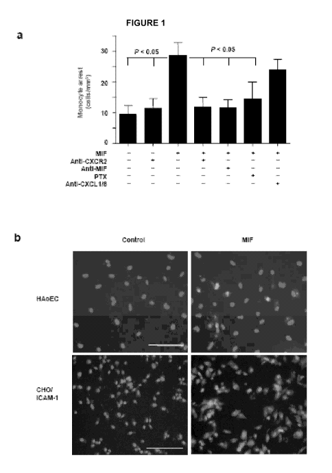
[0006] Figure 1 is an illustration that MIF-triggered mononuclear cell arrest
is mediated by
CXCR2/CXCR4 and CD74. Human aortic endothelial cells (HAoECs), CHO cells
stably expressing
ICAM-1 (CHO/ICAM-1) and mouse microvascular endothelial cells (SVECs) were
preincubated
with or without MIF (together with antibody to MIF, antibodies to CXCLI and
CXCL8, or isotype
control), CXCL8, CXCLIO or CXCL12 for 2 h as indicated. Mononuclear cells were
pretreated with
antibodies to CXCRI, CXCR2, 02, CXCR4, CD74, or isotype controls for 30 min,
or pertussis toxin
(PTX) for 2 has indicated. (a) HAoECs were perfused with primary human
monocytes. (b)
Immunofluorescence using antibody to MIF revealed surface presentation of MIF
(green) on
HAoECs and CHO/ICAM-1 cells after pretreatment for 2 h, but not 30 min (not
shown); in contrast,
MIF was absent in buffer-treated cells (control). Scale bar, 100 gm. (c,d)
CHO/ICAM-l cells were
perfused with MonoMac6 cells. (e) HAoECs were perfused with T cells. (f,g)
CHO/ICAM-l cells
were perfused with Jurkat T cells (f), and with Jurkat CXCR2 transfectants or
vector controls (g). In
c,d,f and g, background binding to vector-transfected CHO cells was
subtracted. (h) Mouse SVECs
were perfused with L1.2 transfectants stably expressing CXCRI, CXCR2 or CXCR3,
and with
controls expressing only endogenous CXCR4, in the presence of the CXCR4
antagonist AMD3465.
Arrest is quantified as cells/mm2 or as percentage of control cell adhesion.
Data in a and c-g
represent mean f s.d. of 3-8 independent experiments; data in h are results
from one representative
experiment of four experiments.
[0007] Figure 2 is an illustration that MIF-triggered mononuclear cell
chemotaxis is mediated by
CXCR2/CXCR4 and CD74. Primary human monocytes (a-e), CD3* T cells (f) and
neutrophils (g h)
-4-
CA 02717071 2010-08-27
WO 2009/117706 PCT/US2009/037883
were individuated to transmigration analysis in the presence or absence of
MIF. CCL2 (a), CXCL8
(a,g,h) and CXCL12 (f) served as positive controls or were used to test
desensitization by MIF (or
by CXCL8, h). The chemotactic effects of MIF, CCL2 and CXCL8 on monocytes (a)
or of MIF on
neutrophils (g) followed bell-shaped dose-response curves. MIF-triggered
chemotaxis of monocytes
was abrogated by an antibody to MIF, boiling (b), or by MIF at indicated
concentrations (in the top
chamber; c). (d) MIF-triggered chemotaxis was mediated by Go;/phosphoinositide-
3-kinase
signaling, as evidenced by treatment with pertussis toxin components A and B
(PTX A + B), PTX
component B alone or Ly294002. (e) MIF-mediated monocyte chemotaxis was
blocked by
antibodies to CD74 or CXCRI/CXCR2. (f) T-cell che.rnotaxis induced by MIF was
blocked by
antibodies to MIF and CXCR4. (g) Neutrophil chemotaxis induced by MIF. (h) MIF-
induced versus
CXCL8-induced neutrophil chemotaxis, effects of antibodies to CXCR2 or CXCR1,
and
desensitization of CXCL8 by MIF. Data in a and f-h are expressed as
chemotactic index; data in c
are expressed as percent of control; and data in b,d and e as percent of
input. Data represent mean
f
s.d. of 4-10 independent experiments, except for panels a,c and g, boiled MIF
in b, and the
antibody-alone controls in b and e, which are means of 2 independent
experiments.
[0008] Figure 3 is an illustration that MIF triggers rapid integrin activation
and calcium signaling.
(a) Human aortic endothelial cells were stimulated with MIF or TNF-a for 2 h.
CXCLI and CXCL8
mRNAs were analyzed by real-time PCR and normalized to control. Supernatant-
derived CXCL8
was assessed by ELISA (n = 3 independent experiments performed in duplicate).
(b) MonoMac6
cells were directly stimulated with MIF or CXCL8 for 1 min and perfused on CHO-
ICAM-1 cells
for 5 min (mean f s.d. of 8 independent experiments). (c) MonoMac6 cells were
stimulated with
MIF for the indicated times. LFA-1 activation (detected by the 327C antibody)
was monitored by
FACSAria, and expressed as the increase in mean fluorescence intensity (MFI).
(d) As in c but for
primary monocytes; data are expressed relative to maximal activation with
Mgt+/EGTA. (e)
MonoMac6 cells were pretreated with antibodies to a,4 integrin, CD74 or CXCR2,
stimulated with
MIF for 1 min, perfused on VCAM-1.Fc for 5 min. Adhesion is expressed as a
percentage of
controls. Arrest data in c-e represent mean s.d. of 5 independent
experiments. (f) Calcium
transients in Fluo-4 AM-labeled neutrophils were stimulated with MIF, CXCL8 or
CXCL7.
Calcium-derived MFI was recorded by FACSAria for 0-240 s. For desensitization,
stimuli were
added 120 a before stimulation. Traces shown represent 4 independent
experiments. (g) Dose-
response curves of calcium-influx triggered by CXCL8, CXCL7 or MIF, at
indicated concentrations,
in Ll.2-CXCR2 transfectants. Data are expressed as the difference between
baseline and peak MFI
(mean s.d. of 4-8 independent experiments).
[0009] Figure 4 is an illustration of MIF-interaction with CXCR2/CXCR4 and
formation of
CXCR2/CD74 complexes. HEK293-CXCR2 transfectants (a) or CXCR4-bearing Jurkat T-
cells (c)
were individuated to receptor binding assays, analyzing competition of
[I'25]CXCL8 (a) or
-5-
CA 02717071 2010-08-27
WO 2009/117706 PCT/US2009/037883
[Iu5]CXCL12 (c) by MIF or cold cognate ligand (mean f s.d., n = 6-10). (b) MIF-
and CXCL8-
induced CXCR2 internalization in HEK293-CXCR2 or RAW264.7-CXCR2 transfectants
(inset
shows representative histograms) as indicated; assessed by FACS analysis of
surface CXCR2
expression (percentage of buffer (Con), mean s.d., n = 5). (d) MIF- and
CXCL12-induced CXCR4
internalization in 7urkat T-cells as in b (mean s.d., n = 4-6). (e) Binding
of fluorescein-MIF to
HEK293-CXCR2 transfectants or vector controls analyzed by FACS. Inset shows
binding of biotin-
MIF to CXCR2 assessed by western blot using antibodies to CXCR2 after
streptavidin (SAv) pull-
down from HEK293-CXCR2 transfectants versus vector controls. (f)
Colocalization of CXCR2 and
CD74 (orange-yellow overlay) in RAW264.7-CXCR2 transfectants stained for
CXCR2, CD74 and
nuclei (Hoechst), analyzed by fluorescence microscopy (top) or confocal laser
scanning microscopy
(bottom). Scale bar, 10 m. (g) Coimmunoprecipitation of CXCR2/CD74 complexes
in CHAPSO-
extracts of HEK293-CXCR2 transfectants expressing His-tagged CD74. Anti-His
immunoprecipitation (IP) followed by anti-CXCR2 or anti-His-CD74 western
blotting (WB; top) or
anti-CXCR2 immunoprecipitation followed by anti-His-M74 or anti-CXCR2 western
blotting
(bottom). Controls: lysates without immunoprecipitation or beads alone. (h) As
in g for L1.2-
CXCR2 transfectants. Anti-CXCR2 immunoprecipitation from LI.2-CXCR2
transfectants followed
by anti-CD74 or anti-CXCR2 western blotting (top). Immunoprecipitation with
isotype IgG or
CXCR2-negative L1.2-cells (bottom) served as controls. Data represent 3
independent experiments
(e-h).
[0010] Figure 5 is an illustration that MIF-induced atherogenic and
microvascular inflammation
through CXCR2 in vivo and effects of MIF blockade on plaque regression. (a)
Monocyte adhesion to
the lumen in vivo and lesional macrophage content in native aortic roots were
determined in
Mif'+Ldlr' and MfLdld mice (n = 4) fed a chow diet for 30 weeks.
Representative images are
shown. Arrows indicate monocytes adherent to the luminal surface. Scale bar,
100 m. (b,c)
Exposure to MIF induced CXCR2-dependent leukocyte recruitment in vivo.
Following intrascrotal
injection of MIF, the cremasteric microvasculature was visualized by
intravital microscopy.
Pretreatment with blocking CXCR2 antibody abrogated adhesion and emigration,
as compared to
IgG control (n = 4). (d) Intraperitoneal injection of MIF or vehicle elicited
neutrophil recruitment in
wild-type mice (n = 3) reconstituted with wild-type, but not I18r1J, bone
marrow. (e-h) Blocking
MIF but not CXCL1 or CXCL12 resulted in regression and stabilization of
advanced atherosclerotic
plaques. Apod mice received a high-fat diet for 12 weeks and were subsequently
treated with
antibodies to MIF, CXCLI or CXCL12, or with vehicle (control) for an
additional 4 weeks of (n =
6-10 mice). Plaques in the aortic root were stained using Oil-Red-O.
Representative images are
shown inc (scale bars, 500 pm). Data in f represent plaque area at baseline
(12 weeks) and after 16
weeks, The relative content of MOMA-2' macrophages is shown in g and the
number of CD3+ T
cells per section in h. Data represent mean s.d.
-6-
CA 02717071 2010-08-27
WO 2009/117706 PCT/US2009/037883
[0011] Figure 6 is an illustration of cellular mechanisms of MIF in the
context of atherogenesis.
MIF expression is induced in cells of the vascular wall and intimal
macrophages by various
proatherogenic stimuli, e.g., oxidized LDL (oxLDL) or angiotensin 11(ATII).
Subsequently, MIF
upregulates endothelial cell adhesion molecules (e.g., vascular [VCAM-1] and
intracellular [ICAM-
1] adhesion molecules) and chemokines (e.g., CCL2) and triggers direct
activation of the respective
integrin receptors (e.g., LFA-1 and VIA-4) by binding and signaling through
its heptahelical
(chemokine) receptors CXCR2 and CXCR4. This entails the recruitment of
mononuclear cells
(monocytes and T cells) and the conversion of macrophages into foam cells,
inhibiting apoptosis and
regulating (e.g., impairing) the migration or proliferation of SMCs. By
inducing MMPs and
cathepsins, MIF promotes elastin and collagen degradation, ultimately leading
to the progression
into unstable plaques. ROS indicates reactive oxygen species; PDGF-BB,
platelet-derived growth
factor-BB.
[0012] Figure 7 is an illustration of signaling via a functional MIF receptor
complex. MIF is
induced by glucocorticoids overriding their function by regulating cytokine
production and, after its
endocytosis, can interact with intracellular proteins, namely JAB-I, thereby
downregulating MAPK
signals and modulating cellular redox homeostasis. In some embodiments,
extracellular MIF binds
to the cell surface protein CD74 (invariant chain Ii). CD74 lacks a signal-
transducing intracellular
domain but interacts with the proteoglycan CD44 and mediates signaling via
CD44 to induce
activation of Src-family RTK and MAPK/extracellular signal-regulated kinase
(ERK), to activate
the PI3K/Akt pathway, or to initiate p53-dependent inhibition of apoptosis.
MIF also binds and
signals through G protein-coupled chemokine receptors (CXCR2 and CXCR4) alone.
Complex
formation of CXCR2 with CD74, enabling accessory binding, facilitates GPCR
activation and
formation of a GPCR-RTK-like signaling complex to trigger calcium influx and
rapid integrin
activation.
[0013] Figure 8 is an illustration of the effects of MIF in myocardial
pathology. In the context of
ischemia-reperfusion, hypoxia, reactive oxygen species (ROS), and endotoxins
(e.g.,
lipopolysaccharide [LPS]) in sepsis induce the secretion of MIF from
cardiomyocytes through a
protein kinase C (PKC)-dependent mechanism and result in extracellular signal-
regulated kinase
(ERK) activation, which contributes to cardiomyocyte apoptosis. Expressed by
surviving
cardiomyocytes or by endothelial progenitor cells (e.g., eEPCs) used for
therapeutic injection, in
some embodiments MIF promotes angiogenesis via its receptors CXCR2 and CXCR4,
requiring
MAPK and P13K activation.
[00141 Figure 9 is an illustration that interference with CXCR4 without
concomitant interference
with CXCR2 aggravates atherosclerosis. Apoe-/- mice receiving a high-fat diet
were continuously
treated with vehicle (control) or AMD3465 via osmotic minipumps for 12 weeks
(n=6 each).
Atherosclerotic plaques were quantified in the aortic root (Fig. 14a) and
thoracoabdominal aorta
-7-
CA 02717071 2010-08-27
WO 2009/117706 PCT/US2009/037883
(Fig. 14b) after oil red O staining. The relative number of neutrophils was
determined by flow
cytometric analysis or standard cytometry in peripheral blood at the indicated
time points (Fig. 14C).
[0015] Figure 10 illustrates the crystal structure of a MIF trimer. The pseudo-
ELR domains form a
ring in the trimer while the N-loop domains extend outward from the pseudo-ELR
ring.
[0016] Figure 11 illustrates the nucleotide sequence of MIF annotated to show
the sequences that
correspond to the N-Loop domain and the pseudo-ELR domain.
DETAILED DESCRIPTION OF THE INVENTION
[0017] Disclosed herein, in certain embodiments, are methods of inhibiting MIF
signaling through
CXCR2 and CXCR4. In some embodiments, MIF signaling through CXCR2 and CXCR4 is
inhibited by occupying the MIF binding domain of CXCR2 and CXCR4 with an
antibody. In some
embodiments MIF signaling through CXCR2 and CXCR4 is inhibited by occupying,
masking, or
otherwise disrupting domains on MIF. In some embodiments, MIF signaling
through CXCR2 and
CXCR4 is inhibited by an antibody occupying, masking, or otherwise disrupting
domains on MIF
and thereby disrupting the binding of CXCR2 and/or CXCR4 to MW. In some
embodiments, MIF
signaling through CXCR2 and CXCR4 is inhibited by an antibody occupying,
masking, or otherwise
disrupting domains on MIF and thereby disrupting MIF trimerization.
[0018] While the art teaches anti-MIF antibodies, the art lacks recognition
that certain portions of
MIF are more important than others with respect to leukocyte interactions. A
problem solved herein
is the identification and raising of antibodies that bind the selective
portions of MIF that are
important to leukocyte chemotaxis.
[0019] Further, the art teaches anti-CXCR2 and anti-CXCR4 antibodies. However,
these receptors
are also involved in interactions with other ligands (e.g., IL-8/CXCL8,
GRObeta/CXCL2 and/or
Stromal Cell-Derived Factor-la (SDF-la)/CXCL12). Detrimental side-effects
often arise if these
interactions are inhibited. A problem solved herein is the failure of the art
to design anti-CXCR2 and
anti-CXCR4 antibodies that selectively inhibit interactions with MIF.
Certain Definitions
[0020] The terms "individual," "subject," or "patient" are used
interchangeably. As used herein,
they mean any mammal (i.e. species of any orders, families, and genus within
the taxonomic
classification animalia: chordata: vertebrata: mammalia), In some embodiments,
the mammal is a
human. In some embodiments, the mammal is a non-human. In some embodiments,
the mammal is a
member of the taxonomic orders: primates (e.g. lemurs, lorids, galagos,
tarsiers, monkeys, apes, and
humans); rodentia (e.g. mice, rats, squirrels, chipmunks, and gophers);
lagomorpha (e.g. hares,
rabbits, and pika); erinaceomorpha (e.g. hedgehogs and gynmures); soricomorpha
(e.g. shrews,
moles, and solenodons); ch iroptera (e.g., bats); cetacea (e.g. whales,
dolphins, and porpoises);
-8-
CA 02717071 2010-08-27
WO 2009/117706 PCT/US2009/037883
carnivora (e.g. cats, lions, and other feliformia; dogs, bears, weasels, and
seals); perissodactyla (e.g.
horse, zebra, tapir, and rhinoceros); artiodactyla (e.g. pigs, camels, cattle,
and deer); proboscidea
(e.g. elephants); sirenia (e.g. manatees, dugong, and sea cows); cingulata
(e.g. armadillos); pilosa
(e.g. anteaters and sloths); didelphimorphia (e.g. american opossums);
paucituberculata (e.g. shrew
opossums); microbiotheria (e.g. Monito del Monte); notoryctemorphia (e.g.
marsupial moles);
dasyuromorphia (e.g. marsupial carnivores); peramelemorphia (e.g. bandicoots
and bilbies); or
diprotodontia (e.g. wombats, koalas, possums, gliders, kangaroos, wallaroos,
and wallabies). In
some embodiments, the animal is a reptile (i.e. species of any orders,
families, and genus within the
taxonomic classification animalia: chordata: vertebrata: reptilia). In some
embodiments, the animal
is a bird (i.e. animalia: chordata: vertebrata: ayes). None of the terms
require or are limited to
situation characterized by the supervision (e.g. constant or intermittent) of
a health care worker (e.g.
a doctor, a registered nurse, a nurse practitioner, a physician's assistant,
an orderly, or a hospice
worker).
[0021] The term "antigen" refers to a substance that is capable of inducing
the production of an
antibody. In some embodiments an antigen is a substance that specifically
binds to an antibody
variable region.
[0022] The terms "antibody" and "antibodies" refer to monoclonal antibodies,
polyclonal
antibodies, bi-specific antibodies, multispecific antibodies, grafted
antibodies, human antibodies,
humanized antibodies, synthetic antibodies, chimeric antibodies, camelized
antibodies, single-chain
Fvs (scFv), single chain antibodies, Fab fragments, F(ab') fragments,
disulfide-linked Fvs (sdFv),
intrabodies, and anti-idiotypic (anti-Id) antibodies and antigen-binding
fragments of any of the
above. In particular, antibodies include immunoglobulin molecules and
immunologically active
fragments of immunoglobulin molecules, i.e., molecules that contain an antigen
binding site.
Depending on the amino acid sequence of the constant domain of their heavy
chains,
immunoglobulins can be assigned to different classes. The heavy-chain constant
domains (Fc) that
correspond to the different classes of immunoglobulins are called a, S, a, y,
and , respectively. The
subunit structures and three-dimensional configurations of different classes
of immunoglobulins are
well known. Immunoglobulin molecules are of any type (e.g., IgG, IgE, IgM,
IgD, IgA and IgY),
class (e.g., IgG 1, IgG 2, IgG 3, IgG 4. IgA 1 and IgA 2) or subclass. The
terms "antibody" and
"immnunoglobulin" are used interchangeably in the broadest sense. In some
embodiments an
antibody is part of a larger molecule, formed by covalent or non-covalent
association of the antibody
with one or more other proteins or peptides.
[0023] With respect to antibodies, the term "variable domain" refers to the
variable
domains of antibodies that are used in the binding and specificity of each
particular antibody for its
particular antigen. However, the variability is not evenly distributed
throughout the variable domains
of antibodies. Rather, it is concentrated in three segments called
hypervariable regions (also known
-9-
CA 02717071 2010-08-27
WO 2009/117706 PCT/US2009/037883
as CDRs) in both the light chain and the heavy chain variable domains. More
highly conserved
portions of variable domains are called the "framework regions" or "FRs." The
variable domains of
unmodified heavy and light chains each contain four FRs (FR1, FR2, FR3 and
FR4), largely
adopting a n-sheet configuration interspersed with three CDRs which form loops
connecting and, in
some cases, part of the n-sheet structure. The CDRs in each chain are held
together in close
proximity by the FRs and, with the CDRs from the other chain, contribute to
the formation of the
antigen-binding site of antibodies (see Kabat et al., Sequences of Proteins of
Immunological Interest,
5th Ed. Public Health Service, National Institutes of Health, Bethesda, Md.
(1991), pages 647-669).
[0024] The terms "hypervariable region" and "CDR" when used herein, refer to
the
amino acid residues of an antibody which are responsible for antigen-binding.
The CDRs comprise
amino acid residues from three sequence regions which bind in a complementary
manner to an
antigen and are known as CDR1, CDR2, and CDR3 for each of the VH and VL
chains. In the light
chain variable domain, the CDRs typically correspond to approximately residues
24-34 (CDRL1),
50-56 (CDRL2) and 89-97 (CDRL3), and in the heavy chain variable domain the
CDRs typically
correspond to approximately residues 31-35 (CDRH1), 50-65 (CDRH2) and 95-102
(CDRH3)
according to Kabat et al., Sequences of Proteins of Immunological Interest,
5th Ed. Public Health
Service, National Institutes of Health, Bethesda, Md. (1991)). It is
understood that the CDRs of
different antibodies may contain insertions, thus the amino acid numbering may
differ. The Kabat
numbering system accounts for such insertions with a numbering scheme that
utilizes letters
attached to specific residues (e.g., 27A, 27B, 27C, 27D, 27E, and 27F of CDRL1
in the light chain)
to reflect any insertions in the numberings between different antibodies.
Alternatively, in the light
chain variable domain, the CDRs typically correspond to approximately residues
26-32 (CDRL1),
50-52 (CDRL2) and 91-96 (CDRL3), and in the heavy chain variable domain, the
CDRs typically
correspond to approximately residues 26-32 (CDRH1), 53-55 (CDRH2) and 96-101
(CDRH3)
according to Chothia and Lesk, J. Mol. Biol., 196: 901-917 (1987)).
[0025] As used herein, "framework region" or "FR" refers to framework amino
acid
residues that form a part of the antigen binding pocket or groove. In some
embodiments, the
framework residues form a loop that is a part of the antigen binding pocket or
groove and the amino
acids residues in the loop may or may not contact the antigen. Framework
regions generally
comprise the regions between the CDRs. In the light chain variable domain, the
FRs typically
correspond to approximately residues 0-23 (FRL1), 35-49 (FRL2), 57-88 (FRL3),
and 98-109 and in
the heavy chain variable domain the FRs typically correspond to approximately
residues 0-30
(FRH1), 36-49 (FRH2), 66-94 (FRH3), and 103-133 according to Kabat et al.,
Sequences of
Proteins of Immunological Interest, 5th Ed. Public Health Service, National
Institutes of Health,
Bethesda, Md. (1991)). As discussed above with the Kabat numbering for the
light chain, the heavy
chain too accounts for insertions in a similar manner (e.g., 35A, 35B of CDRH1
in the heavy chain).
-10-
CA 02717071 2010-08-27
WO 2009/117706 PCT/US2009/037883
Alternatively, in the light chain variable domain, the FRs typically
correspond to approximately
residues 0-25 (FRL1), 33-49 (FRL2) 53-90 (FRL3), and 97-109 (FRL4), and in the
heavy chain
variable domain, the FRs typically correspond to approximately residues 0-25
(FRHI), 33-52
(FRH2), 56-95 (FRH3), and 102-113 (FRH4) according to Chothia and Lesk, J.
Mol. Biol., 196:
901-917 (1987)).
[0026] The loop amino acids of a FR can be assessed and determined by
inspection of
the three-dimensional structure of an antibody heavy chain and/or antibody
light chain. The three-
dimensional structure can be analyzed for solvent accessible amino acid
positions as such positions
are likely to form a loop and/or provide antigen contact in an antibody
variable domain. Some of the
solvent accessible positions can tolerate amino acid sequence diversity and
others (e.g., structural
positions) are, generally, less diversified. The three dimensional structure
of the antibody variable
domain can be derived from a crystal structure or protein modeling.
[0027] Constant domains (Fc) of antibodies are not involved directly in
binding an
antibody to an antigen but, rather, exhibit various effector functions, such
as participation of the
antibody in antibody-dependent cellular toxicity via interactions with, for
example, Fc receptors
(FcR). Fc domains can also increase bioavailability of an antibody in
circulation following
administration to a patient.
[0028] The "light chains" of antibodies (immunoglobulins) from any vertebrate
species
can be assigned to one of two clearly distinct types, called kappa or ("u")
and lambda or CV), based
on the amino acid sequences of their constant domains.
[0029] The term "derivative" in the context of an antibody refers to an
antibody that comprises an
amino acid sequence which has been altered by the introduction of amino acid
residue substitutions,
deletions or additions. The term "derivative" also refers to an antibody which
has been modified,
i.e., by the covalent attachment of any type of molecule to the antibody. For
example, in some
embodiments an antibody is modified, e.g., by glycosylation, acetylation,
pegylation,
phosphorylation, anridation, derivatization by protecting/blocking groups,
proteolytic cleavage,
linkage to a cellular ligand or other protein, etc. In some embodiments,
antibodies and derivatives
thereof are produced by chemical modifications using suitable techniques,
including, but not limited
to specific chemical cleavage, acetylation, formylation, metabolic synthesis
of tunicamycin, etc. In
some embodiments, a derivative of an antibody possesses a similar or identical
function as the
antibody from which it was derived.
[0030] The terms `full length antibody," "intact antibody" and "whole
antibody" are used herein
interchangeably, to refer to an antibody in its substantially intact form, and
not antibody fragments
as defined below. These terms particularly refer to an antibody with heavy
chains contains Fc
regions. In some embodiments an antibody variant provided herein is a full
length antibody. In some
embodiments the full length antibody is human, humanized, chimeric, and/or
affinity matured.
-11-
CA 02717071 2010-08-27
WO 2009/117706 PCT/US2009/037883
[0031] An "affinity matured" antibody is one having one or more alteration in
one or more CDRs
thereof which result in an improvement in the affinity of the antibody for
antigen, compared to a
parent antibody which does not possess those alteration(s). Preferred affinity
matured antibodies will
have nanomolar or even picomolar affinities for the target antigen. Affinity
matured antibodies are
produced by suitable procedures. See, for example, Marks et at., (1992)
Biotechnology 10:779-783
that describes affinity maturation by variable heavy chain (VH) and variable
light chain (VL)
domain shuffling. Random mutagenesis of CDR and/or framework residues is
described in: Barbas,
et al. (1994) Proc. Nat. Acad. Sci, USA 91:3809-3813; Shier et al., (1995)
Gene 169:147-155;
Yelton et al., 1995, J. Immoral. 155:1994-2004; Jackson et al., 1995, J.
Immunol. 154(7):3310-9;
and Hawkins at al, (19920, J. Mol. Biol. 226:889-896, for example.
[0032] The terms "binding fragment," "functional fragment," "antibody
fragment" or "antigen
binding fragment" are used herein, for purposes of the specification and
claims, to mean a portion or
fragment of an intact antibody molecule, preferably wherein the fragment
retains antigen-binding
function. Examples of antibody fragments include Fab, Fab', F(ab'}2, Fd (Vn
and CHI domains), Fd'
and Fv (the VL and VH domains of a single arm of an antibody) fragments,
diabodies, linear
antibodies (Zapata et al. (1995) Protein Eng. 10: 1057), variable light chains
(VL), variable heavy
chains (VH), single-chain antibody molecules, single-chain binding
polypeptides, scFv, scFv2 (a
tandem linkage of two scFv molecules head to tail in a chain), bivalent scFv,
tetravalent scFv, one-
half antibodies, dAb fragments, variable NAR domains, and bispecific or
multispecific antibodies
formed from antibody fragments (e.g., a bi-specific Fab2, and a tri-specific
Fab3, etc.).
[0033] "Fab" fragments are typically produced by papain digestion of
antibodies resulting in the
production of two identical antigen-binding fragments, each with a single
antigen-binding site and a
residual "Fc" fragment. Pepsin treatment yields a F(ab')2 fragment that has
two antigen-combining
sites capable of cross-linking antigen. An 'Tv" is the minimum antibody
fragment that contains a
complete antigen recognition and binding site. In a two-chain Fv species, this
region consists of a
dimer of one heavy- and one light-chain variable domain in tight, non-covalent
association. In a
single-chain Fv (scFv) species, one heavy- and one light-chain variable domain
are covalently linked
by a flexible peptide linker such that the light and heavy chains associate in
a "dimeric" stricture
analogous to that in a two-chain Fv species. It is in this configuration that
the three CDRs of each
variable domain interact to define an antigen-binding site on the surface of
the VH-VL dimer.
Collectively, the six CDRs confer antigen-binding specificity to the antibody.
However, even a
single variable domain (or half of an Fv comprising only three CDRs specific
for an antigen) has the
ability to recognize and bind antigen, although usually at a lower affinity
than the entire binding site.
[0034] The Fab fragment also contains the constant domain of the light chain
and the first constant
domain (CHI) of the heavy chain. Fab fragments differ from Fab' fragments by
the addition of a few
residues at the carboxy terminus of the heavy-chain CHI domain including one
or more cysteines
-12-
CA 02717071 2010-08-27
WO 2009/117706 PCT/US2009/037883
from the antibody hinge region. Fab'-SH is the designation herein for Fab' in
which the cysteine
residue(s) of the constant domains bear a free thiol group. F(ab)2 antibody
fragments originally were
produced as pairs of Fab' fragments that have hinge cysteines between them.
Other chemical
couplings of antibody fragments are also suitable. Methods for producing the
various fragments
from monoclonal Abs include, e.g., Pluckthun, 1992, Immunol. Rev. 130:152-188.
[00351 "Fv" refers to an antibody fragment which contains a complete antigen-
recognition and antigen-binding site. This region consists of a dieter of one
heavy chain and one
light chain variable domain in tight, non-covalent association. It is in this
configuration that the three
CDRs of each variable domain interact to define an antigen-binding site on the
surface of the VH-VL
dieter. Collectively, a combination of one or more of the CDRs from each of
the VH and VL chains
confer antigen-binding specificity to the antibody. For example, it would be
understood that, for
example, the CDRH3 and CDRL3 could be sufficient to confer antigen-binding
specificity to an
antibody when transferred to VH and VL chains of a recipient antibody or
antigen-binding fragment
thereof and this combination of CDRs can be tested for binding, affinity, etc.
using any of the
techniques described herein. Even a single variable domain (or half of an Fv
comprising only three
CDRs specific for an antigen) has the ability to recognize and bind antigen,
although likely at a
lower affinity than when combined with a second variable domain. Furthermore,
although the two
domains of a Fv fragment (VL and VH), are coded for by separate genes, they
can be joined using
recombinant methods by a synthetic linker that enables them to be made as a
single protein chain in
which the VL and Vii regions pair to form monovalent molecules (known as
single chain Fv (scFv);
Bird et al. (1988) Science 242:423-426; Huston et al. (1988) Proc. Natl. Acad.
Sci. USA 85:5879-
5883; and Osbourn et al. (1998) Nat. Biotechnol. 16:778). Such scFvs are also
intended to be
encompassed within the term "antigen-binding portion" of an antibody. Any VH
and VL sequences of
specific scFv can be linked to an Fc region cDNA or genomic sequences, in
order to generate
expression vectors encoding complete Ig (e.g., IgG) molecules or other
isotypes. VH and VL can also
be used in the generation of Fab, Fv or other fragments of Igs using either
protein chemistry or
recombinant DNA technology.
[00361 "Single-chain Fv" or "sFv" antibody fragments comprise the Vii and VL
domains of an antibody, wherein these domains are present in a single
polypeptide chain. In some
embodiments, the Fv polypeptide further comprises a polypeptide linker between
the VH and VL
domains which enables the sFv to form the desired structure for antigen
binding. For a review of
sFvs see, e.g., Pluckthun in The Pharmacology of Monoclonal Antibodies, Vol.
113, Rosenburg and
Moore eds. Springer-Verlag, New York, pp. 269-315 (1994).
[00371 The term "Avimer" refers to a class of therapeutic proteins of human
origin,
which are unrelated to antibodies and antibody fragments, and are composed of
several modular and
reusable binding domains, referred to as A-domains (also referred to as class
A module, complement
-13-
CA 02717071 2010-08-27
WO 2009/117706 PCT/US2009/037883
type repeat, or LDL-receptor class A domain). They were developed from human
extracellular
receptor domains by in vitro exon shuffling and phage display (Silverman et
al., 2005, Nat.
Biotechnol. 23:1493-1494; Silverman et al., 2006, Nat. Biotechnol. 24:220).
The resulting proteins
can contain multiple independent binding domains that can exhibit improved
affinity (in some cases,
sub-nanomolar) and specificity compared with single-epitope binding proteins.
See, for example,
U.S. Patent Application Publ. Nos. 200 5 /022 1 3 8 4, 2005/0164301,
2005/0053973 and
2005/0089932, 2005/0048512, and 2004/0175756, each of which is hereby
incorporated by
reference herein in its entirety.
[00381 Each of the known 217 human A-domains comprises -35 amino acids (-.4
kDa); and domains are separated by linkers that average five amino acids in
length. Native A-
domains fold quickly and efficiently to a uniform, stable structure mediated
primarily by calcium
binding and disulfide formation. A conserved scaffold motif of only 12 amino
acids is required for
this common structure. The end result is a single protein chain containing
multiple domains, each of
which represents a separate function. Each domain of the proteins specifically
binds independently
and the energetic contributions of each domain are additive. These proteins
were called
"AvimersT from avidity multimers.
[0039] The term "diabodies" refers to small antibody fragments with two
antigen-
binding sites, which fragments comprise a heavy chain variable domain (V1)
connected to a light
chain variable domain (V L) in the same polypeptide chain (VH-VL). By using a
linker that is too
short to allow pairing between the two domains on the same chain, the domains
are forced to pair
with the complementary domains of another chain and create two antigen-binding
sites. Diabodies
are described more fully in, for example, EP 404,097; WO 93/11161; and
Hollinger et al., Proc.
Natl. Acad. Sci. USA 90:6444 6448 (1993).
[0040] Antigen-binding polypeptides also include heavy chain dimers such as,
for
example, antibodies from camelids and sharks. Camelid and shark antibodies
comprise a
homodimeric pair of two chains of V-like and C-like domains (neither has a
light chain). Since the
Vu region of a heavy chain dimer IgG in a camelid does not have to make
hydrophobic interactions
with a light chain, the region in the heavy chain that normally contacts a
light chain is changed to
hydrophilic amino acid residues in a camelid. VH domains of heavy-chain dieter
IgGs are called VHH
domains. Shark Ig-NARs comprise a homodimer of one variable domain (termed a V-
NAR domain)
and five C-like constant domains (C-NAR domains). In camelids, the diversity
of antibody
repertoire is determined by the CDRs 1, 2, and 3 in the VH or VHH regions. The
CDR3 in the camel
VHH region is characterized by its relatively long length, averaging 16 amino
acids (Muyldennans at
al., 1994, Protein Engineering 7(9): 1129). This is in contrast to CDR3
regions of antibodies of
many other species. For example, the CDR3 of mouse VH has an average of 9
amino acids. Libraries
of camelid-derived antibody variable regions, which maintain the in vivo
diversity of the variable
-14-
CA 02717071 2010-08-27
WO 2009/117706 PCT/US2009/037883
regions of a camelid, can be made by, for example, the methods disclosed in
U.S. Patent Application
Ser. No. 20050037421.
[0041] The term "monoclonal antibody" refers to an antibody obtained from a
population of
substantially homogeneous antibodies, i.e., the individual antibodies
comprising the population are
identical except for possible naturally occurring mutations that are present
in minor amounts. In
some embodiments, monoclonal antibodies are made, for example, by the
hybridoma method first
described by Kohler and Milstein (1975) Nature 256:495, or are made by
recombinant methods,
e.g., as described in U.S. Pat. No. 4,816,567. In some embodiments, monoclonal
antibodies are
isolated from phage antibody libraries using the techniques described in
Clackson et al., Nature
352:624-628 (1991), as well as in Marks et al., J. Mol. Biol. 222:581-597
(1991).
[0042] The antibodies herein include monoclonal, polyclonal, recombinant,
chimeric, humanized,
bi-specific, grafted, human, and fragments thereof including antibodies
altered by any means to be
less immunogenic in humans. Thus, for example, the monoclonal antibodies and
fragments, etc.,
herein include "chimeric" antibodies and "humanized" antibodies. In general,
chimeric antibodies
include a portion of the heavy and/or light chain that is identical with or
homologous to
corresponding sequences in antibodies derived from a particular species or
belonging to a particular
antibody class or subclass, while the remainder of the chain(s) is identical
with or homologous to
corresponding sequences in antibodies derived from another species or
belonging to another
antibody class or subclass, so long as they exhibit the desired biological
activity (U.S. Pat. No.
4,816,567); Morrison et al. Proc. Nall Acad. Sci. 81:6851-6855 (1984). For
example, in some
embodiments, a chimeric antibody contains variable regions derived from a
mouse and constant
regions derived from human in which the constant region contains sequences
homologous to both
human IgG2 and human IgG4.
[00431 "Humanized" forms of non-human (e.g., murine) antibodies or fragments
are chimeric
immunoglobulins, immunoglobulin chains or fragments thereof (such as Fv, Fab,
Fab', F(ab')2 or
other antigen-binding subsequences of antibodies) which contain minimal
sequence derived from
non-human immunoglobulin. Humanized antibodies include, grafted antibodies or
CDR grafted
antibodies wherein part or all of the amino acid sequence of one or more
complementarity
determining regions (CDRs) derived from a non-human animal antibody is grafted
to an appropriate
position of a human antibody while maintaining the desired binding specificity
and/or affinity of the
original non-human antibody. In some embodiments, corresponding non-human
residues replace Fv
framework residues of the human immunoglobulin. In some embodiments humanized
antibodies
comprise residues that are found neither in the recipient antibody nor in the
imported CDR or
framework sequences. These modifications are made to further refine and
optimize antibody
performance. In some embodiments, the humanized antibody comprises
substantially all of at least
one, and typically two, variable domains, in which all or substantially all of
the CDR regions
-15-
CA 02717071 2010-08-27
WO 2009/117706 PCT/US2009/037883
correspond to those of a non-human immunoglobulin and all or substantially all
of the FR regions
are those of a human immunoglobulin consensus sequence. For further details,
see, e.g.: Jones et al.,
Nature 321: 522-525 (1986); Reichmann et al., Nature 332: 323-329 (1988) and
Presta, Curr. Op.
Struct. Biol. 2: 593-596 (1992).
[0044] As used herein, the term "affinity" refers to the equilibrium constant
for the reversible
binding of two agents and is expressed as Kd. Affinity of a binding protein to
a ligand such as
affinity of an antibody for an epitope can be, for example, from about 100
nanomolar (nM) to about
0.1 nM, from about 100 nM to about 1 picomolar (pM), or from about 100 nM to
about 1
femtomolar (IM). As used herein, the term "avidity" refers to the resistance
of a complex of two or
more agents to dissociation after dilution.
[0045] The phrase "specifically specifically binds " when referring to the
interaction between an
antibody or other binding molecule and a protein or polypeptide or epitope,
typically refers to an
antibody or other binding molecule that recognizes and detectably specifically
binds with high
affinity to the target of interest. Preferably, under designated or
physiological conditions, the
specified antibodies or binding molecules bind to a particular polypeptide,
protein or epitope yet
does not bind in a significant or undesirable amount to other molecules
present in a sample. In other
words the specified antibody or binding molecule does not undesirably cross-
react with non-target
antigens and/or epitopes. A variety of immunoassay formats are used to select
antibodies or other
binding molecule that are immunoreactive with a particular polypeptide and
have a desired
specificity. For example, solid-phase ELISA immunoassays, BlAcore (Surface
Plasmon
Resonance), flow cytometry and radioimmunoassays are used to select monoclonal
antibodies
having a desired immunoreactivity and specificity. See, Harlow, 1988,
ANTIBODIES, A
LABORATORY MANUAL, Cold Spring Harbor Publications, New York (hereinafter,
"Harlow"), for a
description of immunoassay formats and conditions that are used to determine
or assess
immunoreactivity and specificity.
[0046] "Selective binding," "selectivity", and the like refer the preference
of an antibody to interact
with one molecule as compared to another, Preferably, interactions between
antibodies, particularly
modulators, and proteins are both specific and selective. Note that in some
embodiments an antibody
is designed to "specifically bind" and "selectively bind" two distinct, yet
similar targets without
binding to other undesirable targets.
[0047] An "epitope" or "binding site" is an amino acid sequence or sequences
that are
"preferentially bound" or "specifically bound" by an antibody or antigen-
binding fragment thereof.
An epitope can be a linear peptide sequence (i.e., "continuous") or can be
composed of
noncontiguous amino acid sequences (i.e., "conformational" or
"discontinuous"). Epitopes
recognized by an antibody or antigen-binding fragment thereof described herein
can be determined
by peptide mapping and sequence analysis techniques well known to one of skill
in the art.
-16-
CA 02717071 2010-08-27
WO 2009/117706 PCT/US2009/037883
[0048] The terms "polypeptide," "peptide" and "protein" are used
interchangeably herein to refer to
a polymer of amino acid residues. The terms apply to naturally occurring amino
acid polymers as
well as amino acid polymers in which one or more amino acid residues is a non-
naturally occurring
amino acid, e.g., an amino acid analog. The terms encompass amino acid chains
of any length,
including full length proteins (i.e., antigens), wherein the amino acid
residues are linked by covalent
peptide bonds.
[0049] The terms "isolated" and "purified" refer to a material that is
substantially or
essentially removed from or concentrated in its natural environment. For
example, an isolated
nucleic acid is one that is separated from at least some of the nucleic acids
that normally flank it or
other nucleic acids or components (proteins, lipids, etc.) in a sample. In
another example, a
polypeptide is purified if it is substantially removed from or concentrated in
its natural environment.
Methods for purification and isolation of nucleic acids and proteins are
documented methodologies.
For example, antibodies can be isolated and purified from the culture
supernatant or ascites
mentioned above by saturated ammonium sulfate precipitation, euglobulin
precipitation method,
caproic acid method, caprylic acid method, ion exchange chromatography (DEAF
or DE52), or
affinity chromatography using anti-Ig column or a protein A, G or L column. .
Embodiments of
"substantially" include at least 20%, at least 40%, at least 50%, at least
75%, at least 85%, at least
90%, at least 95%, or at least 99%.
[0050] The terms "treat," "treating" or "treatment," and other grammatical
equivalents as used
herein, include alleviating, inhibiting or reducing symptoms, reducing or
inhibiting severity o1
reducing incidence of, prophylactic treatment of, reducing or inhibiting
recurrence of preventing,
delaying onset of, delaying recurrence of, abating or ameliorating a disease
or condition symptoms,
ameliorating the underlying metabolic causes of symptoms, inhibiting the
disease or condition, e.g.,
arresting the development of the disease or condition, relieving the disease
or condition, causing
regression of the disease or condition, relieving a condition caused by the
disease or condition, or
stopping the symptoms of the disease or condition. The terms further include
achieving a therapeutic
benefit. By therapeutic benefit is meant eradication or amelioration of the
underlying disorder being
treated, and/or the eradication or amelioration of one or more of the
physiological symptoms
associated with the underlying disorder such that an improvement is observed
in the individual.
[0051] The terms "prevent," "preventing" or "prevention," and other
grammatical equivalents as
used herein, include preventing additional symptoms, preventing the underlying
metabolic causes of
symptoms, inhibiting the disease or condition, e.g., arresting the development
of the disease or
condition and are intended to include prophylaxis. The terms further include
achieving a
prophylactic benefit. For prophylactic benefit, the compositions are
optionally administered to an
individual at risk of developing a particular disease, to an individual
reporting one or more of the
physiological symptoms of a disease, or to an individual at risk of
reoccurrence of the disease.
-17-
CA 02717071 2010-08-27
WO 2009/117706 PCT/US2009/037883
[0052] The terms "effective amount' 'or "therapeutically effective amount" as
used
herein, refer to a sufficient amount of at least one agent being administered
which achieve a desired
result, e.g., to relieve to some extent one or more symptoms of a disease or
condition being treated.
In certain instances, the result is a reduction and/or alleviation of the
signs, symptoms, or causes of a
disease, or any other desired alteration of a biological system. In specific
instances, the result is a
decrease in the growth of, the killing of, or the inducing of apoptosis in at
least one abnormally
proliferating cell, e.g., a cancer stem cell. In certain instances, an
"effective amount" for therapeutic
uses is the amount of the composition comprising an agent as set forth herein
required to provide a
clinically significant decrease in a disease. An appropriate "effective"
amount in any individual case
is determined using any suitable technique, such as a dose escalation study.
[0053] The terms "administer," "administering," "administration," and the
like, as used herein,
refer to the methods that are used to enable delivery of agents or
compositions to the desired site of
biological action. These methods include, but are not limited to oral routes,
intraduodenal routes,
parenteral injection (including intravenous, subcutaneous, intraperitoneal,
intramuscular,
intravascular or infusion), topical and rectal administration. Administration
techniques that are
optionally employed with the agents and methods described herein, include
e.g., as discussed in
Goodman and Gilman, The Pharmacological Basis of Therapeutics, current ed.;
Pergamon; and
Remington's, Pharmaceutical Sciences (current edition), Mack Publishing Co.,
Easton, Pa. In certain
embodiments, the agents and compositions described herein are administered
orally.
[0054] The term "pharmaceutically acceptable" as used herein, refers to a
material that does not
abrogate the biological activity or properties of the agents described herein,
and is relatively
nontoxic (i.e., the toxicity of the material significantly outweighs the
benefit of the material). In
some instances, a pharmaceutically acceptable material is administered to an
individual without
causing significant undesirable biological effects or significantly
interacting in a deleterious manner
with any of the components of the composition in which it is contained.
Macrophage Migration Inhibitory Factor (MIF)
[0055] In some embodiments, a method and/or composition disclosed herein
inhibits (partially or
fully) the activity of MIF. In certain instances, MIF is a pro-inflammatory
cytokine. In certain
instances, it is secreted by activated immune cells (e.g. a lymphocyte (T-
cell)) in response to an
infection, inflammation, or tissue injury. In certain instances, MIF is
secreted by the anterior
pituitary gland upon stimulation of the hypothalamic-pituitary-adrenal axis.
In certain instances,
MIF is secreted together with insulin from the pancreatic beta-cells and acts
as an autocrine factor to
stimulate insulin release. In certain instances, MIF is a ligand for the
receptors CXCR2, CXCR4,
and CD74. In some embodiments, a method and/or composition disclosed herein
inhibits (partially
or fully) the activity of CXCR2 CXCR4, and/or CD74.
-18-
CA 02717071 2010-08-27
WO 2009/117706 PCT/US2009/037883
[0056] In certain instances, MIF induces chemotaxis in nearby leukocytes (e.g.
lymphocytes,
granulocytes, monocytes/macrophages, and TH-17 cells) along a MIF gradient. In
some
embodiments, a method and/or composition disclosed herein prevents chemotaxis
along a MIF
gradient, or reduces chemotaxis along a MIF gradient. In certain instances,
MIF induces the
chemotaxis of a leukocyte (e.g. lymphocytes, granulocytes,
monocytes/macrophages, and TH-17
cells) to the site of an infection, inflammation or tissue injury. In some
embodiments, a method
and/or composition disclosed herein prevents or decreases the chemotaxis of a
leukocyte to the site
of an infection, inflammation or tissue injury. In certain instances, the
chemotaxis of a leukocyte
(e.g. lymphocytes, granulocytes, monocytes/macrophages, and TH-17 cells) along
a MIF gradient
results in inflammation at the site of infection, inflammation, or tissue
injury. In some embodiments,
a method and/or composition disclosed herein treats inflammation at the site
of infection,
inflammation, or tissue injury. In certain instances, the chemotaxis of
monocytes along a RANTES
gradient results in monocyte arrest (i.e., the deposition of monocytes on
epithelium) at the site of
injury or inflammation. In some embodiments, a method and/or composition
disclosed herein
prevents or decreases monocyte arrest at the site of injury or inflammation.
In some embodiments, a
method and/or composition disclosed herein inhibits treats a lymphocyte
mediated disorder. In some
embodiments, a method and/or composition disclosed herein treats a granulocyte
mediated disorder.
In some embodiments, a method and/or composition disclosed herein treats a
macrophage mediated
disorder. In some embodiments, a method and/or composition disclosed herein
treats a Th-17
mediated disorder. In some embodiments, a method and/or composition disclosed
herein treats a
pancreatic beta-cell mediated disorder.
[0057] In certain instances, MIF is inducible by glucocorticoids, a mechanism
implicated in an
acceleration of atherosclerosis associated with many diseases requiring
glucocorticoid therapy.
Thus, in some embodiments, the compositions and methods described herein
inhibit the induction of
MIF expression by glucocorticoids.
[0058] In certain instances, a human MIF polypeptide is encoded by a
nucleotide sequence located
on chromosome 22 at the cytogenic band 22ql 1.23. In certain instances, a MIF
protein is a 12.3 kDa
protein. In certain instances, a MIF protein is a homotrimer comprising three
polypeptides of 115
amino acids. In certain instances, a MIF protein comprises a pseudo-ELR motif
that mimics the ELR
motif found in chemokines. In certain instances, the pseudo-ELR motif
comprises two nonadjacent
but adequately spaced residues (Arg12 and Asp45 & see Fig. 11). In some
embodiments the pseudo-
ELR motif comprises the amino acid sequence from amino acid 12 to amino acid
45 (such
numbering includes the first methionine residue). This is equivalent to a
pseudo-ELR motif from
amino acid 11 to amino acid 44 in which the first methionine residue is not
counted (in such
instances, the pseudo-ELR motif comprises Arg 11 and Asp 44). In some
embodiments, a method
-19-
CA 02717071 2010-08-27
WO 2009/117706 PCT/US2009/037883
and/or composition disclosed herein treats a MIF-mediated disorder by
inhibiting binding of the
pseudo-ELR motif to CXCR2 and/or CXCR4.
[0059] In certain instances, a MIF protein comprises a 10- to 20-residue N-
terminal Loop motif (N-
loop). In certain instances, a M1F N-loop mediates binding to a CXCR2 and/or
CXCR4 receptor. In
certain instances, the N-loop motif of MIF comprises the sequential residues
(47-56) of MIF (i.e.
L47 M48 A49 F50 G51 G52 S53 S54 E55 P56; see Fig. 11). In certain instances,
the N-loop motif
of MIF comprises amino acids 45-60. In certain instances, the N-loop motif of
MIF comprises
amino acids 44-61. In certain instances, the N-loop motif of MIF comprises
amino acids 43-62. In
certain instances, the N-loop motif of MIF comprises amino acids 42-63. In
certain instances, the N-
loop motif of MIF comprises amino acids 41-64. In certain instances, the N-
loop motif of MIF
comprises amino acids 40-65. In certain instances, the N-loop motif of MIF
comprises amino acids
46-59. In certain instances, the N-loop motif of MIF comprises amino acids 47-
59. In certain
instances, the N-loop motif of MIF comprises amino acids 48-59. In certain
instances, the N-loop
motif of MIF comprises amino acids 50-59. In certain instances, the N-loop
motif of MIF comprises
amino acids 47-58. In certain instances, the N-loop motif of MIF comprises
amino acids 47-57. In
certain instances, the N-loop motif of MIF comprises amino acids 47-56. In
certain instances, the N-
loop motif of MIF comprises amino acids 48-58. In some embodiments the N-Loop
motif comprises
amino acids 48-57. In some embodiments, a method and/or composition disclosed
herein treats a
MIF-mediated disorder by inhibiting binding of the N-loop motif to CXCR2
and/or CXCR4.
[0060] In some embodiments, a method and/or composition disclosed herein
treats a MIF-mediated
disorder by inhibiting (1) binding of the N-loop motif to CXCR2 and/or CXCR4;
and (2) binding of
the pseudo-ELR motif to CXCR2 and/or CXCR4.
[0061] In certain instances, CD74 activates G-protein coupled receptors
(GPCRs), activates
CXCR2, and/or associates with these molecules to form signaling complex. Thus,
in some
embodiments, a method and/or composition disclosed herein treats a MIF-
mediated disorder by
inhibiting the activation GPCRs or CXCR2 by CD74.
[0062] In certain instances, MEP is expressed by endothelial cells, SMCs,
mononuclear cells, and/or
macrophages following arterial injury. In some embodiments, a method and/or
composition
disclosed herein inhibits the expression of MIF by endothelial cells, SMCs,
mononuclear cells,
and/or macrophages following arterial injury. In certain instances, MIF is
expressed by endothelial
cells, SMCs, mononuclear cells, macrophages following exposure to oxidized low-
density
lipoprotein (oxLDL), CD40 ligand, angiotensin II, or combinations thereof. In
some embodiments, a
method and/or composition disclosed herein inhibits the expression of MIF by
endothelial cells,
SMCs, mononuclear cells, and/or macrophages following exposure to oxidized low-
density
lipoprotein, CD40 ligand, angiotensin II, or combinations thereof.
-20-
CA 02717071 2010-08-27
WO 2009/117706 PCT/US2009/037883
[0063] In certain instances, MIF induces expression of CCL2, TNF, and/or ICAM-
1 in endothelial
cells. In some embodiments, a method and/or composition disclosed herein
inhibits the MIF-induced
expression of CCL2, TNF, and/or ICAM-1 in endothelial cells.
[0064] In certain instances, MIF induces expression of MMPs and cathepsins in
SMCs. In some
embodiments, a method and/or composition disclosed herein inhibits the MIF-
induced expression of
MMPs and cathepsins in SMCs.
[0065] In certain instances, MIF triggers a calcium influx through CXCR2 or
CXCR4, induces a
rapid activation of integrins, induces MAPK activation, and mediates the Gai-
and integrin
dependent arrest and the chemotaxis of monocytes and T cells (Figures 2 and
3). Thus, In some
embodiments, a method and/or composition disclosed herein inhibits calcium
influx in monocytes
and/or T cells, inhibit activation of MAPK, inhibit activation of integrins,
inhibit Gal- and integrin
dependent arrest of monocytes and T cells, or combinations thereof.
[0066] In some embodiments, the methods described herein comprise an anti-
CXCR2 antibody; an
anti-CXCR4 antibody; an anti-MIF antibody; or combinations thereof. In some
embodiments, an
antibody disclosed herein inhibits the binding of MIF to CXCR2 and/or CXCR4 by
binding to a
pseudo-ELR motif of MIF. In some embodiments, an antibody disclosed herein
inhibits the binding
of MIF to CXCR2 and/or CXCR4 by binding to an N-loop motif of MIF. In some
embodiments, an
antibody disclosed herein inhibits the binding of MIF to CXCR2 and/or CXCR4 by
simultaneously
binding to both an N-loop motif AND a pseudo-ELR motif of MIF. In some
embodiments, an
antibody disclosed herein is an anti-MIF antibody.
[0067] In certain instances, monocyte recruitment induced by MIF involves the
MIF-binding
protein CD74. In certain instances, the MIF-binding protein CD74 induces
calcium influx, mitogen-
activated protein kinase (MAPK) activation, or Gai-dependent integrin
activation (Figure 7). In
some embodiments the present invention comprises a method of inhibiting MIF
mediated MAPK
kinase activation in an individual in need thereof. In some embodiments the
present invention
comprises a method of inhibiting MIF mediated Gai-dependent integrin
activation in an individual
in need thereof.
[0068] In certain instances, MIF-induced signaling via CD74 involves CD44 and
Src kinases. In
some embodiments, a method and/or composition disclosed herein inhibits CD74-
mediated Src
kinase activation.
[0069] In certain instances, MIF taken up by endocytosis interacts directly
with JAB-l. In some
embodiments, a method and/or composition disclosed herein inhibits endocytosis
of MIF.
[0070] In certain instances, arrestins facilitate the recruitment of G protein-
coupled receptors to the
clathrin-coated vesicles that mediate MIF internalization. Thus, in some
embodiments, a method
and/or composition disclosed herein further comprises an arrestin antagonist.
Examples of agents
-21-
CA 02717071 2010-08-27
WO 2009/117706 PCT/US2009/037883
that inhibit arrestin binding to a GPCR comprise carvedilol, isoprenaline,
isoproterenol, formoterol,
cimeterol, clenbuterol, L-epinepherine, spinophilin and sahneterol.
[0071] In certain instances, ubiquitylation of MIF results in (either
partially or fully) the rapid
internalization and subsequent degradation of MIF. Thus, in some embodiments,
a method and/or
composition disclosed herein further comprises inhibiting ubiquitylation of
MIF. Examples of agents
that inhibit ubiquitylation include, but are not limited to, PYR-41 and
related pyrazones.
[0072] In certain instances, MIF enters cells using clathrin-mediated
endocytosis. Thus, in some
embodiments, a method and/or composition disclosed herein further comprises
inhibiting clathrin-
mediated endocytosis of MIF.
[0073] In certain instances, MIF negatively regulates MAPK signaling or
modulates cell functions
by regulating cellular redox homeostasis through JAB-1. In certain instances,
MIF downregulates
p53 expression. In certain instances, MIF downregulation of p53 expression
results in inhibition of
apoptosis and prolonged survival of macrophages. Thus, in some embodiments, a
method and/or
composition disclosed herein inhibits MIF-modulated survival of macrophages.
[0074] In certain instances, MIF induces MMP-l and MW-9 in vulnerable plaques.
In certain
instances, the induction of MMP-l and MMP-9 in vulnerable plaques results in
(either partially or
fully) collagen degradation, a weakening of the fibrous cap, and plaque
destabilization. In some
embodiments, a method and/or composition disclosed herein inhibits (either
partially or fully)
collagen degradation, weakening of the fibrous cap, and plaque
destabilization.
Inhibitors of MIF signaling through CXCR2 and CXCR4
[0075] Disclosed herein, in certain embodiments, are methods of inhibiting MIF
signaling through
CXCR2 and CXCR4. In some embodiments, MIF signaling through CXCR2 and CXCR4 is
inhibited by occupying the MIF binding domain of CXCR2 and CXCR4 (i.e., the
GPCR antagonist
approach) with an antibody. In some embodiments MIF signaling through CXCR2
and CXCR4 is
inhibited by occupying, masking, or otherwise disrupting domains on MIF (i.e.,
the cytokine
inhibitor approach). In some embodiments, MIF signaling through CXCR2 and
CXCR4 is inhibited
by an antibody occupying, masking, or otherwise disrupting domains on MIF and
thereby disrupting
the binding of CXCR2 and/or CXCR4 to MIF. In some embodiments, MIF signaling
through
CXCR2 and CXCR4 is inhibited by an antibody occupying, masking, or otherwise
disrupting
domains on MIF and thereby disrupting MIF trimerization. In certain instances,
occupying, masking,
or otherwise disrupting domains on MIF does not affect CXCR2 and CXCR4
signaling mediated by
other agonists/ligands (e.g., IL-8/CXCL8, GRObeta/CXCL2 and/or Stromal Cell-
Derived Factor-la
(SDF-1 a)/CXCL12).
MIF Domain Disrupting Agents
-22-
CA 02717071 2010-08-27
WO 2009/117706 PCT/US2009/037883
[0076] In some embodiments MIF signaling through CXCR2 and CXCR4 is inhibited
by
occupying, masking, or otherwise disrupting domains on MIF (e.g., the N-loop
and/or the pseudo-
ELR motif). In some embodiments, MIF signaling through CXCR2 and CXCR4 is
inhibited by an
antibody occupying, masking, or otherwise disrupting domains on MIF and
thereby disrupting the
binding of CXCR2 and/or CXCR4 to MIF. In some embodiments, an antibody
inhibits (i) MIF
binding to CXCR2 and CXCR4 and/or (ii) MIF-activation of CXCR2 and CXCR4; or
(iii) any
combination of (i) and (ii). In certain instances, occupying, masking, or
otherwise disrupting
domains on MIF does not affect CXCR2 and CXCR4 signaling mediated by other
agonists/ligands
(e.g., IL-8/CXCL8, GRObeta/CXCL2 and/or Stromal Cell-Derived Factor-la (SDF-
la)/CXCL12).
[0077] In certain instances, the N-terminal extracellular domain as well as
the first and/or second
extracellular loop are mediators of ligand binding to MW. In some embodiments,
an antibody
inhibits the binding of MIF to CXCR2 and/or CXCR4 by binding to a pseudo-ELR
motif of MIF. In
some embodiments, an antibody inhibits the binding of MIF to CXCR2 and/or
CXCR4 by binding
to an N-loop motif of MIF. In some embodiments, an antibody modulates critical
residues and/or
invokes a conformational change in MIF that prevents receptor or substrate
interactions. In some
embodiments an antibody interferes with motifs relevant for CXCR2 and/or CXCR4
binding and
signaling.
MIF Trimerization Disrupting Agents
]0078] Disclosed herein, in certain embodiments, are methods of inhibiting MIF
signaling through
CXCR2 and CXCR4. In some embodiments MIF signaling through CXCR2 and CXCR4 is
inhibited by occupying, masking, or otherwise disrupting domains on MIF. In
some embodiments,
MIF signaling through CXCR2 and CXCR4 is inhibited by an antibody occupying,
masking, or
otherwise disrupting domains on MIF and thereby disrupting MIF trimerization.
In some
embodiments, impairing the ability of a MWF peptide to form a homotrimer
disrupts (partially or
fully) the ability of MIF to bind to a receptor (e.g., CXCR2, or CXCR4). In
certain instances,
occupying, masking, or otherwise disrupting domains on MIF does not affect
CXCR2 and CXCR4
signaling mediated by other agonists/ligands (e.g., IL-8/CXCL8, GRObeta/CXCL2
and/or Stromal
Cell-Derived Factor-la (SDF-la)/CXCL12).).
[0079] In certain instances, MIF comprises three MIF polypeptide sequences
(i.e., a trimer). In
certain instances, the pseudo-ELR motifs of each MIT polypeptide form a ring
in the trimer. In
certain instances, the N-loop motifs of each MIF polypeptide extend outwards
from the pseudo-ELR
ring (see Figure 10). In certain instances, disruption of the trimer disrupts
the high affinity binding
of MIT to its target receptors. In certain instances, residues 38-44 (beta-2
strand) of one subunit
interact with residues 48-50 (beta-3 strand) of a second subunit. In certain
instances, residues 96-102
(beta-5 strand) of one subunit interact with residues 107-109 (beta-6 strand)
of a second subunit. In
-23-
CA 02717071 2010-08-27
WO 2009/117706 PCT/US2009/037883
certain instances, a domain on one subunit formed by N73 R74 S77 K78 C81
interacts with N111
S 112 TI 13 of a second subunit.
[0080] In some embodiments, an anti-MIF antibody is derived from and/or
specifically binds to any
or all of amino acid residues 38-44 (beta-2 strand) of MIF. In some
embodiments, an anti-MIF
antibody is derived from and/or specifically binds to any or all of amino acid
residues 48-50 (beta-3
strand) of MIF. In some embodiments, an anti-MIF antibody is derived from
and/or specifically
binds to any or all of amino acid residues 96-102 (beta-5 strand) of MIF. In
some embodiments, an
anti-MIF antibody is derived from and/or specifically binds to any or all of
amino acid residues 107-
109 (beta-6 strand) of MIF. In some embodiments, an anti-MIF antibody is
derived from and/or
specifically binds to any or all of amino acid residues N73, R74, S77, K78,
and C81 of MIF. In
some embodiments, an anti-MIF antibody is derived from and/or specifically
binds to any or all of
amino acid residues Nl 11, S112, and T113 of MIF.
Antibodies
[00811 Disclosed herein, in certain embodiments, is a method of treating a MIF-
mediated disorder
in an individual in need thereof. In some embodiments, the method comprises
administering a
therapeutically-effective amount of an anti-CXCR2 antibody; an anti-CXCR4
antibody; an anti-M1F
antibody; or combinations thereof. In some embodiments, the methods described
herein comprise an
anti-CXCR2 antibody. In some embodiments, the methods described herein
comprise an anti-
CXCR4 antibody. In some embodiments, the methods described herein comprise an
anti-MIF
antibody.
[0082] In some embodiments, the antibody is an antibody that specifically
binds to all or part of the
pseudo-ELR motif of MIF. In some embodiments, the part of the pseudo-ELR motif
of MIF that is
bound by the antibody is a part of the pseudo-ELR motif that is exposed or on
the outside of a MIF
trimer. In some embodiments, the antibody specifically binds to all or a
portion of a peptide
sequence as follows: PRASVPDGFLSELTQQLAQATGKPPQYIAVHVVPDQ and the
corresponding feature/domain of at least one of a MIF monomer or MIF trimer.
In some
embodiments, the antibody specifically binds to all or a portion of an amino
acid sequence from
amino acid 11 to amino acid 44 (See Seq ID No. 1) and the corresponding
feature/domain of at least
one of a MIF monomer or MIF trimer.
[0083] In some embodiments, the antibody is an antibody that specifically
binds to all or part of the
N-loop motif of MIF. In some embodiments, the part of the N-loop motif of MIF
that is bound by
the antibody is a part of the N-loop motif that is exposed or on the outside
of a MIF trimer. In some
embodiments, the antibody specifically binds to all or a portion of a peptide
sequence as follows:
DQIMAFGGSSEPCALCSL and the corresponding featureldomain of at least one of a
MIF
monomer or MIF trimer. In some embodiments, the antibody specifically binds to
all or a portion all
-24-
CA 02717071 2010-08-27
WO 2009/117706 PCT/US2009/037883
or a portion of an amino acid sequence from amino acid 40 to amino acid 65
(See Seq ID No. 1) and
the corresponding featureldomain of at least one of a MIF monomer or MIF
trimer.
10084] In some embodiments, the antibody is an antibody that specifically
binds to all or a portion
of the pseudo-ELR motif of MIF and all or a portion of the N-loop motif of
MIF. In some
embodiments, the parts of the N-loop and pseudo-ELR motifs of MIF that are
bound by the antibody
are part that are exposed or on the outside of a MIF trimer. In some
embodiments, the antibody
specifically binds to all or a portion of a peptide sequence as follows:
PRASVPDGFLSELTQQLAQATGKPPQYIAVHVVPDQLMAFGGSSEPCALCSL and the
corresponding feature/domain of at least one of a MIF monomer or MIF trimer.
In some
embodiments, the antibody specifically binds to all or a portion all or a
portion of an amino acid
sequence from amino acid 11 to amino acid 65 (See Seq ID No. 1) and the
corresponding
feature/domain of at least one of a MIF monomer or MIF trimer.
[0085] In some embodiments, the antibody specifically binds to the CXCR2
binding domain of
MIF.
[0086] In some embodiments, the antibody specifically binds to the CXCR4
binding domain of
MIF.
[00871 In some embodiments, the antibody inhibits the formation of a MIF
trimer.
[00881 In some embodiments, the antibody is an anti-CD74 antibody. In some
embodiments, the
antibody inhibits the binding of MIF to CD74. In some embodiments, the anti-
CD74 antibody is or
is derived from M-B741 (Pharmingen).
[0089] In some embodiments, the antibody is an anti-Jab-1 antibody. In some
embodiments, the
antibody inhibits the binding of MIF to JAB-1. In some embodiments, the
antibody specifically
binds to all or a portion of an amino acid sequence from amino acid 50 to
amino acid 65 (See Seq ID
No. 1) and the corresponding feature/domain of at least one of a MIF monomer
or MIF trimer. In
some embodiments, the antibody specifically binds to all or a portion of a
peptide sequence as
follows: FGGSSEPCALCSLHSI and the corresponding feature/domain of at least one
of a MIF
monomer or MIF trimer.
[0090] In some embodiments, the antibody is an anti-CXCR2 antibody. In some
embodiments, the
antibody antagonist is a monoclonal antibody. In some embodiments, the
antibody antagonist is a
polyclonal antibody. In some embodiments, the antibody antagonist is selected
from CXCR2
antibody, clone 48311.211; CXCR2 antibody, clone 5E8/CXCR2; CXCR2 antibody,
clone 19; or
derivatives thereof.
[00911 In some embodiments, the antibody is an anti-CXCR4 antibody selected
from CXCR4
antibody, clone 701; CXCR4 antibody, clone 708; CXCR4 antibody, clone 716;
CXCR4 antibody,
clone 717; CXCR4 antibody, clone 718; CXCR4 antibody, clone 12G5; CXCR4
antibody, clone
4G10; or combinations thereof.
-25-
CA 02717071 2010-08-27
WO 2009/117706 PCT/US2009/037883
[0092] In some embodiments, the antibody is an anti-MIF antibody selected from
MIF antibody,
clone IID.9; MIF antibody, clone IOD,9; MIF antibody, clone XIF7; MIF
antibody, clone 131; MIF
antibody, clone IV2.2; MIF antibody, clone X17; MIF antibody, clone X1115.6;
MIF antibody, clone
XIV 15,4; or combinations thereof.
Production ofMonoclonal Antibodies
[0093] In some embodiments, monoclonal antibodies (mAb) against a peptide
disclosed herein are
produced via the use of a hybridoma. In certain instances, a hybridoma is an
immortalized antibody
producing cell. In some embodiments, a laboratory animal (e.g., a mouse or a
rabbit) is inoculated
with an antigen. In some embodiments, B-cells from the laboratory animal's
spleen are extracted. In
some embodiments, a hybridoma is generated by fusing (1) an extracted B-cell
with (2) a myeloma
cell (i.e., hypoxanthine-guanine-phosphoribosyl transferase negative,
immortalized myeloma cells).
In some embodiments, the B-cell and the myeloma cells are cultured together
and exposed to an
agent that renders their cell membranes more permeable (e.g., PEG).
[0094] In some embodiments, the culture comprises a plurality of hybridoma, a
plurality of
myeloma cells, and a plurality of B-cells. In some embodiments, the cells are
subjected to culturing
conditions that select for hybridoma (e.g., culturing with HAT media).
[0095] In some embodiments, an individual hybridoma (i.e., the clone) is
isolated and cultured. In
some embodiments, the hybridoma is injected into a laboratory animal (e.g., a
rabbit or rat). In some
embodiments, the hybridoma are cultured in a cell culture.
[0096] In some embodiments, the methods described herein comprise a humanized
monoclonal
antibody. In some embodiments, a humanized monoclonal antibody comprises heavy
and light chain
constant regions from a human source and variable regions from a murine
source.
[0097] In some embodiments, humanized immunoglobulins, including humanized
antibodies, are
constructed by genetic engineering. In some embodiments, humanized
immunoglobulins comprise a
framework that is identical to the framework of a particular human
immunoglobulin chain (i.e., an
acceptor or recipient), and three CDRs from a non-human (donor) immunoglobulin
chain. In some
embodiments, a limited number of amino acids in the framework of a humanized
immunoglobulin
chain are identified and chosen to be the same as the amino acids at those
positions in the donor
rather than in the acceptor.
[00981 In some embodiments, a framework is used from a particular human
immunoglobulin that is
homologous to the donor immunoglobulin to be humanized For example, comparison
of the
sequence of a mouse heavy (or light) chain variable region against human heavy
(or light) variable
regions in a data bank (for example, the National Biomedical Research
Foundation Protein
Identification Resource or the protein sequence database of the National
Center for Biotechnology
Information - NCBI) shows that the extent of homology to different human
regions can vary greatly,
for example from about 40% to about 60%, about 70%, about 80%, or higher. By
choosing as the
-26-
CA 02717071 2010-08-27
WO 2009/117706 PCT/US2009/037883
acceptor immunoglobulin one of the human heavy chain variable regions that is
most homologous to
the heavy chain variable region of the donor immunoglobulin, fewer amino acids
will be changed in
going from the donor immunoglobulin to the humanized immunoglobulin. By
choosing as the
acceptor immunoglobulin one of the human light chain variable regions that is
most homologous to
the light chain variable region of the donor immunoglobulin, fewer amino acids
will be changed in
going from the donor immunoglobulin to the humanized immunoglobulin.
[0099] In some embodiments, a humanized immunoglobulin comprises light and
heavy chains from
the same human antibody as acceptor sequences. In some embodiments, a
humanized
immunoglobulin comprises light and heavy chains from different human antibody
germline
sequences as acceptor sequences; when such combinations are used, one can
readily determine
whether the VH and VL bind an epitope of interest using conventional assays
(e.g., an ELISA). In
some embodiments, the human antibody will be chosen in which the light and
heavy chain variable
regions sequences, taken together, are overall most homologous to the donor
light and heavy chain
variable region sequences. In some embodiments, higher affinity is achieved by
selecting a small
number of amino acids in the framework of the humanized immunoglobulin chain
to be the same as
the amino acids at those positions in the donor rather than in the acceptor.
[00100] Any suitable method of modifying a framework region is contemplated
herein. In some
embodiments, the relevant framework amino acids to change are selected based
on differences in
amino acid framework residues between the donor and acceptor molecules. In
some embodiments,
the amino acid positions to change are residues known to be important or to
contribute to CDR
conformation (e.g., canonical framework residues are important for CDR
conformation and/or
structure). In some embodiments, the relevant framework amino acids to change
are selected based
on frequency of an amino acid residue at a particular framework position
(e.g., comparison of the
selected framework with other framework sequences within its subfamily can
reveal residues that
occur at minor frequencies at a particular position or positions). In some
embodiments, the relevant
framework amino acids to change are selected based on proximity to a CDR. In
some embodiments,
the relevant framework amino acids to change are selected based on known or
predicted proximity
to the antigen-CDR interface or predicted to modulate CDR activity. In some
embodiments, the
relevant framework amino acids to change are framework residues that are known
to, or predicted
to, form contacts between the heavy (VH) and light (VL) chain variable region
interface. In some
embodiments, the relevant framework amino acids to change are framework
residues that are
inaccessible to solvent.
[00101] In some embodiments, amino acid changes at some or all of the selected
positions are
incorporated into encoding nucleic acids for the acceptor variable region
framework and donor
CDRs. In some embodiments, altered framework or CDR sequences are individually
made and
tested, or are sequentially or simultaneously combined and tested.
-27-
CA 02717071 2010-08-27
WO 2009/117706 PCT/US2009/037883
[00102] In some embodiments, the variability at any or all of the altered
positions is from a few to a
plurality of different amino acid residues, including all twenty naturally
occurring amino acids or
functional equivalents and analogues thereof. In some embodiments, non-
naturally occurring amino
acids are considered.
[00103] In some embodiments, the humanized antibody sequence is cloned into a
vector. In some
embodiments, any suitable vector is used. In some embodiments, the vector is a
plasmid, viral e.g.
`phage, or phagemid, as appropriate. For further details we, for example,
Molecular Cloning: a
Laboratory Manual: 2nd edition, Sambrook et al., 1989, Cold Spring Harbor
Laboratory Press.
Many known techniques and protocols for manipulation of nucleic acid, for
example in preparation
of nucleic acid constructs, mutagenesis, sequencing, introduction of DNA into
cells and gene
expression, and analysis of proteins, are described in detail in Short
Protocols in Molecular Biology,
Second Edition, Ausubel at at. eds., John Wiley & Sons, 1992. The disclosures
of Sambrook at al.
and Ausubel at al. are incorporated herein by reference for such disclosure.
[001041 In some embodiments, any suitable host cell is transformed with the
vector expressing the
humanized antibody sequence. In some embodiments, the host cell is bacteria,
mammalian cells,
yeast and baculovirus systems. The expression of antibodies and antibody
fragments in prokaryotic
cells such as E. coli is well established in the art. For a review, see for
example Pluckthun, A.
Bio/Technology 9: 545-551 (1991). Expression in eukaryotic cells in culture is
also available to
those skilled in the art as an option for production of the antibodies and
antigen-binding fragments
described herein, see for recent reviews, for example Raff, M.E. (1993) Curr.
Opinion Biotech. 4:
573-576; Trill J.J. at al. (1995) Curr. Opinion Biotech 6: 553-560, each of
which is which is
incorporated herein by reference for such disclosure.
[00105] In some embodiments, a mammalian expression system is used. In some
embodiments, the
mammalian expression system is dehydrofolate reductase deficient ("dhfr- ")
Chinese hamster ovary
cells. In some embodiments, dhfr- CHO cells are transfected with an expression
vector containing a
functional DHFR gene, together with a gene that encodes a desired humanized
antibody.
[00106] In some embodiments, DNA is transformed by any suitable method. For
eukaryotic cells,
suitable techniques include, for example, calcium phosphate transfection, DEAE
Dextran,
electroporation, liposome-mediated transfection and transduction using
retrovirus or other virus,
e.g., vaccinia or, for insect cells, baculovirus. For bacterial cells,
suitable techniques include, for
example, calcium chloride transformation, electroporation and transfection
using bacteriophage.
[00107] In some embodiments, a DNA sequence encoding an antibody or antigen-
binding fragment
thereof is prepared synthetically rather than cloned. In some embodiments, the
DNA sequence is
designed with the appropriate codons for the antibody or antigen-binding
fragment amino acid
sequence. In general, one will select preferred codons for the intended host
if the sequence will be
used for expression. In some embodiments, the complete sequence is assembled
from overlapping
-28-
CA 02717071 2010-08-27
WO 2009/117706 PCT/US2009/037883
oligonucleotides prepared by standard methods and assembled into a complete
coding sequence.
See, e.g., Edge, Nature, 292:756 (1981); Nambair et al., Science, 223:1299
(1984); Jay et al., J. Biol.
Chem., 259:6311 (1984), each of which is which is incorporated herein by
reference for such
disclosure.
Cell Lines
[001081 Disclosed herein, in certain embodiments, is a cell line that
expresses a recombinant human
CXCR4 plus human CD74. In some embodiments, the cell line that expresses a
recombinant human
CXCR4 plus human CD74 is a human cell line (e.g., HEK293). In some
embodiments, the cell line
that expresses a recombinant human CXCR4 plus human CD74 is a non-human cell
line (e.g.,
CHO).
Inflammation
[001091 In some embodiments, a method and/or composition described herein
treats a MIF-mediated
disorder. In some embodiments, a method and/or composition described herein
treats inflammation
(e.g., acute or chronic). In some embodiments, a method and/or composition
described herein treats
inflammation resulting from (either partially or fully) an infection. In some
embodiments, a method
and/or composition described herein treats inflammation resulting from (either
partially or fully)
damage to a tissue (e.g., by a bum, by frostbite, by exposure to a cytotoxic
agent, or by trauma). In
some embodiments, a method and/or composition described herein treats
inflammation resulting
from (either partially or fully) an autoimmune disorder. In some embodiments,
a method and/or
composition described herein treats inflammation resulting from (either
partially or fully) the
presence of a foreign body (e.g., a splinter). In some embodiments, a method
and/or composition
described herein treats inflammation resulting from exposure to a toxin and/or
chemical irritant.
[001101 As used herein, "acute inflammation" refers to inflammation
characterized in that it
develops over the course of a few minutes to a few hours, and ceases once the
stimulus has been
removed (e.g., an infectious agent has been killed by an immune response or
administration of a
therapeutic agent, a foreign body has been removed by an immune response or
extraction, or
damaged tissue has healed). The short duration of acute inflammation results
from the short half-
lives of most inflammatory mediators.
[00111] In certain instances, acute inflammation begins with the activation of
leukocytes (e.g.,
dendritic cells, endothelial cells and mastocytes). In certain instances, the
leukocytes release
inflammatory mediators (e.g., histamines, proteoglycans, serine proteases,
eicosanoids, and
cytokines). In certain instances, inflammatory mediators result in (either
partially or fully) the
symptoms associated with inflammation. For example, in certain instances an
inflammatory
mediator dilates post capillary venules, and increases blood vessel
permeability. In certain instances,
-29-
CA 02717071 2010-08-27
WO 2009/117706 PCT/US2009/037883
the increased blood flow that follows vasodilation results in (either
partially or fully) rubor and
calor. In certain instances, increased permeability of the blood vessels
results in an exudation of
plasma into the tissue leading to edema. In certain instances, the latter
allows leukocytes to migrate
along a chemotactic gradient to the site of the inflammatory stimulant.
Further, in certain instances,
structural changes to blood vessels (e.g., capillaries and venules) occur. In
certain instances, the
structural changes are induced (either partially or fully) by monocytes and/or
macrophages. In
certain instances, the structural changes include, but are not limited to,
remodeling of vessels, and
angiogenesis. In certain instances, angiogenesis contributes to the
maintenance of chronic
inflammation by allowing for increased transport of leukocytes. Additionally,
in certain instances,
histamines and bradykinin irritate nerve endings leading to itching and/or
pain.
[00112] In certain instances, chronic inflammation results from the presence
of a persistent stimulant
(e.g., persistent acute inflammation, bacterial infection (e.g., by
Mycobacterium tuberculosis),
prolonged exposure to chemical agents (e.g., silica, or tobacco smoke) and
autoimmune reactions
(e.g., rheumatoid arthritis)). In certain instances, the persistent stimulant
results in continuous
inflammation (e.g., due to the continuous recruitment of monocytes, and the
proliferation of
macrophages). In certain instances, the continuous inflammation further
damages tissues which
results in the additional recruitment of mononuclear cells thus maintaining
and exacerbating the
inflammation. In certain instances, physiological responses to inflammation
further include
angiogenesis and fibrosis.
[00113] In some embodiments, a method and/or composition described herein
treats a disorder
associated with inflammation (i.e., inflammatory disorders). Inflammatory
disorders include, but are
not limited to, Atherosclerosis; Abdominal aortic aneurysm; Acute disseminated
encephalomyelitis;
Moyamoya disease; Takayasu disease; Acute coronary syndrome; Cardiac-allograft
vasculopathy;
Pulmonary inflammation; Acute respiratory distress syndrome; Pulmonary
fibrosis; Addison's
disease; Ankylosing spondylitis; Antiphospholipid antibody syndrome;
Autoimmune hemolytic
anemia; Autoimmune hepatitis; Autoimmune inner ear disease; Bullous
pemphigoid; Chagas
disease; Chronic obstructive pulmonary disease; Coeliac disease;
Dermatomyositis; Diabetes
mellitus type 1; Diabetes mellitus type 2; Endometriosis; Goodpasture's
syndrome; Graves' disease;
Guillain-Barrel syndrome; Hashimoto's disease; Idiopathic thrombocytopenic
purpura; Interstitial
cystitis; Systemic lupus erythematosus (SLE); Metabolic syndrome, Multiple
sclerosis; Myasthenia
gravis; Myocarditis, Narcolepsy; Obesity; Pemphigus Vulgaris; Pernicious
anaemia; Polymyositis;
Primary biliary cirrhosis; Rheumatoid arthritis; Schizophrenia; Scleroderma;
Sjogren's syndrome;
Vasculitis; Vitiligo; Wegener's granulomatosis; Allergic rhinitis; Prostate
cancer; Non-small cell
lung carcinoma; Ovarian cancer; Breast cancer; Melanoma; Gastric cancer;
Colorectal cancer; Brain
cancer; Metastatic bone disorder; Pancreatic cancer; a Lymphoma; Nasal polyps;
Gastrointestinal
cancer; Ulcerative colitis; Crohn's disorder; Collagenous colitis; Lymphocytic
colitis; Ischaemic
-30-
CA 02717071 2010-08-27
WO 2009/117706 PCT/US2009/037883
colitis; Diversion colitis; Behpet's syndrome; Infective colitis;
Indeterminate colitis; Inflammatory
liver disorder, Endotoxin shock, Septic shock, Rheumatoid spondylitis,
Ankylosing spondylitis,
Gouty arthritis, Polymyalgia rheumatica, Alzheimer's disorder, Parkinson's
disorder, Epilepsy, AIDS
dementia, Asthma, Adult respiratory distress syndrome, Bronchitis, Cystic
fibrosis, Acute
leukocyte-mediated lung injury, Distal proctitis, Wegener's granulomatosis,
Fibromyalgia,
Bronchitis, Cystic fibrosis, Uveitis, Conjunctivitis, Psoriasis, Eczema,
Dermatitis, Smooth muscle
proliferation disorders, Meningitis, Shingles, Encephalitis, Nephritis,
Tuberculosis, Retinitis, Atopic
dermatitis, Pancreatitis, Periodontal gingivitis, Coagulative Necrosis,
Liquefactive Necrosis,
Fibrinoid Necrosis, Neointimal hyperplasia, Myocardial infarction; Stroke;
Organ transplant
rejection; or combinations thereof.
Atherosclerosis
[001141 In some embodiments, a method and/or composition described herein
treats atherosclerosis.
As used herein, "atherosclerosis" means inflammation of an arterial wall and
includes all phases of
atherogenesis (e.g., lipid deposition, intima-media thickening, and subintimal
infiltration with
monocytes) and all atherosclerotic lesions (e.g., Type I lesions to Type VIII
lesions). In certain
instance, atherosclerosis results from (partially or fully) the accumulation
of macrophages. In some
embodiments, the methods and compositions described herein prevent the
accumulation of
macrophages, decrease the number of accumulated macrophages, and/or decrease
the rate at which
macrophages accumulate. In certain instances, atherosclerosis results from
(partially or fully) the
presence of oxidized LDL. In certain instances, oxidized LDL damages an
arterial wall. In some
embodiments, the methods and compositions described herein prevent oxidized
LDL-induced
damage to an arterial wall, decrease the portion of an arterial wall damaged
by oxidized LDL,
decrease the severity of the damage to an arterial wall, and/or decrease the
rate at which an arterial
wall is damaged by oxidized LDL. In certain instances, monocytes respond to
(i.e., follow a
chemotactic gradient to) the damaged arterial wall. In certain instances, the
monocytes differentiate
macrophages. In certain instances, macrophages endocytose the oxidized-LDL
(cells such as
macrophages with endocytosed LDL are called "foam cells"). In some
embodiments, the methods
and compositions described herein prevent the formation of foam cells,
decrease the number of foam
cells, and/or decrease the rate at which foam cells are formed. In certain
instances, a foam cell dies
and subsequently ruptures. In certain instances, the rupture of a foam cell
deposits oxidized
cholesterol into the artery wall. In some embodiments, the methods and
compositions described
herein prevent the deposition of oxidized cholesterol deposited onto an artery
wall, decrease the
amount of oxidized cholesterol deposited onto an artery wall, and/or decrease
the rate at which
oxidized cholesterol is deposited onto an arterial wall. In certain instances,
the arterial wall becomes
inflamed due to the damage caused by the oxidized LDL. In some embodiments,
the methods and
compositions described herein prevent arterial wall inflammation, decrease the
portion of an arterial
-31-
CA 02717071 2010-08-27
WO 2009/117706 PCT/US2009/037883
wall that is inflamed, and/or decrease the severity of the inflammation. In
certain instances, the
inflammation of arterial walls results in (either partially or full) the
expression of matrix
metalloproteinase (MMP)-2, CD40 ligand, and tumor necrosis factor (TNF)-a. In
some
embodiments, the methods and compositions described herein prevent the
expression of matrix
metalloproteinase WP)-2, CD40 ligand, and tumor necrosis factor (TNF)-a, or
decrease the
amount of matrix metalloproteinase (MW)-2, CD40 ligand, and tumor necrosis
factor (TNF)-a
expressed. In certain instances, cells form a hard covering over the inflamed
area. In some
embodiments, the methods and compositions described herein prevent the
formation of the hard
covering, decrease the portion of an arterial wall affected by the hard
covering, and/or decrease the
rate at which the hard covering is formed. In certain instances, the cellular
covering narrows an
artery. In some embodiments, the methods and compositions described herein
prevent arterial
narrowing, decrease the portion of an artery that is narrowed, decrease the
severity of the narrowing,
and/or decrease the rate at which the artery is narrowed..
[001151 In certain instances, an atherosclerotic plaque results (partially or
fully) in stenosis (i.e., the
narrowing of blood vessel). In certain instances, stenosis results (partially
or fully) in decreased
blood flow. In some embodiments, a method and/or composition described herein
treats stenosis
and/or restinosis. In certain instances, the mechanical injury of stenotic
atherosclerotic lesions by
percutaneous intervention (e.g., balloon angioplasty or stenting) induces the
development of
neointimal hyperplasia. In certain instances, the acute injury of the vessel
wall induces acute
endothelial denudation and platelet adhesion, as well as apoptosis of SMCs in
the medial vessel
wall. In certain instances, the accumulation of phenotypically unique SMCs
within the intimal layer
in response to injury functions to restore the integrity of the arterial
vessel wall but subsequently
leads to the progressive narrowing of the vessel. In certain instances,
monocyte recruitment triggers
a more sustained and chronic inflammatory response. In some embodiments,
methods and
compositions disclosed herein inhibit the accumulation of phenotypically
unique SMCs within the
intimal layer. In some embodiments, methods and compositions disclosed herein
inhibit the
accumulation of phenotypically unique SMCs within the intimal layer in an
individual treated by
balloon angioplasty or stenting.
[001161 In certain instances, the rupture of an atherosclerotic plaque results
(partially or fully) in an
infarction (e.g., myocardial infarction or stroke) to a tissue. In certain
instances, myocardial MIF
expression is upregulated in surviving cardiomyocytes and macrophages
following cute myocardial
ischemic injury. In certain instances, hypoxia and oxidative stress induce the
secretion of MIF from
cardiomyocytes through an atypical protein kinase C-dependent export mechanism
and result in
extracellular signal-regulated kinase activation. In certain instances,
increased serum concentrations
of MIF are detected in individuals with acute myocardial infarction. In
certain instances, MIF
contributes to macrophage accumulation in infarcted regions and to the
proinflammatory role of
-32-
CA 02717071 2010-08-27
WO 2009/117706 PCT/US2009/037883
myocyte-induced damage during infarction. In some embodiments, a method and/or
composition
described herein treats an infarction. In certain instances, reperfusion
injury follows an infarction. In
some embodiments, a method and/or composition described herein treats
reperfusion injury.
[00117] In some embodiments, an antibody disclosed herein is administered to
identify and/or locate
an atherosclerotic plaque. In some embodiments, the antibody is labeled for
imaging. In some
embodiments, the antibody is labeled for medical imaging. In some embodiments,
the antibody is
labeled for radio-imaging, PET imaging, MRI imaging, and fluorescent imaging.
In some
embodiments, the antibody localizes to areas of the circulatory system with
high concentrations of
MIF. In some embodiments, an area of the circulatory system with high
concentrations of MIF is an
atherosclerotic plaque. In some embodiments, the labeled antibodies are
detected by any suitable
method (e.g., by use of a gamma camera, MRI, PET scanner, x-ray computed
tomography (CT),
functional magnetic resonance imaging (fMRI), and single photon emission
computed tomography
(SPECT)).
Abdominal Aortic Aneurysm
[00118] In certain instances, an atherosclerotic plaque results (partially or
fully) in the development
of an aneurysm. In some embodiments, the methods and compositions described
herein are
administered to treat an aneurysm. In some embodiments, the methods and
compositions described
herein are administered to treat an abdominal aortic aneurysm ("AAA"). As used
herein, an
"abdominal aortic aneurysm' is a localized dilatation of the abdominal aorta
characterized by at
least a 50% increase over normal arterial diameter. In some embodiments, the
methods and
compositions described herein decrease the dilation of the abdominal aorta.
[00119] In certain instances, abdominal aortic aneurysms result (partially or
fully) from a breakdown
of structural proteins (e.g., elastin and collagen). In some embodiments, a
method and/or
composition disclosed herein partially or fully inhibits the breakdown of a
structural protein (e.g.,
elastin and collagen). In some embodiments, a method and/or composition
disclosed herein
facilitates the regeneration of a structural protein (e.g., elastin and
collagen). In certain instances, the
breakdown of structural proteins is caused by activated MMPs. In some
embodiments, a method
and/or composition disclosed herein partially or fully inhibits the activation
of an MMP. In some
embodiments, a composition and/or method disclosed herein inhibits the
upregulation of MMP-1,
MMP-9 or MMP-12. In certain instances, MMPs are activated following
infiltration of a section of
the abdominal aorta by leukocytes (e.g., macrophages and neutrophils).
[00120] In some embodiments, the methods and compositions described herein
decrease the
infiltration of leukocytes. In certain instances, the MIF is upregulated in
early abdominal aortic
aneurysm. In certain instances, leukocytes follow a MIF gradient to a section
of the abdominal aorta
that is susceptible to the development of an AAA (e.g., the section of the
aorta affected by an
atherosclerotic plaque, infection, cystic medial necrosis, arteritis, trauma,
an anastomotic disruption
-33-
CA 02717071 2010-08-27
WO 2009/117706 PCT/US2009/037883
producing pseudoaneurysms). In some embodiments, a method and/or composition
disclosed herein
partially or fully inhibits the activity of MIF. In some embodiments, a method
and/or composition
disclosed herein partially or fully inhibits the ability of MIF to function as
a chemokine for
macrophages and neutrophils.
[00121] In some embodiments, an antibody disclosed herein is administered to
identify and/or locate
an AAA in an individual in need thereof. In some embodiments, an individual in
need thereof
displays one or more risk factors for developing an AAA (e.g., 60 years of age
or older; male;
cigarette smoking; high blood pressure; high serum cholesterol; diabetes
mellitus; atherosclerosis).
In some embodiments, the antibody is labeled for imaging. In some embodiments,
the antibody is
labeled for medical imaging. In some embodiments, the antibody is labeled for
radio-imaging, PET
imaging, MRI imaging, and fluorescent imaging. In some embodiments, the
antibody localizes to
areas of the circulatory system with high concentrations of MIF. In some
embodiments, an area of
the circulatory system with high concentrations of MIF is an AAA. In some
embodiments, the
labeled antibodies are detected by any suitable method (e.g., by use of a
gamma camera, MRI, PET
scanner, x-ray computed tomography (CT), functional magnetic resonance imaging
(fMRI), and
single photon emission computed tomography (SPECT)).
[00122]
Miscellaneous Disorders
[00123] In some embodiments, a method and/or composition described herein
treats a Tell
mediated autoimmune disorder. In certain instances, a T-cell mediated
autoimmune disorder is
characterized by a Tell mediated immune response against self (e.g., native
cells and tissues).
Examples of T-cell mediated autoimmune disorders include, but are not limited
to colitis, multiple
sclerosis, arthritis, rheumatoid arthritis, osteoarthritis, juvenile
arthritis, psoriatic arthritis, acute
pancreatitis, chronic pancreatitis, diabetes, insulin-dependent diabetes
mellitus (IDDM or type I
diabetes), insulitis, inflammatory bowel disease, Crohn's disease, ulcerative
colitis, autoimmune
hemolytic syndromes, autoimmune hepatitis, autoimmune neuropathy, autoimmune
ovarian failure,
autoimmune orchitis, autoimmune thrombocytopenia, reactive arthritis,
ankylosing spondylitis,
silicone implant associated autoimmune disease, Sjogren's syndrome, systemic
lupus erythematosus
(SLE), vasculitis syndromes (e.g., giant cell arteritis, Behcet's disease &
Wegener's granulomatosis),
vitiligo, secondary hematologic manifestation of autoimmune diseases (e.g,
anemias), drug-induced
autoimmunity, Hashimoto's thyroiditis, hypophysitis, idiopathic thrombocytic
pupura, metal-induced
autoimmunity, myasthenia gravis, pemphigus, autoimmune deafness (e.g.,
Meniere's disease),
Goodpasture's syndrome, Graves' disease, HIV-related autoimmune syndromes and
Gullain-Barre
disease.
-34-
CA 02717071 2010-08-27
WO 2009/117706 PCT/US2009/037883
[00124] In some embodiments, a method and/or composition described herein
treats pain. Pain
includes, but is not limited to acute pain, acute inflammatory pain, chronic
inflammatory pain and
neuropathic pain.
[00125] In some embodiments, a method and/or composition described herein
treats
hypersensitivity. As used herein, "hypersensitivity" refers to an undesireable
immune system
response. Hypersensitivity is divided into four categories. Type I
hypersensitivity includes allergies
(e.g., Atopy, Anaphylaxis, or Asthma). Type 11 hypersensitivity is
cytotoxic/antibody mediated (e.g.,
Autoimmune hemolytic anemia, Thrombocytopenia, Erythroblastosis fetalis, or
Goodpasture's
syndrome). Type III is immune complex diseases (e.g., Serum sickness, Arthus
reaction, or SLE).
Type IV is delayed-type hypersensitivity (DTH), Cell-mediated immune memory
response, and
antibody-independent (e.g., Contact dermatitis, Tuberculin skin test, or
Chronic transplant rejection).
[00126] As used herein, "allergy" means a disorder characterized by excessive
activation of mast
cells and basophils by IgE. In certain instances, the excessive activation of
mast cells and basophils
by IgE results (either partially or fully) in an inflammatory response. In
certain instances, the
inflammatory response is local. In certain instances, the inflammatory
response results in the
narrowing of airways (i.e., bronchoconstriction). In certain instances, the
inflammatory response
results in inflammation of the nose (i.e., rhinitis). In certain instances,
the inflammatory response is
systemic (i.e., anaphylaxis).
[00127] In some embodiments, a method and/or composition described herein
treats angiogenesis.
As used herein, "angiogenesis" refers to the formations of new blood vessels.
In certain instances,
angiogenesis occurs with chronic inflammation. In certain instances,
angiogenesis is induced by
monocytes and/or macrophages. In some embodiments, a method and/or composition
disclosed
herein inhibits angiogenesis. In certain instances, MIF is expressed in
endothelial progenitor cells. In
certain instances, MIF is expressed in tumor-associated neovasculature.
(00128] In some embodiments the present invention comprises a method of
treating a neoplasia. In
certain instances, a neoplastic cell induces an inflammatory response. In
certain instances, part of the
inflammatory response to a neoplastic cell is angiogenesis. In certain
instances, angiogenesis
facilitates the development of a neoplasia. In some embodiments, the neoplasia
is: angiosarcoma,
Ewing sarcoma, osteosarcoma, and other sarcomas, breast carcinoma, cecum
carcinoma, colon
carcinoma, lung carcinoma, ovarian carcinoma, pharyngeal carcinoma,
rectosigmoid carcinoma,
pancreatic carcinoma, renal carcinoma, endometrial carcinoma, gastric
carcinoma, liver carcinoma,
head and neck carcinoma, breast carcinoma and other carcinomas, Hodgkins
lymphoma and other
lymphomas, malignant and other melanomas, parotid tumor, chronic lymphocytic
leukemia and
other leukemias, astrocytomas, gliomas, hemangiomas, retinoblastoma,
neuroblastoma, acoustic
neuroma, neurofibroma, trachoma and pyogenic granulomas.
-35-
CA 02717071 2010-08-27
WO 2009/117706 PCT/US2009/037883
[00129] Disclosed herein, in some embodiments, are methods of promoting
neovascularization
comprising administering to said individual MIF or a MIF analogue.
[00130] As used herein, "sepsis" is a disorder characterized by whole-body
inflammation. In certain
instances, inhibiting the expression or activity of MIF increases the survival
rate of individuals with
sepsis. In some embodiments, a method and/or composition described herein
treats sepsis. In certain
instances, sepsis results in (either partially or fully) myocardial
dysfunction (e.g., myocardial
dysfunction). In some embodiments, a method and/or composition described
herein treats
myocardial dysfunction (e.g., myocardial dysfunction) resulting from sepsis.
[00131] In certain instances, MIF induces kinase activation and
phosphorylation in the heart (i.e.,
indicators of cardiac depression). In some embodiments, a method and/or
composition described
herein treats myocardial dysfunction (e.g., myocardial dysfunction) resulting
from sepsis.
[00132] In certain instances, LPS induces the expression of MIF. In certain
instances, MIF is
induced by endotoxins during sepsis and functions as an initiating factor in
myocardial
inflammatory responses, cardiac myocyte apoptosis, and cardiac dysfunction
(Figure 8).
[00133] In some embodiments, the methods and compositions described herein
inhibit myocardial
inflammatory responses resulting from endotoxin exposure. In some embodiments,
the methods and
compositions described herein inhibit cardiac myocyte apoptosis resulting from
endotoxin exposure.
In some embodiments, the methods and compositions described herein inhibit
cardiac dysfunction
resulting from endotoxin exposure.
[00134] In certain instances, inhibition of MIF results in (either partially
or fully) a significant
increase in survival factors (e.g., Bcl-2, Bax, and phospho-Akt) and an
improvement in
cardiomyocyte survival and myocardial function. In some embodiments, the
methods and
compositions described herein increase the expression of Bcl-2, Bax or phospho-
Akt.
[00135] In certain instances, MIF mediates the late and prolonged cardiac
depression after bum
injury associated and/or major tissue damage. In some embodiments, a method
and/or composition
described herein treats prolonged cardiac depression after bum injury. In some
embodiments, a
method and/or composition described herein treats prolonged cardiac depression
after major tissue
damage.
[00136] In certain instances, MIF is released from the lungs during sepsis.
[00137] In certain instances, antibody neutralization of MIF inhibits the
onset of and reduced the
severity of autoimmune myocarditis. In some embodiments, a method and/or
composition described
herein treats autoimmune myocarditis.
Combinations
[00138] Disclosed herein, in certain embodiments, are methods and
pharmaceutical compositions for
modulating a disorder of a cardiovascular system, comprising a synergistic
combination of (a) an
-36-
CA 02717071 2010-08-27
WO 2009/117706 PCT/US2009/037883
antibody that inhibits (i) MIF binding to CXCR2 and CXCR4 and/or (ii) MIF-
activation of CXCR2
and CXCR4; (iii) the ability of MIF to form a homomultimer; or a combination
thereof; and (b) a
second active agent.
[00139] Disclosed herein, in certain embodiments, are methods and
pharmaceutical compositions for
modulating a disorder of a cardiovascular system, comprising a synergistic
combination of (a) an
antibody that inhibits (i) MIF binding to CXCR2 and CXCR4 and/or (ii) MIF-
activation of CXCR2
and CXCR4; (iii) the ability of MIF to form a homomultimer; or a combination
thereof; and (b) a
second active agent selected from an agent that treats a disorder a component
of which is
inflammation.
[00140] Disclosed herein, in certain embodiments, are methods and
pharmaceutical compositions for
modulating a disorder of a cardiovascular system, comprising a synergistic
combination of (a) an
antibody that inhibits (i) MIF binding to CXCR2 and CXCR4 and/or (ii) MIF-
activation of CXCR2
and CXCR4; (iii) the ability of MIF to form a homomultimer; or a combination
thereof ; and (b) a
second active agent selected from an agent a side-effect of which is undesired
inflammation. In
certain instances, statins (e.g., atorvastatin, lovastatin and simvastatin)
induce inflammation. In
certain instances, administration of a statin results (partially or fully) in
myositis.
[00141] As used herein, the terms "pharmaceutical combination," "administering
an additional
therapy," "administering an additional therapeutic agent" and the like refer
to a pharmaceutical
therapy resulting from the mixing or combining of more than one active
ingredient and includes
both fixed and non-fixed combinations of the active ingredients. The term
"fixed combination"
means that at least one of the agents described herein, and at least one co-
agent, are both
administered to an individual simultaneously in the form of a single entity or
dosage. The term
"non-fixed combination" means that at least one of the agents described
herein, and at least one co-
agent, are administered to an individual as separate entities either
simultaneously, concurrently or
sequentially with variable intervening time limits, wherein such
administration provides effective
levels of the two or more agents in the body of the individual. In some
instances, the co-agent is
administered once or for a period of time, after which the agent is
administered once or over a
period of time. In other instances, the co-agent is administered for a period
of time, after which, a
therapy involving the administration of both the co-agent and the agent are
administered. In still
other embodiments, the agent is administered once or over a period of time,
after which, the co-
agent is administered once or over a period of time. These also apply to
cocktail therapies, e.g. the
administration of three or more active ingredients.
[00142] As used herein, the terms "coadministration," "administered in
combination with" and their
grammatical equivalents are meant to encompass administration of the selected
therapeutic agents to
a single individual, and are intended to include treatment regimens in which
the agents are
administered by the same or different route of administration or at the same
or different times. In
-37-
CA 02717071 2010-08-27
WO 2009/117706 PCT/US2009/037883
some embodiments the agents described herein will be co-administered with
other agents. These
terms encompass administration of two or more agents to an animal so that both
agents and/or their
metabolites are present in the animal at the same time. They include
simultaneous administration in
separate compositions, administration at different times in separate
compositions, and/or
administration in a composition in which both agents are present. Thus, in
some embodiments, the
agents described herein and the other agent(s) are administered in a single
composition. In some
embodiments, the agents described herein and the other agent(s) are admixed in
the composition.
[00143] Where combination treatments or prevention methods are contemplated,
it is not intended
that the agents described herein be limited by the particular nature of the
combination. For example,
the agents described herein are optionally administered in combination as
simple mixtures as well as
chemical hybrids. An example of the latter is where the agent is covalently
linked to a targeting
carrier or to an active pharmaceutical. Covalent binding can be accomplished
in many ways, such as,
though not limited to, the use of a commercially available cross-linking
agent. Furthermore,
combination treatments are optionally administered separately or
concomitantly.
[00144] In some embodiments, the co-administration of (a) an antibody
disclosed herein; and (b) a
second active agent allows (partially or fully) a medical professional to
increase the prescribed
dosage of the inflammatory disorder agent. In certain instances, statin-
induced myositis is dose-
dependent. In some embodiments, prescribing the active agent allows (partially
or fully) a medical
professional to increase the prescribed dosage of statin.
[00145] In some embodiments, the co-administration of (a) an antibody; and (b)
a second active
agent enables (partially or fully) a medical professional to prescribe the
second active agent (i.e., co-
administration rescues the inflammatory disorder agent).
[00146] In some embodiments, the second active agent is an active agent that
targets HDL levels by
indirect means (e.g. CETP inhibition). In some embodiments, combining a non-
selective HDL
therapy with an antibody disclosed herein; (2) a modulator of an interaction
between RANTES and
Platelet Factor 4; or (3) combinations thereof converts the second active
agent that targets HDL
levels by indirect means into a more efficacious therapy.
[00147] In some embodiments, the second active agent is administered before,
after, or
simultaneously with the modulator of inflammation.
Pharmaceutical Therapies
[00148] In some embodiments, the second active agent is niacin, a fibrate, a
statin, a Apo-Al
mimetic peptide (e.g., DF-4, Novartis), an apoA-I transcriptional up-
regulator, an ACAT inhibitor, a
CETP modulator, Glycoprotein (GP) Hb/IIIa receptor antagonists, P2Y12 receptor
antagonists, Lp-
PLA2-inhibitors, an anti-TNF agent, an IL-1 receptor antagonist, an IL-2
receptor antagonist, a
cytotoxic agent, an immunomodulatory agent, an antibiotic, a T-cell co-
stimulatory blocker, a
disorder-modifying anti-rheumatic agent, a B cell depleting agent, an
immunosuppressive agent, an
-38-
CA 02717071 2010-08-27
WO 2009/117706 PCT/US2009/037883
anti-lymphocyte antibody, an alkylating agent, an anti-metabolite, a plant
alkaloid, a terpenoids, a
topoisomerase inhibitor, an anti-tumor antibiotic, a monoclonal antibody, a
hormonal therapy (e.g.,
aromatase inhibitors), or combinations thereof.
[001491 In some embodiments, the second active is niacin, bezafibrate;
ciprofibrate; clofibrate;
gemfibrozil; fenofibrate; DF4 (Ac-D-W-F-K-A-F-Y-D-K-V-A-E-K-F-K-E-A-F-NH2);
DF5; RVX-
208 (Resverlogix); avasimibe; pactimibe sulfate (CS-505); CI-1011 (2,6-
diisopropylphenyl [(2, 4,6-
triisopropylphenyl)acetyl]sulfamate); CI-976 (2,2-dimethyl-N-(2,4,6-
trimethoxyphenyl)dodecanamide); VULM1457 (1-(2,6-diisopropyl-phenyl)-3-[4-(4'-
nitrophenylthio)phenyl] urea); CI-976 (2,2-dimethyl-N-(2,4,6-
trimethoxyphenyl)dodecanamide); E-
5324 (n-butyl-N'-(2-(3-(5-ethyl-4-phenyl-lH-imidazol-1-yl)propoxy)-6-
methylphenyl)urea); HI-
004 (N-(2,6-diisopropylphenyl) tetradecylthioacetamide); KY-455 (N-(4,6-
dimethyl-l-
pentylindolin-7-yl)-2,2-dimethylpropanamide); FY-087 (N-[2-[N'-pentyl-(6,6-
dimethyl-2,4-
heptadiynyl)amino]ethyl]-(2-methyl-l-naphthyl-thio)acetamide); MCC-147
(Mitsubishi Pharma); F
12511 ((S)-2',3',5'-timethyl-4'-hydroxy-alpha-dodecylthioacetanilide); SMP-500
(Sumitomo
Pharmaceuticals); CL 277082 (2,4-difluoro-phenyl-N[[4-(2,2-
dimethylpropyl)phenyl]methyl]-N-
(hepthyl)urea); F-1394 ((ls,2s)-2-[3-(2,2-dimethylpropyl)-3-
nonylureido]aminocyclohexane-1-yl 3-
[N-(2,2,5,5-tetramethyl-1,3-dioxane-4-carbonyl)amino]propionate); CP- 113818
(N-(2,4-
bis(methylthio)-6-methylpyridin-3-yl)-2-(hexylthio)decanoic acid amide); YM-
750; torcetrapib;
anacetrapid; JTT-705 (Japan Tobacco/Roche); abciximab; eptifibatide;
tirofiban; roxifiban;
variabilin; XV 459 (N(3)-(2-(3-(4-formamidinophenyl)isoxazolin-5-yl)acetyl)-
N(2)-(l-
butyloxycarbonyl)-2,3-diaminopropionate); SR 121566A (3-[N-{4-[4-
(aminoiminomethyl)phenyl ]-
1 ,3-thiazol-2-yl}-N-(l -carboxymethylpiperid-4-yl) aminol propionic acid,
trihydrochloride);
FK419 ((S)-2-acetylamino-3-[(R)-[l-[3-(piperidin-4-yl) propionyl] piperidin-3-
ylcarbonyl] amino]
propionic acid trihydrate); clopidogrel; prasugrel; cangrelor; AZD6140
(AstraZeneca); MRS 2395
(2,2-Dimethyl-propionic acid 3-(2-chloro-6-methylaminopurin-9-yl)- 2-(2,2-
dimethyl-
propionyloxymethyl)-propyl ester); BX 667 (Berlex Biosciences); BX 048 (Berlex
Biosciences);
darapladib (SB 480848); SB435495 (GlaxoSmithKline); SB-222657
(GlaxoSmithKline); SB-
253514 (GlaxoSmithKline); alefacept, efalizumab, methotrexate, acitretin,
isotretinoin,
hydroxyurea, mycophenolate mofetil, sulfasalazine, 6-Thioguanine, Dovonex,
Taclonex,
betamethasone, tazarotene, hydroxychloroquine, sulfasalazine, etanercept,
adalimumab, infliximab,
abatacept, rituximab, trastuzumab, Anti-CD45 monoclonal antibody AHN-12 (NCI),
Iodine-131
Anti-B1 Antibody (Corixa Corp.), anti-CD66 monoclonal antibody BW 250/183
(NCI,
Southampton General Hospital), anti-CD45 monoclonal antibody (NCI, Baylor
College of
Medicine), antibody anti-anb3 integrin (NCI), BIW-8962 (BioWa Inc.), Antibody
BC8 (NCI),
antibody muJ591 (NCI), indium In 111 monoclonal antibody MN-14 (NCI), yttrium
Y 90
monoclonal antibody MN-14 (NC!, F105 Monoclonal Antibody (NIAID), Monoclonal
Antibody
-39-
CA 02717071 2010-08-27
WO 2009/117706 PCT/US2009/037883
RAV12 (Raven Biotechnologies), CAT-192 (Human Anti-TGF-Betal Monoclonal
Antibody,
Genzyme), antibody 3F8 (NCI), 177Lu-J591 (Weill Medical College of Cornell
University), TB-403
(Biolnvent International AB), anakinra, azathioprine, cyclophosphamide,
cyclosporine A,
leflunomide, d-penicillamine, amitriptyline, or nortriptyline, chlorambucil,
nitrogen mustard,
prasterone, LJP 394 (abetimus sodium), LJP 1082 (La Jolla Pharmaceutical),
eculizmnab,
belibumab, rhuCD40L (MAID), epratuzumab, sirolimus, tacrolimus, pimecrolimus,
thalidomide,
antithymocyte globulin-equine (Atgam, Pharmacia Upjohn), antithymocyte
globulin-rabbit
(Thymoglobulin, Genzyme), Muromonab-CD3 (FDA Office of Orphan Products
Development),
basiliximab, daclizumab, riluzole, cladribine, natalizumab, interferon beta-
lb, interferon beta-la,
tizanidine, baclofen, mesalazine, asacol, pentasa, mesalamine, balsalazide,
olsalazine, 6-
mercaptopurine, AIN457 (Anti IL-17 Monoclonal Antibody, Novartis),
theophylline, D2E7 (a
human anti-TNF mAb from Knoll Pharmaceuticals), Mepolizumab (Anti-IL-5
antibody, SB
240563), Canakinumab (Anti-IL-1 Beta Antibody, NIAMS), Anti-IL-2 Receptor
Antibody
(Daclizumab, NHLBI), CNTO 328 (Anti IL-6 Monoclonal Antibody, Centocor),
ACZ885 (fully
human anti-interleukin-lbeta monoclonal antibody, Novartis), CNTO 1275 (Fully
Human Anti-IL-
12 Monoclonal Antibody, Centocor), (3S)-N-hydroxy-4-({4-[(4-hydroxy-2-
butynyl)oxy]phenyl} sulfonyl)-2,2-dimet- hyl-3-thiomorpholine carboxamide
(apratastat),
golimumab (CNTO 148), Onercept, BG9924 (Biogen Ides), Certolizumab Pegol
(CDP870, UCB
Pharma), AZD9056 (AstraZeneca), AZD5069 (AstraZeneca), AZD9668 (AstraZeneca),
AZD7928
(AstraZeneca), AZD2914 (AstraZeneca), AZD6067 (AstraZeneca), AZD3342
(AstraZeneca),
AZD8309 (AstraZeneca),), [(1R)-3-methyl-l-({(2S)-3-phenyl-2-[(pyrazin-2-
ylcarbonyl)amino]propanoyl}amino)butyl]boronic acid (Bortezomib), AMG-714,
(Anti-IL 15
Human Monoclonal Antibody, Amgen), ABT-874 (Anti IL-12 monoclonal antibody,
Abbott Labs),
MRA(Tocilizumab, an Anti IL-6 Receptor Monoclonal Antibody, Chugai
Pharmaceutical), CAT-
354 (a human anti-interleukin-13 monoclonal antibody, Cambridge Antibody
Technology,
Medlmmune), aspirin, salicylic acid, gentisic acid, choline magnesium
salicylate, choline salicylate,
choline magnesium salicylate, choline salicylate, magnesium salicylate, sodium
salicylate,
diflunisal, carprofen, fenoprofen, fenoprofen calcium, flurobiprofen,
ibuprofen, ketoprofen,
nabutone, ketolorac, ketorolac tromethamine, naproxen, oxaprozin, diclofenac,
etodolac,
indomethacin, sulindac, tolmetin, meclofenamate, meclofenamate sodium,
mefenamic acid,
piroxicam, meloxicam, celecoxib, rofecoxib, valdecoxib, parecoxib, etoricoxib,
lumiracoxib, CS-
502 (Sankyo), JTE-522 (Japan Tobacco Inc.), L-745,337 (Almirall), NS398
(Sigma), betamethasone
(Celestone), prednisone (Deltasone), alclometasone, aldosterone, amcinonide,
beclometasone,
betamethasone, budesonide, ciclesonide, clobetasol, clobetasone, clocortolone,
cloprednol,
cortisone, cortivazol, deflazacort, deoxycorticosterone, desonide,
desoximetasone, desoxycortone,
dexamethasone, diflorasone, diflucortolone, difluprednate, fluclorolone,
fludrocortisone,
-40-
CA 02717071 2010-08-27
WO 2009/117706 PCT/US2009/037883
fludroxycortide, flumetasone, flunisolide, fluocinolone acetonide,
fluocinonide, fluocortin,
fluocortolone, fluorometholone, fluperolone, fluprednidene, fluticasone,
formocortal, formoterol,
halcinonide, halometasone, hydrocortisone, hydrocortisone aceponate,
hydrocortisone buteprate,
hydrocortisone butyrate, loteprednol, medrysone, meprednisone,
methylprednisolone,
methylprednisolone aceponate, mometasone furoate, paramethasone,
prednicarbate, prednisone,
rimexolone, tixocortol, triamcinolone, ulobetasol; cisplatin; carboplatin;
oxaliplatin;
mechlorethamine; cyclophosphamide; chlorambucil; vincristine; vinblastine;
vinorelbine; vindesine;
azathioprine; mercaptopurine; fludarabine; pentostatin; cladribine; 5-
fluorouracil (5FU); floxuridine
(FUDR); cytosine arabinoside; methotrexate; trimethoprim; pyrimethamine;
pemetrexed; paclitaxel;
docetaxel; etoposide; teniposide; irinotecan; topotecan; amsacrine; etoposide;
etoposide phosphate;
teniposide; dactinomycin; doxorubicin; daunorubicin; vaarubicine; idarubicine;
epirubicin;
bleomycin; plicamycin; mitomycin; trastuzumab; cetuximab; rituximab;
bevacizumab; finasteride;
goserelin; aminoglutethimide; anastrozole; letrozole; vorozole; exemestane; 4-
androstene-3,6,17-
trione ("6-OXO"; 1,4,6-androstatrien-3,17-dione (ATD); formestane;
testolactone; fadrozole;
milatuzumab; milatuzumab conjugated to doxorubicin; or combinations thereof.
Gene Therapy
[00150] Disclosed herein, in certain embodiments, is a composition for
modulating an inflammatory
disorder, comprising a combination of (a) an antibody disclosed herein; and
(b) gene therapy.
Disclosed herein, in certain embodiments, is a method for modulating an
inflammatory disorder,
comprising co-administering a combination of (a) an antibody disclosed herein;
and (b) gene
therapy.
[00151] In some embodiments, the gene therapy comprises modulating the
concentration of a lipid
and/or lipoprotein (e.g., HDL) in the blood of an individual in need thereof.
In some embodiments,
modulating the concentration of a lipid and/or lipoprotein (e.g., HDL) in the
blood comprises
transfecting DNA into an individual in need thereof. In some embodiments, the
DNA encodes an
Apo Al gene, an LCAT gene, an LDL gene, an 11-4 gene, an IL-10 gene, an IL-lra
gene, a galectin-
3 gene, or combinations thereof. In some embodiments, the DNA is transfected
into a liver cell.
[00152] In some embodiments, the DNA is transfected into a liver cell via use
of ultrasound. For
disclosures of techniques related to transfecting ApoAl DNA via use of
ultrasound see U.S. Patent
No. 7,211,248, which is hereby incorporated by reference for those
disclosures.
[00153] In some embodiments, an individual is administered a vector engineered
to carry the human
gene (the "gene vector"). For disclosures of techniques for creating an LDL
gene vector see U.S.
Patent No. 6,784,162, which is hereby incorporated by reference for those
disclosures. In some
embodiments, the gene vector is a retrovirus. In some embodiments, the gene
vector is not a
retrovirus (e.g. it is an adenovirus; a lentivirus; or a polymeric delivery
system such as
-1-
CA 02717071 2010-08-27
WO 2009/117706 PCT/US2009/037883
METAFECTENE, SUPERFECT , EFFECTENE , or MIRUS TRANSIT). In certain instances,
a
retrovirus, adenovirus, or lentivirus will have a mutation such that the virus
is rendered incompetent.
[00154] In some embodiments, the vector is administered in vivo (i.e., the
vector is injected directly
into the individual, for example into a liver cell), ex vivo (i.e., cells from
the individual are grown in
vitro and transduced with the gene vector, embedded in a carrier, and then
implanted in the
individual), or a combination thereof.
[00155] In certain instances, after administration of the gene vector, the
gene vector infects the cells
at the site of administration (e.g. the liver). In certain instances the gene
sequence is incorporated
into the individual's genome (e.g. when the gene vector is a retrovirus). In
certain instances the
therapy will need to be periodically re-administered (e.g. when the gene
vector is not a retrovirus).
In some embodiments, the therapy is re-administered annually. In some
embodiments, the therapy is
re-administered semi-annually. In some embodiments, the therapy is re-
administered when the
individual's HDL level decreases below about 60 mg/dL. In some embodiments,
the therapy is re-
administered when the individual's HDL level decreases below about 50 mg/dL.
In some
embodiments, the therapy is re-administered when the individual's HDL level
decreases below
about 45 mg/dL. In some embodiments, the therapy is re-administered when the
individual's HDL
level decreases below about 40 mg/dL. In some embodiments, the therapy is re-
administered when
the individual's HDL level decreases below about 35 mg/dL. In some
embodiments, the therapy is
re-administered when the individual's HDL level decreases below about 30
mg/dL.
RNAi Therapies
[00156] Disclosed herein, in certain embodiments, is composition for
modulating an inflammatory
disorder, comprising a combination of (a) an antibody disclosed herein; and
(b) an RNAi molecule
designed to silence the expression of a gene that participates in the
development and/or progression
of a MIF-mediated disorder (the "target gene"). Disclosed herein, in certain
embodiments, is a
method for modulating an inflammatory disorder, comprising administering a
combination of (a) an
antibody disclosed herein; and (b) ) an RNAi molecule designed to silence the
expression of a gene
that participates in the development and/or progression of a MIF-mediated
disorder(the "target
gene"). In some embodiments, the target gene is Apolipoprotein B (Apo B), Heat
Shock Protein 110
(Hsp 110), Proprotein Convertase Subtilisin Kexin 9 (Pcsk9), CyDI, TNF-a, IL-
1(3, Atrial
Natriuretic Peptide Receptor A (NPRA), GATA-3, Syk, VEGF, MIP-2, FasL, DDR-1,
C5aR, AP-1,
or combinations thereof.
[00157] In some embodiments, the target gene is silenced by RNA interference
(RNAi). In some
embodiments, the RNAi therapy comprises use of an siRNA molecule. In some
embodiments, a
double stranded RNA (dsRNA) molecule with sequences complementary to an mRNA
sequence of
a gene to be silenced (e.g., Apo B, Hsp 110 and Pcsk9) is generated (e.g by
PCR). In some
embodiments, a 20-25 bp siRNA molecule with sequences complementary to an mRNA
sequence of
-42-
CA 02717071 2010-08-27
WO 2009/117706 PCT/US2009/037883
a gene to be silenced is generated. In some embodiments, the 20-25 bp siRNA
molecule has 2-5 bp
overhangs on the 3' end of each strand, and a 5' phosphate terminus and a 3'
hydroxyl terminus. In
some embodiments, the 20-25 bp siRNA molecule has blunt ends. For techniques
for generating
RNA sequences see Molecular Cloning: A Laboratory Manual, second edition
(Sambrook et at,
1989) and Molecular Cloning: A Laboratory Manual, third edition (Sambrook and
Russel, 2001),
jointly referred to herein as "Sambrook"); Current Protocols in Molecular
Biology (F. M. Ausubel et
at, eds., 1987, including supplements through 2001); Current Protocols in
Nucleic Acid Chemistry
John Wiley & Sons, Inc., New York, 2000) which are hereby incorporated by
reference for such
disclosure.
[00158] In some embodiments, an siRNA molecule is "fully complementary" (i.e.,
100%
complementary) to the target gene. In some embodiments, an antisense molecule
is "mostly
complementary" (e.g., 99%, 98%, 97%, 96%, 95%, 94%, 93%, 92%, 91%, 90%, 85%,
80%, 75%, or
70% complementary) to the target gene. In some embodiments, there is a 1 bp
mismatch, a 2 bp
mismatch, a 3 bp mismatch, a 4 bp mismatch, or a 5 bp mismatch.
[00159] In certain instances, after administration of the dsRNA or siRNA
molecule, cells at the site
of administration (e.g. the cells of the liver and/or small intestine) are
transformed with the dsRNA
or siRNA molecule. In certain instances following transformation, the dsRNA
molecule is cleaved
into multiple fragments of about 20-25 bp to yield siRNA molecules. In certain
instances, the
fragments have about 2bp overhangs on the 3' end of each strand.
[00160] In certain instances, an siRNA molecule is divided into two strands
(the guide strand and the
anti-guide strand) by an RNA-induced Silencing Complex (RISC). In certain
instances, the guide
strand is incorporated into the catalytic component of the RISC (i.e.
argonaute). In certain instances,
the guide strand specifically binds to a complementary RBI mRNA sequence. In
certain instances,
the RISC cleaves an mRNA sequence of a gene to be silenced. In certain
instances, the expression of
the gene to be silenced is down-regulated.
[00161] In some embodiments, a sequence complementary to an mRNA sequence of a
target gene is
incorporated into a vector. In some embodiments, the sequence is placed
between two promoters. In
some embodiments, the promoters are orientated in opposite directions. In some
embodiments, the
vector is contacted with a cell. In certain instances, a cell is transformed
with the vector. In certain
instances following transformation, sense and anti-sense strands of the
sequence are generated. In
certain instances, the sense and anti-sense strands hybridize to form a dsRNA
molecule which is
cleaved into siRNA molecules. In certain instances, the strands hybridize to
form an siRNA
molecule. In some embodiments, the vector is a plasmid (e.g pSUPER;
pSUPER.neo;
pSUPER.neo+gfp).
-43-
CA 02717071 2010-08-27
WO 2009/117706 PCT/US2009/037883
[00162] In some embodiments, an siRNA molecule is administered to in vivo
(i.e., the vector is
injected directly into the individual, for example into a liver cell or a cell
of the small intestine, or
into the blood stream).
[001631 In some embodiments, a siRNA molecule is formulated with a delivery
vehicle (e.g., a
liposome, a biodegradable polymer, a cyclodextrin, a PLGA microsphere, a PLCA
microsphere, a
biodegradable nanocapsule, a bioadhesive microsphere, or a proteinaceous
vector), carriers and
diluents, and other pharmaceutically-acceptable excipients. For methods of
formulating and
administering a nucleic acid molecule to an individual in need thereof see
Akhtar et al., 1992,
Trends Cell Bio., 2, 139; Delivery Strategies for Antisense Oligonucleotide
Therapeutics, ed.
Akhtar, 1995; Maurer et al., 1999, Mol. Membr. Biol., 16, 129-140; Hofland and
Huang, 1999,
Handb. Exp. Pharmacol., 137, 165-192; Lee et al., 2000, ACS Symp. Ser., 752,
184-192; Beigelman
et al., U.S. Pat. No. 6,395,713; Sullivan et al., PCT WO 94/02595; Gonzalez et
al., 1999,
Bioconjugate Chem., 10, 1068-1074; Wang et al., International PCT publication
Nos. WO 03/47518
and WO 03/46185; U.S. Pat. No. 6,447,796; US Patent Application Publication
No. US
2002130430; O Hare and Normand, International PCT Publication No. WO 00/53722;
and U.S.
Patent Application Publication No. 20030077829; U.S. Provisional patent
application No.
60/678,531, all of which are hereby incorporated by reference for such
disclosures.
[00164] In some embodiments, an siRNA molecule described herein is
administered to the liver by
any suitable manner (see e.g., Wen et al., 2004, World J Gastroenterol., 10,
244-9; Murao et al.,
2002, Pharm Res., 19, 1808-14; Liu et al., 2003, Gene Ther., 10, 180-7; Hong
at al., 2003, J Pharm
Pharmacol., 54, 51-8; Herrmann et al., 2004, Arch Viml., 149, 1611-7; and
Matsuno et al., 2003,
Gene Ther., 10, 1559-66).
[00165] In some embodiments, an siRNA molecule described herein is
administered
iontophoretically, for example to a particular organ or compartment (e.g., the
liver or small
intestine). Non-limiting examples of iontophoretic delivery are described in,
for example, WO
03/043689 and WO 03/030989, which are hereby incorporated by reference for
such disclosures.
[00166] In some embodiments, an siRNA molecule described herein is
administered systemically
(i.e., in vivo systemic absorption or accumulation of an siRNA molecule in the
blood stream
followed by distribution throughout the entire body). Administration routes
contemplated for
systemic administration include, but are not limited to, intravenous,
subcutaneous, portal vein,
intraperitoneal, and intramuscular. Each of these administration routes
exposes the siRNA molecules
of the invention to an accessible diseased tissue (e.g., liver).
[00167] In certain instances the therapy will need to be periodically re-
administered. In some
embodiments, the therapy is re-administered annually. In some embodiments, the
therapy is re-
administered semi-annually. In some embodiments, the therapy is administered
monthly. In some
embodiments, the therapy is administered weekly. In some embodiments, the
therapy is re-
-44-
CA 02717071 2010-08-27
WO 2009/117706 PCT/US2009/037883
administered when the individual's HDL level decreases below about 60 mg/dL.
In some
embodiments, the therapy is re-administered when the individual's HDL level
decreases below
about 50 mg/dL. In some embodiments, the therapy is re-administered when the
individual's HDL
level decreases below about 45 mg/dL. In some embodiments, the therapy is re-
administered when
the individual's HDL level decreases below about 40 mg/dL. In some
embodiments, the therapy is
re-administered when the individual's HDL level decreases below about 35
mg/dL. In some
embodiments, the therapy is re-administered when the individual's HDL level
decreases below
about 30 mg/dL.
[00168] For disclosures of techniques related to silencing the expression of
Apo B and/or Hspl 10
see U.S. Pub. No. 200 7/029 3 45 1 which is hereby incorporated by reference
for such disclosures. For
disclosures of techniques related to silencing the expression of Pcsk9 see
U.S. Pub. No.
2007/0173473 which is hereby incorporated by reference for such disclosures.
Antisense Therapies
[00169] Disclosed herein, in certain embodiments, is a composition for
modulating an inflammatory
disorder, comprising a combination of (a) an antibody disclosed herein; and
(b) an antisense
molecule designed to inhibit the expression of and/or activity of a DNA or RNA
sequence that
participates in the development and/or progression of a MIF-mediated disorder
(the "target
sequence"). Disclosed herein, in certain embodiments, is a method for
modulating an inflammatory
disorder, comprising co-administering (a) an antibody disclosed herein; and
(b) an antisense
molecule designed to inhibit the expression of and/or activity of a DNA or RNA
sequence that
participates in the development and/or progression of a MIF-mediated
disorder(the "target
sequence"). In some embodiments, inhibiting the expression of and/or activity
of a target sequence
comprises use of an antisense molecule complementary to the target sequence.
In some
embodiments, the target sequence is microRNA-122 (miRNA-122 or mRNA-122),
secretory
phospholipase A2 (sPLA2), intracellular adhesion molecule-1 (ICAM-1), GATA-3,
NF-ic B, Syk, or
combinations thereof. In certain instances, inhibiting the expression of
and/or activity of miRNA-122
results (partially or fully) in a decrease in the concentration of cholesterol
and/or lipids in blood.
[00170] In some embodiments, an antisense molecule that is complementary to a
target sequence is
generated (e.g. by PCR). In some embodiments, the antisense molecule is about
15 to about 30
nucleotides. In some embodiments, the antisense molecule is about 17 to about
28 nucleotides. In
some embodiments, the antisense molecule is about 19 to about 26 nucleotides.
In some
embodiments, the antisense molecule is about 21 to about 24 nucleotides. For
techniques for
generating RNA sequences see Molecular Cloning: A Laboratory Manual, second
edition
(Sambrook et al., 1989) and Molecular Cloning: A Laboratory Manual, third
edition (Sambrook and
Russel, 2001), jointly referred to herein as "Sambrook"); Current Protocols in
Molecular Biology (F.
M. Ausubel et al., eds., 1987, including supplements through 2001); Current
Protocols in Nucleic
-45-
CA 02717071 2010-08-27
WO 2009/117706 PCT/US2009/037883
Acid Chemistry John Wiley & Sons, Inc., New York, 2000) which are hereby
incorporated by
reference for such disclosure.
[00171] In some embodiments, the antisense molecules are single- stranded,
double- stranded,
circular or hairpin. In some embodiments, the antisense molecules contain
structural elements (e.g.,
internal or terminal bulges, or loops).
[00172] In some embodiments, an antisense molecule is "fully complementary"
(i.e., 100%
complementary) to the target sequence. In some embodiments, an antisense
molecule is "mostly
complementary" (e.g., 99%,98%,97%,96%,95%,94%, 93%,92%,91%,900/o, 85%,80%,75%,
or
70% complementary) to the target RNA sequence. In some embodiments, there is a
1 bp mismatch,
a 2 bp mismatch, a 3 bp mismatch, a 4 bp mismatch, or a 5 bp mismatch.
[00173] In some embodiments, the antisense molecule hybridizes to the target
sequence. As used
herein, "hybridize" means the pairing of nucleotides of an antisense molecule
with corresponding
nucleotides of the target sequence. In certain instances, hybridization
involves the formation of one
or more hydrogen bonds (e.g., Watson-Crick, Hoogsteen or reversed Hoogsteen
hydrogen bonding)
between the pairing nucleotides.
[00174] In certain instances, hybridizing results (partially or fully) in the
degradation, cleavage,
and/or sequestration of the RNA sequence.
[00175] In some embodiments, a siRNA molecule is formulated with a delivery
vehicle (e.g., a
liposome, a biodegradable polymer, a cyclodextrin, a PLGA microsphere, a PLCA
microsphere, a
biodegradable nanocapsule, a bioadhesive microsphere, or a proteinaceous
vector), carriers and
diluents, and other pharmaceutically-acceptable excipients. For methods of
formulating and
administering a nucleic acid molecule to an individual in need thereof see
Akhtar et al., 1992,
Trends Cell Bio., 2, 139; Delivery Strategies for Antisense Oligonucleotide
Therapeutics, ed.
Akhtar, 1995; Maurer et al., 1999, Mol. Membr. Biol., 16, 129-140; Hofland and
Huang, 1999,
Handb. Exp. Pharmacol., 137,165-192; Lee et al., 2000, ACS Symp. Set., 752,
184-192; Beigelman
et al., U.S. Pat. No. 6,395,713; Sullivan et al., PCT WO 94/02595; Gonzalez et
al., 1999,
Bioconjugate Chem., 10, 1068-1074; Wang et al., International PCT publication
Nos. WO 03/47518
and WO 03/46185; U.S. Pat. No. 6,447,796; US Patent Application Publication
No. US
2002130430; OHare and Normand, International PCT Publication No. WO 00/53722;
and U.S.
Patent Application Publication No. 20030077829; U.S. Provisional patent
application No.
60/678,531, all of which are hereby incorporated by reference for such
disclosures.
[00176] In some embodiments, an siRNA molecule described herein is
administered to the liver by
any suitable manner (see e.g., Wen et al., 2004, World J Gastroenterol., 10,
244-9; Murao et al.,
2002, Pharm Res., 19, 1808-14; Liu et al., 2003, Gene Ther., 10, 180-7; Hong
at al., 2003, J Pharm
Pharmacol., 54, 51-8; Herrmann at al., 2004, Arch Virol., 149, 1611-7; and
Matsuno at al., 2003,
Gene Ther., 10, 1559-66).
-46-
CA 02717071 2010-08-27
WO 2009/117706 PCT/US2009/037883
100177] In some embodiments, an siRNA molecule described herein is
administered
iontophoretically, for example to a particular organ or compartment (e.g., the
liver or small
intestine). Non-limiting examples of iontophoretic delivery are described in,
for example, WO
03/043689 and WO 03/030989, which are hereby incorporated by reference for
such disclosures.
[001781 In some embodiments, an siRNA molecule described herein is
administered systemically
(i.e., in vivo systemic absorption or accumulation of an siRNA molecule in the
blood stream
followed by distribution throughout the entire body). Administration routes
contemplated for
systemic administration include, but are not limited to, intravenous,
subcutaneous, portal vein,
intraperitoneal, and intramuscular. Each of these administration routes
exposes the siRNA molecules
of the invention to an accessible diseased tissue (e.g., liver).
[00179] In certain instances the therapy will need to be periodically re-
administered. In some
embodiments, the therapy is re-administered annually. In some embodiments, the
therapy is re-
administered semi-annually. In some embodiments, the therapy is administered
monthly. In some
embodiments, the therapy is administered weekly. In some embodiments, the
therapy is re-
administered when the individual's HDL level decreases below about 60 mg/dL.
In some
embodiments, the therapy is re-administered when the individual's HDL level
decreases below
about 50 mg/dL. In some embodiments, the therapy is re-administered when the
individual's HDL
level decreases below about 45 mg/dL. In some embodiments, the therapy is re-
administered when
the individual's HDL level decreases below about 40 mg/dL. In some
embodiments, the therapy is
re-administered when the individual's HDL level decreases below about 35
mg/dL. In some
embodiments, the therapy is re-administered when the individual's HDL level
decreases below
about 30 mg/dL.
[001801 For disclosures of techniques related to silencing the expression of
miRNA-122 see WO
07/027775A2 which is hereby incorporated by reference for such disclosures.
Device-Mediated Therapies
[00181] In some embodiments, the device mediated strategy comprises removing a
lipid from an
HDL molecule in an individual in need thereof (delipification), removing an
LDL molecule from the
blood or plasma of an individual in need thereof (delipification), or a
combination thereof. For
disclosures of techniques for removing a lipid from an HDL molecule and
removing an LDL
molecule from the blood or plasma of an individual in need thereof see U.S.
Pub. No.
2008/0230465, which is hereby incorporated by reference for those disclosures.
[00182] In certain instances, the delipification therapy will need to be
periodically re-administered.
In some embodiments, the delipification therapy is re-administered annually.
In some embodiments,
the delipification therapy is re-administered semi-annually. In some
embodiments, the delipification
therapy is re-administered monthly. In some embodiments, the delipification
therapy is re-
administered semi-weekly. In some embodiments, the therapy is re-administered
when the
-47-
CA 02717071 2010-08-27
WO 2009/117706 PCT/US2009/037883
individual's HDL level decreases below about 60 mg/dL. In some embodiments,
the therapy is re-
administered when the individual's HDL level decreases below about 50 mg/dL.
In some
embodiments, the therapy is re-administered when the individual's HDL level
decreases below
about 45 mg/dL. In some embodiments, the therapy is re-administered when the
individual's HDL
level decreases below about 40 mg/dL. In some embodiments, the therapy is re-
administered when
the individual's HDL level decreases below about 35 mg/dL. In some
embodiments, the therapy is
re-administered when the individual's HDL level decreases below about 30
mg/dL.
Pharmaceutical Compositions
[00183) Disclosed herein, in certain embodiments, is a pharmaceutical
composition for modulating
an inflammation and/or a MIF-mediated disorder comprising a therapeutically-
effective amount of
an antibody disclosed herein.
[00184] Pharmaceutical compositions herein are formulated using one or more
physiologically
acceptable carriers including excipients and auxiliaries which facilitate
processing of the active
agents into preparations which are used pharmaceutically. Proper formulation
is dependent upon the
route of administration chosen. A summary of pharmaceutical compositions is
found, for example,
in Remington: The Science and Practice of Pharmacy, Nineteenth Ed (Easton,
Pa.: Mack Publishing
Company, 1995); Hoover, John E., Remington's Pharmaceutical Sciences, Mack
Publishing Co.,
Easton, Pennsylvania 1975; Liberman, H.A. and Lachman, L., Eds.,
Pharmaceutical Dosage Forms,
Marcel Decker, New York, N.Y., 1980; and Pharmaceutical Dosage Forms and Drug
Delivery
Systems, Seventh Ed. (Lippincott Williams & Wilkins, 1999).
[00185] In certain embodiments, the pharmaceutical composition for modulating
a disorder of a
cardiovascular system further comprises a pharmaceutically acceptable
diluent(s), excipient(s), or
carrier(s). In some embodiments, the pharmaceutical compositions includes
other medicinal or
pharmaceutical agents, carriers, adjuvants, such as preserving, stabilizing,
wetting or emulsifying
agents, solution promoters, salts for regulating the osmotic pressure, and/or
buffers. In addition, the
pharmaceutical compositions also contain other therapeutically valuable
substances.
[00186] The pharmaceutical formulations described herein are optionally
administered to an
individual by multiple administration routes, including but not limited to,
oral, parenteral (e.g.,
intravenous, subcutaneous, intramuscular), intranasal, buccal, topical,
rectal, or transdemral
administration routes. The pharmaceutical formulations described herein
include, but are not limited
to, aqueous liquid dispersions, self-emulsifying dispersions, solid solutions,
liposomal dispersions,
aerosols, solid dosage forms, powders, immediate release formulations,
controlled release
formulations, fast melt formulations, tablets, capsules, pills, delayed
release formulations, extended
release formulations, pulsatile release formulations, multiparticulate
formulations, and mixed
immediate and controlled release formulations.
-8-
CA 02717071 2010-08-27
WO 2009/117706 PCT/US2009/037883
[00187] The pharmaceutical compositions described herein are formulated into
any suitable dosage
form, including but not limited to, aqueous oral dispersions, liquids, gels,
syrups, elixirs, slurries,
suspensions and the like, for oral ingestion by an individual to be treated,
solid oral dosage forms,
aerosols, controlled release formulations, fast melt formulations,
effervescent formulations,
lyophilized formulations, tablets, powders, pills, dragees, capsules, modified
release formulations,
delayed release formulations, extended release formulations, pulsatile release
formulations,
multiparticulate formulations, and mixed immediate release and controlled
release formulations.
[00188] In some embodiments, the pharmaceutical compositions described herein
are formulated as
multiparticulate formulations. In some embodiments, the pharmaceutical
compositions described
herein comprise a first population of particles and a second population of
particles. In some
embodiments, the first population comprises an active agent. In some
embodiments, the second
population comprises an active agent. In some embodiments, the dose of active
agent in the first
population is equal to the dose of active agent in the second population. In
some embodiments, the
dose of active agent in the first population is not equal to (e.g., greater
than or less than) the dose of
active agent in the second population.
[001891 In some embodiments, the active agent of the first population is
released before the active
agent of the second population. In some embodiments, the second population of
particles comprises
a modified-release (e.g., delayed-release, controlled-release, or extended
release) coating. In some
embodiments, the second population of particles comprises a modified-release
(e.g., delayed-release,
controlled-release, or extended release) matrix.
[00190] Coating materials for use with the pharmaceutical compositions
described herein include,
but are not limited to, polymer coating materials (e.g., cellulose acetate
phthalate, cellulose acetate
trimaletate, hydroxy propyl methylcellulose phthalate, polyvinyl acetate
phthalate); ammonio
methacrylate copolymers (e.g., Eudragit RS and RL); poly acrylic acid and
poly acrylate and
metbacrylate copolymers (e.g., Eudragite S and L, polyvinyl acetaldiethylamino
acetate,
hydroxypropyl methylcellulose acetate succinate, shellac); hydrogels and gel-
forming materials
(e.g., carboxyvinyl polymers, sodium alginate, sodium carmellose, calcium
carmellose, sodium
carboxymethyl starch, poly vinyl alcohol, hydroxyethyl cellulose, methyl
cellulose, gelatin, starch,
hydoxypropyl cellulose, hydroxypropyl methylcellulose, polyvinylpyrrolidone,
crosslinked starch,
microcrystalline cellulose, chitin, aminoacryl-methacrylate copolymer,
pullulan, collagen, casein,
agar, gum arabic, sodium carboxymethyl cellulose, (swellable hydrophilic
polymers)
poly(hydroxyalkyl methacrylate) (m. wt. "5 k-5,000 k), polyvinylpyrrolidone
(m. wt. -10 k-360 k),
anionic and cationic hydrogels, polyvinyl alcohol having a low acetate
residual, a swellable mixture
of agar and carboxymethyl cellulose, copolymers of maleic anhydride and
styrene, ethylene,
propylene or isobutylene, pectin (m. wt. -30 k-300 k), polysaccharides such as
agar, acacia, karaya,
tragacanth, algins and guar, polyacrylamides, Polyox polyethylene oxides (m.
wt. -100 k-5,000 k),
-49-
CA 02717071 2010-08-27
WO 2009/117706 PCT/US2009/037883
AquaKeep acrylate polymers, diesters of polyglucan, crosslinked polyvinyl
alcohol and poly N-
vinyl-2-pyrr didone, sodium starch; hydrophilic polymers (e.g.,
polysaccharides, methyl cellulose,
sodium or calcium carboxymethyl cellulose, hydroxypropyl methyl cellulose,
hydroxypropyl
cellulose, hydroxyethyl cellulose, nitro cellulose, carboxymethyl cellulose,
cellulose ethers,
polyethylene oxides, methyl ethyl cellulose, ethylhydroxy ethylcellulose,
cellulose acetate, cellulose
butyrate, cellulose propionate, gelatin, collagen, starch, maltodextrin,
pullulan, polyvinyl
pyrrolidone, polyvinyl alcohol, polyvinyl acetate, glycerol fatty acid esters,
polyacrylamide,
polyacrylic acid, copolymers of methacrylic acid or methacrylic acid, other
acrylic acid derivatives,
sorbitan esters, natural gums, lecithins, pectin, alginates, ammonia alginate,
sodium, calcium,
potassium alginates, propylene glycol alginate, agar, arabic gum, karaya gum,
locust bean gum,
tragacanth gum, carrageens gum, guar gum, xanthan gum, scleroglucan gum); or
combinations
thereof. In some embodiments, the coating comprises a plasticiser, a
lubricant, a solvent, or
combinations thereof. Suitable plasticisers include, but are not limited to,
acetylated
-glycerides; butyl phthalyl butyl glycolate; dibutyl tartrate; diethyl
phthalate; dimethyl
phthalate; ethyl phthalyl ethyl glycolate; glycerin; propylene glycol;
triacetin; citrate; tripropioin;
diacetin; dibutyl phthalate; acetyl monoglyceride; polyethylene glycols;
castor oil; triethyl citrate;
polyhydric alcohols, glycerol, acetate esters, gylcerol triacetate, acetyl
triethyl citrate, dibenzyl
phthalate, dihexyl phthalate, butyl octyl phthalate, diisononyl phthalate,
butyl octyl phthalate,
dioctyl azelate, epoxidised tallate, triisoctyl trimellitate, diethylhexyl
phthalate, di-n-octyl phthalate,
di-i-octyl phthalate, di-i-decyl phthalate, di-n-undecyl phthalate, di-n-
tridecyl phthalate, tri-2-
ethy1hexyl trimellitate, di-2-ethylhexyl adipate, di-2-ethylhexyl sebacate, di-
2-ethylhexyl azelate,
dibutyl sebacate.
[00191] In some embodiments, the second population of particles comprises a
modified release
matrix material. Materials for use with the pharmaceutical compositions
described herein include,
but are not limited to microcrytalline cellulose, sodium
carboxymethylcellulose,
hydoxyalkylcelluloses (e.g., hydroxypropylmethylcellulose and
hydroxypropylcellulose),
polyethylene oxide, alkylcelluloses (e.g., methylcellulose and
ethylcellulose), polyethylene glycol,
polyvinylpyrrolidone, cellulose acteate, cellulose acetate butyrate, cellulose
acteate phthalate,
cellulose acteate trimellitate, polyvinylacetate phthalate,
polyalkylmethacrylates, polyvinyl acetate,
or combinations thereof.
[00192] In some embodiments, the first population of particles comprises a
cardiovascular disorder
agent. In some embodiments, the second population of particles comprises a (1)
a modulator of MIF;
(2) a modulator of an interaction between RANTES and Platelet Factor 4; or (3)
combinations
thereof. In some embodiments, the first population of particles comprises a
(1) a modulator of MIF;
(2) a modulator of an interaction between RANTES and Platelet Factor 4; or (3)
combinations
-50-
CA 02717071 2010-08-27
WO 2009/117706 PCT/US2009/037883
thereof. In some embodiments, the second population of particles comprises a
cardiovascular
disorder agent.
[00193] Dragee cores are provided with suitable coatings. For this purpose,
concentrated sugar
solutions are generally used, which optionally contain gum arabic, talc,
polyvinylpyrrolidone,
carbopol gel, polyethylene glycol, and/or titanium dioxide, lacquer solutions,
and suitable organic
solvents or solvent mixtures. Dyestuffs or pigments are optionally added to
the tablets or dragee
coatings for identification or to characterize different combinations of
active agent doses.
[00194] In some embodiments, the solid dosage forms disclosed herein are in
the form of a tablet,
(including a suspension tablet, a fast-melt tablet, a bite-disintegration
tablet, a rapid-disintegration
tablet, an effervescent tablet, or a caplet), a pill, a powder (including a
sterile packaged powder, a
dispensable powder, or an effervescent powder) a capsule (including both soft
or hard capsules, e.g.,
capsules made from animal-derived gelatin or plant-derived HPMC, or "sprinkle
capsules"), solid
dispersion, solid solution, bioerodible dosage form, controlled release
formulations, pulsatile release
dosage forms, multiparticulate dosage forms, pellets, granules, or an aerosol.
In other embodiments,
the pharmaceutical formulation is in the form of a powder. In still other
embodiments, the
pharmaceutical formulation is in the form of a tablet, including but not
limited to, a fast-melt tablet.
Additionally, pharmaceutical formulations disclosed herein are optionally
administered as a single
capsule or in multiple capsule dosage form. In some embodiments, the
pharmaceutical formulation
is administered in two, or three, or four, capsules or tablets.
[00195] In another aspect, dosage forms include microencapsulated
formulations. In some
embodiments, one or more other compatible materials are present in the
microencapsulation
material. Exemplary materials include, but are not limited to, pH modifiers,
erosion facilitators, anti-
foaming agents, antioxidants, flavoring agents, and carrier materials such as
binders, suspending
agents, disintegration agents, filling agents, surfactants, solubilizers,
stabilizers, lubricants, wetting
agents, and diluents.
[00196] Exemplary microencapsulation materials useful for delaying the release
of the formulations
including a MIF receptor inhibitor, include, but are not limited to,
hydroxypropyl cellulose ethers
(HPC) such as Klucel or Nisso HPC, low-substituted hydroxypropyl cellulose
ethers (L-HPC),
hydroxypropyl methyl cellulose ethers (HPMC) such as Seppifilm-LC, Pharmacoat
, Metolose SIR,
Methocel -E, Opadry YS, PrimaFlo, Benecel MP824, and Benecel MP843,
methylcellulose
polymers such as Methocel -A, hydroxypropylmethylcellulose acetate stearate
Aqoat (HF-LS, HF-
LG,HF-MS) and Metolose , Ethylcelluloses (EC) and mixtures thereof such as
E461, Ethocel ,
Aqualon -EC, Surelease , Polyvinyl alcohol (PVA) such as Opadry AMB,
hydroxyethylcelluloses
such as Natrosol , carboxymethylcelluloses and salts of
carboxymethylcelluloses (CMC) such as
Aqualon -CMC, polyvinyl alcohol and polyethylene glycol co-polymers such as
Kollicoat IRO,
monoglycerides (Myverol), triglycerides (KLX), polyethylene glycols, modified
food starch, acrylic
-51-
CA 02717071 2010-08-27
WO 2009/117706 PCT/US2009/037883
polymers and mixtures of acrylic polymers with cellulose ethers such as
Eudragit EPO, Eudragit
DOD-55, Eudragit FS 30D Eudragit L100-55, Eudragit L100, Eudragit Sl00,
Eudragit
RD100, Eudragit E100, Eudragit L12.5, Eudragit S12.5, Eudragit NE30D, and
Eudragit
NE 40D, cellulose acetate phthalate, sepifilms such as mixtures of HPMC and
stearic acid,
cyclodextrins, and mixtures of these materials.
[00197] Liquid formulation dosage forms for oral administration are optionally
aqueous suspensions
selected from the group including, but not limited to, pharmaceutically
acceptable aqueous oral
dispersions, emulsions, solutions, elixirs, gels, and syrups. See, e.g., Singh
et al., Encyclopedia of
Pharmaceutical Technology, 2nd Ed., pp. 754-757 (2002). In addition to a MIF
receptor inhibitor,
the liquid dosage forms optionally include additives, such as: (a)
disintegrating agents; (b)
dispersing agents; (c) wetting agents; (d) at least one preservative, (e)
viscosity enhancing agents, (f)
at least one sweetening agent, and (g) at least one flavoring agent. In some
embodiments, the
aqueous dispersions further include a crystal-forming inhibitor.
100198] In some embodiments, the pharmaceutical formulations described herein
are elf-emulsifying
drug delivery systems (SEDDS). Emulsions are dispersions of one immiscible
phase in another,
usually in the form of droplets. Generally, emulsions are created by vigorous
mechanical dispersion.
SEDDS, as opposed to emulsions or microemulsions, spontaneously form emulsions
when added to
an excess of water without any external mechanical dispersion or agitation. An
advantage of SEDDS
is that only gentle mixing is required to distribute the droplets throughout
the solution. Additionally,
water or the aqueous phase is optionally added just prior to administration,
which ensures stability of
an unstable or hydrophobic active ingredient. Thus, the SEDDS provides an
effective delivery
system for oral and parenteral delivery of hydrophobic active ingredients. In
some embodiments,
SEDDS provides improvements in the bioavailability of hydrophobic active
ingredients. Methods of
producing self-emulsifying dosage forms include, but are not limited to, for
example, U.S. Pat. Nos.
5,858,401, 6,667,048, and 6,960,563.
[00199] Suitable intranasal formulations include those described in, for
example, U.S. Pat. Nos.
4,476,116, 5,116,817 and 6,391,452. Nasal dosage forms generally contain large
amounts of water
in addition to the active ingredient. Minor amounts of other ingredients such
as pH adjusters,
emulsifiers or dispersing agents, preservatives, surfactants, gelling agents,
or buffering and other
stabilizing and solubilizing agents are optionally present.
[00200] For administration by inhalation, the pharmaceutical compositions
disclosed herein are
optionally in a form of an aerosol, a mist or a powder. Pharmaceutical
compositions described
herein are conveniently delivered in the form of an aerosol spray presentation
from pressurized
packs or a nebuliser, with the use of a suitable propellant, e.g.,
dichlorodifluoromethane,
trichlorofluoromethane, dicblorotetrafluoroethane, carbon dioxide or other
suitable gas. In the case
of a pressurized aerosol, the dosage unit is determined by providing a valve
to deliver a metered
-52-
CA 02717071 2010-08-27
WO 2009/117706 PCT/US2009/037883
amount. Capsules and cartridges of, such as, by way of example only, gelatin
for use in an inhaler or
insufflator are formulated containing a powder mix and a suitable powder base
such as lactose or
starch.
[00201] Buccal formulations include, but are not limited to, U.S. Pat. Nos.
4,229,447, 4,596,795,
4,755,386, and 5,739,136. In addition, the buccal dosage forms described
herein optionally further
include a bioerodible (hydrolysable) polymeric carrier that also serves to
adhere the dosage form to
the buccal mucosa. The buccal dosage form is fabricated so as to erode
gradually over a
predetermined time period. Buccal drug delivery avoids the disadvantages
encountered with oral
drug administration, e.g., slow absorption, degradation of the active agent by
fluids present in the
gastrointestinal tract and/or first-pass inactivation in the liver. The
bioerodible (hydrolysable)
polymeric carrier generally comprises hydrophilic (water-soluble and water-
swellable) polymers
that adhere to the wet surface of the buccal mucosa. Examples of polymeric
carriers useful herein
include acrylic acid polymers and co, e.g., those known as "carbomers"
(Carbopol(t, which is
obtained from B.F. Goodrich, is one such polymer). Other components also be
incorporated into the
buccal dosage forms described herein include, but are not limited to,
disintegrants, diluents, binders,
lubricants, flavoring, colorants, preservatives, and the like. For buccal or
sublingual administration,
the compositions optionally take the form of tablets, lozenges, or gels
formulated in a conventional
manner.
[00202] Transdermal formulations of a pharmaceutical compositions disclosed
here are administered
for example by those described in U.S. Pat. Nos. 3,598,122, 3,598,123,
3,710,795, 3,731,683,
3,742,951, 3,814,097, 3,921,636, 3,972,995, 3,993,072, 3,993,073, 3,996,934,
4,031,894, 4,060,084,
4,069,307, 4,077,407, 4,201,211, 4,230,105, 4,292,299, 4,292,303, 5,336,168,
5,665,378, 5,837,280,
5,869,090, 6,923,983, 6,929,801 and 6,946,144.
[00203] The transdermal formulations described herein include at least three
components: (1) an
active agent; (2) a penetration enhancer; and (3) an aqueous adjuvant. In
addition, transdermal
formulations include components such as, but not limited to, gelling agents,
creams and ointment
bases, and the like. In some embodiments, the transdemral formulation fiuther
includes a woven or
non-woven backing material to enhance absorption and prevent the removal of
the transdermal
formulation from the skin. In other embodiments, the transdermal formulations
described herein
maintain a saturated or supersaturated state to promote diffusion into the
skin.
[00204] In some embodiments, formulations suitable for transdermal
administration employ
transdermal delivery devices and transdermal delivery patches and are
lipophilic emulsions or
buffered, aqueous solutions, dissolved and/or dispersed in a polymer or an
adhesive. Such patches
are optionally constructed for continuous, pulsatile, or on demand delivery of
pharmaceutical agents.
Still further, transdermal delivery is optionally accomplished by means of
iontophoretic patches and
the like. Additionally, transdermal patches provide controlled delivery. The
rate of absorption is
-53-
CA 02717071 2010-08-27
WO 2009/117706 PCT/US2009/037883
optionally slowed by using rate-controlling membranes or by trapping an active
agent within a
polymer matrix or gel. Conversely, absorption enhancers are used to increase
absorption. An
absorption enhancer or carrier includes absorbable pharmaceutically acceptable
solvents to assist
passage through the skin. For example, transdermal devices are in the form of
a bandage comprising
a backing member, a reservoir containing an active agent optionally with
carriers, optionally a rate
controlling barrier to deliver a an active agent to the skin of the host at a
controlled and
predetermined rate over a prolonged period of time, and means to secure the
device to the skin.
1002051 Formulations suitable for intramuscular, subcutaneous, or intravenous
injection include
physiologically acceptable sterile aqueous or non-aqueous solutions,
dispersions, suspensions or
emulsions, and sterile powders for reconstitution into sterile injectable
solutions or dispersions.
Examples of suitable aqueous and non-aqueous carriers, diluents, solvents, or
vehicles including
water, ethanol, polyols (propyleneglycol, polyethylene-glycol, glycerol,
cremophor and the like),
suitable mixtures thereof vegetable oils (such as olive oil) and injectable
organic esters such as ethyl
oleate. Proper fluidity is maintained, for example, by the use of a coating
such as lecithin, by the
maintenance of the required particle size in the case of dispersions, and by
the use of surfactants.
Formulations suitable for subcutaneous injection also contain optional
additives such as preserving,
wetting, emulsifying, and dispensing agents.
[00206] For intravenous injections, an active agent is optionally formulated
in aqueous solutions,
preferably in physiologically compatible buffers such as Hank's solution,
Ringer's solution, or
physiological saline buffer. For transmucosal administration, penetrants
appropriate to the barrier to
be permeated are used in the formulation. For other parenteral injections,
appropriate formulations
include aqueous or nonaqueous solutions, preferably with physiologically
compatible buffers or
excipients,
[00207]Parenteral injections optionally involve bolus injection or continuous
infusion. Formulations
for injection are optionally presented in unit dosage form, e.g., in ampoules
or in multi dose
containers, with an added preservative. In some embodiments, the
pharmaceutical composition
described herein are in a form suitable for parenteral injection as a sterile
suspensions, solutions or
emulsions in oily or aqueous vehicles, and contain formulatory agents such as
suspending,
stabilizing and/or dispersing agents. Pharmaceutical formulations for
parenteral administration
include aqueous solutions of an active agent in water soluble form.
Additionally, suspensions are
optionally prepared as appropriate oily injection suspensions.
[00208] In some embodiments, an active agent disclosed herein is administered
topically and
formulated into a variety of topically administrable compositions, such as
solutions, suspensions,
lotions, gels, pastes, medicated sticks, balms, creams or ointments. Such
pharmaceutical
compositions optionally contain solubilizers, stabilizers, tonicity enhancing
agents, buffers and
preservatives.
-54-
CA 02717071 2010-08-27
WO 2009/117706 PCT/US2009/037883
[002091 An active agent disclosed herein is also optionally formulated in
rectal compositions such as
enemas, rectal gels, rectal foams, rectal aerosols, suppositories, jelly
suppositories, or retention
enemas, containing conventional suppository bases such as cocoa butter or
other glycerides, as well
as synthetic polymers such as polyvinylpyrrolidone, PEG, and the like. In
suppository forms of the
compositions, a low-melting wax such as, but not limited to, a mixture of
fatty acid glycerides,
optionally in combination with cocoa butter is first melted.
[00210] An active agent disclosed herein is optionally used in the preparation
of medicaments for the
prophylactic and/or therapeutic treatment of inflammatory conditions or
conditions that would
benefit, at least in part, from amelioration. In addition, a method for
treating any of the diseases or
conditions described herein in an individual in need of such treatment,
involves administration of
pharmaceutical compositions containing an active agent disclosed herein, or a
pharmaceutically
acceptable salt, pharmaceutically acceptable N-oxide, pharmaceutically active
metabolite,
pharmaceutically acceptable prodrug, or pharmaceutically acceptable solvate
thereof, in
therapeutically effective amounts to said individual.
[00211] In the case wherein the individual's condition does not improve, upon
the doctor's
discretion the administration of an active agent disclosed herein is
optionally administered
chronically, that is, for an extended period of time, including throughout the
duration of the
individual's life in order to ameliorate or otherwise control or limit the
symptoms of the individual's
disease or condition.
[00212] In the case wherein the individual's status does improve, upon the
doctor's discretion the
administration of an active agent disclosed herein is optionally given
continuously; alternatively, the
dose of drug being administered is temporarily reduced or temporarily
suspended for a certain length
of time (i.e., a "drug holiday"). The length of the drug holiday optionally
varies between 2 days and
1 year, including by way of example only, 2 days, 3 days, 4 days, 5 days, 6
days, 7 days, 10 days, 12
days, 15 days, 20 days, 28 days, 35 days, 50 days, 70 days, 100 days, 120
days, 150 days, 180 days,
200 days, 250 days, 280 days, 300 days, 320 days, 350 days, or 365 days. The
dose reduction during
a drug holiday includes from 10%-100%, including, by way of example only, 10%,
15%,20%,25%,
30%,35%,40%,45%,50%,55%,60%,65%,70%,75%,80%,85%,90%,95%, or 100%.
[00213] Once improvement of the individual's conditions has occurred, a
maintenance dose is
administered if necessary. Subsequently, the dosage or the frequency of
administration, or both, is
reduced, as a function of the symptoms, to a level at which the improved
disease, disorder or
condition is retained. In some embodiments, individuals require intermittent
treatment on a long-
term basis upon any recurrence of symptoms.
[00214] In some embodiments, the pharmaceutical composition described herein
is in unit dosage
forms suitable for single administration of precise dosages. In unit dosage
form, the formulation is
divided into unit doses containing appropriate quantities of an active agent
disclosed herein. In some
-55-
CA 02717071 2010-08-27
WO 2009/117706 PCT/US2009/037883
embodiments, the unit dosage is in the form of a package containing discrete
quantities of the
formulation. Non-limiting examples are packaged tablets or capsules, and
powders in vials or
ampoules. In some embodiments, aqueous suspension compositions are packaged in
single-dose
non-reclosable containers. Alternatively, multiple-dose reclosable containers
are used, in which case
it is typical to include a preservative in the composition. By way of example
only, formulations for
parenteral injection are presented in unit dosage form, which include, but are
not limited to
ampoules, or in multi dose containers, with an added preservative.
[00215] The daily dosages appropriate for an active agent disclosed herein are
from about 0.01 to 3
mg/kg per body weight. An indicated daily dosage in the larger mammal,
including, but not limited
to, humans, is in the range from about 0.5 mg to about 100 mg, conveniently
administered in divided
doses, including, but not limited to, up to four times a day or in extended
release form. Suitable unit
dosage forms for oral administration include from about 1 to 50 mg active
ingredient. The foregoing
ranges are merely suggestive, as the number of variables in regard to an
individual treatment regime
is large, and considerable excursions from these recommended values are not
uncommon. Such
dosages are optionally altered depending on a number of variables, not limited
to the activity of the
MIF receptor inhibitor used, the disease or condition to be treated, the mode
of administration, the
requirements of the individual, the severity of the disease or condition being
treated, and the
judgment of the practitioner.
[00216] Toxicity and therapeutic efficacy of such therapeutic regimens are
optionally determined in
cell cultures or experimental animals, including, but not limited to, the
determination of the LD50
(the dose lethal to 50% of the population) and the ED50 (the dose
therapeutically effective in 50%
of the population). The dose ratio between the toxic and therapeutic effects
is the therapeutic index,
which is expressed as the ratio between LD50 and ED50. An active agent
disclosed herein
exhibiting high therapeutic indices is preferred. The data obtained from cell
culture assays and
animal studies are optionally used in formulating a range of dosage for use in
human. The dosage of
such an active agent disclosed herein lies preferably within a range of
circulating concentrations that
include the ED50 with minimal toxicity. The dosage optionally varies within
this range depending
upon the dosage form employed and the route of administration utilized.
EXAMPLES
[00217] The following specific examples are to be construed as illustrative,
and not limiting of the
disclosure or the claims.
EXAMPLEI
Cell Lines and Reagents
[00218] Human aortic (Schober, A., et al. (2004) Circulation 109, 380-385) and
umbilical vein
(Weber, K.S., et al. (1999) Eur. J. Immunol. 29 700-712) endothelial cells
(PromoCell),
-56-
CA 02717071 2010-08-27
WO 2009/117706 PCT/US2009/037883
MonoMac6 cells (Weber, C., et al. (1993) Eur. J. Immunol. 23, 852-859) and
Chinese hamster
ovary(CHO) ICAM-1-transfectants (Ostermann, G., et al. (2002) Nat. Immunol. 3
151-158) were
used as described. Jurkat cells and RAW264.7 macrophages were transfected with
pcDNA3-
CXCR2. HL-60 cells were transfected with pcDNA3.1/V5- HisTOPO-TA-CD74 or
vector control
(Nucleofector Kit V, Amaxa). L1.2 cells were transfected with pcDNA3-CXCRs or
pcDNA-CCRS
(UMR cDNA Resource Center) for assays on simian virus-40-transformed mouse
microvascular
endothelial cells (SVECs). Peripheral blood mononuclear cells were prepared
from bully coats,
monocytes by adherence or immunomagnetic separation (Miltenyi), primary T
cells by
phytohaemaglutinin/interleukin-2 (Biosource) stimulation and/or immunomagnetic
selection
(antibody to CD3/ M-450 Dynabeads), and neutrophils by Ficoll gradient
centrifugation. Human
embryonal kidney-CXCR2 transfectants (HEK293-CXCR2) have been described
previously (Ben-
Baruch, A., et al. (1997) Cytokine 9, 37-45).
[00219] Recombinant MIF was expressed and purified as described (Bernhagen,
J., at al. (1993)
Nature 365, 756-759). Chemokines were from PeproTech. Human VCAM-1.Fc chimera,
blocking
antibodies to CXCR1 (42705, 5A12), CXCR2 (48311), CXCR4 (44708, FABSP2
cocktail, R&D),
human MIF and mouse MIF (NIIIIII.D.9) (Lan, H.Y., at at. (1997) J. Exp. Med.
185 1455-1465),
CD74 (M-B741, Pharmingen), [i2 integrin (TS1/18), a4 integrin (HP2/1) (Weber,
C., et al. (1996) J.
Cell Biol. 134 1063-1073) and CXCR2 (RU! 15), and antibody to aL integrin
(327C) (Shanui, R., at
al. (2005) Nat. Immunol. ¾, 497-506) were used. PTX and B-oligomer were from
Merck.
Methods Used in Examples
Adhesion assays.
[00220] Arrest of calcein-AM (Molecular Probes)-labeled monocytes, T cells and
L1.2 transfectants
was quantified in parallel-wall chambers in flow (1.5 dynes/cm2, 5 min)
(Schober, A., et at. (2004)
Circulation 109 380-385; Ostermann, G., at at. (2002) Nat. Immunol. 3 151-158;
Weber, C., at at.
(1996) J. Cell Biol. 134,1063-1073). Confluent endothelial cells, CHO-ICAM-1
cells, VCAM-
1.Fc-coated plates and leukocytes were pretreated with MIF, chemokines or
antibodies. CHO-
ICAM-1 cells incubated with MIF (2 h) were stained with antibody to MIF Ka565
(Leng, L., at al.
(2003) J. Exp. Med. 197 1467-1476) and FITC-conjugated antibody.
Chemotaxis assays.
[00221] Using Transwell chambers (Costar), we quantified primary leukocyte
migration toward MIF
or chemokines by fluorescence microscopy or using calcein-AM labeling and
FluoroBlok filters
(Falcon). Cells were pretreated with PTX/B-oligomer, L}294002, MIF (for
desensitization),
antibodies to CXCRs or CD74, or isotype IgG. Pore sizes and intervals were 5
Lur- and 3 h
(monocytes), 3 m and 1.5 h (T cells), and 3 mm and 1 h (neutrophils).
Q-PCR and ELISA.
-57-
CA 02717071 2010-08-27
WO 2009/117706 PCT/US2009/037883
[00222] RNA was reverse-transcribed using oligo-dT primers. RTPCR was
performed using
QuantiTect Kit with SYBRGreen (Qiagen), specific primers and an MJ Opticon2
(Biozym). CXCL8
was quantified by Quantilrine ELISA (R&D).
aL,1i2 integrin activation assay.
[00223] Monocytes stimulated with MIF or Mgz+/EGTA (positive control) were
fixed, reacted with
the antibody 327C and an FITC-conjugated antibody to mouse IgG. LFA-1
activation analyzed by
flow cytometry is reported as the increase in mean fluorescent intensity (MFI)
or relative to the
positive control (Sharon, R., et al. (2005) Nat. Immunol. 6, 497-506).
Calcium mobilization.
[00224]Neutrophils or L1.2 CXCR2 transfectants were labeled with Fluo-4 AM
(Molecular Probes).
After the addition of the first or a subsequent stimulus (MIF, CXCL8 or
CXCL7), MFI was
monitored as a measure of cytosolic Ca2+ concentrations for 120 s using a BD
FACSAria. L1.2
controls showed negligible calcium influx.
Receptor-binding assays.
[00225] Because iodinated MIF is inactive (Leng, L., at al. (2003) J. Exp.
Med. 197 1467-1476;
Kleemann, R., at al. (2002) J. Interferon Cytokine Res. 22, 351-363),
competitive receptor binding
(Hayashi, S., at al. (1995) J. Immunol. 154, 814-824) were performed using
radioiodinated tracers
(Amersham): [I125]CXCL8, reconstituted at 4 nM (80.tCi/mi) to a final
concentration of 40 pM;
[I125]CXCL12, reconstituted at 5 nM (100 LCi/ml) to a final concentration of
50 pM. For
competition of [I] 25]CXCL8 with MIF for CXCR2 binding or competition of
[I'21]CXCL12 with
MIF for CXCR4 binding in equilibrium binding assays, cold MIF and/or CXCL with
tracers to
HEK293-CXCR2 or CXCR4-bearing Jurkat cells were added. The analysis was
performed by liquid
scintillation counting. To calculate EC50 and Kd values, a one-site receptor-
ligand binding model was
assumed and the Cheng/Prusoff-equation and GraphPad Prism were used.
[00226] For pull-down of biotin-MIF-CXCR complexes, HEK293-CXCR2 transfectants
or controls
were incubated with biotin-labeled MIF (Kleemann, R., at al. (2002) J.
Interferon Cytokine Res. 22,
351-363), washed and lysed with coimmunoprecipitation (CoIP) buffer. Complexes
were isolated
from cleared lysates by streptavidin-coated magnetic beads (M280, Dynal) and
analyzed by western
blotting with antibody to CXCR2 or streptavidin-peroxidase. For flow
cytometry, HEK293-CXCR2
transfectants or Jurkat cells pretreated with AMD3465 and/or a 20-fold excess
of unlabeled MIF
were incubated with fluorescein-labeled MIF and analyzed using a BD
FACSCalibur.
CXCR internalization assays.
[002271 HEK293-CXCR2 or Jurkat cells were treated with CXCL8 or CXCL12,
respectively,
treated with MIF, washed with acidic glycine-buffer, stained with antibodies
to CXCR2 or CXCR4,
and analyzed by flow cytometry. Internalization was calculated relative to
surface expression of
-58-
CA 02717071 2010-08-27
WO 2009/117706 PCT/US2009/037883
buffer treated cells (100% control) and isotype control staining (0% control):
geometric
MFI[experimental]-MFI[0% control]/MFI[100% control]-MFI[0% control] x 100.
Co localization of CXCR2 and CD74.
[00228] RAW264.7-CXCR2 transfectants were co stained with CXCR2 and rat
antibody to mouse
CD74 (In-1, Pharmingen), followed by FITC-conjugated antibody to rat IgG and
Cy3-conjugated
antibody to mouse IgG, and were analyzed by confocal laser scanning microscopy
(Zeiss).
Coimmunoprecipitation of CXCR2 and CD74.
[00229]HEK293-CXCR2 cells transiently transfected with pcDNA3.IN5-HisTOPO-TA-
CD74
were lysed in nondenaturing CoIP buffer. Supernatants were incubated with the
CXCR2 antibody
RII115 or an isotype control, and were preblocked with protein G-sepharose
overnight. Proteins
were analyzed by western blots using an antibody to the His-tag (Santa Cruz).
Similarly, ColPs and
immunoblots were performed with antibodies to the His-tag and CXCR2,
respectively. Ll.2-CXCR2
cells were subjected to immunoprecipitation with antibody to CXCR2 and
inununoblotting with an
antibody to mouse CD74.
Ex vivo perfusion and intravital microscopy of carotid arteries.
[00230]Mif' Ldlr 4 mice and MirLdlr-4- littermate controls, crossbred from
Miff- (Fingerle-
Rowson, G., et al. (2003) Proc. Natl. Acad. Sci. USA 100 9354-9359) and Ldlr4-
mice (Charles
River), and Apoe4- mice were fed an atherogenic diet (21% fat; Altromin) for 6
weeks. All single
knockout strains had been back-crossed in the C57BIJ6 background ten times. M
" and Mii mice
were treated with TNF-ft (intraperitoneally (i.p.), 4 h). Explanted arteries
were transferred onto the
stage of an epifluorescence microscope and perfused at 4 il/min with calcein-
AM-labeled
MonoMac6 cells treated with antibodies to CD74 or CXCR2, isotype control IgG,
or left untreated
(Huo, Y., et al. (2001) J. Clin. Invest. 108, 1307-1314). Untreated monocytic
cells were perfused
after blockade with antibody to MIF for 30 min. For intravital microscopy,
rhodamine-G (Molecular
Probes) was administered intravenously (i.v.), and carotid arteries were
exposed in anesthetized
mice. Arrest (>30 s) of labeled leukocytes was analyzed by epifluorescence
microscopy (Zeiss
Axiotech, 20x water immersion). All studies were approved by local authorities
(Bezirksregiermg
Koln), and complied with German animal protection law Az: 50.203.2-AC 36,
19/05.
Mouse model of atherosclerotic disease progression.
[00231] Apoe- - mice fed an atherogenic diet for 12 weeks were injected (3
injections per week, each
50 g) with antibodies to MIF (NIHIIID.9), CXCL12 (79014) or CXCL1 (124014,
R&D) (n = 6-10
mice) for an additional 4 weeks. Aortic roots were fixed by in situ perfusion
and atherosclerosis was
quantified by staining transversal sections with Oil-Red-O. Relative
macrophage and T-cell contents
were determined by staining with antibodies to MOMA-2 (MCA519, Serotec) or to
CD3 (PC3/
188A, Dako) and FITC-conjugated antibody. In Mif Ldf-- andWf ~'Ldlr4- mice fed
a chow diet
for 30 weeks, the abundance of luminal monocytes and lesional macrophages in
aortic roots was
-59-
CA 02717071 2010-08-27
WO 2009/117706 PCT/US2009/037883
determined as described (Verschuren, L., et al. (2005) Arterioscler. Thromb.
Vase. Biol. 25, 161-
167).
Cremaster microcirculation model.
[002321 Human MIF (1 g) was injected intra-scrotally and the cremaster muscle
was exteriorized in
mice treated with antibody to CXCR2 (100 gg i.p.). After 4 h, intravital
microscopy (Zeiss
Axioplan; 20x) was performed in postcapillary venules (Gregory, J.L., et al.
(2004) Arthritis Rheum.
50, 3023-3034; Keane, M.P., et al. (2004) J. Immunol. 172, 2853-2860).
Adhesion was measured as
leukocytes stationary for more than 30 s, emigration as the number of
extravascular leukocytes per
field.
Bone marrow transplantation.
[00233] Femurs and tibias were aseptically removed from donor Il8rb~- (Jackson
Laboratories) or
BALB/c mice. The cells, flushed from the marrow cavities, were administered
i.v. into Miif" or Mif
/- mice 24 h after ablative whole-body irradiation (Zernecke, A., et al.
(2005) Circ. Res. 96, 784-
791).
Model of acute peritonitis.
[002341 Mice repopulated with Il8rb"' or Il8rb7~- bone marrow were injected
i.p. with MIF (200 ng).
After 4 h, peritoneal lavage was performed and Gr-l'CD115-F4/80- neutrophils
were quantified by
flow cytometry using the relevant conjugated antibodies.
Statistical analysis.
1002351 Statistical analysis was performed using either a one-way analysis of
variance (ANOVA)
and Newman-Keuls post-hoc test or an unpaired Student's t-test with Welch's
correction (GraphPad
Prism).
EXAMPLE 2:
Surface-bound MIF induced monocvte arrest through CXCR2
[00236] Monoclonal antibodies and pertussis toxin (PTX) were used to explore
whether MIF-
induced monocyte arrest depends on G ,-coupled activities of CXCR2. Human
aortic endothelial
cells that had been pretreated with recombinant MIF for 2 h substantially
increased the arrest of
primary human monocytes under flow conditions, an effect blocked by an
antibody to MIF (Fig. Ia).
Notably, MIF-triggered, but not spontaneous, monocyte arrest was ablated by an
antibody to
CXCR2 or by PTX, implicating Go-coupled CXCR2. The ability of MIF to induce
monocyte arrest
through CXCR2 was confirmed using monocytic Mono-Mac6 cells and this activity
was associated
with an immobilization of MIF on aortic endothelial cells (Fig. lb). This data
indicated that MIF
was presented on the endothelial cell surface and exerted a chemokine-like
arrest function as a
noncognate CXCR2 ligand. Blocking classical CXCR2 agonists (CXCLI/CXCL8)
failed to interfere
with these effects of MIF (Fig. la).
-60-
CA 02717071 2010-08-27
WO 2009/117706 PCT/US2009/037883
[00237] Chinese hamster ovary (CHO) transfectants that express the 02 integrin
ligand, ICAM-1
(intercellular adhesion molecule 1), were used to dissect the mechanisms by
which MIF promotes
integrin-dependent arrest. As quantified under flow conditions, the exposure
of CHO transfectants to
MIF for 2 h resulted in its surface presentation (Fig. lb) and, like exposure
of the transfectants to
CXCL8, increased monocytic cell arrest (Fig. ic). This effect was fully
sensitive to PTX and an
antibody to [i2 integrin (Fig. 1 c), confirming a role of (1 in 02 integrin-
mediated arrest induced by
MIF. Primary monocytes and MonoMac6 cells express both CXCR1 and CXCR2 (Weber,
K.S., et
al. (1999) Eur. J. Immunol. 22,700-712). Whereas blocking CXCR1 had no effect,
blocking
CXCR2 substantially but not fully impaired MIF-triggered and CXCL8-triggered
monocytic cell
arrest. Addition of antibodies to both CXCR1 and CXCR2 completely inhibited
the arrest functions
of MIF or CXCL8 (Fig. Id & Fig. 8). The use of antibodies to CD74 implicated
this protein, along
with CXCR2, in MIF-induced arrest (Fig. Id). Spontaneous arrest was unaffected
(Fig. 8). Thus,
CXCR2 assisted by CD74 mediates MIF-induced arrest.
MIF induced T-Cell arrest through CXCR4
[00238] Either MIF or CXCL12 immobilized on aortic endothelial cells triggered
the arrest of
primary human effector T cells (Fig. le). MIF-induced, but not spontaneous, T-
cell arrest was
sensitive to PTX and was inhibited by an antibody to CXCR4 (Fig. le). Although
less pronounced
than in monocytes expressing CXCR2 (Fig. 1 d), presentation of MIF (or CXCL12)
on CHO
transfectants expressing ICAM-1 elicited ar(32-dependent arrest of Jurkat T
cells, an effect mediated
by CXCR4 (Fig. If).
[00239] Ectopic expression of CXCR2 in Jurkat T cells increased MIF-triggered
arrest (Fig. 1g),
corroborating the idea that CXCR2 imparts responsiveness to MIF in leukocytes.
L1.2 pre-B
lymphoma transfectants expressing CXCRI, CXCR2 or CXCR3, and controls using
cells expressing
endogenous CXCR4 only were used in the presence of the CXCR4 antagonist
AMD3465. MIF
triggered the arrest of CXCR2 transfectants and CXCR4-bearing controls on
endothelial cells with a
similar efficacy to that of the canonical ligands CXCL8 and CXCL12, whereas
CXCR1 and CXCR3
transfectants were responsive to CXCL8 and CXCLIO, respectively, but not to
MIF (Fig. lh). This
data established that CXCR2 and CXCR4, but not CXCRI or CXCR3, support MIF-
induced arrest.
EXAMPLE3
MIF-induced leukocyte chemotaxis through CXCR2I4 activation
1002401 Chemokines have been eponymously defined as inducers of chemotaxis
(Baggiolini, m., et
al. (1994) Adv. Immunol. 5197-179; Weber, C., et al. (2004) Arterioscler.
Thromb. Vase. Biol. 24,
1997-2008). Paradoxically, MIF was initially thought to interfere with
`random' migration
(Calandra, T., et al. (2003) Nat. Rev. Immunol. 1, 791-800). Although this may
be attributable to
active repulsion or desensitization of directed emigration, specific
mechanisms evoked by MIF to
-61-
CA 02717071 2010-08-27
WO 2009/117706 PCT/US2009/037883
regulate migration remain to be clarified. Our results showing that MIF
induced Ga;-mediated
functions of CXCR2 and CXCR4 prompted us to test if MIF directly elicits
leukocyte chemotaxis
through these receptors.
(00241] Using a transwell system, the promigratory effects of MIF and CXCL8
were compared on
primary human peripheral blood mononuclear cell-derived monocytes. CCL2 was
also used as a
prototypic chemokine for monocytes. Similar to CXCL8 and CCL2, adding MIF to
the lower
chamber induced migration, which followed a bell-shaped dose-response curve
typical for
chemokines, with an optimum at 25-50 ng/ml, albeit with a lower peak migratory
index (Fig. 2a).
Heat treatment or a neutralizing antibody to MIF abolished MIF-induced
transmigration. In contrast,
isotype-matched immunoglobulin (IgG) had no effect (Fig. 2b). When added to
the upper chamber,
MIF dose-dependently desensitized migration toward M1F in the lower chamber
(Fig. 2c) but did
not elicit migration when present in the upper chamber only, suggesting that
MIF evokes true
chemotaxis rather than chemokinesis. Consistent with Gm-dependent signaling
through
phosphoinositide-3-kinase, MIF-induced monocyte chemotaxis was sensitive to
PTX and abrogated
by Ly294002 (Fig. 2d). Both CXCR2 and CD74 specifically contributed to MIF-
triggered monocyte
chemotaxis (Fig. 2e). The role for CXCR2 was confirmed by showing MWF-mediated
cross-
desensitization of CXCL8-induced chemotaxis in CXCR2-transfected L1.2 cells.
The chemotactic
activity of MIF was verified in RAW264.7 macrophages (Fig. 8) and THP-1
monocytes. These data
demonstrate that M1F triggers monocyte chemotaxis through CXCR2.
[00242] To substantiate functional MIF-CXCR4 interactions, the transmigration
of primary CD3' T
lymphocytes devoid of CXCR1 and CXCR2 was evaluated. Similar to CXCL12, a
known CXCR4
ligand and T-cell chemoattractant, MIF dose-dependently induced
transmigration, a process that was
chemotactic and transduced through CXCR4, as shown by antibody blockade and
cross-
desensitization of CXCL12 (Fig. 2f & Fig. 8). Thus, MIF elicits directed T-
cell migration through
CXCR4. In primary human neutrophils, a major cell type bearing CXCR2, MIF
exerted CXCR2-
but not CXCR1-mediated chemotactic activity, exhibiting a bell-shaped dose-
response curve and
cross-densensitizing CXCL8 (Fig. 2g,h). The moderate chemotactic activity of
neutrophils towards
MIF is likely to be related to an absence of CD74 on neutrophils, as its
ectopic expression in CD74-
promyelocytic HL-60 cells enhanced MIF-induced migration (Fig. 8). Although
MIF, like other
CXCR2 ligands, functions as an arrest chemokine, the present data revealed
that MIF also has
appreciable chemotactic properties on mononuclear cells and neutrophils.
EXAMPLE 4
MIF t iggers rapid integrin activation and calcium flux
[00243] Arrest functions of MIF may reflect direct MIF/CXCR signaling, but it
cannot be entirely
excluded that MIF induces other arrest chemokines during the time required for
MIF
-62-
CA 02717071 2010-08-27
WO 2009/117706 PCT/US2009/037883
immobilization. To consolidate evidence that MIF directly induces leukocyte
arrest (Fig. 1), real-
time PCR and ELISAs were performed and found that 2-h-long preincubation of
human aortic (or
venous) endothelial cells with MIF failed to upregulate typical arrest
chemokines known to engage
CXCR2 (Fig. 3a).
[00244] Short-term exposure to chemokines present in solution or immobilized
in juxtaposition to
integrin ligands (for example, vascular cell adhesion molecule (VCAM)-1) can
rapidly upregulate
integrin activity, which mediates leukocyte arrest (Laudanna, C., et al.
(2006) Thromb. Haemost. 95,
5-11). This is accomplished by clustering (for example, a4p1) or
conformational changes (for
example, ad32) immediately preceding ligand binding. Stimulation of monocytic
cells with MIF (or
CXCL8) for 1-5 min triggered aL2-dependent arrest on CHO/ICAM-1 cells (Fig.
3b). To obtain
evidence for a direct stimulation of monocyte integrins, the reporter antibody
327C, which
recognizes an extended high-affinity conformation of aL02, was used (Shamri,
R., et al. (2005) Nat.
Immunol. 61497-506). These assays revealed that aLP2 activation in MonoMac6
cells (Fig. 3c) and
human blood monocytes (Fig. 3d) occurred as early as 1 min after exposure to
MIF and persisted
over 30 min. To evaluate whether MIF's effects were restricted to aLP2, a4[31-
dependent monocytic
cell arrest on VCAM-1 was studied. Exposure to M1F for 1-5 min induced marked
arrest, which was
mediated by CXCR2, CD74 and a4[31(Fig. 3e). Similarly to the effect of CXCL12,
stimulation of
Jurkat T cells with MIF for 1-5 min triggered CXCR4-dependent adhesion on VCAM-
1 (Fig. 8).
[00245] As CXCR2 can mediate increases in cytosolic calcium elicited by CXCL8
(Jones, S.A., et
al. (1997) J. Biol. Chem. 272 16166-16169), the ability of MIF to stimulate
calcium influx and
desensitize CXCL8 signals was tested. Indeed, like CXCL8, MIF induced calcium
influx in primary
human neutrophils and desensitized calcium transients in response to either
CXCL8 or MIF (Fig.
31), confirming that MIF activates GPCR/GC signaling. The partial
desensitization of CXCL8
signaling by MIF seen in neutrophils parallels findings with other CXCR2
ligands (Jones, S.A., et al.
(1997) J. Biol. Chem. 272, 16166-16169) and reflects the presence of CXCR1. In
L1.2 transfectants
expressing CXCR2, MIF fully desensitized CXCL8-induced calcium influx, and in
neutrophils, MWF
desensitized transients induced by the selective CXCR2 ligand CXCL7 (and CXCL7
desensitized
transients induced by MIF) (Fig. 3f). In CXCR2 transfectants, MWF dose-
dependently induced
calcium influx, and was slightly less potent and effective than CXCL8 or CXCL7
(Fig. 3g). In
conclusion, MWF acted on CXCR2 and CXCR4 to elicit rapid integrin activation
and calcium influx.
EXAMPLE 5
MIF interacts with CXCR2 and CXCR4
[00246] To assess the physical interactions of MIF with CXCR2 and CXCR4, we
performed
receptor-binding competition and internalization studies. In HEK293 cells
ectopically expressing
CXCR2, MIF strongly competed with 12'I-labeled CXCL8 for CXCR2 binding under
equilibrium
-63-
CA 02717071 2010-08-27
WO 2009/117706 PCT/US2009/037883
conditions. Binding of the CXCL8 tracer to CXCR2 was inhibited by MIF with an
effector
concentration for half-maximum response (ECso) of 1.5 nM (Fig. 4a). The
affinity of CXCR2 for
MIF (Kd = 1.4 nM) was close to that for CXCLB (Kd = 0.7 nM) and within the
range of the MIF
concentration that induced optimal chemotaxis (2-4 nM). To confirm binding to
CXCR2, we used a
receptor internalization assay that reports specific receptor-ligand
interactions. FACS analysis of
surface CXCR2 on stable HEK293 transfectants showed that MIF induced CXCR2
internalization
with a dose response resembling that of CXCLB (Fig. 4b). Comparable data was
obtained in
CXCR2-transfected RAW264.7 macrophages (inset in Fig. 4b).
[00247] To verify an interaction of MIF with CXCR4, receptor-binding studies
were performed in
Jurkat T cells, which endogenously express CXCR4. MIF competed with 'nI-
labeled CXCL12 for
CXCR4 binding (Kd for CXCL12 = 1.5 nM; EC5o =19.9 nM, K. for MIF = 19.8 nM)
(Fig. 4c). The
Kd was in accordance with MIF concentrations that induce T-cell chemotaxis.
Consistently, MIF,
like CXCL12, elicited CXCR4 internalization in a dose-dependent fashion (Fig.
4d). MIF-induced
internalization of CXCR2 and CXCR4 was specific to these receptors, as MIF,
unlike the cognate
ligand CCL5, was unable to induce CCR5 internalization in L1.2 CCR5
transfectants.
[00248] To corroborate its interactions with CXCRs, MIF was labeled with
biotin or fluorescein,
which, in contrast to iodinated MIF, allows for direct receptor-binding
assays. CXCR2 transfectants,
but not vector controls, supported direct binding of labeled MIF, as evidenced
by flow cytometry
(Fig. 4e), pull down with streptavidin beads (inset in Fig. 4e) and
fluorescence microscopy. In
addition, the specific binding of fluorescein-MIF to CXCR4-bearing Jurkat
cells was inhibited by
the CXCR4 antagonist AMD3465.
Complex formation between CXCR2 and CD74
[002491 Our data suggests the possibility that a functional MIF receptor
complex involves both
GPCRs and CD74. Thus, the colocalization of endogenous CD74 and CXCR2 was
visualized using
confocal fluorescence microscopy in RAW264.7 macrophages expressing human
CXCR2. Using
this technique, prominent colocalization was observed in a polarized pattern
in =50% of cells (Fig.
4f).
[002501 In addition, coimmunoprecipitation assays revealed that CXCR2
physically interacts with
CD74. CXCR2/CD74 complexes were detected in HEK293 cells stably overexpressing
CXCR2 and
transiently expressing His-tagged CD74. These complexes were observed by
precipitation with an
antibody to CXCR2 and by detecting coprecipitated CD74 by western blot against
the His-tag.
Coprecipitation was also seen when the order of the antibodies used was
reversed (Fig. 4g).
Complexes were also detected with CD74 in L1.2 transfectants stably expressing
human CXCR2, as
assessed by coimmunoprecipitation with an antibody to CXCR2. In contrast, no
complexes were
observed with LI.2 controls or the isotype control (Fig. 4h). The data are
consistent with a model in
which CD74 forms a signaling complex with CXCR2 to mediate MEP functions.
-64-
CA 02717071 2010-08-27
WO 2009/117706 PCT/US2009/037883
EXAMPLE 6
CXCR2 mediates MIF-induced monocvse arrest in arteries
[002511 MIF promotes the formation of complex plaques with abundant cell
proliferation,
macrophage infiltration and lipid deposition (Weber, C., et al. (2004)
Arterioscler. Thromb. Vasc.
Biol. 24 1997-2008; Morand, E.F., et al. (2006) Nat. Rev. Drug Discov. 5 399-
410). This has been
related to the induction of endothelial MIF by oxLDL, triggering monocyte
arrest (Schober, A., et al.
(2004) Circulation 109 380-385). The CXCR2 ligand CXCLI can also elicit a411-
dependent
monocyte accumulation in ex vivo-perfused carotid arteries of mice with early
atherosclerotic
endothelium (Huo, Y., et al. (2001) J. Clin. Invest. 108, 1307-1314). This
system was used to test
whether MIF acts via CXCR2 to induce recruitment. Monocyte arrest in carotid
arteries of Apoe-4-
mice fed a high-fat diet was inhibited by antibodies to CXCR2, CD74 or MIF
(Fig. 5a & Fig. 9),
indicating that MIF contributed to atherogenic recruitment via CXCR2 and CD74.
Following the
blockade of MIF, CXCR2 and CD74 for 24 h, a similar pattern was observed for
monocyte arrest in
arteries of wild-type mice treated with tumor necrosis factor (TNF)- a,
mimicking acute vascular
inflammation (Fig. 5b). In arteries of TNF-a-treated Mif - mice, inhibitory
effects on CD74 were
attenuated and blocking MIF was ineffective, whereas there was residual CXCR2
inhibition,
implying the involvement of other inducible ligands (Fig. 5c). Compared to the
effect of MIF
deficiency observed with TNF- a stimulation, monocyte accumulation was more
clearly impaired by
MIF deficiency in arteries ofM f Ldlr~ mice (compared to atherogenic Mif
/`Ldlr4- mice; Fig.
5d,e). In the absence of MIF, there was no apparent contribution of CXCR2.
Moreover, blocking
MIF had no effect (Fig. 5d,e). The inhibitory effects of blocking CXCR2 were
restored by loading
exogenous MIF (Fig. 5f).
[00252] To provide further evidence for the idea that CXCR2 is required for
MIF-mediated
monocyte recruitment in vivo, intravital microscopy was performed on carotid
arteries of chimeric
wild-type MU and Mijf'- mice reconstituted with wild-type or Il8rb ~- bone
marrow (Il8rb encodes
CXCR2; Fig. 5g,h). After treatment with TNF- a for 4 h, the accumulation of
rhodamine G-labeled
leukocytes was attenuated in M - mice reconstituted with wild-type bone marrow
compared to that
in wild-type mice reconstituted with wild-type bone marrow. The reduction in
leukocyte
accumulation due to deficiency in bone marrow CXCR2 was more marked in
chimeric wild-type
mice than in chimeric Mif'- mice (Fig. 5g,h).
EXAMPLE 7
MIF-induced inflammation in vivo relied on CXCR2
[00253] The importance of CXCR2 for MIF-mediated leukocyte recruitment under
atherogenic or
inflammatory conditions was corroborated in vivo. The adhesion of monocytes to
the luminal
-65-
CA 02717071 2010-08-27
WO 2009/117706 PCT/US2009/037883
surface of aortic roots was reduced in M( LdIr4- versus Mi 'Ldlr mice with
primary
atherosclerosis, and this was mirrored by a marked decrease in lesional
macrophage content (Fig.
6a). Intravital microscopy of microcirculation in the cremaster muscle
revealed that injecting MIF
adjacent to the muscle caused a marked increase in (mostly CD68) leukocyte
adhesion and
emigration in postcapillary venules, which was inhibited by an antibody to
CXCR2 (Fig. 6b,c).
Circulating monocyte counts were unaffected.
[00254] Next a model of MIF-induced peritonitis was used in chimeric mice
reconstituted with wild-
type or 118rb-/- bone marrow. Intraperitoneal injection of MIF elicited
neutrophil recruitment after 4
h in mice with wild-type bone marrow, which was abrogated in mice with Jl8rb-~-
bone marrow (Fig.
6d). Collectively, these results demonstrated that MIF triggers leukocyte
recruitment under
atherogenic and inflammatory conditions in vivo through CXCR2.
Targeting MIF resulted in regression of atherosclerosis
[00255] As described herein, MIF acted through both CXCR2 and CXCR4. Given the
role of MIF
and CXCR2 in the development of atherosclerotic lesions, targeting MIF, rather
than CXCLI or
CXCL12, was investigated as a method to modify advanced lesions and their
content of CXCR2'
monocytes and CXCR4' T cells. Apoe4 mice, which had received a high-fat diet
for 12 weeks and
had developed severe atherosclerotic lesions, were treated with neutralizing
antibodies to MIF,
CXCL1 or CXCL12 for 4 weeks. Immunoblotting and adhesion assays were used to
verify the
specificity of the MIF antibody. These assays confirmed that the MIF antibody
blocked MIF-
induced, but not CXCL1- or CXCL8-induced, arrest (Fig. 10).
[00256] Blockade of MIF, but not CXCL1 or CXCL12, resulted in a reduced plaque
area in the
aortic root at 16 weeks and a significant (P < 0.05) plaque regression
compared to baseline at 12
weeks (Fig. 6e,f). In addition, blockade of MIF, but not CXCLI or CXCL12, was
associated with
less of an inflammatory plaque phenotype at 16 weeks, as evidenced by a lower
content of both
macrophages and CD3' T cells (Fig. 6g,h). Therefore, by targeting MIF and
inhibiting the activation
of CXCR2 and CXCR4, therapeutic regression and stabilization of advanced
atherosclerotic lesions
was achieved. In some embodiments, the present invention comprises a method of
reducing plaque
area in an individual in need thereof, comprising administering to said
individual one or more agents
that inhibit (i) MIF binding to CXCR2 and/or CXCR4 and/or (ii) MIF-activation
of CXCR2 and/or
CXCR4; or (iii) any combination of (i) and (ii).
EXAMPLE 8
Interference with CXCR4 aggravates atherosclerosis.
[00257] To explore the role of CXCR4 in atherosclerosis, Apoe-/- mice fed an
atherogenic diet are
continuously treated with the CXCR4 antagonist AMD3465 or vehicle (controls)
via osmotic
minipumps, and atherosclerotic plaque formation is analyzed after 12 weeks.
Compared with
-66-
CA 02717071 2010-08-27
WO 2009/117706 PCT/US2009/037883
controls, AMD3465 treatment significantly exacerbates lesion formation in oil
red O-stained aortic
root sections (Figure 9a) and in thoracoabdominal aortas prepared en face
(Figure 9b). In addition
continuous treatment of Apoe-/- mice with AMD3465 induces a pronounced
peripheral blood
leukocytosis within 2 days, which is sustained throughout the study period,
and an expansion in the
relative number of circulating neutrophils, which further increases during
disease progression
(Figure 9c).
EXAMPLE 9
Blocking Th-17 development in a mouse model of Multiple Sclerosis
[00258] Eight- to twelve-week-old C57BL/6 mice ( obtained from The Jackson
Laboratory, Bar
Harbor, Main, USA) are pretreated on day -1 and weekly thereafter with
intraperitoneal injections
of 5 mg/kg of either a control antibody (group 1), an antagonistic anti-mouse
MIF antibody (group
2), an antibody to CXCR2 that blocks MIF binding and/or activation of CXCR2
(group 3), an
antibody to CXCR4 that blocks MIF binding and/or activation of CXCR4 (group 4)
or an antibody
to CXCR4 that blocks MIF binding and/or activation of CXCR4 and an antibody to
CXCR2 that
blocks MIF binding and/or activation of CXCR2 (group 5). Mice (n = 30 per
group) are immunized
the following day (day 0) by two subcutaneous injections on the back totaling
200 l of an
emulsification of MOG35-55 peptide (MEVGWYRSPFSRVVHLYRNGK; Bachem AG,
Bubendorf, Switzerland) in CFA. The final concentrations of peptide and M.
tuberculosis are 150
gmouse and 1 mg/mouse, respectively. PTX (400 ng; LIST Biological Laboratories
Inc.,
Campbell, California, USA) is injected intraperitoneally on days 0 and 2. The
disease is monitored
daily by measuring paralysis on a 0-6 scale as described above. Average
maximal disease scores are
compared between groups using a one-way ANOVA.
[00259] Paralysis measurements are compared between group 2 mice and group 1
to determine the
efficacy of an antagonistic anti-MIF antibody, for treating or preventing EAE.
Group 5 mice are
compared to group 1 mice to determine the efficacy of an agent that blocks MIF
binding and/or
activation of CXCR2 and CXCR4, for treating or preventing EAE. Group 5 mice
are compared to
groups 3 & 4 to determine the effect of blocking MIF binding and/or activation
of both CXCR2 and
CXCR4 to the effect of blocking CXCR2 or CXCR4 individually.
[00260] Mixed T cells are prepared from draining lymph nodes and spleen on day
7-11 after
immunization. Viable cells (3.75 x 10⁶/ml) are cultured in complete medium
with (re-stimulated) or
without MOO peptide (amino acids 35-55) at various concentrations.
Supernatants from activated
cells are collected 72 h later and TNF, IFN-y, IL-23 & IL-17 are measured by
ELISA (BD
Pharmingen). High IL-17 and IL-23 levels indicate the development of a Th-l7
cells and a Th-17
mediated disease phenotype. Inhibition of these cytokines by treatment of mice
or cell cultures with
MIF blocking antibodies (group 2), or by blocking MIF binding and/or
activation of both CXCR2
-67-
CA 02717071 2010-08-27
WO 2009/117706 PCT/US2009/037883
and CXCR4 (group 5) illustrates a key regulatory role of MIF in the
development of Th-17 cells and
in the progression of a Th-17 mediated inflammatory disease (i.e. multiple
sclerosis).
[00261] For intracellular cytokine staining, spleen and lymph node cells from
immunized mice are
stimulated for 24 h with peptide antigen, and GolgiPlug (BD Pharmingen) is
added in the last 5 h or
GolgiPlug plus 500 ng/ml of ionomycin and 50 ng/ml of phorbol 12-myristate 13-
acetate (PMA;
Sigma-Aldrich) are added for 5 h. For cell staining, cells are permeabilized
with the
Cytofix/Cytoperm Plus Kit (BD Pharmingen) according to the manufacturer's
protocol. Gated CD4-
posivtive T-cells are analyzed for the presence of intracellular IL-17, IL-23
or cell surface IL23
receptor (IL23R) by flow cytometry. The presence of CD4+, IL-17+ double
positive T-cells
indicates development of a Th-17 phenotype that is driving disease
progression. Further the up-
regulation of IL-23Rs on CD4+, IL-17 double positive cells provides supportive
evidence of a Th-17
phenotype. The presence of high intracellular IL-23 in CD4+, IL-17 double
positive cells or in any
leukocyte provides additional supportive evidence for IL-23 driving Th-17 cell
expansion and/or
maintenance. Inhibition of Th-17 cell development, as determined by lower
levels of IL-17, IL-23R
or IL-23, as described in the above experiment, by treating mice with MIF
blocking agents (group 2
mice) or agents that block MIF binding/or activation of CXCR2 and CXCR4 (group
5 mice)
demonstrates a dominant role for MLF in driving the progression of Th-17
mediated autoimmune
disease. The inhibition of Th-17 cell development and the inhibition of the
progression of EAE in
mice by blocking MIF demonstrates the valuable utility of agents that inhibit
(i) MIF binding to
CXCR2 and/or CXCR4 and/or (ii) MIF-activation of CXCR2 and/or CXCR4; or (iii)
any
combination of (i) and (ii) for the treatment and/or prevention of Th-17
mediated autoimmune
diseases such as multiple sclerosis.
EXAMPLE 10
Human Clinical Trial for Treatment of Homozygous Familial Hyoercholesterolemia
[00262] Study Objective(s): The primary objective of this study is to assess
efficacy of anti-MIF
antibody 1 (AB1) in individuals with homozygous familial hypercholesterolemia
(HoFH). AB1
specifically binds to the MIF peptide sequence DQLMAFGGSSEPCALCSL.
METHODS
[00263] Study Design: This is a multi-center, open-label, single-group study
of AB1 in male and
female individuals 218 years of age with HoFH. After initial screening,
eligible individuals enter a
4-week screening period, consisting of 2 visits (Weeks -4 and -1), during
which all lipid-lowering
drugs are discontinued (except for bile acid sequestrants and cholesterol
absorption inhibitors) and
therapeutic lifestyle change counseling (TLC) according to National
Cholesterol Education Program
(NCEP) Adult Treatment Panel (ATP-III) clinical guidelines or equivalent are
initiated. Individuals
already on apheresis continue their treatment regimen maintaining consistent
conditions and
-68-
CA 02717071 2010-08-27
WO 2009/117706 PCT/US2009/037883
intervals during the study. At Visit 3 (Week 0), baseline efficacy/safety
values are determined and
individuals begin treatment with the initial dose of ABI . Treatment frequency
is once per week, for
12 weeks. Study visits are timed with individuals' apheresis treatments to
occur immediately before
the visit procedures, where applicable. When the intervals between aphereses
are misaligned with a
study drug treatment period, the individuals are kept in the same drug
treatment period until the next
scheduled apheresis, and until the intervals are brought back to the original
length of time. Efficacy
measures are done at least 2 weeks after the previous apheresis and just
before the apheresis
procedure scheduled for the day of study visit.
[00264] Number of Participants: Between 30 and 50 individuals.
[00265] Diagnosis and Main Criteria for Inclusion: Men and women 18 years of
age or older with
definite evidence of the familial hypercholesterolemia (FH) homozygote per
World Health
Organization guidelines, and with serum fasting triglyceride (TG) X00 mg/dL
(4.52 mmol/L) for
individuals aged >20 years and 200 mg/dL (2.26 mmol/L) for individuals aged 18-
20 years, are
screened for study participation.
[00266] Study Treatment: The initial administration of AB 1 is infused into
subject at a rate of 50
mg/hr. In the absence of infusion toxicity, increase infusion rate by 50 mg/hr
increments every 30
minutes, to a maximum of 400 mg/hr. Each week thereafter, AB1 is infused at a
rate of 100 mg/hr.
In the absence of infusion toxicity, increase rate by 100 mg/hr increments at
30-minute intervals, to
a maximum of 400 mg/hr.
[00267] Efficacy Evaluations: The primary endpoints are the mean percent
changes in HDL-C and
LDL-C from baseline to week 3, week6, and week 12. A lipid profile which
includes HDL-C and
LDL-C is obtained at each study visit.
EXAMPLE 11
Animal Model for Treatment of Abdominal Aortic Aneurysms (AAA)
[00268] Animal models are prepared as follows. An adult, male rat at is
subjected to infusion of
elastase for 2 hours. Histological analysis is performed 12-24 hours after
infusion to confirm
presence of fragmented and disorganized elastin. Ultrasound is performed daily
to identify and
monitor areas of aortic enlargement.
[00269] 2 weeks after administration of elastase, the rat is administered AB1
(binds to the MIF
peptide sequence DQLMAFGGSSEPCALCSL). The initial administration of ABl is
infused into
subject at a rate of 0.5 mg/hr. In the absence of infusion toxicity, increase
infusion rate by 0.5 mg/hr
increments every 30 minutes, to a maximum of 2.0 mg/hr. Each week thereafter,
AB1 is infused at a
rate of 1.0 mg/hr. In the absence of infusion toxicity, increase rate by 1.0
mg/hr increments at 30-
minute intervals, to a maximum of 4.0 mg/hr.
-69-
CA 02717071 2010-08-27
WO 2009/117706 PCT/US2009/037883
Efficacy Evaluations: The primary endpoints are the mean percent changes in
AAA size (i.e., aortic
diameter) from baseline to weeks 3, 6, and 12.
EXAMPLE 12
Human Clinical Trial for Treatment of Abdominal Aortic Aneurysms (AAA)
[00270] Study Objective(s): The primary objective of this study is to assess
efficacy of anti-MIF
antibody 1 (AB 1) in individuals with early AAA. AB l specifically binds to
the MIF peptide
sequence DQLMAFGGSSEPCALCSL.
METHODS
[00271] Study Design: This is a multi-center, open-label, single-group study
of AB1 in male and
female individuals zl8 years of age with early AAA. Presence of early AAA is
confirmed with
serial cross-sectional imaging. At Week 0, baseline efficacy/safety values are
determined and
individuals begin treatment with the initial dose of ABl. Subjects are
administered AB1 once a
week for 12 weeks.
[00272] Number of Participants: Between 30 and 50 individuals.
[00273] Study Treatment: The initial administration of ABI is infused into
subject at a rate of 50
mg/hr. In the absence of infusion toxicity, increase infusion rate by 50 mg/hr
increments every 30
minutes, to a maximum of 400 mg/hr. Each week thereafter, AB1 is infused at a
rate of 100 mg/hr.
In the absence of infusion toxicity, increase rate by 100 mg/hr increments at
30-minute intervals, to
a maximum of 400 mg/hr.
[00274] Efficacy Evaluations: The primary endpoints are the mean percent
changes in AAA size
(i.e., aortic diameter) from baseline to weeks 3, 6, and 12.
EXAMPLE 13
Raising Polyclonal anti-MIF Antibodies
[00275] Antibodies are generated against the following peptide sequence:
PRASVPDGFLSELTQQLAQATGKPPQYIAVHVVPDQLMAFGGSSEPCALCSL. New Zealand
White rabbits are the host animal used to generate the antibodies.
[00276] A peptide BSA conjugate is generated via GMBS conjugation. The
conjugate is then
formulated as a solution using Freund's complete adjuvant.
[00277] On Day 0, the rabbits are bled (25mL). Then the rabbits are immunized
with 0.2 mg of the
antigenic composition. On day 21, the rabbits are administered an additional
dose of is 0.1 mg. On
day 32, the rabbits are bled (25mis). The interaction of antibodies raised
against the specific antigens
of a MIF monomer or MIF trimer is confirmed by comparing interaction of serum
from the rabbits
obtained on day 0 with interaction of serum from the rabbits obtained on day
32 by Western blot.
-70-
CA 02717071 2010-08-27
WO 2009/117706 PCT/US2009/037883
[00278] While preferred embodiments of the present invention have been shown
and described
herein, it will be obvious to those skilled in the art that such embodiments
are provided by way of
example only. Numerous variations, changes, and substitutions will now occur
to those skilled in the
art without departing from the invention. It should be understood that various
alternatives to the
embodiments of the invention described herein may be employed in practicing
the invention. It is
intended that the following claims define the scope of the invention and that
methods and structures
within the scope of these claims and their equivalents be covered thereby.
-71-