Note: Descriptions are shown in the official language in which they were submitted.
CA 02717081 2010-08-26
WO 2009/107152 PCT/IT2008/000129
System for diagnosing multiple sclerosis
Field of the invention
This disclosure concerns a system for determining
obstructions to venous flow at an extracranial level.
This disclosure was devised by paying specific
attention to its possible use in the diagnosis of
multiple sclerosis.
Description of the related art
Multiple sclerosis (MS) is an inflammatory
demyelinating disease of the central nervous system of
unknown origin, which is widely considered to be
autoimmune in nature.
MS is currently considered as one of the most
invalidating diseases with prevalence about 5-
30/100.000 patients worldwide yearly and incidence
about 1-3/100.000 patients worldwide yearly in medium
risk zones.
Current therapy of MS includes the use of
immunomodulatory drugs (e.g. interferon beta,,
glatiramer acetate), immunosuppressive drugs (e.g.
mitoxantrone, azathioprine, cyclophosphamide),
monoclonal antibodies (e.g. anti-alfa 4 integrin
antibodies) and in some cases symptomatic treatments
only.
Current diagnosis of MS includes clinical
findings, magnetic resonance imaging of brain and
spinal cord, laboratory screening for systemic
autoimmune diseases, and cerebrospinal fluid analysis
(see e.g. Polman et al. Ann. Neurol. 2005; 58:840-846).
CA 02717081 2010-08-26
WO 2009/107152 PCT/IT2008/000129
2
Object and summary of the invention
The need is therefore felt for improved solutions
enabling as early as possible reliable detection of
possible development of MS.
The object of this disclosure is providing such
improved solutions.
According to the invention, the above object is
achieved thanks to the subject matter recalled
specifically in the ensuing claims, which are
understood as forming an integral part of this
disclosure.
An embodiment of the invention provides an
effective means to verify the presence and identify the
nature of venous obstruction, and to determine whether
this may represent a distinctive characteristic of MS.
An embodiment of the invention provides a
description of congenital and/or developmental
malformations of the steno-obstructive type affecting
the main pathways of extracranial cerebrospinal venous
drainage and their association with clinically defined
multiple sclerosis (CDMS).
An embodiment of the invention is a system for
diagnosing multiple sclerosis based , on the
determination of the rate of reflux, increased indices
of resistance in the cerebral veins for providing
clinical data useful for diagnosing multiple sclerosis.
Brief description of the drawings
The invention will now be described, by way of
example only, with reference to the enclosed figures of
drawing, wherein:
- Figure 1 shows the B-mode detection of venous
obstruction;
CA 02717081 2010-08-26
WO 2009/107152 PCT/IT2008/000129
3
- Figure 2 shows the flow velocity in the IJVs;
Figure 3 shows the CSA and 1CSA in MS and in
controls;
Figure 4 shows the location of the steno-
obstructive malformations;
Figure 5 shows the classification and
distribution of the venous malformations;
Figure 6 shows the selective venography in RR
and SP case;
- Figure 7 shows the selective venography in PP
cases; and
Figure 8 is a schematic block diagram of a
system as described herein;
Figure 9 shows the selective venography and
reflux along the azygous system.
Detailed description of exemplary embodiments
In the following description, numerous specific
details are given to provide a thorough understanding
of embodiments. The embodiments can be practiced
without one or more of the specific details, or with
other methods, components, materials, etc. In other
instances, well-known structures, materials, or
operations are not shown or described in detail to
avoid obscuring aspects of the embodiments.
Reference throughout this specification to "one
embodiment" or "an embodiment" means that a particular
feature, structure, or characteristic described in
connection with the embodiment is included in at least
one embodiment. Thus, the appearances of the phrases
"in one embodiment" or "in an embodiment" in various
places throughout this specification are not
necessarily all referring to the same embodiment.
Furthermore, the particular features, structures, or
CA 02717081 2010-08-26
WO 2009/107152 PCT/IT2008/000129
4
characteristics may be combined in any suitable manner
in one or more embodiments.
The headings provided herein are for convenience
only and do not interpret the scope or meaning of the
embodiments.
Since the time of their earliest description (see
e.g. Charcot, JM. Histology of `sclerose en plaque'
Gazette Hosp (Paris) 1868; 41: 554-566), it is known
that plaques, which constitute the fundamental lesion
in multiple sclerosis, have a vein running through
their center. These veins are dilated and split the MS
lesions longitudinally, as demonstrated by autopsic
studies and magnetic resonance venography. Histological
examination of the veins involved reveals unequivocally
the presence of characteristic signs of impaired venous
drainage, such as perivenous iron deposits and fibrin
cuffs, particular to chronic venous disease (CVD).
MS and CVD have in common many other aspects that
are involved in inflammatory processes and tissue
degeneration, such as expression of adhesion molecules,
matrix metalloproteinases hyperactivation (MMP5),
macrophage and T lymphocyte infiltration and increased
iron deposition.
The present disclosure involves the- investigation
of cerebral venous hemodynamics. By means of
transcranial color-coded duplex sonography (TCCS) - a
technique which demonstrated that physiological
intracranial venous flow is monodirectional, and
characterized by a slow velocity, and low resistance
index - the inventors have noted (see e.g. Zamboni, P,
Menegatti E, Bartolomei I, Galeotti R, Malagoni A.M,
Tacconi G, Salvi F. Intracranial venous haemodynamics
in multiple sclerosis. Curr Neurovasc Res 2007; 4)
that, in the deep cerebral veins (DCVs) anatomically
related to plaque disposition, the hemodynamic
CA 02717081 2010-08-26
WO 2009/107152 PCT/IT2008/000129
parameters are consistently, altered, with a high
frequency of inversion of the physiological flow
direction. In addition, the inventors have also noted
the indices of resistance were dramatically increased
5 with respect to those of healthy control subjects,
suggesting an obstruction to venous flow that was
localized at an extracranial level.
An association is generally known to exist between
extracranial venous obstructive malformations and
clinically undefined disabling neurological disorders,
or between vascular malformations different from those
herein described and a disease clinically mimicking MS.
However, although the relationship between the
venous system and MS lesions seems to be proved, the
inventors have noted that to date no information on
intracranial venous haemodynamics nor on extracranial
venous obstructive malformations has been made
available in MS patients.
Magnetic resonance imaging (MRI) has limitations
when evaluating cerebral venous haemodynamics in
relation to the physiological mechanisms impacting on
the local flow patterns, especially during changes of
posture and activation of the respiratory thoracic
pump.
For this reason the inventors investigated the
intracranial venous haemodynamics in MS using
transcranial color-coded duplex sonography (TCCS) and
extracranial EchoColor-Doppler (ECD). The inventors
thus developed an original description of venous
strictures affecting the main pathways of extracranial
cerebrospinal venous drainage and their association
with clinically defined multiple sclerosis (CDMS).
In that respect, the inventors also noted the
associations between extracranial venous obstructive
malformations and clinically undefined disabling
CA 02717081 2010-08-26
WO 2009/107152 PCT/IT2008/000129
6
neurological disorders, and between vascular
malformations different from those herein described and
a disease clinically mimicking MS without however
revealing any association with CDMS.
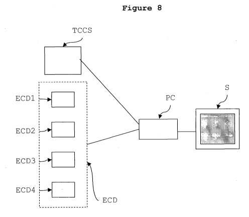
Figure 8 is a schematic block diagram of an
exemplary embodiment of a system as described herein,
i.e. a system for determining at least one index out of
a blood reflux rate index and increased blood
resistance index in cerebral veins in a patient. As
detailed in the following, the presence of such indexes
may be indicative of suspected patient's exposure to
CDMS (Clinically Defined Multiple Sclerosis).
Specifically, the diagram of Figure 8 illustrates
Transcranial Color-coded duplex Sonography apparatus
(TCCS) and Extracranial EchoColor-Doppler apparatus
(ECD).
Such apparatus is well known in the art and
currently used in clinical practice. Exemplary of such
equipment are e.g. the TCCS apparatus available under
the trade designation of MYLAB25 or TECHNOS provided
with high resolution probes of 2.5 MHz and the ECD
apparatus available under the trade designation of
MYLAB25 or TECHNOS provided with high resolution probes
of 7.5-13 MHz from ESAOTE BIOMEDICA (Italy).
The TCCS and ECD apparatus in question may be
connected to a processing equipment such as e.g. a
personal computer (PC) to process the detection signals
produced by the TCCS and ECD apparatus that form a
detection source set for the system herein.
Specifically, the TCCS apparatus comprises a first
detection source adapted to detect (in manner known per
se) a blood reflux in at least one of the deep middle
cerebral veins. Similarly, the ECD apparatus comprises
a second detection source adapted to detect at least
one of:
CA 02717081 2010-08-26
WO 2009/107152 PCT/IT2008/000129
7
- i) a blood reflux in at least one of the
internal jugular and/or vertebral veins;
- ii) a stenose in at least one of the internal
jugular vein;
- iii) a lack of doppler detectable blood flow
in at least one of the internal jugular and/or
vertebral veins; and
iv) a negative difference between the cross-
sectional area of at least one of the internal jugular
veins in the supine posture and in the erect posture of
said patient.
To that end, the ECD apparatus may include (again
in a manner known per se) separate modules ECD1 to
ECD4.
As a function of the detection data provided by
the detection sources (TCCS, ECD) the processing module
PC is thus in a condition to sense the condition where
the set of detection sources detect the subsistence of
at least two of the entities sensed, namely:
- the blood reflux in at least one of the deep
middle cerebral veins is detected by said first
detection source (as detected, e.g. by the TCCS
apparatus), and
- the blood reflux in at least one of the internal
jugular and/or vertebral veins,
- the stenose in at least one of the internal
jugular vein,
- the lack of doppler detectable blood flow in at
least one of the internal jugular and/or vertebral
veins, and
- the negative difference between the cross-
sectional area of at least one of the internal jugular
veins in the supine posture and in the erect posture of
said patient (being the last four entities detected
e.g. by the ECD apparatus).
CA 02717081 2010-08-26
WO 2009/107152 PCT/IT2008/000129
8
When such a situation is sensed, the processing
unit (PC) emits - for instance in the form of a visual
message on an associated screen S - a corresponding
advice signal.
In the exemplary embodiment to which Figure 8
refers, the detection source set of the system thus
includes two separate detection sources.
The first detection source is comprised of the
TCCS apparatus, adapted for detecting the presence of
blood reflux in at least one of the deep middle
cerebral veins.
The second detection source is comprised of the
ECD apparatus, adapted for detecting.(at least one of);
- blood reflux in at least one of the internal
jugular and/or vertebral veins;
- a stenose in at least one of the internal
jugular vein;
- the lack of doppler detectable blood flow in
at least one of the internal jugular and/or vertebral
veins; and
- a negative difference between the cross-
sectional area.
Those of skill in the art will otherwise
appreciate that the one illustrated is just one
exemplary embodiment of the system disclosed herein.
Embodiments of the system disclosed herein may
include recourse to different detection sources (i.e.
different types of apparatus providing the same or
equivalent detection information), or a different
partition of the detection actions (i.e. the set of
detection sources being comprised of a single
integrated detection system) . Also, embodiments of the
system disclosed herein may provide for off-line
provision of the detection signals, i.e. detection of
the various entities concerned being effected
CA 02717081 2010-08-26
WO 2009/107152 PCT/IT2008/000129
9
separately and the values detected stored in view of
subsequent processing.
The malformations identified in the present
disclosure are responsible for chronic extracranial
venous outflow obstruction (CEVO), causing severe
hemodynamic alterations, as documented in the first
part of this disclosure. The physiological regulation
of venous return is significantly altered by the
presence of these malformed obstructions, with
parameters that are significantly different from those
of all control groups (Fig. 2, 3) . CEVO must therefore
be considered an exclusive characteristic of MS.
In fact, the hemodynamic parameters of venous
obstruction adopted in the present study, confirmed in
all cases by selective phlebography, were not present
in the first control population, matched for age and
gender with the MS population (60 healthy controls).
For greater certainty, the inventors selected a second
control population that was older than the average age
of European MS patients, according to censuses in
recent epidemiological studies (72 healthy aged
subjects) . Had the venous obstruction been present in
this group, the inventors would not have been able to
maintain that they have a role in the pathogenesis of
MS, given that this control group had never had any
neurological manifestations, nor other important
diseases.
From this point of view, the fact of not having
found impediments to cerebrospinal venous drainage even
in the third control population, which was represented
by other miscellaneous neurological diseases (45
patients) is of great interest. This finding would
indicate that in no other disease of the nervous system
can this chronically obstructed venous drainage be
observed, not even in other pathologies that, like MS,
CA 02717081 2010-08-26
WO 2009/107152 PCT/IT2008/000129
present neurodegenerative, neuroimmunitary, and
neurovascular aspects.
Some human diseases present similar malformations
involving other venous segments. For example,
5 membranous obstruction of the inferior vena cava
upstream to the outlet of the suprahepatic veins is
morphologically similar to the azygous membrane
depicted in figure 6e; in the former position
determines the appearance of Budd-Chiari syndrome.
10 Venous septum, observed in the present study in the
IJVs (Fig. 6d), has been also described in the inferior
vena cava and in the iliac veins, where it brings about
severe CVD in the lower limbs. Thus, outside the
_. central nervous system, it is widely accepted that
these venous malformations cause cellular degeneration
and sclerosis: in the tissues drained by the affected
veins the inventors respectively found hepatic
cirrhosis, and lipodermatosclerosis with ulcer of the
leg, as well as perivenous iron depositions.
Among the anomalies identified in the present
disclosure, certainly the persistence of the reflux in
any position of the head is the criterion most
significantly associated with MS, with an Odds ratio
(OR) of more than 6 (Table III) . This reflux has a
mechanism that differs from that caused by incompetence
of the jugular valve. In the latter case, valvular
insufficiency tested with Valsalva can be related to a
picture of transient global amnesia. In the present
disclosure the reflux occurred naturally in any body
position without the need to sollicit it by a forced
movement. It is not the expression of valvular
insufficiency but rather of the stenosing lesion that
cannot be crossed with postural or respiratory
mechanisms, becoming a long lasting reverse flow (Fig.
1). In other cases reflux is an expression of the
CA 02717081 2010-08-26
WO 2009/107152 PCT/IT2008/000129
11
opening of collateral circles that compensate for the
reduced flow of the obstructed venous segment, in a
direction that is opposite to the physiologic one.
In the present disclosure stenosing lesions are
defined as obstructions because the propelling
mechanisms of cerebrospinal venous return (i.e., the
muscular thoracic pump and postural variations) are
functionally incapable of overcoming the stenoses. The
example in Figure 9 is especially illuminating since it
illustrates that a stenosis not morphologically closed
in the proximal tract of the azygous vein in the supine
position gives place to a reflux that is transmitted
downward to the level of the lumbar plexuses. In order
to be drained, this portion of countercurrent blood
- enters the intrarachidian plexuses, which become a
substitute circle. Instead, the caval system gains in
part, re-entering the inferior vena cava through the
renal vein.
The present disclosure shows that within the MS
group there exists a correlation between the
topographies of the obstructive malformations and those
of the MS lesions. In the relapsing-remitting (RR) and
secondary progressive (SP) forms, with lesions mostly
involving the brain and the cervical medulla, the
venous obstructions are principally found in the
jugular veins and in the proximal azygous vein (Fig.
4a, 6). In the PP form, with MRI documentation of
thoraco-lumbar lesions, obstruction involves the veins
that drain that particular territory, such as the
distal azygous, hemiazygous, and lumbar veins (Fig. 4b,
7). The different topography of the MS lesions in the
primary progressive (PP) form as compared to the RR and
SP courses, and the further correspondence with the
topography of the obstructed venous segments, suggests
a pivotal role of venous drainage in the complex
CA 02717081 2010-08-26
WO 2009/107152 PCT/IT2008/000129
12
etiopathogenesis of this disease.
For example, the lack of drainage through the
lumbar veins and/or the distal azygous discovered in
PPMS would cause an ascending drainage through the
intrarachidian veins. Such a circumstance is confirmed
by the highest rate of intracranial reflux demonstrated
in this subgroup (Table III), and could be related to
the preferential distribution of plaques in the
medulla. In contrast, the hampered drainage through the
IJVs as well as in the proximal azygous found in the
RR-SP patients seems to be related` to the preferential
onset and distribution of plaques in the brain.
The present disclosure also shows that within the
MS group of patients pharmacologically treated with
immunomodulating and/or immunosuppressive drugs
(particularly with respect to RR-SP group) the
obstructive malformations are not reduced as compared
to the non-treated patients.
The venous strictures, demonstrated in patients
who underwent phlebography, are responsible for CEVO,
causing severe hemodynamic alterations. CEVO must
therefore be considered an exclusive characteristic of
MS, and dramatically increases the risk of MS by 36
fold (OR 36, 95% CI 22-57, p<0.0001).
The physiological regulation of venous return is
significantly altered by the presence of these
malformed obstructions, with parameters that are
significantly different from those of all control
groups, each one significantly increasing the risk of
MS (Tab. III, Figs. 1 and 3).
METHODS
FIRST PHASE: NONINVASIVE SCREENING
Patients and Controls
109 consecutive patients affected by clinically
CA 02717081 2010-08-26
WO 2009/107152 PCT/IT2008/000129
13
defined MS (CDMS), diagnosed according to the
recommended criteria (as set forth in e.g. Polman, CH;
Reingold, SC; Edan, G; Filippi, M; Hartung, H-P.
Diagnostic Criteria for Multiple Sclerosis: 2005
Revisions to the "McDonald Criteria" Ann Neurol 2005;
58: 840-846) were admitted to the first part of the
study. They were subdivided into 69 with a relapsing-
remitting (RR) clinical course, 31 secondary
progressive (SP), and 9 primary progressive (PP),
attributing to each group a relative expanded
disability disease score (EDDS).
These patients were compared with a blind design
to 177 controls, subdivided into three groups: the
first group included 60 healthy subjects matched for
age and -gender with MS patients (HM-C); the second
control group included 72 healthy subjects older than
the median age of the European MS population (HA-C),
and the third group included 45 patients affected by
other neurological diseases (OND) (Table I) . The OND
patients were subdivided into patients affected by
neurodegenerative disorders (Parkinson's disease and
amyotrophic lateral sclerosis-ALS), other
neuroimmunitary disorders (OIND, including myasthenia
gravis and multifocal motor neuropathy, MMN), and
cerebro-vascular disease (ischemic stroke, transient
ischemic attack-TIA) (Table II).
Exclusion criteria
We excluded from the study those subjects having,
or showing the potential for developing, a nervous
system pathology of a venous refluxive and/or
obstructive nature, including:
1. Chronic venous insufficiency of the lower
limbs-CVI
2. History of venous thrombosis and\or post-
CA 02717081 2010-08-26
WO 2009/107152 PCT/IT2008/000129
14
thrombotic syndrome
3. Genetic thrombophilia
4. Congenital angiodysplasias
5. Congenital vascular malformations
6. Budd-Chiari syndrome
7. Behcet disease
8. Other Vasculitis
Patients and controls blindly underwent to a non-
invasive study of cerebro-spinal venous return.
By applying the above stated exclusion criteria
109 CDMS patients entered the study; they were
subdivided into 69 with a relapsing-remitting (RR)
clinical course, 31 secondary progressive (SP), and 9
primary progressive (PP), attributing to each group a
relative expanded disability disease score (EDSS) . The
control groups included 60 HM-C, 72 HA-C, and 45 OND
patients (Table I).
Table I.
All MS Group Group Group Group Group:
Patients HM-C HA-C MS-RR MS-SP MS-PP
(n=109) (n=60) (n=72) (n=69) (n=31) (n=9)
AGE
58
Median (yy) 40 37 38 44 57 (25th-75th (34-46) (28-49) (50.5- (30-43) (40-51)
(46-60)
71.5)
percentile)
Sex
41% 46% 40% 40.5% 42% 44%
%M
45/64 28/32 29/43 28/41 13/18 4/5
M/F
EDDS
1.5 5
median 2 5.5
(0.5- (3.5-
(25th-75th ( 1-4) (4-7)
1.5) 6.5)
percentile)
CA 02717081 2010-08-26
WO 2009/107152 PCT/IT2008/000129
All MS Group Group Group Group Group:
Patients HM-C HA-C MS-RR MS-SP MS-PP
(n=109) (n=60) (n=72) (n=69) (n=31) (n=9)
DISEASE
DURATION(yy) 9
6 4 13
median (4-
(2-12) (2-7) (8-19)
(25th-75th 14.5)
percentile)
5 Table II.
Group Subgroup Subgroup Subgroup
_- OND Neurodegenerat}ve OIND Cerebro-vascular
Disease (Myasthenia;MMN) Disease
(n=45) (ALS;Parkinson's) (n=7) (Stroke; TIA)
(n=19) (n=19)
AGE
median 60 64 50 69
(25th_75th (51-77) (52-76) (45-57) (53-78)
percentile)
Sex
55.5% 47% 57% 63%
%M
25/20 9/10 4/3 12/7
M/F
Study of cerebrospinal venous drainage
Venous cerebrospinal return was examined, with the
operators blinded of the diagnosis, with the subjects
10 positioned on a tilt bed by combining the TCCS
methodology for studying the deep cerebral veins (DCVs)
with that of extracranial EchoColor-Doppler (ECD) for
insonating the internal jugular veins (IJVs) and
vertebral veins (VVs), both previously described. In
15 particular we assessed the cerebrospinal venous return,
CA 02717081 2010-08-26
WO 2009/107152 PCT/IT2008/000129
16
focusing on the detection of five findings suggesting
the presence of chronic extracranial venous outflow
obstruction (CEVO). The detection of at least two
findings was used for non invasive screening of highly
suspected CEVO. Particularly we assessed:
1) Flow direction
Each measurement was preceded by a complete ECD
high resolution B-mode exploration of the cervical
vessels. We assessed the presence of reflux in the IJVs
and VVs. According to a recent study on reflux time
cut-off values, we considered reflux a flow reversal
from its physiological direction for a duration > 0.88
sec. Flow direction was assessed during a short period
of apnea following a normal exhalation, as previously
reported, and not in a forced condition as Valsalva
manouevre.
Furthermore, we assessed the eventual persistence
of reflux with the head positioned at 0 , +15 , +30 ,
+45 , +90 in the four extracranial venous drainage
pathways.
Therefore, TCCS investigation allows to detect,
through the transtemporal window, the presence of
reflux in at least one of the deep cerebral veins
(DCVs)including the Galen, the basal, and the internal
cerebral vein, eliciting venous flow by inviting the
subject under examination to breathe, as previously
reported.
2) Flow velocity.
We measured, at the level of the IJVs-VVs,
respectively, in sitting and supine positions, the peak
systolic velocity (PSV) and the peak diastolic velocity
(PDV), both expressed in cm/sec. Measurement was
derived from a 5 sec. recording of the Doppler spectrum
CA 02717081 2010-08-26
WO 2009/107152 PCT/IT2008/000129
17
analysis. PSV was the highest flow velocity recorded in
systole, and PDV the highest flow velocity recorded in
diastole during 5 sec of apnea, following normal
expiration.
3) Postural control of cerebral venous outflow route
ECD clarified that physiologically the IJV is the
predominant outflow pathway in the supine position,
confirmed by an increased cross-sectional area (CSA)
related to increased blood volume in that posture;
redirection of venous flow to the VVs occurs in the
upright position, with compliant reduction of the CSA
of the IJV. Consequently we measured:
= The cross-sectional area (CSA) of both IJVs, in
supine and sitting postures.
= The difference in CSA (delta CSA) obtained by
subtracting the CSA measured in the supine from that
in the erect position.
ECD-TCCS criteria for venography
In Table III we report the list of the five
criteria assessed through the ECD-TCCS protocol above
described, and used for detection of significant
cerebro-spinal venous flow disturbances and hampered
venous outflow in patients and controls populations.
Diagnosis of highly suspected CEVO required to full-
fill at least two of the five listed criteria. Highly
suspected obstruction of cerebrospinal venous outflow
pathways was taken as an indication to continue the
study using .selective venography in all identified
subjects.
SECOND PHASE: SELECTIVE VENOGRAPHY
Patients and Treatments
Table IV shows the characteristics of the CDMS
CA 02717081 2010-08-26
WO 2009/107152 PCT/IT2008/000129
18
population under study. No significant differences were
found in gender distribution among the three subgroups
RR, SP, and PP, whereas age, EDSS, and disease duration
were, of course, significantly higher in both
progressive courses with respect to RR (p<0.05). HLA 2
DR15 genetic analysis was available in 44/51 patients,
and CSF in 35/51, the latter mainly due to patient
refusal. Finally, 48/51 patients fulfilled the revised
MRI criteria of McDonald, whereas 3 patients, all
belonging to the RR subgroup, did not. However, they
fully satisfied either the clinical presentation or the
additional CSF and MRI requirement. Table V lists the
treatments administered in this cohort in the last
three years. Due to lack of evidence, no treatments are
listed for PP patients, and 14/44 patients RR/SP
refused any pharmacological therapy.
Table IV.
MS Patient
MS RR MS SP MS PP
population
N =29 N =15 N =7
N =51
AGE
39 34 44 57
median
(25 th -75 th percentile) (33-45) (29-39) (41-52) (42-60)
Sex %M 47% 48% 47% 42%
M/F 24/27 14/15 7/8 3/4
EDSS 4.5 5
2 1.5
median (3.5- (3.5-
(25th-75th percentile) (1-4.5) (0.5-2) 6.5) 7.5)
DISEASE DURATION(yy)
6 4 13 12
median
(25 th-75 th percentile) (2-13) (1-7) (5-19) (2-15)
CA 02717081 2010-08-26
WO 2009/107152 PCT/IT2008/000129
19
MS Patient
MS RR MS SP MS PP
population
N =29 N =15 N =7
N =51
HLA2 (DR15)
Haplotype carriers (C) 35% 42% 21% 67%
% 15/28 10/14 3/11 2/3
C/no C
CSF Oligoclonal
Bands + 91% 89% 100% 75%
% 32/35 16/18 13/13 3/4
+/Tot
Compliance with at
least 3 of 4 MRI
revised 96% 90% 100% 100%
Mc Donald criteria 49/51 26/29 15/15 7/7
%
+/Tot
Table V.
N MS
Drugs
Cases
Immunosuppressants
(Mitoxantrone,
18.
Cyclophosphamide,
Azathioprine)
Immunomodulators
(Interferon Beta, 21
Glatiramer acetate,)
Corticosteroids
(I.V. high doses
Methylprednisolone) 88
N cycles in acute
exacerbations
Treatment refusal 14
CA 02717081 2010-08-26
WO 2009/107152 PCT/IT2008/000129
N MS
Drugs
Cases
PP cases
(no available effective 7
treatment)
Statistical Analysis
Clinical and demographic characteristics are
expressed as median and 25th-75th percentile. CSA in
5 sitting and supine postures, delta CSA, PSV, and PDV
are expressed as mean SD. Differences among groups
were tested for significance with the ANOVA analysis of
variance, with Bonferroni correction when p is <0.05.
The two-tailed Fisher's exact test followed by
10 Odds ratio 95% Confidence Interval (CI) was used for
determining the associated risk of MS in case of
positive ultrasonographic criteria for CEVO, by
comparing the whole MS group with a group including all
controls.
15 The Odds ratio is a widely used statistic to
compare the frequency of exposure to risk factors in
epidemiological studies. Odds ratios compare the
retrospective/posterior odds of exposure to a given
risk factor in two groups of individuals. Odds ratios
20 are interpreted with reference to a confidence interval
(e. g. 95%). One can say that a given risk factor is a
significant risk to a disease if the odds ratio is
greater than one and the lower bound of the confidence
interval does not go below one.
The two-tailed Fisher's exact test was also used
for analyzing the different pattern of distribution of
extra-cranial venous strictures in the RR/SP and PP
groups, respectively, as well as for testing
differences in the number of extracranial venous
CA 02717081 2010-08-26
WO 2009/107152 PCT/IT2008/000129
21
strictures between MS patients treated and not treated
with drugs.
P-values up to 0.05 were considered statistically
significant.
RESULTS
FIRST PHASE: NONINVASIVE SCREENING
Patients and Controls
Tables I and II show clinical and demographic
characteristics for the entire group of MS patients,
and for the subgroups. Significant differences were
found in the following:
= Age: SP vs RR, p<0.01. PP vs RR, p<0.01 (ANOVA).
= EDSS: SP vs RR, p<0.01. PP vs RR, p<0.01 (ANOVA).
= Disease duration: SP vs RR, p<Ø01 (ANOVA).
Study of cerebrospinal venous drainage
1) Flow direction
The persistence of reflux with the head positioned
at 0 , +15 , +30 , +45 , +90 in at least one IJV
and/or VV venous segments was never observed in any
subject among the three control populations. By
contrast, results were positive in 77/109 MS patients
(70%), particularly in those with RR and SP courses, in
46/69 (66%) and in 28/31 (90%), respectively. Table
III reports the sensitivity and specificity of such
ECD-TCCS finding, in differentiating MS patients from
controls.
In the PP course, reflux in the DCVs was more
frequently observed as compared to RR-SP, 78% vs. 41%,
respectively, and never in controls (p<0.0001).
Table III.
CA 02717081 2010-08-26
WO 2009/107152 PCT/IT2008/000129
22
Odds Ratio
MS-RR CONTROL All MS
ECD-TCCS MS-PP
MS-SP POPULATIONS vs. P
CRITERIA (N;%)
(N;%) (N;%) All Controls
(95% C.I.)
1.
Re flux
constantly
present in an
outflow pathway 74/100 2/9 0/177 6.4
< 0.0001
(IJV and\or VV) 74% 22% 0% (4.7-8.7)
with the head in
any position
(0 , +15 , +30 ,
+45 , +90 )
2.
Re flux
41/100 7/9 0/177 3.9
propagated < 0.0001
41% 78% 0% (3.1-4.8)
upward to the
DCVs
3.
High resolution
27/100 3/9 0/177 3.2
B-mode evidence < 0.0001
27% 33% 0% (2.7-3.9)
of proximal IJV
stenoses
4.
Flow not Doppler
detectable in
35/100 3/9 0/177 3.5
the IJVs and/or < 0.0001
35% 33% 0% (2.9-4.3)
Ws despite
numerous deep
inspirations
5.
58/100 3/9 21/177 3.2
Negative ACSA in < 0.0001
58% 33% 12% (2.4-4.2)
the IJV
CA 02717081 2010-08-26
WO 2009/107152 PCT/IT2008/000129
23
Finally, B-Mode analysis at high resolution
allowed for the direct observation, in 30 MS patients,
of the presence of closed stenosis in the proximal
segment of an IJV, almost always the left (Fig. 1).
Figure 1 shows the detection of venous obstruction in
one MS patient by means of ECD (transversal access)
with a probe 7,5 MHz; in A) the right cervical side has
been observed, wherein common carotid artery (CC) and
right internal jugular vein (IJVr) are shown. In B) the
same patient has been evaluated with identification of
stenosis of the left internal jugular vein (IJV1) due
to annulus (black arrows encircling the stenosis),
while the left common carotid artery (CC) appears
normal.
2) Flow velocity
Figure 2 reports the highly significant
differences in PSV and PDV values measured in the IJVs
in the sitting position in the entire MS population and
its subgroups, as compared to the three control
populations. A further finding never seen in controls
and recorded in 33% of RR-SP, and in 35% of PP cases,
respectively, was the lack of flow velocity Doppler
detectable in the IJVs and/or VVs despite numerous deep
inspirations (Tab. III). It suggests a functional
venous obstruction at the thoracic level.
Similarly, significant differences were also
observed even with the head at 0 , and at the level of
the VVs.
3) Postural control of cerebral venous outflow route
In Figure 3, the physiologic postural control of
cerebral venous outflow route in the IJVs in both
healthy control populations (HM-C, HA-C) is well
CA 02717081 2010-08-26
WO 2009/107152 PCT/IT2008/000129
24
apparent. CSA values in the sitting position are
consistently lower than those assessed in the supine
position, resulting in a rather big CSA. The same
correct physiologic response to a change in hydrostatic
pressure condition was also demonstrated in the OND
group, with no significant differences from the CSA
assessed in the HM-C and HA-C groups (p<0.0001). In
contrast, Figure 3 shows an overturning of this
physiologic mechanism of postural regulation in the
entire MS population. Redistribution of blood in the
supine posture, in accordance with the principle of
communicating vessels, seemed to be impeded in MS
patients, and CSA at 0 was significantly lower in the
MS patients than in the healthy controls, and even in
the OND patients (p<0.0001). Consequently, the LCSA
levels were significantly reduced in MS as compared to
- the three control groups, as shown in Figure 3.
Finally, LCSA was negative in 56% of MS cases vs. 120
of the three control groups, as shown in Table III.
ECD-TCCS criteria for venography
In Table III we report the list of the ECD-TCCS
findings used for suspecting the presence of CEVO, and
the relative distribution in RR-SP cases, PP cases, and
in the controls. Each of the five findings demonstrated
a noteworthy specificity and appreciable sensitivity in
differentiating CDMS from the three control groups
(Tab. III) . As above reported, 70% of CDMS presented
with reflux in any body posture in at least one of the
four extracranial cerebral outflow routes vs. 0% of the
control groups. However, in the 30% MS cases in which
the ultrasound examination results had been negative
for persistence of reflux in the IJVs/VVs in any body
position, at least two of the other criteria were
consistently positive. The control population never
CA 02717081 2010-08-26
WO 2009/107152 PCT/IT2008/000129
resulted positive for two criteria resulting in a 100%
specificity of the five proposed criteria (Tab. III).
Thus, by fulfilling the condition of two positive ECD-
TCCS diagnostic criteria for hampered venous outflow,
5 the proposed test was positive in 100% of the MS
population, as opposed to 0% of all controls, both
blindly investigated. Consequently, also the
sensitivity by adopting the complex of the five
criteria raised to 100%.
10 As a conclusion:
1St Criterion: Reflux in the IJVs and/or VVs with
the head in any position.
The positiveness of this criterion increases
dramatically the risk of MS by more than sixfold (OR
15 6.4, 95% CI 4.7-8.7, p<0.0001, Fisher's exact test).
2nd Criterion: Reflex in the DCVs
The presence of this finding increases
significantly the risk of MS (OR 3.9, 95% CI 3.1-4.8,
p< 0.0001, Fisher's exact test).
20 3rd Criterion: High resolution B-mode evidence of
proximal IJV stenosis
The presence of closed stenosis in the proximal
segment of an IJV increases the risk of MS (OR 3.2, 95%
CI 2.7-3.9, p<0.0001, Fisher's exact test).
25 4th Criterion: Flow not Doppler detectable in the
IJVs and/or VVs
This suggests a functional venous obstruction at
the thoracic level and increases significantly the risk
of MS, as indicated in Table III.
5th Criterion: Reverted postural control of the
main cerebral venous outflow pathway
The OCSA levels significantly reduced in MS as
compared to the three control groups results in a
significantly increased risk of MS (Table III).
On the whole, the positiveness of two ECD-TCSS
CA 02717081 2010-08-26
WO 2009/107152 PCT/IT2008/000129
26
criteria of suspected CEVO dramatically increase the
risk of CDMS (Odds Ratio= 77745, 95% Confidence
Interval: 1530,1 to 3950364, p<0.0001) due to their
exclusive detection in the MS group.
Stated otherwise, the embodiment described herein
provides for the processing module PC to allot
respective risk factor values to any of criteria
discussed in the foregoing, i.e.:
- said blood reflux in at least one of the deep
cerebral veins;
- said blood reflux in at least one of the
internal jugular and/or vertebral veins,
- said stenose- in at least one of the internal
jugular vein,
- said lack of Doppler detectable blood flow in at
least one of the internal jugular and/or vertebral
veins, and
- said negative difference between the cross-
sectional area
as detected by said detection source set ECD,
TCCS.
The processing module PC is thus in condition to
derive and display for use by the practitioner a
cumulative risk factor value, which is a function of
the respective risk factor values.
While the cumulative risk factor value will tend
to be higher if a higher number of risk criteria are
met, the values of the risk factors allotted to each
criterion met will also play a role in defining the
cumulative risk factor of developing MS.
Table III above may thus be exemplary of an
embodiment where the blood reflux in at least one of
the internal jugular and/or vertebral veins is allotted
a respective risk factor which is (substantially)
higher - i.e. 6.4 - than any other respective risk
CA 02717081 2010-08-26
WO 2009/107152 PCT/IT2008/000129
27
factors allotted the other criteria (which may all fall
in the range 3.2 to 3.9).
SECOND PHASE: SELECTIVE VENOGRAPHY
Patients and Treatments
Table IV shows the characteristics of the CDMS
population under study, while table VI shows the
results of the non invasive screening of this
population (namely the number of positive TCCS-ECD
criteria) performed according to the present invention.
Table VI.
MS Patient
MS RR MS SP MS PP
population
N =29 N =15 N =7
N =51
Number of positive
TCCS-ECD criteria 3 3 3 2
median (2-3) (2-3) (3-3) (2-3)
(25th -75t" percentile)
Selective venography of the azygous and jugular venous
system
Selective phlebography confirmed that the
detection of at least 2/5 TCCS-ECD criteria of
suspected CEVO, never measured in the control
populations, were always related to a severe steno-
obstruction, generally at the thoracic level, of the
principal cerebrospinal venous segments. Interestingly,
pattern of venous obstruction were significantly
different located in RR-SP patients as compared to PP,
and are given in Table VII, (p<0.0001, Fisher's exact
test).
This result, when comparing ultrasonographic
screening with invasive venography, confirmed the
highly significant level of specificity of the former
CA 02717081 2010-08-26
WO 2009/107152 PCT/IT2008/000129
28
also in detecting CEVO (100%, P<0.0001).
Table VII.
Multiple
CEVO CEVO in
Confined Azygous p
to the and/or in
Azygous the IJVs
territory
RR-SP GROUP
0 44 < 0.0001
44 PATIENTS
PP GROUP
2 < 0.0001
7 PATIENTS
5 Selective venography in RR and SP cases
This investigation showed the presence of
obstructions in the proximal azygous vein and/or
internal jugular veins in 100% of cases having RR and
SP clinical courses (Table VII). Rarely (2/51, 4%) did
these obstructions occur in only one IJV segment;
almost always, two or three of the main venous outflow
pathways were involved, thus severely compromising
cerebrospinal venous drainage and demonstrating to be a
multilevel pathology (Fig. 4a). Figure 4 shows the
location in the cerebrospinal outflow veins of the
steno-obstructive malformations in RR-SP (A), and PP
cases (B), respectively. It should be emphasized that
isolated obstruction is quite rare and that venous
return is impaired by the combination of two or three
stenoses. In addition, the topography of vein
obstruction in the RR-SP groups (A) is different from
that in the PP group (B) (IJV=internal jugular vein,
left-1 and right-r; AZY=azygous vein; distal
AZY=segment of the azygous vein below the
emiazygousvein outlet; EMIAZY-Lumb= emiazygous vein and
CA 02717081 2010-08-26
WO 2009/107152 PCT/IT2008/000129
29
lumbar plexus) . The number of extracranial venous wall
stenoses did not differ significantly in patients
treated with immunosuppressant/immunomodulator agents
or in never-treated patients. In treated patients of
the RR-SP class, the present inventors discovered up to
2 lesions in 36% of cases and > 2 lesions in 34%; in
not treated patients, up to 2 lesions in 18% and > 2
lesions in 11% of cases (p=ns. Fisher's exact test).
Association between IJVs' and proximal azygous
obstruction was discovered in the vast majority (37/44,
84%) of cases (Fig. 4a), and azygous obstruction was
mainly located at the junction with the superior vena
cava and/or in its arch (Fig. 6E).
As to the morphology of these obstructions,
selective venography revealed six principal venous
malformations: annulus, agenesia, atresia, septum,
membrane, and twisting. Figure 5 shows the relative
distribution of the malformations found in the
extracranial venous segments. Annulus was more likely
to be found in the jugular system, whereas membranous
obstruction seems to be typical of the azygous vein.
The panel in figure 6 provides the relative
morphological details of the six malformation patterns,
wherein in A) Annulus (arrow) at the level of the left
internal jugular vein (IJV1) located immediately below
the competent valve (VV) at the outlet with the
brachiocephalic trunk (BCT) is shown. In B) Annulus
(arrow) at the level of the right internal jugular vein
(IJVr)and C) Combination of IJV1 atresia (arrow A) and
annulus (arrow), compensated by two distinct collateral
circles (CC) are shown. Unfortunately, the proximal CC
re-enters at the level of the proximal annulus, thereby
reducing its outflow contribution. In figure 6 D)
Septum (arrow S) at the level of the IJVr, above the
anonymous trunk (AnT), E) Membranous obstruction (arrow
CA 02717081 2010-08-26
WO 2009/107152 PCT/IT2008/000129
M) of the outlet of the azygous vein (AZY) in the
superior vena cava (SVC), and F) Agenesia (Ag) of the
distal segment of the right IJV, visible immediately
above the tip of the catheter are shown. Supply flow
5 through the condylar veins feed collateral circles, CCl
and CC2, in turn drained respectively into the external
jugular vein (EJV), and into the thyroid veins (TyVs).
Selective venography in PP cases
10 Also 7 PP cases with MRI-evident lesions, mostly
at the level of the spinal cord, underwent venography.
These patients presented, as a, distinctive
characteristic, a particular topography of the stenosed
venous lesions that invariably involved the azygous
15 vein, with a reduced association with the IJVs when
compared to the RR and SP cases (Table VII) (Fig. 4b).
In PP cases we identified a further association of
proximal azygous stenoses with more distal venous
obstructions, mainly at the level of the distal
20 azygous, the hemiazygous vein, and often
atresia/agenesia of the lumbar veins, not present in
the RR and SP subjects (Fig. 4b, 5 and 7). This
condition compromises drainage of the spinal cord at
the thoracic-lumbar level, suggesting a strict
25 relationship between the localization of venous
obstructions and the clinical and MRI documentation of
the site of the MS lesions.
In figure 7 venous lesions by means of selective
venography in PP cases are shown. In A) Twisting of the
30 proximal AZY (twisted arrow) with evident venous
dilation below, involving also the emiazygous vein
(Emiazy) is depicted. In B) Combination of atresia (At)
and agenesia (Ag) involving the lumbar veins below the
distal segment of the azygous vein (Distal Azy) and C)
Atresia of the emiazygous vein (At) with normal azygous
CA 02717081 2010-08-26
WO 2009/107152 PCT/IT2008/000129
31
vein (AZY) from the outlet with the superior vena cava
(SVC) to the distal segment (Distal Azy) are depicted.
Naturally, while the principle of the invention
remains the same, the details of construction and the
embodiments may widely vary with respect to what has
been described and illustrated purely by way of
example, without departing from the scope of the
present invention.