Note: Descriptions are shown in the official language in which they were submitted.
CA 02717656 2010-08-18
WO 2008/103947 PCT/US2008/054785
ACTIVATION OF HUMAN ANTIGEN-PRESENTING CELLS THROUGH CLEC-6
TECHNICAL FIELD OF THE INVENTION
The present invention relates in general to the field of antigen presentation
and immune cell
activation, and more particularly, to the activation of immune cells through
the CLEC-6 C-type
lectin.
BACKGROUND OF THE INVENTION
Without limiting the scope of the invention, its background is described in
connection with
dendritic cells.
Dendritic cells play a pivotal role in controlling the interface of innate and
acquired immunity by
providing soluble and intercellular signals, followed by recognition of
pathogens. These
functions of DCs are largely dependent on the expression of specialized
surface receptors,
`pattern recognition receptors' (PRRs), represented, most notably, by toll-
like receptors (TLRs)
and C-type lectins or lectin-like receptors (LLRs) (1-3). In the current
paradigm, a major role of
TLRs is to alert DCs to produce interleukin 12 (IL-12) and other inflammatory
cytokines for
initiating immune responses. C-type LLRs operate as constituents of the
powerful antigen
capture and uptake mechanism of macrophages and DCs (1). Compared to TLRs,
however,
LLRs might have broader ranges of biological functions that include cell
migrations (4),
intercellular interactions (5). These multiple functions of LLRs might be due
to the facts that
LLRs, unlike TLRs, can recognize both self and nonself. However, the
complexity of LLRs,
including the redundancy of a number of LLRs expressed in immune cells, has
been one of the
major obstacles to understand the detailed functions of individual LLRs. In
addition, natural
ligands for most of these receptors remain unidentified. Nonetheless, evidence
from recent
studies suggests that LLRs, in collaboration with TLRs, may contribute to the
activation of
immune cells during microbial infections (6-14).
SUMMARY OF THE INVENTION
The present invention includes compositions and methods for using anti-human
CLEC-6
monoclonal antibodies (mAbs) and characterized their biological functions that
are the basis of
envisioned therapeutic applications of anti-CLEC-6 mAbs and their surrogates.
The invention
includes contacting antigen presenting cells, such as dendritic cells (DCs)
that express CLEC-6,
and that it plays a role in the uptake of antigens associated with particular
DC activation that
CA 02717656 2010-08-18
WO 2008/103947 PCT/US2008/054785
2
results in altered Immoral and cellular immune responses. The inventors have
developed and
characterized unique agents capable of activating cells bearing CLEC-6, as
well as the effect of
the resulting changes in cells receiving these signals regards action on other
cells in the immune
system. These effects (either alone, or in concert with other signals (i.e.,
co-stimulation)) are
highly predictive of therapeutic outcomes for certain disease states or for
augmenting protective
outcomes in the context of vaccination.
It was found that CLEC-6, one of the LLRs, is functional in terms of cell
(including DC)
activation by either alone or in collaboration with other cellular signals.
CLEC-6-mediated cell
activation was induced by anti-CLEC-6 mAbs, and therefore anti-human CLEC-6
mAbs or their
surrogates will be useful for developing reagents against diseases.
The present invention includes compositions and methods for increasing the
effectiveness of
antigen presentation by a CLEC-6-expressing antigen presenting cell by
contacting the antigen
presenting cell with an anti-CLEC-6-specific antibody or fragment thereof,
wherein the antigen
presenting cell is activated. The antigen presenting cell may be an isolated
dendritic cell, a
peripheral blood mononuclear cell, a monocyte, a myeloid dendritic cell and
combinations
thereof. In one specific embodiment, the antigen presenting cell is an
isolated dendritic cell, a
peripheral blood mononuclear cell, a monocyte, a B cell, a myeloid dendritic
cell and
combinations thereof that have been cultured in vitro with GM-CSF and IL-4,
interferon alpha,
antigen and combinations thereof. The method may also include the step of
activating the
antigen presenting cells with GM-CSF and IL-4, wherein contact with the CLEC-6-
specific
antibody or fragment thereof increases the surface expression of CD86 and HLA-
DR on the
antigen presenting cell.
It has been found that the present invention can be used to activate antigen
presenting cells with
the CLEC-6-specific antibody or fragment thereof to increases the surface
expression of CD86,
CD80, and HLA-DR on the antigen presenting cell. If the antigen presenting
cells are dendritic
cells (DCs), DCs activated with the CLEC-6-specific antibody and GM-CSF and IL-
4 to have
the gene expression pattern of Figure 4. The antigen presenting cells
activated with a CLEC-6-
specific antibody secrete IL-6, MIP-la, MCP-1, IP-l0, TNFa and combinations
thereof, and if
the APCs are dendritic cells, they secrete IL-6, MIP-la, MCP-1, IP-l0, TNFa,
IL-12p40, IL-la,
IL-lb and combinations thereof. When activating dendritic cell that has been
contacted with
GM-CSF and IL-4 or Interferon alpha, the CLEC-6-specific antibody or fragment
thereof and
the CD40 ligand further increase the activation of the dendritic cells. When
contacted with GM-
CA 02717656 2010-08-18
WO 2008/103947 PCT/US2008/054785
3
CSF and IL-4 or Interferon alpha and the CLEC-6-specific antibody or fragment
the DCs
increased their co-stimulatory activity.
In another embodiment, the method of the present invention can be used to
activate antigen
presenting cells by co-activating the antigen presenting cell through the TLR9
receptor and the
CLEC-6 lectin, wherein the cells increase cytokine and chemokine production,
and even trigger
B cells proliferation. It has also been found that co-activating antigen
presenting cells with
CLEC-6 and LOX-1 in the presence of B cells, induce the B cell immunoglobulin
to class-
switch. The TLR9 receptor may be activated with at least one of a TLR9 ligand,
an anti-TLR9
antibody of fragments thereof, an anti-TLR9-anti-CLEC-6 hybrid antibody or
fragment thereof,
an anti-TLR9-anti-CLEC-6 ligand conjugate. Examples of the CLEC-6-specific
antibody or
fragment thereof may be selected from clone 12H7, 12E3, 9D5, 20H8 and
combinations thereof.
Dendritic cells activated through the CLEC-6-receptor with the CLEC-6-specific
antibody or
fragment thereof also activate monocytes, dendritic cells, peripheral blood
mononuclear cells, B
cells and combinations thereof.
Yet another embodiment of the present invention includes CLEC-6-specific
antibodies or
fragment thereof bound to one half of a Cohesin/Dockerin pair. The CLEC-6-
specific antibody
or fragment thereof may be bound to one half of a Cohesin/Dockerin pair and
the
complementary half may be bound to an antigen. The antigen may be a molecule,
a peptide, a
protein, a nucleic acid, a carbohydrate, a lipid, a cell, a virus or portion
thereof, a bacteria or
portion thereof, a fungi or portion thereof, a parasite or portion thereof. In
another embodiment,
the CLEC-6-specific antibody or fragment thereof is bound to one half of a
Cohesin/Dockerin
pair and the other half of the pair is bound to one or more cytokines selected
from interleukins,
transforming growth factors (TGFs), fibroblast growth factors (FGFs), platelet
derived growth
factors (PDGFs), epidermal growth factors (EGFs), connective tissue activated
peptides
(CTAPs), osteogenic factors, and biologically active analogs, fragments, and
derivatives of such
growth factors, B/T-cell differentiation factors, B/T-cell growth factors,
mitogenic cytokines,
chemotactic cytokines and chemokines, colony stimulating factors, angiogenesis
factors, IFN-a,
IFN-(3, IFN-y, IL1, IL2, IL3, IL4, IL5, IL6, IL7, IL8, IL9, IL10, IL11, IL12,
IL13, IL14, IL15,
IL16, IL17, IL18, etc., leptin, myostatin, macrophage stimulating protein,
platelet-derived
growth factor, TNF-a, TNF-(3, NGF, CD40L, CD137L/4-1BBL, human lymphotoxin-0,
G-CSF,
M-CSF, GM-CSF, PDGF, IL-la, IL1- 0, IP-10, PF4, GRO, 9E3, erythropoietin,
endostatin,
angiostatin, VEGF, transforming growth factor (TGF) supergene family include
the beta
transforming growth factors (for example TGF-01, TGF-02, TGF-(33); bone
morphogenetic
CA 02717656 2010-08-18
WO 2008/103947 PCT/US2008/054785
4
proteins (for example, BMP-1, BMP-2, BMP-3, BMP-4, BMP-5, BMP-6, BMP-7, BMP-8,
BMP-9); heparin-binding growth factors (fibroblast growth factor (FGF),
epidermal growth
factor (EGF), platelet-derived growth factor (PDGF), insulin-like growth
factor (IGF)); Inhibins
(for example, Inhibin A, Inhibin B); growth differentiating factors (for
example, GDF-1); and
Activins (for example, Activin A, Activin B, Activin AB).
The present invention also includes a method for separating myeloid dendritic
cells from
plasmacytoid dendritic cells by using CLEC-6 expression to isolate myeloid
dendritic cells, B
cells or monocytes that express CLEC-6 from plasmacytoid dendritic cells which
do not express
CLEC-6.
The invention includes a hybridoma that expressed a CLEC-6-specific antibody
or fragment
thereof, wherein the CLEC-6-specific antibody or fragment thereof activates an
antigen
presenting cell to express new surface markers, secrete one or more cytokines
or both, for
example, clone 12H7, 12E3, 9D5, 20H8 and combinations thereof. The antibodies
produced by
anti CELC-6 hybridomas may be used in a method for enhancing B cell immune
responses by
triggering a CLEC-6 receptor on a B cell to increase antibody production,
secrete cytokines,
increase B cell activation surface marker expression and combinations thereof.
The B cells
secrete IL-8, MIP-la and combinations thereof and/or increases production of
IgM, IgG and
IgA.
The present invention also includes a method for enhancing T cell activation
by triggering a
CLEC-6 receptor on a dendritic cell with a CLEC-6 specific antibody or
fragment and
contacting a T cell to the CLEC-6 activated dendritic cell, wherein T cell
activation is enhanced.
The T cell may be a naive CD8+ T cell and the dendritic cells may be contacted
with GM-CSF
and IL-4, interferon alpha, antigen and combinations thereof. It has been
found that the T-cells
activated by the CLEC-6 activated DCs increases T cell secretion of IL-10, IL-
15, and surface
expression of 4-1BBL and combinations thereof. The T cells may also
proliferate upon
exposure to dendritic cells activated with anti-CLEC-6 antibodies or fragments
thereof.
The present invention also includes an anti-CLEC-6 immunoglobulin or portion
thereof that is
secreted from mammalian cells and an antigen bound to the immunoglobulin. The
anti-CELC-6
antigen specific domain may be a full length antibody, an antibody variable
region domain, an
Fab fragment, a Fab' fragment, an F(ab)2 fragment, and Fv fragment, and Fabc
fragment and/or
a Fab fragment with portions of the Fc domain. The anti-CELC-6 antibody may
also be used to
CA 02717656 2010-08-18
WO 2008/103947 PCT/US2008/054785
make a vaccine that includes a dendritic cell activated with a CLEC-6-specific
antibody or
fragment thereof.
The present invention also includes use of agents that engage the CLEC-6
receptor on immune
cells, alone or with co-activating agents, the combination activating antigen-
presenting cells for
5 therapeutic applications; use of a CLEC-6 binding agent linked to one or
more antigens, with or
without activating agents, on immune cells to make a vaccine; use of anti-CLEC-
6 agents as co-
activating agents of immune cells for the enhancement of immune responses
directed through a
cell surface receptor other than CLEC-6 expressed on immune cells; use of anti-
CLEC-6
antibody V-region sequences capable of binding to and activating immune cells
through the
CLEC-6 receptor and/or use of DC-CLEC-6 binding agents linked to one or more
toxic agents
for therapeutic purposes in the context of diseases known or suspected to
result from
inappropriate activation of immune cells via CLEC-6 or in the context of
pathogenic cells or
tissues that express CLEC-6.
Yet another embodiment includes a modular rAb carrier that includes a CLEC-6-
specific
antibody binding domain linked to one or more antigen carrier domains that
comprise one half of
a cohesin-dockerin binding pair. The antigen-specific binding domain may
includes at least a
portion of an antibody and/or at least a portion of an antibody in a fusion
protein with the one
half of the cohesin-dockerin binding pair. In one embodiment, the rAb may also
include a
complementary half of the cohesin-dockerin binding pair bound to an antigen
that forms a
complex with the modular rAb carrier, or a complementary half of the cohesin-
dockerin binding
pair that is a fusion protein with an antigen. The antigen specific domain of
the rAb may be a
full length antibody, an antibody variable region domain, an Fab fragment, a
Fab' fragment, an
F(ab)2 fragment, and Fv fragment, and Fabc fragment and/or a Fab fragment with
portions of the
Fc domain.
BRIEF DESCRIPTION OF THE DRAWINGS
For a more complete understanding of the features and advantages of the
present invention,
reference is now made to the detailed description of the invention along with
the accompanying
figures and in which:
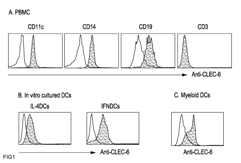
Figures IA and lB show both in vivo and in vitro-cultured DCs express CLEC-6.
Figure IA
shows PBMCs from normal donors were stained with anti-CD l 1 c, CD 14, CD 19,
and CD3 with
anti-CLEC-6 mAbs. Cells stained with individual antibodies were gated to
measure the
expression levels of CLEC-6. Figure lB shows monocytes from normal donors were
cultured in
CA 02717656 2010-08-18
WO 2008/103947 PCT/US2008/054785
6
the presence of GM-CSF with IL-4 (IL-4DCs) or IFNa (IFNDCs), and cells were
stained with
anti-CLEC-6 mAb or isotype control antibody. C. myeloid DCs (Lin-HLA-
DR+CD1lc+CD123-
) were purified from blood by FACS sorter, and stained with anti-CLEC-6 mAbs.
Open and
closed histograms represent cells stained with, respectively, isotype control
and anti-CLEC-6
mAb.
Figure 2 shows that Anti-CLEC-6 mAbs activate DCs. IFNDCs (1x10⁵/200u1/well)
were
cultured in the plates coated with different clones of mAbs for 18 h. Culture
supernatants were
analyzed to measure cytokines and chemokines by Luminex.
Figures 3A and 3B show that anti-CLEC-6 mAbs activate DCs. Figure 3A shows IL-
4DCs
(1x10⁵/well/200 ul) stimulated with anti-CLEC-6 for 18 h, and then cells were
stained with anti-
CD86 and HLA-DR. Figure 3B shows myeloid DCs purified from blood by FACS
sorting.
mDCs (lxl0e5/well/200 ul) were stimulated with anti-CLEC-6 mAbs for 18 h, and
cells were
stained with anti-CD86, CD80, and HLA-DR.
Figure 4 shows the gene expression profile for IL-4DCs stimulated with either
anti-CLEC-6 or
control mAbs for 12 h. Total RNA extracted with RNeasy columns (Qiagen), and
analyzed with
the 2100 Bioanalyser (Agilent). Biotin-labeled cRNA targets were prepared
using the Illumina
totalprep labeling kit (Ambion) and hybridized to Sentrix Human6 BeadChips
(46K transcripts).
These microarrays consist of 50mer oligonucleotide probes attached to 3um
beads which are
lodged into microwells etched at the surface of a silicon wafer. After
staining with Streptavidin-
Cy3, the array surface is imaged using a sub-micron resolution scanner
manufactured by
Illumina (Beadstation 500X). A gene expression analysis software program,
GeneSpring,
Version 7.1 (Agilent), was used to perform data analysis.
Figures 5A and 5B show DCs activated with anti-CLEC-6 produce increased
amounts of
cytokines and chemokines. In vitro-cultured IL-4DCs and purified mDCs
(1x10⁵/200 ul), as
described in Fig. 1 legend, were cultured in the plates coated with anti-CLEC-
6 mAb (2 ug/well)
for 18 h. Culture supernatants were analyzed to measure cytokine and
chemokines by Luminex.
Figures 6A and 6B show that CLEC-6 and CD40 synergize to activate DCs. IL-4DCs
(2x10⁵/200 ul/well) were cultured in the 96-well plates coated with anti-CLEC-
6 in the presence
or absence of soluble CD40L (20 ng/ml) for 18 h. Control mAbs were also
tested. After 18 h,
cells were stained with anti-CD83 and culture supernatants were analyzed to
measure cytokines
and chemokines by Luminex.
CA 02717656 2010-08-18
WO 2008/103947 PCT/US2008/054785
7
Figures 7A to 7C show that CLEC-6 expressed on DCs contributes to enhanced
Immoral
immune responses. Six day GM/IL-4 DCs, 5x10³/well, were incubated in 96 well
plates coated
with anti-CLEC-6 or control mAbs for 16-18 h, and then 1x10⁵ autologous CD19+
B cells
stained with CFSE were co-cultured in the presence of 20 units/ml IL-2 and 50
nM CpG. Figure
7A: on day six, cells were stained with fluorescently labeled antibodies. CD3+
and 7-AAD+
cells were gated out. CD38+ and CFSE- cells were purified by FACS sorter and
Giemsa staining
was performed. Figure 7B shows the culture supernatants on day thirteen were
analyzed for total
IgM, IgG, and IgA by sandwich ELISA. Figure 7C shows that six day GM/IL-4 DCs
cultured in
mAb-coated plates for 48 h, and expression levels of APRIL were determined by
intracellular
staining of the cells. Dotted lines are cells stained with control antibody.
Thin and thick lines
represent cells incubated in the plates coated with anti-CLEC-6 or control
mAb, respectively.
Data are representative of two separate experiments using cells from three
different normal
donors each time.
Figures 8A and 8B show that CLEC-6 expressed on B cells contributes to B cell
activation and
immunoglobulin production. Figure 8A shows CD19+ B cells (2x10⁵/well/200 ul)
were cultured
in plates coated with the mAbs for 16-18 h, and then culture supernatants were
analyzed for
cytokines and chemokines by Luminex. Figure 8B shows 1x10⁵ CD19+ B cells were
cultured in
plates coated with the mAbs for thirteen days. Total Ig levels were measured
by ELISA. Data
are representative of two repeat experiments using cells from three different
normal donors.
Figures 9A to 9E show that CLEC-6 expressed on DCs contributes to enhanced
antigen specific
T cell responses. Figure 9A. 5x10³ of six day IFNDCs were cultured in the
plates coated with
anti-CLEC-6 or control mAbs for 16-18 h, and then purified allogeneic T cells
were co-cultured.
Cells were pulsed with 3[H]-thymidine, 1 uCi/well, for 18 h before harvesting.
3[H]-thymidine
uptake was measured by a beta-counter. Figure 9B. IL-4DCs (5x10³/well) were
incubated in
plates coated with the mAbs in the presence of 100 nM Flu Ml peptide (HLA-A2
epitope)
(upper two panels) or recombinant Flu Ml protein (lower two panels) for 16 h.
2x10⁶ purified
autologous CD8 T cells were co-cultured for 7 days. On day two, 20 units/ml IL-
2 and 10
units/ml of IL-7 were added to the culture. Cells were stained with anti-CD8
and Flu Ml-
tetramer. Figure 9C. IL-4DCs (5x10³/well) were incubated in plates coated with
the mAbs in the
presence of 20 uM Mart-1 peptide (HLA-A2 epitope)(upper two panels) or
recombinant Mart-1
protein (lower two panels) for 16 h. 2x10⁶ purified autologous CD8 T cells
were co-cultured for
10 days. On day two, 20 units/ml IL-2 and 10 units/ml of IL-7 were added to
the culture. Cells
were stained with anti-CD8 and Mart-l-tetramer. Figure 9D. IL-4DCs were loaded
with 10 nM
CA 02717656 2010-08-18
WO 2008/103947 PCT/US2008/054785
8
of anti-CLEC-6-Mart-1 complex or control Ig-Mart-1 complex for 2 h. 2x10⁶
purified
autologous CD8 T cells were co-cultured for 10 days. Cells were stained with
anti-CD8 and Flu
Ml-specific tetramer. Cells in the lower two panels were stimulated with 20
ng/ml LPS from E.
coli. Figure 9E. Purified mDCs loaded with 10 nM of anti-CLEC-6-Flu HA1 or
control Ig-Flu
HA1 complexes for 2 h. 2x10⁶ purified autologous CD4 T cells labeled with CFSE
were co-
cultured for 7 days. Cells were stained with anti-CD4, and cell proliferation
was measured by
analyzing CFSE dilution. Cells in lower two panels were stimulated with 20
ng/ml LPS from E.
coli.
Figure 10 shows PBMC from non-human primates (Cynomolgus) were stained with
anti-CLEC-
6 mAb and antibodies to cell surface markers and analyzed by FACS.
DETAILED DESCRIPTION OF THE INVENTION
While the making and using of various embodiments of the present invention are
discussed in
detail below, it should be appreciated that the present invention provides
many applicable
inventive concepts that can be embodied in a wide variety of specific
contexts. The specific
embodiments discussed herein are merely illustrative of specific ways to make
and use the
invention and do not delimit the scope of the invention.
To facilitate the understanding of this invention, a number of terms are
defined below. Terms
defined herein have meanings as commonly understood by a person of ordinary
skill in the areas
relevant to the present invention. Terms such as "a", "an" and "the" are not
intended to refer to
only a singular entity, but include the general class of which a specific
example may be used for
illustration. The terminology herein is used to describe specific embodiments
of the invention,
but their usage does not delimit the invention, except as outlined in the
claims.
The Dectin-1 gene cluster contains lectin-like oxidized low-density
lipoprotein receptor (LOX)-
1, C-type lectin-like receptor (CLEC)-1 and 2, as well as MICL. CLEC-1 is
expressed
intracellularly when transfected into culture cells, and, therefore,
requirement of some adaptor
molecule was predicted for its surface expression (M. Colonna et al. Eur J
Immunol 30 (2000),
pp. 697-704). However, no cationic amino acid is present in its transmembrane
portion. Instead,
one tyrosine residue is present in its cytoplasmic portion, but the signaling
effect through this
tyrosine is unknown. CLEC-2 contains one DxYxxL (aspartic acid-any-tyrosine-
any-any-
leucine) motif in its cytoplasm and is expressed on the transfected cell
surface. This motif is
known to encourage efficient endocytosis and basolateral expression of ASGPR-
1, and is highly
homologous to the second tyrosine-based motif of dectin-1. In fact, Syk is
recruited to the
CA 02717656 2010-08-18
WO 2008/103947 PCT/US2008/054785
9
phosphotyrosine of CLEC-2, induced by its ligand, the snake venom rhodocytin
(aggretin) (K.
Suzuki-Inoue et al. Blood 107 (2006), pp. 542-549). This observation confirms
the presence of a
unique single YxxL sequence in C-type lectin receptors, which provides a
docking site for Syk
when tyrosine-phosphorylated. MICL (CLEC 12A) has been identified as an ITIM-
containing
molecule homologous to dectin-1 and LOX-1 (A.S. Marshall et al. JBiol Chem 279
(2004), pp.
14792-14802). Its expression is primarily restricted to monocytes,
granulocytes and immature
DCs. Functionally, MICL recruits SHP-1 and 2 upon stimulation and an ITIM-
dependent
inhibitory effect has been observed using a chimeric receptor containing
cytoplasmic MICL
(A.S. Marshall et al. J Biol Chem 279 (2004), pp. 14792-14802). In a recent
report, however,
after ligation of MICL on immature DCs, an altered protein tyrosine
phophorylation pattern as
well as serine phosphorylation of p38 MAPK and ERK were observed, and,
furthermore, CCR7
expression and cytokine production were noted without upregulation of
maturation marker such
as CD83, 86 and DC-LAMP (C.H. Chen et al. Blood 107 (2006), pp. 1459-1467).
Indeed, such
CCR7+ costimulationbow semi-mature phenotype is considered to represent the
steady-state
migrating DCs (L. Ohl et al. Imunity 21 (2004), pp. 279-288). Though still
uncharacterized, the
genes coding for CLEC9A and CLEC12B are also located in the dectin-1 gene
cluster (G.D.
Brown, Nat Rev Immunol 6 (2006), pp. 33-43). CLEC12B contains ITIM in its
cytoplasmic tail,
while CLEC9A bears an ExYxxL (glutamic acid-any-tyrosine-any-any-leucine)
sequence, which
might act as an activation motif. The functions of these molecules remain to
be investigated.
Arce et al., Eur. J. Immunol. (2004) identified and characterized the human
CLEC-6 protein,
related to mouse Mcl/Clecsf8.Human CLEC-6 codes for a type II membrane
glycoprotein of 215
amino acids that belongs to the human calcium-dependent lectin family (C-type
lectin). The
CLEC-6 extracellular region shows a single carbohydrate recognition domain
(CRD).
Biochemical analysis of CLEC-6 on transiently transfected cells showed a
glycoprotein of 30
kDa and cross-linking of the receptor leads to a rapid internalization
suggesting that CLEC-6 is
an endocytic receptor (Arce et al., 2004). Unlike CLEC-1, -2, -9A, -12A, and -
12B, CLEC-6
does not contain a YxxL motif or other consensus signaling motifs. No study
has been done to
characterize the biological function of CLEC-6.
DCs can cross-present protein antigens (Rock KL Immunol Rev. 2005 Oct;207:166-
83). In vivo,
DCs take up antigens by the means of a number of receptors and present
antigenic peptides in
both class I and II. In this context, DC lectins, as pattern recognition
receptors, contribute to the
efficient uptake of antigens as well as cross-presentation of antigens.
CA 02717656 2010-08-18
WO 2008/103947 PCT/US2008/054785
As used herein, the term "modular rAb carrier" is used to describe a
recombinant antibody
system that has been engineered to provide the controlled modular addition of
diverse antigens,
activating proteins, or other antibodies to a single recombinant monoclonal
antibody (mAb), in
this case, an anti-CLEC-6 monoclonal antibody. The rAb may be a monoclonal
antibody made
5 using standard hybridoma techniques, recombinant antibody display, humanized
monoclonal
antibodies and the like. The modular rAb carrier can be used to, e.g., target
(via one primary
recombinant antibody against an internalizing receptor, e.g., a human
dendritic cell receptor)
multiple antigens and/or antigens and an activating cytokine to dendritic
cells (DC). The
modular rAb carrier may also be used to join two different recombinant mAbs
end-to-end in a
10 controlled and defined manner.
The antigen binding portion of the "modular rAb carrier" may be one or more
variable domains,
one or more variable and the first constant domain, an Fab fragment, a Fab'
fragment, an F(ab)2
fragment, and Fv fragment, and Fabc fragment and/or a Fab fragment with
portions of the Fc
domain to which the cognate modular binding portions are added to the amino
acid sequence
and/or bound. The antibody for use in the modular rAb carrier can be of any
isotype or class,
subclass or from any source (animal and/or recombinant).
In one non-limiting example, the modular rAb carrier is engineered to have one
or more modular
cohesin-dockerin protein domains for making specific and defined protein
complexes in the
context of engineered recombinant mAbs. The mAb is a portion of a fusion
protein that includes
one or more modular cohesin-dockerin protein domains carboxy from the antigen
binding
domains of the mAb. The cohesin-dockerin protein domains may even be attached
post-
translationally, e.g., by using chemical cross-linkers and/or disulfide
bonding.
The term "antigen" as used herein refers to a molecule that can initiate a
Immoral and/or cellular
immune response in a recipient of the antigen. Antigen may be used in two
different contexts
with the present invention: as a target for the antibody or other antigen
recognition domain of the
rAb or as the molecule that is carried to and/or into a cell or target by the
rAb as part of a
dockerin/cohesin-molecule complement to the modular rAb carrier. The antigen
is usually an
agent that causes a disease for which a vaccination would be advantageous
treatment. When the
antigen is presented on MHC, the peptide is often about 8 to about 25 amino
acids. Antigens
include any type of biologic molecule, including, for example, simple
intermediary metabolites,
sugars, lipids and hormones as well as macromolecules such as complex
carbohydrates,
phospholipids, nucleic acids and proteins. Common categories of antigens
include, but are not
CA 02717656 2010-08-18
WO 2008/103947 PCT/US2008/054785
11
limited to, viral antigens, bacterial antigens, fungal antigens, protozoal and
other parasitic
antigens, tumor antigens, antigens involved in autoimmune disease, allergy and
graft rejection,
and other miscellaneous antigens.
The modular rAb carrier is able to carry any number of active agents, e.g.,
antibiotics, anti-
infective agents, antiviral agents, anti-tumoral agents, antipyretics,
analgesics, anti-inflammatory
agents, therapeutic agents for osteoporosis, enzymes, cytokines,
anticoagulants, polysaccharides,
collagen, cells, and combinations of two or more of the foregoing active
agents. Examples of
antibiotics for delivery using the present invention include, without
limitation, tetracycline,
aminoglycosides, penicillins, cephalosporins, sulfonamide drugs,
chloramphenicol sodium
succinate, erythromycin, vancomycin, lincomycin, clindamycin, nystatin,
amphotericin B,
amantidine, idoxuridine, p-amino salicyclic acid, isoniazid, rifampin,
antinomycin D,
mithramycin, daunomycin, adriamycin, bleomycin, vinblastine, vincristine,
procarbazine,
imidazole carboxamide, and the like.
Examples of anti-tumor agents for delivery using the present invention
include, without
limitation, doxorubicin, Daunorubicin, taxol, methotrexate, and the like.
Examples of
antipyretics and analgesics include aspirin, Motrin , Ibuprofen , naprosyn,
acetaminophen, and
the like.
Examples of anti-inflammatory agents for delivery using the present invention
include, without
limitation, include NSAIDS, aspirin, steroids, dexamethasone, hydrocortisone,
prednisolone,
Diclofenac Na, and the like.
Examples of therapeutic agents for treating osteoporosis and other factors
acting on bone and
skeleton include for delivery using the present invention include, without
limitation, calcium,
alendronate, bone GLa peptide, parathyroid hormone and its active fragments,
histone H4-
related bone formation and proliferation peptide and mutations, derivatives
and analogs thereof.
Examples of enzymes and enzyme cofactors for delivery using the present
invention include,
without limitation, pancrease, L-asparaginase, hyaluronidase, chymotrypsin,
trypsin, tPA,
streptokinase, urokinase, pancreatin, collagenase, trypsinogen,
chymotrypsinogen, plasminogen,
streptokinase, adenyl cyclase, superoxide dismutase (SOD), and the like.
Examples of cytokines for delivery using the present invention include,
without limitation,
interleukins, transforming growth factors (TGFs), fibroblast growth factors
(FGFs), platelet
derived growth factors (PDGFs), epidermal growth factors (EGFs), connective
tissue activated
peptides (CTAPs), osteogenic factors, and biologically active analogs,
fragments, and
CA 02717656 2010-08-18
WO 2008/103947 PCT/US2008/054785
12
derivatives of such growth factors. Cytokines may be B/T-cell differentiation
factors, B/T-cell
growth factors, mitogenic cytokines, chemotactic cytokines, colony stimulating
factors,
angiogenesis factors, IFN-a, IFN-(3, IFN-y, ILl, IL2, IL3, IL4, IL5, IL6, IL7,
IL8, IL9, IL10,
IL 11, IL12, IL13, IL14, IL 15, IL16, IL17, IL18, etc., leptin, myostatin,
macrophage stimulating
protein, platelet-derived growth factor, TNF-a, TNF-(3, NGF, CD40L, CD137L/4-
1BBL, human
lymphotoxin-0, G-CSF, M-CSF, GM-CSF, PDGF, IL-la, ILl- 0, IP-l0, PF4, GRO,
9E3,
erythropoietin, endostatin, angiostatin, VEGF or any fragments or combinations
thereof. Other
cytokines include members of the transforming growth factor (TGF) supergene
family include
the beta transforming growth factors (for example TGF-01, TGF-02, TGF-(33);
bone
morphogenetic proteins (for example, BMP-1, BMP-2, BMP-3, BMP-4, BMP-5, BMP-6,
BMP-
7, BMP-8, BMP-9); heparin-binding growth factors (for example, fibroblast
growth factor
(FGF), epidermal growth factor (EGF), platelet-derived growth factor (PDGF),
insulin-like
growth factor (IGF)); Inhibins (for example, Inhibin A, Inhibin B); growth
differentiating factors
(for example, GDF-1); and Activins (for example, Activin A, Activin B, Activin
AB).
Examples of growth factors for delivery using the present invention include,
without limitation,
growth factors that can be isolated from native or natural sources, such as
from mammalian
cells, or can be prepared synthetically, such as by recombinant DNA techniques
or by various
chemical processes. In addition, analogs, fragments, or derivatives of these
factors can be used,
provided that they exhibit at least some of the biological activity of the
native molecule. For
example, analogs can be prepared by expression of genes altered by site-
specific mutagenesis or
other genetic engineering techniques.
Examples of anticoagulants for delivery using the present invention include,
without limitation,
include warfarin, heparin, Hirudin, and the like. Examples of factors acting
on the immune
system include for delivery using the present invention include, without
limitation, factors which
control inflammation and malignant neoplasms and factors which attack
infective
microorganisms, such as chemotactic peptides and bradykinins.
Examples of viral antigens include, but are not limited to, e.g., retroviral
antigens such as
retroviral antigens from the human immunodeficiency virus (HIV) antigens such
as gene
products of the gag, pol, and env genes, the Nef protein, reverse
transcriptase, and other HIV
components; hepatitis viral antigens such as the S, M, and L proteins of
hepatitis B virus, the
pre-S antigen of hepatitis B virus, and other hepatitis, e.g., hepatitis A, B,
and C, viral
components such as hepatitis C viral RNA; influenza viral antigens such as
hemagglutinin and
CA 02717656 2010-08-18
WO 2008/103947 PCT/US2008/054785
13
neuraminidase and other influenza viral components; measles viral antigens
such as the measles
virus fusion protein and other measles virus components; rubella viral
antigens such as proteins
El and E2 and other rubella virus components; rotaviral antigens such as VP7sc
and other
rotaviral components; cytomegaloviral antigens such as envelope glycoprotein B
and other
cytomegaloviral antigen components; respiratory syncytial viral antigens such
as the RSV fusion
protein, the M2 protein and other respiratory syncytial viral antigen
components; herpes simplex
viral antigens such as immediate early proteins, glycoprotein D, and other
herpes simplex viral
antigen components; varicella zoster viral antigens such as gpl, gpII, and
other varicella zoster
viral antigen components; Japanese encephalitis viral antigens such as
proteins E, M-E, M-E-
NS1, NS1, NS1-NS2A, 80% E, and other Japanese encephalitis viral antigen
components; rabies
viral antigens such as rabies glycoprotein, rabies nucleoprotein and other
rabies viral antigen
components. See Fundamental Virology, Second Edition, eds. Fields, B. N. and
Knipe, D. M.
(Raven Press, New York, 1991) for additional examples of viral antigens.
Antigenic targets that may be delivered using the rAb-DC/DC-antigen vaccines
of the present
invention include genes encoding antigens such as viral antigens, bacterial
antigens, fungal
antigens or parasitic antigens. Viruses include picornavirus, coronavirus,
togavirus, flavirvirus,
rhabdovirus, paramyxovirus, orthomyxovirus, bunyavirus, arenavirus, reovirus,
retrovirus,
papilomavirus, parvovirus, herpesvirus, poxvirus, hepadnavirus, and spongiform
virus. Other
viral targets include influenza, herpes simplex virus 1 and 2, measles,
dengue, smallpox, polio or
HIV. Pathogens include trypanosomes, tapeworms, roundworms, helminthes,
malaria. Tumor
markers, such as fetal antigen or prostate specific antigen, may be targeted
in this manner. Other
examples include: HIV env proteins and hepatitis B surface antigen.
Administration of a vector
according to the present invention for vaccination purposes would require that
the vector-
associated antigens be sufficiently non-immunogenic to enable long term
expression of the
transgene, for which a strong immune response would be desired. In some cases,
vaccination of
an individual may only be required infrequently, such as yearly or biennially,
and provide long
term immunologic protection against the infectious agent. Specific examples of
organisms,
allergens and nucleic and amino sequences for use in vectors and ultimately as
antigens with the
present invention may be found in U.S. Patent No. 6,541,011, relevant portions
incorporated
herein by reference, in particular, the tables that match organisms and
specific sequences that
may be used with the present invention.
Bacterial antigens for use with the rAb vaccine disclosed herein include, but
are not limited to,
e.g., bacterial antigens such as pertussis toxin, filamentous hemagglutinin,
pertactin, FIM2,
CA 02717656 2010-08-18
WO 2008/103947 PCT/US2008/054785
14
FIM3, adenylate cyclase and other pertussis bacterial antigen components;
diptheria bacterial
antigens such as diptheria toxin or toxoid and other diptheria bacterial
antigen components;
tetanus bacterial antigens such as tetanus toxin or toxoid and other tetanus
bacterial antigen
components; streptococcal bacterial antigens such as M proteins and other
streptococcal
bacterial antigen components; gram-negative bacilli bacterial antigens such as
lipopolysaccharides and other gram-negative bacterial antigen components,
Mycobacterium
tuberculosis bacterial antigens such as mycolic acid, heat shock protein 65
(HSP65), the 30 kDa
major secreted protein, antigen 85A and other mycobacterial antigen
components; Helicobacter
pylori bacterial antigen components; pneumococcal bacterial antigens such as
pneumolysin,
pneumococcal capsular polysaccharides and other pneumococcal bacterial antigen
components;
haemophilus influenza bacterial antigens such as capsular polysaccharides and
other
haemophilus influenza bacterial antigen components; anthrax bacterial antigens
such as anthrax
protective antigen and other anthrax bacterial antigen components; rickettsiae
bacterial antigens
such as rompA and other rickettsiae bacterial antigen component. Also included
with the
bacterial antigens described herein are any other bacterial, mycobacterial,
mycoplasmal,
rickettsial, or chlamydial antigens. Partial or whole pathogens may also be:
haemophilus
influenza; Plasmodium falciparum; neisseria meningitidis; streptococcus
pneumoniae; neisseria
gonorrhoeae; salmonella serotype typhi; shigella; vibrio cholerae; Dengue
Fever;
Encephalitides; Japanese Encephalitis; lyme disease; Yersinia pestis; west
nile virus; yellow
fever; tularemia; hepatitis (viral; bacterial); RSV (respiratory syncytial
virus); HPIV 1 and HPIV
3; adenovirus; small pox; allergies and cancers.
Fungal antigens for use with compositions and methods of the invention
include, but are not
limited to, e.g., candida fungal antigen components; histoplasma fungal
antigens such as heat
shock protein 60 (HSP60) and other histoplasma fungal antigen components;
cryptococcal
fungal antigens such as capsular polysaccharides and other cryptococcal fungal
antigen
components; coccidiodes fungal antigens such as spherule antigens and other
coccidiodes fungal
antigen components; and tinea fungal antigens such as trichophytin and other
coccidiodes fungal
antigen components.
Examples of protozoal and other parasitic antigens include, but are not
limited to, e.g.,
plasmodium falciparum antigens such as merozoite surface antigens, sporozoite
surface
antigens, circumsporozoite antigens, gametocyte/gamete surface antigens, blood-
stage antigen pf
155/RESA and other plasmodial antigen components; toxoplasma antigens such as
SAG-1, p30
and other toxoplasmal antigen components; schistosomae antigens such as
glutathione-S-
CA 02717656 2010-08-18
WO 2008/103947 PCT/US2008/054785
transferase, paramyosin, and other schistosomal antigen components; leishmania
major and
other leishmaniae antigens such as gp63, lipophosphoglycan and its associated
protein and other
leishmanial antigen components; and trypanosoma cruzi antigens such as the 75-
77 kDa antigen,
the 56 kDa antigen and other trypanosomal antigen components.
5 Antigen that can be targeted using the rAb of the present invention will
generally be selected
based on a number of factors, including: likelihood of internalization, level
of immune cell
specificity, type of immune cell targeted, level of immune cell maturity
and/or activation and the
like. Examples of cell surface markers for dendritic cells include, but are
not limited to, MHC
class I, MHC Class II, B7-2, CD18, CD29, CD31, CD43, CD44, CD45, CD54, CD58,
CD83,
10 CD86, CMRF-44, CMRF-56, DCIR and/or DECTIN-1 and the like; while in some
cases also
having the absence of CD2, CD3, CD4, CD8, CD14, CD15, CD16, CD 19, CD20, CD56,
and/or
CD57. Examples of cell surface markers for antigen presenting cells include,
but are not limited
to, MHC class I, MHC Class II, CD40, CD45, B7-1, B7-2, IFN-y receptor and IL-2
receptor,
ICAM-1 and/or Fey receptor. Examples of cell surface markers for T cells
include, but are not
15 limited to, CD3, CD4, CD8, CD 14, CD20, CD1 lb, CD16, CD45 and HLA-DR.
Target antigens on cell surfaces for delivery includes those characteristic of
tumor antigens
typically will be derived from the cell surface, cytoplasm, nucleus,
organelles and the like of
cells of tumor tissue. Examples of tumor targets for the antibody portion of
the present
invention include, without limitation, hematological cancers such as leukemias
and lymphomas,
neurological tumors such as astrocytomas or glioblastomas, melanoma, breast
cancer, lung
cancer, head and neck cancer, gastrointestinal tumors such as gastric or colon
cancer, liver
cancer, pancreatic cancer, genitourinary tumors such cervix, uterus, ovarian
cancer, vaginal
cancer, testicular cancer, prostate cancer or penile cancer, bone tumors,
vascular tumors, or
cancers of the lip, nasopharynx, pharynx and oral cavity, esophagus, rectum,
gall bladder, biliary
tree, larynx, lung and bronchus, bladder, kidney, brain and other parts of the
nervous system,
thyroid, Hodgkin's disease, non-Hodgkin's lymphoma, multiple myeloma and
leukemia.
Examples of antigens that may be delivered alone or in combination to immune
cells for antigen
presentation using the present invention include tumor proteins, e.g., mutated
oncogenes; viral
proteins associated with tumors; and tumor mucins and glycolipids. The
antigens may be viral
proteins associated with tumors would be those from the classes of viruses
noted above. Certain
antigens may be characteristic of tumors (one subset being proteins not
usually expressed by a
tumor precursor cell), or may be a protein which is normally expressed in a
tumor precursor cell,
CA 02717656 2010-08-18
WO 2008/103947 PCT/US2008/054785
16
but having a mutation characteristic of a tumor. Other antigens include mutant
variant(s) of the
normal protein having an altered activity or subcellular distribution, e.g.,
mutations of genes
giving rise to tumor antigens.
Specific non-limiting examples of tumor antigens include: CEA, prostate
specific antigen (PSA),
HER-2/neu, BAGE, GAGE, MAGE 1-4, 6 and 12, MUC (Mucin) (e.g., MUC-1, MUC-2,
etc.),
GM2 and GD2 gangliosides, ras, myc, tyrosinase, MART (melanoma antigen), Pmel
17(gp 100),
GnT-V intron V sequence (N-acetylglucoaminyltransferase V intron V sequence),
Prostate Ca
psm, PRAME (melanoma antigen), 0-catenin, MUM-1-13 (melanoma ubiquitous
mutated gene
product), GAGE (melanoma antigen) 1, BAGE (melanoma antigen) 2-10, c-ERB2
(Her2/neu),
EBNA (Epstein-Barr Virus nuclear antigen) 1-6, gp75, human papilloma virus
(HPV) E6 and
E7, p53, lung resistance protein (LRP), Bcl-2, and Ki-67. In addition, the
immunogenic
molecule can be an autoantigen involved in the initiation and/or propagation
of an autoimmune
disease, the pathology of which is largely due to the activity of antibodies
specific for a molecule
expressed by the relevant target organ, tissue, or cells, e.g., SLE or MG. In
such diseases, it can
be desirable to direct an ongoing antibody-mediated (i.e., a Th2-type) immune
response to the
relevant autoantigen towards a cellular (i.e., a Thl-type) immune response.
Alternatively, it can
be desirable to prevent onset of or decrease the level of a Th2 response to
the autoantigen in a
subject not having, but who is suspected of being susceptible to, the relevant
autoimmune
disease by prophylactically inducing a Thl response to the appropriate
autoantigen.
Autoantigens of interest include, without limitation: (a) with respect to SLE,
the Smith protein,
RNP ribonucleoprotein, and the SS-A and SS-B proteins; and (b) with respect to
MG, the
acetylcholine receptor. Examples of other miscellaneous antigens involved in
one or more types
of autoimmune response include, e.g., endogenous hormones such as luteinizing
hormone,
follicular stimulating hormone, testosterone, growth hormone, prolactin, and
other hormones.
Antigens involved in autoimmune diseases, allergy, and graft rejection can be
used in the
compositions and methods of the invention. For example, an antigen involved in
any one or
more of the following autoimmune diseases or disorders can be used in the
present invention:
diabetes, diabetes mellitus, arthritis (including rheumatoid arthritis,
juvenile rheumatoid arthritis,
osteoarthritis, psoriatic arthritis), multiple sclerosis, myasthenia gravis,
systemic lupus
erythematosis, autoimmune thyroiditis, dermatitis (including atopic dermatitis
and eczematous
dermatitis), psoriasis, Sjogren's Syndrome, including keratoconjunctivitis
sicca secondary to
Sjogren's Syndrome, alopecia areata, allergic responses due to arthropod bite
reactions, Crohn's
disease, aphthous ulcer, iritis, conjunctivitis, keratoconjunctivitis,
ulcerative colitis, asthma,
CA 02717656 2010-08-18
WO 2008/103947 PCT/US2008/054785
17
allergic asthma, cutaneous lupus erythematosus, scleroderma, vaginitis,
proctitis, drug eruptions,
leprosy reversal reactions, erythema nodosum leprosum, autoimmune uveitis,
allergic
encephalomyelitis, acute necrotizing hemorrhagic encephalopathy, idiopathic
bilateral
progressive sensorineural hearing loss, aplastic anemia, pure red cell anemia,
idiopathic
thrombocytopenia, polychondritis, Wegener's granulomatosis, chronic active
hepatitis, Stevens-
Johnson syndrome, idiopathic sprue, lichen planus, Crohn's disease, Graves
ophthalmopathy,
sarcoidosis, primary biliary cirrhosis, uveitis posterior, and interstitial
lung fibrosis. Examples of
antigens involved in autoimmune disease include glutamic acid decarboxylase 65
(GAD 65),
native DNA, myelin basic protein, myelin proteolipid protein, acetylcholine
receptor
components, thyroglobulin, and the thyroid stimulating hormone (TSH) receptor.
Examples of
antigens involved in allergy include pollen antigens such as Japanese cedar
pollen antigens,
ragweed pollen antigens, rye grass pollen antigens, animal derived antigens
such as dust mite
antigens and feline antigens, histocompatiblity antigens, and penicillin and
other therapeutic
drugs. Examples of antigens involved in graft rejection include antigenic
components of the
graft to be transplanted into the graft recipient such as heart, lung, liver,
pancreas, kidney, and
neural graft components. The antigen may be an altered peptide ligand useful
in treating an
autoimmune disease.
As used herein, the term "epitope(s)" refer to a peptide or protein antigen
that includes a
primary, secondary or tertiary structure similar to an epitope located within
any of a number of
pathogen polypeptides encoded by the pathogen DNA or RNA. The level of
similarity will
generally be to such a degree that monoclonal or polyclonal antibodies
directed against such
polypeptides will also bind to, react with, or otherwise recognize, the
peptide or protein antigen.
Various immunoassay methods may be employed in conjunction with such
antibodies, such as,
for example, Western blotting, ELISA, RIA, and the like, all of which are
known to those of
skill in the art. The identification of pathogen epitopes, and/or their
functional equivalents,
suitable for use in vaccines is part of the present invention. Once isolated
and identified, one
may readily obtain functional equivalents. For example, one may employ the
methods of Hopp,
as taught in U.S. Pat. No. 4,554,101, incorporated herein by reference, which
teaches the
identification and preparation of epitopes from amino acid sequences on the
basis of
hydrophilicity. The methods described in several other papers, and software
programs based
thereon, can also be used to identify epitopic core sequences (see, for
example, Jameson and
Wolf, 1988; Wolf et al., 1988; U.S. Pat. No. 4,554,101). The amino acid
sequence of these
SUBSTITUTE SHEET (RULE 26)
CA 02717656 2010-08-18
WO 2008/103947 PCT/US2008/054785
18
"epitopic core sequences" may then be readily incorporated into peptides,
either through the
application of peptide synthesis or recombinant technology.
The preparation of vaccine compositions that includes the nucleic acids that
encode antigens of
the invention as the active ingredient, may be prepared as injectables, either
as liquid solutions
or suspensions; solid forms suitable for solution in, or suspension in, liquid
prior to infection can
also be prepared. The preparation may be emulsified, encapsulated in
liposomes. The active
immunogenic ingredients are often mixed with carriers which are
pharmaceutically acceptable
and compatible with the active ingredient.
The term "pharmaceutically acceptable carrier" refers to a carrier that does
not cause an allergic
reaction or other untoward effect in subjects to whom it is administered.
Suitable
pharmaceutically acceptable carriers include, for example, one or more of
water, saline,
phosphate buffered saline, dextrose, glycerol, ethanol, or the like and
combinations thereof. In
addition, if desired, the vaccine can contain minor amounts of auxiliary
substances such as
wetting or emulsifying agents, pH buffering agents, and/or adjuvants which
enhance the
effectiveness of the vaccine. Examples of adjuvants that may be effective
include but are not
limited to: aluminum hydroxide, N-acetyl-muramyl-L-threonyl-D-isoglutamine
(thr-MDP), N-
acetyl-nor-muramyl-L-alanyl-D-isoglutamine, MTP-PE and RIBI, which contains
three
components extracted from bacteria, monophosporyl lipid A, trehalose
dimycolate and cell wall
skeleton (MPL+TDM+CWS) in a 2% squalene/Tween 80 emulsion. Other examples of
adjuvants include DDA (dimethyldioctadecylammonium bromide), Freund's complete
and
incomplete adjuvants and QuilA. In addition, immune modulating substances such
as
lymphokines (e.g., IFN-y, IL-2 and IL-12) or synthetic IFN-y inducers such as
poly I:C can be
used in combination with adjuvants described herein.
Pharmaceutical products that may include a naked polynucleotide with a single
or multiple
copies of the specific nucleotide sequences that bind to specific DNA-binding
sites of the
apolipoproteins present on plasma lipoproteins as described in the current
invention. The
polynucleotide may encode a biologically active peptide, antisense RNA, or
ribozyme and will
be provided in a physiologically acceptable administrable form. Another
pharmaceutical
product that may spring from the current invention may include a highly
purified plasma
lipoprotein fraction, isolated according to the methodology, described herein
from either the
patients blood or other source, and a polynucleotide containing single or
multiple copies of the
specific nucleotide sequences that bind to specific DNA-binding sites of the
apolipoproteins
SUBSTITUTE SHEET (RULE 26)
CA 02717656 2010-08-18
WO 2008/103947 PCT/US2008/054785
19
present on plasma lipoproteins, prebound to the purified lipoprotein fraction
in a physiologically
acceptable, administrable form.
Yet another pharmaceutical product may include a highly purified plasma
lipoprotein fraction
which contains recombinant apolipoprotein fragments containing single or
multiple copies of
specific DNA-binding motifs, prebound to a polynucleotide containing single or
multiple copies
of the specific nucleotide sequences, in a physiologically acceptable
administrable form. Yet
another pharmaceutical product may include a highly purified plasma
lipoprotein fraction which
contains recombinant apolipoprotein fragments containing single or multiple
copies of specific
DNA-binding motifs, prebound to a polynucleotide containing single or multiple
copies of the
specific nucleotide sequences, in a physiologically acceptable administrable
form.
The dosage to be administered depends to a great extent on the body weight and
physical
condition of the subject being treated as well as the route of administration
and frequency of
treatment. A pharmaceutical composition that includes the naked polynucleotide
prebound to a
highly purified lipoprotein fraction may be administered in amounts ranging
from 1 gg to 1 mg
polynucleotide and 1 gg to 100 mg protein.
Administration of an rAb and rAb complexes a patient will follow general
protocols for the
administration of chemotherapeutics, taking into account the toxicity, if any,
of the vector. It is
anticipated that the treatment cycles would be repeated as necessary. It also
is contemplated that
various standard therapies, as well as surgical intervention, may be applied
in combination with
the described gene therapy.
Where clinical application of a gene therapy is contemplated, it will be
necessary to prepare the
complex as a pharmaceutical composition appropriate for the intended
application. Generally
this will entail preparing a pharmaceutical composition that is essentially
free of pyrogens, as
well as any other impurities that could be harmful to humans or animals. One
also will generally
desire to employ appropriate salts and buffers to render the complex stable
and allow for
complex uptake by target cells.
Aqueous compositions of the present invention may include an effective amount
of the
compound, dissolved or dispersed in a pharmaceutically acceptable carrier or
aqueous medium.
Such compositions can also be referred to as inocula. The use of such media
and agents for
pharmaceutical active substances is well known in the art. Except insofar as
any conventional
media or agent is incompatible with the active ingredient, its use in the
therapeutic compositions
is contemplated. Supplementary active ingredients also can be incorporated
into the
SUBSTITUTE SHEET (RULE 26)
CA 02717656 2010-08-18
WO 2008/103947 PCT/US2008/054785
compositions. The compositions of the present invention may include classic
pharmaceutical
preparations. Dispersions also can be prepared in glycerol, liquid
polyethylene glycols, and
mixtures thereof and in oils. Under ordinary conditions of storage and use,
these preparations
contain a preservative to prevent the growth of microorganisms.
5 Disease States. Depending on the particular disease to be treated,
administration of therapeutic
compositions according to the present invention will be via any common route
so long as the
target tissue is available via that route in order to maximize the delivery of
antigen to a site for
maximum (or in some cases minimum) immune response. Administration will
generally be by
orthotopic, intradermal, subcutaneous, intramuscular, intraperitoneal or
intravenous injection.
10 Other areas for delivery include: oral, nasal, buccal, rectal, vaginal or
topical. Topical
administration would be particularly advantageous for treatment of skin
cancers. Such
compositions would normally be administered as pharmaceutically acceptable
compositions that
include physiologically acceptable carriers, buffers or other excipients.
Vaccine or treatment compositions of the invention may be administered
parenterally, by
15 injection, for example, either subcutaneously or intramuscularly.
Additional formulations which
are suitable for other modes of administration include suppositories, and in
some cases, oral
formulations or formulations suitable for distribution as aerosols. In the
case of the oral
formulations, the manipulation of T-cell subsets employing adjuvants, antigen
packaging, or the
addition of individual cytokines to various formulation that result in
improved oral vaccines with
20 optimized immune responses. For suppositories, traditional binders and
carriers may include,
for example, polyalkylene glycols or triglycerides; such suppositories may be
formed from
mixtures containing the active ingredient in the range of 0.5% to 10%,
preferably 1%-2%. Oral
formulations include such normally employed excipients as, for example,
pharmaceutical grades
of mannitol, lactose, starch magnesium stearate, sodium saccharine, cellulose,
magnesium
carbonate, and the like. These compositions take the form of solutions,
suspensions, tablets,
pills, capsules, sustained release formulations or powders and contain 10%-95%
of active
ingredient, preferably 25-70%.
The antigen encoding nucleic acids of the invention may be formulated into the
vaccine or
treatment compositions as neutral or salt forms. Pharmaceutically acceptable
salts include the
acid addition salts (formed with free amino groups of the peptide) and which
are formed with
inorganic acids such as, for example, hydrochloric or phosphoric acids, or
with organic acids
such as acetic, oxalic, tartaric, maleic, and the like. Salts formed with the
free carboxyl groups
SUBSTITUTE SHEET (RULE 26)
CA 02717656 2010-08-18
WO 2008/103947 PCT/US2008/054785
21
can also be derived from inorganic bases such as, for example, sodium,
potassium, ammonium,
calcium, or ferric hydroides, and such organic bases as isopropylamine,
trimethylamine, 2-
ethylamino ethanol, histidine, procaine, and the like.
Vaccine or treatment compositions are administered in a manner compatible with
the dosage
formulation, and in such amount as will be prophylactically and/or
therapeutically effective.
The quantity to be administered depends on the subject to be treated,
including, e.g., capacity of
the subject's immune system to synthesize antibodies, and the degree of
protection or treatment
desired. Suitable dosage ranges are of the order of several hundred micrograms
active ingredient
per vaccination with a range from about 0.1 mg to 1000 mg, such as in the
range from about 1
mg to 300 mg, and preferably in the range from about 10 mg to 50 mg. Suitable
regiments for
initial administration and booster shots are also variable but are typified by
an initial
administration followed by subsequent inoculations or other administrations.
Precise amounts of
active ingredient required to be administered depend on the judgment of the
practitioner and
may be peculiar to each subject. It will be apparent to those of skill in the
art that the
therapeutically effective amount of nucleic acid molecule or fusion
polypeptides of this
invention will depend, inter alia, upon the administration schedule, the unit
dose of antigen
administered, whether the nucleic acid molecule or fusion polypeptide is
administered in
combination with other therapeutic agents, the immune status and health of the
recipient, and the
therapeutic activity of the particular nucleic acid molecule or fusion
polypeptide.
The compositions can be given in a single dose schedule or in a multiple dose
schedule. A
multiple dose schedule is one in which a primary course of vaccination may
include, e.g., 1-10
separate doses, followed by other doses given at subsequent time intervals
required to maintain
and or reinforce the immune response, for example, at 1-4 months for a second
dose, and if
needed, a subsequent dose(s) after several months. Periodic boosters at
intervals of 1-5 years,
usually 3 years, are desirable to maintain the desired levels of protective
immunity. The course
of the immunization can be followed by in vitro proliferation assays of
peripheral blood
lymphocytes (PBLs) co-cultured with ESAT6 or ST-CF, and by measuring the
levels of IFN-y
released from the primed lymphocytes. The assays may be performed using
conventional labels,
such as radionucleotides, enzymes, fluorescent labels and the like. These
techniques are known
to one skilled in the art and can be found in U.S. Pat. Nos. 3,791,932,
4,174,384 and 3,949,064,
relevant portions incorporated by reference.
SUBSTITUTE SHEET (RULE 26)
CA 02717656 2010-08-18
WO 2008/103947 PCT/US2008/054785
22
The modular rAb carrier and/or conjugated rAb carrier-(cohesion/dockerin
and/or dockerin-
cohesin)-antigen complex (rAb-DC/DC-antigen vaccine) may be provided in one or
more "unit
doses" depending on whether the nucleic acid vectors are used, the final
purified proteins, or the
final vaccine form is used. Unit dose is defined as containing a predetermined-
quantity of the
therapeutic composition calculated to produce the desired responses in
association with its
administration, i.e., the appropriate route and treatment regimen. The
quantity to be
administered, and the particular route and formulation, are within the skill
of those in the clinical
arts. The subject to be treated may also be evaluated, in particular, the
state of the subject's
immune system and the protection desired. A unit dose need not be administered
as a single
injection but may include continuous infusion over a set period of time. Unit
dose of the present
invention may conveniently may be described in terms of DNA/kg (or protein/Kg)
body weight,
with ranges between about 0.05, 0.10, 0.15, 0.20, 0.25, 0.5, 1, 10, 50, 100,
1,000 or more
mg/DNA or protein/kg body weight are administered. Likewise the amount of rAb-
DC/DC-
antigen vaccine delivered can vary from about 0.2 to about 8.0 mg/kg body
weight. Thus, in
particular embodiments, 0.4 mg, 0.5 mg, 0.8 mg, 1.0 mg, 1.5 mg, 2.0 mg, 2.5
mg, 3.0 mg, 4.0
mg, 5.0 mg, 5.5 mg, 6.0 mg, 6.5 mg, 7.0 mg and 7.5 mg of the vaccine may be
delivered to an
individual in vivo. The dosage of rAb-DC/DC-antigen vaccine to be administered
depends to a
great extent on the weight and physical condition of the subject being treated
as well as the route
of administration and the frequency of treatment. A pharmaceutical composition
that includes a
naked polynucleotide prebound to a liposomal or viral delivery vector may be
administered in
amounts ranging from 1 gg to 1 mg polynucleotide to 1 gg to 100 mg protein.
Thus, particular
compositions may include between about 1 g, 5 g, 10 g, 20 g, 30 g, 40 g,
50 g, 60 g,
70 g, 80 g, 100 g, 150 g, 200 g, 250 g, 500 g, 600 g, 700 g, 800 g,
900 gg or 1,000
gg polynucleotide or protein that is bound independently to 1 g, 5 g, 10 g,
20 g, 3.0 g, 40
gg 50 g, 60 g, 70 g, 80 g, 100 g, 150 g, 200 g, 250 g, 500 g, 600 g,
700 g, 800
g, 900 g, 1 mg, 1.5 mg, 5 mg, 10 mg, 20 mg, 30 mg, 40 mg, 50 mg, 60 mg, 70
mg, 80 mg, 90
mg or 100 mg vector.
The present invention was tested in an in vitro cellular system that measures
immune stimulation
of human Flu-specific T cells by dendritic cells to which Flu antigen has been
targeted. The
results shown herein demonstrate the specific expansion of such antigen
specific cells at doses of
the antigen which are by themselves ineffective in this system.
The present invention may also be used to make a modular rAb carrier that is,
e.g., a
recombinant humanized mAb (directed to a specific human dendritic cell
receptor) complexed
SUBSTITUTE SHEET (RULE 26)
CA 02717656 2010-08-18
WO 2008/103947 PCT/US2008/054785
23
with protective antigens from Ricin, Anthrax toxin, and Staphylococcus B
enterotoxin. The
potential market for this entity is vaccination of all military personnel and
stored vaccine held in
reserve to administer to large population centers in response to any biothreat
related to these
agents. The invention has broad application to the design of vaccines in
general, both for human
and animal use. Industries of interest include the pharmaceutical and
biotechnology industries.
The present invention includes compositions and methods, including vaccines,
that specifically
target (deliver) antigens to antigen-presenting cells (APCs) for the purpose
of eliciting potent
and broad immune responses directed against the antigen. These compositions
evoke protective
or therapeutic immune responses against the agent (pathogen or cancer) from
which the antigen
was derived. In addition the invention creates agents that are directly, or in
concert with other
agents, therapeutic through their specific engagement of the CLEC-6 receptor
that is expressed
on antigen-presenting cells.
Materials and Methods
Antibodies and tetramers -Antibodies (Abs) for surface staining of DCs and B
cells, including
isotype control Abs, were purchased from BD Biosciences (CA). Abs for ELISA
were
purchased from Bethyl (TX). Anti-BLyS and anti-APRIL were from PeproTech (NJ).
Tetramers,
HLA-A*0201-GILGFVFTL (Flu Ml) and HLA-A*0201-ELAGIGILTV (Mart-1), were
purchased from Beckman Coulter (CA).
Cells and cultures - Monocytes (1x10⁶/ml) from normal donors were cultured in
Cellgenics
(France) media containing GM-CSF (100 ng/ml) and IL-4 (50 ng/ml) (R&D, CA).
For day three
and day six, DCs, the same amounts of cytokines were supplemented into the
media on day one
and day three, respectively. B cells were purified with a negative isolation
kit (BD). CD4 and
CD8 T cells were purified with magnetic beads coated with anti-CD4 or CD8
(Milteniy, CA).
PBMCs were isolated from Buffy coats using PercollTM gradients (GE Healthcare
UK Ltd,
Buckinghamshire, UK) by density gradient centrifugation. For DC activation,
lx105 DCs were
cultured in the mAb-coated 96-well plate for 16-18 h. mAbs (1-2 ug/well) in
carbonate buffer,
pH 9.4, were incubated for at least 3 h at 37 C. Culture supernatants were
harvested and
cytokines / chemokines were measured by Luminex (Biorad, CA). For gene
analysis, DCs were
cultured in the plates coated with mAbs for 8 h. In some experiments, soluble
50 ng/ml of
CD40L (R&D, CA) or 50 nM CpG (InVivogen, CA) was added into the cultures. In
the DCs and
B cell co-cultures, 5x10³ DCs resuspended in RPMI 1640 with 10% FCS and
antibiotics
(Biosource, CA) were first cultured in the plates coated with mAbs for at
least 6 h, and then
SUBSTITUTE SHEET (RULE 26)
CA 02717656 2010-08-18
WO 2008/103947 PCT/US2008/054785
24
1x10⁵ purified autologous B cells labeled with CFSE (Molecular Probes, OR)
were added. In
some experiments, DCs were pulsed with 5 moi (multiplicity of infection) of
heat-inactivated
influenza virus (A/PR/8 H1N1) for 2 h, and then mixed with B cells. For the
DCs and T cell co-
cultures, 5x10³ DCs were cultured with 1x10⁵ purified autologous CD8 T cells
or mixed
allogeneic T cells. Allogeneic T cells were pulsed with 1 uCi/well 3[H]-
thymidine for the final
18 h of incubation, and then cpm were measured by a beta-counter (Wallac, MN).
5x10⁵ PBMCs
/well were cultured in the plates coated with mAbs. The frequency of Mart-1
and Flu Ml
specific CD8 T cells was measured by staining cells with anti-CD8 and
tetramers on day ten and
day seven of the cultures, respectively. 10 uM of Mart-1 peptide (ELAGIGILTV)
and 20 nM of
recombinant protein containing Mart-1 peptides (see below) were added to the
DC and CD8 T
cell cultures. 20 nM purified recombinant Flu Ml protein (see below) was add
to the PBMC
cultures.
Monoclonal antibodies - Mouse mAbs were generated by conventional technology.
Briefly, six-
week-old BALB/c mice were immunized i.p. with 20 g of receptor
ectodomain.hIgGFc fusion
protein with Ribi adjuvant, then boosts with 20 g antigen ten days and
fifteen days later. After
three months, the mice were boosted again three days prior to taking the
spleens. Alternately,
mice were injected in the footpad with 1-10 g antigen in Ribi adjuvant every
three to four days
over a thirty to forty day period. Three to four days after a final boost,
draining lymph nodes
were harvested. B cells from spleen or lymph node cells were fused with SP2/O-
Ag 14 cells.
Hybridoma supernatants were screened to analyze Abs to the receptor ectodomain
fusion protein
compared to the fusion partner alone, or the receptor ectodomain fused to
alkaline phosphatase
(15). Positive wells were then screened in FACS using 293F cells transiently
transfected with
expression plasmids encoding full-length receptor cDNAs. Selected hybridomas
were single cell
cloned and expanded in CELLine flasks (Integra, CA). Hybridoma supernatants
were mixed
with an equal volume of 1.5 M glycine, 3 M NaCl, Ix PBS, pH 7.8 and tumbled
with MabSelect
resin. The resin was washed with binding buffer and eluted with 0.1 M glycine,
pH 2.7.
Following neutralization with 2 M Tris, mAbs were dialyzed versus PBS.
ELISA - Sandwich ELISA was performed to measure total IgM, IgG, and IgA as
well as flu-
specific immunoglobulins (Igs). Standard human serum (Bethyl) containing known
amounts of
Igs and human AB serum were used as standard for total Igs and flu-specific
Igs, respectively.
Flu specific Ab titers, units, in samples were defined as dilution factor of
AB serum that shows
an identical optical density. The amounts of BAFF and BLyS were measured by
ELISA kits
(Bender MedSystem, CA).
SUBSTITUTE SHEET (RULE 26)
CA 02717656 2010-08-18
WO 2008/103947 PCT/US2008/054785
RNA purification and gene analysis - Total RNA extracted with RNeasy columns
(Qiagen), and
analyzed with the 2100 Bioanalyser (Agilent). Biotin-labeled cRNA targets were
prepared using
the Illumina totalprep labeling kit (Ambion) and hybridized to Sentrix Human6
BeadChips (46K
transcripts). These microarrays consist of 50mer oligonucleotide probes
attached to 3um beads
5 which are lodged into microwells etched at the surface of a silicon wafer.
After staining with
Streptavidin-Cy3, the array surface is imaged using a sub-micron resolution
scanner
manufactured by Illumina (Beadstation 500X). A gene expression analysis
software program,
GeneSpring, Version 7.1 (Agilent), was used to perform data analysis.
Expression and purification of recombinant Flu Ml and MART-1 proteins - PCR
was used to
10 amplify the ORF of Influenza A/Puerto Rico/8/34/Mount Sinai (H1N1) Ml gene
while
incorporating an Nhe I site distal to the initiator codon and a Not I site
distal to the stop codon.
The digested fragment was cloned into pET28b(+) (Novagen), placing the Ml ORF
in-frame
with a His6 tag, thus encoding His.Flu Ml protein. A pET28b (+) derivative
encoding an N-
terminal 169 residue cohesin domain from C. thermocellum (unpublished)
inserted between the
15 Nco I and Nhe I sites expressed Coh.His. For expression of Cohesin-Flex-
hMART-1-PeptideA-
His, the sequence
GACACCACCGAGGCCCGCCACCCCCACCCCCCCGTGACCACCCCCACCACCACCGA
CCGGAAGGGCACCACCGCCGAGGAGCTGGCCGGCATCGGCATCCTGACCGTGATCC
TGGGCGGCAAGCGGACCAACAACAGCACCCCCACCAAGGGCGAATTCTGCAGATAT
20 CCATCACACTGGCGGCCG (SEQ ID NO.:1)(encoding
DTTEARHPHPPVTTPTTDRKGTTAEELAGIGILTVILGGKRTNNSTPTKGEFCRYPSHWR
P (SEQ ID NO.:2)- the shaded residues are the immunodominant HLA-A2-restricted
peptide
and the underlined residues surrounding the peptide are from MART-1) was
inserted between
the Nhe I and Xho I sites of the above vector. The proteins were expressed in
E. coli strain BL21
25 (DE3) (Novagen) or T7 Express (NEB), grown in LB at 37 C with selection for
kanamycin
resistance (40 g/ml) and shaking at 200 rounds/min to mid log phase growth
when 120 mg/L
IPTG was added. After three hours, the cells were harvested by centrifugation
and stored at -
80 C. E. coli cells from each 1 L fermentation were resuspended in 30 ml ice-
cold 50 mM Tris, 1
mM EDTA pH 8.0 (buffer B) with 0.1 ml of protease inhibitor Cocktail II
(Calbiochem, CA).
The cells were sonicated on ice 2x 5 min at setting 18 (Fisher Sonic
Dismembrator 60) with a 5
min rest period and then spun at 17,000 r.p.m. (Sorvall SA-600) for 20 min at
4 C. For His.Flu
Ml purification the 50 ml cell lysate supernatant fraction was passed through
5 ml Q Sepharose
beads and 6.25 ml 160 mM Tris, 40 mM imidazole, 4 M NaCl pH 7.9 was added to
the Q
SUBSTITUTE SHEET (RULE 26)
CA 02717656 2010-08-18
WO 2008/103947 PCT/US2008/054785
26
Sepharose flow through. This was loaded at 4 ml/min onto a 5 ml HiTrap
chelating HP column
charged with Ni++. The column-bound protein was washed with 20 mM NaPO4, 300
MM NaCl
pH 7.6 (buffer D) followed by another wash with 100 mM H3COONa pH 4Ø Bound
protein
was eluted with 100 mM H3COONa pH 4Ø The peak fractions were pooled and
loaded at 4
ml/min onto a 5 ml HiTrap S column equilibrated with 100 mM H3COONa pH 5.5,
and washed
with the equilibration buffer followed by elution with a gradient from 0 - 1 M
NaCl in 50 mM
NaPO4 pH 5.5. Peak fractions eluting at about 500 mM NaCl were pooled. For
Coh.Flu M1.His
purification, cells from 2 L of culture were lysed as above. After
centrifugation, 2.5 ml of Triton
Xl 14 was added to the supernatant with incubation on ice for 5 min. After
further incubation at
25 C for 5 min, the supernatant was separated from the Triton X114 following
centrifugation at
25 C. The extraction was repeated and the supernatant was passed through 5 ml
of Q Sepharose
beads and 6.25 ml 160 mM Tris, 40 mM imidazole, 4 M NaCl pH 7.9 was added to
the Q
Sepharose flow through. The protein was then purified by Ni-'-'- chelating
chromatography as
described above and eluted with 0-500 mM imidazole in buffer D.
Particular sequence corresponding to the L and H variable regions of an anti-
CLEC-6 mAb -
The invention encompasses a particular amino acid sequence shown below
corresponding to
anti-CLEC-6 monoclonal antibody that is a desirable component (in the context
of e.g.,
humanized recombinant antibodies) of therapeutic or protective products. The
following are
such sequences in the context of chimeric mouse V region (underlined) - human
C region (bold)
recombinant antibodies.
rAB-pIRES2 [mAnti_hCLEC_6_9B9.2G 12_Kv-V-hIgGK-C]
DIOMTOTTSSLSASLGDRVTISCRASODISNYLNWYOOKPDGTVKLLIYYTSILOLGVPSRFSGSGSETDYSL
TISNLEOEDIATYFCOOGDSLPFTFGSGTKLEIKRTVAAPSVFIFPPSDEQLKSGTASV VCLLNNFYPREA
KVQWKVDNALQSGNSQESVTEQDSKDSTYSLSSTLTLSKADYEKHKVYACEVTHQGLSSPVTKSFNR
GEC (SEQ ID NO.:3)
rAB-pIRES2[mAnti_hCLEC_6_9B9.2G12_Hv-LV-hIgG4H-C]
OVTLKESGPGILOPSOTLSLTCSFSGFSLSTSGMSVGWIROPSGKGLEWLAHIWWNDDKYYNPVLKSRLTIS
KETSNNOVFLKIASV V SADTATYYCARFYGNCLDYWGOGTTLTV S SAKTKGPS VFPLAPCSRSTSESTAA
LGCLVKDYFPEPVTVSWNSGALTSGVHTFPAVLQSSGLYSLSSVVTVPSSSLGTKTYTCNVDHKPSNT
KVDKRVESKYGPPCPPCPAPEFEGGPSVFLFPPKPKDTLMISRTPEVTCVVVDVSQEDPEVQFNWYV
DGVEVHNAKTKPREEQFNSTYRVVSVLTVLHQDWLNGKEYKCKVSNKGLPSSIEKTISKAKGQPRE
PQVYTLPPSQEEMTKNQVSLTCLVKGFYPSDIAVEWESNGQPENNYKTTPPVLDSDGSFFLYSRLTVD
KSRWQEGNVFSCSVMHEALHNHYTQKSLSLSLGKAS (SEQ ID NO.:4)
The present invention includes the use of the V-region sequences and related
sequences
modified by those well versed in the art to e.g., enhance affinity for CLEC-6
and/or integrated
into human V-region framework sequences to be engineered into expression
vectors to direct the
expression of protein forms that can bind to CLEC-6 on antigen presenting
cells. Fig 7E shows
SUBSTITUTE SHEET (RULE 26)
CA 02717656 2010-08-18
WO 2008/103947 PCT/US2008/054785
27
engineered forms for use in, e.g., preclinical in vitro analysis).
Furthermore, the other mAbs
disclosed in the invention (or derived using similar methods and screens for
the unique biology
disclosed herein), can be via similar means (initially via PCR cloning and
sequencing of mouse
hybridoma V regions) be rendered into expression constructs encoding similar
recombinant
antibodies (rAbs). Such anti-CLEC-6 V regions can furthermore, by those well
versed in the art,
be `humanized (i.e., mouse -specific combining sequences grafted onto human V
region
framework sequences) so as to minimize potential immune reactivity of the
therapeutic rAb.
Engineered recombinant anti-CLEC-6 recombinant antibody - antigen fusion
proteins
(rAb.antigen) are efficacious prototype vaccines in vitro - Expression vectors
can be constructed
with diverse protein coding sequence e.g., fused in-frame to the H chain
coding sequence. For
example, antigens such as Influenza HAS, Influenza Ml, HIV gag, or immuno-
dominant
peptides from cancer antigens, or cytokines, can be expressed subsequently as
rAb.antigen or
rAb.cytokine fusion proteins, which in the context of this invention, can have
utility derived
from using the anti-CLEC-6 V-region sequence to bring the antigen or cytokine
(or toxin)
directly to the surface of the antigen presenting cell bearing CLEC-6. This
permits
internalization of e.g., antigen - sometimes associated with activation of the
receptor and
ensuing initiation of therapeutic or protective action (e.g., via initiation
of a potent immune
response, or via killing of the targeted cell. A vaccine based on this concept
could use a H chain
vector encoding sequences such as those shown below cells. Fig 7E above shows
one example
of the rAb for preclinical in vitro analysis:
rAB-pIRES2[mAnti_hCLEC_6_9B9.2G12 (underlined) _Hv-LV-hIgG4H-C (bold) -Flex-
F1uHA1-1- (italicized) 6xHis]
OVTLKESGPGILOPSOTLSLTCSFSGFSLSTSGMSVGWIROPSGKGLEWLAHIWWNDDKYYNPVLKSRLTIS
KETSNNOVFLKIASV V SADTATYYCARFYGNCLDYWGOGTTLTV S SAKTKGPS VFPLAPCSRSTSESTAA
LGCLVKDYFPEPVTVSWNSGALTSGVHTFPAVLQSSGLYSLSSVVTVPSSSLGTKTYTCNVDHKPSNT
KVDKRVESKYGPPCPPCPAPEFEGGPSVFLFPPKPKDTLMISRTPEVTCVVVDVSQEDPEVQFNWYV
DGVEVHNAKTKPREEQFNSTYRVVSVLTVLHQDWLNGKEYKCKVSNKGLPSSIEKTISKAKGQPRE
PQVYTLPPSQEEMTKNQVSLTCLVKGFYPSDIAVEWESNGQPENNYKTTPPVLDSDGSFFLYSRLTVD
KSRWQEGNVFSCSVMHEALHNHYTQKSLSLSLGKASDTTEPATPTTPVTTDTICIGYHANNSTDTVDTVL
EKNVTVTHSVNLLEDSHNGKLCRLKGIAPLQLGKCNIAGWLLGNPECDPLLPVRSWSYIVETPNSENGICYPGDFI
DYEELREQLSSVSSFERFEIFPKESSWPNHNTNGVTAACSHEGKSSFYRNLLWLTEKEGSYPKLKNSYVNKKGKEV
LVLWGIHHPPNSKEQQNLYQNENAYVSVVTSNYNRRFTPEIAERPKVRDQAGRMNYYWTLLKPGDTIIFEANGNLI
APMYAFALSRGFGSGIITSNASMHECNTKCQTPLGAINSSLPYQNIHPVTIGECLKYVRSAKLRMVHHHHHH (SEQ
ID NO.: 5)
rAB-pIRES2[mAnti_hCLEC_6_9B9.2G12_ (underlined) Hv-LV-hIgG4H-C- (bold) Flex-
F1uHA5-1- (italicized) 6xHis]
OVTLKESGPGILOPSOTLSLTCSFSGFSLSTSGMSVGWIROPSGKGLEWLAHIWWNDDKYYNPVLKSRLTIS
KETSNNOVFLKIASV V SADTATYYCARFYGNCLDYWGOGTTLTV SSAKTKGPS VFPLAPCSRSTSESTAA
LGCLVKDYFPEPVTVSWNSGALTSGVHTFPAVLQSSGLYSLSSVVTVPSSSLGTKTYTCNVDHKPSNT
SUBSTITUTE SHEET (RULE 26)
CA 02717656 2010-08-18
WO 2008/103947 PCT/US2008/054785
28
KVDKRVESKYGPPCPPCPAPEFEGGPSVFLFPPKPKDTLMISRTPEVTCVVVDVSQEDPEVQFNWYV
DGVEVHNAKTKPREEQFNSTYRVVSVLTVLHQDWLNGKEYKCKVSNKGLPSSIEKTISKAKGQPRE
PQVYTLPPSQEEMTKNQVSLTCLVKGFYPSDIAVEWESNGQPENNYKTTPPVLDSDGSFFLYSRLTVD
KSRWQEGNVFSC SVMHEALHNHYTQKSLSLSLGKASDTTEPATPTTPVTTDQICIGYHANNSTEQ VDTIM
EKNVTVTHAQDILEKKHNGKLCDLDGVKPLILRDCSVAGWLLGNPMCDEFINVPEWSYIVEKANPVNDLCYPGD
FNDYEELKHLLSRINHFEKIQIIPKSSWSSHEASLGVSSACPYQGKSSFFRNVVWLIKKNSTYPTIKRSYNNTNQEDL
LVLWGIHHPNDAAEQTKLYQNPTTYISVGTSTLNQRLVPRIATRSKVNGQSGRMEFFWTILKPNDAINFESNGNFI
APEYAYKIVKKGDSTIMKSELEYGNCNTKCQTPMGAINSSMPFHNIHPLTIGECPKYVKSNRLVLAHHHHHH
(SEQ ID NO.:6)
rAB-pIRES2[mAnti_hCLEC_6_9B9.2G12_ (underlined) Hv-LV-hIgG4H-C (bold) -
Dockerin
(italicized) ]
OVTLKESGPGILOPSOTLSLTCSFSGFSLSTSGMSVGWIROPSGKGLEWLAHIWWNDDKYYNPVLKSRLTIS
KETSNNOVFLKIASVVSADTATYYCARFYGNCLDYWGOGTTLTVSSAKTKGPSVFPLAPCSRSTSESTAA
LGCLVKDYFPEPVTVSWNSGALTSGVHTFPAVLQSSGLYSLSSVVTVPSSSLGTKTYTCNVDHKPSNT
KVDKRVESKYGPPCPPCPAPEFEGGPSVFLFPPKPKDTLMISRTPEVTCVVVDVSQEDPEVQFNWYV
DGVEVHNAKTKPREEQFNSTYRVVSVLTVLHQDWLNGKEYKCKVSNKGLPSSIEKTISKAKGQPRE
PQVYTLPPSQEEMTKNQVSLTCLVKGFYPSDIAVEWESNGQPENNYKTTPPVLDSDGSFFLYSRLTVD
KSRWQEGNVFSCSVMHEALHNHYTQKSLSLSLGKASNSPQNEVLYGDVNDDGKVNSTDLTLLKRYVLKAV
STLPSSKAEKNADVNRDGRVNSSDVTILSRYLIRVIEKLPI (SEQ ID NO.:7)
Methods relating to the construction of prototype vaccines based on anti-CLEC-
6 recombinant
antibodies:
cDNA cloning and expression of chimeric mouse/human mAbs - Total RNA was
prepared from
hybridoma cells (RNeasy kit, Qiagen) and used for cDNA synthesis and PCR
(SMART RACE
kit, BD Biosciences) using supplied 5' primers and gene specific 3' primers:
mIgGK, 5'ggatggtgggaagatggatacagttggtgcagcatc3' (SEQ ID NO.: 8);
mIgGX, 5'ctaggaacagtcagcacgggacaaactcttctccacagtgtgaccttc3' (SEQ ID NO.:9);
mIgG1, 5'gtcactggctcagggaaatagcccttgaccaggcatc3' (SEQ ID NO.: 10);
mIgG2a, 5'ccaggcatcctagagtcaccgaggagccagt3' (SEQ ID NO.: 11); and
mIgG2b, 5'ggtgctggaggggacagtcactgagctgctcatagtgt3' (SEQ ID NO.: 12).
PCR products were cloned (pCR2.1 TA kit, Invitrogen) and characterized by DNA
sequencing.
Using the derived sequences for the mouse H and L chain V-region cDNAs,
specific primers
were used to PCR amplify the signal peptide and V-regions while incorporating
flanking
restriction sites for cloning into expression vectors encoding downstream
human IgGK or IgG4H
regions. The vector for expression of chimeric mVK-hIgK was built by
amplifying residues 401-
731 (gil631019371) flanked by Xho I and Not I sites and inserting this into
the Xho I - Not I
interval of pIRES2-DsRed2 (BD Biosciences). PCR was used to amplify the mAb Vk
region
from the initiator codon, appending a Nhe I or Spe I site then CACC, to the
region encoding
(e.g., residue 126 of gil767792941), appending a Xho I site. The PCR fragment
was then cloned
SUBSTITUTE SHEET (RULE 26)
CA 02717656 2010-08-18
WO 2008/103947 PCT/US2008/054785
29
into the Nhe I - Not I interval of the above vector. The vector for chimeric
mVK-hIgx using the
mSLAM leader was built by inserting the sequence
5'
ctagttgctggctaatggaccccaaaggctccctttcctggagaatacttctgtttctctccctggcttttgagttgtc
gtacggattaattaag
ggcccactcgag3' (SEQ ID NO.:13) into the Nhe I - Xho I interval of the above
vector. PCR was
used to amplify the interval between the predicted mature N-terminal codon
(defined using the
SignalP 3.0 Server) (Bendtsen, Nielsen et al. 2004) and the end of the mVK
region (as defined
above) while appending 5'tcgtacgga3'. The fragment digested with Bsi WI and
Xho I was
inserted into the corresponding sites of the above vector. The control hIgK
sequence corresponds
to giJ492578871 residues 26-85 and giJ216694021 residues 67-709. The control
hIgG4H vector
corresponds to residues 12-1473 of giJ196840721 with S229P and L236E
substitutions, which
stabilize a disulphide bond and abrogate residual FcR binding (Reddy, Kinney
et al. 2000),
inserted between the pIRES2-DsRed2 vector Bgl II and Not I sites while adding
the sequence
5'gctagctgattaattaa3' (SEQ ID NO.:14) instead of the stop codon. PCR was used
to amplify the
mAb VH region from the initiator codon, appending CACC then a Bgl II site, to
the region
encoding residue 473 of gil196840721. The PCR fragment was then cloned into
the Bgl II - Apa
I interval of the above vector. The vector for chimeric mVH-hIgG4 sequence
using the mSLAM
leader was built by inserting the sequence
5'
ctagttgctggctaatggaccccaaaggctccctttcctggagaatacttctgtttctctccctggcttttgagttgtc
gtacggattaattaag
ggccc3' (SEQ ID NO.:15) into the Nhe I - Apa I interval of the above vector.
PCR was used to
amplify the interval between the predicted mature N-terminal codon and the end
of the mVH
region while appending 5'tcgtacgga3'. The fragment digested with Bsi WI and
Apa I was
inserted into the corresponding sites of the above vector.
Various antigen coding sequences flanked by a proximal Nhe I site and a distal
Not I site
following the stop codon were inserted into the Nhe I - Pac I - Not I interval
of the H chain
vectors. Flu HAl-1 was encoded by Influenza A virus (A/Puerto Rico/8/34(H1N1))
hemagglutinin giJ216931681 residues 82-1025 (with a C982T change) with
proximal
5' gctagcgatacaacagaacctgcaacacctacaacacctgtaacaa3' sequence (a Nhe I site
followed by
sequence encoding cipA cohesin-cohesin linker residues) and distal
5'caccatcaccatcaccattgagcggccgc3' sequence (encoding His6, a stop codon, and a
Not I site). Flu
HA5-1 was encoded by giJ502960521 Influenza A virus (A/Viet
Nam/1203/2004(H5N1))
hemagglutinin residues 49-990 bound by the same sequences as Flu HAl-1. Doc
was encoded
by giJ40671 1 celD residues 1923-2150 with proximal Nhe I and distal Not I
sites. PSA was
encoded by giJ347848121 prostate specific antigen residues 101-832 with
proximal sequence
SUBSTITUTE SHEET (RULE 26)
CA 02717656 2010-08-18
WO 2008/103947 PCT/US2008/054785
5' gctagcgatacaacagaacctgcaacacctacaacacctgtaacaacaccgacaacaacacttctagcgc3'
(SEQ ID
NO.:16)(Nhe I site and cipA spacer) and a distal Not I site. Flu Ml-PEP was
encoded by
5'
gctagccccattctgagccccctgaccaaaggcattctgggctttgtgtttaccctgaccgtgcccagcgaacgcaagg
gtatacttggat
tcgttttcacacttacttaagcggccgc3' (SEQ ID NO.:17). This and all other peptide-
encoding sequences
5 were created via mixtures of complimentary synthetic DNA fragments with ends
compatible for
cloning into Nhe I and Not I-restricted H chain vectors, or Nhe I - Xho I-
restricted Coh.His
vector. Preferred human codons were always used, except where restriction
sites needed to be
incorporated or in CipA spacer sequences.
Production levels of rAb expression constructs were tested in 5 ml transient
transfections using
10 -2.5 gg each of the L chain and H chain construct and the protocol
described above.
Supernatants were analyzed by anti-hIgG ELISA (AffiniPure Goat anti-human IgG
(H+L),
Jackson ImmunoResearch). In tests of this protocol, production of secreted rAb
was independent
of H chain and L chain vectors concentration over a -2-fold range of each DNA
concentration
(i.e., the system was DNA saturated).
15 The present invention includes the development, characterization and use of
novel anti-human
CLEC-6 reagents and their use to discover novel biology that is the basis of
the invention and its
envisioned applications. In summary, novel anti-CLEC-6 monoclonal antibodies
(mAbs) were
developed and used to uncover previously unknown biology associated with this
cell surface
receptor that is found on antigen-presenting cells. This novel biology is
highly predictive of the
20 application of anti-CLEC-6 agents that activate this receptor for diverse
therapeutic and
protective applications. Data presented below strongly support the initial
predictions and
demonstrate the pathway to reducing the discoveries revealed herein to
clinical application.
Development of high affinity monoclonal antibodies against human CLEC-6 -
Receptor
ectodomain.hIgG (human IgGiFc) and AP (human placental alkaline phosphatase)
fusion
25 proteins were produced for immunization of mice and screening of mAbs,
respectively. An
expression construct for DCIR ectodomain.IgG was described previously (15) and
used the
mouse SLAM (mSLAM) signal peptide to direct secretion (16). A similar
expression vector for
hDCIR ectodomain.AP was generated using PCR to amplify AP resides 133-1581
(gbIB00096471) while adding a proximal in-frame Xho I site and a distal TGA
stop codon and
30 Not I site. This Xho I - Not I fragment replaced the IgG coding sequence in
the above DCIR
ectodomain.IgG vector. CLEC-6 ectodomain constructs in the same Ig and AP
vector series
contained inserts encoding CLEC-6 (bp 317-838, gil375771201. CLEC-6 fusion
proteins were
SUBSTITUTE SHEET (RULE 26)
CA 02717656 2010-08-18
WO 2008/103947 PCT/US2008/054785
31
produced using the FreeStyleTM 293 Expression System (Invitrogen) according to
the
manufacturer's protocol (1 mg total plasmid DNA with 1.3 ml 293 Fectin reagent
/L of
transfection). For rAb production, equal amounts of vector encoding the H and
L chain were co-
transfected. Transfected cells are cultured for 3 days, the culture
supernatant was harvested and
fresh media added with continued incubation for two days. The pooled
supernatants were
clarified by filtration. Receptor ectodomain.hIgG was purified by HiTrap
protein A affinity
chromatography with elution by 0.1 M glycine pH 2.7 and then dialyzed versus
PBS. rAbs
(recombinant antibodies described later)were purified similarly, by using
HiTrap MabSelectTM
columns. Mouse mAbs were generated by conventional cell fusion technology.
Briefly, 6-week-
old BALB/c mice were immunized intraperitonealy with 20 g of receptor
ectodomain.hIgGFc
fusion protein with Ribi adjuvant, then boosts with 20 g antigen 10 days and
15 days later.
After 3 months, the mice were boosted again three days prior to taking the
spleens. Alternately,
mice were injected in the footpad with 1-10 g antigen in Ribi adjuvant every
3-4 days over a
30-40 day period. 3-4 days after a final boost, draining lymph nodes were
harvested. B cells
from spleen or lymph node cells were fused with SP2/O-Ag 14 cells (17) using
conventional
techniques. ELISA was used to screen hybridoma supernatants against the
receptor ectodomain
fusion protein compared to the fusion partner alone, or versus the receptor
ectodomain fused to
AP (15). Positive wells were then screened in FACS using 293F cells
transiently transfected
with expression plasmids encoding full-length receptor cDNAs. Selected
hybridomas were
single cell cloned, adapted to serum-free medium, and expanded in CELLine
flasks (Intergra).
Hybridoma supernatants were mixed with an equal volume of 1.5 M glycine, 3 M
NaCl, Ix PBS,
pH 7.8 and tumbled with MabSelect resin. The resin was washed with binding
buffer and eluted
with 0.1 M glycine, pH 2.7. Following neutralization with 2 M Tris, mAbs were
dialyzed versus
PBS.
Characterization of purified anti-CLEC-6 monoclonal antibodies by direct and
indirect ELISA:
The hybridoma clones were tested for relative affinities of several anti-CLEC-
6 mAbs by ELISA
(i.e., CLEC-6.Ig protein is immobilized on the microtiter plate surface and
the antibodies are
tested in a dose titration series for their ability to bind to CLEC-6.Ig (as
detected by an anti-
mouse IgG.HRP conjugate reagent). The panels are mAb reactivity to CLEC-6.Ig
protein; (A
and D), mAb reactivity to hIgGFc protein, and (B and E) mAb reactivity to CLEC-
6.alkaline
phosphatase fusion protein (C and F). In the latter case, the mAbs are plate
bound (through an
anti-mouse IgG reagent) and bind a constant amount of CLEC-6.AP in solution.
The results
show that the anti-CLEC-6 mAbs react specifically to CLEC-6 ectodomain with
high affinity.
SUBSTITUTE SHEET (RULE 26)
CA 02717656 2010-08-18
WO 2008/103947 PCT/US2008/054785
32
Characterization of purified anti-CLEC-6 monoclonal antibodies FACS versus
293F cells
expressing full-length CLEC-6: Testing of the relative affinities of several
anti-CLEC-6 mAbs
was conducted by FACS (i.e., CLEC-6.mAbs at various concentrations are
incubated with 293F
cells expressing CLEC-6; after washing, cells were stained with anti-mouse IgG
reagent
derivatized with PE.; results are mean fluorescence intensity corrected for
staining to 293F cells
not expressing CLEC-6). The 4 mAbs shown all stain CLEC-6-bearing cells
specifically, with a
rank order of staining potency of 12H7-12E3>9D5>20H8.
In vivo and in vitro-cultured DCs express CLEC-6- The expression levels of
CLEC-6 on
PBMCs from normal donors was measure by FACS. As shown in Fig. la, antigen
presenting
cells, including CDllc+ DCs, CD14+ monocytes, and CD19+ B cells express CLEC-
6.
However, CD3+ T cells do not express CLEC-6. CD56+ NK cells did not express
CLEC-6 (data
not shown). Expression levels of CLEC-6 on in vitro-cultured DCs, as well as
purified blood
myeloid (mDCs) and plasmacytoid DCs (pDCs) were also determined. Data in Fig.
lb show that
both IL-4DCs and IFNDCs express significant levels of CLEC-6. The expression
of CLEC-6 on
in vitro cultured DCs is significant since it permits use of these cells in
experiments directed to
uncovering the function of CLEC-6. mDCs also express high levels of CLEC-6,
but pDCs do not
express CLEC-6 (data not shown). The latter observations are particularly
important since they
apply to cells isolated directly from blood and show that CLEC-6 is not
present on all DC types
- thus suggesting that biology directed through CLEC-6 can address specific DC
types, which
are known to have different immune functions.
Selection of anti-CLEC-6 mAbs that can activate DCs- 12 different hybridoma
clones that
produce mouse anti-human CLEC-6 mAbs were isolated and the mAbs they produce
were tested
for ability to activate DCs by measuring DC phenotypes and cytokines and
chemokines secreted
from DCs. Data in Fig. 2 show an example permitting identification of such
mAbs that activate
DCs. Of four anti-CLEC-6 mAbs, Ab49 could activate DCs and induce DCs to
produce
significant amounts of secreted IL-6, MIP-la, MCP-l, IP-l0, and TNFa. These
anti-CLEC-6
mAbs also stimulate DCs to produce IL-12p40, IL-la, and IL-lb (data not
shown). Three other
anti-CLEC-6 mAbs also activate DCs, and each mAb stimulates DCs to produce
different levels
of cytokines and chemokines.
These data demonstrate that only certain high affinity anti-CLEC-6 mAbs can
activate human
DC - a previously unknown biology. This ability to elicit cytokine secretion
by DC suggests
such anti-CLEC-6 agents could influence immune responses in vivo.
SUBSTITUTE SHEET (RULE 26)
CA 02717656 2010-08-18
WO 2008/103947 PCT/US2008/054785
33
Signaling through CLEC-6 activates DC cell surface markers -DCs are the
primary immune cells
that determine the results of immune responses, either induction or tolerance,
depending on their
activation (18). Some of anti-CLEC-6 mAbs generated in this study could
activate in vitro-
cultured IFNDCs (Fig. 2), the role of CLEC-6 in the activation of different
subsets of DCs (IL-
4DCs and blood mDCs. IL-4DCs) was also tested. IL-4DCs were stimulated with
anti-CLEC-6
mAb, and the data in Fig. 3a show that signals through CLEC-6 activate IL-
4DCs, resulting in
increased expression of cell surface markers CD86 and HLA-DR. Anti-CLEC-6 mAbs
also
activate in vivo DCs - purified mDCs were stimulated with anti-CLEC-6 for 18
h, and then cells
were stained with anti-CD86, CD80, and HLA-DR. As shown in Fig. 3b, anti-CLEC-
6 mAbs
activate mDCs to express increased levels of CD86, CD80, and HLA-DR. The data
in Figure
3A and 3B demonstrate DC activation by specific anti-CLEC-6 mAbs to include up-
regulation
of cell surface molecules that are well known to be important in DC function.
Signaling through CLEC-6 specific activates DC genes - Consistently, DCs
stimulated with anti-
CLEC-6 mAbs express increased levels of multiple genes, including co-
stimulatory molecules as
well as chemokine and cytokine-related genes (Fig. 4). Compared to signals
through other
lectins, including DC-ASGPR and LOX-1 (data not shown), anti-CLEC-6 mAbs
activate DCs in
a unique fashion, suggesting that DCs activated through CLEC-6 should result
in unique
Immoral and cellular immune responses.
Signaling through CLEC-6 activates genes in different DC subsets - Both in
vitro cultured IL-
4DCs and mDCs produce significantly increased amounts of secreted IL-12p40,
MCP-1, and IL-
8 when they were stimulated with anti-CLEC-6 mAbs. Increased levels of other
cytokines and
chemokines, including TNFa, IL-6, MIP-la, IL-la, and IL-lb, were also observed
in the culture
supernatants of DCs stimulated with anti-CLEC-6 (not shown). Such cytokines
are well known
to be key mediators of immune responses and the discovery that specific anti-
CLEC-6 agents
elicit their production provides context to likely therapeutic application of
such agents.
Signaling through CLEC-6 augments signaling through CD40 - Signals through
CLEC-6
synergize with the signal through CD40 for enhanced activation of DCs (Fig.
6). CLEC-6
engagement during CD40-CD40L interaction results in dramatically increased
expression of cell
surface CD83 (Fig 6A) and production of secreted IL-12p70 and IL-12p40 (Fig
6B). Other
cytokines and chemokines, including TNFa, IL-6, MCP-1, MIP-la, IL-la, and IL-
lb were also
significantly increased (not shown). This is important because CLEC-6 can
serve as a co-
stimulatory molecule during in vivo DC activation. Taken together, data
presented from Fig. 1 to
SUBSTITUTE SHEET (RULE 26)
CA 02717656 2010-08-18
WO 2008/103947 PCT/US2008/054785
34
Fig. 6 prove that signaling through CLEC-6 can activate DCs and that CLEC-6
serves as a
potent co-stimulatory molecule for the activation of DCs.
DCs stimulated through CLEC-6 induce potent humoral immune responses -DCs play
an
important role in humoral immune responses by providing signals for both T-
dependent and T-
independent B cell responses (20-23) and by transferring antigens to B cells
(24, 25). In addition
to DCs, signaling through TLR9 as a third signal is necessary for efficient B
cell responses (26,
27). Therefore, we tested the role of CLEC-6 in DCs-mediated humoral immune
responses in the
presence of TLR9 ligand, CpG. Six day GM/IL-4 DCs were stimulated with anti-
CLEC-6 mAb,
and then purified B cells were co-cultured. As shown in Fig. 7a, DCs activated
with anti-CLEC-
6 mAb resulted in remarkably enhanced B cell proliferation (measured via CFSE
dilution) and
plasma cell differentiation (increase in the CD38+CD20- population), compared
to DCs
stimulated with control mAb in the presence of CpG. CD38+CD20- B cells have a
typical
morphology of plasma cells, but they do not express CD138 (data not shown).
The majority of
proliferating cells do not express CCR2, CCR4, CCR6, or CCR7 (data not shown).
The amounts of total immunoglobulins (Igs) produced were measured by ELISA
(Fig. 7b). Anti-
CLEC-6 was compared with mAbs to other lectins, LOX-1 and DC-ASGPR. Consistent
with the
data in Fig. 7a, B cells cultured with anti-CLEC-6-stimulated DCs to
significantly increase
production of total IgM, IgG, and IgA. DCs stimulated with anti-LOX-1 resulted
in similar
levels of IgM, IgG, and IgA productions from B cells. Unlike DCs stimulated
with anti-CLEC-6
and anti-LOX-1 mAbs, DCs stimulated with anti-DC-ASGPR mAb resulted in
significantly
decreased amounts of IgG and IgA, suggesting that signals through CLEC-6 and
LOX-1 induce
B cell immunoglobulin class-switching. In addition to the total Igs, DCs
activated by triggering
LOX-1 are more potent than DCs stimulated with control mAb for the production
of influenza-
virus-specific IgM, IgG, and IgA (data not shown).
The mechanism by which DCs activated with anti-CLEC-6 result in the enhanced B
cell
responses involves a proliferation-inducing ligand (APRIL). DC-derived B
lymphocyte
stimulator protein (BLyS, BAFF) and APRIL are important molecules by which DCs
can
directly regulate human B cell proliferation and function (28-31). Data in
Fig. 7c show that DCs
stimulated through CLEC-6 expressed increased levels of intracellular APRIL as
well as
secreted APRIL, but not BLyS (not shown). Expression levels of BLyS and APRIL
receptors on
B cells in the mixed cultures were measured, but there was no significant
change (not shown).
SUBSTITUTE SHEET (RULE 26)
CA 02717656 2010-08-18
WO 2008/103947 PCT/US2008/054785
Anti-CLEC-6 mAbs have direct effects on human B cells - CD19+ B cells express
CLEC-6 (Fig.
1) suggesting a role for CLEC-6 in B cell biology. Data in Fig. 8a show that
triggering CLEC-6
on B cells results in increased production of secreted IL-8 and MIP-la,
showing that CLEC-6
can also contribute to B cell activation. In addition to IL-8 and MIP-la,
slight increases in IL-6
5 and TNFa were also observed when B cells were stimulated with the anti-CLEC-
6 mAb,
compared to control mAb (not shown). B cells activated with anti-CLEC-6 mAb
secreted
increased amounts of total IgG, IgM, and IgA (Fig 8b).
These observations demonstrate the direct action of CLEC-6 in the above
studies of indirect
effects (i.e., acting through DC) of anti-CLEC-6 agents on B cell biology.
Taken together, these
10 data reveal a high likelihood that such agents administered in vivo will
stimulate antibody
production - e.g., as an adjuvant in vaccination, or (as is shown below) as a
direct vehicle for
targeting antigens to DC and other antigen presenting cells to elicit potent
antigen-specific
antibody responses.
Role of CLEC-6 in T cell responses - DCs stimulated through CLEC-6 express
enhanced levels
15 of co-stimulatory molecules and produce increased amounts of cytokines and
chemokines (Fig.
1, 2, and 3), suggesting that CLEC-6 contributes to cellular immune responses
as well as
Immoral immune responses. This was tested by a mixed lymphocyte reaction
(MLR).
Proliferation of purified allogeneic T cells was significantly enhanced by DCs
stimulated with
mAb specific for CLEC-6 (Fig. 9a).
20 DCs activated through CLEC-6 also result in enhanced Flu Ml specific CD8 T
cell responses
when DCs are pulsed with HLA-A2 epitope of Flu Ml (upper two panels in Fig.
9B) as well as
recombinant Flu Ml protein (Lower two panels in Fig. 9B), suggesting that DCs
activated with
anti-CLEC-6 enhance cross-presentation of protein antigens. For therapeutic
applications such as
vaccine, it would be beneficial if signaling through CLEC-6 results in
alterations of the capacity
25 of DCs for naive CD8 T cell priming and cross-priming. Indeed, data in Fig.
9C show that DCs
activated with anti-CLEC-6 mAb result in significantly enhanced Mart-1
specific CD8 T cell
priming (upper two panels in Fig. 9C) as well as cross-priming (lower two
panels in Fig. 9C).
Taken together, the data in Fig. 9A, B, and C indicates that CLEC-6 plays an
important role in
enhancing DC functions, resulting in the enhanced antigen specific CD8 T cell
responses.
30 To validate the potential utility of CLEC-6 in a vaccine context, anti-CLEC-
6 rAb-antigen
complexes were compared with control rAb-antigen complexes for antigen-
specific CD8 T cell
responses. IFNDCs were loaded with 10 nM of the rAb-Mart-1 fusion proteins,
and autologous
SUBSTITUTE SHEET (RULE 26)
CA 02717656 2010-08-18
WO 2008/103947 PCT/US2008/054785
36
CD8 T cells were co-cultured for 10 days. Cells were then stained with anti-
CD8 and Mart-1
tetramer. Data in Fig. 9d show that anti-CLEC-6 rAb-antigen induced
significantly enhanced
Mart-1 specific CD8 T cell responses compared to control (upper two panels in
Fig. 9D). Data in
the lower two panels in Fig. 9D was generated in the presence of 20 ng/ml LPS
(from E. coli).
To further test the application of anti-CLEC-6 rAbs for vaccine application,
mDCs were loaded
with anti-CLEC-6-Flu HAlcomplexes or control rAb-Flu HAl complexes. Purified
autologous
CD4 T cells were co-cultured for 7 days, and then HAl-specific CD4 T cell
proliferation
appraised by measuring CFSE dilution. As shown in Fig. 9E (upper two panels),
anti-CLEC-6
rAb- HAlinduced greater HAl-specific CD4 T cell proliferation than control rAb-
HA1. Data in
the lower two panels in Fig. 9E was generated in the presence of 20 ng/ml LPS
(from E. coli)
which masks the CLEC-6-specific effect.
The data shown below serve as preclinical validation of using anti-CLEC-6-
antigen complexes
for vaccination purposes. Taken together they show that such prototype
vaccines can direct
antigen to target DC, and presumably together with associated activation
through engaging
CLEC-6, to take up, process, and present antigen to specific memory and naive
T cells and elicit
their subsequent expansion. This property alone is sufficient to elicit
antigen-specific cellular
responses that are key components of cancer vaccines (to kill the cancer
cells) or viral vaccines
(to clear infected cells). Furthermore, the expansion of HAl-specific CD4
cells teaches that the
anti-CLEC-6 prototype vaccine expands the type of T cell population that is
key to eliciting
antigen-specific Immoral (antibody) responses. Data above show that the action
of anti-CLEC-6
agents on Ig class switching further reinforces the high potential unique
properties of such
vaccines.
In vivo DCs in non-human primate express CLEC-6- To test whether blood DCs in
non-human
primates (Cynomolgus) are reactive to the anti-human CLEC-6 mAbs, monkey PBMC
were
stained with anti-CLEC-6 mAbs and antibodies to other cellular markers, CD3,
CD 14, CD 11 c,
CD27, CD56, and CD 16. Data in Fig. 10 show that both CD14 and CD1lc+ cells
were stained
with anti-LOX-1 mAbs. However, CD3+, CD16+, CD27+, and CD56+ cells did not
express
CLEC-6.
These data are important since validate monkey as a relevant model for pre-
clinical studies of
efficacy and safety of the diverse therapeutic anti-CLEC-6 agents that are
envisioned in this
invention.
SUBSTITUTE SHEET (RULE 26)
CA 02717656 2010-08-18
WO 2008/103947 PCT/US2008/054785
37
It is contemplated that any embodiment discussed in this specification can be
implemented with
respect to any method, kit, reagent, or composition of the invention, and vice
versa.
Furthermore, compositions of the invention can be used to achieve methods of
the invention.
It will be understood that particular embodiments described herein are shown
by way of
illustration and not as limitations of the invention. The principal features
of this invention can
be employed in various embodiments without departing from the scope of the
invention. Those
skilled in the art will recognize, or be able to ascertain using no more than
routine
experimentation, numerous equivalents to the specific procedures described
herein. Such
equivalents are considered to be within the scope of this invention and are
covered by the claims.
All publications and patent applications mentioned in the specification are
indicative of the level
of skill of those skilled in the art to which this invention pertains. All
publications and patent
applications are herein incorporated by reference to the same extent as if
each individual
publication or patent application was specifically and individually indicated
to be incorporated
by reference.
The use of the word "a" or "an" when used in conjunction with the term
"comprising" in the
claims and/or the specification may mean "one," but it is also consistent with
the meaning of
"one or more," "at least one," and "one or more than one." The use of the term
"or" in the
claims is used to mean "and/or" unless explicitly indicated to refer to
alternatives only or the
alternatives are mutually exclusive, although the disclosure supports a
definition that refers to
only alternatives and "and/or." Throughout this application, the term "about"
is used to indicate
that a value includes the inherent variation of error for the device, the
method being employed to
determine the value, or the variation that exists among the study subjects.
As used in this specification and claim(s), the words "comprising" (and any
form of comprising,
such as "comprise" and "comprises"), "having" (and any form of having, such as
"have" and
"has"), "including" (and any form of including, such as "includes" and
"include") or
"containing" (and any form of containing, such as "contains" and "contain")
are inclusive or
open-ended and do not exclude additional, unrecited elements or method steps.
The term "or combinations thereof' as used herein refers to all permutations
and combinations
of the listed items preceding the term. For example, "A, B, C, or combinations
thereof' is
intended to include at least one of. A, B, C, AB, AC, BC, or ABC, and if order
is important in a
particular context, also BA, CA, CB, CBA, BCA, ACB, BAC, or CAB. Continuing
with this
example, expressly included are combinations that contain repeats of one or
more item or term,
SUBSTITUTE SHEET (RULE 26)
CA 02717656 2010-08-18
WO 2008/103947 PCT/US2008/054785
38
such as BB, AAA, MB, BBC, AAABCCCC, CBBAAA, CABABB, and so forth. The skilled
artisan will understand that typically there is no limit on the number of
items or terms in any
combination, unless otherwise apparent from the context.
All of the compositions and/or methods disclosed and claimed herein can be
made and executed
without undue experimentation in light of the present disclosure. While the
compositions and
methods of this invention have been described in terms of preferred
embodiments, it will be
apparent to those of skill in the art that variations may be applied to the
compositions and/or
methods and in the steps or in the sequence of steps of the method described
herein without
departing from the concept, spirit and scope of the invention. All such
similar substitutes and
modifications apparent to those skilled in the art are deemed to be within the
spirit, scope and
concept of the invention as defined by the appended claims.
REFERENCES
1. Figdor, C. G., Y. van Kooyk, and G. J. Adema. 2002. C-type lectin receptors
on dendritic
cells and Langerhans cells. Nat Rev Immunol 2:77-84.
2. Pyz, E., A. S. Marshall, S. Gordon, and G. D. Brown. 2006. C-type lectin-
like receptors
on myeloid cells. Ann Med 38:242-251.
3. Brown, G. D. 2006. Dectin-1: a signalling non-TLR pattern-recognition
receptor. Nat
Rev Immunol 6:33-43.
4. Geijtenbeek, T. B., D. J. Krooshoop, D. A. Bleijs, S. J. van Vliet, G. C.
van Duijnhoven,
V. Grabovsky, R. Alon, C. G. Figdor, and Y. van Kooyk. 2000. DC-SIGN-ICAM-2
interaction
mediates dendritic cell trafficking. Nat Immunol 1:353-357.
5. Geijtenbeek, T. B., R. Torensma, S. J. van Vliet, G. C. van Duijnhoven, G.
J. Adema, Y.
van Kooyk, and C. G. Figdor. 2000. Identification of DC-SIGN, a novel
dendritic cell-specific
ICAM-3 receptor that supports primary immune responses. Cell 100:575-585.
6. d'Ostiani, C. F., G. Del Sero, A. Bacci, C. Montagnoli, A. Spreca, A.
Mencacci, P.
Ricciardi-Castagnoli, and L. Romani. 2000. Dendritic cells discriminate
between yeasts and
hyphae of the fungus Candida albicans. Implications for initiation of T helper
cell immunity in
vitro and in vivo. J Exp Med 191:1661-1674.
7. Fradin, C., D. Poulain, and T. Jouault. 2000. beta-1,2-linked
oligomannosides from
Candida albicans bind to a 32-kilodalton macrophage membrane protein
homologous to the
mammalian lectin galectin-3. Infect Immun 68:4391-4398.
SUBSTITUTE SHEET (RULE 26)
CA 02717656 2010-08-18
WO 2008/103947 PCT/US2008/054785
39
8. Cambi, A., K. Gijzen, J. M. de Vries, R. Torensma, B. Joosten, G. J. Adema,
M. G.
Netea, B. J. Kullberg, L. Romani, and C. G. Figdor. 2003. The C-type lectin DC-
SIGN (CD209)
is an antigen-uptake receptor for Candida albicans on dendritic cells. Eur J
Immunol 33:532-538.
9. Netea, M. G., J. W. Meer, I. Verschueren, and B. J. Kullberg. 2002.
CD40/CD40 ligand
interactions in the host defense against disseminated Candida albicans
infection: the role of
macrophage-derived nitric oxide. Eur J Immunol 32:1455-1463.
10. Lee, S. J., S. Evers, D. Roeder, A. F. Parlow, J. Risteli, L. Risteli, Y.
C. Lee, T. Feizi, H.
Langen, and M. C. Nussenzweig. 2002. Mannose receptor-mediated regulation of
serum
glycoprotein homeostasis. Science 295:1898-1901.
11. Maeda, N., J. Nigou, J. L. Herrmann, M. Jackson, A. Amara, P. H. Lagrange,
G. Puzo, B.
Gicquel, and O. Neyrolles. 2003. The cell surface receptor DC-SIGN
discriminates between
Mycobacterium species through selective recognition of the mannose caps on
lipoarabinomannan. J Biol Chem 278:5513-5516.
12. Tailleux, L., O. Schwartz, J. L. Herrmann, E. Pivert, M. Jackson, A.
Amara, L. Legres,
D. Dreher, L. P. Nicod, J. C. Gluckman, P. H. Lagrange, B. Gicquel, and O.
Neyrolles. 2003.
DC-SIGN is the major Mycobacterium tuberculosis receptor on human dendritic
cells. J Exp
Med 197:121-127.
13. Geijtenbeek, T. B., S. J. Van Vliet, E. A. Koppel, M. Sanchez-Hernandez,
C. M.
Vandenbroucke-Grauls, B. Appelmelk, and Y. Van Kooyk. 2003. Mycobacteria
target DC-SIGN
to suppress dendritic cell function. J Exp Med 197:7-17.
14. Cooper, A. M., A. Kipnis, J. Turner, J. Magram, J. Ferrante, and I. M.
Orme. 2002. Mice
lacking bioactive IL-12 can generate protective, antigen-specific cellular
responses to
mycobacterial infection only if the IL-12 p40 subunit is present. J Immunol
168:1322-1327.
15. Bates, E. E., N. Fournier, E. Garcia, J. Valladeau, I. Durand, J. J. Pin,
S. M. Zurawski, S.
Patel, J. S. Abrams, S. Lebecque, P. Garrone, and S. Saeland. 1999. APCs
express DCIR, a
novel C-type lectin surface receptor containing an immunoreceptor tyrosine-
based inhibitory
motif. J Immunol 163:1973-1983.
16. Bendtsen, J. D., H. Nielsen, G. von Heijne, and S. Brunak. 2004. Improved
prediction of
signal peptides: SignalP 3Ø J Mol Biol 340:783-795.
17. Shulman, M., C. D. Wilde, G. Kohler, M. J. Shulman, N. S. Rees, D. Atefi,
J. T. Homey,
S. B. Eaton, W. Whaley, J. T. Galambos, H. Hengartner, L. R. Shapiro, and L.
Zemek. 1978.
SUBSTITUTE SHEET (RULE 26)
CA 02717656 2010-08-18
WO 2008/103947 PCT/US2008/054785
18. Banchereau, J., F. Briere, C. Caux, J. Davoust, S. Lebecque, Y. J. Liu, B.
Pulendran, and
K. Palucka. 2000. Immunobiology of dendritic cells. Annu Rev Immunol 18:767-
811.
19. Jeannin, P., B. Bottazzi, M. Sironi, A. Doni, M. Rusnati, M. Presta, V.
Maina, G.
Magistrelli, J. F. Haeuw, G. Hoeffel, N. Thieblemont, N. Corvaia, C. Garlanda,
Y. Delneste, and
5 A. Mantovani. 2005. Complexity and complementarity of outer membrane protein
A recognition
by cellular and humoral innate immunity receptors. Immunity 22:551-560.
20. Wykes, M., and G. MacPherson. 2000. Dendritic cell-B-cell interaction:
dendritic cells
provide B cells with CD40-independent proliferation signals and CD40-dependent
survival
signals. Immunology 100:1-3.
10 21. Balazs, M., F. Martin, T. Zhou, and J. Kearney. 2002. Blood dendritic
cells interact with
splenic marginal zone B cells to initiate T-independent immune responses.
Immunity 17:341-
352.
22. Kikuchi, T., S. Worgall, R. Singh, M. A. Moore, and R. G. Crystal. 2000.
Dendritic cells
genetically modified to express CD40 ligand and pulsed with antigen can
initiate antigen-
15 specific humoral immunity independent of CD4+ T cells. Nat Med 6:1154-1159.
23. Dubois, B., J. M. Bridon, J. Fayette, C. Barthelemy, J. Banchereau, C.
Caux, and F.
Briere. 1999. Dendritic cells directly modulate B cell growth and
differentiation. J Leukoc Biol
66:224-230.
24. Qi, H., J. G. Egen, A. Y. Huang, and R. N. Germain. 2006. Extrafollicular
activation of
20 lymph node B cells by antigen-bearing dendritic cells. Science 312:1672-
1676.
25. Bergtold, A., D. D. Desai, A. Gavhane, and R. Clynes. 2005. Cell surface
recycling of
internalized antigen permits dendritic cell priming of B cells. Immunity
23:503-514.
26. Ruprecht, C. R., and A. Lanzavecchia. 2006. Toll-like receptor stimulation
as a third
signal required for activation of human naive B cells. Eur J Immunol 36:810-
816.
25 27. Bernasconi, N. L., E. Traggiai, and A. Lanzavecchia. 2002. Maintenance
of serological
memory by polyclonal activation of human memory B cells. Science 298:2199-
2202.
28. Moore, P. A., O. Belvedere, A. Orr, K. Pieri, D. W. LaFleur, P. Feng, D.
Soppet, M.
Charters, R. Gentz, D. Parmelee, Y. Li, O. Galperina, J. Giri, V. Roschke, B.
Nardelli, J. Carrell,
S. Sosnovtseva, W. Greenfield, S. M. Ruben, H. S. Olsen, J. Fikes, and D. M.
Hilbert. 1999.
SUBSTITUTE SHEET (RULE 26)
CA 02717656 2010-08-18
WO 2008/103947 PCT/US2008/054785
41
BLyS: member of the tumor necrosis factor family and B lymphocyte stimulator.
Science
285:260-263.
29. Gross, J. A., J. Johnston, S. Mudri, R. Enselman, S. R. Dillon, K. Madden,
W. Xu, J.
Parrish-Novak, D. Foster, C. Lofton-Day, M. Moore, A. Littau, A. Grossman, H.
Haugen, K.
Foley, H. Blumberg, K. Harrison, W. Kindsvogel, and C. H. Clegg. 2000. TACI
and BCMA are
receptors for a TNF homologue implicated in B-cell autoimmune disease. Nature
404:995-999.
30. Craxton, A., D. Magaletti, E. J. Ryan, and E. A. Clark. 2003. Macrophage-
and dendritic
cell--dependent regulation of human B-cell proliferation requires the TNF
family ligand BAFF.
Blood 101:4464-4471.
31. MacLennan, I., and C. Vinuesa. 2002. Dendritic cells, BAFF, and APRIL:
innate players
in adaptive antibody responses. Immunity 17:235-238.
SUBSTITUTE SHEET (RULE 26)