Note: Descriptions are shown in the official language in which they were submitted.
CA 02718520 2015-07-22
IDENTIFICATION OF MICRO-RNAS INVOLVED IN NEUROMUSCULAR SYNAPSE
MAINTENANCE AND REGENERATION
STATEMENT OF GOVERNMENT SUPPORT
This invention was made with grant support under grant no. HL53351-06 from the
National Institutes of Health. The government has certain rights in the
invention.
FIELD OF THE INVENTION
The present invention relates generally to the fields of developmental
biology,
neurobiology, pathology and molecular biology. More particularly, it concerns
altered miRNA
expression in skeletal muscle tissues that impacts neuromuscular synaptic
maintenance and
.. regeneration in injury or disease. The invention provides methods of
treating a subject afflicted
with a denervating disease by administering agonists of particular miRNAs
(e.g. miR-206 and
miR-1).
BACKGROUND OF THE INVENTION
Amyotrophic lateral sclerosis (ALS), familiarly known as Lou Gehrig's disease,
is the
most common adult motor neuron disease, affecting approximately 30,000 persons
in the United
States (Bruijn et al., 2004). About 90% of ALS cases are sporadic and the
other 10% occur as a
result of an inherited mutation (Boillee et al., 2006). Regardless of the
manner in which ALS is
acquired, similar symptoms characterize the progression of the disease. The
symptoms include
the denervation of target skeletal muscles through the selective loss and
degeneration of motor
neurons, which leads to muscle atrophy and paralysis in the limbs and
respiratory muscles.
Although ALS is the most common motor neuron disease, there is no cure or
effective treatment
that can prevent the loss of motor neurons or significantly improve survival
after diagnosis. The
identification of signaling pathways and downstream molecules that regulate
the initiation and
progression of ALS remains a significant challenge in the search for novel
therapeutics
(Dunckley et al., 2007).
1
CA 02718520 2010-09-15
WO 2009/117418
PCT/US2009/037405
The transcriptional and post-translational regulatory networks that control
neuromuscular
synapse assembly and maintenance have been well characterized (Sanes and
Lichtman, 2001);
however, a role for post-transcriptional mechanisms in regulating this process
has not been
described. In this regard, microRNAs (miRNAs or miRs) are being recognized as
major post-
transcriptional regulators of many biological processes (Bartel, 2004; Van
Rooij et al., 2007a).
MiRNAs are small, non-protein coding RNAs of about 18 to about 25 nucleotides
in length that
regulate gene expression in a sequence-specific manner. MiRNAs act as
repressors of target
mRNAs by promoting their degradation, when their sequences are perfectly
complementary, or
by inhibiting translation, when their sequences contain mismatches.
MiRNAs are transcribed by RNA polymerase II (pol II) or RNA polymerase III
(pol III;
see Qi etal. (2006) Cellular & Molecular Immunology Vol. 3:411-419) and arise
from initial
transcripts, termed primary miRNA transcripts (pri-miRNAs), that are generally
several thousand
bases long and are derived from individual miRNA genes, from introns of
protein coding genes,
or from poly-cistronic transcripts that often encode multiple, closely related
miRNAs. See review
of Carrington et al. (2003). Pri-miRNAs are processed in the nucleus by the
RNase Drosha into
about 70- to about 100-nucleotide hairpin-shaped precursors (pre-miRNAs).
Following transport
to the cytoplasm, the hairpin pre-miRNA is further processed by Dicer to
produce a double-
stranded miRNA (Lee etal., 1993). The mature miRNA strand is then incorporated
into the
RNA-induced silencing complex (RISC), where it associates with its target
mRNAs by base-pair
complementarity. In the relatively rare cases in which a miRNA base pairs
perfectly with an
mRNA target, it promotes mRNA degradation. More commonly, miRNAs form
imperfect
heteroduplexes with target mRNAs, affecting either mRNA stability or
inhibiting mRNA
translation. Loss of function mutations in vertebrates have demonstrated that
miRNAs are key
regulators of diverse biological processes including cardiac hypertrophy,
heart morphogenesis,
and lymphocyte development (Van Rooij et al., 2007b; Zhao et al., 2007; Xiao
et al., 2007).
However, the relationship of miRNAs to neuromuscular synapse function and
signaling remains
to be established.
SUMMARY OF THE INVENTION
The present invention is based, in part, on the discovery that miR-206
regulates
neuromuscular junction stability and regeneration following denervation as a
result of injury or
2
CA 02718520 2010-09-15
WO 2009/117418
PCT/US2009/037405
neurodegenerative disease. Accordingly, the present invention provides a
method of treating a
subject afflicted with a denervating neuropathic state. In one embodiment, the
method comprises
administering to the subject an agonist of miR-206 and/or miR-1. In another
embodiment, the
agonist is a polynucleotide comprising a mature sequence of miR-206 and/or miR-
1. In another
embodiment, the agonist is encoded by an expression vector. The denervating
neuropathic state
can be spinal cord injury, myasthenia gravis, amyotrophic lateral sclerosis,
Friedreich's ataxia,
spinal muscular atrophy, or spinocerebellar ataxia.
The present invention also includes a method for diagnosing a denervating
neuropathic
state (e.g. spinal cord injury or ALS) in a subject. In one embodiment, the
method comprises (a)
obtaining a skeletal muscle tissue sample from the subject; (b) assessing
activity or expression of
miR-206 and/or miR-133b in said sample; and (c) comparing the activity or
expression in step
(b) with the activity or expression of miR-206 and/or miR-133b in a normal
tissue sample,
wherein an increase in the activity or expression of miR-206 and/or miR-133b
as compared to
the activity or expression of miR-206 and/or miR-133b in a normal tissue
sample is diagnostic of
a denervating neuropathic state. Assessing miR-206 and/or miR-133b activity
can comprise
assessing the activity of one or more genes regulated by miR-206 and/or miR-
133b. In one
embodiment, the one or more genes regulated by miR-206 are selected from the
group consisting
of HDAC4, Dach2, or myogenin.
The present invention also provides a method for identifying a modulator of
miR-206
and/or miR-1 activity in skeletal muscle. In one embodiment, the method
comprises (a)
contacting a skeletal muscle cell with a candidate compound; (b) assessing miR-
206 and/or miR-
1 activity or expression; and (c) comparing the activity or expression in step
(b) with the activity
or expression in the absence of the candidate compound, wherein a difference
between the
measured activities or expression indicates that the candidate compound is a
modulator of miR-
206 and/or miR-1. The cell may be contacted with the candidate compound in
vitro or in vivo.
The modulator of miR-206 and/or miR-1 may be an agonist of miR-206 and/or miR-
1 or an
inhibitor of miR-206 and/or miR-1.
The present invention also encompasses a pharmaceutical composition comprising
an
agonist of miR-206 and/or miR-1 and a pharmaceutically acceptable carrier. In
some
embodiments, the pharmaceutical composition may be administered with a second
therapy for a
denervating neuropathic state.
3
CA 02718520 2010-09-15
WO 2009/117418
PCT/US2009/037405
BRIEF DESCRIPTION OF THE DRAWINGS
The following drawings form part of the present specification and are included
to
further demonstrate certain aspects of the present invention. The invention
may be better
understood by reference to one or more of these drawings in combination with
the detailed
description of specific embodiments presented herein.
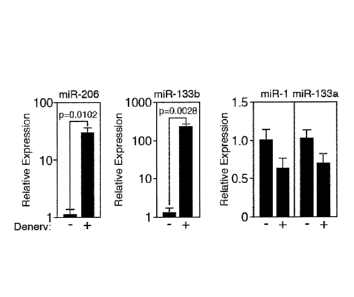
Figure 1. Upregulation of miR-206 in denervated muscle. (A) Northern blot
analysis
showing skeletal muscle specific expression of mature miR-206 in adult mouse
tissues. The
northern blot was re-probed with U6 as a loading control. (B) Northern blot
analysis of miR-1
and miR-206 expression in adult mouse muscle tissues 10-days after sciatic
nerve transection.
The contra-lateral leg was used as a control. Northern blots were re-probed
with U6 as a loading
control. EDL=extensor digitorum longus, TA=tibialis anterior,
GP=gastrocnemius/plantaris. (C)
Transcripts of miR-206, miR-133b, miR-1, and miR-133a were detected by real
time PCR in TA
muscles following 10-days of denervation (+). The contra-lateral muscle was
used as a control
(-).
Figure 2. Generation of miR-206 mutant mice. (A) Diagram of the miR-206/133b
murine locus. (B) Targeting strategy to delete miR-206 from the miR-206/133b
locus by
replacing the pre-miR-206 sequence with a neomycin cassette flanked by loxP
sites. Positions of
5' and 3' probes used for Southern blots are shown. (C) Southern blot analysis
of genomic DNA
from wild-type and heterozygous mice using an external 5' probe. Genomic DNA
was digested
with BamHI. (D) Northern blot analysis of mature miR-206 transcript expression
in
gastrocnemius/plantaris muscle of the indicated miR-206 genotypes. U6 was used
as loading
control. (E) RT-PCR using gene specific primers for pre-miR-206, pre-miR-133b,
pre-miR-1-1,
and pre-miR-1-2 in the soleus muscle of control and miR-206 mutant mice after
denervation. (F)
Hematoxylin and eosin (H&E) and metachromatic ATPase staining show no
difference in the
skeletal muscle architecture and distribution of Type I (dark blue) and Type
II (light blue)
skeletal myofibers in the soleus muscles of wild-type (WT) and miR-206-/- (KO)
mice.
Figure 3. Delayed reinnervation in miR-206 mutant mice. (A) Following sciatic
nerve
transection (as indicated in weeks), a delay in reinnervation is observed in
miR-206 -/- (KO)
mice compared to wild-type (WT) mice as detected by the superimposition of
anti-ZNP staining
(green) with BTX (red). Note the lack of anti-ZNP (green) staining in miR-206 -
/- mice. (B)
4
CA 02718520 2010-09-15
WO 2009/117418
PCT/US2009/037405
Time course and quantification of the number of reinnervated synaptic sites in
WT and miR-206
KO mice following sciatic nerve transection. (C) Immunohistochemistry using
BTX (red) and
anti-ZNP (green) shows a delay in reinnervation of NMJs in miR-206 -/- mice
compared to WT
mice following 7 and 18 days after nerve crush. Note the lack of anti-ZNP
(green) staining in
miR-206-/- mice.
Figure 4. MiR-206 targets HDAC4. (A) Schematic diagram of the Hdac4 3' UTR
from
several species with sequence homologies of the two predicted miR-206 binding
sites. (B)
Luciferase activity of COSI cells co-transfected with wild-type (WT) or mutant
HDAC4 3'UTR-
luciferase constructs with increasing amounts of miR-206 expression plasmid.
Mutation of the
.. predicted miR-206 binding sites in the 3'UTR alleviates the inhibitory
activity of miR-206.
Values are normalized to 13-galactosidase activity. (C) Western blot analysis
showing increased
HDAC4 expression in muscle lysates isolated from wild-type (WT) and miR-206 -/-
(KO) mice
3-weeks following denervation. Control (Ctrl.) refers to HDAC4 mK0 protein
lysate. GAPDH
protein was detected as a control. Relative protein expression compared to WT
is indicated
below the blot. (D) Transcripts of Hdac4 were detected in wild-type (WT) and
miR-206 -/- (KO)
muscles after 3-weeks of denervation. (E) Transcripts of Dach2 were detected
in wild-type (WT)
and miR-206 -/- (KO) muscles after 3-weeks of denervation. (F) Transcripts of
myogenin were
detected in wild-type (WT) and miR-206 -/- (KO) TA muscles after 3-weeks of
denervation. (G)
Immunohistochemistry using BTX (red) and anti-ZNP (green) shows an increase in
.. reinnervation in HDAC4 mK0 mutant mice compared to WT mice 7 days following
nerve crush.
Note the increase in anti-ZNP (green) staining in HDAC4 mK0 mice.
Figure 5. Upregulation of miR-206 in ALS mice. (A) Heat plot of miRNA array
profiling of miRNAs in wild-type (WT) and G93A-SOD1 (ALS) mice. (B) Northern
blot
analysis of miR-1 and miR-206 expression in 7-month old wild-type (WT) and
G93A-SOD1
.. (ALS) TA muscles. Northern blots were re-probed with U6 as a loading
control. (C) ALS
pathogenesis is increased in miR-206/G93A-SOD1 double mutant mice.
Representative image
of a G93A-SOD1 mouse and a miR-206/G93A-SOD1 double mutant mouse. (D) Muscle
degeneration is increased in miR-206/G93A-SOD1 double mutant mice. Hematoxylin
and eosin
(H&E) staining of gastrocnemius/plantaris muscles of wild-type (WT), miR-206
mutant, G93A-
SOD1, and miR-206/G93A-SOD1 double mutant mice.
5
CA 02718520 2010-09-15
WO 2009/117418
PCT/US2009/037405
DETAILED DESCRIPTION
The present invention is based, in part, on the discovery that miR-206
regulates
neuromuscular junction stability and regeneration following injury. MiR-206 is
upregulated in
skeletal muscles of a mouse model of ALS and in response to denervation
following nerve
transection or crush. In addition, the related molecule, miR-133b, is
upregulated in a similar
fashion to miR-206, while miR-1 and miR-133a exhibit a reduced expression in
the ALS disease
model and in response to surgical denervation. Through the creation of miR-206
null mice, the
inventors established an essential role for miR-206 in regulating
neuromuscular junction stability
and regeneration following injury. These results describe the first role of an
miRNA in regulating
neuromuscular synapse function, and point to miR-206 and miR-1 as key
components in
pathogenesis of ALS and other denervating diseases and injuries. Accordingly,
the present
invention provides novel therapeutic approaches for treating neurodegenerative
diseases and
nerve damage by manipulating expression levels of miR-206 and/or miR-1.
Invertebrates, three pairs of muscle-specific miRNAs, miR-1-1/133a-2, miR-1-
2/133a-1,
and miR-206/133b are transcribed as bicistronic transcripts on separate
chromosomes (Liu etal.,
2007). MiR-1 has been shown to regulate cardiac myocyte proliferation and
heart morphogenesis
through the repression of the transcription factor, Hand2 (Zhao et al., 2005;
2007). In skeletal
myoblasts, miR-1 was shown to promote differentiation through the repression
of histone
deacetylase 4 (HDAC4), a repressor of myogenesis; paradoxically, miR-133 was
shown to
.. repress differentiation through the repression of serum response factor
(SRF), an activator of
myogenesis (Chen etal., 2006). MiR-1-1 is co-transcribed with miR-133a-2 from
human
chromosome 20, while miR-1-2 is co-transcribed with miR-133a-1 from human
chromosome 18.
In addition, miR-206, an evolutionarily divergent member of the miR-1 family
of miRNAs, has
been shown to promote myoblast differentiation through the repression of
various target genes,
including a subunit of the DNA polymerase a (Polal), connexin 43 (Cx43),
follistatin-like 1
(Fst11), and utrophin (Utm) (Kim et al., 2006; Rosenberg et al., 2006). MiR-
206 is generated with
miR-133b from a bicistronic transcript from an intergenic region of human
chromosome 6. MiR-
133b was shown to be decreased in patients with Parkinson's disease and to
regulate maturation
of dopaminergic neurons (Kim etal., 2007). MiR-1-1 and miR-1-2 are identical
to each other
and differ from miR-206 by four nucleotides. MiR-133a-1 and miR-133a-2 are
identical to each
6
CA 02718520 2010-09-15
WO 2009/117418
PCT/US2009/037405
other and differ from miR-133b by two nucleotides The stem-loop and mature
sequences for
miR-206, miR-1, miR-133a, and miR-133b are shown below:
Human miR-206 stem-loop (SEQ ID NO: 1):
UGCUUCCCGAGGCCACAUGCUUCUUUAUAUCCCCAUAUGGAUUACUUUGCUAUGG
AAUGUAAGGAAGUGUGUGGUUUCGGCAAGUG
Human mature miR-206 (SEQ ID NO: 2):
UGGAAUGUAAGGAAGUGUGUGG
Human miR-1 stem-loop (SEQ ID NO: 3):
ACCUACUCAGAGUACAUACUUCUUUAUGUACCCAUAUGAACAUACAAUGCUAUG
GAAUGUAAAGAAGUAUGUAUUUUUGGUAGGC
Human mature miR-1 (SEQ ID NO: 4):
UGGAAUGUAAAGAAGUAUGUAU
Human miR-133a stem-loop (SEQ ID NO: 5):
ACAAUGCUUUGCUAGAGCUGGUAAAAUGGAACCAAAUCGCCUCUUCAAUGGAUU
UGGUCCCCUUCAACCAGCUGUAGCUAUGCAUUGA
Human mature miR-133a (SEQ ID NO: 6):
UUUGGUCCCCUUCAACCAGCUG
Human miR-133b stem-loop (SEQ ID NO: 7):
CCUCAGAAGAAAGAUGCCCCCUGCUCUGGCUGGUCAAACGGAACCAAGUCCGUCU
UCCUGAGAGGUUUGGUCCCCUUCAACCAGCUACAGCAGGGCUGGCAAUGCCCAGU
CCUUGGAGA
Human mature miR-133b (SEQ ID NO: 8):
UUUGGUCCCCUUCAACCAGCUA
7
CA 02718520 2010-09-15
WO 2009/117418
PCT/US2009/037405
In one embodiment, the present invention provides a method of treating a
subject
afflicted with a denervating neuropathic state comprising administering to the
subject an agonist
of miR-206 and/or miR-1. An "agonist" can be any compound or molecule that
increases the
activity or expression of the particular miRNA. For example, in certain
embodiments, an agonist
of miR-206 and/or miR-1 can be a polynucleotide comprising a mature miR-206
and/or miR-1
sequence. In some embodiments, the polynucleotide comprises the sequence of
SEQ ID NO: 2,
and/or SEQ ID NO: 4. In another embodiment, the agonist of miR-206 and/or miR-
1 can be a
polynucleotide comprising the pri-miRNA or pre-miRNA sequence for miR-206
and/or miR-1.
In such an embodiment, the polynucleotide can comprise a sequence of SEQ ID
NO: 1 and/or
SEQ ID NO: 3. The polynucleotide comprising the mature sequence, the pre-miRNA
sequence,
or the pri-miRNA sequence for miR-206 and/or miR-1 can be single stranded or
double stranded.
The polynucleotides can contain one or more chemical modifications, such as
locked nucleic
acids, peptide nucleic acids, sugar modifications, such as 2' -0-alkyl (e.g.
2'-0-methyl, 2' -0-
methoxyethyl), 2' -fluoro, and 4' thio modifications, and backbone
modifications, such as one or
more phosphorothioate, morpholino, or phosphonocarboxylate linkages and
combinations
comprising the same. In one embodiment, the polynucleotide comprising a miR-
206 and/or miR-
1 sequence is conjugated to cholesterol.
In another embodiment, the agonist of miR-206 and/or miR-1 can be an agent
distinct
from miR-206 and/or miR-1 that acts to increase, supplement, or replace the
function of miR-206
and/or miR-1. For instance, MyoD and the bHLH protein E12, both of which up-
regulate
expression of miR-206, can be agonists of miR-206. Other transcription factors
or signaling
proteins that up-regulate the expression of miR-206 and/or miR-1 are likewise
contemplated as
agonists of miR-206 and/or miR-1.
In another embodiment, the agonist of miR-206 and/or miR-1 can be expressed in
vivo
from a vector. A "vector" is a composition of matter which can be used to
deliver a nucleic acid
of interest to the interior of a cell. Numerous vectors are known in the art
including, but not
limited to, linear polynucleotides, polynucleotides associated with ionic or
amphiphilic
compounds, plasmids, and viruses. Thus, the term "vector" includes an
autonomously replicating
plasmid or a virus. Examples of viral vectors include, but are not limited to,
adenoviral vectors,
adeno-associated virus vectors, retroviral vectors, and the like. An
8
CA 02718520 2010-09-15
WO 2009/117418
PCT/US2009/037405
expression construct can be replicated in a living cell, or it can be made
synthetically. For
purposes of this application, the terms "expression construct," "expression
vector," and "vector,"
are used interchangeably to demonstrate the application of the invention in a
general, illustrative
sense, and are not intended to limit the invention.
In one embodiment, an expression vector for expressing miR-206 and/or miR-1
comprises a promoter "operably linked" to a polynucleotide encoding miR-206
and/or miR-1.
The phrase "operably linked" or "under transcriptional control" as used herein
means that the
promoter is in the correct location and orientation in relation to a
polynucleotide to control the
initiation of transcription by RNA polymerase and expression of the
polynucleotide. The
polynucleotide encoding miR-206 and/or miR-1 may encode the primary miRNA
sequence (pri-
miRNA), the precursor-miRNA sequence (pre-miRNA), or the mature miRNA sequence
for
miR-206 and/or miR-1. In another embodiment, the expression vector comprises a
polynucleotide operably linked to a promoter, wherein said polynucleotide
comprises the
sequence of SEQ ID NO: 1. In another embodiment, the expression vector
comprises a
polynucleotide operably linked to a promoter, wherein said polynucleotide
comprises the
sequence of SEQ ID NO: 2. In another embodiment, the expression vector
comprises a
polynucleotide operably linked to a promoter, wherein said polynucleotide
comprises the
sequence of SEQ ID NO: 3. In another embodiment, the expression vector
comprises a
polynucleotide operably linked to a promoter, wherein said polynucleotide
comprises the
sequence of SEQ ID NO: 4. The polynucleotide comprising the sequence of SEQ ID
NO: 1, SEQ
ID NO: 2, SEQ ID NO: 3, or SEQ ID NO: 4 may be about 18 to about 2000
nucleotides in
length, about 70 to about 200 nucleotides in length, about 20 to about 50
nucleotides in length, or
about 18 to about 25 nucleotides in length.
In certain embodiments, the nucleic acid encoding a gene product is under
transcriptional
control of a promoter. A "promoter" refers to a DNA sequence recognized by the
synthetic
machinery of the cell, or introduced synthetic machinery, required to initiate
the specific
transcription of a gene. The term promoter will be used here to refer to a
group of transcriptional
control modules that are clustered around the initiation site for RNA
polymerase I, II, or III.
In some embodiments, the human cytomegalovirus (CMV) immediate early gene
.. promoter, the 5V40 early promoter, the Rous sarcoma virus long terminal
repeat, rat insulin
promoter and glyceraldehyde-3-phosphate dehydrogenase can be used to obtain
high-level
9
CA 02718520 2010-09-15
WO 2009/117418
PCT/US2009/037405
expression of the polynucleotide sequence of interest. The use of other viral
or mammalian
cellular or bacterial phage promoters which are well-known in the art to
achieve expression of a
polynucleotide sequence of interest is contemplated as well, provided that the
levels of
expression are sufficient for a given purpose. In certain embodiments, a
tissue-specific
.. promoter, such as a skeletal muscle-specific promoter, can be used to
obtain tissue-specific
expression of the polynucleotide sequence of interest.
By employing a promoter with well-known properties, the level and pattern of
expression
of the protein of interest following transfection or transformation can be
optimized. Further,
selection of a promoter that is regulated in response to specific physiologic
signals can permit
inducible expression of the polynucleotide. Tables 1 and 2 list several
regulatory elements that
may be employed, in the context of the present invention, to regulate the
expression of the
polynucleotide of interest (e.g. agonists of miR-206 and/or miR-1). This list
is not intended to be
exhaustive of all the possible elements involved in the promotion of gene
expression but, merely,
to be exemplary thereof.
Below is a list of viral promoters, cellular promoters/enhancers and inducible
promoters/enhancers that could be used in combination with the polynucleotide
of interest in an
expression construct (Table 1 and Table 2). Additionally, any
promoter/enhancer combination
(as per the Eukaryotic Promoter Data Base EPDB) could also be used to drive
expression of the
polynucleotide. Eukaryotic cells can support cytoplasmic transcription from
certain bacterial
promoters if the appropriate bacterial polymerase is provided, either as part
of the delivery
complex or as an additional genetic expression construct.
TABLE 1
Promoter and/or Enhancer
Promoter/Enhancer References
Immunoglobulin Heavy Chain Banerji etal., 1983; Gilles etal., 1983;
Grosschedl
et al., 1985; Atchinson et al., 1986, 1987; Imler
etal., 1987; Weinberger etal., 1984; Kiledjian
etal., 1988; Porton etal.; 1990
Immunoglobulin Light Chain Queen etal., 1983; Picard et al., 1984
CA 02718520 2010-09-15
WO 2009/117418
PCT/US2009/037405
TABLE 1
Promoter and/or Enhancer
Promoter/Enhancer References
T-Cell Receptor Luria etal., 1987; Winoto etal., 1989; Redondo
etal.; 1990
HLA DQ a and/or DQ 13 Sullivan etal., 1987
13-Interferon Goodbourn etal., 1986; Fujita etal., 1987;
Goodbourn et al., 1988
Interleukin-2 Greene etal., 1989
Interleukin-2 Receptor Greene etal., 1989; Lin etal., 1990
MHC Class II 5 Koch et al., 1989
MHC Class II HLA-DRa Sherman et al., 1989
13-Actin Kawamoto etal., 1988; Ng etal.; 1989
Muscle Creatine Kinase (MCK) Jaynes etal., 1988; Horlick etal., 1989;
Johnson
etal., 1989
Prealbumin (Transthyretin) Costa et al., 1988
Elastase 1 Ornitz etal., 1987
Metallothionein (MTII) Karin et al., 1987; Culotta et al., 1989
Collagenase Pinkert etal., 1987; Angel etal., 1987a
Albumin Pinkert etal., 1987; Tronche etal., 1989, 1990
oc-F etoprotein Godbout etal., 1988; Campere etal., 1989
t-Globin Bodine etal., 1987; Perez-Stable etal., 1990
13-Globin Trudel etal., 1987
c-fos Cohen etal., 1987
c-HA-ras Triesman, 1986; Deschamps etal., 1985
Insulin Edlund et al., 1985
Neural Cell Adhesion Molecule Hirsh et al., 1990
(NCAM)
c1-Antitrypain Latimer et al., 1990
H2B (TH2B) Histone Hwang et al., 1990
Mouse and/or Type I Collagen Ripe et al., 1989
11
CA 02718520 2010-09-15
WO 2009/117418
PCT/US2009/037405
TABLE 1
Promoter and/or Enhancer
Promoter/Enhancer References
Glucose-Regulated Proteins Chang et al., 1989
(GRP94 and GRP78)
Rat Growth Hormone Larsen etal., 1986
Human Serum Amyloid A (SAA) Edbrooke etal., 1989
Troponin I (TN I) Yutzey etal., 1989
Platelet-Derived Growth Factor Pech etal., 1989
(PDGF)
Duchenne Muscular Dystrophy Klamut et al., 1990
SV40 Banerji etal., 1981; Moreau etal., 1981; Sleigh et
al., 1985; Firak et al., 1986; Herr et al., 1986;
Imbra et al., 1986; Kadesch et al., 1986; Wang et
al., 1986; Ondek etal., 1987; Kuhl etal., 1987;
Schaffner etal., 1988
Polyoma Swartzendruber etal., 1975; Vasseur etal., 1980;
Katinka etal., 1980, 1981; Tyndell etal., 1981;
Dandolo etal., 1983; de Villiers etal., 1984; Hen
etal., 1986; Satake etal., 1988; Campbell and/or
Villarreal, 1988
Retroviruses Kriegler etal., 1982, 1983; Levinson etal., 1982;
Kriegler etal., 1983, 1984a, b, 1988; Bosze etal.,
1986; Miksicek etal., 1986; Celander etal., 1987;
Thiesen et al., 1988; Celander etal., 1988; Choi
etal., 1988; Reisman etal., 1989
Papilloma Virus Campo etal., 1983; Lusky etal., 1983; Spandidos
and/or Wilkie, 1983; Spalholz et al., 1985; Lusky
et al., 1986; Cripe et al., 1987; Gloss et al., 1987;
Hirochika etal., 1987; Stephens etal., 1987
Hepatitis B Virus Bulla etal., 1986; Jameel etal., 1986; Shaul
etal.,
1987; Spandau etal., 1988; Vannice etal., 1988
Human Immunodeficiency Virus Muesing et al., 1987; Hauber et al., 1988;
Jakobovits et al., 1988; Feng et al., 1988; Takebe
etal., 1988; Rosen etal., 1988; Berkhout etal.,
1989; Laspia etal., 1989; Sharp etal., 1989;
Braddock etal., 1989
12
CA 02718520 2010-09-15
WO 2009/117418
PCT/US2009/037405
TABLE 1
Promoter and/or Enhancer
Promoter/Enhancer References
Cytomegalovirus (CMV) Weber etal., 1984; Boshart et al., 1985; Foecking
etal., 1986
Gibbon Ape Leukemia Virus Holbrook et al., 1987; Quinn et al., 1989
TABLE 2
Inducible Elements
Element Inducer References
MT II Phorbol Ester (TFA) Palmiter et al., 1982;
Heavy metals Haslinger et al., 1985;
Searle etal., 1985; Stuart
etal., 1985; Imagawa
etal., 1987, Karin etal.,
1987; Angel etal., 1987b;
McNeall et al., 1989
MMTV (mouse mammary Glucocorticoids Huang etal., 1981; Lee
tumor virus) etal., 1981; Majors etal.,
1983; Chandler et al.,
1983; Ponta etal., 1985;
Sakai et al., 1988
13-Interferon poly(rI)x Tavernier et al., 1983
poly(rc)
Adenovirus 5 E2 ElA Imperiale et al., 1984
Collagenase Phorbol Ester (TPA) Angel etal., 1987a
Stromelysin Phorbol Ester (TPA) Angel etal., 1987b
5V40 Phorbol Ester (TPA) Angel etal., 1987b
Murine MX Gene Interferon, Newcastle Hug etal., 1988
Disease Virus
GRP78 Gene A23187 Resendez etal., 1988
ot-2-Macroglobulin IL-6 Kunz et al., 1989
Vimentin Serum Rittling etal., 1989
MHC Class I Gene H-2kb Interferon Blanar etal., 1989
HSP70 ElA, 5V40 Large T Taylor et al., 1989, 1990a,
13
CA 02718520 2010-09-15
WO 2009/117418
PCT/US2009/037405
TABLE 2
Inducible Elements
Element Inducer References
Antigen 1990b
Proliferin Phorbol Ester-TPA Mordacq et al., 1989
Tumor Necrosis Factor PMA Hensel et al., 1989
Thyroid Stimulating Thyroid Hormone Chatterjee et al., 1989
Hormone a Gene
Of particular interest are muscle specific promoters, which include the myosin
light
chain-2 promoter (Franz etal., 1994; Kelly et al., 1995), the a-actin promoter
(Moss et al..,
1996), the troponin 1 promoter (Bhaysar et al., 1996); the Na /Ca2+ exchanger
promoter (Barnes
etal., 1997), the dystrophin promoter (Kimura etal., 1997), the a7 integrin
promoter (Ziober
and Kramer, 1996), and the muscle creatine kinase (MCK) promoter (Jaynes et
al., 1988; Horlick
etal., 1989; Johnson etal., 1989).
A polyadenylation signal may be included to effect proper polyadenylation of
the gene
transcript where desired. The nature of the polyadenylation signal is not
believed to be crucial to
the successful practice of the invention, and any such sequence may be
employed such as human
growth hormone and 5V40 polyadenylation signals. Also contemplated as an
element of the
expression cassette is a terminator. These elements can serve to enhance
message levels and to
minimize read through from the cassette into other sequences.
In certain embodiments of the invention, the cells contain nucleic acid
constructs of
the present invention, a cell may be identified in vitro or in vivo by
including a marker in the
expression construct. Such markers would confer an identifiable change to the
cell permitting
easy identification of cells containing the expression construct. Usually the
inclusion of a drug
selection marker aids in cloning and in the selection of transformants, for
example, genes that
confer resistance to neomycin, puromycin, hygromycin, DHFR, GPT, zeocin and
histidinol are
useful selectable markers. Alternatively, enzymes such as herpes simplex virus
thymidine kinase
(tk) or chloramphenicol acetyltransferase (CAT) may be employed. Immunologic
markers also
can be employed. The selectable marker employed is not believed to be
important, so long as it is
14
CA 02718520 2010-09-15
WO 2009/117418
PCT/US2009/037405
capable of being expressed simultaneously with the nucleic acid encoding a
gene product.
Further examples of selectable markers are well known to one of skill in the
art.
There are a number of ways in which expression vectors may be introduced into
cells.
In certain embodiments of the invention, the expression construct comprises a
virus or
engineered construct derived from a viral genome. The ability of certain
viruses to enter cells via
receptor-mediated endocytosis, to integrate into host cell genome and express
viral genes stably
and efficiently have made them attractive candidates for the transfer of
foreign genes into
mammalian cells (Ridgeway, 1988; Nicolas and Rubenstein, 1988; Baichwal and
Sugden, 1986;
Temin, 1986).
One of the preferred methods for in vivo delivery involves the use of an
adenovirus
expression vector. "Adenovirus expression vector" is meant to include those
constructs
containing adenovirus sequences sufficient to (a) support packaging of the
construct and (b)
to express a polynucleotide that has been cloned therein. The expression
vector comprises a
genetically engineered form of adenovirus. Knowledge of the genetic
organization of adenovirus,
a 36 kB, linear, double-stranded DNA virus, allows substitution of large
pieces of adenoviral
DNA with foreign sequences up to 7 kB (Grunhaus and Horwitz, 1992). In
contrast to retrovirus,
the adenoviral infection of host cells does not result in chromosomal
integration because
adenoviral DNA can replicate in an episomal manner without potential
genotoxicity. Also,
adenoviruses are structurally stable, and no genome rearrangement has been
detected after
extensive amplification. Adenovirus can infect virtually all epithelial cells
regardless of their cell
cycle stage.
Adenovirus is particularly suitable for use as a gene transfer vector because
of its mid-
sized genome, ease of manipulation, high titer, wide target cell range and
high infectivity. Both
ends of the viral genome contain 100-200 base pair inverted repeats (ITRs),
which are cis
elements necessary for viral DNA replication and packaging.
Other than the requirement that the adenovirus vector be replication
defective, or at
least conditionally defective, the nature of the adenovirus vector is not
believed to be crucial
to the successful practice of the invention. The adenovirus may be of any of
the 42 different
known serotypes or subgroups A-F. Adenovirus type 5 of subgroup C is the
preferred
starting material in order to obtain the conditional replication-defective
adenovirus vector for
use in the present invention. This is because Adenovirus type 5 is a human
adenovirus about
CA 02718520 2010-09-15
WO 2009/117418
PCT/US2009/037405
which a great deal of biochemical and genetic information is known, and it has
historically
been used for most constructions employing adenovirus as a vector.
The typical vector according to the present invention is replication defective
and will not
have an adenovirus El region. Thus, it will be most convenient to introduce
the polynucleotide
encoding the gene of interest at the position from which the El-coding
sequences have been
removed. However, the position of insertion of the construct within the
adenovirus sequences is
not critical to the invention. The polynucleotide encoding the gene of
interest may also be
inserted in lieu of the deleted E3 region in E3 replacement vectors, as
described by Karlsson et
al. (1986), or in the E4 region where a helper cell line or helper virus
complements the E4 defect.
Adenovirus vectors have been used in eukaryotic gene expression (Levrero et
al.,
1991; Gomez-Foix etal., 1992) and vaccine development (Grunhaus and Horwitz,
1992;
Graham and Prevec, 1991). Recently, animal studies suggested that recombinant
adenovirus
could be used for gene therapy (Stratford-Perricaudet and Perricaudet, 1991;
Stratford-
Perricaudet etal., 1990; Rich etal., 1993). Studies in administering
recombinant adenovirus
to different tissues include trachea instillation (Rosenfeld et al., 1991;
Rosenfeld et al.,
1992), muscle injection (Ragot etal., 1993), peripheral intravenous injections
(Herz and
Gerard, 1993) and stereotactic inoculation into the brain (Le Gal La Salle et
al., 1993).
Retroviral vectors are also suitable for expressing agonists of miR-206 and/or
miR-1 in
cells. The retroviruses are a group of single-stranded RNA viruses
characterized by an ability to
convert their RNA to double-stranded DNA in infected cells by a process of
reverse-transcription
(Coffin, 1990). The resulting DNA then stably integrates into cellular
chromosomes as a provirus
and directs synthesis of viral proteins. The integration results in the
retention of the viral gene
sequences in the recipient cell and its descendants. The
retroviral genome contains three genes, gag, pol, and env that code for capsid
proteins,
polymerase enzyme, and envelope components, respectively. A sequence found
upstream
from the gag gene contains a signal for packaging of the genome into virions.
Two long
terminal repeat (LTR) sequences are present at the 5' and 3' ends of the viral
genome. These
contain strong promoter and enhancer sequences and are also required for
integration in the
host cell genome (Coffin, 1990).
In order to construct a retroviral vector, a nucleic acid encoding a gene of
interest is
inserted into the viral genome in the place of certain viral sequences to
produce a virus that is
16
CA 02718520 2010-09-15
WO 2009/117418
PCT/US2009/037405
replication-defective. In order to produce virions, a packaging cell line
containing the gag,
pol, and env genes but without the LTR and packaging components is constructed
(Mann et
al., 1983). When a recombinant plasmid containing a cDNA, together with the
retroviral
LTR and packaging sequences is introduced into this cell line (by calcium
phosphate
precipitation for example), the packaging sequence allows the RNA transcript
of the
recombinant plasmid to be packaged into viral particles, which are then
secreted into the
culture media (Nicolas and Rubenstein, 1988; Temin, 1986; Mann etal., 1983).
The media
containing the recombinant retroviruses is then collected, optionally
concentrated, and used
for gene transfer. Retroviral vectors are able to infect a broad variety of
cell types. However,
integration and stable expression require the division of host cells (Paskind
et al., 1975).
Other viral vectors may be employed as expression constructs in the present
invention. Vectors derived from viruses such as vaccinia virus (Ridgeway,
1988; Baichwal
and Sugden, 1986; Coupar etal., 1988) adeno-associated virus (AAV) (Ridgeway,
1988;
Baichwal and Sugden, 1986; Hermonat and Muzycska, 1984) and herpesviruses may
be
employed. They offer several attractive features for various mammalian cells
(Friedmann,
1989; Ridgeway, 1988; Baichwal and Sugden, 1986; Coupar etal., 1988; Horwich
etal.,
1990).
In order to effect expression of gene constructs, the expression construct
must be
delivered into a cell. This delivery may be accomplished in vitro, as in
laboratory procedures for
transforming cells lines, or in vivo or ex vivo, as in the treatment of
certain disease states. One
mechanism for delivery is via viral infection where the expression construct
is encapsidated in an
infectious viral particle.
Several non-viral methods for the transfer of expression constructs into
cultured
mammalian cells also are contemplated by the present invention. These include
calcium
phosphate precipitation (Graham and Van Der Eb, 1973; Chen and Okayama, 1987;
Rippe etal.,
1990) DEAE-dextran (Gopal, 1985), electroporation (Tur-Kaspa etal., 1986;
Potter etal., 1984),
direct microinjection (Harland and Weintraub, 1985), DNA-loaded liposomes
(Nicolau and
Sene, 1982; Fraley etal., 1979) and lipofectamine-DNA complexes, cell
sonication (Fechheimer
etal., 1987), gene bombardment using high velocity microprojectiles (Yang
etal., 1990), and
receptor-mediated transfection (Wu and Wu, 1987; Wu and Wu, 1988). Some of
these
techniques may be successfully adapted for in vivo or ex vivo use.
17
CA 02718520 2010-09-15
WO 2009/117418
PCT/US2009/037405
Once the expression construct has been delivered into the cell, the nucleic
acid encoding
the gene of interest may be positioned and expressed at different sites. In
certain embodiments,
the nucleic acid encoding the gene may be stably integrated into the genome of
the cell. This
integration may be in the cognate location and orientation via homologous
recombination (gene
replacement) or it may be integrated in a random, non-specific location (gene
augmentation). In
yet further embodiments, the nucleic acid may be stably maintained in the cell
as a separate,
episomal segment of DNA. Such nucleic acid segments or "episomes" encode
sequences
sufficient to permit maintenance and replication independent of or in
synchronization with the
host cell cycle. How the expression construct is delivered to a cell and where
in the cell the
nucleic acid remains is dependent on the type of expression construct
employed.
In yet another embodiment of the invention, the expression construct may
simply
consist of naked recombinant DNA or plasmids. Transfer of the construct may be
performed
by any of the methods mentioned above which physically or chemically
permeabilize the cell
membrane. This is particularly applicable for transfer in vitro but it may be
applied to in vivo
use as well. Dubensky etal. (1984) successfully injected polyomavirus DNA in
the form of
calcium phosphate precipitates into liver and spleen of adult and newborn mice
demonstrating
active viral replication and acute infection. Benvenisty and Neshif (1986)
also demonstrated that
direct intraperitoneal injection of calcium phosphate-precipitated plasmids
results in expression
of the transfected genes. It is envisioned that DNA encoding a gene of
interest may also be
transferred in a similar manner in vivo and express the gene product.
In still another embodiment of the invention for transferring a naked DNA
expression
construct into cells may involve particle bombardment. This method depends on
the ability
to accelerate DNA-coated microprojectiles to a high velocity allowing them to
pierce cell
membranes and enter cells without killing them (Klein et al., 1987). Several
devices for
accelerating small particles have been developed. One such device relies on a
high voltage
discharge to generate an electrical current, which in turn provides the motive
force (Yang et al.,
1990). The microprojectiles used have consisted of biologically inert
substances such as
tungsten or gold beads.
Selected organs including the liver, skin, and muscle tissue of rats and mice
have
been bombarded in vivo (Yang et al., 1990; Zelenin et al., 1991). This may
require surgical
exposure of the tissue or cells, to eliminate any intervening tissue between
the gun and the
18
CA 02718520 2010-09-15
WO 2009/117418
PCT/US2009/037405
target organ, i.e., ex vivo treatment. Again, DNA encoding a particular gene
may be
delivered via this method and still be incorporated by the present invention.
In a further embodiment of the invention, the expression construct may be
entrapped
in a liposome. Liposomes are vesicular structures characterized by a
phospholipid bilayer
membrane and an inner aqueous medium. Multilamellar liposomes have multiple
lipid layers
separated by aqueous medium. They form spontaneously when phospholipids are
suspended
in an excess of aqueous solution. The lipid components undergo self-
rearrangement before
the formation of closed structures and entrap water and dissolved solutes
between the lipid
bilayers (Ghosh and Bachhawat, 1991). Also contemplated are lipofectamine-DNA
complexes.
In certain embodiments of the invention, the liposome may be complexed with a
hemagglutinating virus (HVJ). This has been shown to facilitate fusion with
the cell
membrane and promote cell entry of liposome-encapsulated DNA (Kaneda etal.,
1989). In
other embodiments, the liposome may be complexed or employed in conjunction
with
nuclear non-histone chromosomal proteins (HMG-1) (Kato etal., 1991). In yet
further
embodiments, the liposome may be complexed or employed in conjunction with
both HVJ
and HMG-1. In that such expression constructs have been successfully employed
in transfer
and expression of nucleic acid in vitro and in vivo, then they are applicable
for the present
invention. Where a bacterial promoter is employed in the DNA construct, it
also will be
desirable to include within the liposome an appropriate bacterial polymerase.
Other expression constructs which can be employed to deliver a nucleic acid
encoding a particular gene into cells are receptor-mediated delivery vehicles.
These take
advantage of the selective uptake of macromolecules by receptor-mediated
endocytosis in
almost all eukaryotic cells. Because of the cell type-specific distribution of
various
receptors, the delivery can be highly specific (Wu and Wu, 1993).
Receptor-mediated gene targeting vehicles generally consist of two components:
a
cell receptor-specific ligand and a DNA-binding agent. Several ligands have
been used for
receptor-mediated gene transfer. The most extensively characterized ligands
are
asialoorosomucoid (ASOR) (Wu and Wu, 1987) and transferrin (Wagner etal.,
1990).
Recently, a synthetic neoglycoprotein, which recognizes the same receptor as
ASOR, has
been used as a gene delivery vehicle (Ferkol etal., 1993; Perales etal., 1994)
and epidermal
19
CA 02718520 2015-07-22
growth factor (EGF) has also been used to deliver genes to squamous carcinoma
cells
(Myers, EPO 0273085).
In other embodiments, the delivery vehicle may comprise a ligand and a
liposome.
For example, Nicolau et al. (1987) employed lactosyl-ceramide, a galactose-
terminal
asialganglioside, incorporated into liposomes and observed an increase in the
uptake of the
insulin gene by hepatocytes. Thus, it is feasible that a nucleic acid encoding
a particular gene
also may be specifically delivered into a cell type by any number of receptor-
ligand systems
with or without liposomes.
In a particular example, the oligonucleotide may be administered in
combination with
a cationic lipid. Examples of cationic lipids include, but are not limited to,
lipofectin,
DOTMA, DOPE, and DOTAP. The publication of W00071096 describes different
formulations,
such as a DOTAP:cholesterol or cholesterol derivative formulation that can
effectively be used
for gene therapy. Other disclosures also discuss different lipid or liposomal
formulations
including nanoparticles and methods of administration; these include, but are
not limited to, U.S.
Patent Publication 20030203865, 20020150626, 20030032615, and 20040048787.
Methods used
for forming particles are also disclosed in U.S. Patents 5,844,107, 5,877,302,
6,008,336,
6,077,835, 5,972,901, 6,200,801, and 5,972,900.
In certain embodiments, gene transfer may more easily be performed under ex
vivo
conditions. Ex vivo gene therapy refers to the isolation of cells from an
animal, the delivery
of a nucleic acid into the cells in vitro, and then the return of the modified
cells back into an
animal. This may involve the surgical removal of tissue/organs from an animal
or the
primary culture of cells and tissues.
In one embodiment, the present invention provides a method of treating a
subject
afflicted with a denervating neuropathic state. A "denervating neuropathic
state" refers to a
condition in which there is a loss of nerve supply to one or more tissues as a
result of a disease or
injury. Denervating neuropathic states can result from degenerative motor
neuron diseases, such
as myasthenia gravis, polio, amyotrophic lateral sclerosis (ALS), Friedreich's
ataxia, spinal
muscular atrophy, or spinocerebellar ataxia.
CA 02718520 2010-09-15
WO 2009/117418
PCT/US2009/037405
In one embodiment, the present invention provides a method of treating a
subject
suffering from myasthenia gravis by administering to the subject an agonist of
miR-206 and/or
miR-1. Myasthenia gravis (MG) is a neuromuscular disease leading to
fluctuating muscle
weakness and fatiguability. It is an autoimmune disorder, in which weakness is
caused by
circulating antibodies that block acetylcholine receptors at the post-synaptic
neuromuscular
junction, inhibiting the stimulative effect of the neurotransmitter
acetylcholine. Myasthenia is
treated medically with cholinesterase inhibitors or immunosuppressants, and,
in selected cases,
thymectomy. The hallmark of myasthenia gravis is muscle weakness that
increases during
periods of activity and improves after periods of rest. Muscles that control
eye and eyelid
movement, facial expression, chewing, talking, and swallowing are especially
susceptible. The
muscles that control breathing and neck and limb movements can also be
affected.
In another embodiment, the present invention includes a method of treating ALS
in a
subject in need thereof comprising administering to the subject an agonist of
miR-206 and/or
miR-1. ALS (also called Lou Gehrig's Disease, or Maladie de Charcot) is a
progressive, usually
fatal, neurodegenerative disease caused by the degeneration of motor neurons.
As one of the
motor neuron diseases, the disorder causes muscle weakness and atrophy
throughout the body as
both the upper and lower motor neurons degenerate, ceasing to send messages to
muscles.
Unable to function, the muscles gradually weaken, develop fasciculations
(twitches) because of
denervation, and eventually atrophy due to that denervation. The patient may
ultimately lose the
ability to initiate and control all voluntary movement except of the eyes.
Riluzole, the first FDA-
approved treatment for ALS, slows the degeneration of motor neurons, but does
not reverse the
damage that has already occurred. Other treatments for ALS are designed to
relieve symptoms
and improve the quality of life for patients.
In another embodiment, the present invention provides a method for treating
spinal
muscular atrophy in a subject in need thereof by administering an agonist of
miR-206 and/or
miR-1. Spinal muscular atrophy (SMA) is an autosomal recessive disorder which
is the leading
hereditary cause of infant death in humans. The disease is characterized by a
progressive muscle
weakness from proximal to distal with lower limbs more greatly affected than
upper limbs.
Three types of SMA have been described based on disease severity and age of
onset. Type I
affects approximately fifty percent of SMA patients with symptoms presenting
within the first
six months after birth. Death typically occurs within the first two years due
to respiratory failure.
21
CA 02718520 2010-09-15
WO 2009/117418
PCT/US2009/037405
Type II SMA has an onset between six months and eighteen months of age, and
length of
survival is dependent on the severity of respiratory impairment. Type III SMA
patients, who
have symptom onset between eighteen months and early childhood, usually do not
have a
decrease in life expectancy, although most are wheel chair bound at some point
in their disease
progression. SMA is characterized by loss of alpha-motor neurons in the
anterior horn of the
spinal cord, which is correlated with muscle paralysis and atrophy. Currently,
there are no
effective therapeutics for SMA disease.
In still another embodiment, the present invention provides a method of
treating
Friedreich's ataxia in a subject in need thereof comprising administering to
the subject an agonist
of miR-206 and/or miR-1. Friedreich's ataxia is an autosomal recessive
congenital disease that
causes progressive damage to the nervous system resulting in symptoms ranging
from gait
disturbance and speech problems to heart disease. The ataxia of Friedreich's
ataxia results from
the degeneration of nerve tissue in the spinal cord, in particular sensory
neurons essential
(through connections with the cerebellum) for directing muscle movement of the
arms and legs.
The spinal cord becomes thinner and nerve cells lose some of their myelin
sheath. Symptoms of
Friedrich's ataxia include any combination, but not necessarily all of the
following: muscle
weakness in the arms and legs, loss of coordination, vision impairment,
hearing loss, slurred
speech, curvature of the spine (scoliosis), high plantar arches, diabetes, and
heart disorders (e.g.,
atrial fibrillation, and resultant tachycardia, and hypertrophic
cardiomyopathy). The symptoms
can be treated but there is no treatment for Friedrich's Ataxia at this time.
In yet another embodiment, the present invention provides a method of treating
spinocerebellar ataxia in a subject in need thereof comprising administering
to the subject an
agonist of miR-206 and/or miR-1. Spinocerebellar ataxia (SCA) is a genetic
disease
characterized by slowly progressive incoordination of gait and often
associated with poor
coordination of hands, speech, and eye movements. Frequently, atrophy of the
cerebellum
occurs. There is no known cure for spinocerebellar ataxia, which is a
progressive disease. As
with other forms of ataxia, SCA results in unsteady and clumsy motion of the
body due to a
failure of the fine coordination of muscle movements, along with other
symptoms. A person with
this disease will usually end up needing to use a wheelchair, and eventually
they may need
assistance to perform daily tasks.
22
CA 02718520 2010-09-15
WO 2009/117418
PCT/US2009/037405
Denervating neuropathic states can also result from nerve injury, such as
spinal cord or
peripheral nerve injury, where one or more nerves are transected or crushed.
Traumatic spinal
cord injury can be classified into different types. Central Cord syndrome is
associated with
greater loss of upper limb function compared to lower limbs. The Brown-Sequard
syndrome
results from injury to one side with the spinal cord, causing weakness and
loss of proprioception
on the side of the injury and loss of pain and thermal sensation of the other
side. The Anterior
Spinal syndrome results from injury to the anterior part of the spinal cord,
causing weakness and
loss of pain and thermal sensations below the injury site but preservation of
proprioception that
is usually carried in the posterior part of the spinal cord. Tabes Dorsalis
results from injury to the
posterior part of the spinal cord, usually from infection diseases such as
syphilis, causing loss
of touch and proprioceptive sensation. Conus Medullaris syndrome results from
injury to the
tip of the spinal cord, located at Li vertebra. Cauda Equina syndrome is an
injury to the spinal
roots below the Ll vertebra. Traumatic spinal cord injury and other types of
nerve injury can be
treated by agonists of miR-206 and/or miR-1, which promote reinervation of
skeletal muscle
following injury.
In another embodiment of the invention, it is envisioned to use an agonist of
miR-206
and/or miR-1 in combination with other therapeutic modalities. Thus, in
addition to the miRNA
agonists of the invention described herein, one may also provide to the
subject "standard"
pharmaceutical therapies. Such standard therapies will depend upon the
particular denervating
neuropathic state to be treated, but can include Riluzole, cholinesterase
inhibitors (e.g.
edrophonium chloride (Tensilon0, Reversol0), neostigmine, pyridostigmine,
physostigmine,
ambenonium, demarcarium, rivastigmine, phenanthrene derivatives, galantamine,
piperidines,
donepezil, and tacrine) and immunosuppressants (e.g. prednisone, cyclosporine,
mycophenolate
mofetil and azathioprine).
Combinations may be achieved by contacting skeletal muscle cells with a single
composition or pharmacological formulation that includes both agents, or by
contacting the
cell with two distinct compositions or formulations, at the same time, wherein
one composition
includes an agonist of miR-206 and/or miR-1 and the other includes the second
agent.
Alternatively, the therapy using an miRNA agonist may precede or follow
administration of the
other agent(s) by intervals ranging from minutes to weeks. In embodiments
where the other
agent and miRNA agonists are applied separately to the cell, one would
generally ensure that a
23
CA 02718520 2010-09-15
WO 2009/117418
PCT/US2009/037405
significant period of time did not expire between the time of each delivery,
such that the agent
and miRNA agonists would still be able to exert an advantageously combined
effect on the cell.
In such instances, it is contemplated that one would typically contact the
cell with both
modalities within about 12-24 hours of each other and, more preferably, within
about 6-12 hours
of each other, with a delay time of only about 12 hours being most preferred.
In some situations,
it may be desirable to extend the time period for treatment significantly,
however, where several
days (2, 3, 4, 5, 6 or 7) to several weeks (1, 2, 3, 4,5,6, 7 or 8) lapse
between the respective
administrations.
It also is conceivable that more than one administration of either a miRNA
agonist, or the
other agent will be desired. In this regard, various combinations may be
employed. By way of
illustration, where the miRNA agonist is "A" and the other agent/therapy is
"B," the following
permutations based on 3 and 4 total administrations are exemplary:
A/B/A B/A/B B/B/A A/A/B B/A/A A/B/B B/B/B/A B/B/A/B
A/A/B/B A/B/A/B A/B/B/A B/B/A/A B/A/B/A B/A/A/B B/B/B/A
A/A/A/B B/A/A/A A/B/A/A A/A/B/A A/B/B/B B/A/B/B B/B/A/B
Other combinations are likewise contemplated.
The present invention also contemplates methods for scavenging or clearing miR-
206
and/or miR-1 agonists following treatment. In one embodiment, the method
comprises
overexpression of binding site regions for miR-206 and/or miR-1 in skeletal
muscle cells using a
muscle specific promoter. The binding site regions preferably contain a
sequence of the seed
region, the 5' portion of a miRNA spanning bases 2-8, for miR-206 and/or miR-
1. In some
embodiments, the binding site may contain a sequence from the 3' UTR of one or
more targets of
miR-206 and/or miR-1. For instance, in one embodiment, a binding site for miR-
206 and/or
miR-1 contains the 3' UTR of HDAC4. In another embodiment, a miR-206 and/or
miR-1
inhibitor may be administered after a miR-206 and/or miR-1 agonist to
attenuate or stop the
function of the microRNA. Such inhibitors can include antagomirs, antisense,
or inhibitory RNA
molecules (e.g. siRNA or shRNA).
The present invention also encompasses pharmaceutical compositions comprising
an
agonist of miR-206 and/or miR-1 and a pharmaceutically acceptable carrier.
Where clinical
applications are contemplated, pharmaceutical compositions will be prepared in
a form
appropriate for the intended application. Generally, this will entail
preparing compositions that
24
CA 02718520 2015-07-22
are essentially free of pyrogens, as well as other impurities that could be
harmful to humans or
animals.
Colloidal dispersion systems, such as macromolecule complexes, nanocapsules,
microspheres, beads, and lipid-based systems including oil-in-water emulsions,
micelles, mixed
micelles, and liposomes, can be used as delivery vehicles for the agonists of
microRNA function
described herein. Commercially available fat emulsions that are suitable for
delivering the
nucleic acids of the invention to tissues, such as skeletal muscle tissue,
include IntralipidO,
LiposynO, LiposynO II, Liposyn III, Nutrilipid, and other similar lipid
emulsions. A preferred
colloidal system for use as a delivery vehicle in vivo is a liposome (i.e., an
artificial membrane
vesicle). The preparation and use of such systems is well known in the art.
Exemplary
formulations are also disclosed in US 5,981,505; US 6,217,900; US 6,383,512;
US 5,783,565;
US 7,202,227; US 6,379,965; US 6,127,170; US 5,837,533; US 6,747,014; and WO
03/093449.
One will generally desire to employ appropriate salts and buffers to render
delivery
vectors stable and allow for uptake by target cells. Buffers also will be
employed when
recombinant cells are introduced into a patient. Aqueous compositions of the
present
invention comprise an effective amount of the delivery vehicle, dissolved or
dispersed in a
pharmaceutically acceptable carrier or aqueous medium. The phrases
"pharmaceutically
acceptable" or "pharmacologically acceptable" refers to molecular entities and
compositions
that do not produce adverse, allergic, or other untoward reactions when
administered to an
animal or a human. As used herein, "pharmaceutically acceptable carrier"
includes solvents,
buffers, solutions, dispersion media, coatings, antibacterial and antifungal
agents, isotonic
and absorption delaying agents and the like acceptable for use in formulating
pharmaceuticals,
such as pharmaceuticals suitable for administration to humans. The use of such
media and agents
for pharmaceutically active substances is well known in the art. Except
insofar as any
conventional media or agent is incompatible with the active ingredients of the
present invention,
its use in therapeutic compositions is contemplated. Supplementary active
ingredients also can be
incorporated into the compositions, provided they do not inactivate the
nucleic acids of the
compositions.
The active compositions of the present invention may include classic
pharmaceutical
CA 02718520 2010-09-15
WO 2009/117418
PCT/US2009/037405
preparations. Administration of these compositions according to the present
invention may be via
any common route so long as the target tissue is available via that route.
This includes oral,
nasal, or buccal. Alternatively, administration may be by intradermal,
transdermal, subcutaneous,
intramuscular, intraperitoneal or intravenous injection, or by direct
injection into skeletal muscle
tissue. Such compositions would normally be administered as pharmaceutically
acceptable
compositions, as described supra.
The active compounds may also be administered parenterally or
intraperitoneally. By
way of illustration, solutions of the active compounds as free base or
pharmacologically
acceptable salts can be prepared in water suitably mixed with a surfactant,
such as
hydroxypropylcellulose. Dispersions can also be prepared in glycerol, liquid
polyethylene
glycols, and mixtures thereof and in oils. Under ordinary conditions of
storage and use, these
preparations generally contain a preservative to prevent the growth of
microorganisms.
The pharmaceutical forms suitable for injectable use include, for example,
sterile
aqueous solutions or dispersions and sterile powders for the extemporaneous
preparation of
sterile injectable solutions or dispersions. Generally, these preparations are
sterile and fluid
to the extent that easy injectability exists. Preparations should be stable
under the conditions
of manufacture and storage and should be preserved against the contaminating
action of
microorganisms, such as bacteria and fungi. Appropriate solvents or dispersion
media may
contain, for example, water, ethanol, polyol (for example, glycerol, propylene
glycol, and
liquid polyethylene glycol, and the like), suitable mixtures thereof, and
vegetable oils. The
proper fluidity can be maintained, for example, by the use of a coating, such
as lecithin, by
the maintenance of the required particle size in the case of dispersion and by
the use of
surfactants. The prevention of the action of microorganisms can be brought
about by various
antibacterial an antifungal agents, for example, parabens, chlorobutanol,
phenol, sorbic acid,
thimerosal, and the like. In many cases, it will be preferable to include
isotonic agents, for
example, sugars or sodium chloride. Prolonged absorption of the injectable
compositions can
be brought about by the use in the compositions of agents delaying absorption,
for example,
aluminum monostearate and gelatin.
Sterile injectable solutions may be prepared by incorporating the active
compounds in
an appropriate amount into a solvent along with any other ingredients (for
example as
enumerated above) as desired, followed by filtered sterilization. Generally,
dispersions are
26
CA 02718520 2015-07-22
prepared by incorporating the various sterilized active ingredients into a
sterile vehicle which
contains the basic dispersion medium and the desired other ingredients, e.g.,
as enumerated
above. In the case of sterile powders for the preparation of sterile
injectable solutions, the
preferred methods of preparation include vacuum-drying and freeze-drying
techniques which
.. yield a powder of the active ingredient(s) plus any additional desired
ingredient from a
previously sterile-filtered solution thereof
The compositions of the present invention generally may be formulated in a
neutral or
salt form. Pharmaceutically-acceptable salts include, for example, acid
addition salts (formed
with the free amino groups of the protein) derived from inorganic acids (e.g.,
hydrochloric or
.. phosphoric acids, or from organic acids (e.g., acetic, oxalic, tartaric,
mandelic, and the like).
Salts formed with the free carboxyl groups of the protein can also be derived
from inorganic
bases (e.g., sodium, potassium, ammonium, calcium, or ferric hydroxides) or
from organic bases
(e.g., isopropylamine, trimethylamine, histidine, procaine and the like).
Upon formulation, solutions are preferably administered in a manner compatible
with
.. the dosage formulation and in such amount as is therapeutically effective.
The formulations
may easily be administered in a variety of dosage forms such as injectable
solutions, drug
release capsules and the like. For parenteral administration in an aqueous
solution, for
example, the solution generally is suitably buffered and the liquid diluent
first rendered
isotonic for example with sufficient saline or glucose. Such aqueous solutions
may be used,
.. for example, for intravenous, intramuscular, subcutaneous and
intraperitoneal administration.
Preferably, sterile aqueous media are employed as is known to those of skill
in the art,
particularly in light of the present disclosure. By way of illustration, a
single dose may be
dissolved in 1 ml of isotonic NaCl solution and either added to 1000 ml of
hypodermoclysis
fluid or injected at the proposed site of infusion, (see for example,
"Remington's Pharmaceutical
.. Sciences" 15th Edition, pages 1035-1038 and 1570-1580). Pharmacological
therapeutic agents
and methods of administration, dosages, etc., are well known to those of skill
in the art (see for
example, the "Physicians Desk Reference," Klaassen's "The Pharmacological
Basis of
Therapeutics," "Remington's Pharmaceutical Sciences," and "The Merck Index,
Eleventh
Edition,"), and may be combined with the invention in light of the disclosures
herein. Suitable
dosages include about 20 mg/kg to about 200 mg/kg, about 40 mg/kg to about 160
mg/kg, or
about 80 mg/kg to about 100 mg/kg. Some
27
CA 02718520 2010-09-15
WO 2009/117418
PCT/US2009/037405
variation in dosage will necessarily occur depending on the condition of the
subject being
treated. The person responsible for administration will, in any event,
determine the appropriate
dose for the individual subject, and such individual determinations are within
the skill of those of
ordinary skill in the art. Moreover, for human administration, preparations
should meet sterility,
pyrogenicity, general safety and purity standards as required by FDA Office of
Biologics
standards.
Any of the compositions described herein may be comprised in a kit. In a non-
limiting
example, a miR-206 and/or miR-1 agonist is included in a kit. The kit may
further include water
and hybridization buffer to facilitate hybridization of the two strands of the
miRNAs. The kit
may also include one or more transfection reagent(s) to facilitate delivery of
the polynucleotide
agonists to cells.
The components of the kits may be packaged either in aqueous media or in
lyophilized
form. The container means of the kits will generally include at least one
vial, test tube, flask,
bottle, syringe or other container means, into which a component may be
placed, and preferably,
suitably aliquoted. Where there is more than one component in the kit
(labeling reagent and label
may be packaged together), the kit also will generally contain a second, third
or other additional
container into which the additional components may be separately placed.
However, various
combinations of components may be comprised in a vial. The kits of the present
invention also
will typically include a means for containing the nucleic acids, and any other
reagent containers
in close confinement for commercial sale. Such containers may include
injection or blow-molded
plastic containers into which the desired vials are retained.
When the components of the kit are provided in one and/or more liquid
solutions, the
liquid solution is an aqueous solution, with a sterile aqueous solution being
particularly
preferred. However, the components of the kit may be provided as dried
powder(s). When
reagents and/or components are provided as a dry powder, the powder can be
reconstituted by
the addition of a suitable solvent. It is envisioned that the solvent may also
be provided in
another container means.
The container means will generally include at least one vial, test tube,
flask, bottle,
syringe and/or other container means, into which the nucleic acid formulations
are placed,
preferably, suitably allocated. The kits may also comprise a second container
means for
containing a sterile, pharmaceutically acceptable buffer and/or other diluent.
28
CA 02718520 2010-09-15
WO 2009/117418
PCT/US2009/037405
Such kits may also include components that preserve or maintain the
miRNAs/polynucleotides or that protect against their degradation. Such
components may be
RNAse-free or protect against RNAses. Such kits generally will comprise, in
suitable means,
distinct containers for each individual reagent or solution.
A kit will also include instructions for employing the kit components as well
the use
of any other reagent not included in the kit. Instructions may include
variations that can be
implemented. A kit may also include utensils or devices for administering the
miRNA agonist by
various administration routes, such as parenteral or intramuscular
administration.
The present invention also includes a method for diagnosing a denervating
neuropathic
state in a subject. In one embodiment, the method comprises (a) obtaining a
skeletal muscle
tissue sample from the subject; (b) assessing activity or expression of miR-
206 and/or miR-133b
in said sample; and (c) comparing the activity or expression in step (b) with
the activity or
expression of miR-206 and/or miR-133b in a normal tissue sample, wherein an
increase in the
activity or expression of miR-206 and/or miR-133b as compared to the activity
or expression of
miR-206 and/or miR-133b in a normal tissue sample is diagnostic of a
denervating neuropathic
state. In another embodiment, the method comprises (a) obtaining a skeletal
muscle tissue
sample from the subject; (b) assessing activity or expression of miR-1 and/or
miR-133a in said
sample; and (c) comparing the activity or expression in step (b) with the
activity or expression of
miR-1 and/or miR-133a in a normal tissue sample, wherein a decrease in the
activity or
expression of miR-1 and/or miR-133a as compared to the activity or expression
of miR-1 and/or
miR-133a in a normal tissue sample is diagnostic of a denervating neuropathic
state. The
denervating neuropathic state can include spinal cord injury, myasthenia
gravis, amyotrophic
lateral sclerosis, Friedreich's ataxia, spinal muscular atrophy, and
spinocerebellar ataxia.
In one embodiment, assessing miR-206 and/or miR-133b activity comprises
assessing the
activity of one or more genes regulated by miR-206 and/or miR-133b. For
instance, in some
embodiments, the one or more genes regulated by miR-206 is HDAC4, Dach2, or
myogenin. In
another embodiment, assessing miR-1 and/or miR-133a activity comprises
assessing the activity
of one or more genes regulated by miR-1 and/or miR-133a. In another
embodiment, the method
further comprises administering to the subject a therapy for said denervating
neuropathic state
and reassessing miR-206/miR-133b and/or miR-1/miR-133a expression or activity.
The
expression or activity of miR-206/miR-133b and/or miR-1/miR-133a obtained
following
29
CA 02718520 2010-09-15
WO 2009/117418
PCT/US2009/037405
treatment can be compared to expression of these miRNAs in a normal tissue
sample or a tissue
sample obtained from the subject previously (e.g. prior to treatment).
The present invention further comprises methods for identifying modulators of
neuromuscular synapse maintenance and regeneration. For instance, in one
embodiment, the
present invention provides a method for identifying a modulator of miR-206
and/or miR-1
activity in skeletal muscle. Identified agonists of the function of miR-206
and/or miR-1 are
useful in the treatment of denervating neuropathic states, such as ALS or
nerve injury.
Modulators (e.g. agonists) of miR-206 and/or miR-1 can be included in
pharmaceutical
compositions for the treatment of denervating neuropathic states according to
the methods of the
.. present invention.
These assays may comprise random screening of large libraries of candidate
substances;
alternatively, the assays may be used to focus on particular classes of
compounds selected with
an eye towards structural attributes that are believed to make them more
likely to inhibit the
expression and/or function of miR-206 and/or miR-1.
To identify a modulator of miR-206 and/or miR-1, one generally will determine
the
function of miR-206 and/or miR-1 in the presence and absence of the candidate
compound,
optionally in the context of a neuromuscular synapse maintenance and
regeneration cell or
animal model. For example, a method generally comprises: (a) contacting a
skeletal muscle cell
with a candidate compound; (b) assessing miR-206 and/or miR-1 activity or
expression; and (c)
comparing the activity or expression in step (b) with the activity or
expression in the absence of
the candidate compound, wherein a difference between the measured activities
or expression
indicates that the candidate compound is a modulator of miR-206 and/or miR-1,
and hence
neuromuscular synapse maintenance and regeneration. Assays also may be
conducted in isolated
cells, organs, or in living organisms.
Assessing the activity or expression of miR-206 and/or miR-1 can comprise
assessing the
expression level of miR-206 and/or miR-1. Those in the art will be familiar
with a variety of
methods for assessing RNA expression levels including, for example, northern
blotting or RT-
PCR. Assessing the activity or expression of miR-206 and/or miR-1 can comprise
assessing the
activity of miR-206 and/or miR-1. In some embodiments, assessing the activity
of miR-206
and/or miR-1 comprises assessing neuromuscular junction stability. In other
embodiments,
assessing the activity of miR-206 and/or miR-1 comprises assessing expression
or activity of a
CA 02718520 2010-09-15
WO 2009/117418
PCT/US2009/037405
gene regulated by miR-206 and/or miR-1. Genes regulated by miR-206 and/or miR-
1 include,
for example, HDAC4, Dach2, and myogenin. Those in the art will be familiar
with a variety of
methods for assessing the activity or expression of genes regulated by miR-206
and/or miR-1.
Such methods include, for example, northern blotting, RT-PCR, ELISA, or
western blotting.
It will, of course, be understood that all the screening methods of the
present invention
are useful in themselves notwithstanding the fact that effective candidates
may not be found. The
invention provides methods for screening for such candidates, not solely
methods of finding
them.
As used herein the term "candidate compound" refers to any molecule that may
potentially modulate the neuromuscular synapse maintenance and regeneration
function of
miR-206 and/or miR-1 . One will typically acquire, from various commercial
sources, molecular
libraries that are believed to meet the basic criteria for useful drugs in an
effort to "brute force"
the identification of useful compounds. Screening of such libraries, including
combinatorially-
generated libraries, is a rapid and efficient way to screen large number of
related (and unrelated)
compounds for activity. Combinatorial approaches also lend themselves to rapid
evolution of
potential drugs by the creation of second, third, and fourth generation
compounds modeled on
active, but otherwise undesirable compounds. Non-limiting examples of
candidate compounds
that may be screened according to the methods of the present invention are
proteins, peptides,
polypeptides, polynucleotides, oligonucleotides or small molecules. Modulators
of miR-206
and/or miR-1 may also be agonists or inhibitors of upstream regulators of miR-
206 and/or miR-
1.
A quick, inexpensive and easy assay to run is an in vitro assay. Such assays
generally
use isolated molecules, can be run quickly and in large numbers, thereby
increasing the amount
of information obtainable in a short period of time. A variety of vessels may
be used to run the
.. assays, including test tubes, plates, dishes and other surfaces such as
dipsticks or beads. For
example, one may assess the hybridization of an oligonucleotide to a target
miRNA.
A technique for high throughput screening of compounds is described in WO
84/03564.
Large numbers of small compounds may be synthesized on a solid substrate, such
as plastic pins
or some other surface. Such molecules can be rapidly screened for their
ability to hybridize to
miR-206 and/or miR-1.
31
CA 02718520 2010-09-15
WO 2009/117418
PCT/US2009/037405
The present invention also contemplates the screening of compounds for their
ability to
modulate miR-206 and/or miR-1 expression and function in cells. Various cell
lines, including
those derived from skeletal muscle cells (e.g. C2C12 cells), can be utilized
for such screening
assays, including cells specifically engineered for this purpose.
In vivo assays involve the use of various animal models of neuromuscular
synapse
maintenance and regeneration (e.g. G93A-SOD1 transgenic mice) . Due to their
size, ease of
handling, and information on their physiology and genetic make-up, mice are a
preferred
embodiment, especially for transgenics. However, other animals are suitable as
well, including
rats, rabbits, hamsters, guinea pigs, gerbils, woodchucks, cats, dogs, sheep,
goats, pigs, cows,
horses and monkeys (including chimps, gibbons and baboons). Assays for
modulators may be
conducted using an animal derived from any of these species, including those
modified to
provide a model of neuromuscular synapse maintenance and regeneration.
Treatment of animals with test compounds will involve the administration of
the
compound, in an appropriate form, to the animal. Administration will be by any
route that could
be utilized for clinical purposes. Determining the effectiveness of a compound
in vivo may
involve a variety of different criteria, including but not limited to
alteration of synapse
architecture or signaling. Also, measuring toxicity and dose response can be
performed in
animals in a more meaningful fashion than in in vitro or in cyto assays.
The present invention includes a method of regulating expression of HDAC4 in a
cell
comprising contacting the cell with a modulator of a miR-206 and/or miR-1. In
one
embodiment, the expression of HDAC4 is decreased in the cell following
administration of a
miR-206 and/or miR-1 agonist. In another embodiment, the expression of HDAC4
is increased
in the cell following administration of a miR-206 and/or miR-1 inhibitor. In
certain
embodiments, the cell is a skeletal muscle cell.
In another embodiment, the present invention provides a method of attenuating
or
eliminating the expression or activity of miR-206 in a cell by delivering to
the cell an inhibitor of
miR-206. The cell may be in vitro or in vivo. Inhibitors of miR-206 can
include antisense
oligonucleotides, antagomirs, and inhibitory RNA molecules (e.g. shRNA and
siRNA).
Antisense oligonucleotides may comprise a sequence that is at least partially
complementary to a
mature miR-206 sequence, e.g. at least about 75%, 80%, 85%, 90%, 95%, 96%,
97%, 98%, or
99% complementary to a mature miR-206 sequence. In some embodiments, the
antisense
32
CA 02718520 2010-09-15
WO 2009/117418
PCT/US2009/037405
oligonucleotide may be substantially complementary to a mature miR-206
sequence, that is at
least about 95%, 96%, 97%, 98%, or 99% complementary to a target
polynucleotide sequence. In
one embodiment, the antisense oligonucleotide comprises a sequence that is
100%
complementary to a mature miR-206 sequence. In some embodiments, the antisense
oligonucleotides are antagomirs. "Antagomirs" are single-stranded, chemically-
modified
ribonucleotides that are at least partially complementary to the miRNA
sequence. Antagomirs
may comprise one or more modified nucleotides, such as 2'-0-methyl-sugar
modifications. In
some embodiments, antagomirs comprise only modified nucleotides. Antagomirs
may also
comprise one or more phosphorothioate linkages resulting in a partial or full
phosphorothioate
.. backbone. To facilitate in vivo delivery and stability, the antagomir may
be linked to a
cholesterol or other moiety at its 3' end. Antagomirs suitable for inhibiting
miRNAs may be
about 15 to about 50 nucleotides in length, more preferably about 18 to about
30 nucleotides in
length, and most preferably about 20 to about 25 nucleotides in length.
"Partially
complementary" refers to a sequence that is at least about 75%, 80%, 85%, 90%,
95%, 96%,
97%, 98%, or 99% complementary to a target polynucleotide sequence. The
antagomirs may be
at least about 75%, 80%, 85%, 90%, 95%, 96%, 97%, 98%, or 99% complementary to
a mature
miR-206 sequence. In some embodiments, the antagomir may be substantially
complementary to
a mature miR-206 sequence, that is at least about 95%, 96%, 97%, 98%, or 99%
complementary
to a target polynucleotide sequence. In other embodiments, the antagomirs are
100%
complementary to the mature miR-206 sequence.
Another approach for inhibiting the function of miR-206 is administering an
inhibitory
RNA molecule having a double stranded region that is at least partially
identical and partially
complementary to a mature sequence of miR-206. The inhibitory RNA molecule may
be a
double-stranded, small interfering RNA (siRNA) or a short hairpin RNA molecule
(shRNA)
.. comprising a stem-loop structure. The double-stranded regions of the
inhibitory RNA molecule
may comprise a sequence that is at least partially identical and partially
complementary, e.g.
about 75%, 80%, 85%, 90%, 95%, 96%, 97%, 98%, or 99% identical and
complementary, to the
mature miR-206 sequence. In some embodiments, the double-stranded regions of
the inhibitory
RNA comprise a sequence that is at least substantially identical and
substantially complementary
to the mature miR-206 sequence. "Substantially identical and substantially
complementary"
refers to a sequence that is at least about 95%, 96%, 97%, 98%, or 99%
identical and
33
CA 02718520 2015-07-22
complementary to a target polynucleotide sequence. In other embodiments, the
double-stranded
regions of the inhibitory RNA molecule may contain 100% identity and
complementarity to the
miR-206 sequence.
The following examples are included to further illustrate various aspects of
the
invention. It should be appreciated by those of skill in the art that the
techniques disclosed in
the examples which follow represent techniques and/or compositions discovered
by the
inventor to function well in the practice of the invention, and thus can be
considered to
constitute preferred modes for its practice.
EXAMPLES
Example 1. The slow skeletal muscle-enriched miR-206 is upregulated in
denervated
skeletal muscle
MiR-206 is a muscle-specific miRNA that is closely related in sequence to miR-
1 and
shares the same seed region. In contrast to miR-1, which is expressed in the
heart and skeletal
muscle, miR-206 is expressed solely in skeletal muscle. Northern blot analysis
of different
skeletal muscles revealed that miR-206 is highly enriched in muscle groups
that contain slow
fibers such as the soleus (Figure 1A). MiR-1 is expressed at similar levels in
all muscle groups
(Figure 1B).
MiRNA expression profiles of skeletal muscles from the lower limbs of normal
adult
mice and mice subjected to surgical resection of the sciatic nerve for 10 days
were compared. Of
320 miRNAs tested, levels of 16 miRNAs were significantly affected (up or
downregulated >2-
fold) in response to denervation. MiR-206 was one of the most dramatically
upregulated
miRNAs in denervated muscle. Northern blot (Figure 1B) and real time PCR
(Figure 1C)
confirmed that miR-206 was upregulated following denervation. Upregulation was
dramatic in
three muscles that contain predominantly fast-twitch fibers, extensor
digitorum longus (EDL),
tibialis anterior (TA) and gastrocnemius/plantaris (G/P) (Figure 1B). MiR-206
levels were
higher in normally innervated soleus, which contains predominantly slow
myofibers, and
upregulation following denervation was correspondingly less striking.
Consistent with its
34
CA 02718520 2010-09-15
WO 2009/117418
PCT/US2009/037405
transcription from the same promoter as miR-206, miR-133b was also upregulated
following
denervation, whereas miR-1 and miR-133a were downregulated approximately 50
percent in
response to denervation (Figure 1B and 1C). These results indicate that miR-
206 may play a role
in muscle repair after nerve injury. In addition, the data suggest that
although miR-206 and miR-
1 are similar in sequence, the expression pattern and differential response to
various stimuli
suggest unique functions for these two miRNAs.
Example 2. Delayed re-innervation of skeletal muscle in miR-206 knockout mice
after
nerve injury
To determine the in vivo function of miR-206, miR-206 knockout mice were
generated.
MiR-206 is transcribed as a bicistronic pre-miRNA with miR-133b. The targeting
strategy was
designed to abolish the expression of miR-206 and retain the expression of miR-
133b (Figure 2A
and B). The 2.7-kb 5' arm was amplified from 129SvEv genomic DNA and digested
with Sac II
and Not I and ligated into pGK1Neo-F2L2DTA targeting vector. The 2.1-kb 3' arm
was digested
with Hind III and Eco RV and ligated between the neomycin resistance and DTA
cassettes of the
targeting vector. Targeted ES-cells were identified by Southern blotting with
5' and 3' external
probes. One clone with a properly targeted miR-206 allele was used for
injection into 3.5 day
C57BL/6 blastocysts and the resulting chimeras were bred to C57BL/6 females
for germline
transmission.
Southern blot of genomic DNA from wild-type and miR-206 heterozygotes
confirmed
correct targeting and germline transmission of the mutant allele (Figure 2C).
The absence of
mature miR-206 in the skeletal muscle of mutant mice was confirmed by Northern
blot analysis
(Figure 2D). Deletion of miR-206 had no effect on expression of linked pre-miR-
133b or the
closely related miR-1-2 or miR-1-2 (Figure 2E). Mice homozygous for the
targeted deletion of
miR-206 were viable and showed no gross abnormalities in weight, behavior, or
the overall
architecture or fiber-type distribution of skeletal muscle as seen by H&E and
metachromatic
ATPase staining (Figure 2F).
A transcript derived from the miR-206/133b locus was originally identified as
a synapse-
associated non-coding RNA (referred to as 7H4) by Merlie and colleagues
(Velleca etal., 1994).
Presumably 7H4 is selectively transcribed by myonuclei associated with the
neuromuscular
junction (NMJ), as has been shown for genes encoding neurotransmitter receptor
genes and other
CA 02718520 2010-09-15
WO 2009/117418
PCT/US2009/037405
components of the postsynaptic apparatus (Sanes etal., 1991; Sunesen and
Changeux, 2003).
Although the reported 7H4 sequence did not include miR-206, RT-PCR
demonstrated that miR-
206 sequences are included in this transcript and confirmed that miR-206, like
7H4, is enriched
in synaptic regions of muscle fibers (data not shown). Therefore, the original
7H4 RNA (Velleca
etal., 1994) appears to represent a partially processed pri-miRNA from the miR-
206/133b locus.
These results, along with the lack of any obvious phenotype in muscle
structure or function,
focused our attention on the NMJ. The architecture of NMJs in the TA, EDL, and
soleus
muscles of neonatal and adult wild-type and miR-206-/- mice was examined. The
post-synaptic
membrane was visualized using fluorescently-tagged Bungarotoxin (BTX), which
binds to
acetylcholine receptors (AChRs). The motor axon and the nerve terminal were
detected with
antibodies to neurofilament proteins and antibodies to the synaptic vesicle
protein,
synaptotagmin 2 (ZNP) (Fox etal., 2007), respectively. The NMJs of neonatal
and adult mutant
mice showed no obvious differences when compared to age-matched wild-type NMJs
(data not
shown). Thus, miR-206 is dispensable for formation and maturation of the NMJ.
Given the robust upregulation of miR-206 in denervated muscle, we next asked
whether
miR-206 might regulate reinnervation following nerve injury. The sciatic nerve
of miR-206-/-
and control wild-type littermates was cut in the mid-thigh and reinnervation
of the TA muscle
was assessed 1-8 weeks later. Regenerating axons preferentially reinnervate
original synaptic
sites following denervation (Sanes and Lichtman, 1999), so the number of
postsynaptic sites
apposed by nerve was quantified. Because the post-synaptic AChRs remain
largely intact
following denervation, reinnervation can be accurately assessed by the
superimposition of BTX
staining (red) with ZNP staining (green). In wild-type mice, reinnervation
began between 2 and
3 weeks after denervation, and was nearly complete by 5 weeks post-injury
(Figure 3A and B).
In contrast, reinnervation of miR-206-/- TA muscles did not begin until 3
weeks post-injury, and
remained retarded at 5 weeks-post injury (Figure 3A and B). Reinnervation was
also delayed
when the nerve was crushed rather than cut; in this procedure, no gap in the
nerve is generated
and regeneration to targets occurs more rapidly than after nerve cut (Figure
3C). Similar results
were obtained in the gastrocnemius and EDL muscles (data not shown). Thus, in
several
muscles and following two types of nerve injury, reinnervation was
significantly delayed in the
absence of miR-206, suggesting that miR-206 has an essential role in
regulating re-innervation of
neuromuscular junctions following injury.
36
CA 02718520 2010-09-15
WO 2009/117418
PCT/US2009/037405
Successful reinnervation following nerve transection involves a series of
steps. First, a
growth program is triggered in axotomized neurons and their axons regenerate
through the distal
stump to reach the muscle. As expected, these steps were unaffected in the
mutant animals as a
similar number of nerve fibers were observed in wild-type and miR-206-/-
nerves, even though
.. few NMJs had yet formed in the mutant muscle (data not shown). These
results indicate that
axonal regeneration per se was unimpaired in the miR-206 knockout animals.
Another set of steps occurs intramuscularly when axons branch, contact and re-
occupy
muscle fibers, and finally form new nerve terminals specialized for
neurotransmitter release. The
prolonged delay in reinnervation in the absence of miR-206 suggests that miR-
206 regulates a
signal emanating from muscle that influences interaction of the motor nerve
with the NMJ
following injury. Consistent with this conclusion, reinnervation of original
synaptic sites on
miR-206-/- muscle fibers was aberrant in multiple ways. First, many synaptic
sites were only
partially re-occupied by the regenerated nerve in the mutant mice. Second,
levels of
synaptotagmin 2 (ZNP) were lower in the terminal regions of miR-206-/- than in
control mice.
Conversely, in preterminal regions of motor axons, levels of synaptotagmin 2
were higher in
mutants than in controls. Thus, the vesicles fail to aggregate properly in
regenerated nerve
terminals of mutants. Finally, motor axons often sprouted beyond miR-206-/-
NMJs, suggesting
a possible lack of "stop" signals emanating from the muscle (data not shown).
Example 3. MiR-206 targets HDAC4 in skeletal muscles
Among the many computationally predicted targets of miR-206, histone
deacetylase 4
(HDAC4) mRNA is among the strongest. The 3' UTR of the mouse Hdac4 mRNA
contains two
evolutionarily conserved sequences with perfect complementarity to the seed
sequence of miR-
206 (Figure 4A). Moreover, HDAC4 has been implicated in the control of
neuromuscular gene
expression (Cohen etal., 2005, Tang etal., 2008). Also, the closely related
miRNA, miR-1, has
been shown to inhibit translation of HDAC4 mRNA in vitro (Chen etal., 2006).
To test if miR-
206 was capable of repressing HDAC4 translation, the 3' UTR of HDAC4 mRNA was
cloned
downstream of a luciferase reporter under control of the CMV promoter.
Transfection of
increasing amounts of miR-206 resulted in a dose-dependent decrease in
luciferase activity, and
mutation of the miR-206 target sequences in the HDAC4 3'UTR prevented
repression by miR-
206 (Figure 4B). HDAC4 protein expression was increased in skeletal muscle of
miR-206 -/-
37
CA 02718520 2010-09-15
WO 2009/117418
PCT/US2009/037405
animals compared to wild-type controls (Figure 4C). Hdac4 mRNA levels were not
changed in
miR-206 -/- mice, indicating that miR-206 acts in this instance by
translational inhibition rather
than by mRNA destabilization (Valencia-Sanchez etal., 2006) (Figure 4D).
Previous work has
demonstrated that HDAC4 induces myogenin expression through the repression of
Dach2
expression, a repressor of myogenin (Cohen et al., 2007, Tang et al., 2009).
As expected, Dach2
transcripts were decreased and myogenin transcripts were increased following
denervation in
miR-206 -/- mice, consistent with increased HDAC4 protein expression and
enhanced repression
of signaling downstream of HDAC4 in denervated miR-206 -/- mice (Figure 4E and
F).
To test whether HDAC4 mediates effects of miR-206 in muscle, mice with a
conditional
.. Hdac4 null allele in which loxP sites flanked exon 6 of the Hdac4 gene were
generated, and the
allele in skeletal muscle was deleted using transgenic mice that express Cre
recombinase
specifically in this tissue (HDAC4 mK0) (Potthoff et al., 2007, Li etal.,
2005). NMJs formed
and matured normally in the absence of HDAC4 (data not shown). However,
muscles of
HDAC4 mutant mice were reinnervated more rapidly than those of controls
following nerve
crush or cut (Figure 4G), a phenotype opposite that of miR-206 -/- mice.
Likewise, synaptic
sites were better covered by regenerating nerve terminals in HDAC4 mutants
than in controls,
whereas deletion of miR-206 hampered complete occupancy of synaptic sites.
These findings are
consistent with the conclusion that miR-206 functions to counter-act the
negative influence of
HDAC4 on reinnervation following injury.
Example 4. Upregulation of miR-206 in a mouse model of amyotrophic lateral
sclerosis
In an effort to identify miRNAs that are involved in the pathological
progression of
amyotrophic lateral sclerosis (ALS), a disease which results in denervation of
muscles, a miRNA
array profiling of GP muscles of G93A-SOD1 transgenic mice, a recognized mouse
model of
ALS (Son et al., 2007), was performed. Hemizygous G93A-SOD1 transgenic mice
develop
progressive neuromuscular deficits by six months, displaying paralysis in one
or more limbs
soon thereafter and death by nine months (Puttaparthi et al., 2002). The array
was performed on
the muscles of 7 month-old G93A-SOD1 transgenic and wild-type mice. Among the
many
miRNAs that were up and down- regulated in the GP muscles of the G93A-SOD1
mice, miR-
206 was found to be the most upregulated (Figure 5A). Northern blot analysis
confirmed the
array results and quantification of the bands revealed an approximate 9-fold
increase in miR-206
38
CA 02718520 2010-09-15
WO 2009/117418
PCT/US2009/037405
expression and 2-fold decrease in miR-1 expression in the end-stage ALS mice
(Figure 5B and
data not shown). Treatment of C2C12 muscle cells with Riluzole, a therapeutic
drug used
to slow the progression of ALS (McGeer and McGeer, 2005), decreased the
expression of
miR-206 and increased the expression of miR-1 (data not shown).
To further elucidate the role of miR-206 in muscle degeneration, double mutant
mice
were generated by crossing miR-206 mutant animals with G93A-SOD1 animals. ALS
pathogenesis was increased in the miR-206/G93A-SOD1 double mutant mice. Figure
5C depicts
a representative image of a G93A-SOD1 mouse and a miR-206/G93A-SOD1 double
mutant
mouse. Note the enhanced paralysis of the hind limbs in the double mutant
mouse. Muscle
degeneration was also increased in miR-206/G93A-SOD1 double mutant mice as
evidenced by
hematoxylin and eosin (H&E) staining of gastrocnemius/plantaris muscles of
wild-type, miR-
206 knockouts, G93A-SOD1 animals, and miR-206/ G93A-SOD1 double mutant mice
(Figure
5D). These results indicate that loss of miR-206 exacerbates neuromuscular
degeneration,
suggesting that manipulation of miR-206 expression may be a viable therapeutic
approach to the
treatment of neurodegenerative disorders, such as ALS.
Dysregulation of protein-coding genes in mouse models and patients with ALS
has been
well described (Boillee etal., 2006; Gonzalez de Aguilar etal., 2007).
However, to date the
expression profile of miRNAs in ALS has not been reported. We found that the
expression of
several miRNAs, most notably miR-1 and miR-206, is significantly changed in
the muscles of
G93A-SOD1 transgenic mice, a recognized mouse model for ALS. Although there
appears to be
a requirement for damage to motor neurons to initiate the ALS phenotype, other
cell types are
clearly involved in the pathological progression of the disease (Boillee et
al., 2006b). These
observations support a role for non-neuronal mRNAs; as well as, miRNAs in
impacting the
pathological gene networks seen in ALS.
Several mechanisms have been proposed to contribute to the progression of ALS,
including oxidative damage, glutamate excitotoxicity, and axonal retrograde
transport defects
(Dunckley et al., 2007). Our findings demonstrate that a change in the
expression of
miRNAs is also a likely mechanism contributing to the progression of ALS.
While the exact
molecular mechanisms that result in motor neuron degeneration in ALS remain
vague, it is clear
that a common convergence and an initial pathological hallmark of the disease
is the denervation
of target muscle. Therefore, successful therapeutics should target these early
steps in the
39
CA 02718520 2015-07-22
progression of the disease. The robust increase in the expression of miR-206
in denervated
muscle, as well as in G93AS0D1 mice suggests that manipulation of the
expression of miR-206
represents a novel potential therapeutic target to treat the clinical symptoms
associated with ALS
and other motor neuron diseases with dysfunction of the neuromuscular
junction.
* * * * * * * * * * * * *
All of the compositions and methods disclosed and claimed herein can be made
and
executed without undue experimentation in light of the present disclosure.
More specifically, it
will be apparent that certain agents which are both chemically and
physiologically related may
be substituted for the agents described herein while the same or similar
results would be
achieved. The scope of the claims should not be limited by the preferred
embodiments set forth
in the examples, but should be given the broadest purposive construction
consistent with the
description as a whole.
The use of the word "a" or "an" when used in conjunction with the term
"comprising" in
the claims and/or the specification may mean "one," but it is also consistent
with the meaning of
"one or more," "at least one," and "one or more than one." It is contemplated
that any
embodiment discussed herein can be implemented with respect to any method or
composition of
the invention, and vice versa. Furthermore, compositions and kits of the
invention can be used to
achieve methods of the invention. Throughout this application, the term
"about" is used to
indicate that a value includes the standard deviation of error for the device
or method being
employed to determine the value.
The use of the term "or" in the claims is used to mean "and/or" unless
explicitly
indicated to refer to alternatives only or the alternatives are mutually
exclusive, although the
disclosure supports a definition that refers to only alternatives and
"and/or."
As used in this specification and claim(s), the words "comprising" (and any
form of
comprising, such as "comprise" and "comprises"), "having" (and any form of
having, such as
CA 02718520 2010-09-15
WO 2009/117418
PCT/US2009/037405
"have" and "has"), "including" (and any form of including, such as "includes"
and "include")
or "containing" (and any form of containing, such as "contains" and "contain")
are inclusive
or open-ended and do not exclude additional, unrecited elements or method
steps.
41
CA 02718520 2015-07-22
REFERENCES
The following references, to the extent that they provide exemplary procedural
or
other details supplementary to those set forth herein.
U.S. Patent 4,873,191
U.S. Patent 5,604,251
U.S. Patent 5,844,107
U.S. Patent 5,877,302
U.S. Patent 5,972,900
U.S. Patent 5,972,901
U.S. Patent 6,008,336
U.S. Patent 6,077,835
U.S. Patent 6,200,801
U.S. Patent Publn. 20020150626
U.S. Patent Publn. 20030032615
U.S. Patent Publn. 20030203865
U.S. Patent Publn. 20040048787
Abraham et al., MoL Med, 8:750-760,2002.
Ambros, Cell, 113(6):673-676,2003.
Angel et aL, Cell, 49:729, 1987b.
Angel et al., Mol. Cell. Biol., 7:2256, 1987a.
Atchison and Perry, Cell, 46:253, 1986.
Atchison and Perry, Cell, 48:121, 1987.
Babak et al., RNA 10:1813-1819, 2004.
Baichwal and Sugden, In: Gene Transfer, Kucherlapati (Ed.), Plenum Press, NY,
117-148,
1986.
Baldwin and Haddad, J AppL Physiol., 90:345-357, 2001.
Banerji et al., Cell, 27(2 Pt 1):299-308, 1981.
Banerji et al., Cell, 33(3):729-740,1983.
42
CA 02718520 2015-07-22
Barad et al., Genome Res. 14:2486-2494, 1997.
Barnes et aL, J Biol. Chem., 272(17):11510-11517, 1997.
Bartel, Cell, 116:281-297,2004.
Benvenisty and Neshif, Proc. Natl. Acad. Sci. USA, 83(24):9551-9555, 1986.
Berkhout et al., Cell, 59:273-282, 1989.
Bhaysar et al., Genomics, 35(1):11-23, 1996.
Biollee et al., Onset and progression in inherited ALS determined by motor
neurons and
microglia. Science. 2006 Jun 2;312(5778):1389-92.
Blanar et aL, EMBO J, 8:1139, 1989.
Bodine and Ley, EMBO J, 6:2997, 1987.
Boshart et aL, Cell, 41:521, 1985.
Bosze et aL, EMBO J, 5(7):1615-1623, 1986.
Braddock et aL, Cell, 58:269, 1989.
Brennecke et al., Cell, 113:25-36,2003.
Brinster et al., Proc. Natl. Acad. Sci. USA, 82(13):4438-4442, 1985.
Bristow, Cardiology, 92:3-6, 1999.
Bruijn LI, Miller TM, Cleveland DW. Unraveling the Mechanisms Involved in
Motor Neuron
Degeneration in ALS. Annu Rev Neurosci 2004;27:723.
Bulla and Siddiqui, J Virol., 62:1437, 1986.
Calin et al., Proc. Natl. Acd Sci. USA, 99:15524-15529, 2002.
Campbell and Villarreal, Mol. Cell. Biol., 8:1993, 1988.
Campere and Tilghman, Genes and Dev., 3:537, 1989.
Campo et al., Nature, 303:77, 1983.
Carrington et al. Science, 301(5631):336-338,2003.
Celander and Haseltine, J Virology, 61:269, 1987.
Celander et aL, J Virology, 62:1314, 1988.
Chandler et al., Cell, 33:489, 1983.
Chang and Karin, Nature, 410(6824):37-40, 2001.
Chang et al., Biochim. Biophys. Acta, 1092(2):153-160, 1991.
Chang et al., Mol. Cell. Biol., 9:2153, 1989.
Chang et al., Nature, 430(7001):785-789, 2004.
43
CA 02718520 2015-07-22
Chatterjee et al., Proc. Natl. Acad. Sci. USA, 86:9114, 1989.
Chen and Okayama, Mol. Cell Biol., 7(8):2745-2752, 1987.
Chen et al., Mol. Cell Endocrinol., 162:45-55, 2000.
Chen et al., Science, 303(5654):83-86,2004.
Chen et al., Nat Genet 2006 Feb:38:228.
Choi et al., Cell, 53:519, 1988.
Coffin, In: Virology, Fields et al. (Eds.), Raven Press, NY, 1437-1500, 1990.
Cohen et al., J CelL Physiol., 5:75, 1987.
Cohen et al., J Biol Chem 2007 Nov 16;282:33752.
Costa et al., Mol. Cell. Biol., 8:81, 1988.
Couch et al., Am. Rev. Resp. Dis., 88:394-403, 1963.
Coupar et al., Gene, 68:1-10, 1988.
Cripe et al., EMBO J, 6:3745, 1987.
Culotta and Hamer, Mol. Cell. Biol., 9:1376, 1989.
Dandolo et al., J Virology, 47:55-64, 1983.
De Villiers et al., Nature, 312(5991):242-246, 1984.
Deschamps et al., Science, 230: 1174-1177, 1985.
Dubensky et al., Proc. NatL Acad. Sci. USA, 81:7529-7533, 1984.
Dunckley et al.,1V. Engl. I Med. 2007;357(8):775-788.
Durand et al., Ann. Med., 27:311-317,1995.
Edbrooke et al., Mol. Cell. Biol., 9:1908, 1989.
Edgerton and Roy, J Appl. PhysioL, 89:1224-1231, 2000.
Edlund et al., Science, 230:912-916, 1985.
Eichhorn and Bristow, Circulation, 94:2285-2296, 1996.
EPO 0273085
Fechheimer, et al., Proc Natl. Acad. Sci. USA, 84:8463-8467, 1987.
Feng and Holland, Nature, 334:6178,1988.
Ferkol et al., FASEB J, 7:1081-1091, 1993.
Firak and Subramanian, Mol. Cell. Biol., 6:3667, 1986.
Fitts et al., J AppL PhysioL, 89:823-839, 2000.
Foecking and Hofstetter, Gene, 45(1):101-105, 1986.
44
CA 02718520 2015-07-22
Fox et al., J Comp Neurol. 2007 Jul 10;503(2):280-96.
Fraley et al., Proc. Natl. Acad. Sci. USA, 76:3348-3352, 1979.
Franz et al., Cardioscience, 5(4):235-43, 1994.
Friedman et al., Genes Devel., 3:1314, 1989.
Fujita et al., Cell, 49:357, 1987.
Ghosh and Bachhawat, In: Liver Diseases, Targeted Diagnosis and Therapy Using
Specific
Receptors and Ligands, Wu et al. (Eds.), Marcel Dekker, NY, 87-104, 1991.
Ghosh-Choudhury et al., EMBO J, 6:1733-1739, 1987.
Gilles et aL, Cell, 33:717, 1983.
Gloss et al., EMBO 6:3735, 1987.
Godbout et al. Mol. Cell. Biol., 8:1169, 1988.
Gomez-Foix et al., J Biol. Chem., 267:25129-25134, 1992.
Gonzalez de Aguilar et al., Physiol Genomics. 2008 Jan 17;32(2):207-18. Epub
2007 Nov 13.
Goodbourn and Maniatis, Proc. Natl. Acad. Sci. USA, 85:1447, 1988.
Goodbourn et al., Cell, 45:601, 1986.
Gopal, Mol. Cell Biol., 5:1188-1190, 1985.
Gopal-Srivastava et al., J MoL Cell. Biol. 15(12):7081-7090,1995.
Graham and Prevec, In: Methods in Molecular Biology: Gene Transfer and
Expression
Protocol, Murray (Ed.), Humana Press, Clifton, NJ, 7:109-128,1991.
Graham and Van Der Eb, Virology, 52:456-467, 1973.
Graham et al., J Gen. Virl., 36(1):59-74, 1977.
Greene et al., Immunology Today, 10:272, 1989
Grishok et al., Cell, 106:23-34,2001.
Grosschedl and Baltimore, Cell, 41:885,1985.
Grunhaus and Horwitz, Seminar in Virology, 3:237-252, 1992.
Harland and Weintraub, J Cell Biol., 101(3):1094-1099, 1985.
Haslinger and Karin, Proc. Natl. Acad Sci. USA, 82:8572, 1985.
Hauber and Cullen, J Virology, 62:673, 1988.
Hen et al., Nature, 321:249,1986.
Hensel et al., Lymphokine Res., 8:347,1989.
Hermonat and Muzycska, Proc. Natl. Acad. Sci. USA, 81:6466-6470,1984.
CA 02718520 2015-07-22
Herr and Clarke, Cell, 45:461, 1986.
Hersdorffer et al., DNA Cell Biol., 9:713-723, 1990.
Herz and Gerard, Proc. Natl. Acad. Sci. USA, 90:2812-2816, 1993.
Hill et al., Circulation, 101 :2863-2869,2000.
Hirochika et al., J Virol., 61:2599,1987.
Hirsch etal., Mol. Cell. Biol., 10:1959, 1990.
Holbrook etal., Virology, 157:211, 1987.
Horlick and Benfield, Mol. Cell. Biol., 9:2396, 1989.
Horwich et al. I ViroL, 64:642-650, 1990.
Huang etal., Cell, 27:245, 1981.
Hug etal., Mol. Cell. Biol., 8:3065, 1988.
Hutvagner et al., PLoS Biol., 2(4):E98, 2004.
Hwang etal., Mol. Cell. Biol., 10:585, 1990.
Imagawa etal., Cell, 51:251, 1987.
Imbra and Karin, Nature, 323:555, 1986.
Imler etal., MoL CelL BioL, 7:2558, 1987.
Imperiale and Nevins, Mol. Cell. Biol., 4:875, 1984.
Ito and Roeder, Trends Endocrinol. Metab., 12:127-134, 2001.
Jakobovits etal., Mol. CelL BioL, 8:2555,1988.
Jameel and Siddiqui, Mol. Cell. BioL, 6:710, 1986.
Jaynes etal., Mol. Cell. Biol., 8:62, 1988.
Johnson etal., MoL CelL Biol., 9:3393, 1989.
Jones and Shenk, Cell, 13:181-188, 1978.
Kadesch and Berg, MoL Cell. Biol., 6:2593, 1986.
Kaneda etal., Science, 243:375-378, 1989.
Karin etal., Mol. Cell. BioL, 7:606, 1987.
Karin etal., Mol. Cell. BioL, 7:606, 1987.
Karlsson etal., EMBO J., 5:2377-2385, 1986.
Katinka etal., Cell, 20:393, 1980.
Katinka etal., Nature, 290:720, 1981.
Kato etal., I Biol. Chem., 266:3361-3364, 1991.
46
CA 02718520 2015-07-22
Kawamoto etal., Mol. Cell. Biol., 8:267, 1988.
Kelly etal., J Cell Biol., 129(2):383-396, 1995.
Kiledjian etal., Mol. Cell. Biol., 8:145, 1988.
Kim etal., J Cell Biol. 2006 Aug 28;174(5):677-87. Epub 2006 Aug 21.
Kim etal., Science. 2007 Aug 31; 317(5842): 1220-1224.
Kimura et al., Dev. Growth Differ. 39(3):257-265, 1997.
Kinugawa et al., Circ. Res., 89:591-598, 2001.
Kinugawa etal., J. Clin. Endocrinol. Metab., 86:5089-5090, 2001.
Kiriazis and Kranias, Annu. Rev. Physiol., 62:321-351, 2000.
Klamut etal., MoL Cell. Biol., 10: 193, 1990.
Klein et al., Nature, 327:70-73, 1987.
Koch et al., Mol. Cell. Biol., 9:303, 1989.
Krek et al., Nature Genetics, 37:495-500, 2005.
Krenz and Robbins, J Am. Co!!. Cardio!., 44:2390-2397, 2004.
Kriegler and Botchan, In: Eukaryotic Viral Vectors, Gluzman (Ed.), Cold Spring
Harbor:
Cold Spring Harbor Laboratory, NY, 1982.
Kriegler and Botchan, Mol. Cell. Biol., 3:325, 1983a.
Kriegler etal., Cell, 38:483, 1984.
Kriegler etal., Cell, 53:45, 1988.
Kriegler et al., In: Gene Expression, Alan Liss (Ed.), Hamer and Rosenberg,
New York,
1983b.
Kriitzfeldt etal., Nature, 438:685-689, 2005.
Kuhl etal., Cell, 50:1057, 1987.
Kunz etal., Nucl. Acids Res., 17:1121, 1989.
Lagos-Quintana etal., Science, 294(5543):853-858, 2001.
LaPointe etal., Hypertension 27(3 Pt 2):715-22, 1996.
LaPointe etal., J Biol. Chem., 263(19):9075-8, 1988.
Larsen etal.., Proc Natl. Acad. Sci. USA., 83:8283, 1986.
Laspia etal., Cell, 59:283, 1989.
Latimer etal., Mol. Cell. Biol., 10:760, 1990.
Lau et al., Science, 294(5543):858-862, 2001.
47
CA 02718520 2015-07-22
Le Gal La Salle etal., Science, 259:988-990, 1993.
Lee and Ambros, Science, 294(5543):862-864, 2001.
Lee et al., Nature, 294:228, 1981.
Lee et al., Nucleic Acids Res., 12:4191-206, 1984.
Leung etal., Proc. Natl. Acad. Sci. USA, 48:18125-18130, 2006.
Levinson etal., Nature, 295:79, 1982.
Levrero etal., Gene, 101:195-202, 1991.
Lin etal., Mol. Cell. Biol., 10:850, 1990.
Liu etal., Proc Natl Acad Sci USA 101 :9740-9744, 2004.
Liu etal., Proc Natl Acad Sci USA. 2007 Dec 26;104(52):20844-9. Epub 2007 Dec
19.
Luria etal., EMBO 1, 6:3307, 1987.
Lusky and Botchan, Proc. Natl. Acad. Sci. USA, 83:3609, 1986.
Lusky etal., Mol. Cell. Biol., 3:1108, 1983.
Macejak and Samow, Nature, 353:90-94, 1991.
Majors and Varmus, Proc. Natl. Acad. Sci. USA, 80:5866, 1983.
Mann etal., Cell, 33:153-159,1983.
Mansen etal., Mol. Endocrinol., 15:2106-2114,2001.
Markowitz et al., J Virol., 62:1120-1124, 1988.
McGeer and McGeer, BioDrugs. 2005;19(1):31-7.
McNeall etal., Gene, 76:81, 1989.
Meister and Tuschl, Nature, 431 :343-9, 2004.
Miksicek et al., Cell, 46:203, 1986.
Molkentin et al., Cell 93 :215-228, 1998.
Mordacq and Linzer, Genes and Dev., 3:760, 1989.
Moreau etal., NucL Acids Res., 9:6047, 1981.
Morkin, Microsc. Res. Tech., 50:522-531, 2000.
Moss etal., Biol. Chem., 271(49):31688-31694, 1996.
Muesing etal., Cell, 48:691, 1987.
Naya et al., J Biol Chem., 275(7):4545-4548, 2000.
Ng etal., Nuc. Acids Res., 17:601, 1989.
48
CA 02718520 2015-07-22
Nicolas and Rubinstein, In: Vectors: A survey of molecular cloning vectors and
their uses,
Rodriguez and Denhardt (Eds), Stoneham: Butterworth, 494-513, 1988.
Nicolau and Sene, Biochim. Biophys. Acta, 721: 185-190, 1982.
Nicolau et al., Methods EnzymoL, 149:157-176, 1987.
Ojamaa etal., Endocrinology, 141:2139-2144, 2000.
Ondek etal., EMBO J, 6:1017, 1987.
Ornitz et al., Mol. Cell. Biol., 7:3466, 1987.
Palmiter etal., Nature, 300:611, 1982.
Pantos etal., Horm. Metab. Res., 38:308-313, 2006.
Park etal., Mol. Cell., 19:643-653, 2005.
Paskind etal., Virology, 67:242-248, 1975.
Pasquinelli and Ruvkun, Ann. Rev. Cell Dev. Biol., 18:495-513, 2002.
Pavri et al., Mol. Cell., 18:83-96, 2005.
PCT Appin. WO 0071096
PCT Appin. WO 84/03564
PCT Appin. WO 98/33791
Pech et al., Mol. Cell. Biol., 9:396, 1989.
Pelletier and Sonenberg, Nature, 334(6180):320-325, 1988.
Perales et al., Proc. Natl. Acad. Sci. USA, 91:4086-4090, 1994.
Perez-Stable and Constantini, Mol. Cell. Biol., 10: 1116, 1990.
Physicians Desk Reference
Picard and Schaffner, Nature, 307:83, 1984.
Pinkert et al., Genes and Dev., 1:268,1987.
Ponta etal., Proc. Natl. Acad. Sci. USA, 82:1020, 1985.
Porton etal., MoL Cell. Biol., 10:1076, 1990.
Potter etal., Proc. Natl. Acad. Sci. USA, 81:7161-7165,1984.
Potthoff et al., J Clin Invest. 2007 Sep;117(9):2459-67.
Puttaparthi etal., J Neurosci. 2002 Oct 15;22(20):8790-6.
Queen and Baltimore, Cell, 35:741, 1983.
Quinn etal., Mol. Cell. Biol., 9:4713, 1989.
Racher etal., Biotechnology Techniques, 9: 169-174, 1995.
49
CA 02718520 2015-07-22
Ragot et al., Nature, 361 :647-650, 1993.
Redondo etal., Science, 247:1225, 1990.
Reisman and Rotter, Mol. Cell. Biol., 9:3571, 1989.
Remington's Pharmaceutical Sciences, 15th ed., pages 1035-1038 and 1570-1580,
Mack
Publishing Company, Easton, PA, 1980.
Renan, Radiother. Oncol., 19:197-218, 1990.
Resendez Jr. et al., Mol. Cell. Biol., 8:4579, 1988.
Rich etal., Hum. Gene Ther., 4:461-476, 1993.
Ridgeway, In: Vectors: A Survey of Molecular Cloning Vectors and Their Uses,
Rodriguez et
al. (Eds.), Stoneham: Butterworth, 467-492, 1988.
Ripe et al., Mol. Cell. Biol., 9:2224, 1989.
Rippe, etal., Mol. Cell Biol., 10:689-695, 1990.
Rittling et al., Nuc. Acids Res., 17:1619, 1989.
Rosen et al., Cell, 41:813, 1988.
Rosenberg et al., J Cell Biol. 2006 Oct 9;175(1):77-85.
Rosenfeld et al., Science, 252:431-434, 1991.
Rosenfeld, etal., Cell, 68:143-155, 1992.
Roux et al., Proc. Natl. Acad. Sci. USA, 86:9079-9083, 1989.
Sakai etal., Genes and Dev., 2: 1144, 1988.
Sambrook and Russell, Molecular Cloning: A Laboratory Manual 3rd Ed., Cold
Spring
Harbor Laboratory Press, 2001.
Sanes JR, Johnson YR, Kotzbauer PT, Mudd J, Hanley T, Martinou JC, Merlie JP.
Development.
1991 Dec;113(4):1181-91.
Sanes JR, Lichtman JW. Development of the vertebrate neuromuscular junction.
Annu Rev
Neurosci 1999;22:389.
Satake etal., J Virology, 62:970, 1988.
Schaffner etal., J MoL Biol., 201 :81, 1988.
Schuyler and Yarbrough, Basic Res. Cardiol., 85:481-494, 1990.
Searle etal., Mol. Cell. Biol., 5:1480, 1985.
Sempere etal., Genome Biol., 5:R13, 2004.
Sharp and Marciniak, Cell, 59:229, 1989.
CA 02718520 2015-07-22
Shaul and Ben-Levy, EMBO J, 6:1913, 1987.
Sherman etal., MoL Cell. Biol., 9:50, 1989.
Shingara et al., RNA 11:1461-1470, 2005.
Sleigh and Lockett, J EMBO, 4:3831, 1985.
Son et al., Overexpression of CCS in G93A-SOD1 mice leads to accelerated
neurological
deficits with severe mitochondrial pathology. Proc Nat! Acad Sci US A. 2007
Apr
3;104(14):6072-7. Epub 2007 Mar 26.
Spalholz etal., Cell, 42:183, 1985.
Spandau and Lee, J Virology, 62:427, 1988.
Spandidos and Wilkie, EMBO j 2:1193, 1983.
Stephens and Hentschel, Biochem. J, 248:1, 1987.
Stratford-Perricaudet and Perricaudet, In: Human Gene Transfer, Eds, Cohen-
Haguenauer
and Boiron, John Libbey Eurotext, France, 51-61, 1991.
Stratford-Perricaudet etal., Hum. Gene. Ther., 1:241-256, 1990.
Stuart etal., Nature, 317:828, 1985.
Sullivan and Peterlin, MoL CelL BioL, 7:3315, 1987.
Sunesen and Changeux, J Neurocytol. 2003 Jun-Sep;32(5-8):677-84.
Swartzendruber and Lehman, J Cell. Physiology, 85:179, 1975.
Takebe etal., MoL CelL BioL, 8:466, 1988.
Tang et al., Mol Biol Cell. 2009 Feb;20(4):1120-31. doi: 10.1091/mbc.E08-07-
0759. Epub 2008
Dec 24.
Tavernier etal., Nature, 301:634, 1983.
Taylor and Kingston, Mol. Cell. BioL, 10:165, 1990a.
Taylor and Kingston, Mol. Cell. Biol., 10:176, 1990b.
Taylor etal., J Biol. Chem., 264:15160, 1989.
Temin, In: Gene Transfer, Kucherlapati (Ed.), NY, Plenum Press, 149-188, 1986.
The Merck Index, Eleventh Edition
Thiesen etal., J Virology, 62:614, 1988.
Top etal., J Infect. Dis., 124:155-160, 1971.
Treisman, Cell, 46(4):567-174, 1986
Tronche etal., Mol. Biol. Med., 7:173,1990.
51
CA 02718520 2015-07-22
Tronche etal., Mol. Cell Biol., 9(11):4759-4766, 1989.
Trudel and Constantini, Genes and Dev., 6:954, 1987.
Tsika etal., Am. J PhysioL Cell Physiol., 283:C1761-C1775, 2002.
Tur-Kaspa et al., Mol. Cell Biol., 6:716-718,1986.
Tyndell etal., Nuc. Acids. Res., 9:6231, 1981.
Vadaszova etal., Physiol. Res. 53(1):S57-61, 2004.
Valencia-Sanchez MA, Liu J, Hannon GJ, Parker R. Genes Dev 2006 Mar 1;20:515.
van Rooij etal., Proc. Natl. Acad. Sci. USA, 103(48):18255-18260, 2006.
van Rooij E, Olson EN. J Clin Invest 2007 Sep;117:2369.
van Rooij etal., Science 316(5824):575-9. 2007.
Vannice and Levinson, J Virology, 62:1305, 1988.
Varmus etal., Cell, 25:23-36, 1981.
Vasseur etal., Proc Natl. Acad Sci. USA, 77:1068, 1980.
Velleca MA, Wallace MC, Merlie JP. Mol Cell Biol 1994 Nov;14:7095.
Wagner etal., Proc. Nat!. Acad. Sci. USA 87(9):3410-3414, 1990.
Wang and Calame, Cell, 47:241, 1986.
Weber etal., Cell, 36:983, 1984.
Wei etal., J EndocrinoL Invest., 28:8-11,2005.
Weinberger etal. Mol. Cell. Biol., 8:988, 1984.
Winoto and Baltimore, Cell, 59:649, 1989.
Wong etal., Gene, 10:87-94, 1980.
Wu and Wu, Adv. Drug Delivery Rev., 12:159-167, 1993.
Wu and Wu, Biochemistry, 27: 887-892, 1988.
Wu and Wu, J Biol. Chem., 262:4429-4432, 1987.
Xiao etal., Cell. 2007 Oct 5;131(1):146-59.
Xu etal., Curr. Biol., 13:790-795, 2003.
Yamauchi-Takihara etal., Proc. Natl. Acad Sci. USA, 86(10):3504-3508, 1989.
Yang and Russell, Proc. Natl. Acad Sci. USA, 87:4144-4148, 1990.
Yao and Eghbali, Circ. Res. 71:831-839, 1992.
Young etal., In: Handbook of Applied Therapeutics, 7.1-7.12 and 9.1-9.10,
1989.
Yutzey etal. Mol. Cell. Biol., 9:1397, 1989.
52
= CA 02718520 2015-07-22
Zelenin et al., FEBS Lett., 287(1-2):118-120, 1991.
Zeng etal., Cancer Res., 62(13):3630-3635, 2002.
Zhao etal., Nature. 2005 Jul 14:436(7048):214-20.
Zhao etal., Cell. 2007 Apr 20;129(2):303-17. Epub 2007 Mar 29.
Ziober and Kramer, J Biol. Chem., 271(37):22915-22, 1996.
53