Note: Descriptions are shown in the official language in which they were submitted.
CA 02718861 2010-10-26
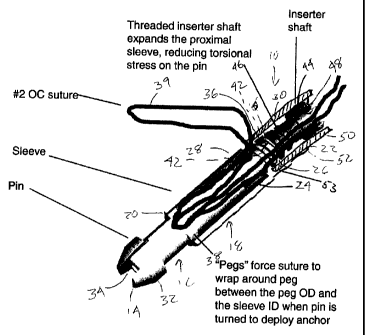
KNOTLESS SUTURE ANCHOR
Background
[0001] The present application relates to suture anchors and more particularly
to a
knotless suture anchor.
[0002] Suture anchors have wide use in surgery particularly for reattaching
soft tissue to
bone. It is preferred to perform most of these surgeries endoscopically. While
working
through a long narrow endoscope knot tying takes on added difficulty.
Accordingly it is
frequently preferred to employ a suture anchor which can capture the suture
without the need
of the surgeon having to tie a knot. It is also preferred that while capturing
the suture to lock
it to the anchor that the anchor not disturb the tension on the suture.
Typically the soft tissue
is carefully positioned just prior to locking the suture and if the act of
locking the suture
causes it to move it can affect the position of the soft tissue.
Summary of the Invention
[0003] A suture anchor according to the present invention comprises an outer
body having
a distal end a proximal end and an axial bore therethrough. An inner body is
receivable
within the outer body. A suture limb is captured between the inner body and
the outer body
by being wrapped around the inner body.
[0004] Preferably, the inner body and the outer body are formed of a
bioabsorbable
material, as for example a material comprising PLGA.
[0005] Preferably, the inner body and outer body are threaded together.
[0006] Preferably, the suture anchor is configured such that as the inner body
is moved
toward the proximal end of the outer body the proximal end of the outer body
expands
outwardly radially.
[00071 Preferably, the suture limb is wrapped around the inner body at least
two times.
Enhanced holding is provided when the suture limb is wrapped around the inner
body at least
five times.
CA 02718861 2010-10-26
[0008] Preferably, the inner body comprises at least one tab extending
outwardly radially
whereby to effect wrapping of the suture about the inner body upon rotation of
the inner
body.
[0009] Preferably, the inner body has a distal end and a proximal end and
wherein the
inner body proximal has outer threads which mate with inner threads on the
proximal end of
the outer body. Preferably, an inserter is provided having a distal end which
mates with the
proximal end of the inner body and which has outer inserter threads engageable
with the
inner threads on the outer body. Preferably, the outer body has a first
configuration in which
at least a portion thereof is radially contracted inwardly and a second
configuration in which
the portion is radially expanded outwardly, and wherein when the outer
inserter threads are
engaged into the outer body inner threads it holds the portion in the second
configuration.
Similarly, when the outer threads on the inner body are engaged into the outer
body inner
threads it holds the portion in the second configuration. Preferably, a
naturally relaxed
position of the portion is in the second configuration. Thus, when the outer
body is expanded
into the bone internal stresses on the outer body are minimized.
[00010] In one aspect of the invention, the suture limb passes into a space
formed between
the inner body and the outer body at their proximal ends, passes out of the
space at their distal
ends and then passes proximally along an outer surface of the outer body.
[00011] Preferably, the outer body has at its proximal end at least one
axially extending slit
therein whereby to relieve stresses upon radially outward expansion of the
outer body
proximal end.
[00012] In one aspect of the invention, at least one more suture limb is
captured between
the inner body and the outer body or perhaps at least three more suture limbs
captured
between the inner body and the outer body.
[00013] A method according to the present invention provides for attaching
tissue to a
bone. It comprises the steps of: passing a limb of suture from the tissue
between a suture
anchor outer body, which has a distal end a proximal end and an axial bore
therethrough, and
an inner body receivable within the outer body; inserting the outer body into
the bone; sliding
the suture between the inner body and the outer body to achieve a desired
tension thereon or
desired position of the tissue; capturing the suture limb between the inner
body and the outer
2
CA 02718861 2010-10-26
body, by wrapping at least a segment of the suture limb about the inner body,
to prevent
sliding of the suture limb therebetween.
[00014] Preferably, the distance between the tissue and the anchor stays
substantially the
same during the step of capturing.
[00015] Preferably, the suture limb is wrapped around the inner body at least
two times.
[00016] Preferably, the inner body has at least one radially extending
projection and the
step of wrapping comprises rotating the inner body within the outer body
during which the
projection engages the suture limb to cause it to wrap about the inner body.
[00017] Preferably, the method further comprises the step of radially
expanding at least a
portion of the outer body to engage the suture anchor into the bone. For
instance when a
proximal end of the inner body has outer threads and the proximal end of the
outer body has
mating inner threads then the step of radially expanding can comprise engaging
the inner
body outer threads with the outer body inner threads.
Brief Description of the Drawings
[00018] FIG. 1 is a perspective cut-away view of a suture anchor according to
the present
invention;
[00019] FIG. 2 is a perspective view of the suture anchor of FIG. 1;
[00020] FIG. 3 is a perspective view of the suture anchor of FIG. 1 pre-loaded
with a suture
capture device;
[00021] FIG. 4 is a side elevation view in cut-away of soft tissue and
associated bone
showing initial insertion of the suture anchor of FIG. 1;
[00022] FIG 5. is a side elevation view in cut away of the soft tissue and
bone of FIG 4
showing free ends of a suture between the soft tissue and the anchor being
pulled to tension
the suture and position the soft tissue;
3
CA 02718861 2010-10-26
[00023] FIG. 6 is a side elevation view in cut away of the soft tissue and
bone of FIG 4
showing the anchor being actuated to lock the suture to the anchor and the
anchor to the
bone;
[00024] FIG. 7 is a side elevation view in cut away of the soft tissue and
bone of FIG 4
showing the anchor fully deployed;
[00025] FIG. 8 is a side elevation view in cut away of the soft tissue and
bone of FIG 4
showing the anchor in partial cut-away in its fully deployed position;
[00026] FIG. 9 is a perspective view of a further embodiment of a suture
anchor according
to the present invention;
[00027] FIG. 10 is a perspective cut-away view of the suture anchor of FIG. 9.
Detailed Description
[00028] FIG. 1 depicts a suture anchor 10 according to the present invention.
It comprises
in gross an inner body 12 having a distal end 14 and proximal end 16 and a
cannulated outer
body 18 having a distal end 20, proximal end 22 and a cannulation 24
therethrough. Towards
the outer body proximal end 22 the cannulation 24 bears internal threads 26
which decrease
in internal diameter at the proximal end 22. On its exterior surface 28 the
outer body 18
bears barb shaped annular flanges 30 to assist in bone fixation.
[00029] The inner body 12 has an annular flange 32 at its distal end 14 with a
groove 34
therethrough passing over the distal end 14. At its proximal end 16 the inner
body 12 has
exterior threads 36 which mate with the inner body threads 26. A pair of
radially extending
projections 38 extend from the inner body 12 toward the outer body 18 at its
distal end 20.
The tolerance between the projections 38 and the outer body 18 should be close
enough to
prevent suture 39 from passing therebetween.
[00030] A tool receiving recess 40 on the inner body proximal end 16 mates
with a driver
head 42 (such as for instance a hex driver) on a distal end of a driver 44.
Just proximal
thereof on the driver 44 are threads 46 which mate with the threads 26 on the
outer body 18.
The threads 46 have a reduced major diameter at a proximal portion 48 which in
its starting
configuration as shown in FIG. 1 sits adjacent the decreased internal diameter
of the outer
4
CA 02718861 2010-10-26
body thread 26 at their proximal end 22. The driver 44 operates within a tube
50 having a
distal end 52 abutting the outer body proximal end 22 with distally projecting
tangs 53
extending into slots 54 in the outer body proximal end. This interface assists
in maintaining
the position of the anchor 10 as it is employed, by resisting both rotation
and proximal
withdrawal thereof.
[00031] Turning also now to FIG. 2, two or more of the stress relief slots 54
extend axially
into the outer body 18 from its proximal end 22. This allows the proximal end
to be made
from somewhat brittle materials yet still be able to expand outwardly radially
to provide
fixation. Both the inner body 12 and outer body 18 are preferably formed of a
bioabsorbable
material such as BIOCRYL RAPIDE available from DePuy Mitek, Inc. of Raynham,
MA.
BIOCRYL RAPIDE is a bioabsorbable polymer formed of homogenous blend of
TriCalcium
Phosphate (TCP) and Polylactic/polyglycolic Acid (PLGA). Other suitable
materials include
without limitation PEEK, PLA, titanium, stainless steel, metals, polymers and
other
biocompatible materials.
[00032] Turning also now to FIGS. 3 to 7, use of the suture anchor 10 will be
described.
The anchor 10 is sterile and packaged in bacteria proof packaging (not shown)
pre-loaded
onto the driver 44 and pre-loaded with a suture capture device 56 comprising
an elongated
filament 58 having a suture capture loop 60 at one end. One example is the
CHIA
PERCPASSER available from DePuy Mitek, Inc. of Raynham, MA. The loop 60 in
FIG. 3 is
shown adjacent the anchor 10 for ease of display but in practice sufficient
length of the
filament 58 would extend from the anchor 10 to allow suture 39 to be pulled
out of a cannula
(not shown) through which the procedure is being endoscopically performed.
[00033] The suture 39 would be loaded into the suture capture loop 60 exterior
of the
patient and the cannula. A tab 62 may be placed on an opposite end of the
filament 58. (This
is also shown adjacent the anchor 10 for ease of display but would more
conveniently be
positioned outside of the cannula.) When the tab is pulled the loop 60 with
the suture 39
captured therein is drawn down between the inner body 12 and outer body 18
pulling the
suture 39 with it. The path of the suture 39 after passing between the inner
body 12 and outer
body 18 goes through the groove 34 to assist in sliding. Additional sutures
can also be
employed, such as additional suture loops in the suture capture loop 60 or
addition suture
loops each with their own suture capture device.
CA 02718861 2010-10-26
[000341 The anchor 10 with the suture 39 therein is now inserted into a pre-
drilled hole 64
in a bone 66 to which a piece of soft tissue 68 is to be attached as shown in
FIG. 4. The
anchor 10 is positioned in the hole 64 such that the suture passes into the
anchor 10 at one of
the stress relief slots 54. The suture 39 is shown looped through the soft
tissue 68 but other
arrangements are possible such as extending from another anchor (not shown and
typically of
a different configuration than anchor 10) which is positioned in the bone 66
below the soft
tissue 68 and up through the soft tissue 68 to the anchor 10, such as in a
dual row rotator cuff
repair. Also, the path from the soft tissue 68 through the anchor 10 could be
reversed.
1000351 Free ends 70 of the suture 39 are drawn through the anchor 10 to
position the soft
tissue 68 and properly tension the suture 39 (see FIG. 5). The tube 50 of the
driver 44 holds
the anchor 10 down and prevents rotation of the outer body 18 while the driver
44 is rotated
to rotate the inner body 18 (see FIG. 6). As the threads 46 of the driver 44
pass through the
reduced inner diameter proximal portion 22 of the outer body 18 it causes it
to expand
outwardly radially to engage the bone 66 and reduces the stress on the inner
body 18.
Preferably, the relaxed condition of the outer body 18 is slightly expanded
radially and as it is
inserted into the hole 64 it is compressed slightly inwardly; the expansion by
the threads 46
move it back to its relaxed configuration thus reducing internal stress. As
the rotation
continues the threads 36 of the inner body move into the reduced inner
diameter proximal
portion 22 to keep the outer body proximal end 22 radially expanded. The
projections 38 on
the inner body 12 cause the suture 39 to wrap around the inner body 12. The
suture 39 feeds
in from the free ends 70, not from the soft tissue 68 so that the position of
the soft tissue 68
and the tension on the suture 39 between the anchor 10 and the soft tissue 68
remains
substantially unchanged as the inner body 12 is rotated. After sufficient
rotation the driver 44
is disengaged from the anchor 10 and removed leaving the suture 39 locked to
the anchor 10
by virtue of its being wrapped around the inner body 12 and the outer body
proximal end 22
is expanded outwardly into the bone 66 to lock the anchor 10 thereto (see
FIGS. 7 and 8).
Tests have shown three to five turns providing good locking of the suture 39.
1000361 FIGS. 9 and 10 illustrate a further preferred embodiment of the
invention which is
essentially similar to that depicted in FIGS. 1 and 2. Like parts are denoted
with like
numerals with the addition of a prime symbol ('). It comprises a suture anchor
10' having an
inner body 12' and cannulated outer body 18' having a short internal thread
24'. The inner
body 12' has an annular flange 32' at its distal end 14' with a groove 34'. It
also carries
6
CA 02718861 2010-10-26
radially extending projections 38'. FIG. 9 especially more clearly illustrates
how a driver
receiving tube 50' abuts a proximal end 22' of the outer body 18' with
distally projecting
tangs 53' extending into stress relief slots 54'. A loop of suture 39' has
free ends which pass
into the outer body 18' from its proximal end 22', preferably through one of
the stress relief
slots 54', passes down between the inner body 12' and outer body 18' and
between the
projections 38', out of the outer body 18' through its distal end 20', through
the groove 34'
on the inner body 12' at its distal end 14' and then back into the outer body
18' between it
and the inner body 12' and also again between the projections 38" and finally
exit through the
opposing stress relief slot 54'. This embodiment is used similarly to the
previous one.
However, the groove 34' assists in wrapping the suture 39' around the inner
body 12' and
one could even dispense with the projections 38' due to the wrapping action
provided by the
groove 34'.
[000371 Various modifications and alterations of this invention will be
apparent to those
skilled in the art without departing from the scope and spirit of this
invention. It should be
understood that the invention is not limited to the embodiments disclosed
herein, and that the
claims should be interpreted as broadly as the prior art allows.
7