Note: Descriptions are shown in the official language in which they were submitted.
CA 02718975 2010-09-17
WO 2009/126306 PCT/US2009/002247
Compositions And Methods For Detecting EGFR Mutations In Cancer
Related Applications
This application claims the benefit of and priority to U.S. provisional patent
application U.S.S.N. 61/123,699, filed April 10, 2008, and U.S. provisional
patent
application U.S.S.N. 61/190,597, filed August 29, 2008, both of which are
hereby
incorporated by reference herein in their entirety.
Background of the Invention
The invention relates generally to the field of mutant proteins and genes
involved
in cancer, and to the detection, diagnosis and treatment of cancer.
Cancer is major cause of death in humans. Lung cancer is a major cause of
cancer-related mortality worldwide and is expected to remain a major health
problem for
the foreseeable future. It is broadly divided into small cell lung cancer
(SCLC, 20% of
lung cancers), and non-small cell lung cancer (NSCLC, 80% of lung cancers).
Somatic
mutations in the epidermal growth factor receptor (EGFR) gene are found in a
subset of
lung adenocarcinomas and are associated with sensitivity to the EGFR tyrosine
kinase
inhibitors (TKI) Gefitinib [Lynch, T.J., et al., N Engl J Med, 2004. 350(21):
p. 2129-39,
and Paez, J.G., et al., Science, 2004. 304(5676): p. 1497-500] and Erlotinib
[Pao, W., et
al., Proc Natl Acad Sci U S A, 2004. 101(36): p. 13306-11]. Many types of EGFR
mutations have been reported, but the most common non-small cell lung cancer
(NSCLC)-associated EGFR mutations are the 15-bp nucleotide in-frame deletion
in exon
19 (E746-A750del) and the point mutation replacing leucine with arginine at
codon 858
in exon 21 (L858R) [Pao, W., et al., Proc Natl Acad Sci U S A, 2004. 101(36):
p. 13306-
11; Riely, G.J., et al., Clin Cancer Res, 2006. 12(24): p. 7232-41; and
Kosaka, T., et al.,
Cancer Res, 2004. 64(24): p. 8919-23. These two mutations represent 85-90% of
EGFR
mutations in NSCLC patients. Importantly, patients with these mutations have
been
shown to respond well to EGFR inhibitors including Gefitinib and Erlotinib
[Riely, G.J.,
et al., Clin Cancer Res, 2006. 12(24): p. 7232-41; Inoue, A., et al., J Clin
Oncol, 2006.
24(21): p. 3340-6; Marchetti, A., et al., J Clin Oncol, 2005. 23(4): p. 857-
65; and
CA 02718975 2010-09-17
WO 2009/126306 PCT/US2009/002247
Mitsudomi, T., et al., J Clin Oncol, 2005. 23(11): p. 2513-20.]. Therefore
detection of
these mutations is an important method to improve treatment of lung cancer
patients.
Since EGFR mutational analysis in lung adenocarcinoma can guide treatment
decisions and to enroll patients on specific arms of clinical trials, direct
DNA sequencing
of PCR amplified products has been developed to detect EGFR mutation in
patient tumor
tissue. However, these tests have not been widely adopted due the high costs
of the
equipment and reagents, the difficulty of performing the assay and the length
of time
required for completion of the test. In addition, DNA sequencing has a limited
sensitivity
for the detection of tumor cells containing an EGFR mutation within a
background of
nonmutant normal cells. A minimum of 50% tumor cells is required to ensure the
accuracy of the EGFR sequencing assay. Recently, other DNA based methods have
been
developed to improve the detection of EGFR mutation in lung cancer specimens,
including TaqMan PCR, Scorpions ARMS, MALDI TOF MS-based genotyping, dHPLC,
and single molecule sequencing. However, these methods are not routine
procedures in
clinical labs and remain expensive and time-consuming. Also they do not
identify
mutation-status on a cellular basis. Therefore, their sensitivity is dependent
on the
percentage tumor cells contained in the sample used to produce the homogenate,
and
samples obtained from standard biopsy are usually not sufficient for DNA
sequencing.
On the other hand, Immunohistochemistry (IHC) is a well-established method of
solid
tumor analysis routinely performed in all clinical laboratories. This method
is a more
accessible technique in clinical diagnosis and the interpretation is less
affected by the
percentage of the cancer cells in the tumor specimens or the amount of tumor
tissue
available for analysis. The method also allows for the simultaneous analysis
of other
proteins or protein modifications. However, total expression level of EGFR by
IHC has
not been shown to predict response to tyrosine kinase inhibitor therapy in
NSCLC
[Meert, A.P., et al., Eur Respir J, 2002. 20(4): p. 975-81]. Thus, development
of
antibodies that specifically detect mutant EGFR protein and that may be used
in IHC will
be a valuable addition to the clinical diagnosis and treatment of lung cancer.
A related challenge facing diagnostic analysis of solid tumor samples
including
lung cancer tumors is access to the tissue sample. Repeated biopsies are not
clinically
feasible for almost all tumor types. Therefore, alternative sources of cancer
cells must be
2
CA 02718975 2010-09-17
WO 2009/126306 PCT/US2009/002247
obtained. This is especially important in the context of targeted therapeutics
in which
repeated tumor analysis may be used to guide the drug therapy. A number of
cancer cell
sources are available in some tumor types including circulating cancer cells
(CTCs),
ascites, bronchial swabs, ductal adenocarincoma is of a cancer tissue type
selected from
the group consisting of lung cancer, colon cancer, breast cancer, cervical
cancer,
pancreatic cancer, prostate cancer, stomach cancer, and esophageal cancer.
circulating proteins may be detected by standard protein assays such as an
ELISA assay.
In this example, the mutation EGFR protein would be captured and detected with
a pair
of antibodies including an antibody against the total protein and an antibody
to the
mutation. Such an assay would enable routine and repeated analysis of treated
patients to
best match the choice of drug and drug regime to the direct affect the therapy
was having
on the patient's tumor.
Summary of the Invention
The invention provides binding agents, such as rabbit monoclonal antibodies,
that
specifically bind to an EGFR molecule with an E746-A750 deletion and an EGFR
molecule with a L858R point mutation.
Accordingly, in a first aspect, the invention provides a binding agent that
specifically binds an epidermal growth factor receptor (EGFR) molecule
comprising a
deletion at position E746-A750. In some embodiments, the epidermal growth
factor
receptor (EGFR) molecule is from a human. In some embodiments, the binding
agent
comprises at least one complementary determining region (CDR), wherein the CDR
comprises a sequence selected from the group consisting of SEQ ID NO: 9, SEQ
ID NO:
10, SEQ ID NO: 11, SEQ ID NO: 16, SEQ ID NO 17, and SEQ ID NO: 18. In some
embodiments, the binding agent specifically binds to an epitope comprising an
amino
acid sequence comprising a threonine-serine-proline sequence. In some
embodiments,
where the binding agent is an antibody, the antibody is produced by the clone
deposited
with the ATCC and given the designation number ATCC No. PTA-9151.
In another aspect, the invention provides a binding agent that specifically
binds to
an epidermal growth factor receptor (EGFR) molecule comprising a point
mutation
substituting leucine with arginine at position 858. In some embodiments, the
epidermal
3
CA 02718975 2010-09-17
WO 2009/126306 PCT/US2009/002247
growth factor receptor (EGFR) molecule is from a human. In some embodiments,
the
binding agent comprises at least one complementary determining region (CDR),
wherein
the CDR comprises an amino acid sequence selected from the group consisting of
SEQ
ID NO: 23, SEQ ID NO: 24, SEQ ID NO: 25, SEQ ID NO: 30, SEQ ID NO 31, and SEQ
ID NO: 32. In some embodiments, the binding agent specifically binds to an
epitope
comprising an amino acid sequence comprising a threonine-aspartic acid-X-
glycine-
arginine sequence, where X is any amino acid residue. In some embodiments,
where the
binding agent is an antibody, the antibody is produced by the clone deposited
with the
ATCC and given the designation number ATCC No. PTA-9152.
In a further aspect, the invention provides a polynucleotide (e.g., a purified
polynucleotide) encoding a binding agent that specifically binds to an
epidermal growth
factor receptor (EGFR) molecule comprising a deletion at position E746-A750.
In a
further aspect, the invention provides a polynucleotide (e.g., a purified
polynucleotide)
encoding a binding agent that specifically binds to an epidermal growth factor
receptor
(EGFR) molecule comprising a point mutation substituting leucine with arginine
at
position 858. In further aspects, the invention provides vectors (e.g.,
expression vectors)
comprising the polynucleotides.
In another aspect, the invention provides methods for identifying a cancer
that
will respond favorably to a therapy targeting aberrant expression of an EGFR
molecule.
The methods comprise comprising (a) contacting a biological sample from the
cancer
with the binding agent that specifically binds to an epidermal growth factor
receptor
(EGFR) molecule comprising a deletion at position E746-A750 to obtain an
amount of
binding and (b) comparing the result of step (a) with an amount of binding
obtained by
contacting a biological sample from a healthy individual with the binding
agent, wherein
a change in the amount of binding from the cancer as compared to the amount of
binding
from the healthy individual indicates the cancer will respond favorably to the
therapy. In
various embodiments, the biological sample from the cancer and the biological
sample
from the healthy individual are of the same tissue type. In some embodiments,
the cancer
is from a human patient. In some embodiments, the cancer is a non-small-cell
lung
cancer (NSCLC). In some embodiments, the cancer is an adenocarcinoma or a
squamous
cell carcinoma. In some embodiments, the cancer is of a tissue type selected
from the
4
CA 02718975 2010-09-17
WO 2009/126306 PCT/US2009/002247
group consisting of lung cancer, colon cancer, breast cancer, cervical cancer,
pancreatic
cancer, prostate cancer, stomach cancer, and esophageal cancer.
In another aspect, the invention provides methods for identifying a cancer
that
will respond favorably to a therapy targeting aberrant expression of an EGFR
molecule.
The methods comprise comprising (a) contacting a biological sample from the
cancer
with the binding agent that specifically binds to an epidermal growth factor
receptor
(EGFR) molecule comprising a point mutation substituting leucine with arginine
at
position 858 to obtain an amount of binding and (b) comparing the result of
step (a) with
an amount of binding obtained by contacting a biological sample from a healthy
individual with the binding agent, wherein a change in the amount of binding
from the
cancer as compared to the amount of binding from the healthy individual
indicates the
cancer will respond favorably to the therapy. In various embodiments, the
biological
sample from the cancer and the biological sample from the healthy individual
are of the
same tissue type. In some embodiments, the cancer is from a human patient. In
some
embodiments, the cancer is a non-small-cell lung cancer (NSCLC). In some
embodiments, the cancer is an adenocarcinoma. In some embodiments, the
adenocarincoma is of a cancer tissue type selected from the group consisting
of lung
cancer, colon cancer, breast cancer, cervical cancer, pancreatic cancer,
prostate cancer,
stomach cancer, and esophageal cancer.
In various embodiments, the amount of binding is determined using an assay
method selected from the group consisting of Western blot, immunofluorescence,
ELISA,
IHC, flow cytometry, immunoprecipitation, autoradiography, scintillation
counting, and
chromatography.
In further aspects, the invention also provides a composition comprising a
binding
agent specifically binds to an epidermal growth factor receptor (EGFR)
molecule
comprising a point mutation substituting leucine with arginine at position
858, a binding
agent that specifically binds to an epidermal growth factor receptor (EGFR)
molecule
comprising a deletion at position E746-A750, or both binding agents. In some
embodiments, the composition further comprises a pharmaceutically acceptable
carrier.
The invention also provides a composition comprising a polynucleotide encoding
a
binding agent specifically binds to an epidermal growth factor receptor (EGFR)
molecule
CA 02718975 2010-09-17
WO 2009/126306 PCT/US2009/002247
comprising a point mutation substituting leucine with arginine at position
858, a
polynucleotide encoding a binding agent that specifically binds to an
epidermal growth
factor receptor (EGFR) molecule comprising a deletion at position E746-A750,
or both
polynucleotides. In some embodiments, the composition further comprises a
pharmaceutically acceptable carrier.
In further aspects, the invention provides a method for treating a patient
having or
suspected of having a cancer that will respond favorably to a therapy
targeting aberrant
expression of an EGFR molecule. The method includes administering to the
patient an
effective amount of a composition of the invention.
Another aspect of the invention discloses method for identifying the L858R
point
mutation and/or E746 - A750 deletion in EGFR status in a patient, said method
comprising the steps of: a) obtaining a biological sample from a patient; b)
screening the
sample with a binding agent that specifically binds the L858R point mutation
and/or
E746 - A750 deletion in EGFR; and c) determining the presence or absence of
the E746
- A750 deletions and/or the L8585R point mutation in EGFR in the sample. In
some
embodiments, the method includes screening the sample with a wildtype EGFR-
specific
antibody. In some embodiments, the method includes screening the sample with a
pan-
keratin antibody (e.g., a pan-cytokeratin antibody).
Another aspect of the invention describes kits for the detection of E746 -
A750
deletion or L858R point mutations in EGFR in a sample, said kit comprising (a)
a binding
agent that specifically binds to the E746 - A750 deletion in EGFR and/or a
binding agent
that specifically binds to the L858R point mutations in EGFR; and b)
instructions for
detecting E746 - A750 deletion or L858R point mutations in EGFR in a sample.
Brief Description of the Drawings
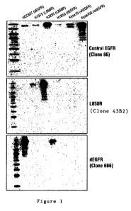
Figure 1 is a representative Western blotting depicting the reactivity of the
antibodies of the invention for EGFR and mutants thereof in the indicated cell
lines. The
control wildtype (wt) EGFR-specific antibody clone 86 (top panel) binds to
(i.e., is
reactive to) lysates prepared from all indicated cell lines, although the
reactivity is
somewhat reduced in the cells expressing mutant EGFR (i.e., HCC827, H1975,
H3255,
and H1650 cells). The EGFR L858R-specific antibody (clone 6B6) is reactive
only to
6
CA 02718975 2010-09-17
WO 2009/126306 PCT/US2009/002247
H175 and H3255 cells (middle panel), while the dEGFR (i.e., EGFR de1746-A750)-
specific antibody (clone 43B2) is reactive only to HCC827 and H1650 cells.
Figure 2 depicts reactivity of the antibodies of the invention by
immunofluorescent immunocytochemistry for EGFR and mutants thereof in the
indicated
cell lines. The control EGFR-specific antibody (top panel) stains (i.e., binds
to) all six
cell lines, regardless of their EGFR mutational status. The EGFR L858R-
specific
antibody stains only the cancer cells with the L858R point mutation in their
EGFR
molecule. Similarly, the dEGFR-specific antibody stains only the cancer cells
with the
deletion in Exon 19 (i.e., E746-A750) in their EGFR molecule.
Figure 3 depicts reactivity of the antibodies of the invention by
immunohistochemistry for EGFR and mutants thereof in sections taken from nude
mice
implanted with the indicated cell lines as xenografts. The control EGFR-
specific antibody
(top panel) stains (i.e., binds to) all six cell lines, regardless of their
EGFR mutational
status. The EGFR L858R-specific antibody stains only the cancer cells with the
L858R
point mutation in their EGFR molecule. Similarly, the dEGFR-specific antibody
stains
only the cancer cells with the deletion in Exon 19 (i.e., E746-A750) in their
EGFR
molecule.
Figure 4 depicts reactivity of the antibodies of the invention by
immunohistochemistry analysis of four representative, non-limiting, pre-typed
NSCLC
samples (i.e., samples whose DNA had been sequenced prior to IHC analysis).
Samples
from patients CL 109 and CL745, which by DNA sequencing were known to harbor
the
EGFR L858R point mutation, stained positive with the L858R-specific antibody,
but
negative for staining with the dEGFR-specific antibody. The samples from
patients
CL495 and CL712, which by DNA sequencing were known to harbor the E746-A750
deletion, stained positive with the dEGFR-specific antibody, but negative for
staining
with the L858R-specific antibody.
Figure 5 depicts reactivity of the antibodies of the invention by
7
CA 02718975 2010-09-17
WO 2009/126306 PCT/US2009/002247
immunohistochemistry of two representative, non-limiting, NSCLC samples of
unknown
genotype (i.e., samples whose DNA had not been sequenced prior to IHC
analysis). The
tumor sample from patient CL761 showed positive staining for Pan-cytokeratin-
specific
antibody, control wild-type EGFR-specific antibody, and L858R-specific
antibody, but
negative with the dEGFR (i.e., the E746-A750de1)-specific antibody. In
contrast, the
tumor sample from patient CL764 stained positive for Pan-cytokeratin-specific
antibody
(positive control), control wildtype EGFR-specific antibody, and dEGFR-
specific
antibody, but negative with the L858R-specific antibody.
Detailed Description of the Preferred Embodiments
The invention relates generally to mutant proteins and genes involved in
cancer,
and to the detection, diagnosis and treatment of cancer utilizing the
antibodies of the
invention disclosed herein.
Higher EGFR protein expression determined by immunohistochemistry is
observed in the majority of squamous cell carcinomas, a small percentage of
large cell
carcinomas, adenocarcinomas, and bronchial pre-neoplastic lesions, implicating
its
significance in lung carcinogenesis [Selvaggi, G., et al., Ann Oncol, 2004.
15(1): p. 28-
32]. There are conflicting data about the prognostic importance of EGFR
protein levels in
NSCLC. A meta-analysis of these studies failed to show a significant
correlation between
EGFR levels and survival [Meert, A.P., et al., Eur Respir J, 2002. 20(4): p.
975-81].
Retrospective evaluations of the relationship between EGFR positive by
immunohistochemistry and response showed that EGFR immunohistochemistry
results
were not predictive of response in the original trial of Gefitinib and later
research data
[Clark, G.M., et al., J Thorac Oncol, 2006. 1(8): p. 837-46; Tsao, M.S., et
al., N Engl J
Med, 2005. 353(2): p. 133-44; Dziadziuszko, R., et al., Ann Oncol, 2007.
18(3): p. 447-
52; and Cappuzzo, F., et al., J Natl Cancer Inst, 2005. 97(9): p. 643-55].
Since the
presence of certain EGFR mutation correlates with clinical response to either
gefitinib or
erlotinib, there is a huge demand for the identification of such EGFR
mutations in
NSCLC patients.
Accordingly, the invention provides rabbit mAbs that were generated, as
described herein, with selective reactivity for EGFR protein with E746-A750de1
and
8
CA 02718975 2010-09-17
WO 2009/126306 PCT/US2009/002247
L858R point mutation. Western blots and immunofluorescence showed the
antibodies
were specific to E746-A750del and L858R mutant EGFR proteins. These antibodies
were further analyzed by IHC in xenograft tumors, cell pellets and molecularly
pre-typed
samples of NSCLC and compared with anti-wtEGFR mAb. The RmAbs were selected to
detect either E746-A750de1 or L858R point mutant EGFR proteins, not wtEGFR or
other
types of EGFR mutations. On the other hand, the anti-wtEGFR Ab was widely
reactive
with a higher proportion of NSCLC. Thus, the binding agents described herein
specifically recognize either E746-A750de1 or L858R mutant EGFR protein.
The invention provides binding agents (such as antibodies) that specifically
bind
to the EGFR L858R mutation and the EGFR E746-A750de1 mutation. The EGFR
mutation-specific antibodies are extremely valuable in the clinical management
(e.g., the
treatment and diagnosis) of cancer patients, particularly patients who have or
are
suspected of having NSCLC or other cancer characterized by aberrant EGFR.
As used in this specification, the singular forms "a," "an" and "the"
specifically
also encompass the plural forms of the terms to which they refer, unless the
content
clearly dictates otherwise.
The term "about" is used herein to mean approximately, in the region of,
roughly,
or around. When the term "about" is used in conjunction with a numerical
range, it
modifies that range by extending the boundaries above and below the numerical
values
set forth. In general, the term "about" is used herein to modify a numerical
value above
and below the stated value by a variance of 20%.
As used herein, unless specifically indicated otherwise, the word "or" is used
in
the "inclusive" sense of "and/or" and not the "exclusive" sense of
"either/or." In the
specification and the appended claims, the singular forms include plural
referents unless
the context clearly dictates otherwise.
As used in this specification, whether in a transitional phrase or in the body
of the
claim, the terms "comprise(s)" and "comprising" are to be interpreted as
having an open-
ended meaning. That is, the terms are to be interpreted synonymously with the
phrases
"having at least" or "including at least". When used in the context of a
process, the term
"comprising" means that the process includes at least the recited steps, but
may include
additional steps. When used in the context of a compound or composition, the
term
9
CA 02718975 2010-09-17
WO 2009/126306 PCT/US2009/002247
"comprising" means that the compound or composition includes at least the
recited
features or components, but may also include additional features or
components.
The patents, published applications, and scientific literature referred to
herein
establish the knowledge of those with skill in the art and are hereby
incorporated by
reference in their entirety to the same extent as if each was specifically and
individually
indicated to be incorporated by reference. Any conflict between any reference
cited
herein and the specific teachings of this specification shall be resolved in
favor of the
latter. Likewise, any conflict between an art-understood definition of a word
or phrase
and a definition of the word or phrase as specifically taught in this
specification shall be
resolved in favor of the latter.
Any suitable materials and/or methods known to those of skill can be utilized
in
carrying out the present invention. However, preferred materials and methods
are
described. Materials, reagents and the like to which reference is made in the
following
description and examples are obtainable from commercial sources, unless
otherwise
noted.
As used herein, the recitation of a numerical range for a variable is intended
to
convey that the invention may be practiced with the variable equal to any of
the values
within that range. Thus, for a variable which is inherently discrete, the
variable can be
equal to any integer value of the numerical range, including the end-points of
the range.
Similarly, for a variable which is inherently continuous, the variable can be
equal to any
real value of the numerical range, including the end-points of the range. As
an example,
a variable which is described as having values between 0 and 2, can be 0, 1 or
2 for
variables which are inherently discrete, and can be 0.0, 0.1, 0.01, 0.001, or
any other real
value for variables which are inherently continuous.
Reference is made hereinafter in detail to specific embodiments of the
invention.
While the invention will be described in conjunction with these specific
embodiments, it
will be understood that it is not intended to limit the invention to such
specific
embodiments. On the contrary, it is intended to cover alternatives,
modifications, and
equivalents as may be included within the spirit and scope of the invention as
defined by
the appended claims. In the following description, numerous specific details
are set forth
in order to provide a thorough understanding of the present invention. The
present
CA 02718975 2010-09-17
WO 2009/126306 PCT/US2009/002247
invention may be practiced without some or all of these specific details. In
other
instances, well known process operations have not been described in detail, in
order not
to unnecessarily obscure the present invention.
The epidermal growth factor receptor (EGFR; also known as ErbB-1 and HER1 in
humans) is the cell-surface receptor for members of the epidermal growth
factor family
(EGF-family) of extracellular protein ligands. The amino acid sequence of wild-
type
human EGFR (including the signal sequence) is provided herein as SEQ ID NO:
47; the
amino acid sequence of wild-type human EGFR (minus the signal sequence) is
provided
herein as SEQ ID NO: 48. Patients of non-small cell lung cancer (NSCLC)
carrying the
somatic mutation of epidermal growth factor receptor (EGFR) have been shown to
be
hyperresponsive to the EGFR tyrosine kinase inhibitor Gefitinib [Lynch, T.J.,
et al., N
Engl J Med, 2004. 350(21): p. 2129-39, and Paez, J.G., et al., Science, 2004.
304(5676):
p. 1497-500] and Erlotinib [Pao, W., et al., Proc Natl Acad Sci U S A, 2004.
101(36): p.
13306-11].
Mutations are known to arise in the EGFR molecule. As used herein, the term
"mutant" or "mutation" refers to a molecule (e.g., a polypeptide or a
polynucleotide) that
has a different structure than the wild-type molecule. That difference in
structure from
the wild-type molecule includes, without limitation, a different sequence
(e.g., a different
amino acid or nucleotide sequence), additional sequences, missing sequences
(i.e., a
portion of the sequence is missing), changes in modification (e.g.,
methylation,
phosphorylation, etc.), and/or fusion of all or part of the wild-type molecule
with another
molecule. By "wild-type" is meant that form of the molecule that naturally
occurs in the
majority of individuals of the species from which the mutant molecule is
derived, and/or
the form of the molecule that naturally occurs in an healthy individual (e.g.,
non-
cancerous) individual of a species from which the mutant molecule is derived.
The
sequence of the wild-type molecule is that typically provided in the GenBank
database.
For example, the amino acid sequence of wild-type human EGFR is provided in
SEQ ID
NO: 47 (without the 24 amino acid long signal sequence) and SEQ ID NO: 48
(with the
signal sequence).
As used herein, an "EGFR mutant" includes any type of mutation (i.e., change)
in
an EGFR molecule that renders the EGFR mutant different than wildtype EGFR.
The
11
CA 02718975 2010-09-17
WO 2009/126306 PCT/US2009/002247
most common NSCLC-associated EGFR mutations are the 15-bp nucleotide in-frame
deletion in exon 19 (E746-A750de1; amino acid sequence (including the signal
sequence)
provided in SEQ ID NO: 49 and without the signal sequence provided in SEQ ID
NO:
50) and the point mutation replacing leucine with arginine at codon 858 in
exon 21
(L858R; amino acid sequence (including the signal sequence) provided in SEQ ID
NO:
51 and without the signal sequence provided in SEQ ID NO: 52). These two EGFR
mutants account for 85-90% EGFR mutations [Riely, G.J., et al., Clin Cancer
Res, 2006.
12(24): p. 7232-41]. The ability to detect mutated gene products in cancer
cells can
identify patients most likely benefit from such therapies, and make clinical
trials more
efficient and informative.
Thus, in a first aspect, the invention provides a binding agent that
specifically
binds an epidermal growth factor receptor (EGFR) molecule comprising a
deletion at
position E746-A750. In some embodiments, the epidermal growth factor receptor
(EGFR) molecule is from a human. In some embodiments, the binding agent
comprises
at least one complementary determining region (CDR), wherein the CDR comprises
a
sequence selected from the group consisting of SEQ ID NO: 9, SEQ ID NO: 10,
SEQ ID
NO: 11, SEQ ID NO: 16, SEQ ID NO 17, and SEQ ID NO: 18. In some embodiments,
the binding agent specifically binds to an epitope comprising an amino acid
sequence
comprising a threonine-serine-proline sequence.
In another aspect, the invention provides a binding agent that specifically
binds to
an epidermal growth factor receptor (EGFR) molecule comprising a point
mutation
substituting leucine with arginine at position 858. In some embodiments, the
epidermal
growth factor receptor (EGFR) molecule is from a human. In some embodiments,
the
binding agent comprises at least one complementary determining region (CDR),
wherein
the CDR comprises an amino acid sequence selected from the group consisting of
SEQ
ID NO: SEQ ID NO: 23, SEQ ID NO: 24, SEQ ID NO: 25, SEQ ID NO: 30, SEQ ID NO
31, and SEQ ID NO: 32. In some embodiments, the binding agent specifically
binds to an
epitope comprising an amino acid sequence comprising a threonine-aspartic acid-
X-
glycine-arginine sequence, where X is any amino acid residue.
As used herein, by "binding agent" is meant a molecule including, without
limitation, an organic molecule such as a polypeptide (e.g., an antibody, as
defined
12
CA 02718975 2010-09-17
WO 2009/126306 PCT/US2009/002247
herein) or a polynucleotide, or an inorganic molecule such as a small chemical
molecule
or a synthetic polymer, that is capable of binding to a reference target
molecule (which
may also be referred to as an antigen). In some embodiments, the binding agent
specifically binds to the reference target molecule. As used herein, by
"specifically
binding" or "specifically binds" means that a binding agent of the invention
(e.g., an
antibody) interacts with its target molecule (e.g., a EGFR E746 - A750
deletion mutant),
where the interaction is interaction is dependent upon the presence of a
particular
structure (i.e., the antigenic determinant or epitope) on the target molecule;
in other
words, the binding agent is recognizing and binding to a specific structure
rather than to
all molecules in general. A binding agent that specifically binds to the
target molecule
may be referred to as a target-specific binding agent. For example, an
antibody that
specifically binds to an EGFR L858R polypeptide may be referred to as an EGFR
L858R-specific antibody (or an EGFR L858R mutant-specific antibody).
In some embodiments, the binding agents of the invention are purified.
By "purified" (or "isolated") refers to a molecule such as a nucleic acid
sequence
(e.g., a polynucleotide) or an amino acid sequence (e.g., a polypeptide) that
is removed or
separated from other components present in its natural environment. For
example, an
isolated antibody is one that is separated from other components of a
eukaryotic cell (e.g.,
the endoplasmic reticulum or cytoplasmic proteins and RNA). An isolated
antibody-
encoding polynucleotide is one that is separated from other nuclear components
(e.g.,
histones) and/or from upstream or downstream nucleic acid sequences (e.g., an
isolated
antibody-encoding polynucleotide may be separated from the endogenous heavy
chain or
light chain promoter). An isolated nucleic acid sequence or amino acid
sequence of the
invention may be at least 60% free, or at least 75% free, or at least 90%
free, or at least
95% free from other components present in natural environment of the indicated
nucleic
acid sequence or amino acid sequence.
In various embodiments of the invention, the reference target molecule to
which
the binding agent specifically binds is an EGFR L858R mutant polypeptide (also
referred
to as a mutation) or an EGFR E746-A750del mutant polypeptide. In some
embodiments,
the EGFR L858R polypeptide has the amino acid sequence set forth in SEQ ID NO:
51 or
13
CA 02718975 2010-09-17
WO 2009/126306 PCT/US2009/002247
SEQ ID NO: 52. In some embodiments, the EGFR E746-A750de1 polypeptide has the
amino acid sequence set forth in SEQ ID NO: 49 or SEQ ID NO: 50.
As used herein, the terms "polypeptide", "peptide" and "protein" are used
interchangeably herein to refer to polymers of amino acids of any length. The
polymer
may be linear or branched, and it may comprise modified amino acids. Where the
amino
acid sequence is provided, unless otherwise specified, the sequence is in an
N'terminal to
C'terminal orientation (e.g., a TSP sequence is N' threonine-serine-proline
C'). In some
embodiments, the polymer may be interrupted by non-amino acids. The terms also
encompass an amino acid polymer that has been modified naturally or by
intervention;
for example, disulfide bond formation, glycosylation, lipidation, acetylation,
phosphorylation, or any other manipulation or modification, such as
conjugation with a
labeling component. Also included within the definition are, for example,
polypeptides
containing one or more analogs of an amino acid (including, for example,
unnatural
amino acids, etc.), as well as other modifications known in the art. It is
understood that,
because the polypeptides of this invention are based upon an antibody, the
polypeptides
can occur as single chains or associated chains.
In some embodiments, a binding agent of the invention has a KD for its target
molecule (e.g., a EGFR L858R polypeptide) of lx 10-6 M or less. In some
embodiments,
a binding agent of the invention binds to its target molecule with a KD of 1
X10-7 M or
less, or a KD of 1 x10⁻⁸ M or less, or a KD of 1 x 10⁻⁹ M or less, or a KD of
1 x 10"10 M or
less, of a KD of I x 10"11 M or less, of a KD of 1 x 10-12 M or less. In
certain embodiments,
the KD of a binding agent of the invention for its target molecule is 1 pM to
500 pM, or
between 500 pM to 1 M, or between 1 M to 100 nM, or between 100 mM to 10 nM.
As used herein, by the term " KD ", is intended to refer to the dissociation
constant of an
interaction between two molecules (e.g., the dissociation constant between a
binding
agent (e.g., an antibody) and its specific target molecule.
In some embodiments, the binding molecule is an antibody.
Naturally occurring antibodies (also called immunoglobulins) are made up of
two
classes of polypeptide chains, light chains and heavy chains. Anon-limiting
antibody of
the invention can be an intact, four immunoglobulin chain antibody comprising
two
heavy chains and two light chains. The heavy chain of the antibody can be of
any isotype
14
CA 02718975 2010-09-17
WO 2009/126306 PCT/US2009/002247
including IgM, IgG, IgE, IgG, IgA or IgD or sub-isotype including IgGI, IgG2,
IgG3,
IgG4, IgEI, IgE2, etc. The light chain can be a kappa light chain or a lambda
light chain.
A single naturally occurring antibody comprises two identical copies of a
light chain and
two identical copies of a heavy chain. The heavy chains, which each contain
one variable
domain (VH) and multiple constant domains, bind to one another via disulfide
bonding
within their constant domains to form the "stem" of the antibody. The light
chains, which
each contain one variable domain (VL) and one constant domain, each bind to
one heavy
chain via disulfide binding. The variable domain of each light chain is
aligned with the
variable domain of the heavy chain to which it is bound. The variable regions
of both the
light chains and heavy chains contain three hypervariable regions sandwiched
between
four more conserved framework regions (FR). These hypervariable regions, known
as the
complementary determining regions (CDRs), form loops that comprise the
principle
antigen binding surface of the antibody (see Kabat, E. A. et a., Sequences of
Proteins of
Immunological Interest, National Institutes of Health, Bethesda, Md., (1987)).
The four
framework regions largely adopt a beta-sheet conformation and the CDRs form
loops
connecting, and in some cases forming part of, the beta-sheet structure. The
CDRs in
each chain are held in close proximity by the framework regions and, with the
CDRs
from the other chain, contribute to the formation of the antigen binding
domain.
Also within the invention are antibody molecules with fewer than 4 chains,
including single chain antibodies, Camelid antibodies and the like and
components of the
antibody, including a heavy chain or a light chain.
Thus, as used herein, the term "antibody" is meant to include intact
immunoglobulin molecules of any isotype or sub-isotype (e.g., IgG, IgGI,
IgG2a, IgG2b,
IgG3, IgG4, IgM, IgD, IgE, IgEl, IgE2, or IgA) from any species (e.g., human,
rodent,
camelid), as well as antigen binding domain fragments thereof, such as Fab,
Fab', F(ab')2;
variants thereof such as scFv, Fv, Fd, dAb, bispecific scFvs, diabodies,
linear antibodies
(see U.S. Pat. No. 5,641,870, Zapata et al., Protein Eng 8 (10): 1057-1062
[1995]);
single-chain antibody molecules; and multispecific antibodies formed from
antibody
fragments; and any polypeptide comprising a binding domain which is, or is
homologous
to, an antibody binding domain. By "antigen binding domain" is meant any
portion of an
antibody that retains specific binding activity of the intact antibody (i.e.,
any portion of
CA 02718975 2010-09-17
WO 2009/126306 PCT/US2009/002247
an antibody that is capable of specific binding to an epitope on the intact
antibody's
target molecule). As used herein, the term "epitope" refers to the smallest
portion of a
target molecule capable of being specifically bond by the antigen binding
domain of a
binding agent (e.g., of an antibody). The minimal size of an epitope may be
about five or
six to seven amino acids. Non-limiting antigen binding domains include
portions of the
heavy chain and/or light chain CDRs of an intact antibody, the heavy and/or
light chain
variable regions of an intact antibody, full length heavy or light chains of
an intact
antibody, or an individual CDR from either the heavy chain or the light chain
of an intact
antibody.
Antibodies of the invention include but are not limited to polyclonal,
monoclonal,
monospecific, polyspecific antibodies and fragments thereof and chimeric
antibodies
comprising an immunoglobulin binding domain fused to another polypeptide.
The term "does not bind," when appeared in context of a binding agent, means
that the binding agent (e.g., an antibody) does not substantially react with
the indicated
molecule. One of skill in the art will appreciate that the expression may be
applicable in
those instances when the binding agent (e.g., a EGFR L858R mutation-specific
antibody)
either does not apparently bind to another target (e.g., wild-type EGFR) as
ascertained in
commonly used experimental detection systems (Western blotting, IHC,
Immunofluorescence, etc.) and compared to a non-specific control antibody
(i.e., an
antibody that is does not specifically bind any molecule or binds to another
target
molecule, such as the pan-cytokeratin-specific antibody described below). A
control
antibody preparation might be, for instance, purified immunoglobulin from a
pre-immune
animal of the same species, an isotype- and species-matched antibody of the
invention.
Tests using control antibodies to demonstrate specificity are recognized by
one of skill in
the art as appropriate and definitive.
In some embodiments of the invention, an antibody that specifically binds to a
target molecule provides a detection signal at least 5-, 10-, or 20-fold
higher than a
detection signal provided with other proteins when used in an immunochemical
assay. In
some embodiments, antibodies that specifically bind to a target molecule do
not detect
other proteins in immunochemical assays and can immunoprecipitate the target
molecule
from solution.
16
CA 02718975 2010-09-17
WO 2009/126306 PCT/US2009/002247
In some embodiments an immunoglobulin chain may comprise in order from 5' to
3', a variable region and a constant region. The variable region may comprise
three
complementarity determining regions (CDRs), with interspersed framework (FR)
regions
for a structure FR1, CDRI, FR2, CDR2, FR3, CDR3 and FR4. Also within the
invention
are heavy or light chain variable regions, framework regions and CDRs. An
antibody of
the invention may comprise a heavy chain constant region that comprises some
or all of a
CHI region, hinge, CH2 and CH3 region. An antibody of the invention may
comprise a
light chain constant region that comprises some or all of a CL region.
An antibody of the invention may have a KD for its target molecule of lx 10"7
M or less.
In other embodiments, the antibody binds to its target molecule with a KD of 1
x10_' M, 1
x 10-9 M, 1 x 10"10 M, 1 x 10"11 M, 1 x 10"12 M or less. In certain
embodiments, the KD is
1 pM to 500 pM, between 500 pM to 1 M, between 1 M to 100 nM, or between
100 mM to 10 nM.
Antibodies of the invention can be derived from any species of animal,
including
mammals. Non-limiting exemplary natural antibodies include antibodies derived
from
human, camelids (e.g., camels and llamas), chickens, goats, and rodents (e.g.,
rats, mice,
hamsters and rabbits), including transgenic rodents genetically engineered to
produce
human antibodies (see, e.g., Lonberg et al., W093/12227; U.S. Pat. No.
5,545,806; and
Kucherlapati, et al., W091/10741; U.S. Pat.No. 6,150,584, which are herein
incorporated
by reference in their entirety). Natural antibodies are the antibodies
produced by a host
animal. "Genetically altered antibodies" refer to antibodies wherein the amino
acid
sequence has been varied from that of a native antibody. Because of the
relevance of
recombinant DNA techniques to this application, one need not be confined to
the
sequences of amino acids found in natural antibodies; antibodies can be
redesigned to
obtain desired characteristics. The possible variations are many and range
from the
changing of just one or a few amino acids to the complete redesign of, for
example, the
variable or constant region. Changes in the constant region will, in general,
be made in
order to improve or alter characteristics, such as complement fixation,
interaction with
membranes and other effector functions. Changes in the variable region will be
made in
order to improve the antigen binding characteristics.
17
CA 02718975 2010-09-17
WO 2009/126306 PCT/US2009/002247
Other antibodies specifically contemplated are oligoclonal antibodies. As used
herein, the phrase "oligoclonal antibodies" refers to a predetermined mixture
of distinct
monoclonal antibodies. See, e.g., PCT publication WO 95/20401; U.S. Patent
Nos.
5,789,208 and 6,335,163. In one embodiment, oligoclonal antibodies consisting
of a
predetermined mixture of antibodies against one or more epitopes are generated
in a
single cell. In other embodiments, oligoclonal antibodies comprise a plurality
of heavy
chains capable of pairing with a common light chain to generate antibodies
with multiple
specificities (e.g., PCT publication WO 04/009618). Oligoclonal antibodies are
particularly useful when it is desired to target multiple epitopes on a single
target
molecule. In view of the assays and epitopes disclosed herein, those skilled
in the art can
generate or select antibodies or mixtures of antibodies that are applicable
for an intended
purpose and desired need.
Recombinant antibodies in the invention are also included in the present
invention. These recombinant antibodies have the same amino acid sequence as
the
natural antibodies or have altered amino acid sequences of the natural
antibodies in the
present application. They can be made in any expression systems including both
prokaryotic and eukaryotic expression systems or using phage display methods
(see, e.g.,
Dower et al., W091/17271 and McCafferty et al., W092/01047; U.S. Pat. No.
5,969,108,
U.S. Pat. No. 6,331,415; US 7,498,024, and U.S. Pat.No. 7,485,291, which are
herein
incorporated by reference in their entirety).
Antibodies can be engineered in numerous ways. They can be made as single-
chain antibodies (including small modular immunopharmaceuticals or SMIPsTM),
Fab
and F(ab')2 fragments, etc. Antibodies can be humanized, chimerized,
deimmunized, or
fully human. Numerous publications set forth the many types of antibodies and
the
methods of engineering such antibodies. For example, see U.S. Patent Nos.
6,355,245;
6,180,370; 5,693,762; 6,407,213; 6,548,640; 5,565,332; 5,225,539; 6,103,889;
and
5,260,203.
The genetically altered antibodies should be functionally equivalent to the
above-
mentioned natural antibodies. In certain embodiments, modified antibodies
provide
improved stability or/and therapeutic efficacy. Examples of modified
antibodies include
those with conservative substitutions of amino acid residues, and one or more
deletions or
18
CA 02718975 2010-09-17
WO 2009/126306 PCT/US2009/002247
additions of amino acids that do not significantly deleteriously alter the
antigen binding
utility. Substitutions can range from changing or modifying one or more amino
acid
residues to complete redesign of a region as long as the therapeutic utility
is maintained.
Antibodies of this invention can be modified post-translationally (e.g.,
acetylation, and/or
phosphorylation) or can be modified synthetically (e.g., the attachment of a
labeling
group).
Antibodies with engineered or variant constant or Fc regions can be useful in
modulating effector functions, such as, for example, antigen-dependent
cytotoxicity
(ADCC) and complement-dependent cytotoxicity (CDC).
In certain embodiments, genetically altered antibodies are chimeric antibodies
and
humanized antibodies.
A chimeric antibody is an antibody having portions derived from different
antibodies. For example, a chimeric antibody may have a variable region and a
constant
region derived from two different antibodies. The donor antibodies may be from
different species. In certain embodiments, the variable region of a chimeric
antibody is
non-human, e.g., murine, and the constant region is human.
The genetically altered antibodies used in the invention include CDR grafted
humanized antibodies. In one embodiment, the humanized antibody comprises
heavy
and/or light chain CDRs of a non-human donor immunoglobulin and heavy chain
and
light chain frameworks and constant regions of a human acceptor
immunoglobulin. The
method of making humanized antibody is disclosed in U.S. Pat. Nos: 5,530,101;
5,585,089; 5,693,761; 5,693,762; and 6,180,370 each of which is incorporated
herein by
reference in its entirety.
In some embodiments, an antibody of the invention will comprise substantially
all
of at least one, and typically two, variable domains (such as Fab, Fab',
F(ab')2, Fabc, Fv)
in which one or more of the CDR regions are synthetic amino acid sequences
that
specifically bind to the target molecule, and all or substantially all of the
framework
regions are those of a human immunoglobulin consensus sequence. The framework
regions can also be those of a native human immunoglobulin sequence. Other CDR
regions in the antibody can be selected to have human immunoglobulin consensus
sequences for such CDRs or the sequence of a native human antibody. The
antibody
19
CA 02718975 2010-09-17
WO 2009/126306 PCT/US2009/002247
optimally also will comprise at least a portion of an immunoglobulin constant
region (Fc)
of a human immunoglobulin. Ordinarily, the antibody will contain both the
light chain as
well as at least the variable domain of a heavy chain. The antibody also may
include the
CH1, hinge, CH2, CH3, and CH4 regions of the heavy chain.
Methods for identifying the CDR regions of an antibody by analyzing the amino
acid sequence of the antibody are well known (see, e.g., Wu, T.T. and Kabat,
E.A. (1970)
J. Exp. Med. 132: 211-250; Martin et al., Methods Enzymol. 203:121-53 (1991);
Morea
et al., Biophys Chem. 68(1-3):9-16 (Oct. 1997); Morea et al., J Mol Biol.
275(2):269-94
(Jan .1998); Chothia et al., Nature 342(6252):877-83 (Dec. 1989); Ponomarenko
and
Bourne, BMC Structural Biology 7:64 (2007).
As one non-limiting example, the following method can be used to identify the
CDRs of an antibody.
For the CDR-L 1, the CDR-L1 is approximately 10-17 amino acid residues in
length. Generally, the start is at approximately residue 24 (the residue
before the 24th
residue is typically a cysteine. The CDR-L 1 ends on the residue before a
tryptophan
residue. Typically, the sequence containing the tryptophan is either Trp-Tyr-
Gln, Trp-
Leu-Gln Trp-Phe-Gln, or Trp-Tyr-Leu, where the last residue within the CDR-L1
domain
is the residue before the TRP in all of these sequences.
For the CDR-L2, the CDR-L2 is typically seven residues in length. Generally,
the
start of the CDR-L2 is approximately sixteen residues after the end of CDR-L1
and
typically begins on the on the residue after the sequences of Ile-Tyr, Val-
Tyr, Ile-Lys, or
Ile-Phe.
For the CDR-L3, the CDR-L3 is typically 7-11 amino acid residues in length.
Generally, the domain starts approximately 33 residues after the end of the
CDR-L2
domain. The residue before the start of the domain is often a cysteine and the
domain
ends on the residue before Phe in the sequence Phe-Gly-XXX-Gly (where XXX is
the
three letter code of any single amino acid).
For the CDR-H 1, the CDR-H1 domain is typically 10-12 amino acid residues in
length and often starts on approximately residue 26. The domain typically
starts four or
five residues after a cysteine residue, and typically ends on the residue
before a Trp (the
Trp is often found in one of the following sequences: Trp-Val, Trp-Ile, or Trp-
Ala.
CA 02718975 2010-09-17
WO 2009/126306 PCT/US2009/002247
For the CDR-H2, the CDR-H2 domain is typically 16 to 19 residues in length and
typically starts 15 residues after the final residue of the CDR-H 1 domain.
The domain
typically ends on the amino acid residue before the sequence Lys/Arg-
Leu/IleNal/Phe/Thr/Ala-Thr/Ser/Ile/Ala (which includes, for example, the
sequences
Lys-Leu-Thr and Arg-Ala-Ala).
For the CDR-H3, the CDR-H3 domain is typically 3-25 amino acids in length and
typically starts 33 amino acid residues after the final residues of the CDR-H2
domain
(which is frequently two amino acid residues after a cysteine residue, e.g., a
cysteine in
the sequence Cys-Ala-Arg). The domain ends on the amino acid immediately
before the
Trp in the sequence Trp-Gly-XXX-Gly (where XXX is the three letter code of any
single
amino acid).
In one embodiment of the application, the antibody fragments are truncated
chains
(truncated at the carboxyl end). In certain embodiments, these truncated
chains possess
one or more immunoglobulin activities (e.g., complement fixation activity).
Examples of
truncated chains include, but are not limited to, Fab fragments (consisting of
the VL, VH,
CL and CH1 domains); Fd fragments (consisting of the VH and CHI domains); Fv
fragments (consisting of VL and VH domains of a single chain of an antibody);
dAb
fragments (consisting of a VH domain); isolated CDR regions; (Fab')2
fragments,
bivalent fragments (comprising two Fab fragments linked by a disulphide bridge
at the
hinge region). The truncated chains can be produced by conventional
biochemical
techniques, such as enzyme cleavage, or recombinant DNA techniques, each of
which is
known in the art. These polypeptide fragments may be produced by proteolytic
cleavage
of intact antibodies by methods well known in the art, or by inserting stop
codons at the
desired locations in the vectors using site-directed mutagenesis, such as
after CH 1 to
produce Fab fragments or after the hinge region to produce (Fab')2 fragments.
Single
chain antibodies may be produced by joining VL- and VH-coding regions with a
DNA
that encodes a peptide linker connecting. the VL and VH protein fragments.
"Fv" usually refers to the minimum antibody fragment that contains a complete
antigen-recognition and -binding site. This region consists of a dimer of one
heavy- and
one light-chain variable domain (i.e., a VL domain and a VH domain) in tight,
non-
covalent association. It is in this configuration that the three CDRs of each
variable
21
CA 02718975 2010-09-17
WO 2009/126306 PCT/US2009/002247
domain interact to define an antigen-binding site on the surface of the VH-VL
dimer.
Collectively, the CDRs confer antigen-binding specificity to the antibody.
However,
even a single variable domain (or half of an Fv comprising three CDRs specific
for an
antigen) has the ability to recognize and bind antigen, although likely at a
lower affinity
than the entire binding site. "Single-chain Fv" or "scFv" antibody fragments
comprise
the VH and VL domains of an antibody, wherein these domains are present in a
single
polypeptide chain. In certain embodiments, the Fv polypeptide further
comprises a
polypeptide linker between the VH and VL domains that enables the scFv to form
the
desired structure for antigen binding. For a review of scFv see Pluckthun in
The
Pharmacology of Monoclonal Antibodies, vol. 113, Rosenburg and Moore, eds.
(Springer-Verlag: New York, 1994), pp. 269-315.
Papain digestion of an intact antibody produces two identical antigen-binding
fragments, called "Fab" fragments, each with a single antigen-binding site,
and a residual
"Fc" fragment, whose name reflects its ability to crystallize readily. The Fab
fragment
contains the entire light chain (i.e., the constant domain (CL) and variable
domain (VL)
of the light chain) together with the first constant domain (CHI) and variable
region (VH)
of the heavy chain. Fab' fragments differ from Fab fragments by the addition
of a few
residues at the carboxy terminus of the heavy chain CH1 domain including one
or more
cysteines from the antibody hinge region. Fab'-SH is the designation herein
for Fab' in
which the cysteine residue(s) of the constant domains bear a free thiol group.
F(ab')2
antibody fragments originally were produced as pairs of Fab' fragments that
have hinge
cysteines between them. For example, pepsin treatment of an antibody yields an
F(ab')2
fragment that has two antigen-combining sites and is still capable of cross-
linking
antigen. In other words, an F(ab')2 fragment comprises two disulfide linked
Fab
fragments. Other chemical couplings of antibody fragments are also known.
Thus, in certain embodiments, the antibodies of the invention may comprise 1,
2, 3, 4, 5,
6, or more CDRS that recognize or specifically bind to the E746 - A750
deletion or that
recognize or specifically bind to the L858R point mutation in EGFR. In some
embodiments, the antibody of the invention that specifically binds to the EGFR
E746-
A750 deletion comprises a comprises at least one complementary determining
region
(CDR), wherein the CDR comprises a sequence selected from the group consisting
of
22
CA 02718975 2010-09-17
WO 2009/126306 PCT/US2009/002247
SEQ ID NO: 9, SEQ ID NO: 10, SEQ ID NO: 11, SEQ ID NO: 16, SEQ ID NO 17, and
SEQ ID NO: 18. In some embodiments, the antibody of the invention that
specifically
binds to the EGFR L858R mutation com comprises at least one complementary
determining region (CDR), wherein the CDR comprises an amino acid sequence
selected
from the group consisting of SEQ ID NO: 23, SEQ ID NO: 24, SEQ ID NO: 25, SEQ
ID
NO: 30, SEQ ID NO 31, and SEQ ID NO: 32.
Another type of antibody of the invention is an SMIP. SMIPs are a class of
single-chain peptides engineered to include an antigen binding domain and
effector
domain (CH2 and CH3 domains). See, e.g., U.S. Patent Application Publication
No.
20050238646. The antigen binding domain may be derived from the variable
region or
CDRs of an antibody, e.g., an EGFR L858R point mutation-specific antibody of
the
invention. Alternatively, the antigen is derived from a protein that
specifically binds the
indicated target (e.g., a non-immunoglobulin molecule that binds to the EGFR
L858R
mutant molecule).
Bispecific antibodies may be monoclonal, human or humanized antibodies that
have binding specificities for at least two different antigens. In the present
case, one of
the binding specificities is for a target molecule of the invention (e.g., a
EGFR L858R
mutant or a EGFR E746-A750de1 mutant), the other one is for any other antigen,
such as
for example, a cell-surface protein or receptor or receptor subunit.
Alternatively, a
therapeutic agent may be placed on chain (e.g., a heavy chain) of the
antibody. The
therapeutic agent can be a drug, toxin, enzyme, DNA, radionuclide, etc.
In some embodiments, the antigen-binding fragment can be a diabody. The term
"diabody" refers to a small antibody fragment with two antigen-binding sites,
which
fragment comprises a heavy-chain variable domain (VH) connected to a light-
chain
variable domain (VL) in the same polypeptide chain (VH-VL). They can be
prepared by
constructing scFv fragments with short linkers (about 5-10 residues) between
the VH and
VL domains such that inter-chain but not intra-chain pairing of the V domains
is
achieved, resulting in a multivalent fragment, i.e., a fragment having two
antigen-binding
sites. Since the linker is too short to allow pairing between the two domains
on the same
chain, the domains are forced to pair with the complementary domains of
another chain
and create two antigen-binding sites. Diabodies are described more fully in,
for example,
23
CA 02718975 2010-09-17
WO 2009/126306 PCT/US2009/002247
EP 404,097; WO 93/11161; and Hollinger et al., Proc. Natl. Acad. Sci. USA, 90:
6444-
6448 (1993).
Camelid antibodies refer to a unique type of antibodies that are devoid of
light
chain, initially discovered from animals of the camelid family. The heavy
chains of these
so-called heavy-chain antibodies bind their antigen by one single domain, the
variable
domain of the heavy immunoglobulin chain, referred to as VHH. VHHs show
homology
with the variable domain of heavy chains of the human VHIII family. The VHHs
obtained from an immunized camel, dromedary, or llama have a number of
advantages,
such as effective production in microorganisms such as Saccharomyces
cerevisiae.
In certain embodiments, single chain antibodies, and chimeric, humanized or
primatized
(CDR-grafted) antibodies, as well as chimeric or CDR-grafted single chain
antibodies,
comprising portions derived from different species, are also encompassed by
the present
disclosure as antigen-binding fragments of an antibody. The various portions
of these
antibodies can be joined together chemically by conventional techniques, or
can be
prepared as a contiguous protein using genetic engineering techniques. For
example,
nucleic acids encoding a chimeric or humanized chain can be expressed to
produce a
contiguous protein. See, e.g., U.S. Pat. Nos. 4,816,567 and 6,331,415; U.S.
Pat.No.
4,816,397; European Patent No. 0,120,694; WO 86/01533; European Patent No.
0,194,276 B1; U.S. Pat.No. 5,225,539; and European Patent No. 0,239,400 B1.
See also,
Newman et al., BioTechnology, 10: 1455-1460 (1992), regarding primatized
antibody.
See, e.g., Ladner et al., U.S. Pat. No. 4,946,778; and Bird et al., Science,
242: 423-426
(1988)), regarding single chain antibodies.
In addition, functional fragments of antibodies, including fragments of
chimeric,
humanized, primatized or single chain antibodies, can also be produced.
Functional
fragments of the subject antibodies retain at least one binding function
and/or modulation
function of the full-length antibody from which they are derived. Since the
immunoglobulin-related genes contain separate functional regions, each having
one or
more distinct biological activities, the genes of the antibody fragments may
be fused to
functional regions from other genes (e.g., enzymes, U.S. Pat. No. 5,004,692,
which is
incorporated by reference in its entirety) to produce fusion proteins or
conjugates having
novel properties.
24
CA 02718975 2010-09-17
WO 2009/126306 PCT/US2009/002247
Non-immunoglobulin binding polypeptides are also contemplated. For example,
CDRs from an antibody disclosed herein may be inserted into a suitable non-
immunoglobulin scaffold to create a non-immunoglobulin binding agent. Suitable
candidate scaffold structures may be derived from, for example, members of
fibronectin
type III and cadherin superfamilies.
Also contemplated are other equivalent non-antibody molecules, such as protein
binding domains or aptamers, which specifically bind to a target molecule
described
herein (e.g., an EGFR mutant). See, e.g., Neuberger et al., Nature 312: 604
(1984).
Aptamers are oligonucleic acid or peptide molecules that bind a specific
target molecule.
DNA or RNA aptamers are typically short oligonucleotides, engineered through
repeated
rounds of selection to bind to a molecular target. Peptide aptamers typically
consist of a
variable peptide loop attached at both ends to a protein scaffold. This double
structural
constraint generally increases the binding affinity of the peptide aptamer to
levels
comparable to an antibody (nanomolar range).
The invention also discloses the use of the antibodies with immunotoxins.
Conjugates that are immunotoxins including antibodies have been widely
described in the
art. The toxins may be coupled to the antibodies by conventional coupling
techniques or
immunotoxins containing protein toxin portions can be produced as fusion
proteins. In
certain embodiments, antibody conjugates may comprise stable linkers and may
release
cytotoxic agents inside cells (see U.S. Patent Nos. 6,867,007 and 6,884,869).
The
conjugates of the present application can be used in a corresponding way to
obtain such
immunotoxins. Illustrative of such immunotoxins are those described by Byers
et al.,
Seminars Cell Biol 2:59-70 (1991) and by Fanger et al., Immunol Today 12:51-54
(1991). Exemplary immunotoxins include radiotherapeutic agents, ribosome-
inactivating
proteins (RIPs), chemotherapeutic agents, toxic peptides, or toxic proteins.
The specific antibodies disclosed in the invention may be used singly or in
combination. The antibodies may also be used in an array format for high
throughput
uses. An antibody microarray is a collection of immobilized antibodies,
typically spotted
and fixed on a solid surface (such as glass, plastic and silicon chip).
In certain embodiments, the antibodies disclosed in the invention are
especially
indicated for diagnostic and therapeutic applications as described herein.
Accordingly,
CA 02718975 2010-09-17
WO 2009/126306 PCT/US2009/002247
the antibodies may be used in therapies, including combination therapies, in
the diagnosis
and prognosis of disease, as well as in the monitoring of disease progression.
The
invention, thus, further includes compositions comprising one or more
embodiments of
an antibody or an antigen binding portion of the invention as described
herein. The
composition may further comprise a pharmaceutically acceptable carrier. The
composition may comprise two or more antibodies or antigen-binding portions,
each with
specificity for a different target site of the invention or two or more
different antibodies
or antigen-binding portions all of which are specific for the same site of the
invention. A
composition of the invention may comprise one or more antibodies or antigen-
binding
portions of the invention and one or more additional reagents, diagnostic
agents or
therapeutic agents.
The present application provides for the polynucleotide molecules encoding the
antibodies and antibody fragments and their analogs described herein. Because
of the
degeneracy of the genetic code, a variety of nucleic acid sequences encode
each antibody
amino acid sequence. The desired nucleic acid sequences can be produced by de
novo
solid-phase DNA synthesis or by PCR mutagenesis of an earlier prepared variant
of the
desired polynucleotide. In one embodiment, the codons that are used comprise
those that
are typical for human or mouse (see, e.g., Nakamura, Y., Nucleic Acids Res.
28: 292
(2000)).
The binding agents of the present invention include the antibodies having the
amino acid sequences set forth herein (whether or not including a leader
sequence), and
binding agent that may comprise at least six contiguous amino acids
encompassing the
amino acid sequence of one or more CDR domains (either from the heavy chain or
the
light chain, or both) of the invention, as well as polypeptides that are at
least 90%
identical, or at least 95% identical, or at least 96%, 97%, 98% or 99%
identical to those
described above (e.g., 90% identical, or at least 95% identical, or at least
96%, 97%, 98%
or 99% identical to SEQ ID NO: 9, SEQ ID NO: 10, SEQ ID NO: 11, SEQ ID NO: 16,
SEQ ID NO 17, SEQ ID NO: 18, SEQ ID NO: 23, SEQ ID NO: 24, SEQ ID NO: 25,
SEQ ID NO: 30, SEQ ID NO 31 or SEQ ID NO: 32.
By "% identical" (or "% identity") for two polypeptides or two polynucleotides
is
intended a similarity score produced by comparing the amino acid sequences of
the two
26
CA 02718975 2010-09-17
WO 2009/126306 PCT/US2009/002247
polypeptides or by comparing the nucleotides sequences of the two
polynucleotides using
the Bestfit program (Wisconsin Sequence Analysis Package, Version 8 for Unix,
Genetics Computer Group, University Research Park, 575 Science Drive, Madison,
Wis.
53711) and the default settings for determining similarity. Bestfit uses the
local
homology algorithm of Smith and Waterman (Advances in Applied Mathematics 2:
482-
489 (1981)) to find the best segment of similarity between two sequences.
In one non-limiting example, a polypeptide having an amino acid sequence that
is
at least, for example, 95% identical to a reference amino acid sequence of a
polypeptide
binding agent of the invention is intended that the amino acid sequence of the
polypeptide
is identical to the reference sequence except that the polypeptide sequence
may include
up to five amino acid alterations per each 100 amino acids of the reference
amino acid
sequence. In other words, to obtain a polypeptide having an amino acid
sequence at least
95% identical to a reference amino acid sequence, up to 5% of the amino acid
residues in
the reference sequence may be deleted or substituted with another amino acid,
or a
number of amino acids up to 5% of the total amino acid residues in the
reference
sequence may be inserted into the reference sequence. These alterations of the
reference
sequence may occur at the amino or carboxy terminal positions of the reference
amino
acid sequence or anywhere between those terminal positions, interspersed
either
individually among residues in the reference sequence or in one or more
contiguous
groups within the reference sequence.
- Similarly, a polynucleotide having a nucleotide sequence at least, for
example,
95% "identical" to a reference nucleotide sequence encoding a binding agent of
the
invention means that the nucleotide sequence of the polynucleotide is
identical to the
reference sequence except that the polynucleotide sequence may include up to
five point
mutations per each 100 nucleotides of the reference nucleotide sequence
encoding the
binding agent or antibody of the invention. For example, to obtain a
polynucleotide
having a nucleotide sequence at least 95% identical to a reference nucleotide
sequence,
up to 5% of the nucleotides in the reference sequence may be deleted or
substituted with
another nucleotide, or a number of nucleotides up to 5% of the total
nucleotides in the
reference sequence may be inserted into the reference sequence. These
mutations of the
reference sequence may occur at the 5' ' terminal positions of the reference
nucleotide
27
CA 02718975 2010-09-17
WO 2009/126306 PCT/US2009/002247
sequence or anywhere between those terminal positions, interspersed either
individually
among nucleotides in the reference sequence or in one or more contiguous
groups within
the reference sequence.
When using Bestfit or any other sequence alignment program to determine
whether a particular sequence is, for instance, 95% identical to a reference
sequence
according to the present invention, the parameters are set, of course, such
that the
percentage of identity is calculated over the full length of the reference
amino acid
sequence or reference nucleotide sequence and that gaps in homology of up to
5% of the
total number of amino acid residues (in a polypeptide) or nucleotide residues
(in a
polynucleotide) in the reference sequence are allowed.
In further aspects, the invention provides a polynucleotide encoding a binding
agent that specifically binds to an epidermal growth factor receptor (EGFR)
molecule
comprising a point mutation substituting leucine with arginine at position 858
or a
binding agent that specifically binds an epidermal growth factor receptor
(EGFR)
molecule comprising a deletion at position E746-A750.
The terms "polynucleotide," "nucleic acid molecule," and "nucleic acid
sequence"
are used interchangeably herein to refer to polymers of nucleotides of any
length, and
include, without limitation, DNA, RNA, DNA/RNA hybrids, and modifications
thereof.
Unless otherwise specified, where the nucleotide sequence is provided, the
nucleotides
are set forth in a 5' to 3' orientation. Thus, the nucleotides can be
deoxyribonucleotides,
ribonucleotides, modified nucleotides or bases, and/or their analogs, or any
substrate that
can be incorporated into a polymer by DNA or RNA polymerase. A polynucleotide
may
comprise modified nucleotides, such as methylated nucleotides and their
analogs. If
present, modification to the nucleotide structure may be imparted before or
after
assembly of the polymer. The sequence of nucleotides may be interrupted by non-
nucleotide components. A polynucleotide may be further modified after
polymerization,
such as by conjugation with a labeling component. Other types of modifications
include,
for example, "caps", substitution of one or more of the naturally occurring
nucleotides
with an analog, internucleotide modifications such as, for example, those with
uncharged
linkages (e.g., methyl phosphonates, phosphotriesters, phosphoamidates,
cabamates, etc.)
and with charged linkages (e.g., phosphorothioates, phosphorodithioates,
etc.), those
28
CA 02718975 2010-09-17
WO 2009/126306 PCT/US2009/002247
containing pendant moieties, such as, for example, proteins (e.g., nucleases,
toxins,
antibodies, signal peptides, ply-L-lysine, etc.), those with intercalators
(e.g., acridine,
psoralen, etc.), those containing chelators (e.g., metals, radioactive metals,
boron,
oxidative metals, etc.), those containing alkylators, those with modified
linkages (e.g.,
alpha anomeric nucleic acids, etc.), as well as unmodified forms of the
polynucleotide(s).
Further, any of the hydroxyl groups ordinarily present in the sugars may be
replaced, for
example, by phosphonate groups, phosphate groups, protected by standard
protecting
groups, or activated to prepare additional linkages to additional nucleotides,
or may be
conjugated to solid supports. The 5' and 3' terminal OH can be phosphorylated
or
substituted with amines or organic capping group moieties of from 1 to 20
carbon atoms.
Other hydroxyls may also be derivatized to standard protecting groups.
Polynucleotides
can also contain analogous forms of ribose or deoxyribose sugars that are
generally
known in the art, including, for example, 2'-O-methyl-, 2'-O-allyl, 2'-fluoro-
or 2'-azido-
ribose, carbocyclic sugar analogs, alpha-anomeric sugars, epimeric sugars such
as
arabinose, xyloses or lyxoses, pyranose sugars, furanose sugars,
sedoheptuloses, acyclic
analogs and abasic nucleoside analogs such as methyl riboside. One or more
phosphodiester linkages may be replaced by alternative linking groups. These
alternative
linking groups include, but are not limited to, embodiments wherein phosphate
is
replaced by P(O)S ("thioate"), P(S)S ("dithioate"), "(O)NR2 ("amidate"),
P(O)R,
P(O)OR', CO or CH2 ("formacetal"), in which each R or R' is independently
H or
substituted or unsubstituted alkyl (1-20 C) optionally containing an ether (--
0--) linkage,
aryl, alkenyl, cycloalkyl, cycloalkenyl or araldyl. Not all linkages in a
polynucleotide
need be identical. The preceding description applies to all polynucleotides
referred to
herein, including RNA and DNA.
The present application also provides the polynucleotide molecules encoding
analogs of the binding agents (e.g., antibodies) described herein. Because of
the
degeneracy of the genetic code, a number of different nucleic acid sequences
may encode
each antibody amino acid sequence. The desired nucleic acid sequences can be
produced
by de novo solid-phase DNA synthesis or by PCR mutagenesis of an earlier
prepared
variant of the desired polynucleotide. In one embodiment, the codons that are
used
29
CA 02718975 2010-09-17
WO 2009/126306 PCT/US2009/002247
comprise those that are typical for human, rabbit, or mouse (see, e.g.,
Nakamura, Y.,
Nucleic Acids Res. 28: 292 (2000)).
In additional, the present invention provides, in part, isolated
polynucleotides that
encode a binding agent of the invention, nucleotide probes that hybridize to
such
polynucleotides, and methods, vectors, and host cells for utilizing such
polynucleotides to
produce recombinant fusion polypeptides. Unless otherwise indicated, all
nucleotide
sequences determined by sequencing a DNA molecule herein were determined using
an
automated DNA sequencer (such as the Model 373 from Applied Biosystems, Inc.),
and
all amino acid sequences of polypeptides encoded by DNA molecules determined
herein
were determined using an automated peptide sequencer. As is known in the art
for any
DNA sequence determined by this automated approach, any nucleotide sequence
determined herein may contain some errors. Nucleotide sequences determined by
automation are typically at least about 90% identical, and more typically at
least about
95% to about 99.9% identical to the actual nucleotide sequence of the
sequenced DNA
molecule. The actual sequence can be more precisely determined by other
approaches
including manual DNA sequencing methods well known in the art. As is also
known in
the art, a single insertion or deletion in a determined nucleotide sequence
compared to the
actual sequence will cause a frame shift in translation of the nucleotide
sequence such
that the predicted amino acid sequence encoded by a determined nucleotide
sequence will
be completely different from the amino acid sequence actually encoded by the
sequenced
DNA molecule, beginning at the point of such an insertion or deletion. Unless
otherwise
indicated, each nucleotide sequence set forth herein is presented as a
sequence of
deoxyribonucleotides (abbreviated A, G, C and T). However, by "nucleotide
sequence"
of a nucleic acid molecule or polynucleotide is intended, for a DNA molecule
or
polynucleotide, a sequence of deoxyribonucleotides, and for an RNA molecule or
polynucleotide, the corresponding sequence of ribonucleotides (A, G, C and U),
where
each thymidine deoxyribonucleotide (T) in the specified deoxyribonucleotide
sequence is
replaced by the ribonucleotide uridine (U). For instance, reference to an RNA
molecule
having the sequence of SEQ ID NO: 1 or set forth using deoxyribonucleotide
abbreviations is intended to indicate an RNA molecule having a sequence in
which each
deoxyribonucleotide A, G or C of SEQ ID NO: 1 has been replaced by the
corresponding
CA 02718975 2010-09-17
WO 2009/126306 PCT/US2009/002247
ribonucleotide A, G or C, and each deoxyribonucleotide T has been replaced by
a
ribonucleotide U.
In some embodiments, the invention provides an isolated polynucleotide (or an
isolated polynucleotide complementary thereto) comprising a nucleotide
sequence at least
about 95% identical to a sequence comprising the sequence of SEQ ID NO: 1, SEQ
ID
NO: 3, SEQ ID NO: 5, or SEQ ID NO: 7. In some embodiments, the invention
provides
an isolated polynucleotide (or an isolated polynucleotide complementary
thereto)
comprising a nucleotide sequence at least about 95% identical to nucleotide
sequence
encoding an antibody (or fragment thereof) comprising the amino acid sequence
of SEQ
ID NO: 2, SEQ ID NO: 4, SEQ ID NO: 6, SEQ ID NO: 8, SEQ ID NO: 9, SEQ ID NO:
10, SEQ ID NO: 11, SEQ ID NO: 16, SEQ ID NO 17, SEQ ID NO: 18, SEQ ID NO: 23,
SEQ ID NO: 24, SEQ ID NO: 25, SEQ ID NO: 30, SEQ ID NO 31, or SEQ ID NO: 32.
Using the information provided herein, such as the nucleotide sequences set
forth
in SEQ ID NOs: 1,3, 5, or 7, a nucleic acid molecule of the present invention
encoding a
polypeptide binding agent (e.g., an antibody) of the invention may be obtained
using
standard cloning and screening procedures, such as those for cloning cDNAs
using
mRNA as starting material.
As indicated, the present invention provides, in part, a full-length antibody.
According to the signal hypothesis, proteins secreted by mammalian cells have
a signal or
secretory leader sequence which is cleaved from the mature protein once export
of the
growing protein chain across the rough endoplasmic reticulum has been
initiated. Most
mammalian cells and even insect cells cleave secreted proteins with the same
specificity.
However, in some cases, cleavage of a secreted protein is not entirely
uniform, which
results in two or more mature species on the protein. Further, it has long
been known that
the cleavage specificity of a secreted protein is ultimately determined by the
primary
structure of the complete protein, that is, it is inherent in the amino acid
sequence of the
polypeptide. Therefore, the present invention provides, in part, nucleotide
sequences
encoding an intact antibody (e.g., comprising two heavy and two light chains)
having the
nucleotide sequence set forth in SEQ ID NOs: 1, 3, 5, or 7, with additional
nucleic acid
residues located 5' to the 5'-terminal residues of SEQ ID NOs: 1, 3, 5, or 7
and encodes
the amino acid sequence of an intact antibody chains having the amino acid
sequence set
31
CA 02718975 2010-09-17
WO 2009/126306 PCT/US2009/002247
forth in SEQ ID NOs: 2, 4, 6, or 8 with additional amino acid residues located
N-
terminally to the N-terminal residue of SEQ ID NOs. 2, 4, 6, or 8. Likewise,
the
invention provides nucleotide sequences encoding CDRs, with additional nucleic
acid
residues located 5' to the 5'-terminal residues of a polynucleotide that
encodes a CDR of
the invention (e.g., a CDR comprising the amino acid sequence set forth in SEQ
ID NO:
9, SEQ ID NO: 10, SEQ ID NO: 11, SEQ ID NO: 16, SEQ ID NO 17, SEQ ID NO: 18,
SEQ ID NO: 23, SEQ ID NO: 24, SEQ ID NO: 25, SEQ ID NO: 30, SEQ ID NO 31, or
SEQ ID NO: 32).
In some embodiments, the antibody-encoding or binding agent-encoding
polynucleotide comprises the nucleotide sequence set forth in SEQ ID NOs: 1,
3, 5, or 7.
In some embodiments, the antibody-encoding or binding agent-encoding
polynucleotide
comprises a nucleotide sequence that encodes a CDR having the amino acid
sequence set
forth in SEQ ID NO: 9, SEQ ID NO: 10, SEQ ID NO: 11, SEQ ID NO: 16, SEQ ID NO
17, SEQ ID NO: 18, SEQ ID NO: 23, SEQ ID NO: 24, SEQ ID NO: 25, SEQ ID NO: 30,
SEQ ID NO 31, or SEQ ID NO: 32. In some embodiments, the polynucleotide
encodes a
polypeptide having the amino acid sequence set forth in SEQ ID NOs: 2, 4, 6,
or 8.
As indicated, polynucleotides of the present invention may be in the form of
RNA, such as mRNA, or in the form of DNA, including, for instance, cDNA and
genomic DNA obtained by cloning or produced synthetically. The DNA may be
double-
stranded or single-stranded. Single-stranded DNA or RNA may be the coding
strand,
also known as the sense strand, or it may be the non-coding strand, also
referred to as the
anti-sense strand.
Isolated polynucleotides of the invention may be nucleic acid molecules, DNA
or
RNA, which have been removed from their native environment. For example,
recombinant DNA molecules contained in a vector are considered isolated for
the
purposes of the present invention. Further examples of isolated DNA molecules
include
recombinant DNA molecules maintained in heterologous host cells or purified
(partially
or substantially) DNA molecules in solution. Isolated RNA molecules include in
vivo or
in vitro RNA transcripts of the DNA molecules of the present invention.
Isolated nucleic
acid molecules according to the present invention further include such
molecules
produced synthetically.
32
CA 02718975 2010-09-17
WO 2009/126306 PCT/US2009/002247
Polynucleotides of the invention include the nucleic acid molecules having the
sequences set forth in SEQ ID NOs: 1, 3, 5, and 7, nucleic acid molecules
comprising the
coding sequence for the antibodies and binding agents of the invention that
comprise a
sequence different from those described above but which, due to the degeneracy
of the
genetic code, still encode an antibody or binding agent of the invention. The
genetic
code is well known in the art, thus, it would be routine for one skilled in
the art to
generate such degenerate variants.
The invention further provides isolated polynucleotides comprising nucleotide
sequences having a sequence complementary to one of the binding agent-encoding
or
antibody-encoding polynucleotides of the invention. Such isolated molecules,
particularly DNA molecules, are useful as probes for gene mapping, by in situ
hybridization with chromosomes, and for detecting expression of the antibody
in tissue
(e.g., human tissue), for instance, by Northern blot analysis.
In some embodiments, the binding agents (e.g., antibodies) of the invention
are
encoded by at least a portion of the nucleotide sequences set forth herein. As
used herein,
a "portion" or "fragment" means a sequence fragment comprising a number of
contiguous amino acid residues (if a polypeptide fragment (which may also be
referred to
herein a peptide)) or a sequence fragment comprising a number of nucleotide
residues (if
a polynucleotide fragment) that is less than the number of such residues in
the whole
sequence (e.g., a 50 nucleotide. sequence is a portion of a 100 nucleotide
long sequence).
In other words, fragment of an indicated molecule that is smaller than the
indicated
molecule. For example, the binding agent-encoding polynucleotides and/or the
antibody-
encoding polynucleotides of the invention may comprise portions of intron
sequences
that do not encode any amino acids in the resulting binding agent or antibody.
A
fragment of a polynucleotide may be at least about 15 nucleotides, or at least
about 20
nucleotides, or at least about 30 nucleotides, or at least about 40
nucleotides in length,
which are useful as diagnostic probes and primers as discussed herein. Of
course, larger
fragments of about 50-1500 nucleotides in length are also useful according to
the present
invention, as are fragments corresponding to most, if not all, of the antibody-
encoding or
binding agent-encoding nucleotide sequence of the cDNAs having sequences set
forth
herein. By "a fragment at least 20 nucleotides in length", for example, is
meant
33
CA 02718975 2010-09-17
WO 2009/126306 PCT/US2009/002247
fragments that include 20 or more contiguous nucleotides from the respective
nucleotide
sequences from which the fragments are derived.
Polynucleotide fragments are useful as nucleotide probes for use
diagnostically
according to conventional DNA hybridization techniques or for use as primers
for
amplification of a target sequence by the polymerase chain reaction (PCR), as
described,
for instance, in Molecular Cloning, A Laboratory Manual, 2nd. edition,
Sambrook, J.,
Fritsch, E. F. and Maniatis, T., eds., Cold Spring Harbor Laboratory Press,
Cold Spring
Harbor, N.Y. (1989), the entire disclosure of which is hereby incorporated
herein by
reference. Of course, a polynucleotide which hybridizes only to a poly A
sequence or to a
complementary stretch of T (or U) resides, would not be included in a
polynucleotide of
the invention used to hybridize to a portion of a nucleic acid of the
invention, since such a
polynucleotide would hybridize to any nucleic acid molecule containing a poly
(A)
stretch or the complement thereof (e.g., practically any double-stranded cDNA
clone).
Generation of such DNA fragments is routine to the skilled artisan, and may be
accomplished, by way of example, by restriction endonuclease cleavage or
shearing by
sonication of DNA obtainable from the cDNA clone described herein or
synthesized
according to the sequence disclosed herein. Alternatively, such fragments can
be directly
generated synthetically.
In another aspect, the invention provides an isolated polynucleotide (e.g., a
nucleotide probe) that hybridizes under stringent conditions to a binding
agent-encoding
or a antibody-encoding polynucleotide of the invention. The term "stringent
conditions"
with respect to nucleotide sequence or nucleotide probe hybridization
conditions is the
"stringency" that occurs within a range from about Tm minus 5 C (i.e., 5 C
below the
melting temperature (Tm) of the probe or sequence) to about 20 C to 25 C
below Tm.
Typical stringent conditions are: overnight incubation at 42 C in a solution
comprising:
50% formamide, 5 X.SSC (750 niM NaCl, 75 mM trisodium citrate), 50 mM sodium
phosphate (pH 7.6), 5X Denhardt's solution, 10% dextran sulfate, and 20
micrograms/ml
denatured, sheared salmon sperm DNA, followed by washing the filters in 0.1X
SSC at
about 65 C. As will be understood by those of skill in the art, the
stringency of
hybridization may be altered in order to identify or detect identical or
related
polynucleotide sequences.
34
CA 02718975 2010-09-17
WO 2009/126306 PCT/US2009/002247
By a polynucleotide or nucleotide probe that hybridizes to a reference
polynucleotide is intended that the polynucleotide or nucleotide probe (e.g.,
DNA, RNA,
or a DNA-RNA hybrid) hybridizes along the entire length of the reference
polynucleotide
or hybridizes to a portion of the reference polynucleotide that is at least
about 15
nucleotides (nt), or to at least about 20 nt, or to at least about 30 nt, or
to about 30-70 nt
of the reference polynucleotide. These nucleotide probes of the invention are
useful as
diagnostic probes and primers (e.g. for PCR) as discussed herein.
Of course, polynucleotides hybridizing to a larger portion of the reference
polynucleotide, for instance, a portion 50-750 nt in length, or even to the
entire length of
the reference polynucleotide, are useful as probes according to the present
invention, as
are polynucleotides corresponding to most, if not all, of the nucleotide
sequence of the
cDNAs described herein or the nucleotide sequences set forth in SEQ ID NOs: 1,
3, 5,
and 7.
As indicated, nucleic acid molecules of the present invention, which encode a
binding agent of the invention, may include but are not limited to those
encoding the
amino acid sequence of the mature intact polypeptide, by itself; fragments
thereof; the
coding sequence for the mature polypeptide and additional sequences, such as
those
encoding the leader or secretory sequence, such as a pre-, or pro- or pre-pro-
protein
sequence; the coding sequence of the mature polypeptide, with or without the
aforementioned additional coding sequences, together with additional, non-
coding
sequences, including for example, but not limited to introns and non-coding 5'
and 3'
sequences, such as the transcribed, non-translated sequences that play a role
in
transcription, mRNA processing, including splicing and polyadenylation
signals, for
example--ribosome binding and stability of mRNA; an additional coding sequence
which
codes for additional amino acids, such as those which provide additional
functionalities.
Thus, the sequence encoding the polypeptide may be fused to a marker sequence,
such as a sequence encoding a peptide that facilitates purification of the
fused
polypeptide. In certain embodiments of this aspect of the invention, the
marker amino
acid sequence is a hexa-histidine peptide, such as the tag provided in a pQE
vector
(Qiagen, Inc.), among others, many of which are commercially available. As
described
in Gentz et al., Proc. Natl. Acad. Sci. USA 86: 821-824 (1989), for instance,
hexa-
CA 02718975 2010-09-17
WO 2009/126306 PCT/US2009/002247
histidine provides for convenient purification of the fusion protein. The "HA"
tag is
another peptide useful for purification which corresponds to an epitope
derived from the
influenza hemagglutinin protein, which has been described by Wilson et al.,
Cell 37: 767
(1984). As discussed below, other such fusion proteins include the binding
agents and/or
antibodies of the invention fused to an Fc domain at the N- or C-terminus.
The present invention further relates to variants of the nucleic acid
molecules of
the present invention, which encode portions, analogs or derivatives of a
binding agent or
antibody disclosed herein. Variants may occur naturally, such as a natural
allelic variant.
By an "allelic variant" is intended one of several alternate forms of a gene
occupying a
given locus on a chromosome of an organism. See, e.g. Genes II, Lewin, B.,
ed., John
Wiley & Sons, New York (1985). Non-naturally occurring variants may be
produced
using art-known mutagenesis techniques.
Such variants include those produced by nucleotide substitutions, deletions or
additions. The substitutions, deletions or additions may involve one or more
nucleotides.
The variants may be altered in coding regions, non-coding regions, or both.
Alterations
in the coding regions may produce conservative or non-conservative amino acid
substitutions, deletions or additions. Some alterations included in the
invention are silent
substitutions, additions and deletions, which do not alter the properties and
activities (e.g.
specific binding activity) of the binding agent and/or antibody disclosed
herein.
Further embodiments of the invention include isolated polynucleotides
comprising a nucleotide sequence at least 90% identical. In some embodiments
of the
invention the nucleotide is at least 95%, 96%, 97%, 98% or 99% identical, to a
binding
agent-encoding or antibody-encoding polynucleotide of the invention.
As a practical matter, whether any particular nucleic acid molecule is at
least
90%, 95%, 96%, 97%, 98% or 99% identical to, for instance, the nucleotide
sequences
set forth in SEQ ID NOs: 1, 3, 5, and 7 or to the nucleotide sequence of the
cDNA clones
encoding the CDRs described herein can be determined conventionally using
known
computer programs such as the Bestfit program (Wisconsin Sequence Analysis
Package,
Version 8 for Unix, Genetics Computer Group, University Research Park, 575
Science
Drive, Madison, Wis. 53711.
36
CA 02718975 2010-09-17
WO 2009/126306 PCT/US2009/002247
Due to the degeneracy of the genetic code, one of ordinary skill in the art
will
immediately recognize that a large number of the nucleic acid molecules having
a
sequence at least 90%, 95%, 96%, 97%, 98%, or 99% identical to the nucleic
acid
sequence of the cDNAs described herein, to the nucleic acid sequences set
forth in SEQ
ID NOs :1, 3, 5, or 7 or to nucleic acid sequences encoding the amino acid
sequences set
forth in SEQ ID NOs: 2, 4, 6, 8, 9, 10, 11, 16, 17, 18, 23, 24, 25, 30, 31, or
32 will encode
a polypeptide having specific binding activity. In fact, since degenerate
variants of these
nucleotide sequences all encode the same polypeptide, this will be clear to
the skilled
artisan even without performing the above described comparison assay. It will
be further
recognized in the art that, for such nucleic acid molecules that are not
degenerate
variants, a reasonable number will also encode a polypeptide that retains the
specific
binding activity of the reference binding agent or antibody of the invention.
This is
because the skilled artisan is fully aware of amino acid substitutions that
are either less
likely or not likely to significantly effect protein function (e.g., replacing
one aliphatic
amino acid with a second aliphatic amino acid). For example, guidance
concerning how
to make phenotypically silent amino acid substitutions is provided in Bowie et
al.,
"Deciphering the Message in Protein Sequences: Tolerance to Amino Acid
Substitutions," Science 247: 1306-1310 (1990), which describes two main
approaches for
studying the tolerance of an amino acid sequence to change. Skilled artisans
familiar
with such techniques also appreciate which amino acid changes are likely to be
permissive at a certain position of the protein. For example, most buried
amino acid
residues require nonpolar side chains, whereas few features of surface side
chains are
generally conserved. Other such phenotypically silent substitutions are
described in
Bowie et al., supra., and the references cited therein.
Methods for DNA sequencing that are well known and generally available in the
art may be used to practice any polynucleotide embodiments of the invention.
The
methods may employ such enzymes as the Klenow fragment of DNA polymerase I,
SEQUENASE (US Biochemical Corp, Cleveland, Ohio), Taq polymerase
(Invitrogen),
thermostable T7 polymerase (Amersham, Chicago, I11.), or combinations of
recombinant
polymerases and proofreading exonucleases such as the ELONGASE Amplification
System marketed by Gibco BRL (Gaithersburg, Md.). The process may be automated
37
CA 02718975 2010-09-17
WO 2009/126306 PCT/US2009/002247
with machines such as the Hamilton Micro Lab 2200 (Hamilton, Reno, Nev.),
Peltier
Thermal Cycler (PTC200; MJ Research, Watertown, Mass.) and the ABI 377 DNA
sequencers (Applied Biosystems).
Polynucleotide sequences encoding a binding agent or antibody of the invention
may be extended utilizing a partial nucleotide sequence and employing various
methods
known in the art to detect upstream sequences such as promoters and regulatory
elements.
For example, one method that may be employed, "restriction-site" PCR, uses
universal
primers to retrieve unknown sequence adjacent to a known locus (Sarkar, G.,
PCR
Methods Applic. 2: 318-322 (1993)). In particular, genomic DNA is first
amplified in the
presence of primer to linker sequence and a primer specific to the known
region.
Exemplary primers are those described in Example 4 herein. The amplified
sequences
are then subjected to a second round of PCR with the same linker primer and
another
specific primer internal to the first one. Products of each round of PCR are
transcribed
with an appropriate RNA polymerase and sequenced using reverse transcriptase.
Inverse PCR may also be used to amplify or extend sequences using divergent
primers based on a known region (Triglia et al., Nucleic Acids Res. 16: 8186
(1988)).
The primers may be designed using OLIGO 4.06 Primer Analysis software
(National
Biosciences Inc., Plymouth, Minn.), or another appropriate program, to be 22-
30
nucleotides in length, to have a GC content of 50% or more, and to anneal to
the target
sequence at temperatures about 68-72 C. The method uses several restriction
enzymes to
generate a suitable fragment in the known region of a gene. The fragment is
then
circularized by intramolecular ligation and used as a PCR template.
Another method which may be used is capture PCR which involves PCR
amplification of DNA fragments adjacent to a known sequence in human and yeast
artificial chromosome DNA (Lagerstrom et al., PCR Methods Applic. 1: 111-119
(1991)). In this method, multiple restriction enzyme digestions and ligations
may also be
used to place an engineered double-stranded sequence into an unknown portion
of the
DNA molecule before performing PCR. Another method which may be used to
retrieve
unknown sequences is that described in Parker et al., Nucleic Acids Res. 19:
3055-3060
(1991)). Additionally, one may use PCR, nested primers, and PROMOTERFINDER
38
CA 02718975 2010-09-17
WO 2009/126306 PCT/US2009/002247
libraries to walk in genomic DNA (Clontech, Palo Alto, Calif.). This process
avoids the
need to screen libraries and is useful in finding intron/exon junctions.
When screening for full-length cDNAs, libraries that have been size-selected
to
include larger cDNAs may be used or random-primed libraries, which contain
more
sequences that contain the 5' regions of genes. A randomly primed library is
useful for
situations in which an oligo d(T) library does not yield a full-length cDNA.
Genomic
libraries may be useful for extension of sequence into the 5' and 3' non-
transcribed
regulatory regions.
Capillary electrophoresis systems, which are commercially available, may be
used
to analyze the size or confirm the nucleotide sequence of sequencing or PCR
products. In
particular, capillary sequencing may employ flowable polymers for
electrophoretic
separation, four different fluorescent dyes (one for each nucleotide) that are
laser
activated, and detection of the emitted wavelengths by a charge coupled device
camera.
Output/light intensity may be converted to electrical signal using appropriate
software
(e.g., GENOTYPERTM and SEQUENCE NAVIGATORTM, Applied Biosystems) and the
entire process from loading of samples to computer analysis and electronic
data display
may be computer controlled. Capillary electrophoresis is useful for the
sequencing of
small pieces of DNA that might be present in limited amounts in a particular
sample.
The present invention also provides recombinant vectors (e.g., an expression
vectors) that comprise an isolated polynucleotide of the present invention,
host cells into
which is introduced the recombinant vectors (i.e., such that the host cells
comprise the
polynucleotide and/or comprise a vector comprising the polynucleotide), and
the
production of recombinant binding agent polypeptides (e.g., antibodies) or
fragments
thereof by recombinant techniques.
As used herein, a "vector" is any construct capable of delivering one or more
polynucleotide(s) of interest to a host cell when the vector is introduced to
the host cell.
An "expression vector" is capable of delivering and expressing the one or more
polynucleotide(s) of interest as encoded polypeptide in a host cell introduced
with the
expression vector. Thus, in an expression vector, the polynucleotide of
interest is
positioned for expression in the vector by being operably linked with
regulatory elements
such as a promoter, enhancer, polyA tail, etc., either within the vector or in
the genome of
39
CA 02718975 2010-09-17
WO 2009/126306 PCT/US2009/002247
the host cell at or near or flanking the integration site of the
polynucleotide of interest
such that the polynucleotide of interest will be translated in the host cell
introduced with
the expression vector. By "introduced" is meant that a vector is inserted into
the host cell
by any means including, without limitation, electroporation, fusion with a
vector-
containing liposomes, chemical transfection (e.g., DEAE-dextran),
transformation,
transvection, and infection and/or transduction (e.g., with recombinant
virus). Thus, non-
limiting examples of vectors include viral vectors (which can be used to
generate
recombinant virus), naked DNA or RNA, plasmids, cosmids, phage vectors, and
DNA or
RNA expression vectors associated with cationic condensing agents.
In some embodiments, the polynucleotide of the invention (e.g., encoding a
EGFR mutant-specific binding agent) may be introduced using a viral expression
system
(e.g., vaccinia or other pox virus, retrovirus, or adenovirus), which may
involve the use of
a non-pathogenic (defective), replication competent virus, or may use a
replication
defective virus. In the latter case, viral propagation generally will occur
only in
complementing virus packaging cells. Suitable systems are disclosed, for
example, in
Fisher-Hoch et al., 1989, Proc. Natl. Acad. Sci. USA 86:317-321; Flexner et
al., 1989,
Ann. N.Y. Acad Sci. 569:86-103; Flexner et al., 1990, Vaccine 8:17-21; U.S.
Pat. Nos.
4,603,112, 4,769,330, and 5,017,487; WO 89/01973; U.S. Pat. No. 4,777,127; GB
2,200,651; EP 0,345,242; WO 91/02805; Berkner-Biotechniques 6:616-627, 1988;
Rosenfeld et al., 1991, Science 252:431-434; Kolls et al., 1994, Proc. Natl.
Acad. Sci.
USA 91:215-219; Kass-Eisler et al., 1993, Proc. Natl. Acad. Sci. USA 90:11498-
11502;
Guzman et al., 1993, Circulation 88:2838-2848; and Guzman et al., 1993, Cir.
Res.
73:1202-1207. Techniques for incorporating DNA into such expression systems
are well
known to those of ordinary skill in the art. The DNA may also be "naked," as
described,
for example, in Ulmer et al., 1993, Science 259:1745-1749, and reviewed by
Cohen,
1993, Science 259:1691-1692. The uptake of naked DNA may be increased by
coating
the DNA onto biodegradable beads, which are efficiently transported into the
cells.
The polynucleotides may be joined to a vector containing a selectable marker
for
propagation in a host. Generally, a plasmid vector is introduced in a
precipitate, such as a
calcium phosphate precipitate, or in a complex with a charged lipid. If the
vector is a
virus, it may be packaged in vitro using an appropriate packaging cell line
and then
CA 02718975 2010-09-17
WO 2009/126306 PCT/US2009/002247
transduced into host cells. The invention may be practiced with vectors
comprising cis-
acting control regions to the polynucleotide of interest. Appropriate trans-
acting factors
may be supplied by the host, supplied by a complementing vector or supplied by
the
vector itself upon introduction into the host. In certain embodiments in this
regard, the
vectors provide for specific expression, which may be inducible and/or cell
type-specific
(e.g., those inducible by environmental factors that are easy to manipulate,
such as
temperature and nutrient additives).
The DNA insert comprising an antibody-encoding or binding agent-encoding
polynucleotide of the invention should be operatively linked to an appropriate
promoter,
such as the phage lambda PL promoter, the E. coli lac, trp and tac promoters,
the SV40
early and late promoters and promoters of retroviral LTRs, to name a few.
Other suitable
promoters are known to the skilled artisan. The expression constructs will
further contain
sites for transcription initiation, termination and, in the transcribed
region, a ribosome
binding site for translation. The coding portion of the mature transcripts
expressed by the
constructs may include a translation initiating at the beginning and a
termination codon
(UAA, UGA or UAG) appropriately positioned at the end of the polypeptide to be
translated.
As indicated, the expression vectors may include at least one selectable
marker.
Such markers include dihydrofolate reductase or neomycin resistance for
eukaryotic cell
culture and tetracycline or ampicillin resistance genes for culturing in E.
coli and other
bacteria. Representative examples of appropriate hosts include, but are not
limited to,
bacterial cells, such as E. coli, Streptomyces and Salmonella typhimurium
cells; fungal
cells, such as yeast cells; insect cells such as Drosophila S2 and Spodoptera
Sf9 cells;
animal cells such as CHO, COS and Bowes melanoma cells; and plant cells.
Appropriate
culture mediums and conditions for the above-described host cells are known in
the art.
Non-limiting vectors for use in bacteria include pQE70, pQE60 and pQE-9,
available from Qiagen; pBS vectors, Phagescript vectors, Bluescript vectors,
pNH8A,
pNH16a, pNH18A, pNH46A, available from Stratagene; and ptrc99a, pKK223-3,
pKK233-3, pDR540, pRIT5 available from Pharmacia. Non-limiting eukaryotic
vectors
include pWLNEO, pSV2CAT, pOG44, pXTI and pSG available from Stratagene; and
41
CA 02718975 2010-09-17
WO 2009/126306 PCT/US2009/002247
pSVK3, pBPV, pMSG and pSVL available from Pharmacia. Other suitable vectors
will
be readily apparent to the skilled artisan.
Non-limiting bacterial promoters suitable for use in the present invention
include
the E. coli lacI and lacZ promoters, the T3 and T7 promoters, the gpt
promoter, the
lambda PR and PL promoters and the trp promoter. Suitable eukaryotic promoters
include the CMV immediate early promoter, the HSV thymidine kinase promoter,
the
early and late SV40 promoters, the promoters of retroviral LTRs, such as those
of the
Rous sarcoma virus (RSV), and metallothionein promoters, such as the mouse
metallothionein-I promoter.
In the yeast, Saccharomyces cerevisiae, a number of vectors containing
constitutive or inducible promoters such as alpha factor, alcohol oxidase, and
PGH may
be used. For reviews, see Ausubel et al. (1989) Current Protocols in Molecular
Biology,
John Wiley & Sons, New York, N.Y, and Grant et al., Methods Enzymol. 153: 516-
544
(1997).
Introduction of the construct into the host cell can be effected by calcium
phosphate transfection, DEAE=dextran mediated transfection, cationic lipid-
mediated
transfection, electroporation, transduction, infection or other methods. Such
methods are
described in many standard laboratory manuals, such as Davis et al., Basic
Methods In
Molecular Biology (1986).
Transcription of DNA encoding a binding agent or antibody of the present
invention by higher eukaryotes may be increased by inserting an enhancer
sequence into
the vector. Enhancers are cis-acting elements of DNA, usually about from 10 to
300 bp
that act to increase transcriptional activity of a promoter in a given host
cell-type.
Examples of enhancers include the SV40 enhancer, which is located on the late
side of
the replication origin at basepairs 100 to 270, the cytomegalovirus early
promoter
enhancer, the polyoma enhancer on the late side of the replication origin, and
adenovirus
enhancers.
For secretion of the translated protein into the lumen of the endoplasmic
reticulum, into the periplasmic space or into the extracellular environment,
appropriate
secretion signals may be incorporated into the expressed polypeptide. The
signals may be
endogenous to the polypeptide or they may be heterologous signals.
42
CA 02718975 2010-09-17
WO 2009/126306 PCT/US2009/002247
The polypeptide (e.g., binding agent or antibody) may be expressed in a
modified
form, such as a fusion protein (e.g., a GST-fusion), and may include not only
secretion
signals, but also additional heterologous functional regions. For instance, a
region of
additional amino acids, particularly charged amino acids, may be added to the
N-terminus
of the polypeptide to improve stability and persistence in the host cell,
during
purification, or during subsequent handling and storage. Also, peptide
moieties may be
added to the polypeptide to facilitate purification. Such regions may be
removed prior to
final preparation of the polypeptide. The addition of peptide moieties to
polypeptides to
engender secretion or excretion, to improve stability and to facilitate
purification, among
others, are familiar and routine techniques in the art.
In one non-limiting example, a binding agent or antibody of the invention may
comprise a heterologous region from an immunoglobulin that is useful to
solubilize
proteins. For example, EP-A-O 464 533 (Canadian counterpart 2045869) discloses
fusion proteins comprising various portions of constant region of immunoglobin
molecules together with another human protein or part thereof. In many cases,
the Fc
part in a fusion protein is thoroughly advantageous for use in therapy and
diagnosis and
thus results, for example, in improved pharmacokinetic properties (EP-A 0232
262). On
the other hand, for some uses it would be desirable to be able to delete the
Fc part after
the fusion protein has been expressed, detected and purified in the
advantageous manner
described. This is the case when Fc portion proves to be a hindrance to use in
therapy and
diagnosis, for example when the fusion protein is to be used as antigen for
immunizations. In drug discovery, for example, human proteins, such as, hIL5-
has been
fused with Fc portions for the purpose of high-throughput screening assays to
identify
antagonists of hIL-5. See Bennett et al., Journal of Molecular Recognition 8:
52-58
(1995) and Johanson et al., The Journal of Biological Chemistry 270(16): 9459-
9471
(1995).
The binding agents and antibodies can be recovered and purified from
recombinant cell cultures by well-known methods including ammonium sulfate or
ethanol precipitation, acid extraction, anion or cation exchange
chromatography,
phosphocellulose chromatography, hydrophobic interaction chromatography,
affinity
chromatography, hydroxylapatite chromatography and lectin chromatography. In
some
43
CA 02718975 2010-09-17
WO 2009/126306 PCT/US2009/002247
embodiments, high performance liquid chromatography ("HPLC") is employed for
purification. Polypeptides of the present invention include naturally purified
products,
products of chemical synthetic procedures, and products produced by
recombinant
techniques from a prokaryotic or eukaryotic host, including, for example,
bacterial, yeast,
higher plant, insect and mammalian cells. Depending upon the host employed in
a
recombinant production procedure, the polypeptides of the present invention
may be
glycosylated or may be non-glycosylated. In addition, polypeptides of the
invention may
also include an initial modified methionine residue, in some cases as a result
of host-
mediated processes.
Accordingly, in another embodiment, the invention provides a method for
producing a recombinant binding agent or antibody by culturing a recombinant
host cell
(as described above) under conditions suitable for the expression of the
fusion
polypeptide and recovering the polypeptide. Culture conditions suitable for
the growth of
host cells and the expression of recombinant polypeptides from such cells are
well known
to those of skill in the art. See, e.g., Current Protocols in Molecular
Biology, Ausubel
FM et al., eds., Volume 2, Chapter 16, Wiley Interscience.
The invention also provides binding agent, particularly antibodies, that
specifically bind to an epitope on a target molecule. Likewise, the invention
provides
epitopes useful for identifying the binding agents that specifically bind to a
target
molecule comprising the epitope. For example, as described herein, an epitope
comprising the sequence (in a N' terminus to C-terminus order), threonine-
serine-proline,
is particularly useful identifying an antibody that will specifically bind to
an epidermal
growth factor receptor (EGFR) molecule comprising a deletion at position E746-
A750.
Epitope mapping can be done using standard methods. For example, phage
display is an in vitro selection technique in which a peptide is genetically
fused to a coat
protein of a bacteriophage resulting in display of a fused protein on the
exterior of the
virion. Biopanning of these virions by incubating the pool of phage displayed
variants
with a specific antibody of interest, which has been immobilized on a plate.
The
unbound phage is then washed away and the specifically bound phage is then
eluted.
The eluted phage is then amplified in E. coli and the process is repeated,
resulting in
enrichment of the phage pool in favor of the tightest binding sequences.
44
CA 02718975 2010-09-17
WO 2009/126306 PCT/US2009/002247
An advantage of this technology is that it allows for the screening of greater
than 109
sequences in an unbiased way. Phage display is especially useful if the
immunogen is
unknown or a large protein fragment.
One of the limitations to phage display includes cross contamination between
phage particles. Cross contamination between phage particles may enrich for
sequences
that do not specifically bind the antibody. Additionally, sequences that are
not found in
nature will be present in the phage displayed peptide library. These sequences
may not
resemble the immunizing peptide at all and may bind tightly to the antibody of
interest.
Retrieving sequences that do not resemble the immunizing peptide can be very
confounding and it is difficult to decipher whether these peptides are
contamination or
unnatural peptides with high binding affinity to the antibody of interest.
The binding agents of the present invention may be employed in various
methods.
For example, the binding agents of the invention may be used in any known
assay
method, such competitive binding assays, direct and indirect sandwich assays,
and
immunoprecipitation assays. Zola, Monoclonal Antibodies: A Manual of
Techniques, pp.
147-158 (CRC Press, Inc. 1987). For use in in vitro assays, the binding agents
may be
detectably labeled (e.g., with a fluorophore such as FITC or phycoerythrin or
with an
enzyme substrate, such as a substrate for horse radish peroxidase) for easy
detection. As
discussed below, the binding agents of the invention may be used for in vivo
diagnostic
assays, such as in vivo imaging. In some embodiments, the antibody is labeled
with a
radionucleotide (such as 3H, 111In, 14C, 32P, or 123I) so that the cells or
tissue of interest
can be localized using immunoscintiography. Methods of conjugating labels to a
binding
agent (such as an antibody) are known in the art. In other embodiments of the
invention,
binding agents of the invention need not be labeled, and the presence thereof
can be
detected using a labeled antibody, which binds to the binding agent of the
invention.
The antibody may also be used as staining reagent in pathology, following
techniques
well known in the art.
The invention also provides immortalized cell lines that produce an antibody
of
the invention. For example, hybridoma clones, constructed as described above,
that
produce monoclonal antibodies to the targeted sties disclosed herein are also
provided.
Similarly, the invention includes recombinant cells producing an antibody of
the
CA 02718975 2010-09-17
WO 2009/126306 PCT/US2009/002247
invention, which cells may be constructed by well known techniques; for
example the
antigen combining site of the monoclonal antibody can be cloned by PCR and
single-
chain antibodies produced as phage-displayed recombinant antibodies or soluble
antibodies in E. coli (see, e.g., Antibody Engineering Protocols, 1995, Humana
Press,
Sudhir Paul editor.).
In another aspect, the invention provides a method for making specific
antibodies.
Polyclonal antibodies of the invention may be produced according to standard
techniques by immunizing a suitable animal (e.g., rabbit, goat, etc.),
collecting immune
serum from the animal, and separating the polyclonal antibodies from the
immune serum,
in accordance with known procedures and screening and isolating a polyclonal
antibody
specific for the site of interest as further described below. Methods for
immunizing non-
human animals such as mice, rats, sheep, goats, pigs, cattle and horses are
well known in
the art. See, e.g., Harlow and Lane, Antibodies: A Laboratory Manual, New
York: Cold
Spring Harbor Press, 1990.
The immunogen may be the full length protein or a peptide comprising the site
of
interest. In some embodiments the immunogen is a peptide of from 7 to 20 amino
acids
in length, such as about 8 to 17 amino acids in length. Peptide antigens
suitable for
producing antibodies of the invention may be designed, constructed and
employed in
accordance with well-known techniques. See, e.g., Antibodies: A Laboratory
Manual,
Chapter 5, p. 75-76, Harlow & Lane Eds., Cold Spring Harbor Laboratory (1988);
Czernik, Methods In Enzymology, 201: 264-283 (1991); Merrifield, J. Am. Chem.
Soc.
85: 21-49 (1962)).
In some embodiments the immunogen is administered with an adjuvant. Suitable
adjuvants will be well known to those of skill in the art. Exemplary adjuvants
include
complete or incomplete Freund's adjuvant, RIBI (muramyl dipeptides) or ISCOM
(immunostimulating complexes).
When the above-described methods are used for producing polyclonal antibodies,
following immunization, the polyclonal antibodies which secreted into the
bloodstream
can be recovered using known techniques. Purified forms of these antibodies
can, of
course, be readily prepared by standard purification techniques, such as for
example,
affinity chromatography with Protein A, anti-immunoglobulin, or the antigen
itself. In
46
CA 02718975 2010-09-17
WO 2009/126306 PCT/US2009/002247
any case, in order to monitor the success of immunization, the antibody levels
with
respect to the antigen in serum will be monitored using standard techniques
such as
ELISA, RIA and the like.
Monoclonal antibodies of the invention may be produced by any of a number of
means that are well-known in the art. In some embodiments, antibody-producing
B cells
are isolated from an animal immunized with a peptide antigen as described
above. The B
cells may be from the spleen, lymph nodes or peripheral blood. Individual B
cells are
isolated and screened as described below to identify cells producing an
antibody of
interest. Identified cells are then cultured to produce a monoclonal antibody
of the
invention.
Alternatively, a monoclonal antibody of the invention may be produced using
standard hybridoma technology, in a hybridoma cell line according to the well-
known
technique of Kohler and Milstein. See Nature 265: 495-97 (1975); Kohler and
Milstein,
Eur. J. Immunol. 6: 511 (1976); see also, Current Protocols in Molecular
Biology,
Ausubel et al. Eds. (1989). Monoclonal antibodies so produced are highly
specific, and
improve the selectivity and specificity of diagnostic assay methods provided
by the
invention. For example, a solution containing the appropriate antigen may be
injected
into a mouse or other species and, after a sufficient time (in keeping with
conventional
techniques), the animal is sacrificed and spleen cells obtained. The spleen
cells are then
immortalized by any of a number of standard means. Methods of immortalizing
cells
include, but are not limited to, transfecting them with oncogenes, infecting
them with an
oncogenic virus and cultivating them under conditions that select for
immortalized cells,
subjecting them to carcinogenic or mutating compounds, fusing them with an
immortalized cell, e.g., a myeloma cell, and inactivating a tumor suppressor
gene. See,
e.g., Harlow and Lane, supra. If fusion with myeloma cells is used, the
myeloma cells
preferably do not secrete immunoglobulin polypeptides (a non-secretory cell
line).
Typically the antibody producing cell and the immortalized cell (such as but
not limited
to myeloma cells) with which it is fused are from the same species. Rabbit
fusion
hybridomas, for example, may be produced as described in U.S Patent No.
5,675,063, C.
Knight, issued October 7, 1997. The immortalized antibody producing cells,
such as
hybridoma cells, are then grown in a suitable selection media, such as
hypoxanthine-
47
CA 02718975 2010-09-17
WO 2009/126306 PCT/US2009/002247
aminopterin-thymidine (HAT), and the supernatant screened for monoclonal
antibodies
having the desired specificity, as described below. The secreted antibody may
be
recovered from tissue culture supernatant by conventional methods such as
precipitation,
ion exchange or affinity chromatography, or the like.
The invention also encompasses antibody-producing cells and cell lines, such
as
hybridomas, as described above.
Polyclonal or monoclonal antibodies may also be obtained through in vitro
immunization. For example, phage display techniques can be used to provide
libraries
containing a repertoire of antibodies with varying affinities for a particular
antigen.
Techniques for the identification of high affinity human antibodies from such
libraries are
described by Griffiths et al., (1994) EMBO J., 13:3245-3260 ; Nissim et al.,
ibid, pp. 692-
698 and by Griffiths et al., ibid, 12:725-734, which are incorporated by
reference.
The antibodies may be produced recombinantly using methods well known in the
art for
example, according to the methods disclosed in U.S. Pat. No. 4,349,893
(Reading) or
U.S. Pat. No. 4,816,567 (Cabilly et al.) The antibodies may also be chemically
constructed by specific antibodies made according to the method disclosed in
U.S. Pat.
No. 4,676,980 (Segel et al.)
Once a desired antibody is identified, polynucleotides encoding the antibody,
such
as heavy, light chains or both (or single chains in the case of a single chain
antibody) or
portions thereof such as those encoding the variable region, may be cloned and
isolated
from antibody-producing cells using means that are well known in the art. For
example,
the antigen combining site of the monoclonal antibody can be cloned by PCR and
single-
chain antibodies produced as phage-displayed recombinant antibodies or soluble
antibodies in E. coli (see, e.g., Antibody Engineering Protocols, 1995, Humana
Press,
Sudhir Paul editor.)
Accordingly, in a further aspect, the invention provides such polynucleotides
encoding the heavy chain, the light chain, a variable region, a framework
region or a
CDR of an antibody of the invention. In some embodiments, the nucleic acids
are
operably linked to expression control sequences. The invention, thus, also
provides
vectors and expression control sequences useful for the recombinant expression
of an
antibody or antigen-binding portion thereof of the invention. Those of skill
in the art will
48
CA 02718975 2010-09-17
WO 2009/126306 PCT/US2009/002247
be able to choose vectors and expression systems that are suitable for the
host cell in
which the antibody or antigen-binding portion is to be expressed.
Monoclonal antibodies of the invention may be produced recombinantly by
expressing the encoding nucleic acids in a suitable host cell under suitable
conditions.
Accordingly, the invention further provides host cells comprising the nucleic
acids and
vectors described above.
Monoclonal Fab fragments may also be produced in Escherichia coli by
recombinant techniques known to those skilled in the art. See, e.g., W. Huse,
Science
246: 1275-81 (1989); Mullinax et al., Proc. Nat'l Acad. Sci. 87: 8095 (1990).
If monoclonal antibodies of a single desired isotype are preferred for a
particular
application, particular isotypes can be prepared directly, by selecting from
the initial
fusion, or prepared secondarily, from a parental hybridoma secreting a
monoclonal
antibody of different isotype by using the sib selection technique to isolate
class-switch
variants (Steplewski, et al., Proc. Nat'l. Acad. Sci., 82: 8653 (1985); Spira
et al., J.
Immunol.Methods, 74: 307 (1984)). Alternatively, the isotype of a monoclonal
antibody
with desirable propertied can be changed using antibody engineering techniques
that are
well-known in the art.
Antibodies of the invention, whether polyclonal or monoclonal, may be screened
for epitope specificity according to standard techniques. See, e.g., Czernik
et al.,
Methods in Enzymology, 201: 264-283 (1991). Peptide competition assays may be
carried out to confirm lack of reactivity with other epitopes. The antibodies
may also be
tested by Western blotting against cell preparations containing the parent
signaling
protein, e.g., cell lines over-expressing the parent protein, to confirm
reactivity with the
desired epitope/target.
In an exemplary embodiment, phage display libraries containing more than 1010
phage clones are used for high-throughput production of monoclonal antibodies
and, for
validation and quality control, high-throughput immunohistochemistry is
utilized to
screen the efficacy of these antibodies. Western blots, protein microarrays
and flow
cytometry can also be used in high-throughput screening of site-specific
polyclonal or
monoclonal antibodies of the present invention. See, e.g., Blow N., Nature,
447: 741-743
(2007).
49
CA 02718975 2010-09-17
WO 2009/126306 PCT/US2009/002247
Antibodies of the invention may exhibit some limited cross-reactivity to
related
epitopes in non-target proteins. This is not unexpected as most antibodies
exhibit some
degree of cross-reactivity, and anti-peptide antibodies will often cross-react
with epitopes
having high homology to the immunizing peptide. See, e.g., Czernik, supra.
Cross-
reactivity with non-target proteins is readily characterized by Western
blotting alongside
markers of known molecular weight.
In certain cases, polyclonal antisera may exhibit some undesirable general
cross-
reactivity which may be removed by further purification of antisera, e.g.,
over a
phosphotyramine column.
Antibodies may be further characterized via immunohistochemical (IHC) staining
using normal and diseased tissues. IHC may be carried out according to well-
known
techniques. See, e.g., Antibodies: A Laboratory Manual, Chapter 10, Harlow &
Lane
Eds., Cold Spring Harbor Laboratory (1988). Briefly, paraffin-embedded tissue
(e.g.,
tumor tissue) is prepared for immunohistochemical staining by deparaffinizing
tissue
sections with xylene followed by ethanol; hydrating in water then PBS;
unmasking
antigen by heating slide in sodium citrate buffer; incubating sections in
hydrogen
peroxide; blocking in blocking solution; incubating slide in primary antibody
and
secondary antibody; and finally detecting using ABC avidin/biotin method
according to
manufacturer's instructions.
Antibodies may be further characterized by flow cytometry carried out
according
to standard methods. See Chow et al., Cytometry (Communications in Clinical
Cytometry) 46: 72-78 (2001). Briefly and by way of example, the following
protocol for
cytometric analysis may be employed: samples may be centrifuged on Ficoll
gradients to
remove lysed erythrocytes and cell debris. Adhering cells may be scrapped off
plates and
washed with PBS. Cells may then be fixed with 2% paraformaldehyde for 10
minutes at
37 C followed by permeabilization in 90% methanol for 30 minutes on ice.
Cells may
then be stained with the primary antibody of the invention (, washed and
labeled with a
fluorescent-labeled secondary antibody. Additional fluorochrome-conjugated
marker
antibodies (e.g., CD45, CD34) may also be added at this time to aid in the
subsequent
identification of specific hematopoietic cell types. The cells would then be
analyzed on a
CA 02718975 2010-09-17
WO 2009/126306 PCT/US2009/002247
flow cytometer (e.g. a Beckman Coulter FC500) according to the specific
protocols of the
instrument used.
Binding agents of the invention may also be advantageously conjugated to
fluorescent dyes (e.g. Alexa488, PE) for use in multi-parametric analyses
along with
other signal transduction (phospho-CrkL, phospho-Erk 1/2) and/or cell marker
(CD34)
antibodies. Methods for making bispecific antibodies are within the purview of
those
skilled in the art. Traditionally, the recombinant production of bispecific
antibodies is
based on the co-expression of two immunoglobulin heavy-chain/light-chain
pairs, where
the two heavy chains have different specificities (Milstein and Cuello,
Nature, 305:537-
539 (1983)). Antibody variable domains with the desired binding specificities
(antibody-
antigen combining sites) can be fused to immunoglobulin constant domain
sequences. In
certain embodiments, the fusion is with an immunoglobulin heavy-chain constant
domain, including at least part of the hinge, CH2, and CH3 regions. DNAs
encoding the
immunoglobulin heavy-chain fusions and, if desired, the immunoglobulin light
chain, are
inserted into separate expression vectors, and are co-transfected into a
suitable host
organism. For further details of illustrative currently known methods for
generating
bispecific antibodies see, for example, Suresh et al., Methods in Enzymology,
121:210
(1986); WO 96/27011; Brennan et al., Science 229:81 (1985); Shalaby et al., J.
Exp.
Med. 175:217-225 (1992); Kostelny et al., J. Immunol. 148(5):1547-1553 (1992);
Hollinger et al., Proc. Natl. Acad. Sci. USA 90:6444-6448 (1993); Gruber et
al., J.
Immunol. 152:5368 (1994); and Tutt et al., J. Immunol. 147:60 (1991).
Bispecific
antibodies also include cross-linked or heteroconjugate antibodies.
Heteroconjugate
antibodies may be made using any convenient cross-linking methods. Suitable
cross-
linking agents are well known in the art, and are disclosed in U.S. Pat. No.
4,676,980,
along with a number of cross-linking techniques.
Various techniques for making and isolating bispecific antibody fragments
directly from recombinant cell culture have also been described. For example,
bispecific
antibodies have been produced using leucine zippers. Kostelny et al., J.
Immunol.,
148(5):1547-1553 (1992). The leucine zipper peptides from the Fos and Jun
proteins may
be linked to the Fab' portions of two different antibodies by gene fusion. The
antibody
homodimers may be reduced at the hinge region to form monomers and then re-
oxidized
51
CA 02718975 2010-09-17
WO 2009/126306 PCT/US2009/002247
to form the antibody heterodimers. This method can also be utilized for the
production of
antibody homodimers. A strategy for making bispecific antibody fragments by
the use of
single-chain Fv (scFv) dimers has also been reported. See Gruber et al., J.
Immunol.,
152:5368 (1994). Alternatively, the antibodies can be "linear antibodies" as
described in
Zapata et al. Protein Eng. 8(10):1057-1062 (1995). Briefly, these antibodies
comprise a
pair of tandem Fd segments (VH -CH1-VH -CH1) which form a pair of antigen
binding
regions. Linear antibodies can be bispecific or monospecific.
To produce the chimeric antibodies, the portions derived from two different
species (e.g., human constant region and murine variable or binding region)
can be joined
together chemically by conventional techniques or can be prepared as single
contiguous
proteins using genetic engineering techniques. The DNA molecules encoding the
proteins
of both the light chain and heavy chain portions of the chimeric antibody can
be
expressed as contiguous proteins. The method of making chimeric antibodies is
disclosed
in U.S. Pat. No. 5,677,427; U.S. Pat.No. 6,120,767; and U.S. Pat. No.
6,329,508, each of
which is incorporated by reference in its entirety.
Fully human antibodies may be produced by a variety of techniques. One example
is trioma methodology. The basic approach and an exemplary cell fusion
partner, SPAZ-
4, for use in this approach have been described by Oestberg et al., Hybridoma
2:361-367
(1983); Oestberg, U.S. Pat. No. 4,634,664; and Engleman et al., U.S. Pat.No.
4,634,666
(each of which is incorporated by reference in its entirety).
Human antibodies can also be produced from non-human transgenic animals
having transgenes encoding at least a segment of the human immunoglobulin
locus. The
production and properties of animals having these properties are described in
detail by,
see, e.g., Lonberg et al., WO93/12227; U.S. Pat. No. 5,545,806; and
Kucherlapati, et al.,
WO91/10741; U.S. Pat.No. 6,150,584, which are herein incorporated by reference
in
their entirety.
Various recombinant antibody library technologies may also be utilized to
produce fully human antibodies. For example, one approach is to screen a DNA
library
from human B cells according to the general protocol outlined by Huse et al.,
Science
246:1275-1281 (1989). The protocol described by Huse is rendered more
efficient in
combination with phage-display technology. See, e.g., Dower et al., WO
91/17271 and
52
CA 02718975 2010-09-17
WO 2009/126306 PCT/US2009/002247
McCafferty et al., WO 92/01047; U.S. Pat. No. 5,969,108, (each of which is
incorporated
by reference in its entirety).
Eukaryotic ribosome can also be used as means to display a library of
antibodies
and isolate the binding human antibodies by screening against the target
antigen, as
described in Coia G, et al., J. Immunol. Methods 1: 254 (1-2):191-7 (2001);
Hanes J. et
al., Nat. Biotechnol. 18(12):1287-92 (2000); Proc. Natl. Acad. Sci. U. S. A.
95(24):14130-5 (1998); Proc. Natl. Acad. Sci. U. S. A. 94(10):4937-42 (1997),
each
which is incorporated by reference in its entirety.
The yeast system is also suitable for screening mammalian cell-surface or
secreted proteins, such as antibodies. Antibody libraries may be displayed on
the surface
of yeast cells for the purpose of obtaining the human antibodies against a
target antigen.
This approach is described by Yeung, et al., Biotechnol. Prog. 18(2):212-20
(2002);
Boeder, E. T., et al., Nat. Biotechnol. 15(6):553-7 (1997), each of which is
herein
incorporated by reference in its entirety. Alternatively, human antibody
libraries may be
expressed intracellularly and screened via the yeast two-hybrid system
(WO0200729A2,
which is incorporated by reference in its entirety).
Recombinant DNA techniques can be used to produce the recombinant specific
antibodies described herein, as well as the chimeric or humanized antibodies,
or any other
genetically-altered antibodies and the fragments or conjugate thereof in any
expression
systems including both prokaryotic and eukaryotic expression systems, such as
bacteria,
yeast, insect cells, plant cells, mammalian cells (for example, NSO cells).
Once produced, the whole antibodies, their dimers, individual light and heavy
chains, or other immunoglobulin forms of the present application can be
purified
according to standard procedures of the art, including ammonium sulfate
precipitation,
affinity columns, column chromatography, gel electrophoresis and the like
(see,
generally, Scopes, R., Protein Purification (Springer-Verlag, N.Y., 1982)).
Once purified,
partially or to homogeneity as desired, the polypeptides may then be used
therapeutically
(including extracorporeally) or in developing and performing assay procedures,
immunofluorescent staining, and the like. (See, generally, Immunological
Methods, Vols.
I and II (Lefkovits and Pernis, eds., Academic Press, NY, 1979 and 1981).
53
CA 02718975 2010-09-17
WO 2009/126306 PCT/US2009/002247
In another aspect, the invention provides methods for identifying a cancer
that
will respond favorably to a EGFR-targeted therapy. The methods comprise
comprising
(a) contacting a biological sample from the cancer with the binding agent that
specifically
binds to either to an epidermal growth factor receptor (EGFR) molecule
comprising a
deletion at position E746-A750 or to an EGFR molecule comprising a L858R point
mutation to obtain an amount of binding and (b) comparing the result of step
(a) with an
amount of binding obtained by contacting a biological sample from a healthy
individual
with the binding agent, wherein a change in the amount of binding from the
cancer as
compared to the amount of binding from the healthy individual indicates the
cancer will
respond favorably to the EGFR-targeted therapy.
By "EGFR-targeted therapy" is meant any intervention, whether physical (e.g.,
surgery), or pharmaceutical (e.g., a compound that inhibits EGFR expression
and/or
activity) that that targets the EGFR molecule (or mutant thereof, such as the
L858R
mutant or the E746-A750de1 mutant) and is given as treatment to a patient
(e.g., a human
patient) suffering from a cancer or is suspected to be susceptible to a cancer
characterized
by aberrant expression of EGFR.
As used herein, by "aberrant expression of EGFR" in an individual or in a
tissue
is meant the overexpression or underexpression of wild-type EGFR, and/or
expression of
a mutant form of the molecule in a tissue as compared to that same tissue in a
non-
diseased individual. For example, expression in a tissue of an EGFR mutant
(e.g., a
EGFR L L858R mutant or the E746-A750del mutant) is aberrant expression of EGFR
in
that tissue. Similarly, an individual is said to aberrantly express EGFR if
that individual
expresses an EGFR molecule in a tissue where, in healthy individuals, EGFR is
not
expressed or is expressed in a different quantity in that same tissue type.
In some embodiments, the cancer is from a human patient. In some embodiments,
.the cancer is a non-small-cell lung cancer (NSCLC). In some embodiments, the
cancer is
an adenocarcinoma or a squamous cell carcinoma. In some embodiments, the
cancer is
of a tissue type selected from the group consisting of lung cancer, colon
cancer, breast
cancer, cervical cancer, pancreatic cancer, prostate cancer, stomach cancer,
and
esophageal cancer.
54
CA 02718975 2010-09-17
WO 2009/126306 PCT/US2009/002247
In various embodiments, the biological sample from the cancer and the
biological
sample from the healthy individual are of the same tissue type. Of course, the
biological
sample from the cancer will be, of course, cancerous (either malignant or
benign), but the
biological sample from the healthy individual may be of the same tissue type
as that of
the cancer. For example, where the cancer is an NSCLC, the biological sample
from the
healthy individual may be a lung tissue sample. Similarly, if the cancer is a
adenocarcinoma from the pancreas, the biological sample from the healthy
individual
may be a pancreas tissue sample.
By "respond favorably" is meant that following treatment with a therapy that
targets a molecule (e.g., an EGFR mutant-targeted therapy), a cancer (which
may be
benign or malignant) decreases in size (e.g., if a solid tumor), decreases in
the number of
neoplastic cells (e.g., if a non-solid tumor such as leukemia), does not
increase in size
(e.g., if a solid tumor), or does not increase in the number of neoplastic
cells (e.g., if a
non-solid tumor). The number of cancer cells can be counted in a blood sample
using,
for example, a hemacytometer. For solid tumors, size can be determined using
calipers
or, if the tumor is excised, by weighing the tumor on a scale.
As used herein, the term "biological sample" or "tissue sample" is used in its
broadest sense, and means any biological sample suspected of containing a
molecule of
interest (e.g., an EGFR molecule or mutant thereof), and may comprise a cell,
chromosomes isolated from a cell (e.g., a spread of metaphase chromosomes),
genomic
DNA (in solution or bound to a solid support such as for Southern analysis),
RNA (in
solution or bound to a solid support such as for northern analysis), cDNA (in
solution or
bound to a solid support), an extract from cells, blood, urine, marrow, or a
tissue, and the
like.
Biological samples useful in the practice of the methods of the invention may
be
obtained from any mammal in which a cancer characterized by the presence of a
molecule of interest is or might be present or developing. As used herein, the
phrase
"characterized by" with respect to a cancer and indicated molecule (e.g.,
aberrantly
expressed EGFR, e.g., overexpressed EGFR or expression of an EGFR mutant) is
meant
a cancer in which the indicated molecule is aberrantly expressed, as compared
to a
cancerous or non-cancerous biological sample of the same tissue type in which
the
CA 02718975 2010-09-17
WO 2009/126306 PCT/US2009/002247
indicated molecule is not aberrantly expressed. The presence of the aberrantly
expressed
EGFR may drive (i.e., stimulate or be the causative agent of), in whole or in
part, the
growth and survival of such cancer.
Any biological sample comprising cells (or extracts of cells) from a mammalian
cancer is suitable for use in the methods of the invention. In one embodiment,
the
biological sample comprises cells obtained from a tumor biopsy. The biopsy may
be
obtained, according to standard clinical techniques, from primary tumors
occurring in an
organ of a mammal, or by secondary tumors that have metastasized in other
tissues. In
another embodiment, the biological sample comprises cells obtained from a fine
needle
aspirate taken from a tumor, and techniques for obtaining such aspirates are
well known
in the art (see Cristallini et al., Acta Cytol. 36(3): 416-22 (1992))
Cellular extracts of the foregoing biological samples may be prepared, either
crude or partially (or entirely) purified, in accordance with standard
techniques, and used
in the methods of the invention. Alternatively, biological samples comprising
whole cells
may be utilized in assay formats such as immunohistochemistry (IHC), flow
cytometry
(FC), and immunofluorescence (IF). Such whole-cell assays are advantageous in
that
they minimize manipulation of the tumor cell sample and thus reduce the risks
of altering
the in vivo signaling/activation state of the cells and/or introducing
artifact signals.
Whole cell assays are also advantageous because they characterize expression
and
signaling only in tumor cells, rather than a mixture of tumor and normal
cells.
As used herein, an "individual," also referred to herein as a "subject," or
"patient"
is a vertebrate animal, such a mammal (e.g., a human. Mammals include, without
limitation, to, farm animals (such as cows, pigs, and chicken), pets (such as
cats, parrots,
turtles, lizards, dogs, and horses), primates (such as chimpanzees and
gorillas), zoo
animals (such as mice and rats. The patient may or may not be afflicted with a
condition
(e.g., cancer) and/or may or may not presently show symptoms. In some
embodiments,
the subject has cancer. In some embodiments, the subject has a tumor or has
had a tumor
removed. It is understood that even if a tumor has been removed from a
subject, tumor
cells may nevertheless, in some instances, remain in the subject. For
instance, although a
tumor from one site may have been removed, the tumor may have metastasized and
spread to other locations in the body. Also, although a tumor may have been
removed
56
CA 02718975 2010-09-17
WO 2009/126306 PCT/US2009/002247
from a subject, a portion of the tumor or some tumor cells may have been
inadvertently
or unavoidably left behind in the subject due to limitations in the surgical
procedure or
the like. In some embodiments, the subject is at risk of developing a tumor
(or cancer). In
some embodiments, the subject is undergoing or has undergone additional
treatment (e.g.,
chemotherapy, surgery, hormone therapy, radiation, or additional
immunotherapy).
Although present methods are primarily concerned with the treatment of human
subjects, the disclosed methods may also be used for the treatment of other
mammalian
subjects such as dogs and cats for veterinary purposes.
In some embodiments, the methods for identifying a cancer that will respond
favorably to an EGFR-targeted therapy may be carried out prior to preliminary
blood
evaluation or surgical surveillance procedures. Such a diagnostic assay may be
employed
to identify patients having EGFR expressed in a tissue where, in a non-
diseased
individual, there is normally no EGFR expressed. The aberrant EGFR-expressing
patient
may have cancer or be at risk for developing cancer, and is identified as a
patient who is
likely to respond favorably to EGFR-directed therapy.
The methods are applicable, for example, where biological samples are taken
from a subject has not been previously diagnosed as having cancer, and/or has
yet
undergone treatment for cancer, and the method is employed to help diagnose
the disease,
or monitor the possible progression of the condition. For example, the methods
are
applicable where a subject patient has been previously diagnosed as having
cancer, and
possibly has already undergone treatment for the disease, and the method is
employed to
monitor the progression of the disease involving aberrant expression of EGFR.
The method of the invention may also be used to assess the risk of the subject
patient from developing cancer (e.g., a patient with a familial history of
cancer but who
has yet to become symptomatic).
In another aspect, the invention provides a method of treating a patient
having or
suspected of having a cancer characterized by aberrant expression of EGFR,
wherein the
method comprising administering to the patient an effective amount of a
binding agent
that specifically binds to either to an epidermal growth factor receptor
(EGFR) molecule
comprising a deletion at position E746-A750 or to an EGFR molecule comprising
a
L858R point mutation, a polynucleotide encoding such a binding agent, a vector
57
CA 02718975 2010-09-17
WO 2009/126306 PCT/US2009/002247
comprising such a polynucleotide, and/or compositions comprising the binding
agent,
polynucleotide, or vector. In some embodiments, the cancer is characterized by
aberrant
EGFR expression.
By "treating" is meant halting, retarding, or inhibiting progression of a
cancer or
preventing development of cancer in a patient. In some embodiments, the cancer
is a
cancer characterized by characterized by the presence of a molecule to which
the
administered binding agent specifically binds.
In some embodiments, the subject has a cancer aberrantly expressing the EGFR
molecule (e.g., over- or under-expresses wt EGFR or expresses an EGFR mutant
molecule such as the EGFR L858R mutant or the EGFR E746-A750 deletion mutant
described herein) or has had such a tumor removed and/or a biopsy taken of
such a
tumor. In some embodiments, regression of the tumor, reduction in metastases,
and/or
reduction in tumor size or reduction in tumor cell count is induced by
administration of
the effective amount of a binding agent (or composition comprising the same)
and/or a
binding agent-encoding polynucleotide (or composition comprising the same).
As used herein, by an "effective amount" is an amount or dosage sufficient to
effect beneficial or desired results including halting, slowing, halting,
retarding, or
inhibiting progression of a cancer in a patient or preventing development of
cancer in a
patient. An effective amount will vary depending upon, e.g., an age and a body
weight of
a subject to which the a binding agent, binding agent-encoding polynucleotide,
vector
comprising the polynucleotide and/or compositions thereof is to be
administered, a
severity of symptoms and a route of administration, and thus administration is
determined
on an individual basis. In general, the daily adult dosage for oral
administration is about
0.1 to 1000 mg, given as a single dose or in divided doses. For continuous
intravenous
administration, the compositions can be administered in the range of 0.01
ug/kg/min to
1.0 ug/kg/min, desirably 0.025 ug/kg/min to 0.1 ug/kg/min.
Thus, in further aspects, the invention also provides a composition comprising
a
binding agent specifically binds to an epidermal growth factor receptor (EGFR)
molecule
comprising a point mutation substituting leucine with arginine at position
858, a binding
agent that specifically binds to an epidermal growth factor receptor (EGFR)
molecule
58
CA 02718975 2010-09-17
WO 2009/126306 PCT/US2009/002247
comprising a deletion at position E746-A750, or both binding agents. In some
embodiments, the composition further comprises a pharmaceutically acceptable
carrier.
The invention also provides a composition comprising a polynucleotide encoding
a binding agent specifically binds to an epidermal growth factor receptor
(EGFR)
molecule comprising a point mutation substituting leucine with arginine at
position 858, a
polynucleotide encoding a binding agent that specifically binds to an
epidermal growth
factor receptor (EGFR) molecule comprising a deletion at position E746-A750,
or both
polynucleotides or vectors containing the same. In some embodiments, the
composition
further comprises a pharmaceutically acceptable carrier.
An effective amount of a binding agent of the invention (e.g., an antibody),
binding agent-encoding polynucleotide, vector containing such a
polynucleotide, or
compositions thereof can be administered in one or more administrations. By
way of
example, an effective amount of a binding agent, such as an EGFR L858R mutant-
specific antibody or an EGFR E746-A750del-specific antibody, is an amount
sufficient to
ameliorate, stop, stabilize, reverse, slow and/or delay progression of a
condition (e.g., a
cancer characterized by aberrant EGFR expression) in a patient or is an amount
sufficient
to ameliorate, stop, stabilize, reverse, slow and/or delay growth of a cell
(e.g., a biospsied
cancer cell) in vitro. As is understood in the art, an effective amount of,
for example, an
EGFR L858R mutant-specific antibody or an EGFR E746-A750del-specific
antibodymay
vary, depending on, inter alia, patient history as well as other factors such
as the type
(and/or dosage) of EGFR L858R mutant-specific antibody or EGFR E746-A750del-
specific antibody used.
Effective amounts and schedules for administering the binding agents, binding
agent-encoding polynucleotides, and/or compositions of the invention may be
determined
empirically, and making such determinations is within the skill in the art.
Those skilled in
the art will understand that the dosage that must be administered will vary
depending on,
for example, the mammal that will receive the binding agents, binding agent-
encoding
polynucleotides, and/or compositions of the invention, the route of
administration, the
particular type of binding agents, binding agent-encoding polynucleotides,
and/or
compositions of the invention used and other drugs being administered to the
mammal.
Where the patient is administered an antibody and/or a composition comprising
an
59
CA 02718975 2010-09-17
WO 2009/126306 PCT/US2009/002247
antibody, guidance in selecting appropriate doses for antibody is found in the
literature on
therapeutic uses of antibodies, e.g., Handbook of Monoclonal Antibodies,
Ferrone et al.,
eds., Noges Publications, Park Ridge, N.J., 1985, ch. 22 and pp. 303-357;
Smith et al.,
Antibodies in Human Diagnosis and Therapy, Haber et al., eds., Raven Press,
New York,
1977, pp. 365-389.
A typical daily dosage of an effective amount of a binding agent used alone
might
range from about 1 ug/kg to up to 100 mg/kg of body weight or more per day,
depending
on the factors mentioned above. Generally, any of the following doses may be
used: a
dose of at least about 50 mg/kg body weight; at least about 10 mg/kg body
weight; at
least about 3 mg/kg body weight; at least about 1 mg/kg body weight; at least
about 750
ug/kg body weight; at least about 500 ug/kg body weight; at least about 250
ug/kg body
weight; at least about 100 ug /kg body weight; at least about 50 ug/kg body
weight; at
least about 10 ug /kg body weight; at least about 1 ug/kg body weight, or
more, is
administered. In some embodiments, a dose of a binding agent (e.g., antibody)
provided
herein is between about 0.01 mg/kg and about 50 mg/kg, between about 0.05
mg/kg and
about 40 mg/kg, between about 0.1 mg and about 30 mg/kg, between about 0.1 mg
and
about 20 mg/kg, between about 0.5 mg and about 15 mg, or between about 1 mg
and 10
mg. In some embodiments, the dose is between about 1 mg and 5 mg. In some
alternative
embodiments, the dose is between about 5 mg and 10 mg.
In some embodiments, the methods described herein further comprise the step of
treating the subject with an additional form of therapy, and/or the
compositions described
herein further comprise additional agents directed toward additional therapy.
In some
embodiments, the additional form of therapy is an additional anti-cancer
therapy (e.g., the
composition may include an anti-cancer agent). In some embodiments the methods
described herein further comprise the step of treating the subject with
chemotherapy,
radiation, surgery, hormone therapy, and/or additional immunotherapy. In some
embodiments, the radiation is external beam radiation or teletherapy. In some
alternative
embodiments, the radiation is administered as internal therapy or
brachytherapy. In some
embodiments, the additional form of therapy comprises administration of one or
more
therapeutic agents, such as inhibitors of kinases. In some embodiments, the
therapeutic
agent is a therapeutic antibody, such as Avastin.RTM, which is an anti-VEGF
antibody,
CA 02718975 2010-09-17
WO 2009/126306 PCT/US2009/002247
Herceptin® (Trastuzumab)(Genentech, Calif.), which is an anti-HER2
antibody,
Zenapax® (daclizumab)(Roche Pharmaceuticals, Switzerland), which is an
anti-
CD25 antibody, and Rituxan.TM. (IDEC Pharm./Genentech, Roche/Zettyaku), which
is
an anti-CD20 antibody.
In some embodiments, the additional therapeutic agent is an angiogenesis
inhibitor.
In some embodiments, the additional therapeutic agent is a cytotoxic compound.
In some embodiments, the binding agents of the invention may also be used to
target
cancer cells for effector-mediated cell death. For example, the binding agents
(e.g.,
antibodies) of the invention may directly kill the cancer cells through
complement-
mediated or antibody-dependent cellular cytotoxicity. The binding agents
(e.g.,
antibodies) disclosed herein may also be administered as a fusion molecule
joined to a
cytotoxic moiety to directly kill cancer cells. The fusion can be achieved
chemically or
genetically (e.g., via expression as a single, fused molecule). As those
skilled in the art
will appreciate, for small molecules, chemical fusion is used, while for
biological
compounds, either chemical or genetic fusion can be used.
Non-limiting examples of cytotoxic compounds include therapeutic drugs,
radiotherapeutic agents, ribosome-inactivating proteins (RIPs),
chemotherapeutic agents,
toxic peptides, toxic proteins, and mixtures thereof Exemplary
chemotherapeutic agents
that may be attached to a binding agent or included in a composition of the
invention
include taxol, doxorubicin, docetaxel, prednisone, cisplatin, mitomycin,
progesterone,
tamoxifen, verapamil, podophyllotoxin, procarbazine, mechlorethamine,
cyclophosphamide, camptothecin, ifosfamide, melphalan, chlorambucil, bisulfan,
nitrosurea, dactinomycin, daunorubicin, bleomycin, plicomycin, etoposide (VP
16),
transplatinum, 5-fluorouracil, vincristin, vinblastin, or methotrexate.
In some embodiments, the addition therapeutic agent is an antinflammatory
agent.
The cytotoxic drugs can be intracellularly acting cytotoxic drugs, such as
short-
range radiation emitters, including, for example, short-range, high-energy a-
emitters.
Enzymatically active toxins and fragments thereof, including ribosome-
inactivating
proteins, are exemplified by saporin, luffin, momordins, ricin, trichosanthin,
gelonin,
abrin, etc. Procedures for preparing enzymatically active polypeptides of the
61
CA 02718975 2010-09-17
WO 2009/126306 PCT/US2009/002247
immunotoxins are described in W084/03508 and W085/03508, which are hereby
incorporated by reference. Certain cytotoxic moieties are derived from
adriamycin,
chlorambucil, daunomycin, methotrexate, neocarzinostatin, and platinum, for
example.
Alternatively, the binding agent can be coupled to high energy radiation
emitters,
for example, a radioisotope, such as 131I, a y-emitter, which, when localized
at the tumor
site, results in a killing of several cell diameters. See, e.g., S. E. Order,
"Analysis,
Results, and Future Prospective of the Therapeutic Use of Radiolabeled
Antibody in
Cancer Therapy", Monoclonal Antibodies for Cancer Detection and Therapy,
Baldwin et
al. (eds.), pp. 303-316 (Academic Press 1985), which is hereby incorporated by
reference.
Other suitable radioisotopes include a-emitters, such as 212Bi, 213Bi, and 21
'At, and 13-
emitters, such as 186Re and 90Y.
The methods described herein (including therapeutic methods) and the
compositions described herein can be administered by a single direct injection
at a single
time point or multiple time points to a single or multiple sites.
Administration can also be
nearly simultaneous to multiple sites. Frequency of administration may be
determined
and adjusted over the course of therapy, and is base on accomplishing desired
results. In
some cases, sustained continuous release formulations of binding agents
(including
antibodies), polynucleotides, and pharmaceutical compositions of the invention
may be
appropriate. Various formulations and devices for achieving sustained release
are known
in the art.
The binding agent (e.g., an antibody), binding agent-encoding polynucleotide,
and/or vector containing such a polynucleotide or compositions containing any
of these
may be administered to the patient in a carrier, for example, a
pharmaceutically-
acceptable carrier. Thus, in further aspects, the invention provides a
composition (e.g., a
pharmaceutical composition) comprising a pharmaceutically acceptable carrier
and (a) a
binding agent of the invention, (b) a binding agent-encoding polynucleotide of
the
invention and/or (c) a vector comprising a binding agent-encoding
polynucleotide.
As used herein, "pharmaceutically acceptable carrier" or "pharmaceutically
acceptable excipient" includes any material which, when combined with an
active
ingredient, allows the ingredient to retain biological activity and is non-
reactive with the
subject's immune system and non-toxic to the subject when delivered. Examples
include,
62
CA 02718975 2010-09-17
WO 2009/126306 PCT/US2009/002247
but are not limited to, any of the standard pharmaceutical carriers such as a
phosphate
buffered saline solution, water, emulsions such as oil/water emulsion, and
various types
of wetting agents. Non-limiting examples of diluents for aerosol or parenteral
administration are phosphate buffered saline, normal (0.9%) saline, Ringer's
solution and
dextrose solution. The pH of the solution may be from about 5 to about 8, or
from about 7
to about 7.5. Further carriers include sustained release preparations such as
semipermeable matrices of solid hydrophobic polymers containing the antibody,
which
matrices are in the form of shaped articles, e.g., films, liposomes or
microparticles. It will
be apparent to those persons skilled in the art that certain carriers may be
more preferable
depending upon, for instance, the route of administration and concentration of
antibody
being administered. Compositions comprising such carriers are formulated by
well
known conventional methods (see, for example, Remington's Pharmaceutical
Sciences,
18th edition, A. Gennaro, ed., Mack Publishing Co., Easton, Pa., 1990; and
Remington,
The Science and Practice of Pharmacy 20th Ed. Mack Publishing, 2000).
While any suitable carrier known to those of ordinary skill in the art may be
employed in the pharmaceutical compositions of this invention, the type of
carrier will
vary depending on the mode of administration. Numerous delivery techniques for
the
pharmaceutical compositions of the invention (i.e., containing a binding agent
or a
binding agent-encoding polynucleotide) are well known in the art, such as
those
described by Rolland, 1998, Crit. Rev. Therap. Drug Carrier Systems 15:143-
198, and
references cited therein.
Composition comprising a binding agent and/or a binding agent-encoding
polynucleotide of the present invention may be formulated for any appropriate
manner of
administration, including for example, systemic, topical, oral, nasal,
intravenous,
intracranial, intraperitoneal, subcutaneous or intramuscular administration,
or by other
methods, such as infusion, which ensure its delivery to the bloodstream in an
effective
form. The composition may also be administered by isolated perfusion
techniques, such
as isolated tissue perfusion, to exert local therapeutic effects. For
parenteral
administration, such as subcutaneous injection, the carrier preferably
comprises water,
saline, alcohol, a fat, a wax or a buffer. For oral administration, any of the
above carriers
or a solid carrier, such as mannitol, lactose, starch, magnesium stearate,
sodium
63
CA 02718975 2010-09-17
WO 2009/126306 PCT/US2009/002247
saccharine, talcum, cellulose, glucose, sucrose, and magnesium carbonate, may
be
employed. In some embodiments, for oral administration, the formulation of the
compositions is resistant to decomposition in the digestive tract, for
example, as
microcapsules encapsulating the binding agent (or binding agent-encoding
polynucleotide
or vector comprising such a polynucleotide) within liposomes. Biodegradable
microspheres (e.g., polylactate polyglycolate) may also be employed as
carriers for the
pharmaceutical compositions of this invention. Suitable biodegradable
microspheres are
disclosed, for example, in U.S. Pat. Nos. 4,897,268 and 5,075,109.
Compositions of the invention may also comprise buffers (e.g., neutral
buffered
saline or phosphate buffered saline), carbohydrates (e.g., glucose, mannose,
sucrose or
dextran), mannitol, proteins, polypeptides or amino acids such as glycine,
antioxidants,
chelating agents such as EDTA or glutathione, adjuvants (e.g., aluminum
hydroxide)
and/or preservatives. Alternatively, compositions of the present invention may
be
formulated as a lyophilizate.
In some embodiments, the binding agent and/or binding agent-encoding
polynucleotide also may be entrapped in microcapsules prepared, for example,
by
coacervation techniques or by interfacial polymerization (for example,
hydroxymethylcellulose or gelatin-microcapsules and poly(methylmethacylate)
microcapsules, respectively), in colloidal drug delivery systems (for example,
liposomes,
albumin microspheres, microemulsions, nano-particles and nanocapsules), or in
macroemulsions. Such techniques are disclosed in Remington's Pharmaceutical
Sciences,
18th edition, A. Gennaro, ed., Mack Publishing Co., Easton, Pa., 1990; and
Remington,
The Science and Practice of Pharmacy 20th Ed. Mack Publishing, 2000. To
increase the
serum half life of the binding agent (e.g., an antibody), one may incorporate
a salvage
receptor binding epitope into the antibody (especially an antibody fragment)
as described
in U.S. Pat. No. 5,739,277, for example. As used herein, the term "salvage
receptor
binding epitope" refers to an epitope of the Fc region of an IgG molecule
(e.g., IgGI,
IgG2, IgG3, and IgG4) that is responsible for increasing the in vivo serum
half-life of the
IgG molecule.
The binding agents (and/or binding agent-encoding polynucleotides) disclosed
herein may also be formulated as liposomes. Liposomes containing the binding
agents
64
CA 02718975 2010-09-17
WO 2009/126306 PCT/US2009/002247
(and/or binding agent-encoding polynucleotides) are prepared by methods known
in the
art, such as described in Epstein et al., 1985, Proc. Natl. Acad. Sci. USA
82:3688; Hwang
et al., 1980, Proc. Natl Acad. Sci. USA 77:4030; and U.S. Pat. Nos. 4,485,045
and
4,544,545. Liposomes with enhanced circulation time are disclosed in U.S. Pat.
No.
5,013,556. Particularly useful liposomes can be generated by the reverse phase
evaporation method with a lipid composition comprising phosphatidylcholine,
cholesterol
and PEG-derivatized phosphatidylethanolamine (PEG-PE). Liposomes are extruded
through filters of defined pore size to yield liposomes with the desired
diameter. In
addition, where the binding agent is an antibody, antibodies (including
antigen binding
domain fragments such as Fab' fragments) can be conjugated to the liposomes as
described in Martin et al., 1982, J. Biol. Chem. 257:286-288, via a disulfide
interchange
reaction. Administration of expression vectors includes local or systemic
administration,
including injection, oral administration, particle gun or catheterized
administration, and
topical administration. One skilled in the art is familiar with administration
of expression
vectors to obtain expression of an exogenous protein in vivo. See, e.g., U.S.
Pat. Nos.
6,436,908; 6,413,942; and 6,376,471.
Targeted delivery of therapeutic compositions comprising a polynucleotide
encoding a binding agent (e.g., an antibody) of the invention can also be
used. Receptor-
mediated DNA delivery techniques are described in, for example, Findeis et
al., Trends
Biotechnol. (1993) 11:202; Chiou et al., Gene Therapeutics: Methods And
Applications
Of Direct Gene Transfer (J. A. Wolff, ed.) (1994); Wu et al., J. Biol. Chem.
(1988)
263:621; Wu et al., J. Biol. Chem. (1994) 269:542; Zenke et al., Proc. Natl.
Acad. Sci.
(USA) (1990) 87:3655; Wu et al., J. Biol. Chem. (1991) 266:338. Therapeutic
compositions containing a polynucleotide are administered in a range of about
100 ng to
about 200 mg of DNA for local administration in a gene therapy protocol.
Concentration
ranges of about 500 ng to about 50 mg, about 1 ug to about 2 mg, about 5 ug to
about 500
ug, and about 20 ug to about 100 ug of DNA can also be used during a gene
therapy
protocol. The therapeutic polynucleotides and polypeptides of the present
invention can
be delivered using gene delivery vehicles. The gene delivery vehicle can be of
viral or
non-viral origin (see generally, Jolly, Cancer Gene Therapy (1994) 1:51;
Kimura, Human
Gene Therapy (1994) 5:845; Connelly, Human Gene Therapy (1995) 1:185; and
Kaplitt,
CA 02718975 2010-09-17
WO 2009/126306 PCT/US2009/002247
Nature Genetics (1994) 6:148). Expression of such coding sequences can be
induced
using endogenous mammalian or heterologous promoters. Expression of the coding
sequence can be either constitutive or regulated.
Viral-based vectors for delivery of a desired polynucleotide and expression in
a
desired cell are well known in the art. Exemplary viral-based vehicles
include, but are not
limited to, recombinant retroviruses (see, e.g., PCT Publication Nos. WO
90/07936; WO
94/03622; WO 93/25698; WO 93/25234; WO 93/11230; WO 93/10218; WO 91/02805;
U.S. Pat. Nos. 5,219,740; 4,777,127; GB Patent No. 2,200,651; and EP 0 345
242),
alphavirus-based vectors (e.g., Sindbis virus vectors, Semliki forest virus
(ATCC VR-67;
ATCC VR-1247), Ross River virus (ATCC VR-373; ATCC VR-1246) and Venezuelan
equine encephalitis virus (ATCC VR-923; ATCC VR-1250; ATCC VR 1249; ATCC
VR-532)), and adeno-associated virus (AAV) vectors (see, e.g., PCT Publication
Nos.
WO 94/12649, WO 93/03769; WO 93/19191; WO 94/28938; WO 95/11984 and WO
95/00655). Administration of DNA linked to killed adenovirus as described in
Curiel,
Hum. Gene Ther. (1992) 3:147 can also be employed.
Non-viral delivery vehicles and methods can also be employed, including, but
not
limited to, polycationic condensed DNA linked or unlinked to killed adenovirus
alone
(see, e.g., Curiel, Hum. Gene Ther. (1992) 3:147); ligand-linked DNA (see,
e.g., Wu, J.
Biol. Chem. (1989) 264:16985); eukaryotic cell delivery vehicles cells (see,
e.g., U.S.
Pat. No. 5,814,482; PCT Publication Nos. WO 95/07994; WO 96/17072; WO
95/30763;
and WO 97/42338) and nucleic charge neutralization or fusion with cell
membranes.
Naked DNA can also be employed. Exemplary naked DNA introduction methods are
described in PCT Publication No. WO 90/11092 and U.S. Pat. No. 5,580,859.
Liposomes
that can act as gene delivery vehicles are described in U.S. Pat. No.
5,422,120; PCT
Publication Nos. WO 95/13796; WO 94/23697; WO 91/14445; and EP 0 524 968.
Additional approaches are described in Philip, Mol. Cell Biol. (1994) 14:2411,
and in
Woffendin, Proc. Natl. Acad. Sci. (1994) 91:1581.
The compositions described herein may be administered as part of a sustained
release formulation (i.e., a formulation such as a capsule or sponge that
effects a slow
release of compound following administration). Such formulations may generally
be
prepared using well known technology and administered by, for example, oral,
rectal or
66
CA 02718975 2010-09-17
WO 2009/126306 PCT/US2009/002247
subcutaneous implantation, or by implantation at the desired target site.
Sustained-release
formulations may contain a polypeptide, polynucleotide or antibody dispersed
in a carrier
matrix and/or contained within a reservoir surrounded by a rate controlling
membrane.
Carriers for use within such formulations are biocompatible, and may also be
biodegradable; preferably the formulation provides a relatively constant level
of active
component release. The amount of active compound contained within a sustained
release
formulation depends upon the site of implantation, the rate and expected
duration of
release and the nature of the condition to be treated.
The compositions of the invention include bulk drug compositions useful in the
manufacture of non-pharmaceutical compositions (e.g., impure or non-sterile
compositions) and pharmaceutical compositions (i.e., compositions that are
suitable for
administration to a subject or patient) which can be used in the preparation
of unit dosage
forms.
In yet another aspect, the invention provides kits for the detection of E746 -
A750
deletion or L858R point mutations in EGFR in a biological sample. The kit
includes a
binding agent that specifically binds to the E746 - A750 deletion in EGFR
and/or a
binding agent that specifically binds to the L858R point mutations in EGFR;
and b)
instructions for detecting E746 - A750 deletion or L858R point mutations in
EGFR in a
sample.
Antibodies and peptides of the invention may also be used within a kit for
detecting the E746 - A750 deletion or L858R point mutation in EGFR. Such a kit
may
further comprise a packaged combination of reagents in predetermined amounts
with
instructions for performing the diagnostic assay. Where the antibody is
labeled with an
enzyme, the kit will include substrates and co-factors required by the enzyme.
In
addition, other additives may be included such as stabilizers, buffers and the
like. The
relative amounts of the various reagents may be varied widely to provide for
concentrations in solution of the reagents that substantially optimize the
sensitivity of the
assay. Particularly, the reagents may be provided as dry powders, usually
lyophilized,
including excipients that, on dissolution, will provide a reagent solution
having the
appropriate concentration.
67
CA 02718975 2010-09-17
WO 2009/126306 PCT/US2009/002247
In particular embodiments, the binding agents (e.g. antibodies) of the present
application are attached to labeling moieties, such as a detectable marker.
One or more
detectable labels can be attached to the antibodies. Exemplary labeling
moieties include
radiopaque dyes, radiocontrast agents, fluorescent molecules, spin-labeled
molecules,
enzymes, or other labeling moieties of diagnostic value, particularly in
radiologic or
magnetic resonance imaging techniques.
A radiolabeled antibody in accordance with this disclosure can be used for in
vitro
diagnostic tests. The specific activity of an antibody, binding portion
thereof, probe, or
ligand, depends upon the half-life, the isotopic purity of the radioactive
label, and how
the label is incorporated into the biological agent. In immunoassay tests, the
higher the
specific activity, in general, the better the sensitivity. Radioisotopes
useful as labels, e.g.,
for use in diagnostics, include iodine (131I or 125I), indium ("In),
technetium (99Tc),
phosphorus (32P), carbon (14C), and tritium (3H), or one of the therapeutic
isotopes listed
above.
Fluorophore and chromophore labeled biological agents can be prepared from
standard moieties known in the art. Since antibodies and other proteins absorb
light
having wavelengths up to about 310 nm, the fluorescent moieties may be
selected to have
substantial absorption at wavelengths above 310 rim, such as for example,
above 400 rim.
A variety of suitable fluorescers and chromophores are described by Stryer,
Science,
162:526 (1968) and Brand et al., Annual Review of Biochemistry, 41:843-868
(1972),
which are hereby incorporated by reference. The antibodies can be labeled with
fluorescent chromophore groups by conventional procedures such as those
disclosed in
U.S. Patent Nos. 3,940,475, 4,289,747, and 4,376,110, which are hereby
incorporated by.
reference.
The control may be parallel samples providing a basis for comparison, for
example, biological samples drawn from a healthy subject, or biological
samples drawn
from healthy tissues of the same subject. Alternatively, the control may be a
pre-
determined reference or threshold amount. If the subject is being treated with
a
therapeutic agent, and the progress of the treatment is monitored by the
change in
expression of a target of the invention, a control may be derived from
biological samples
drawn from the subject prior to, or during the course of the treatment.
68
CA 02718975 2010-09-17
WO 2009/126306 PCT/US2009/002247
In certain embodiments, binding agent conjugates for diagnostic use in the
present
application are intended for use in vitro, where the binding agent (e.g., an
antibody) is
linked to a secondary binding ligand or to an enzyme (an enzyme tag) that will
generate a
colored product upon contact with a chromogenic substrate. Examples of
suitable
enzymes include urease, alkaline phosphatase, (horseradish) hydrogen
peroxidase and
glucose oxidase. In certain embodiments, secondary binding ligands are biotin
and avidin
or streptavidin compounds.
Binding agents (e.g., antibodies) of the invention may also be optimized for
use in
a flow cytometry (FC) assay to determine the rylation status of a target in
subjects before,
during, and after treatment with a therapeutic agent rein. For example, bone
marrow cells
or peripheral blood cells from patients may be analyzed by flow cytometry as
well as for
markers identifying various hematopoietic cell types. In this manner,
activation status of
the malignant cells may be specifically characterized. Flow cytometry may be
carried out
according to standard methods. See, e.g., Chow et al., Cytometry
(Communications in
Clinical Cytometry) 46: 72-78 (2001).
Alternatively, antibodies of the invention may be used in immunohistochemical
(IHC) staining to detect differences in signal transduction or protein
activity using normal
and diseased tissues. IHC may be carried out according to well-known
techniques. See,
e.g., Antibodies: A Laboratory Manual, supra.
Peptides and antibodies of the invention may be also be optimized for use in
other
clinically-suitable applications, for example bead-based multiplex-type
assays, such as
IGEN, LuminexTM and/or BioplexTM assay formats, or otherwise optimized for
antibody
arrays formats, such as reversed-phase array applications (see, e.g. Paweletz
et al.,
Oncogene 20(16): 1981-89 (2001)). Accordingly, in another embodiment, the
invention
provides a method for the multiplex detection of the targets in a biological
sample, the
method comprising utilizing two or more binding agents of the invention.
In another aspect, the present application concerns immunoassays for binding,
purifying, quantifying and otherwise generally detecting the target molecule.
Thus, In
various embodiments, the amount of binding is determined using an assay method
including, without limitation, Western blotting, immunofluorescence, ELISA,
IHC, flow
69
CA 02718975 2010-09-17
WO 2009/126306 PCT/US2009/002247
cytometry, immunoprecipitation, autoradiography, scintillation counting, and
chromatography.
Assays may be homogeneous assays or heterogeneous assays. In a homogeneous
assay the immunological reaction usually involves an antibody of the
invention, a labeled
analyte, and the sample of interest. The signal arising from the label is
modified, directly
or indirectly, upon the binding of the antibody to the labeled analyte. Both
the
immunological reaction and detection of the extent thereof are carried out in
a
homogeneous solution. Immunochemical labels that may be used include free
radicals,
radioisotopes, fluorescent dyes, enzymes, bacteriophages, coenzymes, and so
forth.
In a heterogeneous assay approach, the reagents are usually the specimen, an
antibody of
the invention, and suitable means for producing a detectable signal. Similar
specimens as
described above may be used. The antibody is generally immobilized on a
support, such
as a bead, plate or slide, and contacted with the specimen suspected of
containing the
antigen in a liquid phase. The support is then separated from the liquid phase
and either
the support phase or the liquid phase is examined for a detectable signal
using means for
producing such signal. The signal is related to the presence of the analyte in
the
specimen. Means for producing a detectable signal include the use of
radioactive labels,
fluorescent labels, enzyme labels, and so forth.
Antibodies disclosed herein may be conjugated to a solid support suitable for
a
diagnostic assay (e.g., beads, plates, slides or wells formed from materials
such as latex
or polystyrene) in accordance with known techniques, such as precipitation.
In certain embodiments, immunoassays are the various types of enzyme linked
immunoadsorbent assays (ELISAs) and radioimmunoassays (RIA) known in the art.
Immunohistochemical detection using tissue sections is also particularly
useful.
However, it will be readily appreciated that detection is not limited to such
techniques,
and Western blotting, dot and slot blotting, FACS analyses, and the like may
also be
used. The steps of various useful immunoassays have been described in the
scientific
literature, such as, e.g., Nakamura et al., in Enzyme Immunoassays:
Heterogeneous and
Homogeneous Systems, Chapter 27 (1987), incorporated herein by reference.
In general, the detection of immunocomplex formation is well known in the art
and may
be achieved through the application of numerous approaches. These methods are
based
CA 02718975 2010-09-17
WO 2009/126306 PCT/US2009/002247
upon the detection of radioactive, fluorescent, biological or enzymatic tags.
Of course,
one may find additional advantages through the use of a secondary binding
ligand such as
a second antibody or a biotin/avidin ligand binding arrangement, as is known
in the art.
The antibody used in the detection may itself be conjugated to a detectable
label, wherein
one would then simply detect this label. The amount of the primary immune
complexes
in the composition would, thereby, be determined.
Alternatively, the first antibody that becomes bound within the primary immune
complexes may be detected by means of a second binding ligand that has binding
affinity
for the antibody. In these cases, the second binding ligand may be linked to a
detectable
label. The second binding ligand is itself often an antibody, which may thus
be termed a
"secondary" antibody. The primary immune complexes are contacted with the
labeled,
secondary binding ligand, or antibody, under conditions effective and for a
period of time
sufficient to allow the formation of secondary immune complexes. The secondary
immune complexes are washed extensively to remove any non-specifically bound
labeled
secondary antibodies or ligands, and the remaining label in the secondary
immune
complex is detected.
An enzyme linked immunoadsorbent assay (ELISA) is a type of binding assay. In
one type of ELISA, antibodies disclosed herein are immobilized onto a selected
surface
exhibiting protein affinity, such as a well in a polystyrene microtiter plate.
Then, a
suspected neoplastic tissue sample is added to the wells. After binding and
washing to
remove non-specifically bound immune complexes, the bound target signaling
protein
may be detected.
In another type of ELISA, the neoplastic tissue samples are immobilized onto
the
well surface and then contacted with the site-specific antibodies disclosed
herein. After
binding and washing to remove non-specifically bound immune complexes, the
bound
antibodies are detected.
Irrespective of the format used, ELISAs have certain features in common, such
as
coating, incubating or binding, washing to remove non-specifically bound
species, and
detecting the bound immune complexes.
The radioimmunoassay (RIA) is an analytical technique which depends on the
competition (affinity) of an antigen for antigen-binding sites on antibody
molecules.
71
CA 02718975 2010-09-17
WO 2009/126306 PCT/US2009/002247
Standard curves are constructed from data gathered from a series of samples
each
containing the same known concentration of labeled antigen, and various, but
known,
concentrations of unlabeled antigen. Antigens are labeled with a radioactive
isotope
tracer. The mixture is incubated in contact with an antibody. Then the free
antigen is
separated from the antibody and the antigen bound thereto. Then, by use of a
suitable
detector, such as a gamma or beta radiation detector, the percent of either
the bound or
free labeled antigen or both is determined. This procedure is repeated for a
number of
samples containing various known concentrations of unlabeled antigens and the
results
are plotted as a standard graph. The percent of bound tracer antigens is
plotted as a
function of the antigen concentration. Typically, as the total antigen
concentration
increases the relative amount of the tracer antigen bound to the antibody
decreases. After
the standard graph is prepared, it is thereafter used to determine the
concentration of
antigen in samples undergoing analysis.
In an analysis, the sample in which the concentration of antigen is to be
determined is mixed with a known amount of tracer antigen. Tracer antigen is
the same
antigen known to be in the sample but which has been labeled with a suitable
radioactive
isotope. The sample with tracer is then incubated in contact with the
antibody. Then it
can be counted in a suitable detector which counts the free antigen remaining
in the
sample. The antigen bound to the antibody or immunoadsorbent may also be
similarly
counted. Then, from the standard curve, the concentration of antigen in the
original
sample is determined.
The following Examples are provided only to further illustrate the invention,
and
are not intended to limit its scope, except as provided in the claims appended
hereto. The
invention encompasses modifications and variations of the methods taught
herein which
would be obvious to one of ordinary skill in the art.
EXAMPLES
Example 1
Generation Of RmAb
72
CA 02718975 2010-09-17
WO 2009/126306 PCT/US2009/002247
New Zealand Rabbits were immunized with synthetic peptides matching the
EGFR sequence with E746-A750de1 or L858R mutations. For the EGFR E746-A750de1,
the amino acid sequence of the immunogen used was CKIPVAIKTSPKANKE (SEQ ID
NO: 53). For the EGFR L858R mutation, the amino acid of the immunogen used was
CKITDFGRAKLLGAE (SEQ ID NO: 54). Note that for both of these immunogens, the
N'terminal cysteine residue is not included in the sequence of EGFR-rather,
this is a
convenient docking point for the carrier, Keyhole limpet hemocyanin (KLH).
Thus, the
immunogenic portion of the immunogen was really KIPVAIKTSPKANKE (SEQ ID NO:
55) for the EGFR E746-A750de1 and KITDFGRAKLLGAE (SEQ ID NO: 56) for the
EGFR L858R. Positive immunoreactive rabbits were identified by Western
blotting and
preliminary IHC screening, and chosen for rabbit monoclonal preparation.
Supernatants
from newly generated clones were screened by ELISA for reactivity with the
immunogen
peptide.
Supernatants thus identified by ELISA having specificity for EGFR with E746-
A750de1 or specificity for the EGFR L858R point mutation were next tested by
Western
blotting analysis of cell extracts made from cells known to harbor the EGFR
with E746-
A750de1 or the EGFR L858R point mutation. A panel of six human cancer cell
lines
expressing either wild type EGFR (wtEGFR) with/without amplification, or EGFR
mutation E746-A750de1 or L858R were used. The H3255 cell line (EGFR
amplification
with L858R point mutation was provided by Dr. Lewis Cantley (Harvard Medical
School, Boston, MA). The H1975 cell line (EGFR L858R point mutation) and the
H1650 cell line (EGFR E746_A750de1) were purchased from the American Type
Culture
Collection, Manassas, VA ('ATCC')). The following cell lines, HCC827 (EGFR
amplification with E746-A750de1), Kyse450 (human esophageal squamous cell
carcinoma cell line with wtEGFR with amplification) and Kyse70 (human
esophageal
squamous cell carcinoma cell line with wtEGFR without amplification) were
obtained
from the Deutsche Sammlung von Mikroorganismen and Zellkulturen GmbH in
Braunschweig, Germany ('DSMZ').
For the Western blotting analysis, cultured cells were washed twice with cold
lx
PBS and then lysed in 1 x cell lysis buffer (20 mM Tris-HCL, pH 7.5, 150 mM
NaCl,
1 mM Na2EDTA, 1 mM EGTA, I% triton, 2.5 mM sodium pyrophosphate, 1 mM beta
73
CA 02718975 2010-09-17
WO 2009/126306 PCT/US2009/002247
glycerophosphate, 1 mM Na3VO4, 1 ug/ml leupeptin) supplemented with Complete,
Mini, EDTA-free protease inhibitor cocktail (Roche). Lysates were sonicated
and
centrifuged at 14000rpm for 5 min. The protein concentration was measured
using
Coomassie protein assay reagent (Pierce Chemical Co., Rockford, IL). Equal
amounts of
total protein were resolved by 8% pre-cast Tris-Glycine gels (Invitrogen).
Protein were
blotted to nitrocellulose membranes and incubated overnight at 4 C with the
RmAb
following standard methods protocols (see, e.g., Ausubel et al., supra).
Specific binding
was detected by HRP-conjugated species-specific secondary antibody and
visualized by
using LumiGLO development and exposed to x-ray film.
As shown in Figure 1, while E746-A750de1(dEGFR) RmAb only detects EGFR
(E746-A750de1) in HCC827 and H1650 cells, L858R RmAb detects EGFR (L858R) in
H3255 and H1975 cells. These two mutation-specific antibodies do not react
with EGFR
in two human esophageal squamous cell carcinoma cell lines (Kyse450 and
Kyse70) that
contain wild type sequence for exon 19 (where the E746-A750 deletion occurs)
and exon
21 (where the L858R point mutation occurs of EGFR. As expected, a control EGFR
RmAb (clone 86) reacted with EGFR in all cases (see Figure. 1).
After hybridoma clones were selected, additional analysis was performed on the
antibodies produced by the hybridoma including immunohistochemistry of
cellular
extracts made from the above-listed cells. Eventually, the clones were tested
for the
ability of the antibodies they produced to specifically bind their targets in
such
applications as flow cytometry and immunofluorescence. Clones that produced
antibodies with the specificity sought were deposited with the ATCC on April
10, 2009.
The E746-A750de1(dEGFR) RmAb-producing clone (clone 6B6F8B10) and EGFR
(L858R)-producing clone (clone 43B2E11E5B2) were assigned ATCC No. PTA-9151
and ATCC No. PTA-9152, respectively.
Example 2
Immunocytochemistry
Next, fluorescence immunocytochemistry analysis was performed using the L858R,
dEGFR, and control EGFR antibodies on slides of H3255, H 1975, H 1650, and
HCC827
cell lines.
74
CA 02718975 2010-09-17
WO 2009/126306 PCT/US2009/002247
For fluorescence immunocytochemistry on cells, cell lines were grown in 8-well
chamber slides (BD, Franklin Lakes, NJ) to approximately 70% confluency. Cells
were
fixed with 4% formaldehyde (Polysciences, Warrington, PA) in PBS for 15
minutes at
room temperature, rinsed in PBS (3x10 min), and then blocked in 5% normal goat
serum
(Sigma-Aldrich, St. Louis, MO) in PBS containing 0.3% Triton X-100
(Mallinckrodt
Baker, Phillipsburg, NJ) for one hour at room temperature. The blocking
solution was
aspirated from the chambers and cells were incubated overnight at 4 C in
primary
antibodies diluted in PBS with 0.3% Triton and 1% BSA (American Bioanalytical,
Natick, MA). Slides were rinsed in PBS (3x10 min) and then incubated for one
hour at
room temperature in AlexaFluor 488 conjugated goat anti-rabbit IgG secondary
antibody (Invitrogen, Carlsbad, CA) diluted in PBS with 0.3% Triton and 1%
BSA.
Slides were rinsed in PBS as before, chambers were removed from the slides and
they
were cover-slipped with Prolong Gold antifade mounting medium (Invitrogen).
Cells
were imaged on a Nikon Cl confocal microscope.
Cell pellets of Kyse70 and Kyse450 cells were used as controls for both
immunoflourescence and immunohistochemistry (IHC) analysis. (Note: Kyse70, and
Kyse450 were paraffin-embedded for IHC analysis-see Example 3 and Figure 3
below.)
As shown in Figure 2, the wtEGFR-specific antibody stained all six cell lines
regardless of their EGFR mutational status (top row). The L858R-specific
antibody
stained (i.e., specifically bound to) only the cancer cells with L858R point
mutation (i.e.,
the H1975 and H3255 cells) (see Figure 2, middle row). The dEGFR-specific
antibody
(i.e., the E746-A750de1-specific antibody) stained only the cancer cells with
E746 A750 mutant EGFR (i.e., the H1650 and HCC827 cells) (Figure 2, bottom
row).
Thus, the L858R-specific antibody was specific for its mutant EGFR (i.e.,
specifically bound to the EGFR mutant containing the L858R point mutation),
and did
not bind to either wildtype EGFR or the EGFR mutant containing the E746-A750
deletion. Similarly, the dEGFR-specific antibody was specific for its mutant
EGFR (i.e.,
specifically bound to the EGFR mutant containing the E746-A750 deletion, and
did not
bind to either wildtype EGFR or the EGFR mutant containing the L858R point
mutation.
Example 3
CA 02718975 2010-09-17
WO 2009/126306 PCT/US2009/002247
Immunohistochemistry on Xeno rg afts
To test the specificity of binding of the rabbit monoclonal antibodies
described in
Example 1, xenografts were prepared of human cancer cells in nude mice.
For xenografts, H3255, H1975, H1650, and HCC827 cells were inoculated
subcutaneously (s.c.) in the right thigh of nude (nu/nu) mice (5x10⁶ to 2x 10⁷
cells per
mouse) and grown until a tumor diameter of about 10 mm was reached.
For immunofluorescence analysis, all analyses were performed on formalin-
fixed,
paraffin-embedded blocks. Serial 4-um-thick tissue sections were cut from TMAs
for
immunohistochemistry study. The slides were baked at 55 C overnight, then
deparaffinized in xylene and rehydrated through a graded series of ethanol
concentrations. Antigen retrieval (microwave boiling for 10 minutes in 1 mM
EDTA)
was performed. Intrinsic peroxidase activity was blocked by 3% hydrogen
peroxide for
min. 5% goat serum (Sigma) solution was used for blocking nonspecific antibody
binding, and the optimally diluted primary antibodies were applied to cover
the specimen.
Slides were incubated at 4 C overnight. After three washes in TBS-T for 5
minutes
each, slides were incubated for 30 min with labeled polymer-HRP anti-rabbit
secondary
antibody at room temperature. Following three additional washes in TBS-T,
slides were
visualized using substrate-chromagen (EnvisionTM + kit, commercially available
from
Dako). Sections were scanned at low magnification. Intensity of the staining
as well as
percentage of positive cells was recorded. Stain intensity was scored from 0
to 3+, based
on the, staining intensity and percentage of positive cells were recorded.
The staining intensity score was established as follows: 0 if tumor cells had
complete absence of staining or faint staining intensity in less than 10% ; 1+
if more than
10% of tumor cells had faint staining; 2+ if tumor cells had moderate
staining; 3+ if
tumor cells had strong staining. Tumors with 1+, 2+, and 3+ expression were
interpreted
as positive for dEGFR or L858R EGFR antibodies expression, and tumors with no
expression (0 score) were interpreted as negative. The distribution of
staining, membrane
or cytoplasm, was also recorded and assessed at high magnification. Table 1
provides a
summary of the staining scoring system.
Table 1: Scores of the Images
Mutant Antibodies (L858R and dEGFR)
76
CA 02718975 2010-09-17
WO 2009/126306 PCT/US2009/002247
Positive Ne ative
Moderate to strong cytoplasm and membrane No staining or a faint/barely
perceptible
staining in no more than 10% of tumor cells. cytoplasm staining in less than
10% of
tumor cells.
Control EGFR and Pan-Keratin Antibodies
Score 1 2 3
Intensity of stainin Weak staining Moderate staining Strong staining
Figure 3 provides the photographs of IHC staining of non-limiting,
representative
samples of H1975 (unamplified L858R mutation), H3255 (amplified L858R
mutation),
H1650 (unamplified E746-A750de1 mutation), HCC827 (amplified E746-A750de1
mutation), Kyse750 (unamplified wildtype EGFR), and Kyse 450 (amplified
wildtype
EGFR) xenografts using wildtype EGFR-specific antibody (top row), the EGFR
L858R-
specific antibody (middle row) and the EGFR dEGFR (i.e., the E746-A750del,
which is
also sometimes referred to a the de1722-726, because )-specific antibody. Note
that the
EGFR E746-A750del mutation is sometimes referred to herein and in the
scientific
literature as the EGFR de1722-726 (i.e., deletion of residues 722-726) because
the
numbering of the amino acid begins on the EGFR mutant that includes the signal
sequence in the EGFR E746-A750de1 and does not include the signal sequence in
the
EGFR del722-726.
As shown in Figure 3, paraffin-embedded xenografts demonstrated appropriate
staining with control and mutation-specific antibodies. All cells were labeled
(i.e.,
stained or bound) with the wtEGFR control antibody (Figure 3, top row). The
signal was
localized to the plasma membrane and cytoplasm, as expected with a
constitutively active
EGF receptor. The fluorescence intensity was proportional to the presumptive
EGFR
expression level - cells with amplified expression (+amp) had brighter signal
than those
lower expression levels (-amp). Staining with mutation-specific antibodies was
only seen
in cancer cells and not in normal tissue, and its localization correlated with
control EGFR
antibody staining. The L858R-antibody only labeled (i.e., bound to) L858R-
positive
cells (H3255 and H1975, middle row) with higher intensity in H3255 xenograft
where the
high L858R EGFR expression is due to EGFR gene amplification. No binding of
the
L858R-specific antibody was seen in wild-type EGFR-expressing (Kyse450 and
Kyse70)
or deletion mutant (HCC827 and H1650) cells with the L858R-specific antibody.
The
77
CA 02718975 2010-09-17
WO 2009/126306 PCT/US2009/002247
deletion-specific antibody (i.e., the dEGFR-specific antibody) labeled only
the cells
expressing the EGFR deletion (HCC827 and H1650) and the intensity was higher
in
HCC827 cells bearing EGFR amplification (compare the middle two panels on the
bottom row of Figure 3). Wild type EGFR-expressing cells (i.e., Kyse450 and
Kyse70)
and L858R mutant (H3255 and H1975) cells were not labeled by the E746-A750
deletion-specific antibody (Figure 3, bottom row).
Note that weak staining was observed with L858R-specific antibody in the
HCC827 xenograft in areas of the tissue expressing high levels of EGFR. This
is likely
the result of cross-reactivity of 43B2 antibody with high levels of wild type
EGFR.
Similarly, weak staining (i.e., binding) of the EGFR E746-A750 -specific
(6B6F8B 10)
antibody was observed in H3255 and H1975 xenografts, which could be attributed
to
background staining due to the use of a sub-optimal working concentration of
this
antibody.
Example 4
Immunohistochemistry (IHC) of Pre-typed Human Tissues
The two EGFR mutation-specific antibodies described herein (i.e., the EGFR
E746-A750 -specific and the EGFR L858R-specific antibodies) were used in
imunohistochemistry on EGFR genotyped NSCLC patient samples. These patients
samples thus had known EGFR mutational status by DNA sequencing prior to being
subjected to IHC analysis.
For these studies, all analyses were performed on formalin-fixed, paraffin
blocks.
Human samples of NSCLC paraffin blocks were provided by the pathological
department
of Second Xiangya Hospital, Central South University (Changsha, Hunan,
P.R.China).
These tissues were examined with hematoxyline and eosin to confirm
histopathological
diagnosis and selected as adequate specimens for further analysis.
Immunohistochemistry by wild type EGFR antibody was used to screen for EGFR
positive samples (++/+++ and +++/+++) for molecular studies.
For sequencing, hematoxylin and eosin-stained sections of formalin-fixed
paraffin-embedded tissue were reviewed to identify regions of tissue
comprising at least
50% tumor cells. Cases where tumor cells comprising less than 50% of the
tissue, or
78
CA 02718975 2010-09-17
WO 2009/126306 PCT/US2009/002247
where the amount of tumor tissue was limited, were excluded for unselected
screen.
Genomic DNA was isolated using the FormaPure kit (Agencourt Bioscience,
Beverly,
MA) according to the manufacturer's instructions. Exon sequences for EGFR
(kinase
domain) were amplified with specific primers by a nested polymerase chain
reaction
(nested PCR). Molecular types of the samples were pre-selected by DNA
sequencing for
exon 19 and exon 21 of EGFR.
The immunohistochemical staining of four representative, non-limiting
molecular
pre-typed NSCLC samples with wtEGFR, E746-A750de1 and L858R mutant EGFR
antibodies is showed in Figure 4. This same IHC analysis was performed on
additional
molecular pre-typed NSCLC paraffin samples, and the IHC results of staining
(i.e.,
binding) by the EGFR mutant-specific antibodies of the invention (i.e., EGFR
L858R
(43B2E11E5B2) Rabbit mAb and the EGFR de1722-726 (D6B6F8B10) Rabbit mAb) of
these samples were scored using the scoring system set forth above in Table 1.
As a
control, staining with a pan-Kerain-specific antibody (Cell Signaling
Technology,
Danvers, MA) was employed, since keratin is exists on all epithelial cells,
including lung
cells. The genes of these samples were sequenced prior to IHC analysis. Table
2
provides the results of the scoring of the IHC results in comparison to the
gene
sequencing results obtained prior to IHC analysis, where the "Failed" category
indicates
that the DNA from the sample was too degraded for sequence to be obtained.
Table 2
IHC (scored as described in Table 1) Gene Sequencing
L858R dEGFR wt Failed
Pan-Keratin wtEGFR L858R (+) 24 2 2
2-3 (+) 2-3 (+) dEGFR (+) 20 2 1
L858R - 35 4
dEGFR (-)
wtEGFR L858R (+)
(-) - (+) dEGFR +
L858R (-) 15 1
dEGFR (-)
Pan-Keratin wtEGFR L858R - 5 4 27 9
(-) - (+) (-) - (+) dEGFR (-)
As shown in Table 2, 5% of the samples which were IHC (+) were unable to be
screened by sequencing (i.e., they were "Failed"). Thus, IHC may detect mutant
tumors
79
CA 02718975 2010-09-17
WO 2009/126306 PCT/US2009/002247
where the DNA of the sample is degraded or damaged to such a degree that DNA
sequencing is impossible, resulting in a "Failed" result. 6.7% of the samples
were IHC
(+) but were wild-type according to the sequence analysis. Real time PCR may
help
confirm the presence of an EGFR mutation (i.e., a L858R or a de1722-726
mutation) in
these samples. Finally, 15% of the samples were IHC (-) and sequence (+). This
finding
may result from low expression level of the EGFR mutant, or from poor tissue
sample
quality. In these samples, the staining with the control pan-keratin antibody
was weak,
which means the quality of these tissue samples was not good for IHC.
Thus, a 100% correlation between IHC data and EGFR mutational status data was
observed among these tumor samples.
Since the interpretation of the immunohistochemistry results depends on the
intensity of staining at individual cancer cells, some tumor samples carrying
the
mutations with low percentage of cancer cells can be detected by IHC with
mutant EGFR
antibodies, but will be missed by direct sequencing. In addition, this assay
enables us to
examine paraffin blocks from small biopsy samples, which are difficult to
extract enough
high quality DNA for sequencing. Thus, this immunohistochemistry assay with
the two
EGFR mutant-specific antibodies described herein is a simple, rapid,
sensitive, and
reliable assay identify the specific EGFR mutations in NSCLC. When a wtEGFR-
specific antibody is included, this immunohistochemistry assay can also
measure total
EGFR protein level.
Thus, IHC-positive tumors by both wtEGFR and mutant EGFR antibodies show
stronger EGFR protein expression in all the xenograft and NSCLC samples,
whereas
IHC-negative by mutant EGFR antibodies, but positive by wtEGFR antibody, show
EGFR overexpression without E746-A750de1 and L858R point mutation. Screening
for
such mutant EGFR proteins in cancer (e.g., lung cancers, such as NSCLC, or
other
cancers, particularly adenocarcinomas) by the immunohistochemistry may
identify
patients who will have response to therapeutic drugs, for example Gefitinib
and Erlotinib.
Example 5
Unselected Tumors
CA 02718975 2010-09-17
WO 2009/126306 PCT/US2009/002247
To determine whether the antibodies of the invention could be used when the
genotype of a patient's sample was not available, IHC was next performed on
NSCLC
tumors that had not been previously subjected to DNA sequence analysis. In
other
words, these tumor samples had an unknown genotype.
For these studies, paraffin embedded tumor specimens from 340 patients with
primary NSCLC were screened for the presence of the EGFR deletion and the EGFR
L858R point mutation by IHC with a panel of four antibodies. These 340
patients were
known to have NSCLC, but the seqence of their EGFR gene had not been
determined.
The panel of antiboides included the two EGFR mutation-specific antibodies, a
control
wiltype EGFR-specific antibody, and a pan-cytokeratin-specific antibody to
verify the
tissue quality of the paraffin blocks. (Keratin, which is present in all
epithelial cells
including the NSCLC lung cancer cells, is bound by the pan-cytokeratin-
specific
antibody.)
The IHC results of two representative NLSCS tumors from two patients, CL761
and CL764, are shown in Figure 5. As shown in Figure 5, the tumor sample from
patient
CL761 showed positive staining for Pan- cytokeratin-specific, control wtEGFR-
specific,
and L858R-specific antibodies, but was negative for staining with the dEGFR-
speicific
antibody. In contrast, the tumor sample from patient CL764 stained positive
for Pan-
cytokeratin, control EGFR, and dEGFR antibody, but was negative with the L858R
antibody.
Following the finding of these results by IHC analysis, DNA sequence analysis
of
these two patient's tumor samples confirmed the presence of the L858R mutation
in
patient CL761's tumor and the E746-A750 deletion in patient CL764's tumors.
IHC was performed on a total of 340 NSCLC samples from patients of unknown
genotype (i.e., samples for which DNA analysis had not previously identified a
mutation
in the EGFR gene) and scored using the scoring criteria set forth in Table 1.
These 340
NSCLC samples were categorized into the sub-types of pathology diagnoses for
NSCLC,
namely adenocarcinoma (AC), squamous cell carcinoma (SCC), and large cell
carcinoma
(LCC).
The results of these IHC analyses are provided in Table 3.
Table 3: IHC staining on molecular unknown tumor samples of NSCLC
81
CA 02718975 2010-09-17
WO 2009/126306 PCT/US2009/002247
340 NSCLC tumor samples were stained with L858R and dEGFR antibodies
Path Diag. No. L858R + dEGFR +
AC 217 28 23
SCC 112 0 1 (SCC?)
LCC 11 0 0
Total 340 28 24
As shown in Table 3, 24 cases (7.1 %) were scored positive with E746-A750
deletion antibody and 28 cases (8.2%) were scored positive with L858R
antibody.
Interestingly, as shown in Table 3, the sub-type of NSCLC that had the highest
number of
either EGFR L858R or dEGFR (i.e., E746-A750) mutation were the adenocarcinoma
cells. Although the adenocarcinomas in Table 3 (and Table 4 below) were
NSCLCs,
adenocarcinoma also occurs in cancers including, without limitation, colon
cancer, breast
cancer, cervical cancer, pancreatic cancer (e.g., most pancreatic cancers are
ductal
adenocarcinomas), prostate cancer, stomach cancer, and esophageal cancer.
Additionally, 52 patients (15.3%) were positive with both EGFR mutation-
specific antibodies. Moderate to strong staining with the control wtEGFR-
specific
antibody was observed in 84.6% of the mutant-EGFR positive cases, confirming
the
results provided above that a wildtypeEGFR-specific antibody is inadequate in
detecting
tumor samples bearing an EGFR mutation.
To confirm the IHC results, direct DNA sequence analysis of the EGFR gene
(exon 19 and 21) was performed on tumor specimens from 244 patients, including
all
adenocarcinoma samples and a small number of the squamous and large cell
carcinoma
samples. These results are provided below in Table 4. Note that the "Failed"
category
indicates that the DNA from these samples was too damaged or degraded to
obtain
adequate sequencing.
Table 4: Direct DNA sequencing results of Tumors Samples
Failed
Pathology No. L858R + dEGFR (+) Wt L858R dEGFR
AC 217 29 23 143 25 22
SCC 19 0 1 17 1 1
LCC 8 0 0 7 1 1
Total 244 29 24 167 27 24
As noted, 51 of the 244 patient tumor samples had DNA that was too degraded to
be sequenced.
82
CA 02718975 2010-09-17
WO 2009/126306 PCT/US2009/002247
As shown in Table 4, all of the EGFR L858R mutations were found in
adenocarcinomas, and 23 out of 24 EGFR E746-A750de1 mutations were found in
adenocarcinomas. Thus, the IHC assay described herein is extremely useful for
detecting
an NSCLC (or another tumor type) that falls into an adenocarcinoma subtype.
In addition, all samples positive with the control EGFR antibody but showing a
discrepancy between IHC and direct DNA sequencing results (nine total samples)
were
genotyped on the Sequenom mass spectrometry (MS)-based system. This technology
has
been reported to have higher accuracy than direct DNA sequencing in the
genotyping of
low quality DNA obtained from formalin-fixed paraffin-embedded tissues (FFPET)
(Jaremko et al., Hum Mutat 25: 232-238, 2005). Table 5 shows the MS sequencing
results from these nine tumor samples that showed a mismatch between IHC
staining and
direct DNA sequencing.
Table 5
EGFR Mutant Status from IHC direct DNA sequencing, and MS sequencing
Exon 19 Deletion E746-A750 L858R Mutation
No. IHC Direct MS No. IHC Direct MS
Sequenc- Sequenc- Sequenc- Sequenc
ing ing ing -ing
CL182 WT Del WT CL182 L858R T847A Failed
CL193 WT WT WT CL193 WT L858R WT
CL472 WT Failed WT CL472 L858R WT Failed
CL508 Del WT Del CL508 WT Failed WT
CL720 Del WT Del CL720 WT L858R WT
CL736 WT Del Del CL736 WT WT WT
(L746-
750
CL742 WT Del WT CL742 WT WT WT
CL761 WT WT WT CL761 L858R WT Failed
CL781 WT WT Del CL781 WT L858R WT
A correlation was made of the results shown in Table 5 between the different
analysis methods used (i.e., IHC staining, direct DNA sequencing, and MS
sequencing),
and the results are provided below in Table 6.
Table 6
Correlction of MS sequencing to detect EGFR mutation with IHC and direct DNA
83
CA 02718975 2010-09-17
WO 2009/126306 PCT/US2009/002247
sequencing
Correlative No. Exon 19 del L858R % Correlation
IHC/MS 7/9 6/6 (3MS 87
sequencing sequencing failed)
Direct 4/9 2/6 (3 MS 40
sequencing/MS sequencing failed
sequencing
As shown in Table 6, a higher correlation between the Sequenom and IHC results
was found than was found between direct DNA sequencing and IHC. This finding
suggests that EGFR mutation-specific IHC might be more accurate than EGFR
direct
DNA sequencing.
Overall, the detection of these two EGFR mutations by IHC was confirmed in 47
of
52 cases by either direct DNA sequencing or Sequenom analysis. Overall, the
sensitivity
of the IHC assay using mutation specific antibodies was found to be 92%, with
a
specificity of 99%. DNA sequence analysis identified an additional 5 cases
containing
EGFR mutations that were negative for IHC by EGFR mutant-specific antibodies.
However, these samples were negative for IHC by either control EGFR or pan-
cytokeratin staining, suggesting that the quality of these samples was too
poor for IHC.
This suggests that PCR amplification and DNA sequencing may improve mutation
detection for cases involving poorly preserved tissue.
Example 6
Sequence Analysis
Using the methods described above, the cDNA and amino acid sequences for the
Heavy chain of the EGFR E746-A750de1(6B6F8B 10 (sometimes referred to as the
D6B6F8B10 clone or just the 6B6 clone) rabbit monoclonal antibody were
determined
and are provided in SEQ ID NO:1 and SEQ ID NO:2, respectively. The cDNA and
amino acid sequences for the Light chain of the EGFR E746-A750de1(clone 6B6F8B
10)
rabbit monoclonal antibody are provided in SEQ ID NO:3 and SEQ ID NO:4,
respectively. The cDNA and amino acid sequences for the Heavy chain of the
EGFR
L858R (clone 43B2E11E5B2) rabbit monoclonal antibody are provided in SEQ ID
NO:5
84
CA 02718975 2010-09-17
WO 2009/126306 PCT/US2009/002247
and SEQ ID NO:6, respectively. The cDNA and amino acid sequences for the Light
chain
of the EGFR L858R (43B2E11E5B2) rabbit monoclonal antibody are provided in SEQ
ID NO:7 and SEQ ID NO:8, respectively.
The Complementarity Determining Regions (CDRs) and Frame Work Regions
(FWRs), as defined by Kabat rules, were determined from the sequence of the
full length
Heavy and Light chains using the method of Wu and Kabat (Wu, T.T. and Kabat,
E.A.
(1970) J. Exp. Med., 132, 211-250) for the EGFR de1722-726 (6B6F8B10) and EGFR
L858R (43B2E11E5B2) rabbit monoclonal antibodies.
The regions for the EGFR E746-A750de1(6B6F8B 10) Rabbit mAb were
determined to have the following amino acid sequences:
Heavy chain Complementarity Determining Regions (CDRs) and Frame Work Regions
(FWRs):
CDR1: FSFSNNDWMC (SEQ ID NO: 9)
CDR2: CIYGGSSIGTNYAGWAKG (SEQ ID NO: 10)
CDR3: DLANL (SEQ ID NO: 11)
FWR1: HCQSLEESGGGLVKPGASLTLTCTASG (SEQ ID NO: 12)
FWR2: WVRQAPGKGLEWIA (SEQ ID NO: 13)
FWR3: RFTISRTSSTTVALQMTSLTVADTATYFCTR (SEQ ID NO: 14)
FWR4: WGPGTLVSVSS (SEQ ID NO: 15)
Light chain Complementarity Determining Regions (CDRs) and Frame Work Regions
(FWRs): as defined by Kabat rules
CDR1: QSSQSVYSDWLS (SEQ ID NO: 16)
CDR2: EASKLAS (SEQ ID NO: 17)
CDR3: LASYDCTRADCLA (SEQ ID NO: 18)
FWR1: AQVLTQTPSSVSAAVGGTVTINC (SEQ ID NO: 19)
FWR2: WYQQKGGQPPRQLIY (SEQ ID NO: 20)
FWR3: GVPSRFSGSGSGTQFTLTINDVQCDDAATYYC (SEQ ID NO: 21)
FWR4: FGGGTEVVVR (SEQ ID NO: 22)
The regions for the 3197 EGFR L858R (43B2E11E5B2) Rabbit mAb were determined
to
have the following amino acid sequences:
3197 EGFR L858R (43B2E11E5B2) Rabbit mAb
Heavy chain Complementarity Determining Regions (CDRs) and Frame Work Regions
(FWRs): as defined by Kabat rules
CA 02718975 2010-09-17
WO 2009/126306 PCT/US2009/002247
CDR1: FSLNTYGVS (SEQ ID NO: 23)
CDR2: YIFTDGQTYYASWAKG (SEQ ID NO: 24)
CDR3: VDI (SEQ ID NO: 25)
FWRI : QCQSVEESGGRLVTPGTPLTLTCTVSG (SEQ ID NO: 26)
FWR2: WVRQAPGKGLEWIG (SEQ ID NO: 27)
FWR3: RFTISKTSSTTVDLKITSPTTEDTATYFCAS (SEQ ID NO: 28)
FWR4: WGPGTPVTVSS (SEQ ID NO: 29)
Light chain Complementarity Determining Regions (CDRs) and Frame Work Regions
(FWRs): as defined by Kabat rules
CDR1: QSSPSVYSNYLS (SEQ ID NO: 30)
CDR2: DASHLAS (SEQ ID NO: 31)
CDR3: LGSYDCSSVDCHA (SEQ ID NO: 32)
FWRI: AQVLTQTPSPVSAAVGSTVTIKC (SEQ ID NO: 33)
FWR2: WYQQKSGQPPKQLIY (SEQ ID NO: 34)
FWR3: GVPSRFSGSGSGTQFTLTISGVQCDDAATYYC (SEQ ID NO: 35)
FWR4: FGGGTEVVVK (SEQ ID NO: 36)
Heavy and light chain V-D-J and V-J assignments were additionally identified.
The heavy and light chain V-D-J and V-J assignments for the EGFR E746-A750de1
(6B6F8B 10) rabbit monoclonal antibody were identified to be as follows.
Heavy chain V-D-J assignment:
V-region is VHla3:
HCQSLEESGGGLVKPGASLTLTCTASGFSFSNNDWMCWVRQAPGKGLEWICIYG
GSSIGTNYAGWAKGRFTISRTSSTTVALQMTSLTVADTATYFCTR (SEQ ID NO:
37)
D-region is too short to identify:
DLA (SEQ ID NO: 38)
J-region is JH4:
NLWGPGTLVSVSS (SEQ ID NO: 39)
Light chain V-J assignment:
V-region is:
MDMRAPTQLLGLLLLWLPGATFAQVLTQTPSSV SAAVGGTVTINCQSSQSV YSD
WLS WYQQKGGQPPRQLIYEASKLASGVPSRFSGSGSGTQFTLTINDV QCDDAAT
YYCLASYDCTRADCL (SEQ ID NO: 40)
86
CA 02718975 2010-09-17
WO 2009/126306 PCT/US2009/002247
J-region is JK2:
AFGGGTEVVVR (SEQ ID NO: 41)
The heavy and light chain V-D-J and V-J assignments for the EGFR L858R
(43B2E11E5B2) rabbit monoclonal antibody were determined to be as follows:
Heavy chain V-D-J assignment:
V-gene is VHlal:
QCQS V EESGGRLVTPGTPLTLTCTV SGFSLNTYGV S WVRQAPGKGLEWIG
YIFTDGQTYYASWAKGRFTISKTSSTTVDLKITSPTTEDTATYFCAS (SEQ ID NO:
42)
D-gene is too short to identify:
VDI (SEQ ID NO: 43)
J-region is JH4:
WGPGTPVTVSS (SEQ ID NO: 44)
Light chain V-J assignment:
V-region:
MDMRAPTQLLGLLLL WLPGATFAQVLTQTPSPV SAAVGSTVTIKCQSSPS
VYSNYLSWYQQKSGQPPKQLIYDASHLASGVPSRFSGSGSGTQFTLTISG
VQCDDAATYYCLGSYDCSSVDCH (SEQ ID NO: 45)
J-region is JK2:
AFGGGTEVVVK (SEQ ID NO: 46)
Example 7
Epitope Mapping by Phase Display
An ELISA plate was coated with 100 ug/ml of antibody in 0.1M NaHCO3 (pH
8.6). Samples of 100 ul of diluted mAb were added to each well and incubated
overnight
at 4 C with gentle agitation. The plate was washed, incubated with blocking
buffer (5
mg/l BSA, 0.02% NaN3 in 0.1 M NaHCO3 (pH 8.6) at 4 C for 1 hour and then
washed
rapidly six times with TBST. The phage displayed libraries; Ph.D.-7 and Ph.D-
12 were
purchased from New England BioLabs (Ipswich, MA). The libraries were diluted
to
2x10¹¹ with 100 ul of TBST, added to the plate and incubated for 60 minutes at
room
temperature with gentle agitation. The plate was then washed 10 times with TB
ST.
87
CA 02718975 2010-09-17
WO 2009/126306 PCT/US2009/002247
Bound phage was eluted with 100 ul of 0.2 M glyine-HC1 (pH 2.2), 1 mg/ml BSA
for 10
minutes. The eluate was neutralized with 15 ul of 1 M Tris-HC1 (pH 9.1).
Eluted phage
was amplified in ER2738 culture at 37 C for 4.5 hours with vigorous shaking.
Amplified
phage was centrifuged for 10 minutes at 10,000 rpm at 4 C and then 80% of the
supernatant was transferred to a fresh tube along with 1/6 volume of PEG/NaCI
[20%
(w/v) PEG-8000, 2.5 M NaCI] was added to precipitate the phage at 4 C
overnight.
Phage was isolated by centrifugation for 20 min at 10,000 g at 4 C to pellet
residual cells.
The supernatant was transferred to a fresh microcentrifuge tube and
reprecipitated with
1/6 vol. PEG/NaCI on ice for 60 min. Phage was isolated by centrifugation at 4
C for 10
minutes and resuspended in 200 ul of TBS, 0.02% NaN3. Isolated phage was
centrifuged
for 1 min to pellet any remaining insoluble matter. The supernatant was
transferred to a
fresh tube and amplified phage was titrated on LB medium plates containing
IPTG and
X-gal. The protocol for second and third-round biopanning was identical to the
first.
The immunogens used for the EGFR L858R and EGFR deletion monoclonal
antibodies were short peptides, 15 and 16 amino acids long, respectively (see
Example 1).
These peptides are conjugated to keyhole limpet hemocyanin (KLH) which is a
complex,
high-molecular weight protein widely used as a carrier protein in antibody
production
because of its excellent immunogenicity it confers to attached antigens. These
immunogens strongly bind to their respective monoclonal antibodies. To
determine the
core epitope of 5-6 amino acids, two different phage display libraries
available from New
England Biolabs (Ipswich, MA) were utilized. The PhD7 library is the best
characterized
and encodes most if not all of the possible 7 residue sequences. With this
library fewer
clones are pulled out but they will be the ones with strong binding affinities
compared to
the other library PhD 12. PhD 12 encodes 12-residue sequences and will pull
down more
clones that may have multiple weak binding contacts.
The EGFR E749-A750de1 rabbit mAb when screened against both the PhD7 and PhD
12
suggested that the "TSP" (Table 7) is a potentially important area within the
immunogen.
The "TSP" site is directly adjacent to the deletion site. These experiments
may be
validated with peptide ELISAs.
Table 7. Epitope mapping of EGFR E746-A750 Del Rabbit mAb
88
CA 02718975 2010-09-17
WO 2009/126306 PCT/US2009/002247
EGFR E746-A750de1 (6B6 clone (i.e., the 6B6F8B 10 Rabbit mAb
Immunogen: CKIPVAIKTSPKANKE
PhD 12
Clone Frequency Amino Acid Sequence
2/9 HKMHSHPRLTSP
1/9 HTSYYTNTDWGR
1/9 WPH VHKHIUR
1/9 HWGHHSKSHP.R
2/9 HWGNHSKSHPQR
1/9 HRGHHS S THR
1 /9 HLKHHPPYKDAT
PhD 7
Clone Frequency Amino Acid Sequence
1/11 GPTADTN
1/11 SAFYQLN
1/11 RPSTSPL
1/11 QLFTSAS
1/11 MPNRNRS
1/11 GDGPLRR
1/11 KHPTYRQ
1/11 MPNRNRS
1/11 KLHQMRT
1/11 KVSRTGR
1/11 VPRAIYH
Phage display using the EGFR L858R rabbit mAb identified a clear consensus
sequence of "TDXGR" using the PhD 12 library. These data are summarized in
Table 8.
These data may be verified with peptide ELISAs.
Table 8. E ito e mapping of EGFR L858R Rabbit mAb
EGFR L858R (43B2 (i.e., the 43B2E11E5B2))
Rabbit mAb
Immuno en : CTDFGRAKL
PhD 12
Clone Frequency Amino Acid Sequence
5/9 MEIITDLGRPML
1/9 AKSSTDFGRAQV
1 /9 YPPAPLGRTTDF
1/9 KR IPSPP WDP
1/9 TFHNKLLLHDWR
89
CA 02718975 2010-09-17
WO 2009/126306 PCT/US2009/002247
Table 9 provides a summary of the consensus sequences for the two EGFR
mutant-specific antibodies.
Table 9. Summary of e ito a sequences
EGFR L858R Amino Acid Sequence
Immunogen CKITDFGRAKLLGAE
Central E ito e TDXGR
EGFR (E746-A750 DEL)
Immunogen CKIPVAIKTSPKANKE
Central E ito e TSP
To validate the consensus sequences obtained via phage display libraries
(Table
9), alanine scanning may be performed by mutating residues within the antigen
to alanine
and analyzing which changes are important for binding. Both of the EGFR mutant
antibodies were immunized with short peptides sequences ranging from 15-16
amino
acids. For these antibodies, epitope mapping may performed with peptide ELISAs
with
mutated versions of these immunogens.