Note: Descriptions are shown in the official language in which they were submitted.
CA 02719004 2012-08-17
METHOD AND APPARATUS FOR DETERMINING A FOCAL POSITION OF AN
IMAGING DEVICE ADAPTED TO IMAGE A BIOLOGIC SAMPLE
BACKGROUND OF THE INVENTION
1. Technical Field
[0002] The present invention relates to imaging biologic samples in
general, and to
focusing the image of biologic samples in particular.
2. Background Information
[0003] Automated microscopy systems that take multiple images over the
surface of a
biologic sample generally require the imaging system to be re-focused for each
field imaged.
Re-focusing is necessary because the required precision of focus for such
systems, at useful
magnifications, can be as little as one micron and it is virtually impossible
to hold the
mechanical tolerances of the sample holder to these dimensions. To completely
image even a
small biologic sample quiescently residing within an analysis chamber such as
that disclosed in
U.S. Patent No. 6,723,290, entitled "Container for Holding Biologic Fluid for
Analysis", it is
necessary to take over a hundred individual images, and to perform this
operation in a reasonable
time, it is necessary to re-focus each image field as rapidly as possible.
[0004] Conventional automatic focusing techniques typically use
characteristics of the
image itself to acquire the proper focus, or may use a device that is
independent of the image
capture device, such as an interferometer or the like to measure and maintain
a set distance
between the objective lens and the subject. In the first case, it is typically
necessary to take
several image capture cycles with the image capture device to calculate the
best point of focus.
This multiple image process is time consuming and therefore not desirable. In
the second case,
the independent device typically adds complication and considerable expense to
the imaging
system. The present invention, in contrast, provides an inexpensive means of
ensuring rapid
focusing that is consistently accurate, one that can be used in a variety of
imaging system
1
CA 02719004 2010-09-17
WO 2009/117678 PCT/US2009/037839
configurations, and one that relies only on the imaging system itself.
SUMMARY OF THE INVENTION
[0005] According to the present invention, a method and apparatus for
focusing an
imaging device is provided, which imaging device is adapted to image a
biologic sample, which
sample has a refractive index.
[0006] According to an aspect of the present invention, a method for
focusing an imaging
device is provided, comprising the steps of: 1) disposing lenslets within a
field of a biologic
sample, which lenslets have a height, and have a refractive index and which
refractive index is
different from that of the sample; 2) wherein one or both of the imaging
device and the sample
are relatively locatable so a focal position of the imaging device can be
moved along the height
of the lenslets; 3) imaging at least a portion of the sample including a
plurality of lenslets using
transmittance at one or more predetermined wavelengths; 4) determining an
average light
transmittance intensity of the sample at the wavelengths; 5) determining an
average light
transmittance intensity of a region of each lenslet at the wavelengths; and 6)
determining the
focal position of the imaging device using the average light transmittance
intensity of the sample
and the average light transmittance intensity of the region of the lenslets.
[0007] According to another aspect of the present invention, an apparatus
for imaging a
biologic sample is provided. The apparatus includes a chamber, a plurality of
lenslets, a field
illuminator, an image dissector, a positioner, and a programmable analyzer.
The chamber is
formed between a first panel and a second panel, which panels are transparent.
The chamber is
operable to quiescently hold the sample. The plurality of lenslets is disposed
within the chamber.
Each lenslet has a height and a refractive index, which refractive index is
different from that of
the sample. The field illuminator is operable to selectively illuminate at
least a field of the
sample. The image dissector is operable to convene an image of light passing
through the field
of the sample and lenslets into an electronic data format. The positioner is
operable to
selectively change the relative position of one or more of the chamber
containing the lenslets, the
field illuminator, and the image dissector to selectively change a focal
position of the apparatus
along the height of the lenslets. The programmable analyzer is adapted to
cooperate with the
field illuminator and the image dissector to image at least the field of the
sample and a plurality
of the lenslets using transmittance at one or more predetermined wavelengths.
The analyzer is
CA 02719004 2010-09-17
WO 2009/117678 PCT/US2009/037839
further adapted to: 1) determine a representative light transmittance
intensity of the sample field
at the wavelengths; 2) determine a representative light transmittance
intensity of at least one
region of the lenslets at the wavelengths; and 3) determine the focal position
of the apparatus
using the representative light transmittance intensity of the sample field and
the representative
light transmittance intensity of the region of the lenslets.
[0008] The present method and apparatus present numerous advantages over
currently
available biologic sample imaging and analysis technology. For example, the
present invention
provides an inexpensive means of ensuring rapid focusing of a biologic sample
that is
consistently accurate, one that can be used in a variety of imaging system
configurations, and
one that relies only on the imaging system itself. The present invention also
can typically
determine from a single image whether or not the imaging system is in perfect
exposure, and if
not, the present invention can often determine the exact amount of movement
required to bring
sample image into perfect focus. The present invention also provides an
imaging method and
apparatus that is insensitive to the actual exposure or image light intensity,
and thereby provides
a more robust method and apparatus.
[0009] The present method and advantages associated therewith will become
more
readily apparent in view of the detailed description provided below, including
the accompanying
drawings.
BRIEF DESCRIPTION OF THE DRAWINGS:
[0010] FIG. 1 is a diagrammatic schematic of an analysis device that may
be used with
the present method.
[0011] FIG. 2 is a diagrammatic planar view of an analysis chamber
embodiment.
[0012] FIG. 3 is a diagrammatic cross-sectional view of an analysis
chamber.
[0013] FIG. 4 is a diagrammatic planar view of an analysis chamber
embodiment.
[0014] FIG. 5 is a diagrammatic magnified view of a biologic fluid sample
disposed
within the chamber of the container shown in FIG. 4.
[0015] FIG 6 is a graphical illustration of a lenslet light transmittance
pattern.
[0016] FIG. 7 is a graphical illustration of the lenslet light
transmittance pattern,
illustrating sections of the pattern.
[0017] FIG. 8 is a block diagram providing steps of a method according to
the present
3
CA 02719004 2010-09-17
WO 2009/117678 PCT/US2009/037839
invention.
DETAILED DESCRIPTION OF EMBODIMENTS OF THE INVENTION
[0018] Now referring to FIGS. 1-5, the present invention provides a method
and
apparatus for focusing an analytical device, which device is operable to image
a biologic sample.
The analytical device is referred to hereinafter as an "imaging device". The
invention can be
used to focus imaging devices operable to analyze a variety of different
biologic sample types,
including liquid samples, tissue samples, smears, etc. The present invention
is particularly useful
for, but is not limited to, focusing imaging devices operable to analyze
liquid biologic samples;
e.g., substantially undiluted samples of anticoagulated whole blood. The tem'
"substantially
undiluted" as used herein describes a sample which is either not diluted at
all or has not been
diluted purposefully, but has had some reagents added thereto for purposes of
the analysis; e.g.,
anticoagulants, colorants, etc.
[0019] Because the properties of images of biologic samples, such as whole
blood, are
quite variable, there is no single metric which can be applied to a single
image which can
determine whether the imaging device is in focus. To overcome this issue,
existing imaging
devices typically utilize an iterative focusing process that requires multiple
images (at least two,
and often times many more) to determine the best focal position relative to
the sample; e.g., the
device will search for a focus depth which provides the highest contrast, the
sharpest edges or the
like. The present invention, in contrast, can determine from a single image
whether or not the
imaging system is in perfect exposure, and if not, the present invention can
often determine the
exact amount of movement required to bring sample image into perfect focus.
[0020] According to an aspect of the present invention, the method
includes the steps of:
1) positioning lenslets 10 relative to a field of a biologic sample 11,
wherein one or both of the
imaging device 12 and the sample 11 are relatively locatable so that a focal
position of the
imaging device 12 can be moved along the height 14 of the lenslets 10; 2)
imaging at least a
portion of the biologic sample 11 containing a plurality of the lenslets 10
using transmittance at
one or more predetermined wavelengths; 3) determining an average light
transmittance intensity
of the sample 11 at the wavelengths; 4) determining an average light
transmittance intensity of a
region of the lenslets 10 at the wavelengths; and 5) determining the focal
position of the imaging
device 12 using the average light transmittance intensity of the sample 11 and
the average light
4
CA 02719004 2010-09-17
WO 2009/117678 PCT/US2009/037839
transmittance intensity of the region of the lenslets 10.
[0021] The lenslets 10 have a height 14, a refractive index, a regular
shape, and a
characteristic light transmittance pattern. The height 14 of the lenslets 10
is parallel to the axis
along which the focal position is adjusted; e.g., typically referred to as the
"Z" axis of an imaging
device 12. The refractive index of the lenslets 10 is different from the
refractive index of the
sample 11. Each of the lenslets 10 has the same regular shape, which shape is
typically
symmetrical (e.g., spherical).
[0022] The characteristic light transmittance pattern of a lenslet 10 is a
function of
several factors, including the refractive index of that lenslet 10. Lenslets
10 act to bend the light
transmitted through the sample 11 away from the image path, and thus make at
least some parts
of the lenslets 10 appear darker than the background. The relative intensity
of the light
transmitted through each part of the lenslet 10 (i.e., the "light
transmittance intensity") is also a
function of the shape of the lenslet 10 and the focal position of the imaging
device 12. If all of
the lenslets 10 have a regular and preferably symmetrical shape, light
transmittance intensity
variability relating to the lenslet 10 geometry can be substantially
eliminated, and the
relationship between relative light transmittance intensity and focal position
can be used to
determine the exact point of focus of the imaging device 12.
[0023] The characteristic light transmittance pattern for a lenslet 10 is
repeatable and
consistent amongst lenslets 10 of the same type, and can be described for
reference. The light
transmittance pattern can be produced using one or more of a plurality of
different wavelengths
of light, provided the wavelengths of light are not appreciably absorbed by
the lenslet 10. The
light transmittance pattern can be described in terms of the relative light
transmittance intensity
through the lenslets 10 as a function of the focal position along the height
14 of the lenslets 10.
The term "relative" is used to describe the light transmittance intensity of
the different regions of
the lenslet 10, relative to each other (e.g., center region vs. outer
regions), and relative to the
average light transmittance intensity of the sample 11 with which the lenslets
10 are disposed.
FIG.S. 6 and 7 show an example of a graphical representation of a
characteristic light
transmittance pattern. The graphical representation is an example of the
pattern and the present
invention is not limited to the same. The pattern can take the form of
mathematical expressions
describing the relative light transmittance curves, or could be in the faun of
a data table, etc.
[0024] An example of an acceptable type of lenslet 10 is a spherical bead
that can be
CA 02719004 2010-09-17
WO 2009/117678 PCT/US2009/037839
intimately mixed with the sample 11. As will be detailed below, spherical
beads made from a
polymeric material function well as lenslets 10 when mixed with a biologic
fluid sample 11 (e.g.,
substantially undiluted, anticoagulated whole blood) that is disposed with a
thin analysis
chamber 17 through which investigating light can be transmitted. Such spheres
can be made of
polystyrene and are commercially available, for example, from Thermo
Scientific of Fremont,
California, U.S.A., catalogue no. 4204A, in four micron (4 m) diameter. The
lenslets 10 are not
limited to a spherical shape, or any particular material or size, provided an
amount of light can be
transmitted there through which is sufficient for the present focusing
process.
[0025] The imaging device 12 includes an objective lens 18, a chamber
holding device
20, a sample illuminator 22, an image dissector 24, and a programmable
analyzer 26. One or
both of the objective lens 18 and chamber holding device 20 are movable toward
and away from
each other to change a relative focal position. The sample illuminator 22 is
positioned to
illuminate the sample 11 by transmitting light at one or more predetermined
wavelengths through
the sample 11. Light transmitted through the chamber 17 is captured by the
image dissector 24
and processed into an image. The image is produced in a manner that permits
the light
transmittance intensity captured within the image to be determined on a per
unit basis. The term
"per unit basis" means an incremental unit of which the image of the sample 11
can be dissected;
e.g., a "pixel" is generally defined as the smallest element of an image that
can be individually
processed within a particular imaging system. The present method is not,
however, limited to
use with any particular imaging device 12. In an alternative embodiment, the
imaging device 12
could include a chamber associated with the device 12 that is intended to be
used multiple times,
as opposed to a disposable, independent chamber that is located within the
imaging device 12 by
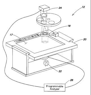
a chamber holding device.
[0026] FIG. 1 illustrates an example of a biologic sample imaging device
12 that can be
adapted for use with the present method, which device 12 includes a sample
illuminator 22, an
image dissector 24, and a programmable analyzer 26. The sample illuminator 22
includes a light
source that selectively produces light along one or more wavelengths that are
not appreciably
absorbed by either the sample 11 or by lenslets 10 disposed within a chamber
17 quiescently
holding the sample 11 to be tested. The imaging device 12 typically includes
optics for
manipulating the light (e.g., magnification, filtering, etc.). The sample
illuminator 22 produces
light along predetermined wavelengths (or along a spectrum of wavelengths
which is
6
CA 02719004 2012-08-17
subsequently limited to the predetermined wavelengths) that is transmitted
through the sample
11. The light transmittance intensity of the lenslets 10 and the sample 11 are
measured, for
example, by positioning a light source on one side of the chamber 17,
directing the light through
the chamber 17, and thereafter capturing the light using the image dissector
24. An example of
an acceptable image dissector 24 is a charge couple device (CCD) type image
sensor that
converts an image of the light passing through the sample 11 into an
electronic data format.
Complimentary metal oxide semiconductors ("CMOS") type image sensors are
another example
of an image sensor that can be used. The present invention is not limited to
either of these
examples. The programmable analyzer 26 includes a central processing unit
(CPU) and is
connected to the sample illuminator 22 and image dissector 24. The CPU is
adapted (e.g.,
programmed) to selectively perform the functions necessary to perform the
present method. It
should be noted that the functionality of programmable analyzer 26 may be
implemented using
hardware, software, firmware, or a combination thereof. A person skilled in
the art would be
able to program the processing unit to perform the functionality described
herein without undue
experimentation. U.S. Patent No. 6,866,823 entitled "Apparatus for Analyzing
Biologic Fluids"
issued March 15, 2005 discloses such an imaging device 12.
[0027] An analysis chamber 17 that may be used within the analytical device
is defined
by a first panel 28 and a second panel 30, spaced apart from one another. The
panels 28, 30 are
both sufficiently transparent to allow the transmission of light along
predetermined wavelengths
there through in an amount sufficient to perform the analysis on the sample
11, including the
focusing methodology described below. The present method can utilize a variety
of different
analysis chamber types having the aforesaid characteristics, and is not
therefore limited to any
particular type of analysis chamber 17.
[0028] An example of an acceptable chamber 17 is shown in FIGS. 2 and 3,
which
chamber 17 includes a first panel 28, a second panel 30, and at least three
separators 32 disposed
between the panels 28, 30. The separators 32 space the panels 28, 30 apart
from one another.
The dimension of a separator 32 that extends between the panels is referred to
herein as the
height 34 of the separator 32. The heights 34 of the separators 32 typically
do not equal one
another exactly (e.g., manufacturing tolerances), but are within commercially
acceptable
tolerance for spacing means used in similar analysis apparatus. Spherical
beads are an example
7
CA 02719004 2012-08-17
of an acceptable separator 32. This example of an acceptable analysis chamber
17 is described in
greater detail in U.S. Patent Application Publication Nos. 2007/0243117 and
2007/0087442.
[0029] In some embodiments, the separators 32 and the lenslets 10 are one
in the same;
i.e., the spherical beads have a size that is acceptable to separate the
panels of the chamber 17,
and have optical properties that make them acceptable as lenslets 10. It can
be advantageous in
some applications to have the spherical beads act as both separators 32 and as
lenslets 10.
Because the spheres act as a separator in those embodiments (e.g., in contact
with or close
proximity to the interior surfaces), it is unlikely that any appreciable
amount of sample 11
material will be disposed between the sphere and either interior surface of
the panels. The
absence of any appreciable sample 11 material decreases or eliminates the
possibility of sample
material interfering with any light transmittance analysis of the spheres.
There is no
requirement, however, that either the separators 32 or lenslets 10 function as
the other; e.g.,
separators 32 can be used independent of and along with lenslets 10.
[0030] Another example of an acceptable chamber 17 is disposed in a
disposable
container as shown in FIG 4. The chamber 17 is formed between a first panel
and a second
panel. Both the first panel and the second panel are transparent to allow
light to pass through the
chamber 17. This chamber 17 embodiment is described in greater detail in U.S.
Patent No.
6,723,290.
[0031] The analysis chambers 17 shown in FIGS. 2-4, represent chambers that
are
acceptable for use in the present method. In both instances, the chamber 17 is
typically sized to
hold about 0. 2 to 1.0 p.1 of sample 11, but the chamber 17 is not limited to
any particular volume
capacity, and the capacity can vary to suit the analysis application. These
chambers 17 are
operable to hold a sample 11 quiescently within the chamber 17. The term
"quiescent" is used to
describe that the sample 11 is deposited within the chamber 17 for analysis,
and is not
purposefully moved during the analysis. To the extent that motion is present
within the blood
sample, it will predominantly be that due to Brownian motion of the blood
sample's formed
constituents, which motion is not disabling of the use of the device of this
invention. The present
method is not, however, limited to these particular chamber 17 embodiments.
8
CA 02719004 2010-09-17
WO 2009/117678 PCT/US2009/037839
Example:
[0032] The following is an illustrative example of focusing an imaging
device 12
according to the present method. FIG.. 8 provides a block diagram according to
a method aspect
of the present invention. The invention is not, however, limited to this
example.
[0033] A sample 11 of biologic fluid (e.g., substantially undiluted
anticoagulated whole
blood), is deposited within a chamber 17. A number of lenslets 10 are
positioned relative to the
sample 11 in a manner such that they will remain at a fixed distance from the
point in the sample
11 where best focus is required for the duration of the analysis. The number
of lenslets 10 can
vary depending upon the application, but there should be enough such that
there are a sufficient
number of lenslets 10 in each field of view to accurately practice the method
(e.g., a number of
lenslets 10 great enough so that statistically acceptable average light
transmittance intensity
values can be calculated, etc.). The lenslets 10 may be dispersed randomly or
in a pattern. The
method is facilitated if the lenslets 10 are disposed within the focal plane
of the sample 11,
including being intimately disposed within the sample 11 itself, such as is
the case in chamber 17
described above and shown in FIGS. 2-5. The method does not require the
lenslets 10 being
disposed within the sample focal plane, however. The focal plane for the
lenslets 10 may be
offset from the focal plane of the sample 11; e.g., the focal planes of the
lenslets 10 and the
sample 11 may differ where each is illuminated using different wavelengths.
The two focal
planes may also be offset if the lenslets 10 are not physically coplanar with
the sample portion of
interest. In cases of focal plane offset, once the focal adjustment distance
is determined for the
lenslets 10, it is added to the amount of the focal plane offset between the
lenslets 10 and the
sample 11, which is known or is determinable, to arrive at the appropriate
focal position.
[0034] The lenslets 10 and the sample 11 are illuminated at one or more
wavelengths that
are not appreciably absorbed by either the lenslets 10 or the sample 11. As an
example, light
produced at wavelengths of about 620 nm or greater will not be appreciably
absorbed by a
sample 11 of substantially undiluted whole blood or by four micron (41.tm)
spherical lenslets 10
consisting of polystyrene. Light at other wavelengths can be used for
alternative analyses, and
the present invention is not limited to using any particular wavelength. An
image of the light
transmittance intensity is captured using a digital image dissector 24, such
as a CCD or CMOS
camera. The light transmittance intensity of the sample 11 is determined for
the imaged sample
11 on a per unit basis, and an average value light transmittance intensity
value for the sample 11
9
CA 02719004 2010-09-17
WO 2009/117678 PCT/US2009/037839
is determined. Although it is not a requirement that light transmittance
intensity values be
determined for the entire area of the sample 11, it is preferable since doing
so typically provides
a more complete analysis of the sample and a concomitant increase in accuracy.
The digital
image is analyzed using conventional image processing techniques to locate the
lenslets 10,
which appear as dark objects within the image. Depending upon where the focal
plane of the
imaging device 12 is initially located, the centers of the lenslets 10 may
appear light relative to
the other portions of the lenslets 10. FIG. 5 shows spherical lenslets 10 that
appear circular, with
lighter colored central regions.
[0035] To facilitate the image analysis, the image can be segmented and
only those
objects having a relative average light transmittance value below a
predetermined cutoff value
(e.g., 0.90) are then selected for analysis. FIG. 7 shows, for example, a
characteristic light
transmittance intensity pattern segmented to define different intensity
quadrants having different
regional characteristics (e.g., off-peak low, target range, off-peak high).
Alternative and/or
additional cutoff criteria can also be used to decrease the amount of analysis
required; e.g., area
corresponding to about that of a lenslet 10. These cutoff values can be chosen
through trial and
error to increase the accuracy and speed of the analysis. The selected objects
are then
collectively further analyzed to determine the light transmittance intensity
values at particular
points on the objects, which objects are the lenslets 10. As indicated above,
the light
transmittance intensity is determined on a per unit basis (e.g., per pixel
basis) within the image.
Consequently, the image of a lenslet 10 is represented by some number of
pixels depending upon
the size of the lenslet 10 and the magnification factor of the imaging device
12 (e.g., a spherical
lenslet 10 that is four microns (411m) in diameter, imaged using a
magnification of 0.5 microns
per pixel, will have a diameter represented by about eight (8) pixels). The
light transmittance
values at each point are averaged. The physical uniformity of the lenslets 10
and the averaging
of the intensity values increase the reliability of the intensity values.
[0036] The existing focal position of the imaging device 12 is
determinable using the
predetermined characteristic light transmittance pattern of the lenslets 10,
the average light
transmittance intensity values of at least one region within the lenslet 10
images, and the average
light transmittance intensity values of the sample 11.
[0037] The predetermined characteristic light transmittance intensity
pattern for a lenslet
is repeatable and consistent for a given type of lenslet 10. The pattern,
which is stored within
CA 02719004 2010-09-17
WO 2009/117678 PCT/US2009/037839
the imaging device 12, is a function of relative light transmittance intensity
values of the
different regions of the lenslet 10, relative to each other, and relative to
the average light
transmittance intensity of the sample 11. The pattern shown in FIGS. 6 and 7,
for example, is
graphical representative of a pattern for a four micron (4 m) diameter
lenslet. The vertical axis
of each graph is the average intensity of a region of interest within the
lenslet 10 relative to the
average light transmittance intensity of the sample 11. The horizontal axis of
each graph is the
relative focal position.
[0038] The characteristic light transmittance intensity pattern shown in
FIGS. 6 and 7
includes a first data line 36 depicting an average value for a lenslet region
having a high intensity
value, a second data line 38 depicting an average value for a lenslet region
having a low intensity
value, a third data line 40 depicting an average intensity value for all of
the regions of the lenslet
10, a fourth data line 42 depicting an average value for a region adjacent,
but outside the lenslet
10, and a fifth data line 44 depicting an average intensity value for the
center region of the
lenslet, all of which lines are depicted as a function of focal position. The
positions of these data
lines relative to one another are a repeatable, consistent characteristic of
the lenslet. Because
these data lines are created as a function of relative light transmittance
intensity (e.g., relative to
each other and relative to the average intensity of the sample 11), the
present method is
insensitive to the actual exposure or image light intensity, and is operable
so long as there is
sufficient signal available for analysis.
[0039] Of particular note is the light transmittance intensity values at
the center of the
lenslet, which vary cyclically from a maximum to a minimum; i.e., an "S" type
plot. The
maximum to minimum intensity values for the center region of the spherical
lenslet 10 occur
within a focal distance corresponding to two lenslet diameters; e.g., for a
four micron (41.tm)
diameter lenslet, the peak-to-valley intensity spread is about eight microns
(8 m). This curve is
highly reproducible, and the relative light transmittance intensity in the
middle of the curve is
used as a target value for an optimum focal position. The middle of the "S"
shaped curve for the
center region is used as a target value because it resides in a relatively
linear, constant slope
portion of the curve, which provides desirable focal position sensitivity. The
data collected for
the center regions of the lenslets 10 also has a higher degree of reliability
than other regions. In
the pattern shown in FIGS. 6 and 7, the middle of the "S" shaped curve for the
center region is
aligned with a value of about 0.75 on the Y-axis; i.e., a position where the
average light
11
CA 02719004 2010-09-17
WO 2009/117678 PCT/US2009/037839
transmittance intensity values of the center region is about three-quarters
(.75) of the average
light transmittance intensity value of the sample 11 in relative terms. The
focal position value
associated with the middle of the "S" curve (i.e., at the .75 intersection)
represents the target
focal position that has been determined to provide the desired focus of the
imaging device 12
relative to the lenslets 10.
[0040] The determination of the existing focal position of the imaging
device 12 is made
by locating on a curve within the characteristic pattern (e.g., the center
region curve, etc.) the
average intensity value of at least one region within the lenslets 10 relative
to the average
intensity value for the sample 11 (e.g. the y-axis value) and finding the
corresponding existing
focal position value (i.e., the x-axis value). The offset between the existing
focal position and
the target focal position (i.e., the position of optimum focus) represents the
adjustment necessary
to bring the imaging device 12 to the target focal position.
[0041] In certain portions of the lenslet characteristic pattern, the
curves can be
intersected at more than one point by a horizontal line extending from the y-
axis. In such cases,
more than one existing focal position may be associated with a relative
intensity value. To
determine the correct existing focal position, another data point from the
characteristic pattern is
determined. This data can be collected from the existing image or a subsequent
image. For
example, the average intensity value from a high value region on the lenslets
10 divided by the
average sample intensity value (e.g. the y-axis value) can be plotted on the
high value curve
within the pattern. Once the position on the high value curve is determined,
the associated
existing focal position value can be determined from the x-axis. The existing
focal position
value from the high curve will agree with one of the focal positions
determined off of the center
region curve, thereby confirming one of the focal positions as the correct
existing focal position.
The same process can be performed relative to another curve within the pattern
to provide
additional confirming data, if desired. Once the existing focal position is
confirmed, the offset
between the existing focal position and the target focal position can be
determined, which offset
represents the adjustment necessary to bring the imaging device 12 to the
target focal position.
[0042] It should be noted that the correct existing focal position (and
therefore the offset
to the optimum focus) can be determined using any of the curves plotted within
the characteristic
pattern. As stated above, however, the center region curve offers advantages
relative to
sensitivity and reliability. Consequently, the accuracy of the process is
enhanced by using the
12
CA 02719004 2012-08-17
center region curve for at least one of the data points used to establish the
existing focal position.
[0043] In some instances, the relative intensity value determined for the
characteristic
pattern may be aligned with curve portions (e.g., substantially horizontal
segments) that can be
associated with a plurality of focal positions on the y-axis. In those
instances, the above
methodology is performed and one of the potential existing focal positions is
chosen. The
methodology is then be repeated with a new image and adjustments made to the
focal position if
necessary.
[0044] In the event the original focal position of the imaging portion of
the analytical
device is so far off that intensity values determined from that position yield
data outside the
characteristic pattern for that lenslet, the imaging portion of the analysis
device can be first
brought into approximate focus by a conventional technique such as maximizing
contrast.
[0045] Although this invention has been shown and described with respect to
the detailed
embodiments thereof, it will be understood by those skilled in the art that
various changes in
form and detail may be made. For example, the characteristic light
transmittance
intensity pattern for the lenslets 10 is described above within the Detailed
Description portion in a graphical form. The pattern is not limited to a
graphical expression, and can take the form of mathematical expressions
describing the relative
light transmittance curves, or could be in the form of a data table, etc. As
another example, the
above detailed embodiments discuss the invention in terms of a biologic sample
11 being
disposed within an independent chamber 17. In alternative embodiments, the
imaging device 12
may incorporate sample handling hardware. As another example, the
characteristic light
transmittance patterns are described in terms of average light transmittance
values. In alternative
embodiments, useful information may be accessed using data from single
lenslets 10, or by
statistical information other than average values.
[0046] What is claimed is:
13