Note: Descriptions are shown in the official language in which they were submitted.
CA 02719247 2015-06-12
CA2719247
IKKi Inhibitor Therapies and Screening Methods, and Related IKKi Diagnostics
FIELD
The present disclosure relates to diagnostics, screening methods, and
treatment methods related
to obesity, insulin resistance, diabetes, weight loss, and related disorders.
In particular, the present
disclosure provides methods of treating such conditions with IKKi inhibitors,
methods of diagnosing
such conditions based on IKKi status, and methods of screening candidate IKKi
inhibitors.
BACKGROUND
Generally, obesity is defined as an excess of adipose tissue. Clinically, it
is generally defined as
that amount of adiposity that imparts a health risk. Even mild obesity, at 20%
over desirable weight
according to standard height-weight charts, may increase the risk for disease
and premature death. While
the etiology of obesity and diabetes is not entirely overlapping, it is now
amply clear that both share
appreciable biochemical and physiological components.
The incidence of the metabolic disorders of diabetes and obesity has reached
epidemic levels. It
has been estimated that over 120 million Americans are clinically over-weight
and more than ten million
Americans are diagnosed with diabetes every year. Moreover, obesity and
diabetes can cause or
contribute to the development of, or at least affect the treatment of, other
diseases and disorders such as
cardiovascular diseases, stroke, hypertension, and kidney failure. The
combined economic burden of
diabetes and obesity and the co-morbidities associated with these disorders is
estimated to be over $100
billion a year. Obesity and diabetes have a major impact on human health and
the various national
healthcare systems all over the world.
Recently launched weight-loss drugs have failed or have demonstrated limited
efficacy and
undesirable side effects. Similarly, despite a tremendous medical need, the
pharmaceutical industry has
realized only limited success developing therapeutics to manage diabetes. The
most common therapeutics
(sulfonylureas) are not effective and the most promising new drugs
(thiazolidinediones) have
demonstrated rare but fatal side effects. Thus, there is an urgent need for a
more comprehensive
understanding of the molecular basis of obesity and diabetes, for diagnosis
tests that allow early detection
of predispositions to the disorders, and for more effective pharmaceuticals
for preventing and treating the
diseases without undesirable side effects.
1
CA 02719247 2015-06-12
CA2719247
SUMMARY
The present disclosure provides diagnostics, screening methods, and treatment
methods
related to obesity, insulin resistance, diabetes, weight loss, and related
disorders. In particular, the
present disclosure provides methods of treating such conditions with IKKi
inhibitors, methods of
diagnosing such conditions based on IKKi status, and methods of screening
candidate IKKi
inhibitors.
Various embodiments disclosed herein provide a pharmaceutical composition
comprising an
IKKi-inhibiting agent and a pharmaceutically acceptable carrier for use in
treatment or prevention, as
described above.
Some embodiments disclosed herein provide methods of treatment comprising:
administering
an IKKi inhibitor to a subject with a condition associated with impaired
insulinreceptor signaling,
wherein the administering causes a reduction in one or more symptoms of the
condition.
In certain disclosed embodiments, the impaired insulin receptor signaling is
in the subject's
adipocyte cells or adipose tissue macrophage cells, or liver or muscle cells.
In particular
embodiments, the impaired insulin receptor signaling causes the subject to
have impaired glucose
metabolism. In further embodiments, the administering causes an increase in
glucose metabolism by
adipocytes and adipose tissue macrophages of the subject. In some embodiments,
the increase in
glucose metabolism is caused by increased insulin receptor signaling in
response to insulin. In
particular embodiments, the administering causes a reduction of body fat in
the subject (e.g., the size
of adipocytes in the subject are reduced). In certain embodiments, the
administration causes the
patient to lose at least 10 pounds (e.g., 10 ... 15 ... 20 ... 35 ... 60 ...
100 ... or 200 or more pounds). In
some embodiments, the administration causes at least a 5% reduction in the
patient's body weight
(e.g., at least 7% ... 10% ... 20% ... 30% ... 50% ... 75% reduction or more).
In some disclosed embodiments, the condition treated is obesity. In other
embodiments, the
condition treated is diabetes (e.g., type I, or type II, or both types I and
II). In further embodiments,
the condition treated is insulin resistance. In particular embodiments, the
subject does not have
diabetes type I or type II. In particular embodiments, the IKKi inhibitor
inhibits the insulin receptor
phosphorylation activity of IKKi. In further embodiments, the IKKi inhibitor
inhibits the insulin
receptor phosphorylation activity of IKKi at the serine in the human insulin
receptor sequence
VKTVNES (SEQ ID NO:15) or at the corresponding serine in the insulin receptor
sequences of
another species (e.g., mouse, cat, dog, rat, horse, cow, etc.) which can be
located, for example, by
performing a sequence alignment. In further embodiments, the IKKi inhibitor
inhibits the insulin
2
CA 02719247 2015-06-12
CA2719247
receptor phosphorylation activity of IKKi at the serine in the human insulin
receptor sequence
VKTVNES (SEQ ID NO:15), or related species, but does not inhibit one or more
other activities of
IKKi (e.g., phosphorylation of one or more of the II(13 proteins). In further
embodiments, the IKKi
inhibitor inhibits the phosphorylations of other proteins that directly or
indirectly contribute to
disregulation of glucose or lipid homeostasis.
In some embodiments, the IKKi inhibitor comprises a benzimidazol substituted
thiopene
derivative. In further embodiments, the IKKi inhibitor operates through RNA
interference. In
particular embodiments, the IKKi inhibitor is an siRNA or antisense
oligonucleotide. In particular
embodiments, the IKKi inhibitor is an anti-IKKi antibody (e.g., monoclonal
antibody or antibody
fragment). In certain embodiments, the IKKi inhibitor is an anti-IKKi antibody
specific for the TLR4
phosphorylated form of IKKi.
In particular embodiments, the present disclosure provides methods of reducing
body fat of a
subject comprising: administering an IKKi inhibitor to a subject under
conditions such that there is a
reduction in body fat of the subject. In further embodiments, the reduction in
body fat is a result of
increased glucose metabolism caused by the IKKi inhibitor. In some
embodiments, the reduction in
body fat is caused by increased insulin receptor signaling.
In certain embodiments, the present disclosure provides diagnostic methods
comprising: a)
measuring the IKKi protein or mRNA expression level in a sample from a
subject, and b) determining
if the subject has, or has an elevated risk for, a condition associated with
impaired insulin receptor
signaling, wherein an elevated IKKi protein or mRNA expression level indicates
that the subject has,
or is at elevated risk for, the condition.
In some embodiments, the sample comprises adipocytes, adipose tissue
macrophages, or
adipose tissue from the subject. In further embodiments, the measuring
comprises the use of an anti-
IKKi antibody or antibody fragment. In particular embodiments, the measuring
comprises the use of
an IKKi nucleic acid probe (e.g., at least a portion of SEQ ID NO:13). In
further embodiments, the
condition diagnosed is obesity or predisposition to obesity. In further
embodiments, the condition
diagnosed is diabetes type I or type II or both. In some embodiments, the
condition diagnosed is
insulin resistance.
In additional embodiments, the present disclosure provides diagnostic methods
comprising: a)
determining the level of insulin receptor phosphorylation in a sample from a
subject, and b)
determining if the subject has, or has an elevated risk for, a condition
associated with impaired insulin
receptor signaling, wherein an elevated level of insulin receptor
phosphorylation in the sample
3
CA 02719247 2015-06-12
CA2719247
indicates that the subject has, or is at elevated risk for, the condition.
In some embodiments, the level of insulin receptor phosphorylation is compared
to a
standard, wherein the standard is either known to be associated with the
condition or is from a healthy
individual without the condition. In particular embodiments, the sample
comprises adipocytes,
adipose tissue macrophages, or adipose tissue from the subject. In further
embodiments, the
condition diagnosed is obesity. In further embodiments, the condition
diagnosed is diabetes type I or
type II or both. In some embodiments, the condition diagnosed is insulin
resistance.
In certain embodiments, the present disclosure provides diagnostic methods
comprising: a)
determining the level of TRL4 mediated IKKi phosphorylation in a sample from a
subject, and b)
determining if the subject has, or has an elevated risk for, a condition
associated with impaired insulin
receptor signaling, wherein an elevated level of TRL4 mediated IKKi
phosphorylation of IKKi in the
sample indicates that the subject has, or is at elevated risk for, the
condition.
In particular embodiments, the present disclosure provides cell-free methods
of screening a
candidate agent comprising: a) combining IKKi (e.g., full protein or active
peptide fragments), an insulin
receptor, labeled phosphorous atoms, and a candidate IKKi inhibitor under
conditions such that the IKKi
can transfer the labeled phosphorous atoms onto the insulin receptor if not
inhibited by the candidate
IKKi inhibitor; and b) determining if the candidate IKKi inhibitor inhibits
the IKKi from phosphorylating
the insulin receptor.
In certain embodiments, the determining comprises evaluating whether the
candidate IKKi
inhibitor inhibits the IKKi from phosphorylating the serine in the insulin
receptor in the sequence
VKTVNES (SEQ ID NO:15) or at the serine in related non-human insulin
sequences. In further
embodiments, the methods further comprises step c) administering the candidate
IKKi inhibitor to an
animal and determining if the IKKi inhibitor promotes glucose metabolism in
the animal. In some
embodiments, the animal is a model for obesity, diabetes, or insulin
resistance. In other embodiments,
the determining comprising weighing the animal before and after treatment.
In some embodiments, the IKKi inhibitor comprises a benzimidazol substituted
thiopene
derivative. In further embodiments, the IKKi inhibitor operates through RNA
interference. In other
embodiments, the IKKi inhibitor is an siRNA or antisense oligonucleotide. In
particular embodiments,
the IKKi inhibitor is an anti-IKKi antibody (e.g., monoclonal antibody or
antibody fragment). In certain
embodiments, the IKKi inhibitor is an anti-IKKi antibody specific for the TLR4
phosphorylated form of
IKKi.
In particular embodiments, the present disclosure provides methods of
screening a candidate
4
CA 02719247 2015-06-12
CA2719247
agent comprising: a) contacting a cell (e.g., adipoctye, macrophage, or 3T3-L1
fibroblast or other
adipocyte cell culture line) with a candidate IKKi inhibitor, wherein the cell
comprises insulin receptors;
and b) determining if the candidate IKKi inhibitor prevents, or reduces the
level of, phosphorylation of
the insulin receptors in the cell. In some embodiments, the methods further
comprise a step of lysing the
cell to generate a cell lysate prior to the determining step. In further
embodiments, the determining
comprises examining the level of phosphorylation at the serine in the insulin
receptor sequence
VKTVNES (SEQ ID NO:15). In certain embodiments, the cell is an adipocyte or
adipose tissue
macrophage.
In some embodiments, the determining comprises the use of an antibody, or
antibody fragment,
that recognizes the phosphorylated form, or the un-phosphorylated forms, of
the insulin receptors. In
further embodiments, the determining comprises the use of an antibody, or
antibody fragment, that
recognizes the form of the insulin receptor that is phosphorylated at the
serine in the insulin receptor
sequence VKTVNES (SEQ ID NO:15) or at corresponding serine in non-human
insulin receptor
sequences. In other embodiments, the determining comprises the use of an
antibody or antibody
fragment that recognizes the form of the insulin receptors that is not
phosphorylated at the serine in the
insulin receptor sequence VKTVNES (SEQ ID NO:15) or at the corresponding
serine in non-human
insulin receptor sequences.
In some embodiments, the methods further comprise step c) administering the
candidate IKKi
inhibitor to an animal and determining if the IKKi inhibitor promotes glucose
metabolism in the animal.
In particular embodiments, the animal is a model for obesity, diabetes, or
insulin resistance. In additional
embodiments, the determining comprising weighing the animal before and after
treatment.
In certain embodiments, the candidate IKKi inhibitor comprises a benzimidazol
substituted
thiopene derivative. In further embodiments, the candidate IKKi inhibitor
operates through RNA
interference. In other embodiments, the IKKi inhibitor is an siRNA or
antisense oligonucleotide. In
additional embodiments, the candidate IKKi inhibitor is an anti-IKKi antibody.
In some embodiments,
the cell is treated with an IKKi inducer prior to the contacting step. In
further embodiments, the IKKi
inducer is selected from the group consisting of: tumor necrosis factor (TNF),
lipopolysaccharide (LPS),
interleukin-1 (IL-1), interleukin-6 (IL-6), interferon-gamma, and phorbol
myristate.
In some embodiments, the present disclosure provides methods of screening a
candidate agent
comprising: a) contacting a cell with a candidate IKKi inhibitor, wherein the
cell is an adipocyte or
adipose tissue macrophage, and wherein the cell comprises activated IKKi
proteins; and b) determining if
the IKKi inhibitor promotes glucose metabolism in the cell. In particular
embodiments, prior to the
5
CA 02719247 2015-06-12
CA2719247
contacting step, the cell is first contacted with an IKKi inducer. In other
embodiments, the IKKi inducer
is selected from the group consisting of: LPS, IL-1, IL-6, interferon-gamma,
and phorbol myristate.
In some embodiments, the determining if the IKKi inhibitor promotes glucose
metabolism in the
cell comprises measuring the uptake of glucose by the cell. In certain
embodiments, the determining if
the IKKi inhibitor promotes glucose metabolism in the cell comprises measuring
the state of
phosphorylation of insulin receptors in the cell. In further embodiments, the
determining if the IKKi
inhibitor promotes glucose metabolism in the cells comprises measuring the
state of phosphorylation of a
protein selected from the group consisting of: Aps, Cbl, and TC10.
In additional embodiments, the determining if the IKKi inhibitor promotes
glucose metabolism in
the cell comprises measuring the ability of GLUT4 to transport glucose. In
further embodiments, the
determining if the IKKi inhibitor promotes glucose metabolism in the cells
comprises measuring the size
of the cell compared to a control cell. In certain embodiments, the methods
further comprise step c)
administering the candidate IKKi inhibitor to an animal and determining if the
IKKi inhibitor promotes
glucose metabolism in the animal. In some embodiments, the animal is a model
for obesity, diabetes, or
insulin resistance. In particular embodiments, the determining comprising
weighing the animal before
and after treatment.
In some embodiments, the present disclosure provides a method of treating
conditions associated
with impaired insulin, comprising: providing a subject experiencing or at risk
for impaired insulin
signaling and administering to the subject a therapeutically effective dose of
an IKKi-inhibiting agent,
wherein the administration results in improved insulin signaling in the
subject. In some embodiments,
the impaired insulin signaling occurs in such as adipocyte cells, adipose
tissue macrophage cells, adipose
tissue, liver cells, and liver tissue. In some embodiments, the subject is
experiencing or is at risk of
experiencing a condition such as obesity, diabetes, and insulin resistance. In
some embodiments, the
administering of an IKKi-inhibiting agent results in an outcome of increased
glucose metabolism,
reduction in body fat, lack of increase in body fat, increased insulin
receptor signaling, decreased level of
insulin receptor phosphorylation, reduction in or prevention of chronic
inflammation in liver, reduction in
or prevention of chronic inflammation in adipose tissue, reduction in or
prevention of hepatic steatosis,
promotion of metabolic energy expenditure, reduction in circulating free fatty
acids, and/or reduction in
cholesterol. In some embodiments, the decreased level of insulin receptor
phosphorylation occurs at the
serine residue of insulin receptor sequence VKTVNES (SEQ ID NO: 15). In some
embodiments, the
IKKi-mediated phosphorylation of 'KB in the subject is unaffected by the IKKi-
inhibiting agent. In some
embodiments, the IKKi inhibitor comprises an agent such as a benzimidazol-
substituted thiopene
6
CA 02719247 2015-06-12
CA2719247
derivative, an siRNA, an antisense oligonucleotide, a non-phospho-specific
anti-IKKi antibody, and a
phospho-specific anti-IKKi antibody.
In some embodiments, the present disclosure provides a method of reducing body
fat or
preventing increase in body fat in a subject, comprising: providing a subject
experiencing or at risk
of overweight or obese body composition, and administering to the subject a
therapeutically effective
dose of an IKKi-inhibiting agent, wherein the administration results in
reduction of or prevention of
increase in body fat in the subject. In some embodiments, the subject is
experiencing or is at risk of
experiencing a condition such as diabetes and insulin resistance. In some
embodiments, the
administering of an IKKi-inhibiting agent results in an outcome such as
increased glucose
metabolism, increased insulin receptor signaling, decreased level of insulin
receptor phosphorylation,
reduction in or prevention of chronic inflammation in liver, reduction in or
prevention of chronic
inflammation in adipose tissue, reduction in or prevention of hepatic
steatosis, promotion of
metabolic energy expenditure, reduction in circulating free fatty acids,
and/or reduction in
cholesterol. In some embodiments, the decreased level of insulin receptor
phosphorylation occurs at
the Ser of insulin receptor sequence VKTVNES (SEQ ID NO: 15). In some
embodiments, the IKKi-
mediated phosphorylation of IkB in the subject is unaffected by the IKKi-
inhibiting agent. In some
embodiments, the IKKi inhibitor comprises an agent such as a benzimidazol-
substituted thiopene
derivative, an siRNA, an antisense oligonucleotide, a non-phospho-specific
anti-IKKi antibody, and a
phosphor-specific anti-IKKi antibody.
In some embodiments, the present disclosure provides a diagnostic method,
comprising:
providing a sample from a subject, and measuring the level in the sample of a
molecule such as IKKi
protein, IKKi transcript, phosphorylated insulin receptor, and phosphorylated
IKKi wherein the IKKi
phosphorylation is mediated by TLR4, and determining if the subject has or has
an elevated risk for a
condition associated with impaired insulin receptor signaling, wherein an
elevated level of said
molecule indicates that said subject has, or is at elevated risk for, a
condition associated with
impaired insulin receptor signaling. In some embodiments, the sample comprises
adipocytes, adipose
tissue macrophages, adipose tissue, liver cells, or liver tissue. In some
embodiments, the measuring
comprises the use of an agent specific to the molecule. This agent may include
a nucleic acid probe, a
non-phospho-specific antibody, and/or a phospho-specific antibody. In some
embodiments, the level
of the molecule is compared to a standard, wherein the standard is either
known to be associated with
the condition or is from a healthy individual without the condition or from
the subject at a prior time
period. In some embodiments, the condition is obesity, diabetes, and/or
insulin resistance.
7
CA 02719247 2015-06-12
CA2719247
In some embodiments the present disclosure provides a method of identifying an
IKKi-
inhibiting agent, comprising: combining a polypeptide comprising IKKi, a
polypeptide comprising an
insulin receptor, labeled phosphorous atoms, and a candidate IKKi inhibitor
under conditions
sufficient to promote phosphorylation of said insulin receptor by the IKKi
polypeptide in absence of
the candidate inhibitor, and determining the activity of the IKKi polypeptide
with regard to
phosphorylation of the insulin receptor. In some embodiments, the decreased
level of insulin receptor
phosphorylation occurs at the serine residue of insulin receptor sequence
VKTVNES (SEQ ID NO:
15). Some embodiments further comprise a step of administering the candidate
IKKi inhibitor to an
animal and determining whether the candidate IKKi inhibitor promotes glucose
metabolism in the
animal.
In some embodiments, the present disclosure provides a method of identifying
an IKKi-
inhibiting agent, comprising: providing a cell or cell lysate comprising
insulin receptors, contacting
the cell with a candidate IKKi inhibitor, and determining whether the
candidate IKKi inhibitor
affected a property such as the rate of glucose metabolism and/or the level of
phosphorylation of said
insulin receptors. In some embodiments, the determination of whether the IKKi
inhibitor affects the
rate of glucose metabolism comprises measuring a feature such as uptake of
glucose by the cell, the
phosphorylation state of insulin receptors, the phosphorylation state of APS,
the phosphorylation state
of Cbl, the phosphorylation state of TC10, the ability of GLUT4 to transport
glucose, the
translocation of GLUT4 to the plasma membrane, and/or the size of the cell
relative to a control cell.
In some embodiments, the IKKi inhibitor comprises an agent such as a
benzimidazol-substituted
thiopene derivative, an siRNA, an antisense oligonucleotide, a non-phospho-
specific anti-IKKi
antibody, and a phospho-specific anti-IKKi antibody. In some embodiments, the
cell is treated with
an IKKi-inducing agent prior to the contacting step. Some embodiments further
comprise the step of
administering the candidate IKKi inhibitor to an animal and determining
whether the candidate IKKi
inhibitor promotes glucose metabolism in the animal.
Conditions and disease states which may be treated by methods and compositions
disclosed
herein include but are not limited to diabetes mellitus, type I diabetes, type
II diabetes, gestational
diabetes, metabolic syndrome, metabolic syndrome X, syndrome X, insulin
resistance syndrome,
Reaven's syndrome, CHAOS, and malnutrition-related diabetes mellitus.
Lipid metabolic conditions and disease states which may be treated using
methods and
compositions disclosed herein include but are not limited to lipodystrophy,
congenital generalized
lipodystrophy (Beradinelli-Seip syndrome), familial partial lipodystrophy,
acquired partial
8
CA 02719247 2015-06-12
CA2719247
lipodystrophy (Barraquer-Simons syndrome), acquired generalized lipodystrophy,
centrifugal
abdominal lipodystrophy (Lipodystrophia centrifugalis abdominalis infantilis),
lipoatrophia annularis
(Ferreira-Marques lipoatrophia), localized lipodystrophy, HIV-associated
lipodystrophy,
hypercholesterolemia, hyperlipidemia, obesity, hypertriglyceridemia. Lipid
metabolic conditions
may occur in concert with or in absence of conditions such as vascular
disease, hypertension,
atherosclerosis, arteriosclerosis, peripheral vascular disease (PVD),
peripheral arterial disease ( also
known as peripheral artery disease or PAD), claudication, intermittent
claudication, vascular diseases,
peripheral arterial occlusive disease (PAOD), coronary artery disease (CAD),
cardiovascular disease,
obesity, metabolic syndrome, and critical limb ischemia.
Methods and treatments disclosed herein find use in the treatment of high
total cholesterol
(hypercholesterolemia). Primary causes of hypercholesterolemia include but are
not limited to high-
fat diet, smoking or tobacco use, hypothyroidism, renal disease, liver
disease, use of progestins, use
of anabolic steroids, and use of glucocorticoids. Hypercholesterolemia may be
polygenic or familial.
Known familial hypercholesterolemia diseases include but are not limited to
familial ligand defective
apoB-100 (FLDB) and autosomal recessive hypercholesterolemia.
Methods and compositions disclosed herein find use in the treatment of hepatic
steatosis
disease, also referred to as fatty liver disease. Fatty liver disease can
range from fatty liver alone
(steatosis) to fatty liver associated with inflammation (steatohepatitis).
This condition can occur with
the use of alcohol (alcohol-related fatty liver) or in the absence of alcohol
(nonalcoholic fatty liver
disease [NAFLD]). Other factors that may lead to fatty liver disease include
but are not limited to
drugs (eg, amiodarone, tamoxifen, methotrexate), alcohol, metabolic
abnormalities (eg, galactosemia,
glycogen storage diseases, homocystinuria, tyrosemia), nutritional status
(e.g., overnutrition, severe
malnutrition, total parenteral nutrition [TPN], starvation diet), or other
health problems (eg, celiac
sprue, Wilson disease). Individuals genetically predisposed to fatty liver
disease may exhibit normal
or underweight body composition.
Subject matter disclosed herein finds use in the treatment or prevention of
overweight and
obesity. The most widely accepted clinical definition of obesity is the World
Health Organization
(WHO) criteria based on BMI. Under this convention for adults, grade 1
overweight (commonly and
simply called overweight) is a BMI of 25-29.9 kg/m2. Grade 2 overweight
(commonly called obesity)
is a BMI of 30-39.9 kg/m2. Grade 3 overweight (commonly called severe or
morbid obesity) is a BMI
greater than or equal to 40 kg/m2. The surgical literature often uses a
different classification to
recognize particularly severe obesity. In this setting, a BMI greater than 40
kg/m2 is described as
9
CA 02719247 2015-06-12
CA2719247
severe obesity, a BMI of 40-50 kg/m2 is termed morbid obesity, and a BMI
greater than 50 kg/m2 is
termed super obese. The definition of obesity in children involves BMIs
greater than the 85th
(commonly used to define overweight) or the 95th (commonly used to define
obesity) percentile,
respectively, for age-matched and sex-matched control subjects. Secondary
causes of obesity include
but are not limited to hypothyroidism, Cushing syndrome, insulinoma,
hypothalamic obesity,
polycystic ovarian syndrome, genetic syndromes (eg, Prader-Willi syndrome,
Alstrom syndrome,
Bardet-Biedl syndrome, Cohen syndrome, Borjeson-Forssman-Lehmann syndrome,
Frohlich
syndrome), growth hormone deficiency, oral contraceptive use, medication-
induced obesity (e.g.,
phenothiazines, sodium valproate, carbamazepine, tricyclic antidepressants,
lithium, glucocorticoids,
megestrol acetate, thiazolidine diones, sulphonylureas, insulin, adrenergic
antagonists, serotonin
antagonists [especially cyproheptadine]), eating disorders (especially binge-
eating disorder, bulimia
nervosa, night-eating disorder), hypogonadism, pseudohypoparathyroidism, and
obesity related to
tube feeding.
In some embodiments, methods and compositions disclosed herein are used to
treat subjects
having one or more of the above diseases or conditions, but lacking at least
one of the following
diseases or conditions: asthma, bronchitis, lung inflammation, osteoarthritis,
juvenile arthritis,
rheumatoid arthritis, spondylo arthopathies, gouty arthritis, chronic
granulomatous diseases such as
tuberculosis, leprosy, sarcoidosis, and silicosis, nephritis, amyloidosis,
ankylosing spondylitis,
chronic bronchitis, scleroderma, systemic lupus erythematosus, polymyositis,
appendicitis,
inflammatory bowel disease, Crohn's disease, gastritis, irritable bowel
syndrome, ulcerative colitis,
colorectal cancer, Sjorgen's syndrome, Reiter's syndrome, psoriasis, pelvic
inflammatory disease,
orbital inflammatory disease, thrombotic disease, menstrual cramps,
tendinitis, bursitis, psoriasis,
eczema, bums, dermatitis and inappropriate allergic responses to environmental
stimuli such as
poison ivy, pollen, insect stings and certain foods, including atopic
dermatitis and contact dermatitis,
migraine headaches, periarteritis nodosa, thyroiditis, aplastic anemia,
Hodgkin's disease, sclerodoma,
rheumatic fever, myasthenia gravis, sarcoidosis, nephrotic syndrome, Behcet's
syndrome,
polymyositis, gingivitis, hypersensitivity, conjunctivitis, swelling occurring
after injury,
lipopolysaccharide-induced septic shock, tissue regeneration,
neurodegenerative disease (e.g.,
Alzheimer's Disease), tissue rejection, osteoporosis, cachexia, and
neurodegeneration. In some
embodiments, methods and compositions disclosed herein are used to treat
subjects not in need of
tissue regeneration. In some embodiments, methods and compositions disclosed
herein are used to
treat subjects lacking cell proliferative disorders such as, for instance,
benign prostate hyperplasia,
CA 02719247 2015-06-12
= CA2719247
familial adenomatosis, polyposis, neuro-fibromatosis, psoriasis, pulmonary
fibrosis, and arthritis
glomerulonephritis.
In some embodiments, methods and compositions disclosed herein are used to
treat subjects
lacking at least one of the following cancers: carcinoma such as bladder,
breast, colon, kidney, liver,
lung, including small cell lung cancer, esophagus, gall-bladder, ovary,
pancreas, stomach, cervix, thyroid,
prostate, and skin, including squamous cell carcinoma; hematopoietic tumors of
lymphoid lineage,
including leukemia, acute lymphocytic leukemia, acute lymphoblastic leukemia,
B-cell lymphoma, T-
cell-lymphoma, Hodgkin's lymphoma, non-Hodgkin's lymphoma, hairy cell lymphoma
and Burkett's
lymphoma ; hematopoietic tumors of myeloid lineage, including acute and
chronic myclogenous
leukemias, myelodysplastic syndrome and promyelocytic leukemia; tumors of
mesenchymal origin,
including fibrosarcoma and rhabdomyosarcoma ; tumors of the central and
peripheral nervous system,
including astrocytoma, neuroblastoma, glioma and schwannomas; other tumors,
including melanoma,
seminoma, teratocarcinoma, osteosarcoma, xeroderma pigmentosum,
keratoxanthoma, thyroid follicular
cancer and Kaposi's sarcoma.
In some embodiments, methods disclosed herein comprise testing a subject for a
disease or
condition such as impaired insulin signaling, obesity, diabetes, insulin
resistance, high cholesterol,
metabolic syndrome, hepatic stenosis, chronic inflammation in liver, and
chronic inflammation in
adipose tissue, followed by administering an IKK i-inhibiting agent. In some
embodiments, methods
disclosed herein comprise administering to a subject an IKKi-inhibiting agent,
followed by testing the
subject for a disease or a condition such as impaired insulin signaling,
obesity, diabetes, insulin
resistance, high cholesterol, metabolic syndrome, hepatic stenosis, chronic
inflammation in liver, and
chronic inflammation in adipose tissue. In some embodiments, methods disclosed
herein comprise
testing a subject for a disease or condition such as impaired insulin
signaling, obesity, diabetes, insulin
resistance, high cholesterol, metabolic syndrome, hepatic stenosis, chronic
inflammation in liver, and
chronic inflammation in adipose tissue, followed by administering an IKKi-
inhibiting agent, followed by
a second round of testing for a disease or condition such as impaired insulin
signaling, obesity, diabetes,
insulin resistance, high cholesterol, metabolic syndrome, hepatic stenosis,
chronic inflammation in liver,
and chronic inflammation in adipose tissue (e.g., to monitor the effect of the
treatment). In some
embodiments, methods disclosed herein comprise testing a subject for a disease
or condition such as
impaired insulin signaling, obesity, diabetes, insulin resistance, high
cholesterol, metabolic syndrome,
hepatic stenosis, chronic inflammation in liver, and chronic inflammation in
adipose tissue, followed by
administering an IKKi-inhibiting agent, followed by a second round of testing
for a disease or condition
11
CA 02719247 2015-06-12
CA2719247
such as impaired insulin signaling, obesity, diabetes, insulin resistance,
high cholesterol, metabolic
syndrome, hepatic stenosis, chronic inflammation in liver, and chronic
inflammation in adipose tissue,
and a second administration of IKKi-inhibiting agent, with this second
administration being modified in
dose, duration, frequency, or administration route in a manner dependent upon
the results of the prior
testing.
Also disclosed herein is use of an IKKi-inhibiting agent in the manufacture of
a medicament for
the treatment of a condition such as impaired insulin signaling, obesity,
diabetes, insulin resistance, high
cholesterol, metabolic syndrome, hepatic stenosis, chronic inflammation in
liver, and chronic
inflammation in adipose tissue.
The claimed invention relates to use of an IKKi-inhibiting agent to antagonize
IKKi mediated
inhibition of insulin signalling in a cell. Such a cell may be a cell of
adipose tissue or of liver tissue.
Such a cell may be in a subject. Thus, such use may be for treatment of
impaired insulin signalling in
a subject or in preparation of a medicament for such a treatment. Such use may
be for reducing body
fat or preventing increase in body fat in a subject experiencing or at risk of
becoming overweight or
obese or in preparation of medicament for such purposes. The claimed invention
also relates to
pharmaceutical compositions comprising an IKK i-inhibiting agent and a
pharmaceutically acceptable
carrier intended for such a use.
BRIEF DESCRIPTION OF THE DRAWINGS
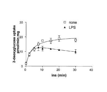
Figure 1 shows that LPS blocks insulin-stimulated glucose uptake in 3T3L1
adipocytes.
Adipocytes were pretreated with or without LPS, and then treated with insulin
and 2-deoxyglucose
uptake was assessed at various times as indicated in the Figure.
Figure 2 shows that LPS blocks insulin-stimulated translocation of the glucose
transporter
Glut4. Cells were transfected with a Glut4-eGFP/Myc reporter gene, to monitor
12
CA 02719247 2010-09-22
WO 2009/120801
PCT/US2009/038287
the translocation of the protein to the cell surface in response to insulin.
Preincubation with
LPS attenuated insulin action.
Figure 3 shows the role of IKK isoforms in LPS action. Figure 3A shows LPS
increases the phosphorylation of IKK isoforms a, 13, and i. Figure 3B shows
knockdown of
IKK isoforms by siRNA does not affect activation of Akt by insulin.
Figure 4 shows inhibition of insulin-stimulated glucose uptake by LPS is
prevented
by knockdown of Ikki. 3T3L1 adiopocytes were transfected with the indicated
siRNA oligos,
and the effect of LPS on insulin-stimulated glucose transport was assessed.
Figure 5 shows overexpression of dominant negative Ikki blocks the inhibitory
effects of LPS. 3T3L1 adipocytes were transfected with wt or kinase-inactive
Ikki and the
inhibitory effects of LPS were assessed on insulin-stimulated glucose uptake.
Figure 6a shows the IKKi inhibitor 5-(5,6-Dimethoxy-1H-benzimidazol-1-y1)-34[2-
(methylsulfonyl)phenyl]methoxy]-2-thiophenecarbonitrile. Figure 6b shows that
an
inhibitor of IKKi prevents LPS decreases in insulin stimulated glucose uptake.
In particular,
3T3L1 adipocytes were treated with 50 nM of 5-(5,6-Dimethoxy-1H-benzimidazol-1-
y1)-3-
[[2-(methylsulfonyl)phenyl]methoxy]-2-thiophenecarbonitrile prior to treatment
with LPS
and insulin.
Figure 7 shows that treatment of adipocytes with LPS had no effect on insulin-
stimulated tyrosine phosphorylation of the insulin receptor (InsR) or IRS-1
(Figure 7A), as
detected by anti-phosphotyrosine immunoblotting, nor was there a reduction in
the amount of
PI-3' kinase that co-immunoprecipitated with IRS-1 after insulin stimulation
(Figure 7B).
Figure 8 shows a Western blot from Example 1 that shows activation of the
protein
kinase Akt by insulin was not affected by LPS pre-treatment of cells.
Figure 9 shows that LPS attenuate the insulin-stimulated tyrosine
phosporylation of
the adapter proteins APS and Cbl. 3T3L1 adipocytes were pretreated with LPS
prior to
treatment with insulin. Cells were lysed and APS and C-Cbl tyrosine
phosphorylation was
assessed by Western blotting as shown in this Figure.
Figure 10 shows a Western blot from Example 1 that shows that LPS treatment
reduces the activation of TC10 by insulin.
Figure 11 shows that LPS stimulates the phosphorylation of the insulin
receptor via
activation of IKKi. Figure 11 a: 3T3L1 cells were incubated with 32P-
orthophosphate, and
stimulated with or without LPS. InsR and APS were immunoprecipitated and
subject to
autoradiography. Figure 1 lb: cells were subject to knock down with the
indicated Ikki
siRNA oligos prior to 32P labelling and immunoprecipitation.
13
CA 02719247 2010-09-22
WO 2009/120801
PCT/US2009/038287
Figure 12 shows contemplated phosphorylation sites on the insulin receptor by
IKKi,
based on known IKKi consensus sites. The following sequences are shown in this
Figure:
InsR: VKTVNES1 35AS (SEQ ID NO:9); p65: VFTDLAS468VD (SEQ ID NO:7) and STAT1:
IKTELIS711VS (SEQ ID NO:8). It is noted that the phosphorylated serine at
position 1035
in the insulin receptor, is position 1062 in the an alternatively spliced
version of the insulin
receptor (which is shown at Figure 26).
Figure 13 shows overexpression of kinsase-inactive IKKi increases the binding
of the
insulin receptor to APS. COS cells were transfected with wildtype or kinase-
inactive IKKi.
Cells were stimulated with or without insulin, lysed and lysates pulled down
with GST-APS
5H2 domain.
Figure 14 shows mutation of Seri 35 in the insulin receptor blocks the
inhibitory
effect of LPS on insulin-stimulated glucose uptake. 3T3L1 adipocytes were
subject to
siRNA knockdown of the insulin receptor, followed by transfection with a
vector containing
wildtype or seri 35 Ala mutant receptor. Cells were pretreated with LPS
followed by insulin,
and A) insulin receptor phosphorylation by 32P incorporation or B) glucose
transport was
assayed.
Figure 15 shows IKKi is upregulated in adipose tissue after high fat feeding
of mice.
Mice were fed a normal chow or high fight diet for 8 weeks, and adipose tissue
was excised
and subject to differential centrifugation to separate adipocytes and a
stromal vascular
fraction that contains adipose tissue macrophages (ATM). Cells were lysed and
subject to
western blotting with anti-IKKi antibodies.
Figure 16 shows that genetic ablation of Ikki prevents weight gain on a high
fat dies.
IKKiKo mice are shown on the left, while wildtype mice are on the right.
Figure 17 shows that IKKi knockout mice (IKKiK0) gained significantly less
weight
than did their wildtype littermates on high fat and normal chow diets, with
quantitation of this
data indicating that this reduction in weight gain was statistically
significant, with a P<0.01.
Figure 18 shows that genetic ablation of IKKi prevents adipose tissue
expansion after
high fat feeding. Epididymal fat pads were excised from wildtype (left) or
IKKiK0 (right)
mice fed a high fat diet for 8 weeks.
Figure 19 shows that the reduction in weight in the IKKiK0 mice was due to
small
fat cells.
Figure 20 shows that genetic ablation of IKKi increases respiration in mice.
Figure 21 shows that genetic ablation of IKKi increases respiration in mice.
Figure 22 shows that IKKiKo mice remain glucose tolerant on a high fat diet.
14
CA 02719247 2010-09-22
WO 2009/120801
PCT/US2009/038287
Figure 23 shows insulin tolerance in IKKi knock out mice after 3 months on a
high
fat diet.
Figure 24A shows the consensus sequence for the murine IKKi amino acid
sequence
(SEQ ID NO:10), and Figure 24B shows the consensus sequence for the murine
Ikki nucleic
acid sequence.
Figure 25A shows the consensus sequence for the human IKKi amino acid sequence
(SEQ ID NO:10), and Figure 25B shows the consensus sequence for the human Ikki
nucleic
acid sequence.
Figure 26 shows the consensus amino acid sequence of one of the two
alternative
splicing forms of human Insulin Receptor (SEQ ID NO:14). Underlined in this
Figure is
VKTVNES (SEQ ID NO:15). The serine in this sequence (shown in bold) is the
site of
phosphorylation of the insulin receptor by IKKi.
Figure 27 shows that IKKi enzyme activity is markedly elevated in adipose
tissue
derived from high fat fed mice compared to control mice.
Figure 28 shows that high-fat diet (HFD) increases NFKB activity in adipose
tissue as
measured by in vivo bioluminescence in live mice. A, Male HLL mice on normal
diet (ND)
and HFD were assessed for bioluminescence after injection of luciferin.
Quantitation of
luminescence collected over the abdominal cavity is presented for ND and HFD
HLL mice.
(n=7 per group). *p-value<0.05. B, Tissues from HLL mice were dissected and
assessed ex
vivo for luminescence. Quantitation of absolute tissue luminescence from HLL
mice. n=7
mice per group. Data was collected serially after dissection to ensure plateau
of luminescent
signal.
Figure 29 shows induction of NFKB expression in adipose tissue macrophage
(ATM)
clusters in obese mice. Epididymal fat pads from ND or HFD fed male C57B1/6
mice were
analyzed for p65/Re1A expression by immunofluorescence showing maximal signal
in ATM
clusters and localization of p65 in ATM nuclei (TOPRO3 co-stain).
Figure 30 shows that high fat diet increases IKKi expression in white adipose
tissue
and liver. A, Quantitative qPCR analysis on the expression of genes encoding
IKK family
members in liver and white adipose tissue. White bars, wild-type mice, normal
diet (ND)
(n=6); gray bar, wild-type mice, high fat diet (HFD) for 4 months (n=6). All
data are
presented as the average SEM normalized to Rp1p0 expression. Average of ND
value was
set as 1. B, Quantitative qPCR analysis on the expression of genes encoding
IKK family
CA 02719247 2010-09-22
WO 2009/120801
PCT/US2009/038287
members in isolated adipocytes and stromal vascular fraction. White bars, ND
(n=6); gray
bar, HFD for 4 months (n=6).
Figure 31 shows additional analysis of high fat diet-induced increases IKKi
expression in white adipose tissue and liver. A, Lysates from liver and white
adipose tissue
(WAT) of wild type (WT) and IKKi knockout mice (IKKi KO) fed with ND or HFD
were
immunoprecipitated with antibody against IKKi as indicated. The expression
level of IKKi
was determined by immunoblotting with same antibody against IKKi. B, Lysates
from liver
and WAT of WT and IKKi KO fed with ND or HFD were immunoprecipitated (IP) with
antibody against IKKi and assayed for kinase activity against myelin basic
protein (MBP) as
substrate. The expression level of IKKi in IP was determined by immunoblotting
with same
antibody against IKKi. Lysates for IP were immunoblotted with antibodies
against Rab5B
and Caveolin 1 as a loading control.
Figure 32 shows that IKKi KO mice are protected from diet-induced weight gain
by
increasing energy expenditure. A, Representative confocal image of caveolin-
stained
epididymal adipose tissue from WT and KO mice fed with HFD. Bar=100 m. B,
Whisker
plot of adipocyte area from evaluation of <500 adipocytes from 3-4 independent
mice.
*p<0.0001 comparing mean adipocyte area.
Figure 33 shows additional analyses indicating that IKKi KO mice are protected
from
diet-induced weight gain by increasing energy expenditure. A, Adipocyte
numbers in fat
pads of WT (gray bar) and IKKi KO mice (black bar) fed with HFD. n=5 mice per
genotype.
*, p-value<0.005. B, (Left) adiponectin levels in serum from WT (gray bar) and
KO (black
bar) mice fed with ND or HFD as indicated. (Right) serum adiponectin
normalized with
body weight. n=12 mice per group. *, p-value<0.05; **, p-value<0.01. C, Leptin
levels in
serum from WT (gray bar) and KO (black bar) mice fed with ND or HFD as
indicated. n=12
mice per group. *, p-value<0.05. D, Food intake was measured for WT (gray bar)
and KO
(black bar) mice fed with ND or HFD as indicated. n=8 mice per group. *, p-
value<0.05.
Figure 34 shows additional analyses indicating that IKKi KO mice are protected
from
diet-induced weight gain by increasing energy expenditure. A, (Top)
quantitative qPCR
analysis on the expression of genes encoding UCP-1 and UCP-2 in WAT. Gray
bars, wild-
type mice (n=6); black bar, IKKi KO mice (n=6) fed with ND or HFD as
indicated. Data are
presented as the average SEM normalized to Rp1p0 expression. *, p-value<0.05.
Average
of WT fed with ND value was set as 1. (Bottom) Protein expression of UCP-1 in
WAT,
measured by immunoblotting with WAT lysates from WT and IKKi KO mice (5 mice
in each
group) fed with HFD as indicated. Rab5 was used as internal loading control.
B, Rectal
16
CA 02719247 2010-09-22
WO 2009/120801
PCT/US2009/038287
temperature measured for WT and KO mice fed with ND (3 months old) or HFD (5
months
old with diet for 2 months). n=10 per group. *, p-value<0.05; **, p-
value<0.01.
Figure 35 shows IKKi KO mice display improved glucose and lipid homeostasis.
A,
Blood glucose and serum insulin levels measured for 18 hr fasting WT (gray
bar) and KO
(black bar) mice fed with ND or HFD as indicated. n=12 mice per group. B,
Serum NEFA,
triglyceride and total cholesterol levels measured for 18 hr-fasting WT (gray
bar) and KO
(black bar) mice fed with ND or HFD as indicated. n=12 mice per group. All
data are
presented as the average S.E.M. (*, p-value<0.05; **, p-value<0.01).
Figure 36 shows additional data indicating that IKKi KO mice display improved
glucose and lipid homeostasis. A, Glucose tolerance test (GTT) measured for 12
hr-fasting
WT (gray) and KO (black) mice fed with ND (left panel) or HFD (right panel).
n=12 mice
per group. B, Serum insulin levels measured for mice during GTT shown in (C)
at time
points 0, 30, 60 and 180 min after injection. All data are presented as the
average S.E.M.
(*, p-value<0.05; **, p-value<0.01).
Figure 37 shows additional data indicating that IKKi KO mice display improved
glucose and lipid homeostasis. Pyruvate tolerance test (PTT) measured for 12
hr fasting WT
(gray) and KO (black) mice fed with HFD. n=12 mice per group. All data are
presented as
the average S.E.M. (*, p-value<0.05; **, p-value<0.01).
Figure 38 shows that IKKi knock out preserves insulin signaling and insulin
sensitivity in liver and adipose cells in mice fed a high fat diet. (A-C) Mice
fasted for 18 hrs
were IP injected with insulin (5mU/g) or saline. Lysates from liver (A), WAT
(B) and
gastrocnemius (C) of WT (duplicate per group) or IKKi KO mice (triplicate per
group) fed
with ND (top) or HFD (bottom) were immunoblotted with indicated antibodies.
Figure 39 shows additional data indicating that IKKi knock out preserves
insulin
signaling and insulin sensitivity in liver and adipose cells in mice fed a
high fat diet. (A-B)
Quantitative qPCR analysis on the expression of genes encoding PDK4 (A) and
glucokinase
(B) in liver of WT and IKKi KO mice fed with ND or HFD as indicated. Gray
bars, wild-
type mice (n=6); black bar, IKKi KO mice (n=6). (C- D) Quantitative qPCR
analysis on the
expression of genes encoding adiponectin (C) and PPARy (D, Top) in WAT of WT
and IKKi
KO mice fed with ND or HFD as indicated. Gray bars, wild-type mice (n=6);
black bar,
IKKi KO mice (n=6). (D, Bottom) protein expression of PPARy in WAT, measured
by
immunoblotting with WAT lysates from WT and IKKi KO mice (5 mice in each
group) fed
17
CA 02719247 2010-09-22
WO 2009/120801
PCT/US2009/038287
with HFD as indicated. Rab5 was used as internal loading control. All data are
presented as
the average S.E.M. (*, p-value<0.05; **, p-value<0.01).
Figure 40 shows additional data indicating that IKKi knock out preserves
insulin
signaling and insulin sensitivity in liver and adipose cells in mice fed a
high fat diet. Top)
quantitative qPCR analysis on the expression of genes encoding the PPARy
targets CD36,
CAP, GLUT4 in WAT of WT and IKKi KO mice fed with ND or HFD as indicated. Gray
bars, wild-type mice (n=6); black bar, IKKi KO mice (n=6). (Bottom) protein
expression of
CD36, CAP and GLUT4 in WAT, measured by immunoblotting with WAT lysates from
WT
(duplicate mice in each group) and IKKi KO mice (triplicate mice in each
group) fed with
ND or HFD as indicated. Rab5 was used as internal loading control. All data
are presented
as the average S.E.M. (*, p-value<0.05; **, p-value<0.01).
Figure 41 shows additional data indicating that IKKi knock out preserves
insulin
signaling and insulin sensitivity in liver and adipose cells in mice fed a
high fat diet. (Left)
qPCR analysis on the expression of gene encoding Lipinl in WAT of WT and IKKi
KO mice
fed with ND or HFD as indicated. Gray bars, wild type mice (n=6); black bar,
IKKi KO mice
(n=6). (Right) protein expression of lipinl in WAT, measured by immunoblotting
with
WAT lysates from WT (duplicate mice in each group) and IKKi KO mice
(triplicate mice in
each group) fed with ND or HFD as indicated. Rab5 was used as internal loading
control.
All data are presented as the average S.E.M. (*, p-value<0.05; **, p-
value<0.01).
Figure 42 shows additional data indicating that IKKi knock out preserves
insulin
signaling and insulin sensitivity in liver and adipose cells in mice fed a
high fat diet. A, Ex
vivo insulin-stimulated glucose incorporation into lipid in adipocytes
isolated from WT and
KO mice fed with ND or HFD as indicated. Cells were untreated (white bar) or
treated with
insulin for 30min (shaded bar). n=3 mice per condition. B, Insulin-stimulated
glucose
uptake in 3T3-L1 adipocytes. Differentiated adipocytes were electroporated
with vector
control (white bar), IKKi WT (gray bar) or IKKi kinase dead (K3 8A) (black
bar) mutant
expression constructs. Cells were untreated (basal) or treated with insulin
for 30min
(Insulin). Amount of14C-2DG uptake in cells was normalized with total amount
of protein.
n=3 for each condition. All data are presented as the average S.E.M. (*, p-
value<0.05; **,
p-value<0.01).
Figure 43 shows that IKKi knockout mice are protected from diet-induced
hepatic
steatosis. A, Liver weight normalized with body weight was measured from WT
(gray bar)
and IKKi KO (black bar) mice fed with ND or HFD as indicated. n=8 for each
group. B,
Representative images of liver from WT and KO mice fed with ND or HFD as
indicated.
18
CA 02719247 2010-09-22
WO 2009/120801
PCT/US2009/038287
Figure 44 shows additional data indicating that IKKi knockout mice are
protected
from diet-induced hepatic steatosis. A, Liver triglyceride content normalized
with liver
weight was measured from WT (gray bar) and IKKi KO (black bar) mice fed with
ND or
HFD in fed or fasted condition as indicated. n=8 for each group (*, p<0.05).
B,
Representative images of hematoxylin and eosin-stained section of liver from
fasting WT or
KO mice fed with HFD for 2 months. Arrows indicate central veins.
Figure 45 shows additional data indicating that IKKi knockout mice are
protected
from diet-induced hepatic steatosis. A, (Left) quantitative qPCR analysis on
the expression
of the gene encoding Lipinl in liver of WT and IKKi KO mice fed with ND or HFD
as
indicated. Gray bars, wild-type mice (n=6); black bar, IKKi KO mice (n=6).
(Right) protein
expression of lipinl in liver, measured by immunoblotting with liver lysates
from WT
(duplicate mice in each group) and IKKi KO mice (triplicate mice in each
group) fed with
ND or HFD as indicated. Rab5 was used as an internal loading control. B, qPCR
analysis
on the expression of genes encoding CD36, FABP4, PPARy in liver of WT and IKKi
KO
mice fed with ND or HFD as indicated. Gray bars, wild-type mice (n=6); black
bar, IKKi
KO mice (n=6).
Figure 46 shows additional data indicating that IKKi knockout mice are
protected
from diet-induced hepatic steatosis. (Top) Immunoblotting with an anti-FLAG
antibody to
detect the overexpression levels of IKKi WT and its kinase-dead mutant (K38A)
in H2.35
hepatoma cells. (Bottom) qPCR analysis on the expression of the indicated
genes. Gene
expression was measured from cells transfected with vector control (white
bar), IKKi WT
(gray bar) or IKKi kinase dead (K3 8A) (black bar) mutant expression
constructs.
Figure 47 shows that obesity-induced inflammation is attenuated in IKKi KO
mice.
A, Serum proinflammatory cytokines MCP-1, TNFa and Rantes secretion were
measured in
WT (gray bar) and IKKi KO mice (black bar) fed with ND or HFD as indicated.
n=8. (**, p-
value <0.01). B, Quantitation of F4/80 ' crown-like structures. Confocal
images were used
to quantitate the percentage of crown-like structures. 3-5 low power fields
analyzed for 3-4
mice per genotype (>1000 adipocyte examined per genotype, *p value<0.001). All
data are
presented as the average S.E.M.
Figure 48 shows additional data indicating that obesity-induced inflammation
is
attenuated in IKKi KO mice. Shown are qPCR analyses on the expression of genes
encoding
TNFa, Rantes, MIP-1 a, IP-10 and MCP-1 in WAT of WT and IKKi KO mice fed with
ND
19
CA 02719247 2010-09-22
WO 2009/120801
PCT/US2009/038287
or HFD as indicated. Gray bars, wild-type mice (n=6); black bar, IKKi KO mice
(n=6). All
data are presented as the average S.E.M. (*, p-value<0.05; **, p-value<0.01).
Figure 49 shows additional data indicating that obesity-induced inflammation
is
attenuated in IKKi KO mice. Shown are qPCR analyses on the expression of genes
encoding
TNFa, MCP-1, MIP-la, JP-10, Rantes, or iNOS in liver of WT and IKKi KO mice
fed with
ND or HFD as indicated. Gray bars, wild-type mice (n=6); black bar, IKKi KO
mice (n=6).
All data are presented as the average S.E.M. (*, p-value<0.05; **, p-
value<0.01).
Figure 50 shows additional data indicating that obesity-induced inflammation
is
attenuated in IKKi KO mice. Shown are protein levels of phospho-JNK, INK, Ix13
were
measured by immunoblotting with lysates from liver, gastrocnemius and WAT of
WT
(duplicate mice in each group) and IKKi KO mice (triplicate mice in each
group) fed with
ND or HFD as indicated. Rab5 and caveolin 1 were used as internal loading
controls.
Figure 51 shows additional data indicating that obesity-induced inflammation
is
attenuated in IKKi KO mice. (Top) serum proinflammatory cytokines MCP-1 and
Rantes
secretion were measured in WT (gray bar) and IKKi KO mice (black bar) injected
with saline
or LPS for 2.5 hrs as indicated. n=8. (Bottom) protein level of phospho-IKKI3,
pIKB (serine
32 or serine 32/36) was measured by immunoblotting with lysates from liver and
WAT of
WT and IKKi KO mice injected with saline or LPS for 2.5 hrs as indicated. Rab5
and
caveolin 1 were used as internal loading control. All data are presented as
the average
S.E.M. (*, p-value<0.05; **, p-value<0.01).
Figure 52 shows activation of luciferase transgene in HLL mice with HFD. A,
Quantitation of luciferase activity corrected for tissue weight. *, p-
value<0.05; **, p
value<0.01 B, HFD leads to submaximal activation of NFKB. Data from ND and HFD
HLL
mice were compared to tissue luminescence obtained 3 hours after IP injection
of
lipopolysaccharide (LPS).
Figure 53 shows characterization of the anti-IKKi antibody used in experiments
conducted during the course of the present invention. FLAG-IKKi WT (lane 2),
kinase-dead
(K38A) (lane 3) and FLAG-TBK1 WT (Lane 4), kinase-dead (K38A) (lane 5)
constructs
were transfected into Cos cells. Lysates were immunoblotted with anti-FLAG,
anti-IKKi and
anti-TBK1 antibodies.
Figure 54 shows measurement of tissue weight for WT and IKKi KO mice fed
normal or high-fat diets. (Top) tissues weights normalized with body weight
were measured
for liver, gastrocnemius, quadriceps and gonadal WAT from WT (gray bar) and
IKKi KO
CA 02719247 2010-09-22
WO 2009/120801
PCT/US2009/038287
mice (black bar) fed with ND or HFD as indicated (*, p-value<0.05). (Bottom)
Representative images of WT and KO mice fed with ND or HFD as indicated.
Figure 55 shows thermogenesis gene expression in brown adipose tissue of WT
and
IKKi KO mice fed normal or high-fat diets. A, (Left) qPCR analysis of the
expression of
genes encoding UCP-1, PGC-la and PPARy in brown adipose tissue (BAT) of WT and
IKKi
KO mice fed with ND or HFD as indicated. Gray bars, wild-type mice (n=6);
black bar,
IKKi KO mice (n=6). (Right) protein level of UCP-1 was measured by
immunoblotting with
lysates from BAT of WT (n=5) and IKKi KO mice (n=5) fed with HFD. B, Protein
levels
of subunits of OXPHOS complex were immunoblotted with anti-OXOPHOS cocktail
antibody with lysates from gastrocnemius, WAT and BAT of WT and KO mice fed
with ND
or HFD.
Figure 56 shows glucose metabolic gene expression in liver of WT and IKKi KO
mice fed normal or high-fat diets. Shown are qPCR analyses on the expression
of glucose
metabolic genes encoding pyruvate kinase, PEPCK, G6Pase in liver of WT and
IKKi KO
mice fed with ND or HFD as indicated. Gray bars, wild-type mice (n=6); black
bar, IKKi
KO mice (n=6).
Figure 57 shows lipid metabolic gene expression in liver of WT and IKKi KO
mice
fed normal or high-fat diets. A, qPCR analysis on the expression of fatty acid
synthesis
genes encoding FAS, ACC1 and SCD1 in liver of WT and IKKi KO mice fed with ND
or
HFD as indicated. Gray bars, wild-type mice (n=6); black bar, IKKi KO mice
(n=6). B,
qPCR analysis on the expression of I3-oxidation genes encoding Acoxl, CPT1,
MCAD and
Acadl in liver of WT and IKKi KO mice fed with ND or HFD as indicated. Gray
bars, wild-
type mice (n=6); black bar, IKKi KO mice (n=6).
Figure 58 shows Inflammatory signaling protein expression in isolated
adipocytes
and SVF from WT and IKKi KO mice fed normal or high-fat diets. Protein level
of
phospho-INK, INK, IKB was measured by immunoblotting with lysates from
isolated
adipocytes and SVF of WT (triplicate mice in each group) and IKKi KO mice
(triplicate mice
in each group) fed with HFD as indicated. Rab5 and caveolin 1 were used as
internal loading
controls.
DEFINITIONS
To facilitate an understanding of the invention, a number of terms are defined
below.
21
CA 02719247 2013-04-05
,
As used herein, the term "IKKi inhibitor" refers to any moiety (e.g.,
compound,
nucleic acid sequence, antibody, etc.) that specifically inhibits the
enzymatic activity of, or
the expression of, IKKi.
As used herein, the terms "detect," "detecting" or "detection" may describe
either the
general act of discovering or discerning or the specific observation of a
detectably labeled
composition.
The term "RNA interference" or "RNAi" refers to the silencing or decreasing of
gene
expression by siNAs (e.g., "short interfering RNA", "siRNA", "short
interfering nucleic acid
molecule", "short interfering oligonucleotide molecule", or "chemically-
modified short
interfering nucleic acid molecule"). It is the process of sequence-specific,
post-
transcriptional gene silencing in animals and plants, initiated by siNA that
is homologous in
its duplex region to the sequence of the silenced gene. The gene (e.g., IKKi)
may be
endogenous or exogenous to the organism, present integrated into a chromosome
or present
in a transfection vector that is not integrated into the genome. The
expression of the gene is
either completely or partially inhibited. RNAi may also be considered to
inhibit the function
of a target RNA; the function of the target RNA may be complete or partial.
The term "short interfering nucleic acid," "siNA," "short interfering RNA,"
"siRNA,"
"short interfering nucleic acid molecule," "short interfering oligonucleotide
molecule," or
"chemically-modified short interfering nucleic acid molecule" as used herein
refers to any
nucleic acid molecule capable of inhibiting or down regulating gene expression
or viral
replication, for example by mediating RNA interference "RNAi" or gene
silencing in a
sequence-specific manner (see, e.g., Bass, 2001, Nature, 411, 428-429;
Elbashir et al., 2001,
Nature, 411, 494-498; and Kreutzer et al., International PCT Publication No.
WO 00/44895;
Zernicka-Goetz et al., International PCT Publication No. WO 01/36646; Fire,
International
PCT Publication No. WO 99/32619; Plaetinck et al., International PCT
Publication No. WO
00/01846; Mello and Fire, International PCT Publication No. WO 01/29058;
Deschamps-
Depaillette, International PCT Publication No. WO 99/07409; and Li et al.,
International
PCT Publication No. WO 00/44914; Allshire, 2002, Science, 297, 1818-1819;
Volpe etal.,
2002, Science, 297, 1833-1837; Jenuwein, 2002, Science, 297, 2215-2218; and
Hall et al.,
2002, Science, 297, 2232-2237; Hutvagner and Zamore, 2002, Science, 297, 2056-
60;
McManus et al., 2002, RNA, 8, 842-850; Reinhart et al., 2002, Gene & Dev., 16,
1616-1626;
and Reinhart & Bartel, 2002, Science, 297, 1831).
In some embodiments, the siNA can be a double-stranded polynucleotide
molecule comprising self-complementary sense and antisense regions, wherein
the antisense
22
CA 02719247 2010-09-22
WO 2009/120801
PCT/US2009/038287
region comprises nucleotide sequence that is complementary to nucleotide
sequence in a
target nucleic acid molecule or a portion thereof and the sense region having
nucleotide
sequence corresponding to the target nucleic acid sequence or a portion
thereof. The siNA
can be assembled from two separate oligonucleotides, where one strand is the
sense strand
and the other is the antisense strand, wherein the antisense and sense strands
are self-
complementary (i.e., each strand comprises nucleotide sequence that is
complementary to
nucleotide sequence in the other strand; such as where the antisense strand
and sense strand
form a duplex or double stranded structure, for example wherein the double
stranded region
is about 19 base pairs); the antisense strand comprises nucleotide sequence
that is
complementary to nucleotide sequence in a target nucleic acid molecule or a
portion thereof
and the sense strand comprises nucleotide sequence corresponding to the target
nucleic acid
sequence or a portion thereof Alternatively, the siNA is assembled from a
single
oligonucleotide, where the self-complementary sense and antisense regions of
the siNA are
linked by means of a nucleic acid based or non-nucleic acid-based linker(s).
The siNA can
be a polynucleotide with a duplex, asymmetric duplex, hairpin or asymmetric
hairpin
secondary structure, having self-complementary sense and antisense regions,
wherein the
antisense region comprises nucleotide sequence that is complementary to
nucleotide sequence
in a separate target nucleic acid molecule or a portion thereof and the sense
region having
nucleotide sequence corresponding to the target nucleic acid sequence or a
portion thereof
The siNA can be a circular single-stranded polynucleotide having two or more
loop structures
and a stem comprising self-complementary sense and antisense regions, wherein
the antisense
region comprises nucleotide sequence that is complementary to nucleotide
sequence in a
target nucleic acid molecule or a portion thereof and the sense region having
nucleotide
sequence corresponding to the target nucleic acid sequence or a portion
thereof, and wherein
the circular polynucleotide can be processed either in vivo or in vitro to
generate an active
siNA molecule capable of mediating RNAi. The siNA can also comprise a single
stranded
polynucleotide having nucleotide sequence complementary to nucleotide sequence
in a target
nucleic acid molecule or a portion thereof (for example, where such siNA
molecule does not
require the presence within the siNA molecule of nucleotide sequence
corresponding to the
target nucleic acid sequence or a portion thereof), wherein the single
stranded polynucleotide
can further comprise a terminal phosphate group, such as a 5'-phosphate (see,
e.g., Martinez
et al., 2002, Cell., 110, 563-574 and Schwarz et al., 2002, Molecular Cell,
10, 537-568), or
5',3'-diphosphate. In certain embodiments, the siNA molecule of the invention
comprises
separate sense and antisense sequences or regions, wherein the sense and
antisense regions
23
CA 02719247 2010-09-22
WO 2009/120801
PCT/US2009/038287
are covalently linked by nucleotide or non-nucleotide linkers molecules as is
known in the
art, or are alternately non-covalently linked by ionic interactions, hydrogen
bonding, van der
waals interactions, hydrophobic intercations, and/or stacking interactions. In
certain
embodiments, the siNA molecules of the invention comprise nucleotide sequence
that is
complementary to nucleotide sequence of a target gene (e.g., IKKi). In another
embodiment,
the siNA molecule of the invention interacts with nucleotide sequence of a
target gene in a
manner that causes inhibition of expression of the target gene. As used
herein, siNA
molecules need not be limited to those molecules containing only RNA, but
further
encompasses chemically-modified nucleotides and non-nucleotides. In certain
embodiments,
the short interfering nucleic acid molecules of the invention lack 2'-hydroxy
(2'-OH)
containing nucleotides. In some embodiments, siNA molecules do not require the
presence
of nucleotides having a 2'-hydroxy group for mediating RNAi and as such, short
interfering
nucleic acid molecules of the invention optionally do not include any
ribonucleotides (e.g.,
nucleotides having a 2'-OH group). Such siNA molecules that do not require the
presence of
ribonucleotides within the siNA molecule to support RNAi can however have an
attached
linker or linkers or other attached or associated groups, moieties, or chains
containing one or
more nucleotides with 2'-OH groups. Optionally, siNA molecules can comprise
ribonucleotides at about 5, 10, 20, 30, 40, or 50% of the nucleotide
positions. The modified
short interfering nucleic acid molecules of the invention can also be referred
to as short
interfering modified oligonucleotides "siMON." As used herein, the term siNA
is meant to
be equivalent to other terms used to describe nucleic acid molecules that are
capable of
mediating sequence specific RNAi, for example short interfering RNA (siRNA),
double-
stranded RNA (dsRNA), micro-RNA (miRNA), short hairpin RNA (shRNA), short
interfering oligonucleotide, short interfering nucleic acid, short interfering
modified
oligonucleotide, chemically-modified siRNA, post-transcriptional gene
silencing RNA
(ptgsRNA), and others. In addition, as used herein, the term RNAi is meant to
be equivalent
to other terms used to describe sequence specific RNA interference, such as
post
transcriptional gene silencing, translational inhibition, or epigenetics. For
example, siNA
molecules of the invention can be used to epigenetically silence genes at both
the post-
transcriptional level or the pre-transcriptional level. In a non-limiting
example, epigenetic
regulation of gene expression by siNA molecules of the invention can result
from siNA
mediated modification of chromatin structure to alter gene expression (see,
e.g., Allshire,
2002, Science, 297, 1818-1819; Volpe et al, 2002, Science, 297, 1833-1837;
Jenuwein, 2002,
Science, 297, 2215-2218; and Hall et al., 2002, Science, 297, 2232-2237).
24
CA 02719247 2010-09-22
WO 2009/120801
PCT/US2009/038287
By "asymmetric hairpin" as used herein is meant a linear siNA molecule
comprising
an antisense region, a loop portion that can comprise nucleotides or non-
nucleotides, and a
sense region that comprises fewer nucleotides than the antisense region to the
extent that the
sense region has enough complimentary nucleotides to base pair with the
antisense region
and form a duplex with loop. For example, an asymmetric hairpin siNA molecule
of the
invention can comprise an antisense region having length sufficient to mediate
RNAi in a cell
or in vitro system (e.g. about 19 to about 22 nucleotides) and a loop region
comprising about
4 to about 8 nucleotides, and a sense region having about 3 to about 18
nucleotides that are
complementary to the antisense region. The asymmetric hairpin siNA molecule
can also
comprise a 5'-terminal phosphate group that can be chemically modified. The
loop portion
of the asymmetric hairpin siNA molecule can comprise nucleotides, non-
nucleotides, linker
molecules, or conjugate molecules as described herein.
By "asymmetric duplex" as used herein is meant a siNA molecule having two
separate
strands comprising a sense region and an antisense region, wherein the sense
region
comprises fewer nucleotides than the antisense region to the extent that the
sense region has
enough complimentary nucleotides to base pair with the antisense region and
form a duplex.
For example, an asymmetric duplex siNA molecule of the invention can comprise
an
antisense region having length sufficient to mediate RNAi in a cell or in
vitro system (e.g.
about 19 to about 22 nucleotides) and a sense region having about 3 to about
18 nucleotides
that are complementary to the antisense region.
As used herein, the term "nucleic acid molecule" refers to any nucleic acid
containing
molecule, including but not limited to, DNA or RNA. The term encompasses
sequences that
include any of the known base analogs of DNA and RNA including, but not
limited to,
4-acetylcytosine, 8-hydroxy-N6-methyladenosine, aziridinylcytosine,
pseudoisocytosine,
5-(carboxyhydroxylmethyl) uracil, 5-fluorouracil, 5-bromouracil, 5-
carboxymethylaminomethy1-2-thiouracil, 5-carboxymethylaminomethyluracil,
dihydrouracil,
inosine, N6-isopentenyladenine, 1-methyladenine, 1-methylpseudouracil, 1-
methylguanine,
1-methylinosine, 2,2-dimethylguanine, 2-methyladenine, 2-methylguanine, 3-
methylcytosine,
5-methylcytosine, N6-methyladenine, 7-methylguanine, 5-
methylaminomethyluracil, 5-
methoxyaminomethy1-2-thiouracil, beta-D-mannosylqueosine,
5'-methoxycarbonylmethyluracil, 5-methoxyuracil, 2-methylthio-N6-
isopentenyladenine,
uracil-5-oxyacetic acid methylester, uracil-5-oxyacetic acid, oxybutoxosine,
pseudouracil,
queosine, 2-thiocytosine, 5-methy1-2-thiouracil, 2-thiouracil, 4-thiouracil, 5-
methyluracil, N-
CA 02719247 2010-09-22
WO 2009/120801
PCT/US2009/038287
uracil-5-oxyacetic acid methylester, uracil-5-oxyacetic acid, pseudouracil,
queosine, 2-
thiocytosine, and 2,6-diaminopurine.
A gene, such as the insulin receptor, may produce multiple RNA species that
are
generated by differential splicing of the primary RNA transcript. cDNAs that
are splice
variants of the same gene will contain regions of sequence identity or
complete homology
(representing the presence of the same exon or portion of the same exon on
both cDNAs) and
regions of complete non-identity (for example, representing the presence of
exon "A" on
cDNA 1 wherein cDNA 2 contains exon "B" instead). Because the two cDNAs
contain
regions of sequence identity they will both hybridize to a probe derived from
the entire gene
or portions of the gene containing sequences found on both cDNAs; the two
splice variants
are therefore substantially homologous to such a probe and to each other.
As used herein, the term "probe" refers to an oligonucleotide (i.e., a
sequence of
nucleotides), whether occurring naturally as in a purified restriction digest
or produced
synthetically, recombinantly or by PCR amplification, that is capable of
hybridizing to at
least a portion of another oligonucleotide of interest. A probe may be single-
stranded or
double-stranded. Probes are useful in the detection, identification and
isolation of particular
gene sequences. In certain embodiments, a probe used in the present invention
will be
labeled with a "reporter molecule," so that is detectable in any detection
system, including,
but not limited to enzyme (e.g., ELISA, as well as enzyme-based histochemical
assays),
fluorescent, radioactive, and luminescent systems. It is not intended that the
present
invention be limited to any particular detection system or label.
The term "isolated" when used in relation to a nucleic acid, as in "an
isolated
oligonucleotide" or "isolated polynucleotide" refers to a nucleic acid
sequence that is
identified and separated from at least one component or contaminant with which
it is
ordinarily associated in its natural source. Isolated nucleic acid is such
present in a form or
setting that is different from that in which it is found in nature. In
contrast, non-isolated
nucleic acids as nucleic acids such as DNA and RNA found in the state they
exist in nature.
For example, a given DNA sequence (e.g., a gene) is found on the host cell
chromosome in
proximity to neighboring genes; RNA sequences, such as a specific mRNA
sequence
encoding a specific protein, are found in the cell as a mixture with numerous
other mRNAs
that encode a multitude of proteins. However, isolated nucleic acid encoding a
given protein
includes, by way of example, such nucleic acid in cells ordinarily expressing
the given
protein where the nucleic acid is in a chromosomal location different from
that of natural
cells, or is otherwise flanked by a different nucleic acid sequence than that
found in nature.
26
CA 02719247 2010-09-22
WO 2009/120801
PCT/US2009/038287
The isolated nucleic acid, oligonucleotide, or polynucleotide may be present
in single-
stranded or double-stranded form. When an isolated nucleic acid,
oligonucleotide or
polynucleotide is to be utilized to express a protein, the oligonucleotide or
polynucleotide
will contain at a minimum the sense or coding strand (i.e., the
oligonucleotide or
polynucleotide may be single-stranded), but may contain both the sense and
anti-sense
strands (i.e., the oligonucleotide or polynucleotide may be double-stranded).
As used herein, the term "purified" or "to purify" refers to the removal of
components
(e.g., contaminants) from a sample. For example, antibodies are purified by
removal of
contaminating non-immunoglobulin proteins; they are also purified by the
removal of
immunoglobulin that does not bind to the target molecule. The removal of non-
immunoglobulin proteins and/or the removal of immunoglobulins that do not bind
to the
target molecule results in an increase in the percent of target-reactive
immunoglobulins in the
sample. In another example, recombinant polypeptides are expressed in
bacterial host cells
and the polypeptides are purified by the removal of host cell proteins; the
percent of
recombinant polypeptides is thereby increased in the sample.
As used herein, the terms "subject" and "patient" refer to any animal, such as
a
mammal like a dog, cat, bird, livestock, and preferably a human (e.g. a human
with a disease
such as obesity, diabetes, or insulin resistance).
As used here, the term "antibody" is used in the broadest sense and
specifically covers
monoclonal antibodies (including full length monoclonal antibodies),
polyclonal antibodies,
multispecific antibodies (e.g., bispecific antibodies), and antibody fragments
so long as they
exhibit the desired biological activity.
As used herein, the term "antibody fragments" refers to a portion of an intact
antibody. Examples of antibody fragments include, but are not limited to,
linear antibodies;
single-chain antibody molecules; Fc or Fc' peptides, Fab and Fab fragments,
and
multispecific antibodies formed from antibody fragments. The antibody
fragments preferably
retain at least part of the hinge and optionally the CH1 region of an IgG
heavy chain. In other
preferred embodiments, the antibody fragments comprise at least a portion of
the CH2 region
or the entire CH2 region.
As used herein, the term "toxic" refers to any detrimental or harmful effects
on a
subject, a cell, or a tissue as compared to the same cell or tissue prior to
the administration of
the toxicant.
As used herein, the term "effective amount" refers to the amount of a
composition
(e.g., inhibitor of IKKi) sufficient to effect beneficial or desired results.
An effective amount
27
CA 02719247 2010-09-22
WO 2009/120801
PCT/US2009/038287
can be administered in one or more administrations, applications or dosages
and is not
intended to be limited to a particular formulation or administration route.
As used herein, the term "administration" refers to the act of giving a drug,
prodrug,
or other agent, or therapeutic treatment (e.g., compositions of the present
invention) to a
subject (e.g., a subject or in vivo, in vitro, or ex vivo cells, tissues, and
organs). Exemplary
routes of administration to the human body can be through the eyes
(ophthalmic), mouth
(oral), skin(transdermal, topical), nose (nasal), lungs (inhalant), oral
mucosa (buccal), ear, by
injection (e.g., intravenously, subcutaneously, intratumorally,
intraperitoneally, etc.) and the
like.
As used herein, the term "co-administration" refers to the administration of
at least
two agent(s) (e.g., IKKi siRNAs or antibodies and one or more other agents) or
therapies to a
subject. In some embodiments, the co-administration of two or more agents or
therapies is
concurrent. In other embodiments, a first agent/therapy is administered prior
to a second
agent/therapy. Those of skill in the art understand that the formulations
and/or routes of
administration of the various agents or therapies used may vary. The
appropriate dosage for
co-administration can be readily determined by one skilled in the art. In some
embodiments,
when agents or therapies are co-administered, the respective agents or
therapies are
administered at lower dosages than appropriate for their administration alone.
Thus, co-
administration is especially desirable in embodiments where the co-
administration of the
agents or therapies lowers the requisite dosage of a potentially harmful
(e.g., toxic) agent(s).
As used herein, the term "pharmaceutical composition" refers to the
combination of
an active agent (e.g., IKKi antibody or IKKi-inhibiting agent) with a carrier,
inert or active,
making the composition especially suitable for diagnostic or therapeutic use
in vitro, in vivo
or ex vivo.
The terms "pharmaceutically acceptable" or "pharmacologically acceptable," as
used
herein, refer to compositions that do not substantially produce adverse
reactions, e.g., toxic,
allergic, or immunological reactions, when administered to a subject.
As used herein, the term "sample" is used in its broadest sense. In one sense,
it is
meant to include a specimen or culture obtained from any source, as well as
biological and
environmental samples. Biological samples may be obtained from animals
(including
humans) and encompass fluids, solids, tissues, and gases. Biological samples
include blood
products, such as plasma, serum and the like. Environmental samples include
environmental
material such as surface matter, soil, water, crystals and industrial samples.
Such examples
28
CA 02719247 2010-09-22
WO 2009/120801
PCT/US2009/038287
are not however to be construed as limiting the sample types applicable to the
present
invention.
The term "homology" refers to a degree of complementarity. There may be
partial
homology or complete homology (i.e., identity). A partially complementary
sequence is one
that at least partially inhibits a completely complementary sequence from
hybridizing to a
target nucleic acid and is referred to using the functional term
"substantially homologous."
The term "inhibition of binding," when used in reference to nucleic acid
binding, refers to
inhibition of binding caused by competition of homologous sequences for
binding to a target
sequence. The inhibition of hybridization of the completely complementary
sequence to the
target sequence may be examined using a hybridization assay (Southern or
Northern blot,
solution hybridization and the like) under conditions of low stringency. A
substantially
homologous sequence or probe will compete for and inhibit the binding (i.e.,
the
hybridization) of a completely homologous to a target under conditions of low
stringency.
This is not to say that conditions of low stringency are such that non-
specific binding is
permitted; low stringency conditions require that the binding of two sequences
to one another
be a specific (i.e., selective) interaction. The absence of non-specific
binding may be tested
by the use of a second target that lacks even a partial degree of
complementarity (e.g., less
than about 30% identity); in the absence of non-specific binding the probe
will not hybridize
to the second non-complementary target.
The art knows well that numerous equivalent conditions may be employed to
comprise low stringency conditions; factors such as the length and nature
(DNA, RNA, base
composition) of the probe and nature of the target (DNA, RNA, base
composition, present in
solution or immobilized, etc.) and the concentration of the salts and other
components (e.g.,
the presence or absence of formamide, dextran sulfate, polyethylene glycol)
are considered
and the hybridization solution may be varied to generate conditions of low
stringency
hybridization different from, but equivalent to, the above listed conditions.
In addition, the
art knows conditions that promote hybridization under conditions of high
stringency (e.g.,
increasing the temperature of the hybridization and/or wash steps, the use of
formamide in
the hybridization solution, etc.).
When used in reference to a double-stranded nucleic acid sequence such as a
cDNA
or genomic clone, the term "substantially homologous" refers to any probe that
can hybridize
to either or both strands of the double-stranded nucleic acid sequence under
conditions of low
stringency as described above.
As used herein, the term "competes for binding" is used in reference to a
first
29
CA 02719247 2010-09-22
WO 2009/120801
PCT/US2009/038287
polypeptide with an activity which binds to the same substrate as does a
second polypeptide
with an activity, where the second polypeptide is a variant of the first
polypeptide or a related
or dissimilar polypeptide. The efficiency (e.g., kinetics or thermodynamics)
of binding by
the first polypeptide may be the same as or greater than or less than the
efficiency substrate
binding by the second polypeptide. For example, the equilibrium binding
constant (KD) for
binding to the substrate may be different for the two polypeptides. The term
"Km" as used
herein refers to the Michaelis-Menton constant for an enzyme and is defined as
the
concentration of the specific substrate at which a given enzyme yields one-
half its maximum
velocity in an enzyme catalyzed reaction.
As used herein, the term "hybridization" is used in reference to the pairing
of
complementary nucleic acids. Hybridization and the strength of hybridization
(i.e., the
strength of the association between the nucleic acids) is impacted by such
factors as the
degree of complementary between the nucleic acids, stringency of the
conditions involved,
the Tm of the formed hybrid, and the G:C ratio within the nucleic acids.
As used herein, the term" Tm " is used in reference to the "melting
temperature." The
melting temperature is the temperature at which a population of double-
stranded nucleic acid
molecules becomes half dissociated into single strands. The equation for
calculating the Tm
of nucleic acids is well known in the art. As indicated by standard
references, a simple
estimate of the Tm value may be calculated by the equation: Tm =81.5+0.41(%
G+C), when a
nucleic acid is in aqueous solution at 1 M NaC1 (See e.g., Anderson and Young,
Quantitative
Filter Hybridization, in Nucleic Acid Hybridization [1985]). Other references
include more
sophisticated computations that take structural as well as sequence
characteristics into
account for the calculation of Tm.
As used herein the term "stringency" is used in reference to the conditions of
temperature, ionic strength, and the presence of other compounds such as
organic solvents,
under which nucleic acid hybridizations are conducted. Those skilled in the
art will
recognize that "stringency" conditions may be altered by varying the
parameters just
described either individually or in concert. With "high stringency"
conditions, nucleic acid
base pairing will occur only between nucleic acid fragments that have a high
frequency of
complementary base sequences (e.g., hybridization under "high stringency"
conditions may
occur between homologs with about 85-100% identity, preferably about 70-100%
identity).
With medium stringency conditions, nucleic acid base pairing will occur
between nucleic
acids with an intermediate frequency of complementary base sequences (e.g.,
hybridization
under "medium stringency" conditions may occur between homologs with about 50-
70%
CA 02719247 2010-09-22
WO 2009/120801
PCT/US2009/038287
identity). Thus, conditions of "weak" or "low" stringency are often required
with nucleic
acids that are derived from organisms that are genetically diverse, as the
frequency of
complementary sequences is usually less.
"High stringency conditions" when used in reference to nucleic acid
hybridization
comprise conditions equivalent to binding or hybridization at 42 C. in a
solution consisting
of 5X SSPE (43.8 g/lNaC1, 6.9 g/1 NaH2P0 4 H20 and 1.85 g/1 EDTA, pH adjusted
to 7.4
with NaOH), 0.5% SDS, 5× Denhardt's reagent and 100 ug/ml denatured
salmon sperm
DNA followed by washing in a solution comprising 0.1X SSPE, 1.0% SDS at 42 C.
when a
probe of about 500 nucleotides in length is employed.
"Medium stringency conditions" when used in reference to nucleic acid
hybridization
comprise conditions equivalent to binding or hybridization at 42 C. in a
solution consisting
of 5X.SSPE (43.8 g/1NaC1, 6.9 g/lNaH 2P0 4 H20 and 1.85 g/1 EDTA, pH adjusted
to 7.4
with NaOH), 0.5% SDS, 5X Denhardt's reagent and 100 g/ml denatured salmon
sperm
DNA followed by washing in a solution comprising 1.0X.SSPE, 1.0% SDS at 42 C.
when a
probe of about 500 nucleotides in length is employed.
"Low stringency conditions" comprise conditions equivalent to binding or
hybridization at 42 C. in a solution consisting of 5X SSPE (43.8 g/lNaC1, 6.9
g/1 NaH 2P0 4
H20 and 1.85 g/1 EDTA, pH adjusted to 7.4 with NaOH), 0.1% SDS, 5X Denhardt's
reagent
[50X Denhardt's contains per 500 ml: 5 g Ficoll (Type 400, Pharamcia), 5 g BSA
(Fraction
V; Sigma)] and 100 g/ml denatured salmon sperm DNA followed by washing in a
solution
comprising 5X SSPE, 0.1% SDS at 42 C. when a probe of about 500 nucleotides
in length
is employed.
The present invention is not limited to the hybridization of probes of about
500
nucleotides in length. The present invention contemplates the use of probes
between
approximately 10 nucleotides up to several thousand (e.g., at least 5000)
nucleotides in
length. One skilled in the relevant understands that stringency conditions may
be altered for
probes of other sizes (See e.g., Anderson and Young, Quantitative Filter
Hybridization, in
Nucleic Acid Hybridization [1985] and Sambrook et al., Molecular Cloning: A
Laboratory
Manual, Cold Spring Harbor Press, NY [1989]).
The following terms are used to describe the sequence relationships between
two or
more polynucleotides: "reference sequence", "sequence identity", "percentage
of sequence
identity", and "substantial identity". A "reference sequence" is a defined
sequence used as a
basis for a sequence comparison; a reference sequence may be a subset of a
larger sequence,
for example, as a segment of a full-length cDNA sequence given in a sequence
listing or may
31
CA 02719247 2010-09-22
WO 2009/120801
PCT/US2009/038287
comprise a complete gene sequence. Generally, a reference sequence is at least
20
nucleotides in length, frequently at least 25 nucleotides in length, and often
at least 50
nucleotides in length. Since two polynucleotides may each (1) comprise a
sequence (i.e., a
portion of the complete polynucleotide sequence) that is similar between the
two
polynucleotides, and (2) may further comprise a sequence that is divergent
between the two
polynucleotides, sequence comparisons between two (or more) polynucleotides
are typically
performed by comparing sequences of the two polynucleotides over a "comparison
window"
to identify and compare local regions of sequence similarity. A "comparison
window", as
used herein, refers to a conceptual segment of at least 20 contiguous
nucleotide positions
wherein a polynucleotide sequence may be compared to a reference sequence of
at least 20
contiguous nucleotides and wherein the portion of the polynucleotide sequence
in the
comparison window may comprise additions or deletions (i.e., gaps) of 20
percent or less as
compared to the reference sequence (which does not comprise additions or
deletions) for
optimal alignment of the two sequences. Optimal alignment of sequences for
aligning a
comparison window may be conducted by the local homology algorithm of Smith
and
Waterman [Smith and Waterman, Adv. Appl. Math. 2: 482 (1981)] by the homology
alignment algorithm of Needleman and Wunsch [Needleman and Wunsch, J. Mol.
Biol.
48:443 (1970)], by the search for similarity method of Pearson and Lipman
[Pearson and
Lipman, Proc. Natl. Acad. Sci. (U.S.A.) 85:2444 (1988)], by computerized
implementations of these algorithms (GAP, BESTFIT, FASTA, and TFASTA in the
Wisconsin Genetics Software Package Release 7.0, Genetics Computer Group, 575
Science
Dr., Madison, Wis.), or by inspection, and the best alignment (i.e., resulting
in the highest
percentage of homology over the comparison window) generated by the various
methods is
selected. The term "sequence identity" means that two polynucleotide sequences
are identical
(i.e., on a nucleotide-by-nucleotide basis) over the window of comparison. The
term
"percentage of sequence identity" is calculated by comparing two optimally
aligned
sequences over the window of comparison, determining the number of positions
at which the
identical nucleic acid base (e.g., A, T, C, G, U, or I) occurs in both
sequences to yield the
number of matched positions, dividing the number of matched positions by the
total number
of positions in the window of comparison (i.e., the window size), and
multiplying the result
by 100 to yield the percentage of sequence identity. The terms "substantial
identity" as used
herein denotes a characteristic of a polynucleotide sequence, wherein the
polynucleotide
comprises a sequence that has at least 85 percent sequence identity,
preferably at least 90 to
95 percent sequence identity, more usually at least 99 percent sequence
identity as compared
32
CA 02719247 2010-09-22
WO 2009/120801
PCT/US2009/038287
to a reference sequence over a comparison window of at least 20 nucleotide
positions,
frequently over a window of at least 25-50 nucleotides, wherein the percentage
of sequence
identity is calculated by comparing the reference sequence to the
polynucleotide sequence
which may include deletions or additions which total 20 percent or less of the
reference
sequence over the window of comparison. The reference sequence may be a subset
of a
larger sequence, for example, as a segment of the full-length sequences of the
compositions
claimed in the present invention (e.g., nucleic acid sequences encoding IKKi
peptide or a
fragment thereof).
As applied to polypeptides, the term "substantial identity" means that two
peptide
sequences, when optimally aligned, such as by the programs GAP or BESTFIT
using default
gap weights, share at least 80 percent sequence identity, preferably at least
90 percent
sequence identity, more preferably at least 95 percent sequence identity or
more (e.g., 99
percent sequence identity). Preferably, residue positions that are not
identical differ by
conservative amino acid substitutions. Conservative amino acid substitutions
refer to the
interchangeability of residues having similar side chains. For example, a
group of amino
acids having aliphatic side chains is glycine, alanine, valine, leucine, and
isoleucine; a group
of amino acids having aliphatic-hydroxyl side chains is serine and threonine;
a group of
amino acids having amide-containing side chains is asparagine and glutamine; a
group of
amino acids having aromatic side chains is phenylalanine, tyrosine, and
tryptophan; a group
of amino acids having basic side chains is lysine, arginine, and histidine;
and a group of
amino acids having sulfur-containing side chains is cysteine and methionine.
Preferred
conservative amino acids substitution groups are: valine-leucine-isoleucine,
phenylalanine-
tyrosine, lysine-arginine, alanine-valine, and asparagine-glutamine.
As used herein, the term "IKKi" refers to inhibitor of kappa light polypeptide
gene
enhancer in B-cells, kinase epsilon and all alternative terms used to
reference said protein and
the gene encoding it, including but not limited to: I kappa-B kinase epsilon,
IkBKE, IKKE,
IKK-E, IKK-epsilon, IKKI, IKK-i, Inducible I kappa-B kinase, Inhibitor of
nuclear factor
kappa-B kinase epsilon subunit, Inhibitor of nuclear factor kappa-B kinase
subunit epsilon,
KIAA0151, MGC125294, MGC125295, MGC125297, AW558201, Ikke, IKKepsilon, Ikki,
IKK-i, Inducible I kappa-B kinase, Inhibitor of nuclear factor kappa-B kinase
epsilon subunit,
Inhibitor of nuclear factor kappa-B kinase subunit epsilon, IKK3, IKK8 and
IKKt.
As used herein, the term "phospho-specific antibody" refers to an antibody
that can
differentiate between an antigen that bears a covalent phosphate modification
and one that
does not. A phospho-specific antibody may be specific for a non-phosphorylated
form of an
33
CA 02719247 2010-09-22
WO 2009/120801
PCT/US2009/038287
antigen, or it may be specific for a phosphorylated form. In some cases, the
epitope
recognized by the phospho-specific antibody is known. In some cases, it is
not. Methods for
the generation and use of phospho-specific antibodies are known in the art
(e.g., Taya et al,
In: Tumor Suppressor Genes: Vol. 2 Regulation, Function, and Medicinal
Applications,
Methods Mol. Biol., 223, 17-26, 2003; U.S. Pat. No. 6,309,863; U.S. Pat. No.
6,924,361;
Sykes et al, Current Proteomics, 3, 113-117, 2006).
DETAILED DESCRIPTION
The present invention provides diagnostics, screening methods, and treatment
methods
related to obesity, insulin resistance, diabetes, weight loss, and related
disorders. In particular,
the present invention provides methods of treating such conditions with IKKi
inhibitors, methods
of diagnosing such conditions based on IKKi status, and methods of screening
candidate IKKi
inhibitors.
Work conducted during the development of embodiments of the present invention
demonstrated that activated IKKi phosphosphorylates the insulin receptor,
thereby blocking the
activity of the insulin receptor in the insulin signaling pathway. Preventing
the activation of
IKKi (e.g., by using IKKi inhibitors) therefore is useful in reducing body
fat, increasing percent
lean body mass, as well as treating diseases and conditions such as obesity,
insulin resistance,
diabetes, and related disorders. While the present invention is not limited to
any particular
mechanism, and an understanding of the mechanism is not necessary to practice
the present
invention, it is believed that inhibition of IKKi allows, for example, the
natural activity of insulin
to promote glucose metabolism, thereby reducing body fat and treating
diabetes, obesity, and
related conditions. Determining the activity level of IKKi, therefore, is also
useful for
diagnosing conditions such as obesity, insulin resistance, and related
disorders. Again, while the
present invention is not limited to any particular mechanism, it is believed
that determining the
activity level of activated IKKi provides information on if, and how much, the
insulin receptor is
being inhibited by IKK. This in turn is diagnostic of certain conditions that
are involved with
improper glucose metabolism.
Insulin resistance is characterized by defects in both insulin-stimulated
glucose
transport in muscle and fat and insulin-dependent suppression of glucose
output in the liver
(Saltiel, 2001; Taniguchi et al., Nat. Rev. Mol. Cell Biol. 7, 85-96. 2006;
Thirone et al.,
Trends Endocrinool. Metab. 17, 72-78,2006). Numerous longitudinal studies
suggest that
insulin resistance is the first step in the development of Type 2 diabetes,
particularly in obese
patients. Obesity is correlated with increased circulating levels of pro-
inflammatory
34
CA 02719247 2010-09-22
WO 2009/120801
PCT/US2009/038287
cytokines including TNFa, IL-6, IL-18, IL-1B, and CRP (Hotamisligil, Nature,
444, 860-
867,2006; Shoelson et al., Gastroentrol, 132, 2169-2180, 2007; Wellen et al.,
J. Clin.
Invest., 115, 1111-1119, 2005). Many of these cytokines can block insulin
action, indicating
a possible inflammatory link between obesity and Type 2 diabetes (Schenk et
al., J. Clin.
Invest., 118, 2992-3002, 2008). Inflammation has been observed in both liver
and adipose
tissue from obese rodents and humans (Odegaard et al., Nat. Clin. Pract.
Endocrinol.
Metab. 11, 212-217, 2008; Schenk et al., J. Clin. Invest., 118, 2992-3002,
2008), and
targeted deletion of genes involved in inflammatory processes, including CCR2
(Tamura et
al., Arterioscler. Thromb. Vasc. Biol. 28, 2195-2201, 2008; Weisberg et al.,
J. Clin.
Invest., 112, 1796-1808, 2006), MCP1 (Kanda et al., J. Clin. Invest., 116,
1494-1505, 2006),
TNFa (Hotamisligil et al., Science, 259, 87-91, 1993; Moller, Trends
Endocrinol. Metabl,
11, 212-217, 2000), TLR4 (Shi et al., J. Clin. Investõ 116, 3015-3025, 2006),
JNK1
(Hirosumi et al., Nature, 420, 333-336, 2002; Sabio et al., Science, 322, 1539-
1543, 2008;
Solinas et al., Cell Metab., 6, 386-397, 2007; Tuncman et al., PNAS, 103,
10741-10746,
2006), CAP (Lesniewski et al., Nat. Med., 13, 455-462, 2007) and others
(Franckhauser et
al., Diabetologia, 51, 1306-1316, 2008; Odegaard et al., Nature, 447, 1116-
1120, 2007;
Wellen et al., Cell, 129, 537-548, 2007), appears to disrupt the link between
dietary or
genetic obesity and insulin resistance. However, the initial steps in the
generation of this
inflammatory state, the primary signals involved and the tissues in which
insulin action is
impaired, remain uncertain.
A number of studies have indicated an important role for NFKB in linking
obesity and
insulin resistance (Tilg et al., Mol. Med., 14, 222-231, 2008; Wunderlich et
al., PNAS, 105,
1297-1302, 2008). This pathway may be activated downstream of the toll-like
receptor-4
(TLR4) due to its interactions with dietary fatty acids (Kim et al., Circ.
Res., 100, 1589-
1596, 2007; Tsukumo et al., Diabetes, 56, 1986-1998, 2007), or as a
consequence of hypoxia
associated with obesity (Schenk et al., J. Clin. Invest., 118, 2992-3002,
2008; Ye et al., Am.
J. Physiol. Endocrinol., Metabl., 293, E1118-1128, 2007). Most studies
implicating NFKB
have relied on the targeted deletion (Arkan et al., Nat. Med., 11, 191-198,
2005; Cai et
al.,Nat. Med., 11, 183-190, 2005; Zhang et al.,Cell, 135, 61-73, 2008) or
pharmacological
inhibition (Yin et al., Nature, 396, 77-80, 1998; Yuan et al., Science, 293,
1673-1677, 2001)
of the kinase IKKB, which lies upstream of the inhibitory IKB proteins. Upon
phosphorylation, IKB undergoes proteasomal degradation, and is released from
the associated
NFKB transcription factor, permitting its translocation to the nucleus and
transcription of
CA 02719247 2010-09-22
WO 2009/120801
PCT/US2009/038287
numerous inflammatory genes (Akira et al., Nat. Rev. Immunol., 4, 499-511,
2004; Kawai
et al., Trends Mol. Med., 13, 460-469, 2007). Hepatocyte-specific IKK13
knockout mice fed
a high fat diet retain liver insulin sensitivity but develop insulin
resistance in fat and muscle.
In contrast, myeloid-specific IKK13 knockout mice are protected from diet-
induced global
insulin resistance, but not obesity (Arkan et al., Nat. Med., 11, 191-198,
2005).
Additionally, high dose salicylates, which inhibit IKK13 activity, improve
glucose tolerance in
obese mice (Kim et al., Circ. Res., 100, 1589-1596, 2007; Yin et al., Nature,
396, 77-80,
1998) and in patients with type 2 diabetes (Fleischman et al., Diabetes Care,
31, 289-294,
2008; Grilli et al., Science, 274, 1383-1385, 1996; Kopp et al., Science, 265,
956-959, 1994;
Koska et al., Diabetologia, 52, 385-393, 2008).
The IKK (IKB Kinase) family of proteins is comprised of four members,
IKKa, IKK13, IKKi (or 8) and TBK1 (TANK Binding Kinase 1) (Hacker et al.,
Science
STKE, 357, rel3õ 2006; Kawai et al., Trends Mol. Med., 13, 460-469, 2007).
While
IKKa and 13 activate the canonical NFKB pathway, the roles of IKKi and TBK1
are less well
understood. Expression of IKKi is induced in myeloid cells after inflammatory
stimuli,
partially as a result of NFKB activation (Shimada et al., Int. Immunol., 11,
1357-1362, 1999),
whereas TBK1 is more ubiquitously expressed (Pomerantz et al., EMBO J., 18,
6694-6704,
1999). These atypical IKKs can enhance NFKB transcriptional activity through
phosphorylation of RelA (Adli et al., J. Biol. Chem., 281, 26976-26984, 2006;
Buss et al., J.
Biol. Chem., 279, 55633-55643, 2004), but are thought to mainly regulate
transcription via
the phosphorylation of the transcription factors Interferon Regulatory Factor-
3 and 7 (IRF3
and IRF7) (Peters et al., Mol. Cell, 5, 513-522, 2000; Sharma et al., Science,
300, 1148-
1151, 2003) .
Despite strong evidence for an inflammatory link between obesity and diabetes,
the
primary site or sites at which the inflammatory response occurs has not yet
been established.
Adipose tissue responds to overnutrition, perhaps through the generation of
endoplasmic
reticulum or oxidative stress (Hotamisligil et al., Nat. Rev. Immunol., 8, 923-
934, 2008;
Wellen et al., J. Clin. Invest., 115, 1111-1119, 2005), by secreting cytokines
or chemokines
that recruit proinflammatory, M1 polarized macrophages to adipose tissue
(Lumeng et al.,
Diabetes, 56, 16-23, 2007). These in turn secrete more cytokines that
attenuate insulin action
in adipocytes, resulting in increased lipolysis and free fatty acid release
(Feingold et al.,
Endocrinol., 130, 10-16, 1992; Green et al., Endocrinol., 134, 2581-2588,
1994). Evidence
indicates that liver also undergoes an inflammatory response due to genetic or
dietary obesity
36
CA 02719247 2010-09-22
WO 2009/120801
PCT/US2009/038287
by secreting proinflammatory cytokines (Ramadori et al., J. Physiol.,
Pharmacol., Suppl. 1,
107-117, 2008). However, the molecular details underlying macrophage
recruitment and
activation, the subtypes involved, their crosstalk with muscle, fat and liver
cells, and the
manner by which they regulate energy expenditure and storage remain uncertain.
Experiments conducted during the development of embodiments of the present
invention
show that high fat diet induces the expression of IKKi in both liver and white
adipose tissue,
and further that mice bearing a targeted deletion of IKKi are surprisingly
protected from diet-
induced obesity, liver and adipose inflammation, hepatic steatosis, and
insulin resistance,
providing an appealing therapeutic target for obesity and type 2 diabetes.
I. Insulin/Insulin-Receptor Signaling Pathway
Insulin-stimulated glucose transport is mediated by the transporter Glut4.
The links between the innate immune system, inflammation and insulin
resistance
suggest that numerous mechanisms have emerged to modulate insulin sensitivity,
reinforcing
the importance of understanding the basic mechanisms of insulin action.
Glucose transport is
the rate-limiting step by which insulin increases glucose storage and
utilization, and is
mediated by the facilitative transporter Glut4. Insulin increases glucose
uptake in muscle and
fat mainly by enriching the concentration of Glut4 proteins at the plasma
membrane. This is
a process of regulated recycling, in which the endocytosis, sorting,
exocytosis, tethering,
docking and fusion of the protein are tightly regulated. In the absence of
insulin, or after its
receptor is inactivated, Glut4 is internalized via classical endocytotic
processes. In
adipocytes, these vesicles are retained in a perinuclear region in the cell
and traffic to discrete
sites at the plasma membrane for tethering, docking and fusion in response to
insulin.
Signaling from the Insulin Receptor.
The insulin receptor (IR) is a heterotetrameric bi-functional complex,
composed of
two extracellular a subunits that bind insulin and two transmembrane 13
subunits with
tyrosine kinase activity. Insulin binding to the a subunit stimulates the
transphosphorylation
of one 13 subunit by another on specific tyrosine residues in an activation
loop, resulting in the
increased catalytic activity of the kinase, and increased autophosphorylation
at other tyrosine
residues in the juxtamembrane regions and intracellular tail. The activated IR
then
phosphorylates intracellular substrates that include the insulin receptor
substrate family
(IRS1-4), APS and Cbl family members. Some of these proteins, such as IRS-1,
are recruited
37
CA 02719247 2010-09-22
WO 2009/120801
PCT/US2009/038287
to a juxtamembrane region in the receptor containing an NPXY motif, while APS
and IRS-2
bind directly to the activation loop. Upon phosphorylation, IR substrates
interact with a
series of effector or adapter molecules containing Src homology 2 (SH2)
domains that
specifically recognize different phosphotyrosine motifs.
The PI 3-Kinase Pathway and Glucose Uptake.
The IRS family of proteins is the best characterized of receptor substrates.
IRS-1-
knockout mice are insulin resistant in peripheral tissues with impaired
glucose tolerance.
IRS-2 knockout mice are insulin resistant in both peripheral tissues and
liver, and develop
type 2 diabetes due to insulin resistance along with decreased 13-cell
function.
Upon tyrosine phosphorylation, IRS proteins interact with the p85 regulatory
subunit
of PI 3-kinase, leading to the activation of the enzyme and its targeting to
the plasma
membrane. The enzyme generates the lipid product phosphatidylinositol 3,4,5-
trisphosphate
(PIP3), which regulates the localization and activity of numerous proteins.
Blockade of the
enzyme with pharmacological inhibitors completely inhibits the stimulation of
glucose
uptake. Overexpression of dominant-interfering forms of PI 3-kinase blocks
glucose uptake,
and overexpression of constitutively active forms partially mimic insulin
action. Targeted
deletion of the p85 PI 3-kinase regulatory subunit in mice increases insulin
sensitivity,
enhancing glucose uptake and disposal, presumably due to increased PI kinase
activity.
Conversely, gene knockout of the catalytic subunit results in insulin
resistance and glucose
intolerance.
Insulin-stimulated increases in PIP3 result in the recruitment of pleckstrin
homology
(PH) domain-containing proteins, including various enzymes, their substrates,
adapter
molecules, and cytoskeletal proteins. Among these is PDK1, which
phosphorylates the
kinases Akt1-3, PKCc/X and SGK. The protein kinase mTOR, complexed to the
regulatory
protein Rictor, has been identified as PDK2. PIP3 mediates the translocation
of Akt to the
plasma membrane, via its PH domain, for phosphorylation. Overexpression of a
membrane-
bound form of Akt in 3T3L1 adipocytes increased the localization of Glut4 to
the plasma
membrane; insulin-stimulated Glut4 translocation was inhibited by expression
of a dominant-
interfering Akt mutant; and knock down or knockout of Akt blocks insulin
action.
Despite the evidence supporting an important role for the PI 3-kinase pathway,
activation of this enzyme is not sufficient for insulin-stimulated glucose
transport.
Stimulation of PI 3-kinase activity by PDGF or interleukin 4 does not increase
glucose
38
CA 02719247 2010-09-22
WO 2009/120801
PCT/US2009/038287
uptake. Numerous insulin receptor mutants have been identified in which the
stimulation of
glucose uptake and PI-kinase are differentially regulated. Overexpression of
the IRS1 PTB
domain decreased IRS1-associated PI 3-kinase activity, but was without effect
on insulin-
stimulated glucose uptake. Moreover, addition of a membrane-permeable analog
of PIP3 did
not stimulate glucose uptake in the absence of insulin. Consistent with this,
overexpression
of constitutively active PI 3-kinase mutants did not fully mimic insulin-
stimulated Glut4
translocation to the plasma membrane. Together these data suggest that PI 3-
kinase
activation is not sufficient to stimulate glucose uptake.
Insulin Signaling from Lipid Rafts
Several studies have shown that a separate insulin signaling pathway is
localized in
lipid raft microdomains, specialized regions of the plasma membrane enriched
in cholesterol,
sphingolipids, glycolipids, GPI-anchored proteins, and lipid-modified
signaling proteins. At
least some of the insulin receptor has been shown to reside in these
microdomains, perhaps
through its interaction with the raft protein caveolin. Activation of the
insulin receptor in
these plasma membrane subdomains stimulates the tyrosine phosphorylation of
the proto-
oncogenes c-Cbl and Cbl-b. This phosphorylation step requires recruitment of
Cbl to the
adapter protein APS (see below). Upon binding to the receptor, APS is
phosphorylated on a
C-terminal tyrosine, resulting in the recruitment of Cbl via the 5H2 domain of
the latter
protein. Cbl subsequently undergoes phosphorylation on three tyrosines.
The Cbl Associated Protein (CAP) is recruited with Cbl to the insulin
receptor:APS
complex. CAP is a bi-functional adapter protein with three 5H3 domains, and an
amino-
terminal region similar to the gut peptide Sorbin, called the Sorbin Homology
(SoHo)
domain. CAP is found in insulin-sensitive tissues, and expression is increased
by activation
of PPARy, the receptor for the thiazolidinedione class of insulin-sensitizing
drugs.
The carboxyl-terminal 5H3 domain of CAP associates with a MOO motif in Cbl,
such that these proteins are constitutively associated. Upon recruitment to
the insulin
receptor, CAP interacts with the lipid raft domain protein flotillin via its
SoHo domain.
Overexpression of dominant-interfering CAP mutants that do not bind to Cbl or
flotillin
blocked translocation of phosphorylated Cbl to lipid rafts, and also prevented
insulin-
stimulated glucose uptake and Glut4 translocation.
Upon tyrosine phosphorylation, Cbl interacts with the protein CrkII, an
5H2/5H3-
containing adapter protein. CrkII binds to Cbl via its 5H2 domain, and is
constitutively
39
CA 02719247 2010-09-22
WO 2009/120801
PCT/US2009/038287
associated with the nucleotide exchange factor C3G via its SH3 domain. Thus,
insulin
stimulates the translocation of both CrkII and C3G to lipid rafts, an effect
of the hormone that
can be blocked by transfection of cells with CAPASH3 or CAPASoHo. Upon its
translocation, C3G catalyzes activation of the small Rho family G proteins,
TC10a and
TC1013. SiRNA-mediated knockdown studies indicate that activation of TC10a but
not 13 is
required for the stimulation of glucose uptake by insulin. Together, these
data indicate that
the CAP/Cbl/TC10 pathway is required for insulin-stimulated glucose uptake in
parallel with,
and independent of the PI 3-kinase signaling cascade. Moreover, numerous
studies have
demonstrated alterations in the CAP/Cbl/TC10 pathway in states of obesity and
insulin
resistance.
II. IKKi
IKKi is a kinase that is related to IKKa and IKK13 (Shimada et al., Int.
Immunol., 11:
1357-1362 (1999)). Although IKKi has homology with IKKa and IKKB, the amino
acid identity
between IKKi and IKK13 is only 24% in the kinase domain. Over- expression of
IKKi activates
NFKB. IKKi is expressed preferentially in immune cells, and is induced in
response to LPS or
inflammatory cytokines. The kinase activity can be regulated by IKKi
expression levels
(Shimada et al.). IKKi phosphorylates the IkB proteins of the complex that
inhibits NFKB
activity. Phosphorylation of these IkB proteins causes them to be degraded,
which allows NFKB
to become active.
As described above, during the development of the present invention, it was
determined
that IKKi is a responsible for phosphorylating the insulin receptor, thereby
inhibiting the role of
insulin in proper glucose metabolism. IKKi is also known as inducible I Kappa
B kinase, as well
as IKK8 and IKK3. In states of overnutrition, inflammatory macrophages
infiltrate adipose
tissue, and secrete cytokine that impair insulin action, in particular
blocking the anti-lipolytic
effects of the hormone, which results in increased lipolysis and fatty acid
production. These
secreted cytokines and fatty acids can activate the NFKB pathway in both
macrophages and
adipocytes. The inducible I Kappa B kinase (IKKi) is a protein kinase that
lies in this pathway,
induced by both fatty acids and cytokines such as TNFa and IL-6, and in turn
initiating an
inflammatory pathway(s). As described in the Example below, upon activation,
IKKi induces
insulin resistance by catalyzing phosphorylation of the insulin receptor,
thereby blocking its
interaction with, and tyrosine phosphorylation of, substrates, in the process
of attenuating insulin
action. IKKi is induced in both adipocytes and adipose tissue macrophages
derived from high
CA 02719247 2010-09-22
WO 2009/120801
PCT/US2009/038287
fat diet-fed mice compared to those from mice fed a control diet. As described
in the Examples
below, mice in which the IKKi gene was ablated were resistant to the effects
of high fat diet.
Unlike their control littermates, these mice do not become obese or insulin
resistant upon
prolonged high fat feeding. The consensus amino acid and nucleic acid
sequences for murine
IKKi are shown in Figure 24, while the consensus amino acid and nucleic acid
sequences for
human IKKi are shown in Figure 25.
III. IKKi Inhibitors
The present invention is not limited by the type of IKKi inhibitors that are
employed
for therapeutic treatment of obesity, diabetes, insulin resistance, glucose
metabolism
disorders and conditions, as well as treatment for reducing body fat and
increasing percent
lean body mass. Exemplary compounds are discussed below. Additional IKKI
inhibitors
may be identified by the screening methods described in part IV. In preferred
embodiments,
the IKKi inhibitors inhibit the insulin receptor phosphorylation activity of
IKKi. In certain
embodiments, the IKKi inhibitors inhibit the insulin receptor phosphorylation
activity of
IKKi at the serine in the insulin receptor sequence VKTVNES (SEQ ID NO:15) or
at a
corresponding serine in non-human insulin receptor sequences (which can be
identified using,
for example, sequence alignments). In some embodiments, the IKKi inhibitors
inhibit the
insulin receptor phosphorylation activity of IKKi, but do not inhibit other
activities, including
other kinase activities, of IKKi.
Exemplary agents that inhibit IKKi expression or activity include small
interfering RNAs
(siRNAs), ribozymes, antisense nucleic acids, kinase inhibitors, anti-IKKi
antibodies, small
molecules, peptides, mutant IKKi polypeptides and the like. In some
embodiments, the IKKi
inhibitor is an nucleic acid sequence that can inhibit the functioning of an
IKKi RNA (e.g., such
as the sequences shown in Figures 24 and 25). Nucleic acids that can inhibit
the function of an
IKKi RNA can be generated from coding and non-coding regions of the IKKi gene.
However,
nucleic acids that can inhibit the function of an IKKi RNA are often selected
to be
complementary to sequences near the 5' end of the coding region. Hence, in
some embodiments,
the nucleic acid that can inhibit the functioning of an IKKi RNA can be
complementary to
sequences near the 5' end of SEQ ID NO:11 (murine) or SEQ ID NO:13 (human). In
other
embodiments, nucleic acids that can inhibit the function of an Ikki RNA from
other species (e.g.,
mouse, rat, cat, dog, goat, pig or a monkey IKKi RNA).
A nucleic acid sequence that can inhibit the functioning of an IKKi RNA need
not be
100% complementary to a selected region of SEQ ID NOS: 11 or 13, or closely
related
41
CA 02719247 2010-09-22
WO 2009/120801
PCT/US2009/038287
sequences. Instead, some variability in the sequence of the nucleic acid that
can inhibit the
functioning of an IKKi RNA is permitted, as functionality can be determined in
the screening
assays described below. For example, a nucleic acid that can inhibit the
functioning of a human
IKKi RNA can be complementary to a nucleic acid encoding a mouse or rat IKKi
gene product.
Nucleic acids encoding mouse IKKi gene product, for example, can be found in
the NCBI
database at GenBank Accession No. AB016589, NM 019777, NT 0399180, and
CCDS15269.1;
a mouse IKKi polypeptide sequence has GenBank Accession No. NP 062751 or
CCDS15269.1;
and a rat IKKi cDNA is GenBank Accession No. XM 344139.
Moreover, nucleic acids that can hybridize under moderately or highly
stringent
hybridization conditions are sufficiently complementary to inhibit the
functioning of an IKK
RNA and can be utilized in the compositions of the invention. Generally,
stringent
hybridization conditions are selected to be about 5 C lower than the thermal
melting point (Tm)
for the specific sequence at a defined ionic strength and pH. However,
stringent conditions
encompass temperatures in the range of about 1 C to about 20 C lower than the
thermal pointing
point of the selected sequence, depending upon the desired degree of
stringency as otherwise
qualified herein. In some embodiments, the nucleic acids that can inhibit the
functioning of
IKKi RNA can hybridize to an IKKi RNA under physiological conditions, for
example,
physiological temperatures and salt concentrations.
Precise complementarity is therefore not required for successful duplex
formation
between a nucleic acid that can inhibit an IKKi RNA and the complementary
coding sequence of
an IKKi RNA. Inhibitory nucleic acid molecules that comprise, for example, 2,
3, 4, or 5 or
more stretches of contiguous nucleotides that are precisely complementary to
an IKKi coding
sequence, each separated by a stretch of contiguous nucleotides that are not
complementary to
adjacent IKKi coding sequences, can inhibit the function of IKKi mRNA.
In general, each stretch of contiguous nucleotides is at least 4, 5, 6, 7, or
8 or more
nucleotides in length. Non-complementary intervening sequences are preferably
1, 2, 3, or 4
nucleotides in length. One skilled in the art can use the calculated melting
point of a nucleic acid
hybridized to a sense nucleic acid to estimate the degree of mismatching that
will be tolerated
between a particular nucleic acid for inhibiting expression of a particular
IKKi RNA.
In some embodiments a nucleic acid that can inhibit the function of an
endogenous IKKi
RNA is an anti-sense oligonucleotide. The anti-sense oligonucleotide is
complementary to at
least a portion of the coding sequence of an IKKi gene sequence, such as SEQ
ID NO: 11 or 13.
Such anti-sense oligonucleotides are generally at least six nucleotides in
length, but can be about
8, 12, 15, 20, 25, 30, 35, 40, 45, or 50 nucleotides long. Longer
oligonucleotides can also be
42
= CA 02719247 2013-04-05
used. IKKi anti-sense oligonucleotides can be provided in a DNA construct, or
expression
cassette and introduced into cells whose division is to be decreased, for
example, into cells
expressing IKKi, such as adipocytes or macrophages.
In one embodiment of the invention, expression of an IKKi gene is decreased
using a
ribozyme. A ribozyme is an RNA molecule with catalytic activity (See, e.g.,
Cech, 1987,
Science 236: 1532-1539; Cech, 1990, Ann. Rev. Biochem. 59: 543-568; Cech,
1992, Curr.
Opin. Struct. Biol. 2: 605-609; and Couture and Stinchcomb, 1996, Trends
Genet. 12: 510-
515. Ribozymes can be used to inhibit gene function by cleaving an RNA
sequence, as is
known in the art (see, e. g., Haseloff et al., U.S. Pat. No. 5,641, 673).
IKKi nucleic acids complementary IKKI sequences, such as SEQ ID NOS: 11 or 13
can
be used to generate ribozymes that will specifically bind to mRNA transcribed
from an IKKi
gene. Methods of designing and constructing ribozymes that can cleave other
RNA molecules
in trans in a highly sequence specific manner have been developed and
described in the art (see
Haseloff et al. (1988), Nature 334: 585-591). For example, the cleavage
activity of ribozymes
can be targeted to specific RNAs by engineering a discrete "hybridization"
region into the
ribozyme. The hybridization region contains a sequence complementary to the
target RNA and
thus specifically hybridizes with the target. The target sequence can be a
segment of about 10,
12, 15, 20, or 50 contiguous nucleotides selected from a nucleotide sequence
such as SEQ ID
NOS: 11 or 13. Longer complementary sequences can be used to increase the
affinity of the
hybridization sequence for the target. The hybridizing and cleavage regions of
the ribozyme can
be integrally related; thus, upon hybridizing to the target RNA through the
complementary
regions, the catalytic region of the ribozyme can cleave the target.
RNA interference (RNAi) involves post-transcriptional gene silencing (PTGS)
induced
by the direct introduction of dsRNA. Small interfering RNAs (siRNAs) are
generally 21-23
nucleotide dsRNAs that mediate post-transcriptional gene silencing.
Introduction of siRNAs can
induce post-transcriptional gene silencing in mammalian cells. siRNAs can also
be produced in
vivo by cleavage of dsRNA introduced directly or via a transgene or virus.
Amplification by an
RNA-dependent RNA polymerase may occur in some organisms. siRNAs are
incorporated into
the RNA-induced silencing complex, guiding the complex to the homologous
endogenous
mRNA where the complex cleaves the transcript.
Rules for designing siRNAs are known in the art (see, e.g., Elbashir et al.,
2001, Nature
411: 494-498; J. Harborth, S. ).
Thus, an effective siRNA can
be made by selecting target sites within an IKKi sequence, such as SEQ ID NOS:
11 or 13 that
begin with AA, that have 3' UU overhangs for both the sense and antisense
siRNA strands, and
43
CA 02719247 2013-04-05
that have an approximate 50% G/C content. In some embodiments, an siRNA can be
a double-
stranded RNA having one of the following sequences:
AAUUACCUGU GGCACACAGA UU (SEQ ID NO:14)
AAGGCCCGCA ACAAGAAAUC CUU (SEQ ID NO:15)
AACAAGAAAU CCtGAGAGCU GUU (SEQ ID NO:16)
AAAUCCGGAG AGCUGGUUGC UU (SEQ ID NO:17)
AAGGUCUUCA ACACUACCAG CU (SEQ ID NO:18).
In some embodiments, the IKKi inhibitor is small interfering RNAs with one of
the
following sequences: 5'-GUGAAGGUCUUCAACACUACC-3' (SEQ ID NO:19) and 5'-
UAGUGUUGAAGACCUUCACAG-3' (SEQ ID NO: 20).
In certain embodiments, the IKKi inhibitor is a small molecule. For example,
in
particular embodiments, the IKKi inhibitor is 5-(5,6-Dimethoxy-1H-benzimidazol-
1-y1)-34[2-
(methylsulfonyl)phenylimethoxy]-2-thiophenecarbonitrile. In particular
embodiments, the IKKi
inhibitor is a benzimidazol substituted thiopene derivative, such as those
described in
W02005/075465. Exemplary IKKi
inhibitors are described by the following formula:
140
0
f
wherein n is 0, 1,2, 3, or 4;
each RI which may be the same or different, independently represents H,
halogen or a
group (X)a (Y)bZ;
X represents -0- or -CONH-;
aisOorl;
Y represents -C1_6 alkylene-
b is 0 or 1;
44
CA 02719247 2010-09-22
WO 2009/120801
PCT/US2009/038287
Z represents hydroxy, C1_6 alkyl, C1_6, haloalkyl, C7 heterocyclyl, C1_6
alkoxyalkyl, C1-6
haloalkoxyalkyl;
R2 represents a group -(Xl)c(Y1)dZ1
Wherein Xl represents -C1_12 alkylene-;
cisOorl;
Y1 represents -0-;
d is 0 or 1;
OZ' represents H, aryl or heteroaryl each of which contains 5-14 ring atoms,
C5_7
heterocyclyl, C5_7 cycloalkyl, C5_7 cycloalkenyl, (each of which aryl,
heteroaryl, heterocyclyl,
cycloalkyl, cycloalkenyl may be optionally substituted by one or more
substituents
independently selected from Ci_6 alkyl, C1_6 haloalkyl, halogen, Ci_6 alkoxy,
Ci_6 haloalkoxy,
S02R3, C1_6 hydroxyalkyl);
R3 represents H or C1-6 alkyl;
or pharmaceutically acceptable salts, solvates or physiologically derivatives
thereof.
In other embodiments, the IKKi inhibitor is selected from a compound shown in
Table 1
below:
TABLE 1
5-[5,6-bis(methyloxy)-1H-benzimidazol-1-y1]-3-({[4-
(hydroxymethyl)phenyl]methyl} oxy)-2-thiophenecarbonitrile
5-[5,6-bis(methyloxy)-1H-benzimidazol-1-y1]-3-{[(1R)-1-(2-
chlorophenyl)ethyl]oxy} -2-thiophenecarbonitrile
5 -(1H-benzimidazol-1-y1)-3 - { [(2-methylphenyl)methyl]oxy} -2-
thiophenecarbonitrile
5-(1H-benzimidazol-1-y1)-3-[(phenylmethyl)oxy]-2-thiophenecarbonitrile
5-[5,6-bis(methyloxy)-1H-benzimidazol-1-y1]-3-({[1-methy1-1H-1,2,3-
benzotriazol-
6-y1)methyl]oxy} -2-thiophenecarbonitrile)
5-(6-(methyloxy)-5-{[2-(4-morpholinyl)ethyl]oxy}-1H-benzimidazol-1-y1)-3-({[2-
(trifluoromethvl)phenyl]methyl} oxy)-2-thiophenecarbonitrile
5-[5,6-bis(methyloxy)-1H-benzimidazol-1-y1]-3- {[(2,5-
difluorophenyl)methyl]oxy}-
2- thiophenecarbonitrile
5-[5,6-bis(methyloxy)-1H-benzimidazol-1-y1]-3-({[2-
(trifluoromethyl)phenyl]methylIoxy)-2- thiophenecarbonitrile
5-[5,6-bis(methyloxy)-1H-benzimidazol-1-y1]-3-({[2-
(methylsulfonyl)phenyl]methyl}oxy)-2- thiophenecarbonitrile
5-(5-chloro-1H-benzimidazol-1-y1)-3-Rphenylmethyl)oxyl-2-thiophenecarbonitrile
5-[5,6-bis(methyloxy)-1H-benzimidazol-1-y1]-3- {[(2,6-
difluorophenyl)methyl]oxy}-
2- thiophenecarbonitrile
5-[5-[(3-hydroxypropyl)oxy]-6-(methyloxy)-1H-benzimidazol-1-y1]-3-({[2-
(trifluoromethvl)phenYllmethyl } oxy)-2-thiophene-carbonitrile
5-[5,6-bis(methyloxy)-1H-benzimidazol-1-y1]-3-{[(3-bromo-4-
pyridinyl)methyl]oxy} -2- thiophenecarbonitrile
CA 02719247 2010-09-22
WO 2009/120801
PCT/US2009/038287
5- [5 ,6-bis(methyloxy)- 1H-benzimidazol- 1 -y1]-3 - [(cyclohexylmethypoxy] -2-
thiophenecarbonitrile
1- [5 -cyano-4-( { [2-(trifluoromethyl)phenyl]methyl} oxy)-2-thienyll-N-[2-(4-
morpholinyl)ethy1]- 1H-benzimidazole-5-carboxamide
5- [5 ,6-bis(methyloxy)- 1 H-b enzimidazol- 1 -y1]-3 -( {(1R)-112-
(trifluoromethyl)phenyl] ethyl} oxy)-2-thiophenecarbonitrile
5- [5 ,6-bis(methyloxy)- 1 H-b enzimidazol- 1-y1]-3 - [(2-phenylethyl)oxy] -2-
thiophenecarbonitrile
5- [5 ,6-bis(methyloxy)-1H-benzimidazol- 1-y1]-3 - [( {2-
[(trifluoromethyl)oxy]phenvllmethyl)oxy]-2-thiophenecarbonitrile
5- [5, 6-bis(methyloxy)-1H-benzimidazol- 1-y1]-3 -( { [2-
(methyloxy)phenyl]methyl}oxy)-2- thiophenecarbonitrile
5- [5 ,6-bis(methyloxy)- 1H-benzimidazol- 1 -y1]-3 - { [2-(4-
morpholinyl)ethyl]oxy} -2-
thiophenecarbonitrile
5- [5 ,6-bis(methyloxy)- 1H-b enzimidazol- 1-y1]-3 - { [2-(2-oxo- 1 -
pyrrolidinyl)ethyl]oxy} -2- thiophenecarbonitrile
5- [5 ,6-bis(methyloxy)- 1 H-b enzimidazol- 1 -y1]-3 - [(tetrahydro-2-
furanylmethyl)oxy]-2-
thiophenecarbonitrile
5- [5 ,6-bis(methyloxy)- 1H-benzimidazol- 1 -y1]-3 - [(tetrahydro-2H-pyran-2-
ylmethyl)oxy]-2- thiophenecarbonitrile
5- [5 ,6-bis(methyloxy)- 1H-b enzimidazol- 1-y1]-3 - { [2-
(phenyloxy)ethyl]oxy} -2-
thiophenecarbonitrile
5- [5 ,6-bis(methyloxy)- 1H-b enzimidazol- 1-y1]-3 - { [( 1 S)- 1 -(2-
chlorophenyl)butyl]oxy} -2- thiophenecarbonitrile
5- [5 ,6-bis(methyloxy)- 1H-benzimidazol- 1 -y1]-3 - [(3 -thienylmethyl)oxy] -
2-
thiophenecarbonitrile
5- [5 ,6-bis(methyloxy)- 1 H-b enzimidazol- 1-y1]-3 - [(2-thienvlmethyl)oxv1-2-
thiophenecarbonitrile
5- [5 ,6-bis(methyloxy)- 1 H-b enzimidazol- 1 -y1]-3 - { [( 1R)- 1 -
methylpropyl] oxy} -
2thiophenecarbonitrile
In certain embodiments, the IKKi inhibitor is an antibody or antibody
fragment, including
polyclonal and monoclonal antibodies. Various procedures known in the art may
be used for the
production of polyclonal antibodies directed against IKKi. For the production
of antibody,
various host animals can be immunized by injection with a peptide
corresponding to an IKKi
epitope including but not limited to rabbits, mice, rats, sheep, goats, etc.
In a preferred
embodiment, the peptide is conjugated to an immunogenic carrier (e.g.,
diphtheria toxoid, bovine
serum albumin (BSA), or keyhole limpet hemocyanin (KLH)). Various adjuvants
may be used
to increase the immunological response, depending on the host species,
including but not limited
to Freund's (complete and incomplete), mineral gels (e.g., aluminum
hydroxide), surface active
substances (e.g., lysolecithin, pluronic polyols, polyanions, peptides, oil
emulsions, keyhole
46
CA 02719247 2010-09-22
WO 2009/120801
PCT/US2009/038287
limpet hemocyanins, dinitrophenol, and potentially useful human adjuvants such
as BCG
(Bacille Calmette-Guerin) and Corynebacterium parvum).
For preparation of monoclonal antibodies directed toward IKKi, it is
contemplated that
any technique that provides for the production of antibody molecules by
continuous cell lines in
culture will find use with the present invention (See e.g., Harlow and Lane,
Antibodies. A
Laboratory Manual, Cold Spring Harbor Laboratory Press, Cold Spring Harbor,
NY). These
include but are not limited to the hybridoma technique originally developed by
Kohler and
Milstein (Kohler and Milstein, Nature 256: 495-497 [1975]), as well as the
trioma technique, the
human B-cell hybridoma technique (See e.g., Kozbor et al., Immunol. Tod., 4:
72 [1983]), and
the EBV-hybridoma technique to produce human monoclonal antibodies (Cole et
al., in
Monoclonal Antibodies and Cancer Therapy, Alan R. Liss, Inc., pp. 77-96
[1985]).
Various commercially available anti-IKKi antibodies may be employed with the
methods
and compositions of the present invention. Exemplary antibodies include, but
are not limited to:
mouse monoclonal antibody 107A1458 from Abcam; mouse monoclonal antibody
72B587 from
Abeam; mouse anti-human monoclonal antibodies 1F7, 2B6, 2F1, and 3D11 from
Abnova
Corporation; mouse anti-human monoclonal antibody 72B587 from ABR-Affinity
BioReagents;
rabbit anti-human polyclonal antibody 701-716 from Calbiochem; and mouse
monoclonal
antibody 107A1458 from Imgenix.
IV. IKKi Inhibitor Screening Methods
The present invention provides screening methods to identify IKKi inhibitors
useful in
reducing body fat, increasing percent lean body mass, as well as treating
diseases such as
obesity, insulin resistance, diabetes, and related disorders. The inhibitors
also find use in
research and diagnostic applications. In preferred embodiments, the IKKi
inhibitors identified
inhibit the insulin receptor phosphorylation activity of IKKi. In certain
embodiments, the IKKi
inhibitors identified inhibit the insulin receptor phosphorylation activity of
IKKi at the serine in
SEQ ID NO:15 (VKTVNES), which is underlined in SEQ ID NO:14 (consensus human
sequence) or corresponding serine in corresponding human or non-human
sequences (which can
be identified using, for example, sequence alignments). In some embodiments,
the IKKi
inhibitors identified inhibit the insulin receptor phosphorylation activity of
IKKi, but do not
inhibit one or more other activities (e.g., other kinase activities) of IKKi.
The present invention provides screening methods to identify agents capable of
inhibiting
the insulin receptor phosphorylation activity of IKKi. For example, in certain
embodiments,
cell-free assays are conducted in which IKKi, IR (insulin receptor), 32P-y-
labeled nucleotides,
47
CA 02719247 2016-05-25
CA2719247
and a candidate agent are combined under conditions in which IKKi can transfer
32P from the
nucleotide onto the insulin receptor (e.g., at the Serine at position 1062 in
SEQ ID NO:14, and as
shown in bold in Figure 26). The level of phosphorylation of IR can then be
assessed to determine if a
candidate agent decreases (or increases) the activity of IKKi relative to a
control that was not contacted
with the candidate agent. It is noted that certain methods for determining the
kinase activity of IKKi
are known in the art and are provided in Shimada etal., Internat. Immunol,
11:1357-1362, 1990 and
W02004/097009.
Cell based assays may also be employed to identify agents capable of
inhibiting the insulin
receptor phosphorylation activity of IKKi. For example, a test cell can be
contacted with a candidate
agent and then the cells can be lysed to produce a cellular lysate. The IKKi
IR kinase activity in the
cellular lysate can be assessed with an in vitro kinase assay to determine if
the candidate agent
increased or decreased the IKKi IR kinase activity within the cell. In certain
embodiments, the
phosphorylation of IR is determined (e.g., at the serine in the sequence
VKTVNES (SEQ ID NO:15)
within the IR). In other embodiments, the phosphorylation state of proteins
downstream from IR are
determined, including Aps, Cbl, and TC10. In particular embodiments, the test
cells is an adipocyte or
adipose tissue macrophage. Antibodies may be employed to determine if a
candidate agent modulates
the activity (e.g., IR kinase activity) of IKKi within a cells. This can be
done, for example, by
obtaining an antibody that recognizes the IR (or Aps, Cbl, or TC10) when it is
phosphorylated (e.g., at
the serine at position 1035/1065), and obtaining another antibody that
recognizes IR (or Aps, Cbl, or
TC10) when it is non-phosphorylated. Such antibodies can be generated, for
example, by immunizing
with peptides comprising SEQ ID NO:15, where the serine in this sequence is
phosphorylated and non-
phosphorylated. According to this method, cells are contacted with a candidate
agent. A lysate is
prepared from the contacted cells. The lysate is then assayed with antibodies
that recognize IR (or Aps,
Cbl, or TC10) in both the phosphorylated and non-phosphorylated forms. The
amount of antibody
binding to the phosphorylated and non-phosphorylated forms of IR is then
compared to the amount of
antibody binding to the phosphorylated and non-phosphorylated form of IR in a
lysate prepared from a
control cells that was not contacted with the candidate agent. A decrease in
the ratio of phosphorylated
to non-phosphorylated IR in a treated cell relative to a control cell will
indicate that the candidate agent
inhibits IKKi IR kinase activity. In some embodiments, the level of
phosphorylated IR (or Aps, Cbl, or
TC10) is detected using a flow cytometric assay, e.g a bead-based assay such
as a Luminex0 xMAPCD
assay. In some embodiments, phospho-specific antibodies are used in the bead-
based assay for
quantification of phosphorylated IR (or
48
CA 02719247 2013-04-05
Aps, Cbl, or TC10) relative to un-phosphorylated IR (or Aps, Cbl, or TC10).
Methods for
quantitative bead-based flow cytometric assays are known in the art and are
taught in, for
example, U.S. Pat. No. 5,981,180, and U.S. Pat. No. 7,049,151.
The ability of a candidate agent to modulate the kinase activity of IKKi can
also be
assessed through use of an in vitro kinase assay. For example, a cell lysate
can be prepared. A
portion of the cell lysate can be contacted with a candidate agent to produce
a contacted lysate.
The IR kinase activity of1KKi in the contacted lysate can then be compared to
the IR kinase
activity of IKKi in the lysate that was not contacted with the candidate agent
to determine if the
candidate agent modulates IKKi IR kinase activity. Conditions under which in
vitro kinase
assays can be conducted with IKKi are described herein and are known in the
art (Shimada et al.,
Internat. Immunol, 11: 1357-1362 (1999) ).
In certain embodiments of the cell based and cell lysate based assays, the
cells are treated
with compounds that will activate IKKi. Such IKKi inducers include, but are
not limited to,
tumor necrosis factor (INF), lipopolysaccharide (LPS), interleukin-1 (IL-1),
interleukin-6 (IL-6),
interferon-gamma, phorbol myristate, and similar agents. In certain
embodiments, such 1KKi
inducers are compounds that activate TLR4, such that TLR4 ldnases IKKi,
thereby activating
IKKi.
In particular embodiments, the present invention provides screening methods
for
identifying IKKi inhibitors using adipocytes or adipose tissue macrophages
that comprise
activated IKKi. In certain embodiments, the adipocytes or macrophages are
contacted with an
IKKi inducer, such as LPS, IL-1, IL-6, interferon-gamma, or phorbol myristate,
or other agent in
order to activate IKKi. Such activation may be by way of activating TLR4,
which activates
IKKi. Activated IKKi, as discussed above, phosphorylates the insulin receptor,
thereby
inhibiting its interaction with insulin (which inhibits glucose metabolism).
Activated adipocytes
and macrophages are then contacted with a candidate IKKi inhibitor (and a
control is not
contacted by a candidate IKKi inhibitor) and the effect of the inhibitor is
measured. In certain
embodiments, the uptake of glucose is monitored (e.g., 2-deoxyglutose is
employed to measure
uptake as shown in Figure 1). In other embodiments, the amount, or state, of
phosphorylation of
the insulin receptor is measured (e.g., phosphorylation of the serine in SEQ
ID NO:15). In
further embodiments, the amount, or state, of phosphorylation of the Aps, Cbl,
or TC10 is
measured. In further embodiments, the ability of GLUT4 to transport glucose is
measured (e.g.,
' labeled GLUT4 is employed as in the Example below to determine the amount of
GLUT4 at the
plasma membrane). In further embodiments, the size of the adipocytes or
macrophages are
49
CA 02719247 2010-09-22
WO 2009/120801
PCT/US2009/038287
measured, as the Examples below shows that IKKi inhibition leads to smaller
adipocytes (see
Figure 19).
In certain embodiments, the present invention provides animal based screening
methods
for identifying or confirming that a candidate agent is an IKKi inhibitor.
Such screening
methods may be used either alone or in combination with the above cell-free
and cell-based
methods. For example, a candidate compound or compound known to inhibit IKKi
activity
(e.g., IR kinase activity) can be administered to a non-human subject, and
lean body mass, fat
mass, or fat-free mass of the subject can be compared to that of a control
subject (e. g., a
corresponding non-human subject to which the test compound was not
administered or to the
baseline fat mass of the subject). Suitable non-human subjects include, for
example, rodents
such as rats and mice, rabbits, guinea pigs, farm animals such as pigs,
turkeys, cows, sheep,
goats, or chickens, or household pets such as dogs or cats. In certain
embodiments, the animal is
fed a high calorie diet. In other embodiments, the animal employed is an
animal model of
obesity, diabetes, or insulin resistance.
A candidate agent can be administered to a subject by any route, including,
without
limitation, oral or parenteral routes of administration such as intravenous,
intramuscular,
intraperitoneal, subcutaneous, intrathecal, intraarterial, nasal, or pulmonary
administration. A
test compound can be formulated as, for example, a solution, suspension, or
emulsion with
pharmaceutically acceptable carriers or excipients suitable for the particular
route of
administration, including sterile aqueous or non-aqueous carriers. Aqueous
carriers include,
without limitation, water, alcohol, saline, and buffered solutions. Examples
of non-aqueous
carriers include, without limitation, propylene glycol, polyethylene glycol,
vegetable oils, and
injectable organic esters. Preservatives, flavorings, sugars, and other
additives such as
antimicrobials, antioxidants, chelating agents, inert gases, and the like also
may be present.
For oral administration, tablets or capsules can be prepared by conventional
means with
pharmaceutically acceptable excipients such as binding agents (e.g.,
pregelatinized maize starch,
polyvinylpyrrolidone or hydroxypropyl methylcellulose), fillers (e.g.,
lactose, microcrystalline
cellulose or calcium hydrogen phosphate), lubricants (e.g., magnesium
stearate, talc or silica),
disintegrants (e.g., potato starch or sodium starch glycolate), or wetting
agents (e.g., sodium
lauryl sulfate). Tablets can be coated using methods known in the art.
Preparations for oral
administration also can be formulated to give controlled release of the
compound. Nasal
preparations can be presented in a liquid form or as a dry product. Nebulised
aqueous
suspensions or solutions can include carriers or excipients to adjust pH
and/or toxicity.
CA 02719247 2010-09-22
WO 2009/120801
PCT/US2009/038287
The effect of a candidate agent on a non-human subject can be evaluated using
a variety
of methods. Fat mass and/or lean mass can be assessed using, for example, DEXA
hydrodensitometry weighing (i.e., underwater weighing), anthropometry (i. e.,
skinfold
measurements using calipers, for example), near infrared interactance (NIR),
magnetic resonance
imaging (MRI), total body electrical conductivity (TOBEC), air displacement
(BOD POD),
bioelectrical impedance (BIA), or computed tomography. The effect of a test
compound on
physical activity of a subject also can be monitored to get a sense of
"exercise" changes and an
initial sense of energy expenditure, since these may change in any animal that
has differences in
body fat and bone strength. The effect of a test compound on other
characteristics related to
metabolism also can be determined. These include, for example, characteristics
related to food
intake (e.g., appetite, taste/smell, pain with eating, satiation, parenteral
nutrition, and enteral
nutrition), characteristics related to digestion in the gastrointestinal tract
(e.g., analyses of villous
surfaces of gut, enzymes in gut, or bile salts), characteristics related to
absorption, changes in
caloric requirements, characteristics related to nutrient loss (e.g., through
feces, hemorrhage,
urine, fistulas, or loss through barriers such as the gastrointestinal tract,
skin, or lung). Energy
expenditure also can be monitored. For example, multi-directional motion can
be monitored
using a Mini Mitter device (Bend, OR). Other characteristics related to energy
expenditure that
can be monitored include step/walking motion, heart rate, breathing, use of
oxygen and output of
carbon dioxide, photobeam monitoring, and charting of physical activity.
The candidate agents of the present invention can be obtained using any of the
numerous approaches in combinatorial library methods known in the art,
including biological
libraries; peptoid libraries (libraries of molecules having the
functionalities of peptides, but
with a novel, non-peptide backbone, which are resistant to enzymatic
degradation but which
nevertheless remain bioactive; see, e.g., Zuckennann et at., J. Med. Chem. 37:
2678-85
[1994]); spatially addressable parallel solid phase or solution phase
libraries; synthetic library
methods requiring deconvolution; the 'one-bead one-compound' library method;
and synthetic
library methods using affinity chromatography selection. The biological
library and peptoid
library approaches are preferred for use with peptide libraries, while the
other four
approaches are applicable to peptide, non-peptide oligomer or small molecule
libraries of
compounds (Lam (1997) Anticancer Drug Des. 12:145).
Examples of methods for the synthesis of molecular libraries can be found in
the art,
for example in: DeWitt et at., Proc. Natl. Acad. Sci. U.S.A. 90:6909 [1993];
Erb et at.,
Proc. Nad. Acad. Sci. USA 91:11422 [1994]; Zuckermann et at., J. Med. Chem.
37:2678
[1994]; Cho et at., Science 261:1303 [1993]; Carrell et at., Angew. Chem. Int.
Ed. Engl.
51
CA 02719247 2013-04-05
33.2059 [1994]; Care11 et al., Angew. Chem. hit. Ed. Engl. 33:2061 [1994]; and
Gallop et
al., J. Med. Chem. 37:1233 [1994].
Libraries of compounds may be presented in solution (e.g., Houghten,
Biotechniques
13:412-421 [1992]), or on beads (Lam, Nature 354:82-84 [1991]), chips (Fodor,
Nature
364:555-556 [1993]), bacteria or spores (U.S. Patent No. 5,223,409),
plasmids (Cull etal., Proc. Nad. Acad. Sci. USA 89:18651869 [1992]) or on
phage (Scott and Smith, Science 249:386-390 [1990]; Devlin Science 249:404-406
[1990];
Cwirla etal., Proc. Nati. Acad. Sci. 87:6378-6382 [1990]; Felici, J. Mol.
Biol. 222:301
[1991]).
V. Treatment Methods
IKKi inhibitors may be used to treat a variety of disease and conditions,
particularly those
related to improper insulin-insulin receptor signaling (e.g., those caused by
increased
phosphorylation of the insulin receptor by IKKi, thereby attenuating the
ability of insulin to bind
to this receptor for normal glucose metabolism). In particular embodiments, an
IKKi inhibitor is
administered to a subject in order to reduce body fat or increase lead body
mass in the subject.
In other embodiments, an IKKi inhibitor is administered to a subject to reduce
the symptoms of
(or eliminate the symptoms of) obesity, insulin resistance, diabetes, and
related disorders. In
some embodiments, an IKKi inhibitor is administered to lower cholesterol or
lipid in a subject or
to prevent elevated cholesterol or lipids in a subject. In preferred
embodiments, the IKKi
inhibitors are used to treat the symptoms of obesity or type 2 diabetes.
Both Types 1 and 2 diabetes mellitus are disorders of dysregulated energy
metabolism, due to inadequate action and/or secretion of insulin. Although it
is more
common in Type 2, patients with both forms of diabetes exhibit insulin
resistance, resulting
from a defect in insulin-stimulated glucose transport in muscle and fat and
suppression of
hepatic glucose output. Obesity is a crucial determinant in the development of
most cases of
Type 2 diabetes, and is associated with increased circulating levels of pro-
inflammatory
cytokines that impair glucose tolerance, such as TNFa, IL-6, IL-18, IL-1B, and
CRP.
Weight loss decreases the circulating levels of these cytokines, suggesting a
direct role of
adipose tissue in regulating systemic inflammation. The inflammatory signaling
cascade
leading to NFKB activation contributes to the development of insulin
resistance in obese
animal models. Haploinsufficiency of IKB Kinase-B (IKKI3) protects mice from
high fat
52
CA 02719247 2010-09-22
WO 2009/120801
PCT/US2009/038287
diet-induced insulin resistance, but does not protect against obesity. High
dose salicylates,
which inhibit IKKB activity, improve glucose tolerance in obese mice.
The present invention further provides pharmaceutical compositions comprising
an IKKi
inhibitor, alone or in combination with at least one other agent, such as a
stabilizing compound,
and may be administered in any sterile, biocompatible pharmaceutical carrier,
including, but not
limited to, saline, buffered saline, dextrose, and water.
As is well known in the medical arts, dosages for any one patient depends upon
many
factors, including the patient's size, body surface area, age, the particular
compound to be
administered, sex, time and route of administration, general health, and
interaction with other
drugs being concurrently administered.
Depending on the condition being treated, these pharmaceutical compositions
may be
formulated and administered systemically or locally. Techniques for
formulation and
administration may be found in the latest edition of "Remington's
Pharmaceutical Sciences"
(Mack Publishing Co, Easton Pa.). Suitable routes may, for example, include
oral or
transmucosal administration; as well as parenteral delivery, including
intramuscular,
subcutaneous, intramedullary, intrathecal, intraventricular, intravenous,
intraperitoneal, or
intranasal administration. Administration of expression vectors to express
therapeutic proteins or
nucleic acids sequences (e.g., siRNA sequences, antisense sequences, etc.) may
also be
employed.
For injection, the pharmaceutical compositions of the invention may be
formulated in
aqueous solutions, preferably in physiologically compatible buffers such as
Hanks' solution,
Ringer's solution, or physiologically buffered saline. For tissue or cellular
administration,
penetrants appropriate to the particular barrier to be permeated are used in
the formulation.
Such penetrants are generally known in the art.
In other embodiments, the pharmaceutical compositions of the present invention
can be
formulated using pharmaceutically acceptable carriers well known in the art in
dosages suitable
for oral administration. Such carriers enable the pharmaceutical compositions
to be formulated
as tablets, pills, capsules, liquids, gels, syrups, slurries, suspensions and
the like, for oral or nasal
ingestion by a patient to be treated.
Pharmaceutical compositions suitable for use in the present invention include
compositions wherein the active ingredients are contained in an effective
amount to achieve the
intended purpose. For example, an effective amount of an IKKi inhibitor may be
that amount
that restores a normal (non-diseased) rate of insulin mediated glucose
metabolism.
53
CA 02719247 2010-09-22
WO 2009/120801
PCT/US2009/038287
Determination of effective amounts is well within the capability of those
skilled in the art,
especially in light of the disclosure provided herein.
In addition to the active ingredients these pharmaceutical compositions may
contain
suitable pharmaceutically acceptable carriers comprising excipients and
auxiliaries that facilitate
processing of the active compounds into preparations that can be used
pharmaceutically. The
preparations formulated for oral administration may be in the form of tablets,
dragees, capsules,
or solutions. The pharmaceutical compositions of the present invention may be
manufactured in
a manner that is itself known (e.g., by means of conventional mixing,
dissolving, granulating,
dragee-making, levigating, emulsifying, encapsulating, entrapping or
lyophilizing processes).
Pharmaceutical formulations for parenteral administration include aqueous
solutions of
the active compounds in water-soluble form. Additionally, suspensions of the
active compounds
may be prepared as appropriate oily injection suspensions. Suitable lipophilic
solvents or
vehicles include fatty oils such as sesame oil, or synthetic fatty acid
esters, such as ethyl oleate or
triglycerides, or liposomes. Aqueous injection suspensions may contain
substances that increase
the viscosity of the suspension, such as sodium carboxymethyl cellulose,
sorbitol, or dextran.
Optionally, the suspension may also contain suitable stabilizers or agents
that increase the
solubility of the compounds to allow for the preparation of highly
concentrated solutions.
Pharmaceutical preparations for oral use can be obtained by combining the
active
compounds with solid excipient, optionally grinding a resulting mixture, and
processing the
mixture of granules, after adding suitable auxiliaries, if desired, to obtain
tablets or dragee cores.
Suitable excipients are carbohydrate or protein fillers such as sugars,
including lactose, sucrose,
mannitol, or sorbitol; starch from corn, wheat, rice, potato, etc; cellulose
such as methyl
cellulose, hydroxypropylmethyl-cellulose, or sodium carboxymethylcellulose;
and gums
including arabic and tragacanth; and proteins such as gelatin and collagen. If
desired,
disintegrating or solubilizing agents may be added, such as the cross-linked
polyvinyl
pyrrolidone, agar, alginic acid or a salt thereof such as sodium alginate.
Dragee cores are provided with suitable coatings such as concentrated sugar
solutions,
which may also contain gum arabic, talc, polyvinylpyrrolidone, carbopol gel,
polyethylene
glycol, and/or titanium dioxide, lacquer solutions, and suitable organic
solvents or solvent
mixtures. Dyestuffs or pigments may be added to the tablets or dragee coatings
for product
identification or to characterize the quantity of active compound, (i.e.,
dosage).
Pharmaceutical preparations that can be used orally include push-fit capsules
made of
gelatin, as well as soft, sealed capsules made of gelatin and a coating such
as glycerol or sorbitol.
The push-fit capsules can contain the active ingredients mixed with a filler
or binders such as
54
CA 02719247 2010-09-22
WO 2009/120801
PCT/US2009/038287
lactose or starches, lubricants such as talc or magnesium stearate, and,
optionally, stabilizers. In
soft capsules, the active compounds may be dissolved or suspended in suitable
liquids, such as
fatty oils, liquid paraffin, or liquid polyethylene glycol with or without
stabilizers.
Compositions comprising a compound of the invention formulated in a
pharmaceutical
acceptable carrier may be prepared, placed in an appropriate container, and
labeled for treatment
of an indicated condition. Conditions indicated on the label may include
treatment of obesity,
diabetes, insulin resistance, or weight loss.
The pharmaceutical composition may be provided as a salt and can be formed
with many
acids, including but not limited to hydrochloric, sulfuric, acetic, lactic,
tartaric, malic, succinic,
etc. Salts tend to be more soluble in aqueous or other protonic solvents that
are the
corresponding free base forms. In other cases, the preferred preparation may
be a lyophilized
powder in 1 mM-50 mM histidine, 0.1%-2% sucrose, 2%-7% mannitol at a pH range
of 4.5 to
5.5 that is combined with buffer prior to use.
For any compound used in the method of the invention, the therapeutically
effective dose
can be estimated initially from cell culture assays. Then, preferably, dosage
can be formulated in
animal models (particularly murine models). A therapeutically effective dose
refers to that
amount of an IKKi inhibitor that ameliorates symptoms of the disease state or
unwanted
condition. Toxicity and therapeutic efficacy of such compounds can be
determined by standard
pharmaceutical procedures in cell cultures or experimental animals, e.g., for
determining the
LD50 (the dose lethal to 50% of the population) and the ED50 (the dose
therapeutically effective
in 50% of the population). The dose ratio between toxic and therapeutic
effects is the
therapeutic index, and it can be expressed as the ratio LD50/ED50. Compounds
that exhibit large
therapeutic indices are preferred.
The data obtained from these cell culture assays and additional animal studies
can be
used in formulating a range of dosage for human use. The dosage of such
compounds lies
preferably within a range of circulating concentrations that include the ED50
with little or no
toxicity. The dosage varies within this range depending upon the dosage form
employed,
sensitivity of the patient, and the route of administration.
The exact dosage is chosen by the individual physician in view of the patient
to be
treated. Dosage and administration are adjusted to provide sufficient levels
of the active moiety
or to maintain the desired effect. Additional factors which may be taken into
account include the
severity of the disease state; age, weight, and gender of the patient; diet,
time and frequency of
administration, drug combination (s), reaction sensitivities, and
tolerance/response to therapy.
= CA 02719247 2013-04-05
Long acting pharmaceutical compositions might be administered every 3 to 4
days, every week,
or once every two weeks depending on half-life and clearance rate of the
particular formulation.
Normal dosage amounts may vary from 0.1 to 100,000 micrograms, up to a total
dose of
about 1 g, depending upon the route of administration. Guidance as to
particular dosages and
methods of delivery is provided in the literature (See, U.S. Pat. Nos.
4,657,760; 5,206,344;
5,225,212; W02004/097009, or W02005/075465).
In some embodiments, the composition comprising an inhibitor of IKKi is co-
administered with an anti-cancer agent (e.g., chemotherapeutic). The present
invention is not
limited by type of anti-cancer agent co-administered. Indeed, a variety of
anti-cancer agents
are contemplated to be useful in the present invention including, but not
limited to, Acivicin;
Aclarubicin; Acodazole Hydrochloride; Acronine; Adozelesin; Adriamycin;
Aldesleukin;
Alitretinoin; Allopurinol Sodium; Altretamine; Ambomycin; Ametantrone Acetate;
Aminoglutethimide; Amsacrine; Anastrozole; Annonaceous Acetogenins;
Anthramycin;
Asimicin; Asparaginase; Asperlin; Azacitidine; Azetepa; Azotomycin;
Batimastat;
Benzodepa; Bexarotene; Bicalutamide; Bisantrene Hydrochloride; Bisnafide
Dimesylate;
Bizelesin; Bleomycin Sulfate; Brequinar Sodium; Bropirimine; Bullatacin;
Busulfan;
Cabergoline; Cactinomycin; Calusterone; Caracemide; Carbetimer; Carboplatin;
Carmustine;
Carubicin Hydrochloride; Carzelesin; Cedefingol; Celecoxib; Chlorambucil;
Cirolemycin;
Cisplatin; Cladribine; Crisnatol Mesylate; Cyclophosphamide; Cytarabine;
Dacarbazine;
DACA (N[2-(Dimethyl-amino)ethyllacridine-4-carboxamide); Dactinomycin;
Daunorubicin
Hydrochloride; Daunomycin; Decitabine; Denilepkin Diftitox; Dexormaplatin;
Dezaguanine;
Dezaguanine Mesylate; Diaziquone; Docetaxel; Doxorubicin; Doxorubicin
Hydrochloride;
Droloxifene; Droloxifene Citrate; Dromostanolone Propionate; Duazomycin;
Edatrexate;
Eflomithine Hydrochloride; Elsamitrucin; Enloplatin; Enpromate; Epipropidine;
Epirubicin
Hydrochloride; Erbulozole; Esorubicin Hydrochloride; Estramustine;
Estramustine Phosphate
Sodium; Etanidazole; Ethiodized Oil 1131; Etoposide; Etoposide Phosphate;
Etoprine;
Fadrozole Hydrochloride; Fazarabine; Fenretinide; Floxuridine; Fludarabine
Phosphate;
Fluorouracil; 5-FdUMP; Flurocitabine; Fosquidone; Fostriecin Sodium; FK-317;
FK-973;
FR-66979; FR-900482; Gemcitabine; Geimcitabine Hydrochloride; Gemtuzumab
Ozogamicin; Gold Au 198; Goserelin Acetate; Guanacone; Hydroxyurea; Idarubicin
Hydrochloride; Ifosfamide; Ilmofosine; Interferon Alfa-2a; Interferon Alfa-2b;
Interferon
Alfa-nl; Interferon Alfa-n3; Interferon Beta-1a; Interferon Gamma-lb;
Iproplatin; Irinotecan
Hydrochloride; Lanreotide Acetate; Letrozole; Leuprolide Acetate; Liarozole
Hydrochloride;
56
CA 02719247 2010-09-22
WO 2009/120801
PCT/US2009/038287
Lometrexol Sodium; Lomustine; Losoxantrone Hydrochloride; Masoprocol;
Maytansine;
Mechlorethamine Hydrochloride; Megestrol Acetate; Melengestrol Acetate;
Melphalan;
Menogaril; Mercaptopurine; Methotrexate; Methotrexate Sodium; Methoxsalen;
Metoprine;
Meturedepa; Mitindomide; Mitocarcin; Mitocromin; Mitogillin; Mitomalcin;
Mitomycin;
Mytomycin C; Mitosper; Mitotane; Mitoxantrone Hydrochloride; Mycophenolic
Acid;
Nocodazole; Nogalamycin; Oprelvekin; Ormaplatin; Oxisuran; Paclitaxel;
Pamidronate
Disodium; Pegaspargase; Peliomycin; Pentamustine; Peplomycin Sulfate;
Perfosfamide;
Pipobroman; Piposulfan; Piroxantrone Hydrochloride; Plicamycin; Plomestane;
Porfimer
Sodium; Porfiromycin; Prednimustine; Procarbazine Hydrochloride; Puromycin;
Puromycin
Hydrochloride; Pyrazofurin; Riboprine; Rituximab; Rogletimide; Rolliniastatin;
Safingol;
Safingol Hydrochloride; Samarium/Lexidronam; Semustine; Simtrazene; Sparfosate
Sodium;
Sparsomycin; Spirogermanium Hydrochloride; Spiromustine; Spiroplatin;
Squamocin;
Squamotacin; Streptonigrin; Streptozocin; Strontium Chloride Sr 89; Sulofenur;
Talisomycin;
Taxane; Taxoid; Tecogalan Sodium; Tegafur; Teloxantrone Hydrochloride;
Temoporfin;
Teniposide; Teroxirone; Testolactone; Thiamiprine; Thioguanine; Thiotepa;
Thymitaq;
Tiazofurin; Tirapazamine; Tomudex; TOP-53; Topotecan Hydrochloride; Toremifene
Citrate; Trastuzumab; Trestolone Acetate; Triciribine Phosphate; Trimetrexate;
Trimetrexate
Glucuronate; Triptorelin; Tubulozole Hydrochloride; Uracil Mustard; Uredepa;
Valrubicin;
Vapreotide; Verteporfin; Vinblastine; Vinblastine Sulfate; Vincristine;
Vincristine Sulfate;
Vindesine; Vindesine Sulfate; Vinepidine Sulfate; Vinglycinate Sulfate;
Vinleurosine Sulfate;
Vinorelbine Tartrate; Vinrosidine Sulfate; Vinzolidine Sulfate; Vorozole;
Zeniplatin;
Zinostatin; Zorubicin Hydrochloride; 2-Chlorodeoxyadenosine; 2'-Deoxyformycin;
9-
aminocamptothecin; raltitrexed; N-propargy1-5,8-dideazafolic acid; 2-chloro-2'-
arabino-
fluoro-2'-deoxyadenosine; 2-chloro-2'-deoxyadenosine; anisomycin; trichostatin
A; hPRL-
G129R; CEP-751; linomide; sulfur mustard; nitrogen mustard (mechlorethamine);
cyclophosphamide; melphalan; chlorambucil; ifosfamide; busulfan; N-methyl-N-
nitrosourea
(MNU); N, N'-Bis(2-chloroethyl)-N-nitrosourea (BCNU); N-(2-chloroethyl)-N'-
cyclohex- yl-
N-nitrosourea (CCNU); N-(2-chloroethyl)-N'-(trans-4-methylcyclohexyl-N--
nitrosourea
(MeCCNU); N-(2-chloroethyl)-N'-(diethyl)ethylphosphonate-N-nit- rosourea
(fotemustine);
streptozotocin; diacarbazine (DTIC); mitozolomide; temozolomide; thiotepa;
mitomycin C;
AZQ; adozelesin; Cisplatin; Carboplatin; Ormaplatin; Oxaliplatin; C1-973; DWA
2114R;
JM216; JM335; Bis (platinum); tomudex; azacitidine; cytarabine; gemcitabine; 6-
Mercaptopurine; 6-Thioguanine; Hypoxanthine; teniposide; 9-amino camptothecin;
Topotecan; CPT-11; Doxorubicin; Daunomycin; Epirubicin; darubicin;
mitoxantrone;
57
CA 02719247 2010-09-22
WO 2009/120801
PCT/US2009/038287
losoxantrone; Dactinomycin (Actinomycin D); amsacrine; pyrazoloacridine; all-
trans retinol;
14-hydroxy-retro-retinol; all-trans retinoic acid; N-(4-Hydroxyphenyl)
retinamide; 13-cis
retinoic acid; 3-Methyl TTNEB; 9-cis retinoic acid; fludarabine (2-F-ara-AMP);
and 2-
chlorodeoxyadenosine (2-Cda).
Other anti-cancer agents include: Antiproliferative agents (e.g., Piritrexim
Isothionate), Antiprostatic hypertrophy agent (e.g., Sitogluside), Benign
prostatic
hypertrophy therapy agents (e.g., Tamsulosin Hydrochloride), Prostate growth
inhibitor
agents (e.g., Pentomone), and Radioactive agents: Fibrinogen 1125;
Fludeoxyglucose F 18;
Fluorodopa F 18; Insulin 1125; Insulin 1131; Iobenguane 1123; Iodipamide
Sodium 1131;
Iodoantipyrine 1131; Iodocholesterol 1131; Iodohippurate Sodium 1123;
Iodohippurate
Sodium 1125; Iodohippurate Sodium 1131; Iodopyracet 1125; Iodopyracet 1131;
Iofetamine
Hydrochloride 1123; Iomethin 1125; Iomethin 1131; Iothalamate Sodium 1125;
Iothalamate
Sodium 1131; Iotyrosine 1131; Liothyronine 1125; Liothyronine 1131; Merisoprol
Acetate
Hg 197; Merisoprol Acetate Hg 203; Merisoprol Hg 197; Selenomethionine Se 75;
Technetium Tc 99m Antimony Trisulfide Colloid; Technetium Tc 99m Bicisate;
Technetium
Tc 99m Disofenin; Technetium Tc 99m Etidronate; Technetium Tc 99m Exametazime;
Technetium Tc 99m Furifosmin; Technetium Tc 99m Gluceptate; Technetium Tc 99m
Lidofenin; Technetium Tc 99m Mebrofenin; Technetium Tc 99m Medronate;
Technetium Tc
99m Medronate Disodium; Technetium Tc 99m Mertiatide; Technetium Tc 99m
Oxidronate;
Technetium Tc 99m Pentetate; Technetium Tc 99m Pentetate Calcium Trisodium;
Technetium Tc 99m Sestamibi; Technetium Tc 99m Siboroxime; Technetium Tc 99m
Succimer; Technetium Tc 99m Sulfur Colloid; Technetium Tc 99m Teboroxime;
Technetium
Tc 99m Tetrofosmin; Technetium Tc 99m Tiatide; Thyroxine 1125; Thyroxine 1131;
Tolpovidone 1131; Triolein 1125; Triolein 1131.
Another category of anti-cancer agents is anti-cancer Supplementary
Potentiating
Agents, including: Tricyclic anti-depressant drugs (e.g., imipramine,
desipramine,
amitryptyline, clomipramine, trimipramine, doxepin, nortriptyline,
protriptyline, amoxapine
and maprotiline); non-tricyclic anti-depressant drugs (e.g., sertraline,
trazodone and
citalopram); Ca'' antagonists (e.g., verapamil, nifedipine, nitrendipine and
caroverine);
Calmodulin inhibitors (e.g., prenylamine, trifluoroperazine and clomipramine);
Amphotericin
B; Triparanol analogues (e.g., tamoxifen); antiarrhythmic drugs (e.g.,
quinidine);
antihypertensive drugs (e.g., reserpine); Thiol depleters (e.g., buthionine
and sulfoximine)
and Multiple Drug Resistance reducing agents such as Cremaphor EL.
58
CA 02719247 2010-09-22
WO 2009/120801
PCT/US2009/038287
Still other anticancer agents are those selected from the group consisting of:
annonaceous acetogenins; asimicin; rolliniastatin; guanacone, squamocin,
bullatacin;
squamotacin; taxanes; paclitaxel; gemcitabine; methotrexate FR-900482; FK-973;
FR-66979;
FK-317; 5-FU; FUDR; FdUMP; Hydroxyurea; Docetaxel; discodermolide;
epothilones;
vincristine; vinblastine; vinorelbine; meta-pac; irinotecan; SN-38; 10-0H
campto; topotecan;
etoposide; adriamycin; flavopiridol; Cis-Pt; carbo-Pt; bleomycin; mitomycin C;
mithramycin;
capecitabine; cytarabine; 2-C1-2'deoxyadenosine; Fludarabine-PO4;
mitoxantrone;
mitozolomide; Pentostatin; and Tomudex.
One particularly preferred class of anticancer agents are taxanes (e.g.,
paclitaxel and
docetaxel). Another important category of anticancer agent is annonaceous
acetogenin.
Other cancer therapies include hormonal manipulation. In some embodiments, the
anti-
cancer agent is tamoxifen or the aromatase inhibitor arimidex (i.e.,
anastrozole).
In some embodiments, the composition comprising an inhibitor of IKKi is co-
administered with an anti-viral agent. The present invention is not limited by
type of anti-
viral agent co-administered. Indeed, a variety of anti-viral agents are
contemplated to be
useful in the present invention including, but not limited to, Zanamivir,
Oseltamivir,
Amantadine, Rimantadine, Acyclovir, Valacyclovir, Famciclovir, Nevirapine
[ViramuneO]m
Delavirdine [Rescriptor0], Efavirenz [Sustiva0 and Stocrin0], Zidovudine [AZT,
ZDV,
azidothymidine, Retrovir0], Didanosine [ddI, Videx0 Videx EC ], Zalcitabine
[ddC,
deoxycytidine, Hivid0], Stavudine [d4T, ZeritO, Zerit XRO], Lamivudine [3TC,
Epivir0],
Abacavir [ABC, Ziagen0], Emtricitabine [FTC, Emtriva0, Coviracil]), Amprenavir
[Agenerase], Fosamprenavir [Lexiva], Indinavir [Crixivan], Iopinavir/ritonavir
[Kaletra],
Ritonavir [Norvir], Saquinavir [Fortovase], Nelfinavir [Viracept], Tenofovir
[tenofovir
disoproxil fumarate, Viread0], Adefovir [bis-POM PMPA, Preveon0 and
Hepsera0]),
Denavir, Efavirenz, Epivir, Famvir, Fortovase, Invirase, Nevirapine, Norvir,
Oseltamivir,
Penciclovir, Preveon, Relenza, Rescriptor, Retrovir, Saquinavir, Sustiva,
Symadine,
Symmetrel, Tamiflu, Valacyclovir, Valtrex, Viracept, Viramune, Zanamivir,
Ziagen, and
Zovirax.
In some embodiments, the composition comprising an inhibitor of IKKi is co-
administered with an antibiotic agent. Examples of antibiotics include, but
are not limited to,
penicillins, aminoglycosides, macrolides, monobactams, rifamycins,
tetracyclines,
chloramphenicol, clindamycin, lincomycin, imipenem, fusidic acid, novobiocin,
fosfomycin,
fusidate sodium, neomycin, polymyxin, capreomycin, colistimethate, colistin,
gramicidin,
59
CA 02719247 2016-05-25
CA2719247
minocycline, doxycycline, vanomycin, bacitracin, kanamycin, gentamycin,
erythromicin and
cephalosporins.
In some embodiments, the composition comprising an inhibitor of IKKi is co-
administered
with an antifungal agent. Examples of antifungal agents include but are not
limited to, azoles (e.g.,
Fluconazole0, Itraconazole0, Ketoconazolee, Miconazole , Clortrimazole ,
Voriconazole 0,
Posaconazole0, Rovuconazolet, etc.), polyenes (e.g., natamycin, lucensomycin,
nystatin,
amphotericin B, etc.), echinocandins (e.g., Cancidase), pradimicins (e.g.,
beanomicins,
nikkomycins, sordarins, allylamines, etc.) and derivatives and analogs
thereof.
In some embodiments, the composition comprising an inhibitor of IKKi is co-
administered
with a cholesterol-lowering agent and/or a lipid-lowering agent. Examples of
cholesterol lowering
drugs include, but are not limited to HMG CoA reductase inhibitors, squalene
synthetase inhibitors,
fibric acid derivatives, probucols, bile acid sequestrants, nicotinic acids
and neomycins. Examples of
HMG CoA reductase inhibitors includes, but is not limited to, pravastatin,
lovastatin, simvastatin,
atorvastatin, fluvastatin and cerivastatin. A fibric acid derivative includes,
but is not limited to,
gemfibrolzil, fenofibrate, clofibrate, bezafibrate, ciprofibrate, and
clinofibrate. Other agents include,
but are not limited to, dextrothyroxine or its sodium salt, colestipol or its
hydrochloride,
cholestyramine, nicotinic acid, neomycin, p-aminoaslicylic acid or AspirinTM.
Representative lipid-
lowering drugs can be found in The Medical Letter on Drugs and Therapeutics,
vol. 43, issue 1105, pp.
43-48, May 28, 2001.
VI. IKKi Diagnostics
The present invention provides diagnostic assays related to IKKi for
diagnosing conditions
such as diabetes, obesity, insulin resistance, and related conditions. For
example, measurements of
IKKi protein and nucleic acid levels and activity in tissues or blood from
patients reveal susceptibility
to these devastating diseases.
In certain embodiments, the IKKi activity level is measured as a diagnostic.
For example, a
sample from a patient (e.g., biopsy, blood sample, or other biological fluid)
is assayed to determine the
level of insulin receptor phosphorylation (e.g., at the serine in the sequence
VKTVNES, which is SEQ
ID NO:15). Methods of detecting the state of phosphorylation of the insulin
receptor are discussed
above. In other embodiments, the phosphorylation state of Aps, Cbl, or TC10 is
determined in a
sample from a patient in order to assess the activity level of IKKi.
CA 02719247 2010-09-22
WO 2009/120801
PCT/US2009/038287
In particular embodiments, the diagnostic assay comprises detecting the
activation level
of IKKi by determining the level of phosphorylation of IKKi itself As
discussed above, the
TRL4 protein phosphorylates IKKi, which activates IKKi, causing IKKi to
phosphorylate the
insulin receptor (thereby inhibiting the action of the insulin receptor with
respect to insulin). As
such, in certain embodiments, detecting the phosphorylation state of IKKi in a
patient sample is
diagnostic of conditions such as obesity, diabetes, insulin resistance, and
related conditions.
A. IKKi Protein Detection
The level of IKKi expression in a patient sample may be detected using a
variety of
techniques known to those of ordinary skill in the art, including but not
limited to: protein
sequencing; and, immunoassays.
1. Sequencing
Illustrative non-limiting examples of protein sequencing techniques include,
but are
not limited to, mass spectrometry and Edman degradation.
Mass spectrometry can, in principle, sequence any size protein but becomes
computationally more difficult as size increases. A protein is digested by an
endoprotease,
and the resulting solution is passed through a high pressure liquid
chromatography column.
At the end of this column, the solution is sprayed out of a narrow nozzle
charged to a high
positive potential into the mass spectrometer. The charge on the droplets
causes them to
fragment until only single ions remain. The peptides are then fragmented and
the mass-
charge ratios of the fragments measured. The mass spectrum is analyzed by
computer and
often compared against a database of previously sequenced proteins in order to
determine the
sequences of the fragments. The process is then repeated with a different
digestion enzyme,
and the overlaps in sequences are used to construct a sequence for the
protein.
In the Edman degradation reaction, the peptide to be sequenced is adsorbed
onto a
solid surface (e.g., a glass fiber coated with polybrene). The Edman reagent,
phenylisothiocyanate (PTC), is added to the adsorbed peptide, together with a
mildly basic
buffer solution of 12% trimethylamine, and reacts with the amine group of the
N-terminal
amino acid. The terminal amino acid derivative can then be selectively
detached by the
addition of anhydrous acid. The derivative isomerizes to give a substituted
phenylthiohydantoin, which can be washed off and identified by chromatography,
and the
cycle can be repeated. The efficiency of each step is about 98%, which allows
about 50
amino acids to be reliably determined.
61
CA 02719247 2010-09-22
WO 2009/120801
PCT/US2009/038287
2. Immunoassays
Illustrative non-limiting examples of immunoassays include, but are not
limited to:
immunoprecipitation; Western blot; ELISA; immunohistochemistry;
immunocytochemistry;
flow cytometry; and, immuno-PCR. Polyclonal or monoclonal antibodies
detectably labeled
using various techniques known to those of ordinary skill in the art (e.g.,
colorimetric,
fluorescent, chemiluminescent or radioactive) are suitable for use in the
immunoassays.
Immunoprecipitation is the technique of precipitating an antigen out of
solution using
an antibody specific to that antigen. The process can be used to identify
protein complexes
present in cell extracts by targeting a protein believed to be in the complex.
The complexes
are brought out of solution by insoluble antibody-binding proteins isolated
initially from
bacteria, such as Protein A and Protein G. The antibodies can also be coupled
to sepharose
beads that can easily be isolated out of solution. After washing, the
precipitate can be
analyzed using mass spectrometry, Western blotting, or any number of other
methods for
identifying constituents in the complex.
A Western blot, or immunoblot, is a method to detect protein in a given sample
of
tissue homogenate or extract. It uses gel electrophoresis to separate
denatured proteins by
mass. The proteins are then transferred out of the gel and onto a membrane,
typically
polyvinyldiflroride or nitrocellulose, where they are probed using antibodies
specific to the
protein of interest. As a result, researchers can examine the amount of
protein in a given
sample and compare levels between several groups.
An ELISA, short for Enzyme-Linked ImmunoSorbent Assay, is a biochemical
technique to detect the presence of an antibody or an antigen in a sample. It
utilizes a
minimum of two antibodies, one of which is specific to the antigen and the
other of which is
coupled to an enzyme. The second antibody will cause a chromogenic or
fluorogenic
substrate to produce a signal. Variations of ELISA include sandwich ELISA,
competitive
ELISA, and ELISPOT. Because the ELISA can be performed to evaluate either the
presence
of antigen or the presence of antibody in a sample, it is a useful tool both
for determining
serum antibody concentrations and also for detecting the presence of antigen.
Immunohistochemistry and immunocytochemistry refer to the process of
localizing
proteins in a tissue section or cell, respectively, via the principle of
antigens in tissue or cells
binding to their respective antibodies. Visualization is enabled by tagging
the antibody with
color producing or fluorescent tags. Typical examples of color tags include,
but are not
limited to, horseradish peroxidase and alkaline phosphatase. Typical examples
of
62
CA 02719247 2010-09-22
WO 2009/120801
PCT/US2009/038287
fluorophore tags include, but are not limited to, fluorescein isothiocyanate
(FITC) or
phycoerythrin (PE).
Flow cytometry is a technique for counting, examining and sorting microscopic
particles suspended in a stream of fluid. It allows simultaneous
multiparametric analysis of
the physical and/or chemical characteristics of single cells flowing through
an
optical/electronic detection apparatus. A beam of light (e.g., a laser) of a
single frequency or
color is directed onto a hydrodynamically focused stream of fluid. A number of
detectors are
aimed at the point where the stream passes through the light beam; one in line
with the light
beam (Forward Scatter or FSC) and several perpendicular to it (Side Scatter
(SSC) and one or
more fluorescent detectors). Each suspended particle passing through the beam
scatters the
light in some way, and fluorescent chemicals in the particle may be excited
into emitting light
at a lower frequency than the light source. The combination of scattered and
fluorescent light
is picked up by the detectors, and by analyzing fluctuations in brightness at
each detector, one
for each fluorescent emission peak, it is possible to deduce various facts
about the physical
and chemical structure of each individual particle. FSC correlates with the
cell volume and
SSC correlates with the density or inner complexity of the particle (e.g.,
shape of the nucleus,
the amount and type of cytoplasmic granules or the membrane roughness).
Immuno-polymerase chain reaction (IPCR) utilizes nucleic acid amplification
techniques to increase signal generation in antibody-based immunoassays.
Because no
protein equivalence of PCR exists, that is, proteins cannot be replicated in
the same manner
that nucleic acid is replicated during PCR, the only way to increase detection
sensitivity is by
signal amplification. The target proteins are bound to antibodies which are
directly or
indirectly conjugated to oligonucleotides. Unbound antibodies are washed away
and the
remaining bound antibodies have their oligonucleotides amplified. Protein
detection occurs
via detection of amplified oligonucleotides using standard nucleic acid
detection methods,
including real-time methods.
B. DNA and RNA Detection
The level of IKKi mRNA can also be detected using a variety of nucleic acid
techniques known to those of ordinary skill in the art, including but not
limited to: nucleic
acid sequencing; nucleic acid hybridization; and, nucleic acid amplification.
The sequence of
murine and human Ikki nucleic acid are provided in Figures 24B and 25B to
provide
guidance for designing primers and probes for detecting IKKi nucleic acid.
63
CA 02719247 2010-09-22
WO 2009/120801
PCT/US2009/038287
1. Sequencing
Illustrative non-limiting examples of nucleic acid sequencing techniques
include, but
are not limited to, chain terminator (Sanger) sequencing and dye terminator
sequencing.
Those of ordinary skill in the art will recognize that because RNA is less
stable in the cell and
more prone to nuclease attack experimentally RNA is usually reverse
transcribed to DNA
before sequencing.
Chain terminator sequencing uses sequence-specific termination of a DNA
synthesis
reaction using modified nucleotide substrates. Extension is initiated at a
specific site on the
template DNA by using a short radioactive, or other labeled, oligonucleotide
primer
complementary to the template at that region. The oligonucleotide primer is
extended using a
DNA polymerase, standard four deoxynucleotide bases, and a low concentration
of one chain
terminating nucleotide, most commonly a di-deoxynucleotide. This reaction is
repeated in
four separate tubes with each of the bases taking turns as the di-
deoxynucleotide. Limited
incorporation of the chain terminating nucleotide by the DNA polymerase
results in a series
of related DNA fragments that are terminated only at positions where that
particular di-
deoxynucleotide is used. For each reaction tube, the fragments are size-
separated by
electrophoresis in a slab polyacrylamide gel or a capillary tube filled with a
viscous polymer.
The sequence is determined by reading which lane produces a visualized mark
from the
labeled primer as you scan from the top of the gel to the bottom.
Dye terminator sequencing alternatively labels the terminators. Complete
sequencing
can be performed in a single reaction by labeling each of the di-
deoxynucleotide chain-
terminators with a separate fluorescent dye, which fluoresces at a different
wavelength.
2. Hybridization
Illustrative non-limiting examples of nucleic acid hybridization techniques
include,
but are not limited to, in situ hybridization (ISH), microarray, and Southern
or Northern blot.
In situ hybridization (ISH) is a type of hybridization that uses a labeled
complementary DNA or RNA strand as a probe to localize a specific DNA or RNA
sequence
in a portion or section of tissue (in situ), or, if the tissue is small
enough, the entire tissue
(whole mount ISH). DNA ISH can be used to determine the structure of
chromosomes.
RNA ISH is used to measure and localize mRNAs and other transcripts within
tissue sections
or whole mounts. Sample cells and tissues are usually treated to fix the
target transcripts in
place and to increase access of the probe. The probe hybridizes to the target
sequence at
elevated temperature, and then the excess probe is washed away. The probe that
was labeled
64
CA 02719247 2016-05-25
CA2719247
with either radio-, fluorescent- or antigen-labeled bases is localized and
quantitated in the tissue
using either autoradiography, fluorescence microscopy or immunohistochemistry,
respectively.
ISH can also use two or more probes, labeled with radioactivity or the other
non-radioactive labels,
to simultaneously detect two or more transcripts.
2.1 FISH
In some embodiments, IKKi nucleic acid is detected using fluorescence in situ
hybridization (FISH). The preferred FISH assays for the present invention
utilize bacterial artificial
chromosomes (BACs). These have been used extensively in the human genome
sequencing project
(see Nature 409: 953-958 (2001)) and clones containing specific BACs are
available through
distributors that can be located through many sources, e.g., NCBI. Each BAC
clone from the
human genome has been given a reference name that unambiguously identifies it.
These names can
be used to find a corresponding GenBank sequence and to order copies of the
clone from a
distributor.
The present invention further provides a method of performing a FISH assay on
human
prostate cells, human prostate tissue or on the fluid surrounding said human
prostate cells or human
prostate tissue.
Specific protocols are well known in the art and can be readily adapted for
the present
invention. Guidance regarding methodology may be obtained from many references
including: In
situ Hybridization: Medical Applications (eds. G. R. Coulton and J. de
Belleroche), Kluwer
Academic Publishers, Boston (1992); In situ Hybridization: In Neurobiology;
Advances in
Methodology (eds. J. H. Eberwine, K. L. Valentino, and J. D. Barchas), Oxford
University
Press Inc., England (1994); Kuo, et al., Am. I Hum. Genet. 49:112-119 (1991);
Klinger, et al.,
Am. I Hum. Genet. 5/:55-65 (1992); and Ward, et al., Am. I Hum. Genet. 52:854-
865
(1993)). There are also kits that are commercially available and that provide
protocols for
performing FISH assays (available from e.g., Oncor, Inc., Gaithersburg, MD).
Patents providing
guidance on methodology include U.S. 5,225,326; 5,545,524; 6,121,489 and
6,573,043.
2.2 Microarrays
Different kinds of biological assays are called microarrays including, but not
limited to:
DNA microarrays (e.g., cDNA microarrays and oligonucleotide microarrays);
protein microarrays;
tissue microarrays; transfection or cell microarrays; chemical compound
microarrays; and, antibody
microarrays. A DNA microarray, commonly known as gene chip, DNA chip, or
biochip, is a
CA 02719247 2016-05-25
CA2719247
collection of microscopic DNA spots attached to a solid surface (e.g., glass,
plastic or silicon chip)
forming an array for the purpose of expression profiling or monitoring
expression levels for
thousands of genes simultaneously. The affixed DNA segments are known as
probes, thousands of
which can be used in a single DNA microarray. Microarrays can be used to
identify disease genes
by comparing gene expression in disease and normal cells. Microarrays can be
fabricated using a
variety of technologies, including but not limiting: printing with fine-
pointed pins onto glass slides;
photolithography using pre-made masks; photolithography using dynamic
micromirror devices;
ink-jet printing; or, electrochemistry on microelectrode arrays.
Southern and Northern blotting is used to detect specific DNA or RNA
sequences,
respectively. DNA or RNA extracted from a sample is fragmented,
electrophoretically separated
on a matrix gel, and transferred to a membrane filter. The filter bound DNA or
RNA is subject to
hybridization with a labeled probe complementary to the sequence of interest.
Hybridized probe
bound to the filter is detected. A variant of the procedure is the reverse
Northern blot, in which the
substrate nucleic acid that is affixed to the membrane is a collection of
isolated DNA fragments and
the probe is RNA extracted from a tissue and labeled.
3. Amplification
IKKi genomic DNA and mRNA may be amplified prior to or simultaneous with
detection.
Illustrative non-limiting examples of nucleic acid amplification techniques
include, but are not
limited to, polymerase chain reaction (PCR), reverse transcription polymerase
chain reaction (RT-
PCR), transcription-mediated amplification (TMA), ligase chain reaction (LCR),
strand
displacement amplification (SDA), and nucleic acid sequence based
amplification (NASBA).
Those of ordinary skill in the art will recognize that certain amplification
techniques (e.g., PCR)
require that RNA be reversed transcribed to DNA prior to amplification (e.g.,
RT-PCR), whereas
other amplification techniques directly amplify RNA (e.g., TMA and NASBA).
The polymerase chain reaction (U.S. Pat. Nos. 4,683,195, 4,683,202, 4,800,159
and
4,965,188), commonly referred to as PCR, uses multiple cycles of denaturation,
annealing of primer
pairs to opposite strands, and primer extension to exponentially increase copy
numbers of a target
nucleic acid sequence. In a variation called RT-PCR, reverse transcriptase
(RT) is used to make a
66
CA 02719247 2013-04-05
complementary DNA (cDNA) from mRNA, and the cDNA is then amplified by PCR to
produce multiple copies of DNA. For other various permutations of PCR see,
e.g., U.S. Pat.
Nos. 4,683,195, 4,683,202 and 4,800,159; Mullis et al., Meth. EnzymoL 155: 335
(1987);
and, Murakawa et al., DNA 7: 287 (1988).
Transcription mediated amplification (U.S. Pat. Nos. 5,480,784 and 5,399,491),
=
commonly referred to as TMA,
synthesizes multiple copies of a target nucleic acid sequence
autocatalytically under
conditions of substantially constant temperature, ionic strength, and pH in
which multiple
RNA copies of the target sequence autocatalytically generate additional
copies. See, e.g.,
U.S. Pat. Nos. 5,399,491 and 5,824,518.
In a variation described in U.S. Publ. No. 20060046265,
TMA optionally incorporates the use of blocking moieties,
terminating moieties, and other modifying moieties to improve TMA process
sensitivity and
accuracy.
The ligase chain reaction (Weiss, R., Science 254: 1292 (1991),
commonly referred to as LCR, uses two sets of complementary
DNA oligonucleotides that hybridize to adjacent regions of the target nucleic
acid. The DNA
oligonucleotides are covalently linked by a DNA ligase in repeated cycles of
thermal
denaturation, hybridization and ligation to produce a detectable double-
stranded ligated
oligonucleotide product.
Strand displacement amplification (Walker, G. et al., Proc. Natl. Acad. ScL
USA
89: 392-396 (1992); U.S. Pat. Nos. 5,270,184 and 5,455,166),
commonly referred to as SDA, uses cycles of
annealing pairs of primer sequences to opposite strands of a target sequence,
primer extension
in the presence of a dNTPaS to produce a duplex hemiphosphorothioated primer
extension
product, endonuclease-mediated nicking of a hemimodified restriction
endonuclease
recognition site, and polymerase-mediated primer extension from the 3' end of
the nick to
displace an existing strand and produce a strand for the next round of primer
annealing,
nicking and strand displacement, resulting in geometric amplification of
product.
Thermophilic SDA (tSDA) uses thermophilic endonucleases and polymerases at
higher
temperatures in essentially the same method (EP Pat. No. 0 684 315).
Other amplification methods include, for example: nucleic acid sequence based
amplification (U.S. Pat. No. 5,130,238), _
67
= CA 02719247 2013-04-05
commonly referred to as NASBA; one that uses an RNA replicase to amplify the
probe
molecule itself (Lizardi et al., BioTechnol. 6: 1197 (1988),
commonly referred to as Q13 replicase; a transcription based amplification
method (Kwoh et al., Proc. Natl. Acad. Sci. USA 86:1173 (1989)); and, self-
sustained
sequence replication (Guatelli et al., Proc. Natl. Acad. Sci. USA 87: 1874
(1990).
For further discussion of known
amplification methods see Persing, David H., "In Vitro Nucleic Acid
Amplification
Techniques" in Diagnostic Medical Microbiology: Principles and Applications
(Persing et
al., Eds.), pp. 51-87 (American Society for Microbiology, Washington, DC
(1993)).
4. Detection Methods
Non-amplified or amplified nucleic acids can be detected by any conventional
means.
For example, IKKi nucleic acid can be detected by hybridization with a
detectably labeled
probe and measurement of the resulting hybrids. Illustrative non-limiting
examples of
detection methods are described below.
One illustrative detection method, the Hybridization Protection Assay (HPA)
involves
hybridizing a chemiluminescent oligonucleotide probe (e.g., an acridinium
ester-labeled (AE)
probe) to the target sequence, selectively hydrolyzing the chemiluminescent
label present on
unhybridized probe, and measuring the chemiluminescence produced from the
remaining
probe in a luminometer. See, e.g., U.S. Pat. No. 5,283,174 and Norman C.
Nelson et al.,
Nonisotopic Probing, Blotting, and Sequencing, ch. 17 (Larry J. Kricka ed., 2d
ed. 1995).
Another illustrative detection method provides for quantitative evaluation of
the
amplification process in real-time. Evaluation of an amplification process in
"real-time"
involves determining the amount of amplicon in the reaction mixture either
continuously or
periodically during the amplification reaction, and using the determined
values to calculate
the amount of target sequence initially present in the sample. A variety of
methods for
determining the amount of initial target sequence present in a sample based on
real-time
amplification are well known in the art. These include methods disclosed in
U.S. Pat. Nos.
6,303,305 and 6,541,205.
Another method for determining the quantity of target sequence initially
present in a sample,
but which is not based on a real-time amplification, is disclosed in U.S. Pat.
No. 5,710,029.
68
= CA 02719247 2013-04-05
Amplification products may be detected in real-time through the use of various
self-
hybridizing probes, most of which have a stem-loop structure. Such self-
hybridizing probes
are labeled so that they emit differently detectable signals, depending on
whether the probes
are in a self-hybridized state or an altered state through hybridization to a
target sequence.
By way of non-limiting example, "molecular torches" are a type of self-
hybridizing probe
that includes distinct regions of self-complementarity (referred to as "the
target binding
domain" and "the target closing domain") which are connected by a joining
region (e.g., non-
nucleotide linker) and which hybridize to each other under predetermined
hybridization assay
conditions. In a preferred embodiment, molecular torches contain single-
stranded base
regions in the target binding domain that are from 1 to about 20 bases in
length and are
accessible for hybridization to a target sequence present in an amplification
reaction under
strand displacement conditions. Under strand displacement conditions,
hybridization of the
two complementary regions, which may be fully or partially complementary, of
the molecular
torch is favored, except in the presence of the target sequence, which will
bind to the single-
stranded region present in the target binding domain and displace all or a
portion of the target
closing domain. The target binding domain and the target closing domain of a
molecular
torch include a detectable label or a pair of interacting labels (e.g.,
luminescent/quencher)
positioned so that a different signal is produced when the molecular torch is
self-hybridized
than when the molecular torch is hybridized to the target sequence, thereby
permitting
detection of probe:target duplexes in a test sample in the presence of
unhybridized molecular
torches. Molecular torches and a variety of types of interacting label pairs
are disclosed in
U.S. Pat. No. 6,534,274.
Another example of a detection probe having self-complementarity is a
"molecular
beacon." Molecular beacons include nucleic acid molecules having a target
complementary
sequence, an affinity pair (or nucleic acid arms) holding the probe in a
closed conformation in
the absence of a target sequence present in an amplification reaction, and a
label pair that
interacts when the probe is in a closed conformation. Hybridization of the
target sequence
and the target complementary sequence separates the members of the affinity
pair, thereby
shifting the probe to an open conformation. The shift to the open conformation
is detectable
due to reduced interaction of the label pair, which may be, for example, a
fluorophore and a
quencher (e.g., DABCYL and EDANS). Molecular beacons are disclosed in U.S.
Pat. Nos.
5,925,517 and 6,150,097.
Other self-hybridizing probes are well known to those of ordinary skill in the
art. By
way of non-limiting example, probe binding pairs having interacting labels,
such as those
69
= CA 02719247 2013-04-05
disclosed in U.S. Pat. No. 5,928,862
might
be adapted for use in the present invention. Additional detection systems
include "molecular
switches," as disclosed in U.S. Publ. No. 20050042638.
Other probes, such as those comprising intercalating dyes and/or
fluorochromes,
are also useful for detection of amplification products in the present
invention. See, e.g.,U U.S.
Pat. No. 5,814,447.
EXAMPLES
The following exampled are provided in order to demonstrate and further
illustrate
certain preferred embodiments and aspects of the present invention and are not
to be
construed as limiting the scope thereof.
Example 1
Activation of IKKi Mediates Phosphorylation of the Insulin Receptor
This Example describes the identification of IKKi as an enzyme responsible for
phosphorylation of the insulin receptor - thereby inhibiting the insulin
receptor. The activation
of IKKi, therefore, was found to attenuate the activity of insulin, which is
associated with
obesity, diabetes, insulin resistance, and related disorders.
Methods
Materials and reagents. All chemicals were obtained from Sigma-Aldrich unless
stated
otherwise. LPS from S. Minnesota R595(Re) (Calbiochem), E.coli 55:B5 and
0111:B4
(Sigma) was dissolved in phosphate buffered saline and subjected to gentle
sonication until
opaque to generate a homogeneous micellular emulsion. Dissolved lOug/uL LPS
was stored
maximally one week at 4 C prior to use as a 1000-fold stock solution. 2-deoxy-
D-
r14
I C]glucose and [32P]orthophosphate(ao were obtained from GE Health. StealthTM
duplex
siRNA was obtained from Invitrogen, using 5'-UGC CAG UGA UGU GUU UCC AUC
UUC U (SEQ ID NO:1) against the mouse InsR (not conserved in human), 5'-CCU
CUU
CUG GCA AUG GAG UAC UGU U (SEQ ID NO:2) against mouse IKKa, 5'-UGG CAC
CCA AUG AUU UGC CAC UGC U (SEQ ID NO:3) against mouse IKK13, 5'CAG CUC
UGA CUU AGA GUC CUC ACU A (SEQ ID NO:4) against mouse IKKi. Primers were
designed using the Block-ITTm program from Invitrogen, as a control the
scrambled primers
suggested by the program were used.
CA 02719247 2016-05-25
CA2719247
Antibodies. Antibodies against InsR, IRS-1, p110, APS, GST and Cbl were from
Santa Cruz.
Phospho-specific antibodies against T308 and S473 of Akt, IKKa/p and IKBa E as
well as
antibodies against Akt and IKKa were obtained from Cell Signalling. 4G10 and
IKKf3 were from
Upstate, caveolin-1 was from Transduction laboratories, IKKi was from ImGenex
and Flag
antibody was from Sigma.
Plasmids and mutagenesis. All IKKi expressing plasmids were kindly provided by
Dr. Hiscott
and Dr. Maniatis [see, Fitzgerald et al., Nature Immunol, 2003, 4 (5): 491-
496, and Sharma et al.,
Science 2003, May 16;300(5622):1148-51. Epub 2003 Apr 17]. A retroviral
construct expressing
myc-GLUT4-enhanced green fluorescent protein, containing a myc-tag in the
first exofacial loop of
GLUT4 (myc-GLUT4-eGFP) was a kind gift of Dr. Lodish [Bogan, et al. Mol Cell
Biol. 2001
Jul;21(14):4785-806]. A human InsR expressing plasmid was kindly provided by
Dr. Pessin,
SUNY Stony Brook, NY, USA. Mutated InsR was generated using Stratagene Quick
ChangeTM
mutagenesis kit according to the manufacturer's protocol using 5'-CTC TCG GAG
ACT GGC
TGC CTC GTT GAC CGT (SEQ ID NO:5) and 5'-ACG GTC AAC GAG GCA GCC AGT CTC
CGA GAG (SEQ ID NO:6) as mutagenic primers.
Cell culture and transfection. 3T3-L1 fibroblasts (American Type Culture
Collection) were
cultured and differentiated as described previously [Reed & Lane, Proc Natl
Acad Sci U S A. 1980
Jan;77(1):285-91. Cells were routinely used within 7 days upon completion of
the differentiation
process, with only cultures in which >95% of cells displaying adipocyte
morphology being used.
CHO-1R and COS-1 cells were maintained in Dulbecco modified Eagle medium
(Gibco) containing
10% foetal bovine serum. CHO-IR and 3T3-L1 cells were routinely transfected
with plasmids and
siRNA by electroporation as described previously (Inoue, M., et al., (2006)
Mol Biol. Cell 17,
2303, Baumann, et al., (2000) Nature 407, 202). Cos-1 cells were transfected
using FuGene6
(Roche Diagnostics) in accordance with the manufacturer's protocol.
Animals and animal care. Male C57BL/6 mice were rendered insulin resistant by
feeding an high
fat diet consisting of 45% of calories from fat(D12451 Research Diets Inc.)
starting at 8 weeks of
age for 20 weeks. Control mice were fed a standard diet consisting of 4.5%
fat(5002 LabDiet).
Animals were housed in a specific pathogen-free facility with a 12-hour
light/12-hour dark cycle
and given free access to food and water. All animal use was in compliance with
the Institute of
71
CA 02719247 2016-05-25
CA2719247
Laboratory Animal Research Guide for the Care and Use of Laboratory Animals
and approved by
the University Committee on Use and Care of Animals at the University of
Michigan.
SVF and primary adipocyte isolation. Epididymal fat pads from male C57BL/6
mice fed a normal
or a high fat diet were excised and minced in PBS with calcium chloride and
0.5% BSA. Tissue
suspensions were centrifuged at 500g for 5 minutes to remove erythrocytes and
free leukocytes.
Collagenase (Sigma-Aldrich) was added to 1 mg/ml and incubated at 37 C for 20
minutes with
shaking. The cell suspension was filtered through a 100 m filter and then spun
at 300g for 5
minutes to separate floating adipocytes from SVF pellet. To ensure proper
isolation adipocyte
fractions were examined by microscopy.
Glucose uptake analysis. 3T3-L1 adipocytes grown in 12-well plates were
subjected to a glucose
uptake assay as described previously [Van den Berghe et al., Mol Cell Biol.
1994 Apr;14(4):2372-
7] using 0.075 uCi/well 2-deoxy-D414C]glucose (GE Health). Samples were
counted in an LS6500
Multi-Purpose Scintillation Counter (Beckman Coulter) using BCS Biodegradable
Counting
Scintillant (GE Health).
Glut4 translocation. 3T3-L1 fibroblasts infected with retroviral myc-GLUT4-
eGFP were selected
by GFP-intensity using flow-cytometry and differentiated into adipocytes as
described above. The
assay was performed as described [Bogan et al., Mol Cell Biol. 2001
Jul;21(14):4785-806] with
small modifications. Briefly, cells were seeded on 96-well plates, and treated
as indicated. Then
cells were fixed for 10 minutes at room-temperature in 10% buffered neutral
formalin (VWR
International). One well was permeabilised with 0.5% Triton-X100 to measure
total myc-signal.
After the cells were washed and fixation was quenched using 50 mM glycine in
phosphate-buffered
saline (pH 7.4) cells were incubated overnight in blocking buffer containing
1% goat serum and 1%
bovine serum albumin on phosphate-buffered saline (pH 7.4). Cells were
incubated for I hour with
anti-myc monoclonal antibody in blocking buffer. Cells were then incubated for
half an hour with
A1exa594 goat-anti-mouse antibody in blocking buffer. After extensive washing
with phosphate-
buffered saline, fluorescence was measured with a Fluorostar Optima plate
reader (BMG Labtech
Inc., Durham, NC) using the appropriate filter sets. The
72
CA 02719247 2010-09-22
WO 2009/120801
PCT/US2009/038287
percentage of GLUT4 at the plasma membrane was calculated for each condition.
GFP
fluorescence was used to correct for variation in cell-density in each well.
Images of the cells
were captured using an FV300 scanning laser confocal microscope (Olympus
America Inc.,
Center Valley, PA)
Immunoprecipitation and Immunoblotting. For radio-immunoprecipitation cells
were
incubated with phosphate-free Dulbecco modified Eagle medium (Gibco) for 16
hours
followed by a 3 hour incubation with 1mCi/plate [32P]orthophosphate. After
stimulation,
cells were washed twice with ice-cold phosphate-buffered saline and were lysed
for 30
minutes at 4 C with buffer containing 1mM Na3VO4, 1mM EGTA, 1mM EDTA, 50mM
Tris-
HC1 pH7.4, 1% NonidetP-40, 0.5% sodium deoxycholate, 150mM NaC1, 5mM NaF in
the
presence of protease inhibitors (Roche Diagnostics). Cell lysate was cleared
from cellular
debris by spinning at 14,000 x g for 10 minutes at 4 C in a table-top
centrifuge after which
supernatans was pushed through a Millex-HA 0.45 um filter (Millipore). Per
immunoprecipitate 1 mg of lysate was subjected to immunoprecipitation using 5
ug antibody
for 1.5 hours at 4 C. Immunocomplexes were harvested by incubation with
ProtA/ProtG
beads (Roche Diagnostics) for 1.5 hours at 4 C. Samples were washed
extensively with lysis
buffer before solubilisation in sodium dodecyl sulfate (SDS) sample buffer.
Bound proteins
were resolved were resolved by SDS-polyacrylamide gel electrophoresis
(Invitrogen) and
transferred to nitrocellulose membranes (BioRad). Individual proteins were
detected with
the specific antibodies and visualised on film using horseradish peroxidase-
conjucated
secondary antibodies (SantaCruz) and Western Lightning Enhanced
Chemoluminescence
(Perkin Elmer Life Sciences).
In vitro pull-down assay. GST fusion proteins containing the 5H2 domain of APS
were
expressed in BL21 E.coli and purified as described previously (Liu et al.,
(2002) Mol. Cell.
Biol. 22, 3599). CHO-IR cells were transfected with wild-type or kinase-dead
IKKi.
Whole-cell lysates were prepared as described above for immunoprecipitation
using buffer
containing 50mM Tris-HC1 pH8, 135 mM NaC1, 1% Triton X-100, 1 mM EDTA, 1 mM
sodium orthophosphate, 10 mM NaF and protease inhibitors (Roche Diagnostics).
Cells
were incubated with GST or GST-APS-5H2 for 1 hour at 4 C. Complexes were
harvested
with glutathione-Sepharose beads (GE Health) for 1 hour at 4 C and subjected
to extensive
washing before resolving the complexes by immunoblotting.
73
CA 02719247 2010-09-22
WO 2009/120801
PCT/US2009/038287
Energy expenditure and respiratory quotient. Oxygen consumption (V02), carbon
dioxide
production (VCO2), spontaneous motor activity and food intake were measured
using the
Comprehensive Laboratory Monitoring System (CLAMS, Columbus Instruments), an
integrated open-circuit calorimeter equipped with an optical beam activity
monitoring
system. Mice were weighed each time before the measurements and individually
placed into
the sealed chambers (7.9" x 4" x 5") at 4:30-5:00pm. Animals are allowed to
stay at least
12-24 hours in the measuring chamber to adapt to the new environment before
the virtual
measurements start. The measurements were carried out continuously for 24-48
hours.
During this time, animals were provided with food and water through the
equipped feeding
and drinking devices located inside the chamber. The amount of food consumed
by each
animal was monitored through a precision balance attached below the chamber.
The system
was routinely calibrated each time before the experiment using a standard gas
(20.5% 02 and
0.5% CO2 in N2). V02 and VCO2 in each chamber were sampled sequentially for 5
seconds
in a 10 minutes interval and the motor activity was recorded every second. The
air flow rate
through the chambers was adjusted at the level to keep the oxygen differential
around 0.3% at
resting conditions. Respiratory quotient (RQ) was calculated as a ratio of
VCO2 to V02.
Energy expenditure and substrate oxidation rates can also be calculated based
on the values
of V02, VCO2, and the protein breakdown estimated from urinary nitrogen
excretion.
Results
Lipopolysaccharide (LPS) attenuates insulin action in adipocytes.
To model the molecular changes associated with obesity in adipose tissue, the
effect
of activation of the Toll-like receptor 4 (TLR4) were examined with
lipopolysaccharide
(LPS) in 3T3L1 adipocytes. Cultured adipocytes were pre-treated with LPS at
concentrations
known to activate TLR4, and insulin-stimulated 2-deoxyglucose uptake was
assessed (Figure
1). LPS produced a 30-40% reduction in glucose uptake stimulated by insulin,
while slightly
elevating the basal transport.
Insulin-stimulated glucose uptake is mediated by the facilitative glucose
transporter
Glut4. Insulin increases glucose uptake by stimulating the translocation of
Glut4 from
intracellular stores to the cell surface. In order to assess the means by
which LPS attenuates
insulin action, 3T3L1 adipocytes were transfected with a construct containing
Glut4 fused to
both enhanced green fluorescent protein and a myc epitope tag (Myc-Glut4-
eGFP), thus
allowing an evaluation of the insulin-dependent translocation of the protein
(GFP-signal) and
74
CA 02719247 2010-09-22
WO 2009/120801
PCT/US2009/038287
membrane insertion (Myc). As shown in Figure 2, LPS blocked the effect of
insulin on both
processes.
Inhibition of insulin action by LPS is mediated by activation of IKKi.
LPS binding to TLR4 leads to activation of a class of protein kinases known as
the
IKK's, comprised of four members, a, 13, t (or 8) and TBK1. To identify which
IKK-isoforms
are required for the attenuation of insulin action by LPS, IKK activation was
analyzed in
3T3L1 adipocytes after treatment with LPS. IKK 0 and i underwent activation
after LPS
treatment (as indicated by incorporation of 32P), whereas TBK1 and IKKa were
unaffected
(Figure 3a). To determine which of these is involved in the inhibitory effects
of LPS, each
IKK isoform was knocked down by siRNA prior to assay of glucose uptake.
Protein levels of all IKK isoforms were efficiently reduced by siRNA
treatment, while
insulin-stimulated Akt phosphorylation was not affected (Figure 3b),
indicating that there
were no non-specific effects of the knockdowns on insulin receptor activation
or signalling.
These cells were then assayed for the ability of LPS to block insulin-
stimulated glucose
uptake. Knockdown of IKKa and IKK13 did not alter the inhibitory effect of LPS
on insulin-
stimulated glucose uptake, whereas knockdown of IKKI, prevented the adverse
effects of LPS
(Figure 4), indicating that this isoform is required for the effects of LPS on
insulin action in
adipocytes.
To further evaluate this effect,wild-type IKKi or a kinase-inactive dominant-
negative
IKKi mutant was ectopically exprssed in 3T3L1 adipocytes (Figure 5). In the
face of ectopic
overexpression of wild-type IKKi, LPS reduced insulin-stimulated glucose
uptake, while
overexpression of the kinase-dead IKKi mutant fully prevented the adverse
effects of LPS.
As a final method to evaluate the importance of IKKi in the deleterious
effects of LPS on
insulin action, cells were treated with a specific IKKi inhibitor prior to
treatment with LPS
and insulin. 3T3L1 adipocytes were treated with 50nM of the IKKi inhibitor 5-
(5,6-
Dimethoxy-1H-benzimidazol-1-y1)-3-[[2-(methylsulfonyl)phenyl]methoxy]-2-
thiophenecarbonitrile (Figure 6a), and then treated with LPS, followed by
insulin (Figure 6b).
The IKKi inhibitor prevented the inhibitory effects of LPS on insulin-
stimulated glucose
uptake, while having little direct effect on insulin-stimulated or basal
transport.
LPS inhibits insulin-stimulated APS and Cbl tyrosine phosphorylation.
Having established that acute LPS treatment interferes with insulin-stimulated
Glut4
translocation in adipocytes, it was then determined where in the signalling
pathway insulin
CA 02719247 2010-09-22
WO 2009/120801
PCT/US2009/038287
action is adversely affected by LPS-induced TLR4 activation. Treatment of
adipocytes with
LPS had no effect on insulin-stimulated tyrosine phosphorylation of the
insulin receptor
(InsR) or IRS-1 (Figure 7A), as detected by anti-phosphotyrosine
immunoblotting, nor was
there a reduction in the amount of PI-3' kinase that co-immunoprecipitated
with IRS-1 after
insulin stimulation (Figure 7B). Likewise, activation of the protein kinase
Akt by insulin was
not affected by LPS pre-treatment of cells (Figure 8). In addition, there was
no effect of LPS
on insulin-stimulated activation of Erk-1/2 or PKC?, or on tyrosine
phosphorylation of
caveolin. However, insulin-stimulated tyrosine phosphorylation of both APS and
Cbl were
markedly reduced by LPS-treatment (Figure 9). Consistent with these
observations, LPS
also reduced insulin-induced activation of TC10, a downstream effector of
APS/Cbl (Figure
10).
IKKi catalyzes the phosphorylation of the insulin receptor to selectively
block
signaling pathways.
Since LPS reduces the insulin-stimulated tyrosine phosphorylation of APS, it
was
hypothesized that TLR4 activation influences insulin signalling at the insulin
receptor-APS
signalling node. To identify the targets of IKKi after LPS activation, in vivo
orthophosphate
labelling of LPS-treated adipocytes was performed followed by
immunoprecipitation of
either the receptor or APS. LPS stimulated the incorporation of radioactive
phosphate into
the InsR, but not APS, suggesting that IKKi catalyzes the direct
phosphorylation of the InsR
(Figure 11a). The LPS-stimulated incorporation of radioactive phosphate into
the InsR was
completely blocked by siRNA-dependent reduction of IKKi, but not any of the
other IKK
isoforms (Figure 1 lb).
The primary sequence of the InsR was compared to primary amino acid sequences
of
known IKKi targets, including p65 and STAT1 (Figure 12). While not necessary
to
understand to practice the present invention, Seri 35 was identified in the
sequence
VKTVNES1 35AS (SEQ ID NO:9) of the InsR as a potential IKKi phosphorylation
site.
Seri 35 is fully solvent-exposed in the crystal structure of the insulin
receptor tyrosine kinase
domain (Hubbard et al., EMBO J. 1997 Sep 15;16(18):5572-81), and resembles the
known
IKKIL target sequences: VFTDLAS468VD (SEQ ID NO:7) in p65 (Mattioli et al., J
Biol Chem.
2006 Mar 10;281(10):6175-83) and IKTELIS711VS (SEQ ID NO:8) in STAT1 (TenOever
et
al., Science. 2007 Mar 2;315(5816):1274-8). To test the hypothesis that this
is the site of
IKKi-catalyzed phosphorylation, a human InsR mutant was generated by replacing
Seri 35
with alanine, and ectopically expressed in COS-cells in conjunction with wild-
type IKKi.
76
CA 02719247 2010-09-22
WO 2009/120801
PCT/US2009/038287
Lysates from these cells were subjected to a pull-down assay with a GST-APS
fusion protein
(Figure 13). In cells expressing the wildtype InsR, IKKi overexpression
reduced the
interaction of the receptor with GST-APS. In contrast, the Seri 35Ala InsR
mutant was
resistant to the negative effects of IKKi on the interaction of the receptor
with GST-APS.
Furthermore, the Seri 35Ala substitution blocked the incorporation of
radioactive phosphate
relative to the wild-type receptor when co-expressed with IKKi (Figure 14A).
To establish whether Seri 35 is a physiologically important site for negative
regulation
of insulin action, the endogenous mouse insulin receptor was knocked down in
3T3L1
adipocytes using siRNA, subsequently ectopically expressed the LPS-resistant,
siRNA-
resistant human Seri 35Ala mutant, and assayed insulin-stimulated glucose
uptake. Cells
subject to the murine InsR knockdown did not respond to insulin, while
reexpression of the
wildtype or Seri 35Ala mutant human receptor restored responsiveness.
Interestingly, the
inhibitory effects of LPS were eliminated in cells expressing the mutant
receptor, confirming
that the phosphorylation of this site was responsible for the inhibitory
effects of IKKi (Figure
14B).
IKKi expression is increased in adipose tissue after high fat feeding.
To evaluate the role of IKKi in obesity and diabetes, its expression was
evaluated in
mice subject to a normal or high fat diet. After 8 weeks on a high fat diet,
male mice were
sacrificed and epididymal fat pads were excised and centrifuged to separate
adipocytes from
the stromal vascular fraction that is highly enriched in adipose tissue
macrophages. Cells
were lysed and IKKi levels were determined by western blotting (Figure 15).
High fat
feeding increased IKKi levels in both adipocytes and adipose tissue
macrophages.
Genetic ablation of IKKi prevents obesity and insulin resistance.
The IKKi gene was deleted in mice by homologous recombination (Tenoever et
al.,
Science, 2007 Mar 2;315(5816):1274-8). Mice were placed on a high fat or
normal chow
diet for 8 weeks. As is seen in Figure 16, IKKi knockout mice (IKKiK0) gained
significantly less weight than did their wildtype littermates on this diet.
Quantitation of this
data revealed that this reduction in weight gain was statistically
significant, with a P<0.01
(Figure 17). Most of the reduction in weight resulted from significantly
smaller adipose
depots, as shown in Figure 18. Further investigation revealed that this
difference was due to
smaller fat cells (Figure 19).
77
CA 02719247 2010-09-22
WO 2009/120801
PCT/US2009/038287
To evaluate the mechanisms underlying the reduction in weight gain of IKKiK0
mice, food intake, energy expenditure and respiratory quotient were evaluated.
While there
was no significant difference in food intake between the KO and WT mice,
IKKiK0 mice
exhibited increased V02 (Figure 20) and VCO2 (Figure 21) during the dark and
light cycles,
indicative of increased energy expenditure.
To evaluate the impact of the changes in energy metabolism on glucose
homeostasis,
glucose and insulin tolerance tests were performed on wildtype and IKKi
knockout mice after
high fat feeding. After 8 weeks on a high fat diet, wildtype and IKKiK0 mice
were fasted
overnight, and injected with a bolus of glucose. Blood was sampled every 30
minutes, and
glucose and insulin levels were assayed (Figure 22). While the wildtype mice
were glucose
intolerant, IKKiK0 mice showed much improved glucose tolerance, with an
approximate
25% reduction in the area under the curve. Insulin tolerance was also
evaluated in the same
mice after a 3 hour fast. While wildtype mice fed a high fat diet were
resistant to the actions
of insulin, IKKiK0 mice remained insulin sensitive (Figure 23).
IKKi enzymatic activity is markedly increased in mice fed a high fat diet
To assess the effect of high fat diet on the enzymatic activity of IKKi in
adipose
tissue, fat tissue was excised from mice in either normal diet or high fat fed
mice, lysed, and
immunoprecipitated with antibodies to IKKi. Following immunoprecipitation, the
activity of
IKKi was assayed by incubation with myelin basic protein and 32P-ATP. Enzyme
activity
was determined by SDS polyacrylamide gel electrophoresis, followed by
autoradiography,
The results, shown in Figure 27, show that IKKi activity was markedly
increased in mice fed
a high fat diet.
Example 2
IKKi Regulates Energy Expenditure, Insulin Sensitivity and Chronic
Inflammation in
Obese Mice
This Example shows that high fat diet can increase NFKB activation in mice,
which
leads to a sustained elevation in level of the Inducible IKB kinase (IKKi) in
liver, adipocytes
and adipose tissue macrophages. In experiments conducted during the course of
the
development of embodiments of the present invention it is shown that IKKi
knockout mice
are protected from high fat diet-induced obesity, chronic inflammation in
liver and fat,
78
CA 02719247 2010-09-22
WO 2009/120801
PCT/US2009/038287
hepatic steatosis and whole-body insulin resistance. These mice show increased
energy
expenditure and thermogenesis on high fat diet compared to wild type mice.
They maintain
insulin sensitivity in liver and fat, without activation of the
proinflammatory JNK pathway
associated with obesity. Gene expression analyses indicate that targeted
deletion of IKKi
increases expression of the uncoupling protein UCP1, reduces expression of
inflammatory
cytokines, and changes expression of certain regulatory proteins and enzymes
involved in
glucose and lipid metabolism. Thus, IKKi is an unexpected new therapeutic
target for
obesity, insulin resistance, diabetes and other complications associated with
these disorders.
Methods
Reagents
All chemicals were obtained from Sigma-Aldrich unless stated otherwise. Anti-
IKKi,
anti-TBK1, anti-phospho-IKKI3, anti-ERK, anti-Akt, anti-phospho-Akt (ser473),
anti-
phospho INK, anti-JNK, anti-IKB, anti-phospho IKB (ser32), anti-phospho IKB
(ser32/36)
were purchased from Cell Signaling. Anti-1R13, anti-NFKB (p65), and anti-Rab5B
were
obtained from Santa Cruz Biotechnology. Anti-MGL1 antibodies purchased from
eBioscience. Anti-F4/80 antibodies were from Abcam. Isolectin-Alexa 568, anti-
Aktl anti-
caveolin 3 and anti-caveolin 1 antibodies were purchased from BD Biosciences.
Anti-
luciferase antibody from Cortex Biochem. MBP, anti-IRS1 monoclonal antibody,
CAP
polyclonal and phospho-tyrosine (4G10) antibodies were purchased from Upstate
Biotechnology. Anti-GLUT4 and anti-UCP-1 were purchased from Alpha Diagnostic.
Anti-
actin and anti-FLAG were purchased from Sigma. Total rodent OXPHOS antibody
was
purchased from MitoSciences. Anti-Lipinl was kindly provided by Dr. Thurl
Harris at
University of Virginia. Enhanced chemiluminesence (ECL) reagents were
purchased from
NEN, Inc. EDTA-free protease inhibitor tablet was purchased from Roche, Inc.
Plasmids and muta genesis
All IKKi expressing plasmids were as previously described (Fitzgerald et al.,
Nat.
Immunol., 4, 491-496, 2003; Sharma et al., Science, 300, 1148-1151, 2003).
Animals and animal care
Wild type or IKKi knockout male C57BL/6 mice were fed a high fat diet
consisting of
45% of calories from fat (D12451 Research Diets Inc.) starting at 8 weeks of
age for 14-18
weeks. Control mice were fed a standard diet consisting of 4.5% fat (5002 Lab
Diet). Unless
79
CA 02719247 2010-09-22
WO 2009/120801
PCT/US2009/038287
mentioned in the Figure legends, most of the experiments were performed 6
hours after
withdraw of food. Animals were housed in a specific pathogen-free facility
with a 12-hour
light/12-hour dark cycle and given free access to food and water. All animal
use was in
compliance with the Institute of Laboratory Animal Research Guide for the Care
and Use of
Laboratory Animals and approved by the University Committee on Use and Care of
Animals
at the University of Michigan.
Energy expenditure and respiratory quotient
WT and IKKi knockout mice (N = 8 per genotype) were placed in standard
metabolic
cages both on ND and after 16 weeks of HFD. Body composition was measured by
NMR
analyzer conducted by the University of Michigan Animal Metabolic Phenotyping
Core.
Food consumption, spontaneous cage activity, V02, VCO2 and RER were measured
during 3
consecutive days (3 dark cycles and 2 light cycles). The mean values for light
and dark
cycles were used in the analyses.
Oxygen consumption (V02), carbon dioxide production (VCO2), spontaneous motor
activity and food intake were measured using the Comprehensive Laboratory
Monitoring
System (CLAMS, Columbus Instruments), an integrated open-circuit calorimeter
equipped
with an optical beam activity monitoring system (Lesniewski et al., Nat. Med.,
13, 455-462,
2007). These studies were conducted by University of Michigan Animal Metabolic
Phenotyping Core. Respiratory quotient (RQ) was calculated as a ratio of VCO2
to V02.
Energy expenditure and substrate oxidation rates can also be calculated based
on the values
of V02, VCO2, and the protein breakdown estimated from urinary nitrogen
excretion.
Rectal temperature measurement
Rectal temperature recordings were determined with YSI 4600 Precision
thermometer
(YSI, Inc., Yellow Springs, OH) around noon.
Whole blood and plasma measurements
Whole blood was collected into heparin tubes. Plasma insulin concentrations
were
measured by insulin ELISA kit (Crystal Chem Inc.). Blood glucose was measured
by
OneTouch Ultra Glucometer. NEFA and cholesterol were measured by colorimetric
assay
(Wako). Triglyceride level was measure by Triglyceride Reagent kit purchased
from Sigma.
Adiponectin, and leptin concentration was measured by ELISA kits purchased
from Cayman
CA 02719247 2010-09-22
WO 2009/120801
PCT/US2009/038287
Chem Inc. Plasma MCP-1, TNFa, Rantes and IL-6 were measured by ELISA kits
purchased
from R&D Systems.
Glucose, pyruvate and insulin tolerance tests
For glucose or pyruvate tolerance tests, mice were injected intraperitoneally
with
1.5mg glucose/g body weight or 2mg sodium pyruvate/g body weight after 12 hrs
of fasting.
Blood glucose was measured at basal, 15, 30, 45, 60, 90, 120 and 180 min from
tail blood
using the One Touch Ultra glucometer (Lifescan). For insulin tolerance tests,
mice were
given an intraperitoneal injection of 0.75 unit insulin/kg body weight after 3
hrs of fasting.
Blood glucose concentrations were determined as described above.
Primary adipocytes and SVF isolation
3-5 male mice per genotype were fasted for 12 hrs before dissection.
Epididymal fat
pads were collected and minced with scissors. Fat pads were then digested with
lmg/mg of
type II collagenase in KRBH buffer (10mM Hepes, ph7.4, 15mM NaHCO3, 120mM
NaC1,
4mM KH2PO4, 1mM Mg504, 1mM CaC12 and 2mM sodium pyruvate) for 10 minutes with
vigorous shaking at 37 C. The cell suspension was filtered through a 70[Lm
filter and then
spun at 300g for 5 minutes to separate floating adipocytes from SVF pellet. To
ensure proper
isolation adipocyte fractions were examined by microscopy. Isolated cells were
resuspended
in appropriate buffer for RNA or protein analysis.
RNA extraction and Real-time RT-PCR analysis
Mouse tissues were isolated, rinsed in Phosphate Buffered Saline (PBS), frozen
in
liquid nitrogen and stored at ¨80 C until extraction. Total RNA was extracted
from tissues
using the RNeasy Lipid Tissue Kit (Qiagen) according to the manufacturer's
instructions with
the inclusion of a DNase digestion step. Total RNA was extracted from primary
adipocytes
and SVF using the RNeasy Kit (Qiagen) with a DNase step. The Superscript First-
Strand
Synthesis System for RT-PCR (Invitrogen) was used with random primers for
reverse
transcription. Real-time PCR amplification of the cDNA was performed on
samples in
triplicate with Power SYBR Green PCR Master Mix (Applied Biosystems) using the
Applied
Biosystems 7900HT Fast Real-time PCR System. Rplp0 was chosen as the internal
control
for normalization after screening several candidate genes; its expression was
not significantly
affected by experimental conditions. Sequences of all primers used in this
study are listed in
81
CA 02719247 2010-09-22
WO 2009/120801
PCT/US2009/038287
Supplementary Figure 8. Data was analyzed using the 2-AAcT method (Livak and
Schmittgen,
2001), and statistical significance was determined using the unpaired
heterocedastic Student's
t-test with one averaged sample value per mouse. For qPCR with tissue samples,
WT fed
with ND was set as 1.
Immunoprecipitation
Tissues were resuspended in lysis buffer (50mM Tris, pH7.5, 5mM EDTA, 250mM
sucrose, 1% NP40, 2mM DTT, 1mM sodium vanadate, 100mM NaF, 10mM Na4P207, and
freshly added protease inhibitor tablet), grinded and rocked for 1 hr in cold
room (Li et al.,
2006). Crude lysates were then centrifuged at 14,000 x g for 15 minutes twice
and the
protein concentration was determined using BioRad Protein Assay Reagent.
Immunoprecipitations were performed with the indicated amount of lysates.
Supernatants
were incubated with 5 ug of polyclonal antibody overnight and then incubated
with protein A
beads for another 1 h at 4 C. Samples were washed extensively with lysis
buffer before
solubilisation in sodium dodecyl sulfate (SDS) sample buffer. Bound proteins
were resolved
by SDS-polyacrylamide gel electrophoresis and transferred to nitrocellulose
membranes
(BioRad). Individual proteins were detected with the specific antibodies and
visualised on
film using horseradish peroxidase-conjugated secondary antibodies (BioRad) and
Western
Lightning Enhanced Chemoluminescence (Perkin Elmer Life Sciences).
Insulin signaling analysis
Epididymal fat, liver and quadriceps muscle tissues were collected from mice
in the
basal state or 10 min after an IP injection of insulin (0.85 U/kg), and
quickly frozen in liquid
nitrogen. Frozen tissues were homogenized on ice in lysis buffer, grinded and
rocked for 1 hr
in cold room. 40 iLig proteins were resolved by a 4-12% or 4-20% SDS-
polyacrylamide gel
electrophoresis and transferred to nitrocellulose membranes (BioRad).
Individual proteins
were detected with the specific antibodies and visualised on film using
horseradish
peroxidase-conjugated secondary antibodies (BioRad) and Western Lightning
Enhanced
Chemoluminescence (Perkin Elmer Life Sciences).
Immunohistochemistry and confocal microscopy
IHC and confocal microscopy were performed as described (Lumeng et al., 2007a;
Lumeng et al., 2008). Adipocyte cross-sectional area from caveolin stained
adipose tissue
82
CA 02719247 2010-09-22
WO 2009/120801
PCT/US2009/038287
images (150-200 adipocytes/mouse, 3 mice/genotype) was calculated using
CellProfiler
image analysis software. Crown-like structures were identified with F4/80
immunostaining
for quantitation. Adipocyte number was calculated from adipocyte diameters
using
established formulas (Hirsch et al., Clin. Endrocrinol. Metab., 5,299-311,
1976).
In vivo bioluminescence
HLL mice were shaved to expose the skin and injected IP with 150mg/kg
luciferin
prior to imaging with a Xenogen IVIS System 100 under sedation. Serial images
were taken
and luminescence quantitated at the plateau of the signal (-15-20 minutes
after injection).
Tissue were harvested after luciferin injection and imaged at the plateau of
the luminescent
signal.
Ex vivo lipo genesis assay
3-5 male mice per genotype were fasted for 3 hours before dissection.
Epididymal fat
pads were collected and minced with scissors. Fat pads were then digested with
lmg/mg of
type I collagenase in KRBH buffer (10mM Hepes, pH7.4, 15mM NaHCO3, 120mM NaC1,
4mM KH2PO4, 1mM Mg504, 1mM CaC12 and 2mM sodium pyruvate) for 10 minutes with
vigorous shaking at 37 degrees. Adipocytes were isolated by removing
supernatants that
contains preadipocytes, macrophage and erythrocytes with centrifugation at
200rpm for 2min.
Isolated cells were resuspended in 1.5m1 of KRBH buffer. 50 1 of cells were
mixed with
110 of It-glucose and 950 1 of KRBH in the absence or presence of various
concentrations
of insulin, followed by shaking at 37 degree for 1 hour. 500 1 of suspension
from each
sample was mixed with 500 1 of PBS and 5m1 of non-aqueous scintillant Ecoscint
0
(National Diagnostics) in a scintillation vial. All experiments were performed
at a glucose
concentration of 4 [iM glucose, a concentration at which glucose uptake is
rate limiting to the
assay, effectively measuring glucose uptake in the isolated adipocytes. Vials
were vortexed
vigorously, and allowed to settle down for separation. The separation
procedure was
repeated 3 times to completely separate It-glucose from non-aqueous phase. Non-
aqueous
phase was transferred to a new vial for counting. Experiments were performed
in triplicate
and normalized to both protein amount and cell number. Results were similar
regardless of
normalization; data are presented as normalized to protein amount. Comparisons
between
groups were made using analysis of student t-test.
83
CA 02719247 2010-09-22
WO 2009/120801
PCT/US2009/038287
Cell culture and transfection
3T3-L1 fibroblasts (American Type Culture Collection) were cultured and
differentiated as described previously (Reed and Lane, PNAS, 77, 285-289,
1980). Cells
were routinely used within 7 days upon completion of the differentiation
process, with only
cultures in which >95% of cells displaying adipocyte morphology being used.
3T3-L1
adipocytes were transfected by electroporation as previously described (Min et
al., Mol. Cell,
3,751-760, 1999). Cos-1 cells and H2.35 hepatoma cells were transfected using
Lipofectamine 2000 (Invitrogen) in accordance with manufacturer's protocol.
Glucose uptake in 3T3-L1 adipocytes
Insulin stimulated glucose uptake was performed following as previously
described
(Baumann et al., Nature, 407, 202-207, 2000).
Results
High fat diet produces the activation of NFkB in transgenic mice
While NFKB activation has been implicated in obesity, the range of tissues
involved
in this activation is unknown. To evaluate how obesity regulates NFKB
activation in living
animals in vivo, the effect of diet-induced obesity on transgenic mice
engineered was
analyzed with a luciferase construct driven by an NFKB responsive promoter
(HLL mice)
(Sadikot et al., Am. J. Respir. Crit. Care. Med., 164, 873-878, 2001). After
injection with
a luciferin substrate, high fat diet-fed HLL mice demonstrated an approximate
2-fold increase
in abdominal luminescence compared to chow-fed controls (Figure 28). To assess
which
tissues were responsible for this increased signal, organs were dissected and
imaged
individually (Figure 28). The luciferase reporter was activated approximately
5-fold in
visceral adipose tissue after high fat diet; this activation persisted after
correction for tissue
weight (Figure 28 and Figure 52). Less pronounced transgene activation was
seen in the
liver, kidney and quadriceps muscle.
It has been proposed that obesity-induced inflammation is chronic and low-
grade
compared to other inflammatory stimuli (Hotamisligil, Nature, 444, 860-867,
2006; Shoelson
et al., Gastroenterol., 132, 2169-2180, 2007; Wellen et al., J. Clin. Invest.,
115, 1111-1119,
2005). To evaluate this, the degree of NFKB activation in normal chow and high
fat diet-fed
HLL mice was compared before and after injection with lipopolysaccharide (LPS)
(Figure
84
CA 02719247 2010-09-22
WO 2009/120801
PCT/US2009/038287
52). For all tissues examined (except for muscle), LPS injection activated the
transgene far
above basal levels. While the present invention is not limited to any
particular mechanism,
and an understanding of the mechanism is not necessary to practice the present
invention, this
supports the notion that the signaling pathways leading to NFKB activation are
sub-
maximally, but chronically activated in obesity.
To determine the cell types within adipose tissue in which NFKB is activated,
immunohistochemical analyses were performed on adipose tissue of HLL mice on
control
and high fat diets. The luciferase reporter was specifically enriched in
adipose tissue
macrophage (ATM) clusters and adipocytes in epididymal fat pads from high fat,
but not
chow-fed mice. NFKB expression (Re1A/p65) was also more concentrated in F4/80
' ATM
clusters by immunofluorescence in C57B1/6 mice fed with high fat diet (Figure
29).
High fat diet increases IKKi expression in white adipose tissue and liver
To investigate the mechanism and consequences of NFKB activation by high fat
diet,
the expression of genes encoding IKK family members in liver and white adipose
tissue was
measured by real-time PCR. As shown in Figure 30, high fat feeding produced a
small but
significant increase in the expression of IKKa, 13 and TBK1 in liver. However,
mRNA
encoding IKKi increased 2.6 fold in mice fed a high-fat diet, compared to
control diet-fed
mice. In white adipose tissue (WAT), IKKa was unaffected, whereas high-fat
diet increased
the expression of IKK13 1.7 fold. Interestingly, IKKi and TBK1 were increased
by 12 and 9
fold, respectively. To determine the cell types in adipose tissue responsible
for these
changes, adipocytes were separated from the SVF by centrifugation. Mice fed a
high fat diet
exhibited a 2 to 3-fold increase in expression of IKKa, IKK13 and TBK1 mRNA in
adipocytes, whereas IKKi expression was increased up to 28-fold compared to
control mice
(Figure 30). However, among the IKK genes, only the expression of IKKi was up
regulated
in SVF isolated from white adipose tissue (1.5 fold), although the number of
macrophages in
adipose tissue from these mice was also significantly increased after high fat
feeding
(Weisberg et al., J. Clin. Invest., 112, 1796-1808, 2003; Xu et al., Diabetes,
55, 3429-3438,
2003), thus resulting in a major overall increase in IKKi.
To assess the cell type specificity of IKKi protein expression in white
adipose tissue
from control and high fat fed mice, immunohistochemistry was performed by
confocal
CA 02719247 2016-05-25
CA2719247
microscopy (Lumeng et al., Diabetes, 56, 16-23, 2007), using an antibody
specific for IKKi. As
previously reported, high fat diet produced increased adipocyte size. Adipose
tissue from control
diet-fed mice exhibited the presence of M2 polarized, MGL1+ adipose tissue
macrophages (ATMs)
(Lumeng etal., J. Clin. Invest., 117, 175-184, 2007), whereas high fat diet
produced increased
infiltration of M1 polarized macrophages, detected in crown-like structures.
While IKKi was
barely detected in adipose tissue from mice fed a control diet, the protein
was observed in both
adipocytes and MGL-, F4/80+ ATMs in adipose tissue from mice fed a high fat
diet, but not in
MGL+ cells. These data indicate that high fat diet induction of IKKi occurred
mainly in M1
polarized ATMs, and was not detected in M2 polarized cells.
IKKi protein levels were also monitored by western blotting (Figure 31). Mice
were fed a
control or high fat diet, and epididymal adipose tissue and liver were
removed, lysed and
immunoblotted with anti-IKKi antibodies. IKKi knockout mice (Tenoever et al.,
Science, 315,
1274-1278, 2007) were also examined as a control. IKKi expression was low in
liver or white
adipose tissue from wild type mice on a chow diet, but was markedly increased
in both tissues from
mice fed a high fat diet, correlating well with RNA levels reported in Figure
30. As a control, no
IKKi was seen in tissues from knockout mice. Interestingly, none of the other
IKK isoforms were
up regulated in IKKi knockout mice, suggesting that no compensation occurred
(Table 2).
TABLE 2
genes Iiissues IND-WT IND-KO IHFD-WT IHFD-KO
IKK" WAT 1 0.038 0.893
0.697 0.756 0.018 0.697 0.031
Liver 1 0.055 1.065
0.051 1.188 0.358 1.351 0.077
IKKP WAT 1 .0042 0.965
0.051 1.699 0.059,1.673 0.062
Liver 1 0.04.3 1.265
0.044_1.410 0.068 1.563 0.053
TBK1 WAT 1 0.097 1.303
0.381 9.122 0.692,10.55 0.650
Liver 1 0.050 0.951
0.097 1.394 0.068 1.250 0.053
The induction of IKKi by high-fat diet prompted the evaluation of IKKi protein
kinase
activity in tissues from mice fed control and high fat diet. In experiments
conducted during the
course of the present invention, an in vitro immune complex kinase assay for
IKKi was developed.
To confirm that the antibody is specific for IKKi but not its related protein
86
== CA 02719247 2013-04-05
TBK1, FLAG-tagged IKKi and TBK1 were expressed in Cos cells individually, and
detected
the proteins with anti-IKKi or anti-FLAG antibody. As shown in Figure 53, anti-
FLAG
antibody detected similar expression of both proteins in the lysates. While
the anti-TBK1
antibody specifically detected FLAG-TBK1, the anti-IKKi antibody specifically
detected
FLAG-IKKi, without cross-reacting with FLAG-TBK1. A comparision was conducted
of the
kinase activity of IKKi immunoprecipitated from lysates prepared from both
adipose tissue
and liver of mice fed with either chow or high fat diet. IKKi kinase activity
increased by 3.7
fold and 1.5 fold in liver and WAT from mice fed a high fat versus chow diet,
although there
was no apparent increase in the specific activity of the enzyme (Figure 31).
IKKi knockout mice display decreased weight gain and increased energy
expenditure
on a high fat diet
While the present invention is not limited to any particular mechanism, and an
understanding of the mechanism is not necessary to practice the present
invention, the
profound increase in expression of the IKKi gene and protein after high fat
diet led to the
contemplation that this protein represented a link between obesity and insulin
resistance.
The role of IKKi in the regulation of energy balance was investigated by
evaluating mice
with a targeted deletion in the IKKi gene (Tenoever etal., Science, 315, 1274-
1278, 2007).
On a normal chow diet, IKKi knockout mice
did not differ significantly from their wild type counterparts (Table 3).
Their weights were
similar, and they had roughly similar circulating levels of glucose, insulin,
and non-esterified
fatty acids, although triglycerides were slightly lower in IKKi knockout mice.
However, after
exposure for 3 months to a high fat diet, wild type controls gained near 20
grams, whereas
IKKi knockout mice gained significantly less (approximately 12 grams),
implying that loss of
IKKi protected mice from diet-induced obesity.
TABLE 3 - Metabolic parameters of WT and IKKi KO mice in normal diet.
Parameters Vfirt-type 1KKi KO p vefue
Body viteigh (2) 32,024,90-,32.110,58- 0$2$
Gliucese (pigidL) 159..3 5.2 144.0 11.2 0,26
bIsulit) (WO 619,348-9.2 916,4 9B.2 0,05
NEFA (01M) 0:0994.977- 0,.$40t0..037 0.175
Trigtpzeride (mgkiL) .3331350 41149i-2.3i 0.03'"
Ctioastero= (mWdO 82,5 3,24 76.2 2.02 0.292
87
CA 02719247 2010-09-22
WO 2009/120801
PCT/US2009/038287
The body composition of the mice was next examined using an NMR analyzer
(Table
4). The percentage of lean and fat mass was similar between wild type and IKKi
knockout
mice on a chow diet. Exposure to 3 months of high fat diet increased the
percentage of fat
mass and decreased that of lean mass in both control and knockout mice. Tissue
weights of
white adipose tissue (WAT) and gastrocnemius/quadricep muscle were also
measured. As
shown in Figure 54, no significant differences were observed in the tissue
weight per body
weight between wild type and knockout mice on normal chow or high fat diet. In
contrast,
while high fat diet produced a large increase in liver weight in control mice,
this diet-induced
increase was not observed in knockout mice.
TABLE 4
maas pet body wez:gN= (%).
Fat Lean
ND HFEI ND I WO
9.4?'.10 31 5? g F0.2,9*0.44Q :53=
1.9*0.652
fl,(K KO 6-21 1 351 .3."_! 1 1 249 F2.5011.1 55 181-1.011
p 0 1490767 0 2fiQ2646 5 0
1702973 0 15233.962
Adipocyte size was next compared between wild type controls and knockout mice
fed
a high fat diet. Cell size was visualized and quantified in white adipose
sections from same
the area of the fat pad to avoid size variation between locations. As shown in
Figure 32,
adipocytes from IKKi KO mice were significantly smaller than those from wild
type mice on
a high fat diet. Interestingly, while adipocytes were smaller, there was a
consistent 10-15%
increase in the number of cells in the epididymal fat pad from mice on a high
fat diet (Figure
33).
Serum adipokine levels were measured, looking first at adiponectin (Figure
33).
While no differences were detected in mice on a chow diet, the serum
adiponectin levels were
significantly higher in knockout mice fed a high fat diet compared to wild
type controls. As
previously reported (Kadowaki et al., J. Clin. Invest., 116, 1784-1792, 2006),
adiponectin
levels per body weight were decreased in wild type mice after high fat feeding
by
approximately 33%. Surprisingly, this high fat diet-induced decrease was
almost completely
prevented in IKKi knockout mice. High fat diet increased serum leptin levels
by 8.5-fold in
wild type mice (Figure 33), while leptin levels were approximately 40% lower
in IKKi
knockout mice exposed to normal chow or high fat diet compared to wild type
mice, probably
reflecting smaller adipocyte size and increased leptin sensitivity.
88
CA 02719247 2010-09-22
WO 2009/120801
PCT/US2009/038287
Body weight represents a net balance of food intake and energy expenditure.
IKKi
knockout mice showed higher daily food intake per body weight compared to wild
type mice,
either on chow or a high-fat diet (Figure 33). While the present invention is
not limited to
any particular mechanism, and an understanding of the mechanism is not
necessary to
practice the present invention, this may be related to the lower circulating
leptin levels in the
knockout mice. Energy expenditure was then examined via indirect calorimetry.
02
consumption was similar in both wild type control and IKKi knockout mice on a
chow diet
during the 72 hrs examined (Figure 20). On a high fat diet, wild type mice
showed little
change in 02 consumption, whereas IKKi knockout mice demonstrated a
significant increase
under these conditions. This difference was consistent throughout light and
dark phases,
indicating an increase in energy expenditure for IKKi knockout mice on high
fat diet. The
respiratory quotient (RQ=VCO2NO2) was also compared, as a measure of fuel-
partitioning
patterns. RQ fluctuated between 0.85 and 1.0 in mice on a chow diet, and
fluctuated
between 0.8 and 0.9 in mice on high fat diet for both genotypes. No
differences in respiratory
quotient were in found in WT and KO mice on either normal or high fat diet
(Figures 20 and
21). Taken together, these data suggest that IKKi KO mice are protected from
diet-induced
obesity, likely due to increased energy expenditure.
The lack of effect of IKKi knockout on RQ suggested that there was no
difference in
fuel selection between carbohydrates and lipids, leading us to explore whether
the increase in
energy expenditure might occur secondarily to increased thermogenesis. To this
end, the
expression of uncoupling proteins was evaluated in white adipose tissue from
wild type and
IKKi knock out mice on a normal chow or high fat diet (Figure 34). In chow-fed
mice,
UCP1 mRNA was barely detectable in WAT in both wild type and IKKi knock out
mice.
High fat diet produced an approximate 2-fold increase in UCP1 mRNA in wild
type mice, but
generated a 10-fold increase in IKKi knock out mice. The expression of UCP2
mRNA was
not changed. Levels of UCP1 mRNA and protein were also evaluated in brown fat
from
these animals, and no discernible difference was detected in IKKi knock out
mice compared
to control animals (Figure 55). In order to determine the physiological
effects of increased
UCP1 expression in white adipose tissue, the rectal temperature of these mice
were measured
(Figure 34). A significant increase in body temperature was found in IKKi
knockout mice
compared to control mice in both diets. IKKi knockout mice were 1.5 C warmer
than their
wild type counterparts on a high fat diet, with a smaller 0.5 C increase seen
in normal chow-
fed mice. However, there was no apparent increase in mitochondrial biogenesis
in muscle,
89
CA 02719247 2010-09-22
WO 2009/120801
PCT/US2009/038287
WAT or BAT in IKKi knock out mice, based upon western blotting of subunits of
OXPHOS
complexes (Figure 55).
Genetic ablation of IKKi improves glucose and lipid homeostasis
Because IKKi knockout mice were protected from diet-induced obesity, whether
this
gene might play a role in glucose homeostasis was considered. Fasting glucose
and insulin
levels were examined. As mentioned above, fasting glucose and insulin levels
were similar
between wild type and IKKi knockout mice fed a normal diet. Chronic exposure
to high fat
diet increased fasting glucose and insulin levels in wild type mice (Figure
35). In contrast,
both glucose and insulin levels were significantly reduced in IKKi knockout
mice compared
to wild type mice fed a high fat diet.
Obesity is commonly associated with hyperlipidemia. High fat diet produced a
major
increase in total cholesterol levels along with a slight increase in
triglycerides in wild type
mice. IKKi knockout mice exhibited reduced fasting serum free fatty acid
levels on high fat
diet relative to wild type mice (Figure 35), but were similar to wild types
regarding fasting
serum triglyceride levels. Surprisingly and unexpectedly, the loss of IKKi
also resulted in
markedly reduced cholesterol levels in high fat diet-fed mice.
To assess the impact of IKKi on systemic glucose homeostasis in more detail,
intraperitoneal (IP) glucose and insulin tolerance tests (GTT) were performed
after 3 months
of chow or high fat diet (Figure 36). Both wild type and knockout mice
exhibited normal
glucose tolerance on chow diet. While wild type mice were glucose intolerant
on a high fat
diet, IKKi knockout mice maintained normal glucose tolerance. Insulin levels
were also
examined at 0, 30, 60 and 180 min during the GTT. Insulin levels were lower in
IKKi
knockout mice at all time points compared to wild type controls (Figure 36).
Insulin
tolerance tests also revealed differences between the mice (Figure 23).
Although there were
no differences detected between the genotypes on a normal diet, high fat-fed
IKKi knockout
mice were more sensitive to IP injection of insulin compared to wild type
controls.
A pyruvate tolerance test (PTT) was also performed on these mice after 3
months of
high fat diet (Figure 37). Pyruvate is a precursor for gluconeogenesis, a
process suppressed
by insulin. Blood glucose was measured at several time points after IP
administration of
pyruvate in wild type and IKKi knock out mice fed a high fat diet. Blood
glucose levels were
CA 02719247 2010-09-22
WO 2009/120801
PCT/US2009/038287
significantly lower in IKKi knockout mice on high fat diet, compared to the
wild type
controls. While the present invention is not limited to any particular
mechanism, and an
understanding of the mechanism is not necessary to practice the present
invention, taken
together, these results indicate that loss of IKKi protected mice from high
fat diet-induced
glucose and pyruvate intolerance and insulin resistance.
IKKi knock out preserves insulin signaling in liver and adipose cells in mice
fed a high
fat diet
To investigate the mechanisms by which targeted disruption of the IKKi gene
protects
mice from the deleterious effects of high fat feeding, insulin signaling
pathways in liver, fat
and muscle ex vivo were investigated. Wild type and IKKi knock out mice fed
normal chow
or high fat diets were injected IP with insulin or saline. Ten minutes later,
liver, skeletal
muscle and adipose tissue was removed for analysis of insulin-stimulated
phosphorylation
events by immunoblotting with phospho-Akt antibody. In liver from both wild
type and
IKKi knockout mice fed a normal chow diet, insulin injection stimulated Akt
phosphorylation
(Figure 38). While high fat diet feeding of mice resulted in a blunted insulin
response in wild
type mice, as previously reported (Khamzina et al., Endocrinol., 146, 1473-
1481, 2005), the
IKKi knock out mice exhibited normal insulin-stimulated Akt phosphorylation.
Insulin-
stimulated Akt phosphorylation was similarly reduced in white adipose tissue
after high fat
feeding in wild type but not IKKi knockout mice (Figure 38). Despite these
differences,
insulin-stimulated Akt phosphorylation was similar between genotypes in muscle
from mice
on either control or high fat diets (Figure 38). While the present invention
is not limited to
any particular mechanism, and an understanding of the mechanism is not
necessary to
practice the present invention,these data suggest that IKKi may be a local
negative regulator
of insulin signaling. Indeed, the levels of IKKi found in muscle were quite
low and high fat
diet had no effect on the expression of IKKi in muscle.
To evaluate downstream pathways that might account for the increased insulin
sensitivity in liver from IKKi knock out mice, hepatic gene expression was
next examined by
microarray, with changes confirmed by real-time PCR. Liver mRNA was prepared
from two
groups of fasting mice: wild type and IKKi knock out mice on high fat diet,
and subject these
to microarray analyses. Results are presented in Table 5. These studies
revealed significant
differences between IKKi knockout and wild type controls on a high fat diet in
levels of
91
CA 02719247 2010-09-22
WO 2009/120801
PCT/US2009/038287
mRNA encoding several proteins. Major changes included two genes involved in
glucose
homeostasis, pyruvate dehydrogenase kinase isoform 4 (PDK4) and glucokinase,
with
smaller changes in fructose 1,6 bisphosphatase and malate dehydrogenase.
Interestingly,
there was little if any change observed in pyruvate kinase, PEPCK or glucose-6-
phosphatase
mRNAs. The lack of effects on these genes was validated by RT-PCR, and is
shown in
Figure 56. Real-time PCR data confirmed that PDK4 mRNA expression was reduced
66%
in IKKi knockout mice compared to controls (Figure 39). PDK4 phosphorylates
and inhibits
the activity of the pyruvate dehydrogenase complex (PDC), which catalyzes
decarboxylation
of pyruvate, thus linking glycolysis to the TCA cycle and fatty acid synthesis
(Sugden et al.,
Am. J. Physiol. Endocrinol. Metab. 284, E855-862, 2003). Increased PDC
activity
increases the substrates available for oxidation but limits those for
gluconeogenesis,
suggesting that reducing PDK4 activity would increase glucose and fatty acid
oxidation but
prevent gluconeogenesis. Moreover, insulin suppresses PDK4 expression in
skeletal muscle
and hepatoma cells (Kwon et al., Diabetes, 53, 899-910, 2004), and PDK4
knockout mice
have lower blood glucose but higher circulating free fatty acid and
triglyceride levels (Jeoung
et al., Biochem. J. 397, 417-425, 2006). Liver glucokinase converts glucose to
glucose-6-
phosphate, and is the rate-limiting enzyme controlling glycolysis. Real-time
PCR data
confirmed that the expression of glucokinase mRNA was 2-fold increased in IKKi
knockout
mice on high fat diet (Figure 39). Thus, a reduction in PDK4 and increase in
glucokinase
would produce increased flux of glucose through glycolysis, and may account
for the
improvement in pyruvate tolerance and the reduced level of free fatty acids
observed in IKKi
KO mice.
92
CA 0271 924 7 2010-09-22
WO 2009/120801
PCT/US2009/038287
TABLE 5
Metabolic Genes
_
Tni,.., rmar,g.R.
SyNti,z): .i..1441,,,,, rN..1:- Ac....,.,,:S.SS:x7 Nt... i.."A'Tj.
P.cf1.&11kr...i
fatty acid Isksidifs
34 ft.:=.), a&.=;i :',f1.:.iii,1 pr.4'110 A:,Ld4x,',---rYk.,
NA1._.02.44C4: 0...--1 FA :i.1;....N1i:ig
;-<>-11 a. 11 =,]{=)<A1:fi,13 1,4*,,,,1,.1-4.7$!-,j.S=? Q.:&4::,
{:..,...:',05.9...,...aty e.Ø. 1i4 fc..4.....ir:
(,=-:.4r). 4:-..ifs,.:' a*..:Or.: W.:1 .2,0M1,'4! 0.542
''E.;:!:1i..a.,,lc, I:r0"=,!4:rp.Ort.,2,
1.pidink)taix:tivr
C.ide-.N c.oh dc$5,11ld.q....:::rig..i..;:tsbs. f:a==,r,zrriza;:wi &ataw.
4:iO3.a s NM,..00 '"i:::,! 0 .e..*I i:pn...p.....i
Jo.loc, :..Ysli dc.5:?.,1114::10...$rim OFFA-11ke oloclot: NI 33
0..013 10.1r.s..16.4'$?,1211
MO rr..3ic.., er..,...;:yrne -3. NiA.1;1'(,)-d-pP...ntliRill.3:. r1Ø-
Ø$1.:ie:: N4+9.....0of4s1.4. (1 .C4 1ris
1Ø. :ipasi:;.. L.,:i.;;.1,...--.4,,:1:..,si: hiNt.:0,..:0;,2ii. I
gim tylatatilorAm
pc.*4i.,..f,i..:N.,ki.:.1.,1=1:.:0N.r:ai,.,o:k...10:5-.;:. i'..$6,.2.11-4,:.0-
11.*: 4 N00-1:1 1.Y4::::
. .õ- . . r). 1111148
11.=:93r.$.1.,.:=J;i1.1.14.:::.i,t. 11x III'
tiVE-;1.Ziiz.'..-.,OPticApi'lQZ414* 1,101,õn4..1V.15 0.0
glvor.134$.0r.wai$
G.:-A, 4.1.-;....-.:014Nn.4 NM ...0-10242
M41:1 ro.i3,1,.,4e c1oa$41.. NA0.4.4101)1'4.1 NM...O413 0..6
APIetabasin
..,k8s-Sel..1.4 aat.:,--Vt,,:o rsdi:4=:,ia,..4t,
i'a:=.c4I:?1,., rn.m;Ar CI:S N1.10...00'.i-01Mi.:: 0.4.ti NLY.P:i
awidursd,n=:t,
C:Ni:f. 1,...arde,ry1 rt 11,:intassa. 3 N10....,17:1041 0.41.4
NADP1.1 t=Nirita.,,do::lato
F' '1 14t,..%:f.:1:::py,..,...: ,idA:asL, NI,;',1U'i.'.."..".f?:
0.2.,:; pyr.i:r,:riirt. frx,.:,i4b4k...61:,..,
3'pri 1:?147pf,Ein14:rt,8ti181;6i,45, 1,4.10_01=Ei.:1$15 1).i4
170:1*,01:it'.6 11:.:118ZYDI8ii5",
Cticilie.31e,roi methoFial
:!-1? 18Fil4-rfly;814.1'..t,A ,,j,..11:1,11:81E; N17 0.4-( bik.-
;, 0....-14,1;411.18i,
===itr...,:,===.:-;:lvi., '.11-1-,0.rvo....i-:..1..-:-
.fKiNyl...';4,..031,,r;':,-C..b..E.sal,...yiki, A .,,-..y.crii414 -''' N I
i' '2 0,...4:....'2,=:-.4>i .1..1-,,...1841i,.-
A4014 =I1r8:I..14p1::14111 .,,,,=;., NM2.):-::17.45t, 0.iiz,E,,
<-..:holS.,,N-13.: 1rE01.0811
1k0$ rsfo<Wiloo
1141-1 4:440146:0xidav, f: NIN1_,.,.ICE84.111. C-..-'(4-'5C.,..f:
4ior.,
C:yi>.5d2tlytoc.N-,u,,na tkft4...41.,a1n e.v.e.ain1fig 2 N411_00I+)240N
(.1.70 ROE.: rze1411it..16
N-snla 0-246. bt.;:;:a polyp.tki.t,- 10,1fZiW'M.; 0 ..e,...Ct
P.V:::::.; :1:::iktiLit=Ei,jr..
1y44814 µfyttxt..,ffsnia P4C, f1:11:?" 4, 111}fraff:::4, a, vii.w.<5.p:id,.:.
I. l'INI_:iCIP:1'22 0 ..:i'...' ROS :r.:.:t);.:ii..is..:-Jor.,
Setoxificatio:n
Gsta2taftoila ..S.tan.v..',..ns-,.?.....3.02 2'..;Y.7.,-.2:;= N:',1_006f32
0.42 84t11.ek4tc.,1
.-,141'.a.4 4:i1at.*,.-sne ;i-tramy..!Ptras,.6., ...1111.5 4
Gsr . 4- ". N.M ,....-.......
.
C 4111 14114t11111
tp3Iiia)p.re 5ianV,,T3w m13 11C.84
0.32 f.:;,:..tzr.v.ika:..tvl
Inflammatory genes
____________________________________________ mirj 111i15:1.9.e
15yrn.1)01 01.1rKi.n41114 .4f.:11,.E84ln N.Q. (KOMI)
ArixT1 wIneV..i Al:
A111182 ar,:risYin A2 NM_ _.00758.5 1...3.:=?:
C4:10 chi=3ralo11*:4: .1:C.,c; imotif.i Ig11.101 9 N:f.10113.33
0.i54
Ctf:2: cher.:0-A1ne .(C-.Cmdkfl rempf.ar 2 NM :00010
C1704 COI4 Riffqen. 'NM_ _.0044.1.41 if..83
(1'C0fI. 0f..0313 arqigt4i. N1....,'1_.000,5-1.3 0.72
CiI:..i2. of111.184e =:.:1-.111. 3 NM...cal:MS:2 0.00
ash cy10k11:e in4ut7.1bii0 S1-1.3-1=-....7.,r.011.4n.p pro:el:: i1
00964 1.71
1.:1401 Cht.Y40k1t:e .111:1,-.X..,4 ifmt1f) liv,r,c1 I N.M_.000120
0.72
Cx.v111a therm:51,317:e .(C-X,C motif) ligard 10 NM....021274 O.:SS
01101i (C-X-C n.,..,c1.i..)104.11ti 14 N.V,L.51956:9 0.73
1.1fi.;f,=.2 creabi4441141.1/14 enzpne 2 NM_01111040
0,134
C.i0. ptok1m.inc11..ic.Ib Q,..:31I2-4-.ortt,;141:-..,qpro;'0
N.M.....11:111..f..1a'i.:15 1.72
1-10tmi:f2: 11Pro,er i!.!-.0m1-,400 2
.(DrØ.Ivap0iili4".: kM2.7.111033 1.03
11120:0 ii-;1i-fe.f4r.-.., aakiatect gene 2013N...f.v.,..11121.348 0.0:7
11127 1114rwfaT: 4f0b4-10:10k1:010...r...,m84irr 27 1102002'..3 0.61
11144 11.=:tr1i-011I4cfildpf1l411=:i 44 NM_133011 0.70
1012 irsier-fefr...v-1nd0:74c1p.otpte.trw....-10,-peptide rf Wk.:OM-32
().fA
I111:3 11-4f=fqR-A-11-;11.001:1 l'_44a1..&r wi1.4.1.Mratf1cop0p0(10 ff.
NM_2153I
ill 46 1101 1e=1.;ki--:, 1 :fete:411x 811I.490.10st N111L001annI 0112
11.2111 .1o1Ã1r1e.0ki:: 3 tocept.i..:-....1;4,,,rii-ro tt.:a:11NM.2,135fa-
:="& (.11,...8
4F1421 14ta1fiege ar..41.ise1&t:00 2 :111e. M.1_1 24030 0.04
mr4;11 ri,..8,.,,rophE:ekpr4f,s..gd.......er,,e.. I, Nkl...01:...1821
0.04
$1..2 U11 4414111 of .cy844Aji46 si..,..;FL-M10.,:a: 2
N.M...0077C4a1.114P. 1316
Tb-1 8411-11k.e !=,3f;.!W,t.',1 1Nm_u.'',1..si,i3,.a 1:1.7.1
11:2 to11--hke feceptor 2 N.M...01100,0 0310
TI:,4 1:,411-11114 7e844;01 4 W4..1101207 111,....710
'111104422 Nr.F-Kg 1e41051I4 f1111001, .a104.1-111Pit:ced p.41.144.1 2
NM__.3003.%1 050
1.1r..,ff1;24 1011101 04.cro:4 f41184:.re11ep84r 584erI41107.
11er01NM_.11113249. (475
93
CA 02719247 2010-09-22
WO 2009/120801
PCT/US2009/038287
Transcription factors
fold change
symbol gone name Accession N. (KOANT) other name
Onecuti one cut doinain, family tnei-nder 1 MM 008262 2.97 1-
iNfe
Thrsp, thyroid hormone.
responsive SPOTI4 NMJIO931 1:69 spot1.41.
:Cebbb CCAATienhanoer binding
protein (ClE Nik0098.83 1:66
Tiel trans&icinAke ,enhancer
of split 1, Ilipr.NM_011599
Hese hairy and enhancer of
spiA 6 (Ondsoph NM_019479 1.53.
N r1114 nuclear
receptor..sobfarn4 1, aroup ft NM 009108 1.50 FXR
In addition to direct effects on hepatic gene expression, the increased
hepatic insulin
sensitivity in IKKi knock out mice might also occur in part secondarily to
increased
circulating levels of adiponectin. This adipokine is mainly produced by
adipocytes and
circulates at high concentrations in serum; its mRNA expression in adipocytes
correlates
inversely with insulin resistance (Kadowaki et al., J. Clin. Invest., 116,
1784-1792, 2006).
As described above, circulating levels of this adipokine were elevated in IKKi
knock out
mice compared to wild types on a high fat diet (Figure 33). Adiponectin mRNA
expression
was examined in white adipose tissue by real-time PCR. As shown in Figure 39,
the
expression of adiponectin mRNA was reduced in white adipose tissue derived
from high fat
diet-fed wild type mice. This reduction was prevented in IKKi knockout mice
compared to
wild type controls, correlating well with circulating levels of adiponectin
(Figure 33).
Because adiponectin is regulated by the activity of PPARy (Semple et al., J.
Clin.
Invest., 116, 581-589, 2006), the expression of PPARy was determined to assess
whether it is
induced in absence of IKKi. PPARy mRNA levels in white adipose tissue were
also reduced
after high fat feeding, and elevated in IKKi knockout mice compared with wild
type controls
(Figure 39). The PPARy-regulated genes CD36, CAP and GLUT4 were also up
regulated in
adipocytes from these mice, as were the encoded proteins (Figure 40), further
indicating that
PPARy activity is increased in adipose tissue from these mice. Recent studies
showed that
lipinl directly interacts with PPARyand increases its transcriptional activity
(Koh et al., J.
Biol. Chem., 283, 34896-34906, 2008). Overexpression of lipinl in 3T3-L1 cells
increased
the mRNA levels of adipogenic genes, including C/EBPa, PPARy, GLUT4 and aP2.
Moreover, mutation of lipinl in mice (ficl) produces a lipodystrophic
phenotype and insulin
resistance, suggesting an indispensable role of lipinl in maintaining insulin
sensitivity (Koh
et al., J. Biol. Chem., 283, 34896-34906, 2008; Phan et al., J. Biol. Chem.,
279, 29558-
94
CA 02719247 2010-09-22
WO 2009/120801
PCT/US2009/038287
29564, 2004). IKKi knockout mice expressed nearly 2-fold greater levels of
lipinl mRNA
and protein compared to control mice on a high fat diet (Figure 41), providing
a clue to the
normal insulin responsiveness in adipose tissue of the knockout mice on high
fat diet.
The insulin sensitivity of isolated adipocytes in vitro was measured by
assaying rates
of lipogenesis at low concentrations of glucose, as a surrogate for glucose
transport
(Lesniewski et al., Nat. Med., 13, 455-462, 2007). Adipocytes from epididymal
fat pads
were isolated from wild type and knockout mice after control chow or high fat
diet feeding.
Cells were incubated with [14C]glucose in the presence or absence of insulin,
and
incorporation into lipid was determined after solvent extraction. As shown in
Figure 42,
adipocytes derived from wild type mice on a control diet responded to insulin
with a two-fold
increase in lipogenesis. However, adipocytes derived from wildtype mice on a
high fat diet
were almost completely unresponsive to insulin. In contrast, insulin-
stimulated lipogenesis
assayed in adipocytes isolated from IKKi knout mice on a high fat diet
remained insulin
responsive, demonstrating a near two-fold stimulation after treatment with
insulin.
To determine whether the resistance of IKKi knockout mice to the deleterious
effects
of high fat diet is cell autonomous, an effort was made to mimic the increased
expression of
IKKi produced by high fat diet by overexpressing the enzyme in 3T3-L1
adipocytes. Cells
were transfected by electroporation with constructs expressing wildtype or a
kinase-inactive
mutant of IKKi, and then assayed for insulin-stimulated glucose uptake (Min et
al., Mol.
Cell, 3, 751-760, 1999) (Figure 42). Insulin treatment produced a 10-fold
increase in
glucose transport in these cells. Transfection of the kinase-dead IKKi mutant
had no effect,
whereas expression of wild type IKKi produced a 50% reduction in insulin-
stimulated
glucose uptake, along with a small increase in basal activity. While the
present invention is
not limited to any particular mechanism, and an understanding of the mechanism
is not
necessary to practice the present invention, these data, along with the
changes in the
expression of CAP (Ribon et al., PNAS, 95, 14751-14756, 1998) and GLUT4
(Armoni et al.,
J. Biol. Chem., 281, 19881-19891, 2006) in vivo, suggest that increased
expression of IKKi
in adipocytes produces a direct, cell-autonomous reduction in insulin
sensitivity.
IKKi knockout mice are protected from diet-induced hepatic steatosis
Chronic exposure of mice to high fat diet causes enlarged liver mass and
accumulation of lipids, leading to fatty liver (steatosis) (Bradbury, Am. J.
Physiol.
CA 02719247 2010-09-22
WO 2009/120801
PCT/US2009/038287
Gastrointest. Liver Physiol., 290, G194-198, 2006; Postic et al., J. Clin.
Invest., 118, 829-
838, 2008). An analysis was conducted to determine whether IKKi knock out
might protect
mice from high fat diet-induced steatosis. High fat diet increased liver mass
in wild type
controls, as shown in Figure 43. Unexpectedly, the liver mass of IKKi KO mice
was
significantly less than that of wild type mice fed a high fat diet. The
absence of steatosis in
IKKi KO mice was apparent from examination of the liver, which was
considerably darker
than that of the wild type counterparts (Figure 43). Triglyceride accumulation
was evaluated
in livers of wild type and IKKi knockout mice on a high fat diet, and examined
morphology
by H-E staining. The triglyceride content of livers from IKKi knockout mice
was
significantly lower than those of wild type mice after feeding with a high fat
diet, either in fed
or fasted conditions (Figure 44). No differences in liver triglycerides were
found between
wild type and knockout mice fed a normal diet. Additionally, high fat diet
caused abundant
macrosteatosis in wild type livers, as visualized by H-E staining. In
contrast, IKKi knockout
liver accumulated significantly less lipid within hepatocytes (Figure 44),
correlating well with
reduced hepatic triglycerides. While the present invention is not limited to
any particular
mechanism, and an understanding of the mechanism is not necessary to practice
the present
invention, these data suggest that IKKi knockout mice are protected from diet-
induced
hepatosteatosis.
Recent studies have indicated a relationship between hepatic steatosis and
insulin
resistance (Postic et al., J. Clin. Invest., 118, 829-838, 2008; Sanyal, Nat.
Clin. Pract.
Gastroenterol. Hepatol., 2, 46-53, 2005). Excessive fat accumulation in the
liver can occur
as a result of increased fat delivery, increased synthesis, reduced oxidation,
and/or reduced fat
export in the form of VLDL (Postic et al., J. Clin. Invest., 118, 829-838,
2008). To explore
in more detail the mechanisms underlying the resistance of IKKi KO mice to the
development of steatosis, the expression of genes in liver involved in lipid
metabolism was
investigated. Interestingly, no significant differences were detected between
wild type and
knockout mice in the expression of lipogenic enzymes, including FAS, ACC1, and
Scdl, or
those involved in beta-oxidation, including Acoxl, Acadl, CPT1a, and MCAD,
although all
of these genes showed the expected response to a high-fat diet (Figure 57).
Lipinl expression
reduces VLDL-triglyceride release from liver (Chen et al.,Arterioscler.
Thromb. Vasc. Biol.
28, 1738-1744, 2008), and its deficiency is associated with fatty liver and
insulin resistance
(Xu et al., Diabetes, 55, 3429-3438, 2006). Interestingly, both mRNA and
protein levels of
lipinl were increased in IKKi knockout mice on both control and high fat diets
(Figure 45).
96
CA 02719247 2010-09-22
WO 2009/120801
PCT/US2009/038287
CD36 is a plasma membrane fatty acid transporter (Ibrahimi et al., Curr. Opin.
Clin.
Nutr. Metab. Care, 5, 139-145, 2002). Recent studies have shown that
activation of
LXRacan induce CD36 expression, thereby contributing to hepatic steatosis,
whereas LXRa-
induced steatosis was abolished in CD36 knockout mice (Zhou et al.,
Gastroenterol., 134,
556-567, 2008). CD36 expression increased in response to high fat feeding in
wild type
mice. Expression of this mRNA was greatly reduced in IKKi knockout mice
compared to
wild types on normal chow and high fat diets, either in the fed or fasted
state (Figure 45).
Additionally, the high fat diet-induced increase in the expression of both
hepatic FABP4 and
PPARy was partially prevented in IKKi knock out mice (Figure 45). Thus,
although it is not
possible to determine which effects were primary or secondary to reduced lipid
accumulation,
while the present invention is not limited to any particular mechanism, and an
understanding
of the mechanism is not necessary to practice the present invention, it is
contemplated that
IKKi knockout mice were protected from diet-induced hepatic steatosis
partially due to direct
inhibition of the expression of CD36, PPARy and FABP4, and increased
expression of
Lipinl.
To determine whether increased levels of IKKi in liver cells can reproduce the
effects
of high fat feeding on gene expression in a cell-autonomous fashion, H2-35
hepatoma cells
were transfected with wild type or kinase inactive IKKi, followed by assaying
of mRNA
levels of selected genes by RT-PCR. Approximately 3-fold overexpression of
both wildtype
and kinase dead IKKi was achieved in these cells, with approximately 20%
efficiency (Figure
46). Interestingly, expression of the wild type kinase produced an approximate
2-fold
increase in the expression of PDK4, with a 5-fold increase in Rantes mRNA
expression. In
contrast, overexpression of a kinase-dead enzyme was without effect. These
data suggest
that the IKKi-dependent changes in hepatic gene expression are likely to be
direct and cell
autonomous.
IKKi knockout mice are protected from chronic, diet-induced, but not acute
inflammation
Recent studies have shown that obesity is associated with a state of chronic,
low-
grade inflammation, characterized by infiltration of proinflammatory Ml-
polarized
macrophages into fat tissue (Lumeng et al., J. Clin. Invest., 117, 175-184,
2007), and
elevated levels of proinflammatory cytokines such as TNFa, MIP-la and IL-6
secreted from
97
CA 02719247 2010-09-22
WO 2009/120801
PCT/US2009/038287
these adipose tissue macrophages (Hotamisligil et al., Nat. Rev. Immunol., 8,
923-964,
2008; Wellen et al., J. Clin. Invest., 115, 1111-1119, 2005). To investigate
the role of IKKi
in the innate immune response in chronic high fat feeding, serum cytokine
levels were
measured in wild type and knockout mice by ELISA. As shown in Figure 47, serum
levels of
TNFa, MCP-1, and Rantes were similar between wild type controls and knockout
mice on
chow diet. Exposure of wild type mice to a high fat diet elevated the
secretion of all three
proinflammatory cytokines; TNFa was elevated up to 3-fold, MCP-1 was elevated
3.7 fold
and Rantes levels were up 2.2 fold. Interestingly, the circulating levels of
all three cytokines
were at near normal levels in the IKKi knockout mice.
Macrophage infiltration in adipose tissue was also examined with cell surface
markers
(Lumeng et al., Diabetes, 56, 16-23, 2007). Immunofluorescence staining was
performed in
adipose tissue sections from wild type and IKKi knockout mice on control and
high fat diets.
Adipose tissue from IKKi knockout mice fed a high fat diet exhibited
significantly less ATM
infiltration compared to wild type controls, as detected with F4/80 antibody
(Figure).
Quantification of the macrophages stained positive with the antibody showed
that ATM
infiltration was attenuated by 90% in IKKi knockout adipose tissue (Figure
47). Chemokine
and cytokine mRNA expression in adipose tissue was also significantly
decreased, correlating
well with decreased ATM infiltration (Figure 48). Levels of mRNAs encoding
TNFa,
Rantes, and MIPla were significantly reduced in IKKi knock out mice compared
to wild
type mice on a high fat diet, although levels of MCP-la and IP-10 mRNA were
unaffected.
While it is clear that obesity can induce inflammation in adipose tissue, it
is possible
that inflammation occurs in liver as well (Sanyal, Nat. Clin. Pract.
Gastroenterol. Hepatol.,
2, 46-53, 2005; Schwabe et al. Am. J. Gastrointest. Liver Physiol., 290, G583-
589, 2006).
RNA ws isolated from liver and real-time PCR was performed to measure the
levels of
mRNAs encoding cytokines and other inflammatory genes (Figure 49). Chronic
high fat diet
increased the mRNA levels of TNFa, MCP-1, MIP-1qRantes and IP-10 in livers of
wild type
controls. The diet-induced increase in mRNA expression of all these
proinflammatory
proteins was markedly reduced in livers of IKKi knockout mice compared with
wild type
controls. As another marker of inflammation, iNOS expression was examined in
livers from
these mice (Figure 49). iNOS expression was markedly elevated upon high fat
feeding of
wild type mice, and this increase was almost completely blocked in livers from
IKKi knock
out mice.
98
CA 02719247 2010-09-22
WO 2009/120801
PCT/US2009/038287
Because inflammation appeared to be reduced in IKKi knock out mice, signaling
pathways in liver, WAT and muscle tissues thought to be associated with
chronic
inflammation were assayed. Numerous studies have shown that high fat diet
stimulates the
activity of the JNK pathway (Hirosumi et al., Nature, 420, 333-336, 2002;
Todoric et al.,
Diabetologia, 49, 2109-2119, 2006; Tuncman et al., PNAS, 103, 10741-10746,
2006) , which
is thought to play a crucial role in linking obesity to insulin resistance
(Nakatani et al., J.
Biol., Chem., 279, 45803-45809, 2004; Singh et al., Hepatology, 49, 87-96,
2009; Solinas et
al., Cell Metab., 6, 386-397, 2007). To assess the role of this pathway in the
resistance of
IKKi knock out mice to the deleterious effects of high fat feeding, lysates
from wild type and
IKKi knockout mice fed a normal chow or high fat diet were immunoblotted and
analyzed
with a phospho-JNK antibody, as a surrogate to assay activation of the kinase.
As shown in
Figure 50, feeding wild type mice a chronic high fat diet produced increased
JNK
phosphorylation. This increase was seen in liver, gastrocnemius muscle, and
white adipose
tissue. Interestingly, IKKi knock out mice exhibited reduced levels of JNK
phosphorylation
in all three tissues comparable to control-fed wild type mice. Similar results
were observed
regarding JNK phosphorylation in isolated adipocytes and cells derived from
the SVF,
suggesting that loss of IKKi affects the inflammatory response in both cell
types (Figure 58).
However, overexpression of IKKi in various cell lines did not lead to
increased
phosphorylation of JNK,suggesting that the reduction in this pathway in knock
out mice was
likely to be secondary to reduced fat accumulation, leading to the conclusion
that the JNK
pathway is not a direct target of IKKi. Moreover, while the present invention
is not limited to
any particular mechanism, and an understanding of the mechanism is not
necessary to
practice the present invention, the level of IKB in these tissues was not
changed by the loss of
IKKi, indicating that IKKi may not play an important role in maintaining the
stability of I B.
Taken together, while the present invention is not limited to any particular
mechanism, and an understanding of the mechanism is not necessary to practice
the present
invention, these data suggest that targeted deletion of the IKKi gene prevents
the generation
of low-grade inflammation in response to high fat feeding. Because IKKi is
known to
catalyze the phosphorylation of IRF3 and 7 that are involved in directly
regulating expression
of certain inflammatory genes, the hypothesis was considered that IKKi
knockout mice might
be unresponsive to acute inflammatory signals as well. Endotoxin, due to its
bacterial cell
wall component, lipopolysaccharide (LPS), stimulates a strong host immune
response via
TLR4 activation, inducing proinflammatory cytokines such as TNFa, MCP-1,
Rantes and IL-
99
CA 02719247 2013-11-06
6. To test the role of IKKi in acute inflammatory responses, wild type and
IKKi knockout
mice were injected with LPS. LPS injection stimulated both IKKI3 and IKB
phosphorylation
in liver and WAT of both wild type and knockout mice, and also led to a
profound elevation
in circulating levels of MCP-1 and Rantes within 2.5 hours (Figure 51).
However, the levels
of these cytokines were not changed in IKKi knockout mice, compared with wild
type
controls. These data are consistent with previous findings (Tenoever et al.,
Science, 315,
1274-1278, 2007). While the present invention is not limited to any particular
mechanism,
and an understanding of the mechanism is not necessary to practice the present
invention, this
suggests that IKKi is not involved in the acute immune response, but may play
a role in
sustaining a state of chronic, low-grade inflammation in obesity.
This description contains a sequence listing in electronic form in ASCII text
format.
A copy of the sequence listing in electronic form is available from the
Canadian Intellectual
Property Office.
100