Note: Descriptions are shown in the official language in which they were submitted.
CA 02719421 2010-09-23
WO 2009/129528 PCT/US2009/041103
MYOPIA CONTROL MEANS
BACKGROUND OF THE INVENTION
Field of the Invention
The current invention relates to means for inhibiting or ameliorating the
progression of myopia, particularly in young people, and includes both
methods and apparatus. The methods include procedures for the prescription,
selection, fitting and supply of contact and spectacle lenses. The apparatus
includes stocks, sets or kits of such lenses and to lenses or lens components
per se.
In this specification contact and spectacle lenses capable of - or
intended for - both correcting central refractive error and inhibiting the
progression (increasing severity) of myopia over time are termed `anti-myopia'
lenses.
Background and Discussion Of Prior Art
Myopia (short- sightedness) is a disorder of the eye in which
accommodation of the natural lens can bring near objects but not distant
objects to focus on the central retina, distant objects being focused in front
of
(anterior to) the retina. That is, the focusing power of the eye is too strong
`at
distance' for the accommodative power of the eye. The condition is corrected
by the use of lenses with negative central refractive power which enable
natural accommodation of the lens to focus both near and distant objects on
the fovea in the central portion of the retina. Hyperopia (long-sightedness)
is a
disorder where distant but not near objects can be clearly focused, the
condition being corrected by the use of positive power lenses.
Progressive myopia, which is generally considered to be caused by
gradually increasing eye length rather than lens power, can be a serious
condition that leads to increasing visual impairment despite the use of
successively stronger corrective lenses. Some countries in Asia are reporting
that more than 80% of youths aged 17 years suffer from myopia and that
many are likely to have or develop the progressive condition.
It is generally agreed that normal eye development - called
emmetropization - is regulated by a feedback mechanism that controls eye
1
CA 02719421 2010-09-23
WO 2009/129528 PCT/US2009/041103
length to allow good central focus by accommodation at both distance and at
near - called emmetropia - during animal growth. It is therefore assumed
that, in progressive myopia, this feedback mechanism goes awry and causes
the eye to continue to lengthen excessively even though good corrective
lenses are used. Many conflicting theories have been advanced about the
nature of the feedback mechanism and, thus, many different treatments for
progressive myopia have been proposed.
It has been proposed, for example, that the feedback mechanism
controlling eye growth is somehow upset by deficiencies in the
accommodative effort of the eye due to excessive near work. The deficiency
is considered to manifest as lag of accommodation (imprecise and insufficient
accommodation) at near resulting in defocus, which stimulates further
undesirable axial elongation of the eye. Bifocal lenses and PALs (progressive
addition lenses) in spectacles were thus employed to relieve the
accommodative stress and defocus in the hope that the stimulus for
elongation would be removed. However, data from clinical studies showed
poor efficacy over the use standard refractive correction using negative power
lenses.
US patent No. 6,752,499 to Aller teaches prescribing commercially
available concentric bifocal contact lenses for myopic eyes that also exhibit
near point esophoria to control the progression of myopia. Both distance-
center and near-center contact lenses were employed. These lenses, in which
both distance and near zones lie within the normal pupil diameter or `optic
zone' of the lens, have the disadvantage that they present two central images
to the retina at all times so that image quality is always degraded. In
addition,
the success of such treatment methods appears to be limited and variable.
In US patent 6,045,578 to Collins et al. propose that emmetropization
is regulated by the degree and direction of spherical aberration present at
the
fovea. It was proposed that young myopes have higher levels of central
negative spherical aberration which promotes inappropriate eye growth and
that the use of therapeutic lenses to impart positive central spherical
aberration will inhibit excessive axial growth and thus the progression of
myopia. We are not aware of the publication of any significant comparative
trial using lenses advocated by Collins et al for controlling the progression
of
2
CA 02719421 2010-09-23
WO 2009/129528 PCT/US2009/041103
myopia. However, we note that the additional spherical aberration further
degrades central image quality for both near and distance vision and is, as
before, inherently undesirable.
In WO 200604440A2, Phillips et al suggest that simple defocus at the
fovea for both distance and near vision inhibits excessive eye growth. They
therefore teach the use of a bifocal contact lens that simultaneously provides
the central retina with (a) clear vision for both distance and near and (b)
myopic defocus for both distance and near. Again, we are not aware of
significant published trials reporting the efficacy of this approach and note
again that central vision is degraded.
In contrast to the above, US patent No 7,025,460 to Smith et al
discloses compelling results of animal trials which demonstrate that it is the
nature of the peripheral image, not the central image, that provides the
feedback stimulus for emmetropization. (These trials and experiments have
been published in prestigious peer-reviewed scientific journals and have
received widespread acceptance in the scientific community.) Thus, Smith et
al, teach that control of off-axis focus by manipulation of the curvature of
field
to move the peripheral image progressively in front of the peripheral retina
with increasing peripheral angle provides a method of abating, retarding or
controlling the progression of myopia. Lenses that manipulate the peripheral
image in this way are therefore called `anti-myopia' lenses as they inhibit
myopia progression as well as providing correction of central refractive
error.
Smith et al noted that hypermetropia or hyperopia (impaired near vision
caused by insufficient eye length) could be addressed by manipulation of the
curvature of field to move the peripheral image progressively behind the
peripheral retina.
International patent application WO/2007/146673 by Holden et al
disclosed two-zone anti-myopia lenses that are more easily designed and
manufactured than those which manipulate peripheral curvature of field in the
manner taught by Smith et al. In such lenses, the central zone that provides
the refractive correction needed for good central vision approximates the
pupil
diameter and is surrounded by a single-focus therapeutic peripheral zone
having a refractive power tailored to move at least portion of the peripheral
image in front of the retina.
3
CA 02719421 2010-09-23
WO 2009/129528 PCT/US2009/041103
While we have confirmed the work of Smith et al and agree with
Holden et al that a two-zone anti-myopia lens is easier to design and
manufacture, the implementation of the Smith/Holden teachings in practice is
still not straight forward as it requires instruments, training and facilities
for the
measurement of peripheral refraction that are not widely available, especially
in the less affluent countries where progressive myopia is a severe problem.
The correct prescription of anti-myopia lenses with a peripheral zone tailored
to a patient's eye requires, for example, (i) a peripheral refractometer that
is
capable of reliably determining peripheral focus, (ii) trained professionals
who
can use such refractometers with appropriate skill and who can accurately
specify the characteristics of corrective lens required for a particular
patient,
as well as (iii) the presence of a lens manufacturing facility that is capable
of
making custom lenses with prescribed central and peripheral profiles to order.
The associated costs may well put such anti-myopia lenses beyond the reach
of those most in need, despite being simpler to design and specify than the
`progressive' anti-myopia lenses of Smith et al.
At this point, three matters of terminology need to be clarified: how the
severity of myopia is indicated, the difference between conventional bifocal
lenses and anti-myopia lenses, and, the use of absolute and relative terms to
indicate the peripheral power of a lens.
First, it is conventional to refer to a patient as, say, a `minus 3D myope'
meaning that the patient needs or wears -3 Diopter ("D") corrective lenses.
This can be confusing because the patient has a +3D refractive error and
could - with some logic - be called `a +3D myope'. Since the conventional
terminology is entrenched, it will be used herein but care will be taken
herein
to indicate whether the refractive error of the eye or the power of the
corrective lens is intended.
Second, a conventional bifocal lens has two central optic zones of
different refractive power, one enabling good central distance vision and the
other enabling good central near vision. In bifocal spectacle lenses, the near
zone is formed in the lower portion of the lens and the distance zone is
formed in the central and/or upper portions of the lens. This allows the
desired
zone and image to be automatically selected by normal eye movement so a
4
CA 02719421 2010-09-23
WO 2009/129528 PCT/US2009/041103
single image is presented to the eye. Because conventional bifocal contact
lenses are located on the cornea and move with the eye, both the distance
and near zones are located in the central portion of the lens that
approximates
normal pupil diameter. Thus, both the corrected distance and near images are
always presented to the fovea simultaneously and it is left to the brain to
direct attention to one or the other, but each image is necessarily degraded
by
the other. Anti-myopia lenses are not inherently - or even preferably -
bifocal
in that they are not concerned to provide good near and distance central
vision using different central optical zones. Instead, anti-myopia lenses
normally have a central refractive zone to correct central myopic refractive
error and provide good central vision and a peripheral `therapeutic'
refractive
zone outside the central zone to inhibit continued eye growth. However, anti-
myopia lenses can be bifocal, in which case they would have two central
zones like a conventional bifocal lens in addition to the therapeutic
peripheral
zone.
Third, the difference between the refractive power of the central and
peripheral zones of an anti-myopia lens is often referred to as `peripheral
defocus' because it is conventional to specify lenses in terms of a base
corrective refractive power applied to the whole optic zone and to regard a
different power in the periphery to be a modification of the base power. Thus,
when the peripheral refractive power is less negative than the central power,
the corrective lens is said to have peripheral `myopic defocus' and, when the
peripheral refractive power is more negative than the central power, the lens
is said to have `hyperopic defocus' in the periphery. This is confusing if the
change in peripheral power improves focus in the periphery. On the other
hand, as the peripheral defocus of many anti-myopia lenses is increased to
ensure that the peripheral image is in front of the retina, these lenses may
cause focal error or blur in the peripheral retina. In this specification,
`peripheral defocus' will be used conventionally for the relative difference
between peripheral and central refractive power of an anti-myopia lens and
'peripheral power' will indicate the absolute refractive power in the
periphery
of the optic zone of a lens. It will be appreciated, however, that peripheral
defocus and peripheral power are essentially equivalent since one can readily
be derived from the other if the central power of the lens is known. It should
5
CA 02719421 2010-09-23
WO 2009/129528 PCT/US2009/041103
also be noted that the peripheral defocus may be different for different
radial
distances on a lens if the peripheral power and/or central power of the lens
is
not constant with radius. Finally, the peripheral mis-focus perceived by a
patient fitted with an anti-myopia contact or spectacle lens will be called
`blur'
or `peripheral blur'.
BRIEF SUMMARY OF THE INVENTION
While we appreciate the scientific contribution of Smith et al, as
published in both the scientific and patent literature, and while we recognize
the practical benefit of the two-zone anti-myopia lenses proposed by Holden
et al, we are nevertheless concerned about the cost of providing
Smith/Holden anti-myopia lenses and therapies to myopes, particularly to
young myopes in developing countries where progressive myopia is common
and debilitating.
Since our research indicated that the optimal area of the peripheral
image to manipulate for a two-zone anti-myopia lens is that affected by an
incident peripheral ray at an angle of about 30 degrees, we undertook
extensive surveys of the eyes of young myopes in Australia and China in
which central and peripheral refractive errors were measured at this angle
both with and without their conventional corrective lenses in place.
Peripheral
error was measured at approximately 30 degrees to the visual axis for the
temporal, nasal and superior quadrants of the retina. From other studies, we
also considered that -as far as the problem of progressive myopia is
concerned - the population of young myopes surveyed is generally
representative of -0.25D to -6D myopes worldwide. This cohort or group can
be termed `normal myopes' to distinguish them from extreme or pathological
myopes that are significantly worse than -6D. In short, we believe that our
strategies for inhibiting progressive myopia, as disclosed herein, can be
generally applied to normal myopes. The survey data is summarized in
Figures 3 - 11 but more detailed and technical publications will occur in the
scientific literature.
In summary, our survey data revealed that:
(i) Surprisingly, and in apparent conflict with the teachings of Smith et
al, almost all unaided eyes with significant myopia (greater than
6
CA 02719421 2010-09-23
WO 2009/129528 PCT/US2009/041103
+1.75D central refractive error) were not hyperopic in the periphery.
Only those with central refractive errors less than +1.75D were
slightly (less than -1.0D) hyperopic in the periphery, and this
tended to be in the temporal quadrant of the eye.
(ii) The degree of peripheral refractive error at 30 degrees (incident) in
the unaided eye is generally positively related to the degree of
central refractive error, being more closely proportional in the nasal
meridian. For unaided eyes with central refractive errors increasing
from about +1.75D to about +3.75D this peripheral refractive error
increased roughly proportionately from zero to about +2D and, for
central errors increasing from about +4.0D to about +6.0D, the
peripheral error increased from about +2.0D to a little over about
+4.0D, again in substantial proportion.
(iii) Thus, instead of the worst unaided myopes being the most
hyperopic in the periphery they were the most myopic; that is, they
should have had the greatest inhibition of eye growth according to
Smith et al.
The apparent conflict between our survey findings and the teachings of
Smith et al is readily resolved when overall refractive errors are measured on
`aided eyes'; that is, with the subject's habitual contact or spectacle lenses
in
place. It is then found that practically all aided myopic eyes are hyperopic
in
the periphery and that the greater the central refractive correction the
greater
the peripheral hyperopia. In other words, by making the power of the
conventional lens sufficiently negative to bring the central focus onto the
retina, the peripheral focus of the aided eye is moved back behind the retina
making the periphery hyperopic and generating the stimulus for further eye
growth. Ironically, for -4D to -6D myopes (ie, at the higher end of normal),
any therapeutic benefit of their substantial (unaided) peripheral myopia is
swamped by the peripheral hyperopia imposed by corrective lenses of
conventional design. In short, the results of our survey strongly support the
basic hypothesis of Smith et al.
7
CA 02719421 2010-09-23
WO 2009/129528 PCT/US2009/041103
Our investigations have shown that the great majority of myopes will
accept contact lenses that have 3.0D myopic peripheral defocus at 30
degrees (incident) and that many will tolerate or get used to a peripheral
defocus as high as 3.5D. Combining this information with the broad survey
findings outlined above showed that there is a very high statistical
probability
(around 95%) that contact-lens-wearing -6D myopes or better can be fitted
from a stock of pre-manufactured anti-myopia contact lenses with a lens that
both corrects central error and has a pre-set myopic peripheral defocus
sufficient to mitigate myopia progression without intolerable peripheral blur.
While the situation with spectacle-wearing myopes is nominally much
the same as for contact lens wearers, their tolerance for peripheral blur may
be somewhat reduced because of `swim'; that is, peripheral blur that changes
with eye movement. However, the amount of swim can be generally reduced
by adjustment of the base curve of the spectacle lenses.
Producing, using and supplying pre-manufactured sets, kits or stocks
of anti-myopia lenses with pre-determined corrective and peripheral powers
therefore comprise one aspect of this invention. Another aspect is pre-
assembled or pre-manufactured sets, kits or stocks in which the lenses are
organized or arrayed according to corrective power and/or according to steps
or levels of peripheral defocus so that use and understanding of anti-myopia
lenses by clinicians and patients is facilitated. For example, understanding
can be facilitated by using only a few steps of peripheral power (so that
multiple lenses within a band of central powers share the same peripheral
power). In addition or alternatively, a kit with multiple anti-myopia lenses
having the same central corrective power but differing levels of peripheral
power enable a clinician to select a level of peripheral power, defocus or
therapeutic effect based on assessed patient propensity for progressive
myopia. Another aspect of the invention therefore relates to methods of
prescription and/or trial fitting using the anti-myopia lenses of the present
invention.
For contact lenses, it is preferable that the central corrective power of
the anti-myopia lenses of the set, kit or stock increases in increments of
about
-0.25 D, giving about 20 increments over -5D or about 24 increments over -
6D, but other increments may be used. A larger number lenses with smaller
8
CA 02719421 2010-09-23
WO 2009/129528 PCT/US2009/041103
increments is possible but generally not cost-effective while fewer lenses
with
large increments - for example, -0.33D or even -0.50D - may save cost but
be less than optimal for the patient. It will be appreciated that the set or
kit of
contact lenses may include multiple copies or batches of each lens to form a
stock of identical lenses to allow multiple identical prescriptions or
fittings
without the need to restock the set. Normally, the contact lenses will be
hygienically packed in sachets identifying the central corrective power,
peripheral power or defocus and the treatment level (amount of peripheral
defocus). Also, it will be appreciated that not every set, kit or stock of
contact
lenses formed in accordance with this invention needs to have a full
complement of lenses from, say, -0.25D to - 6.0D, as smaller kits may be
more appropriate for specialist clinicians who prefer to treat only certain
classes of patients.
The stocks, sets or kits for spectacle lenses can be of quite a different
character to those indicated above for contact lenses because they may only
comprise a few add-on (eg, clip-on or stick-on) lenses applied to the
spectacles used by the patient. Such add-on lenses can have piano center
zones and offer the choice of just a few levels of peripheral myopic defocus
or
power to be selected according to desired therapeutic effect and/or patient
tolerance to swim. Here, `piano' means contributing negligible refractive
power
to the combined lens. Thus add-on lenses with piano central zones maybe
transparent lenticular discs in which the central material has negligible
optical
power, or they may be ring-like in that there is a central hole rather than
any
central material. The former are preferred because the physical edge of a hole
is avoided and the transition between the piano center and the selected
peripheral power can be made gradual. Also, if the add-on lens is not rigid
(as
in a clip-on lens) but is floppy (as in a peel-off and stick-on sheet-like
lens),
thin stick-on discs can be more easily handled than thin rings. From another
aspect, the invention also comprises add-on spectacle lenses of the type
indicated for converting standard spectacle lenses into anti-myopia lenses.
Alternatively, sets, kits or stocks of pre-manufactured finished anti-
myopia spectacle lenses with increments of central power and with steps
and/or levels of peripheral power (as described above for contact lenses) may
be provided or used with the advantage of precision, despite the additional
9
CA 02719421 2010-09-23
WO 2009/129528 PCT/US2009/041103
cost involved. Further, bearing in mind the problem of swim in some spectacle
lenses, the number or range of pre-manufactured spectacle lenses may be
less than for contact lenses. The precise matching of the peripheral and
central powers enabled by complete kits of finished trial spectacle lenses can
be of particular value for large clinics that have turnkey facilities for
finishing
base lenses in-house. It will also be appreciated that both contact and
spectacle lenses envisaged herein may be rotationally symmetric or
asymmetric; that is, some may have substantially the same peripheral
defocus in all quadrants while others may have different levels of defocus in
different quadrants.
The need for a clinician to measure peripheral refraction of an eye,
calculate the adjustment required to secure the desired therapeutic effect,
specify a custom lens and have it supplied is thus avoided by the availability
of such sets of pre-manufactured anti-myopia lenses. While it is preferable
for
the clinician to have a set of anti-myopia lenses for trial fitting and/or
supply to
patients, it may suffice for the clinician to simply determine the central
refractive error and the fit or style of the lens and to then order an anti-
myopia
lens with the appropriate central correction and shape from a stock or kit of
lenses held by a manufacturer or wholesaler.
From another aspect, the invention comprises a set, kit or stock of pre-
manufactured lenses for providing an anti-myopia lens for an eye of a myopic
patient, where each lens has a central optical axis and a central optical zone
with a corrective refractive power of less than about -6.0D and each lens has
a peripheral optical zone lying outside the central zone that includes
incident
angles of around 30 degrees and that has myopic defocus of not more than
about 3.5D. The lenses may be rotationally symmetric having the same
peripheral defocus in all quadrants or they may be asymmetric in that the
peripheral defocus is concentrated in selected quadrants, the nasal and
temporal quadrants of the lenses being preferred. The lenses of the set are
arranged in an orderly manner so that a clinician, by selecting a lens for
central corrective power, is able to provide a lens which inhibits myopia
progression without needing to measure peripheral refractive error in the eye
and prescribe a lens with customized peripheral power.
CA 02719421 2010-09-23
WO 2009/129528 PCT/US2009/041103
From another aspect, the invention comprises an anti-myopia
spectacle lens formed from a base lens with a central corrective optical zone
of at least normal pupil diameter, and a therapeutic lens with a piano center
attached to the base lens. The therapeutic lens has an annular peripheral
zone surrounding the piano center of sufficient size to include incident
angles
of around 30 degrees and has refractive power that is more positive than that
of the central corrective zone of the base lens.
From another aspect, the invention comprises a method of supplying or
selecting an anti-myopia lens for a myopic eye which includes the steps of:
measuring the central refractive error of the myopic eye, assessing the
propensity of the patient for progressive myopia by having regard to patient
history, selecting from a set, kit or stock of pre-manufactured lenses a first
lens having (i) a central corrective refractive power that best matches the
measured central refractive error and (ii) a level of peripheral myopic
defocus
that best matches the assessed propensity for progressive myopia, trying the
first lens on the eye to determine whether the peripheral blur is acceptable
and, if so, supplying or prescribing the first lens. If the level of myopic
defocus
is unacceptable, a second trial lens is selected from the set, kit or stock of
with the same the same central corrective power but a reduced level of
peripheral myopic defocus.
From another aspect, the method may employ a set, kit or stock of
lenses having multiple lenses with the same central corrective refractive
power but with different levels of myopic peripheral defocus, the method then
comprising the steps of: measuring the central refractive error of the myopic
eye, taking the patient's history to assess the patient's propensity for
progressive myopia, and supplying, prescribing or selecting a lens having (i)
a central refractive power to correct the measured refractive error and (ii)
the
level of myopic peripheral defocus corresponding to assessed propensity for
progressive myopia.
From another aspect, the invention can involve a method of providing
an anti-myopia spectacle lens having the steps of: measuring the central
refractive error of the eye, judging the propensity of the patient for
progressive
myopia from the patient history, prescribing and fitting a conventional
spectacle lens for the eye to correct the error, selecting an auxiliary lens
with
11
CA 02719421 2010-09-23
WO 2009/129528 PCT/US2009/041103
a piano central zone surrounded by a peripheral zone with a positive
peripheral power appropriate to the judged propensity of the patient for
progressive myopia, and coaxially attaching the auxiliary lens to the
conventional lens so that the combination of the conventional and auxiliary
lens generates a peripheral defocus for inhibiting the progression of myopia
in
the eye.
From another aspect the invention provides an ophthalmic device, for
example an ophthalmic lens, such as a contact lens, for reducing the
progression of myopia of an eye, the ophthalmic device comprising a
predetermined central sphere power which is defined by an amount of myopia
of an eye, and includes a predetermined peripheral power profile which
effects a relative peripheral refraction of a corrected eye and which
peripheral
power profile defines a peripheral defocus. The peripheral defocus is a
differential between the central sphere power and the peripheral sphere
power along the peripheral power profile, wherein the peripheral defocus is a
function of the central sphere power.
From another aspect the invention provides a method for reducing the
progression of myopia of an eye, the method comprising placing an
ophthalmic device, for example a contact lens, on an eye wherein the device
comprises a predetermined central sphere power which is defined by an
amount of myopia of an eye, the device further including a predetermined
peripheral power profile which effects a myopic defocus, and including a
peripheral defocus of the peripheral power profile, wherein the peripheral
defocus is a differential between the central sphere power and the peripheral
power along the peripheral power profile, wherein the peripheral defocus is a
function of the central sphere power.
From another aspect the invention provides a ophthalmic device, for
example a contact lens, for reducing the progression of myopia of an eye, the
device including a predetermined central sphero-cylindrical power which is
defined by an amount of myopia of an eye, a predetermined peripheral power
profile which effects a relative peripheral refraction of a corrected eye and
a
peripheral defocus of the peripheral power profile, wherein the peripheral
defocus is a differential between the central sphero-cylindrical power and the
12
CA 02719421 2010-09-23
WO 2009/129528 PCT/US2009/041103
peripheral sphere power along the peripheral power profile, and wherein the
peripheral defocus is a function of the central sphero-cylindrical power.
In embodiments of these aspects, the peripheral defocus may be
defined by the average amount of relative peripheral refraction in a
population
by sphere power. The peripheral defocus may be approximately first order
linear as a constant function of the central sphere power, or may be non-
linear as a function of the central sphere power, or may increase non-linearly
or decrease non-linearly as a function of the central sphere power. The
peripheral defocus up to 30 degrees from the central axis may be between
about 0.25D and 4.OOD, and/or the peripheral defocus up to 40 degrees from
the central axis may be between about 0.5D and about 6.OOD. Also, the
ophthalmic device may be part of a series of ophthalmic devices comprising
an ophthalmic device having an average peripheral defocus, an ophthalmic
device having an above average peripheral defocus and an ophthalmic device
having a below average peripheral defocus, wherein the average peripheral
defocus is determined by a mean from a defined population.
These and other features and advantages of the invention will be
understood with reference to the drawing figures and detailed description
herein, and will be realized by means of the various elements and
combinations particularly pointed out in the appended claims. It is to be
understood that both the foregoing general description and the following brief
description of the drawings and detailed description of the invention are
exemplary and explanatory of preferred embodiments of the invention, and
are not restrictive of the invention, as claimed.
DESCRIPTION
Brief Description of the Drawings
Figure 1 is a diagrammatic sectional plan of a emmetropic human eye
showing various incident light rays to clarify the meaning of terms used in
this
specification.
Figure 2 is a similar diagram to that of Figure 1 but illustrates - in an
exaggerated manner - refractive errors of a typical myopic eye.
13
CA 02719421 2010-09-23
WO 2009/129528 PCT/US2009/041103
Figure 3 is a similar diagram to that of Figure 2 but illustrates the use of
a normal lens for correcting central refractive error.
Figure 4 is a similar diagram to that of Figure 2 but illustrates the use of
an anti-myopia contact lens.
Figure 5 is a similar diagram to that of Figure 2 but illustrates the use of
an add-on lens and a normal spectacle lens as a combined anti-myopia lens.
Figure 6 is a scatter plot of spherical equivalent corrective power
needed for peripheral rays in the temporal retina against central spherical
equivalent corrective power of unaided eyes obtained from a large survey of
youthful Australian and Chinese myopes.
Figure 7 is a scatter plot of spherical equivalent corrective power for
the peripheral nasal retina quadrant against central spherical equivalent
corrective power obtained from the survey of Figure 6.
Figure 8 is a scatter plot of spherical equivalent corrective power for
the peripheral superior retina quadrant against central spherical equivalent
corrective power obtained from the survey of Figures 6 and 7.
Figure 9 is a scatter plot of central corrective power vs. mean
horizontal peripheral corrective power and central corrective power vs.
temporal peripheral corrective power, with best-fit linear regression lines
shown for each, obtained from the survey of Figures 6 and 7.
Figure 10 is a scatter plot of central corrective power vs. mean
horizontal corrective peripheral power and central corrective power vs. nasal
quadrant corrective power, with best-fit linear regression lines shown for
each,
obtained from the survey of Figures 6 and 7.
Figure 11 is a scatter plot of mean horizontal corrective power against
spherical corrective central power, obtained from the survey of Figure 6.
Figure 12 is a tabulation based upon the survey results of Figures 6 -
11 relating measured central and median peripheral refractive errors to
different sets of anti-myopia lens characteristics.
Figure 13 is a tabulation also based upon the survey results of Figures
6 - 11 relating measured, central, nasal and temporal refractive errors to
additional different sets of anti-myopia lens characteristics.
14
CA 02719421 2010-09-23
WO 2009/129528 PCT/US2009/041103
Figure 14 is a graph showing peripheral defocus power curves of four
exemplary contact lens designs that are consistent with the "Mild"
prescription
option of the table of Figure 12.
Figure 15 is a diagrammatic representation of a two part trial kit of
lenses suitable for use by a practitioner for both correcting myopia and
inhibiting the progression of myopia in patients.
Figure 16 is a diagrammatic representation of a trial and dispensing kit,
set or stock of contact lenses.
Figure 17 is a diagrammatic representation of a small kit of add-on
spectacle lenses.
Figure 18 represents the central and peripheral auto refraction of an
eye which is emmetropic with power (D) in the y axis and offset (in degrees)
from the central axis in the x axis.
Figure 19 shows the peripheral auto refraction of an eye which is highly
(with a subjective central refraction of about -6.00D) myopic with power (D)
in
the y axis and offset (in degrees) from the central axis in the x axis.
Figure 20 shows the peripheral auto refraction of an eye myopic eye
with a subjective central refraction of about -1.50D and the peripheral auto
refraction through a soft contact lens with a high peripheral defocus with
power (D) in the y axis and offset (in degrees) from the central axis in the x
axis.
Figure 21 shows the peripheral auto refraction of same highly myopic
eye as in Figure 19 and the peripheral auto refraction through a soft contact
lens with a high peripheral defocus with power (D) in the y axis and offset
(in
degrees) from the central axis in the x axis.
Figure 22 shows the results of a study of Schmid in which the sphere
power in minus cylinder notation was measured centrally and at 20 degrees in
the nasal, temporal, inferior and superior retina with central sphere power
(D)
in the X axis and peripheral differential power (D) in the Y axis.
Figure 23 shows more details on the study of Schmid and separates
the nasal, temporal, inferior and superior data with central sphere power (D)
in
the X axis and peripheral differential power (D) in the Y axis.
Figure 24 is a representation of the effect of peripheral refraction in
terms of sphere refraction and sphere equivalent on rated side vision quality.
CA 02719421 2010-09-23
WO 2009/129528 PCT/US2009/041103
Figure 25 is a graph plotting central sphere equivalent refraction
against the refractive difference between central and 30 degree nasally offset
autorefractions, for both sphere meridian and sphere equivalents.
Figure 26 is a graph plotting central sphere equivalent refraction
against the refractive difference (sphere equivalent) between central and 30
degree nasally offset autorefractions, for two test populations.
Figure 27 is a representation of an example lens provision scheme
having "Low", "Average" and "High" target correction series.
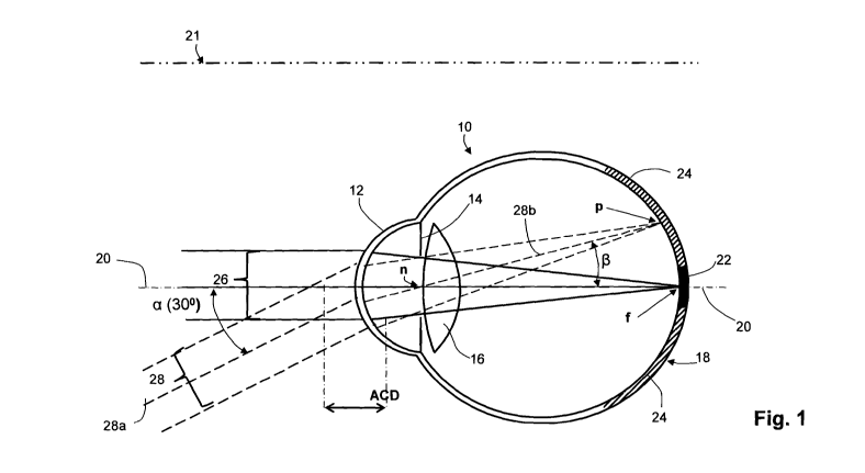
Figure 1 is a greatly simplified diagrammatic sectional plan of a normal
left human eye 10 having a cornea 12, iris 14, lens 16, retina 18 and visual
axis 20, the nasal plane between the eyes (or mid-visual axis) being indicated
at 21. Retina 18 is divided into (i) a central portion 22 (solid black) that
is used
for central vision and includes the fovea, the most sensitive portion of the
retina, and (ii) an annular peripheral portion 24 (hatched) which is much
larger
in area than central portion 22 but is less sensitive. In a normal or
emmetropic
eye with a straight-ahead gaze (on axis 20) directed at distance, an axial
central beam 26 from a distant object will be brought to focus at f on the
fovea
in the middle of central region 22 of retina 18 providing good visual acuity.
At
the same time, a peripheral or off-axis beam 28 from a distant object will be
brought to focus at point p on peripheral retina 24, it being assumed that the
central ray 28a of peripheral beam 28 intersects visual axis 20 at the axial
center n of pupil 14 (sometimes referred to as the nodal point of eye 10).
When the gaze is directed at a near on-axis object, the lens 16 changes
shape and optical power in a process called `accommodation' to (ideally) also
bring beam 26 from the near object to focus at point f. Similarly, beam 28
from
a near off-axis object ideally will be brought to focus at point p on
peripheral
retina 24. In fact, an emmetropic eye will normally exhibit astigmatism for
peripheral off-axis objects so that there will be two slightly different foci
near p
on the peripheral retina for both near and distance images.
It will be noted by inspection of Figure 1 that peripheral beam 28 enters
eye 10 from the temporal side or quadrant of the eye (and head) and is
focused on the nasal side or quadrant of peripheral retina 24. Conversely,
though not shown, peripheral rays entering eye 10 from the nasal quadrant
16
CA 02719421 2010-09-23
WO 2009/129528 PCT/US2009/041103
will impinge on the temporal quadrant of peripheral retina 24. Of course,
peripheral rays can enter the eye from the superior quadrant (above) or the
inferior quadrant (below).
The work of Smith et al has shown that out-of-focus images on the
peripheral retina 24 provide an important stimulus for the regulation of eye
growth. Accordingly, the measurement of the refractive power of the eye at
off-axis angles is now considered to be of critical importance for correctly
prescribing lenses for myopes suffering from progressive myopia. In this
specification, the angle a at which central ray 28a of peripheral beam 28
intersects axis 20 at point n is the peripheral angle (or off-axis angle) of
that
ray or beam. Because of refraction at the cornea 12 and lens 16, the angle R
which the emergent central peripheral ray 28b makes with optic axis 20 within
eye 10 is less than a and is difficult to determine in vivo with normally
available instruments. It is noted that the axial distance between the
anterior
surface of cornea 12 and the plane of iris 14 - often referred to as the
anterior
chamber depth, or ACD - is generally taken to be 3.5mm. This distance is
identified as ACD in Figures 1 - 3 and is used in measuring the peripheral
powers of anti-myopia lenses.
Our research suggests that an incident angle a of 30 degrees in any
meridian will place point p far enough into the peripheral retina to provide
the
desired stimulus for eye growth but is not so oblique as to be excessively
difficult to use. Effectively, angle a can be regarded as a solid angle.
Accordingly, in our extensive surveys of myopic youth in Australia and China
we used an incident peripheral angle a of 30 degrees when measuring the
peripheral refraction of aided and unaided eyes. The findings of these surveys
have therefore been used to design and test corrective symmetrical and
asymmetrical anti-myopia lenses for myopic patients generally.
Figure 2 is essentially the same diagram as that of Figure 1 but shows
(very much exaggerated) a myopic eye 1 Oa that is also prone to progressive
myopia. (The same reference numerals are used for the same parts as in eye
10 of Figure 1.) The axial length of eye 1 Oa is too long for accommodation to
focus on-axis beam 26 from a distant object onto central retina 22. Instead,
it
is focused at point f' in front of central retina 22 and distant images will
therefore be out-of-focus. For convenience, this problem is commonly
17
CA 02719421 2010-09-23
WO 2009/129528 PCT/US2009/041103
regarded as `refractive error' because can be corrected by fitting a negative
power lens (see Figure 3). However, myopes can typically focus near on-axis
objects on central the retina 22 achieve good near vision. Eye 10a illustrates
another `refractive error' common in myopic eyes - peripheral hyperopic
defocus - in which off-axis beam 28 is focused behind peripheral retina 24, at
p'. Smith et al have shown that hyperopic defocus increases the stimulus for
excessive eye growth and contributes to progressive myopia.
Figure 3 shows myopic eye 10a fitted with a conventional corrective
lens. Though a spectacle lens 30 is illustrated, the same considerations apply
to conventional contact lenses. Lens 30 has negative power that nicely
corrects central vision to let accommodation of natural lens 16 bring on-axis
beam 26 from a distant object to focus on the fovea at f. However, lens 30
shifts the focus of off-axis beam 28 to a point p" further behind peripheral
retina 24, generating an even greater stimulus for continued eye growth and
progressive myopia. Figure 4 illustrates the effect of an anti-myopia
corrective lens on myopic eye 10a, in this case a contact lens 32. Lens 32 has
a central optic zone 32a of about pupil size (normally 4 - 5mm diameter for
youths under standard room lighting) that corrects central vision to let
accommodation of natural lens 16 bring distance central focus onto central
retina 22 at point f. Lens 32 has an annular peripheral therapeutic optic zone
32b surrounding central zone 32a with sufficient myopic defocus to bring
peripheral beam 28 to focus at point p" in front of peripheral retina 24,
thereby
generating a stimulus to inhibit eye growth and progression of myopia in eye
10a. However, the out-of-focus peripheral image can cause peripheral blur.
Contact lens 32 has a non-optic zone 32c surrounding peripheral zone 32b to
enhance fitting and comfort.
Figure 5 shows the conversion of conventional spectacle lens 30 in
Figure 3 to an anti-myopia lens with the same effect as contact lens 32 by the
addition of an add-on lens 34. Lens 34 has a piano central optic zone 36 that
does not affect the central refractive power of base spectacle lens 30 so that
central beam 26 can still be brought to focus at point f on the fovea of
central
retina 22. However, add-on lens 34 has an annular a peripheral refractive
zone 38 with positive power that, despite the negative power of lens 30,
18
CA 02719421 2010-09-23
WO 2009/129528 PCT/US2009/041103
generates sufficient peripheral myopic defocus to bring off-axis beam 28 to
focus at point p"' in front of peripheral retina 24 (as in the case of contact
lens
32, Figure 4). Add-on lens 34 can be attached by mechanical clips 40 to base
lens 30 (or to a spectacle frame, not shown), or it may be attached by a
suitable adhesive.
Those skilled in the art will appreciate that there are known ways in
which the refractive powers of the different zones of multi-zone artificial
lenses
can be measured in different quadrants, and in which the peripheral and
central focal points of aided and un-aided eyes can be determined.
International patent applications WO/2008116270 and PCT/AU2008/000434
by Erhmann et al respectively disclose techniques for mapping the refractive
power of lenses and the eye at large peripheral angles.
The extensive surveys of youth in Australia and China that we have
conducted in which we measured peripheral refractive error at 30 degrees
(incident) in the nasal, temporal and superior quadrants confirmed that most
(but not all myopes) have hyperopic defocus, but the amount was not as great
as anticipated and did not increase as dramatically as expected for high
myopes. Data from the myopic eyes included in these surveys has been
condensed into the graphs or scatter charts of Figures 6 - 11 in which the
refractive power of an eye is measured in terms of the spherical equivalent,
and in which all peripheral measurements were made at 30 degrees (incident)
in various quadrants. These graphs can be summarized as follows.
Figure 6 plots peripheral corrective refractive power (as spherical
equivalent) of the surveyed eyes at 30 degrees in the temporal quadrant
against central (on-axis) corrective refractive power (also as spherical
equivalent), indicating a generally linear relationship. It will be seen that
there
is a 3D spread in peripheral focus for mild myopes (better than -2D), with
most eyes exhibiting hyperopic (relative) defocus in the periphery. Those
exhibiting hyperopic defocus would be considered to be a much greater risk
for progressive myopia than those with myopic defocus. Similar results are
evident for measurements of corrective peripheral powers in the nasal and
superior quadrants - Figures 7 and 8 respectively - but with a significantly
greater spread of corrective peripheral powers, especially in the superior
quadrant.
19
CA 02719421 2010-09-23
WO 2009/129528 PCT/US2009/041103
Figures 9 and 10 present the corrective temporal and nasal power data
of Figures 6 and 7. Figure 9 plots corrective central vs. corrective mean
horizontal power and corrective central vs. corrective temporal power, with
best-fit linear regression lines shown for each. Figure 10 plots corrective
central vs. corrective mean horizontal power and corrective central vs.
corrective nasal power, with best-fit linear regression lines shown for each.
Finally, if the mean corrective horizontal power is plotted against the
corrective spherical central power, as in Figure 11, it will be seen that
almost
all of the survey population fall within a 3D spread.
Clearly, this data supports the basis of the present invention; namely,
that the peripheral power of anti-myopia lenses for normal myopes can be
pre-set according to central power without peripheral defocus exceeding 3D,
thus avoiding the need to measure peripheral refractive error in the eye and
prescribe customized lenses to both correct central refractive error and to
appropriately control peripheral refraction for therapeutic purposes.
Furthermore, the data can be reduced to useful look-up tables or rules of
thumb that correlate the median sphere difference between central and
peripheral power for rotationally symmetrical lenses (as in Figure 12) or for
rotationally asymmetric lenses having different temporal and nasal powers (as
in Figure 13).
Referring more specifically to Figure 12, the left hand column [Central
Refractive Error (D)] lists measured central refractive error in increasing
increments of +0.25 D up to +6.00 D for the unaided eyes of the surveyed
population. The second column from the left [Median Peripheral Refractive
Error (D)] reports the measured median peripheral refractive error at 30
degrees (incident). Thus, -0.25 D myopes (those with a central refractive
error of +0.25 D) were found, on average, to be -0.71 D hyperopic periphery
and -5D myopes of the population (those with a central refractive error of
+5.00 D) were found, on average, to be +2.83D myopic in the periphery. The
third column [Median Script (Survey) Cent / Periph.] of the table of Figure 12
indicates the absolute central and the peripheral defocus (respectively) of a
customized prescription lens appropriate, on average, for the part of the
surveyed population having the corresponding central and peripheral
refractive errors indicated in the first two columns of Figure 12. Thus a -
0.25
CA 02719421 2010-09-23
WO 2009/129528 PCT/US2009/041103
D myope (with a +0.25 measured central error) requires a lens with a
corrective central power of -0.25 D and a peripheral defocus of +0.96D to (i)
provide both good central vision and (ii) bring the peripheral focus (at 30
degrees incident) in front of the retina so as to substantially eliminate the
stimulus for excessive eye growth. Similarly, a -5.0D myope requires a
corrective central power of -5.00 D and a peripheral defocus of +2.17D for
good vision and to substantially eliminate the stimulus for eye growth. Thus,
the third column of Figure 12 defines a pre-manufactured set, kit or stock of
anti-myopia lenses with minimal pre-set peripheral powers. Conveniently,
these lenses can be rotationally symmetric.
The two-part fourth column [Add Stepped Peripheral Power / Defocus
to Lens] of the table of Figure 12 indicates mild and high treatment options
(levels of peripheral defocus) that may be used to provide corresponding
medium and high levels of corrective stimulus for reducing eye growth. The
`mild' option adds +1.00 D, +1.50 D, +2.00 D and +2.50 D in four discrete
steps of peripheral defocus, which increase the level of defocus over the
minimal-treatment lenses of the third column of Figure 12, while the `high'
level adds +1.50 D, +2.00 D, +2.50 D and +3.00 D in four steps of peripheral
defocus. The use of steps in peripheral defocus (ie, where multiple lenses
with the same central power have difference peripheral powers) is intended to
simplify understanding and prescription by patients, clinicians and
manufacturers. The lenses of the two-part fourth column of Figure 12 are also
conveniently rotationally asymmetric.
Referring more particularly to Figure 13, the table of this Figure
provides information useful in the case of manufacturing rotationally
asymmetric lenses having different temporal and nasal powers. The left hand
column [Central Refractive Error (D)] again lists increments of measured
central refractive error in increasing increments of +0.25D up to +6.00 D (for
-
0.25D to -6.0D myopes). The second column from the left [Median Temporal
Refractive Error (D)] reports the median measured temporal refractive error
for those of the surveyed population having the corresponding increment of
measured central refractive error listed in the left hand column of Figure 13.
The third column from the left [Median Nasal Refractive Peripheral Error (D)]
reports the measured median nasal refractive error for subjects having the
21
CA 02719421 2010-09-23
WO 2009/129528 PCT/US2009/041103
corresponding central refractive error listed in the left-most column. It is
noted
that the surveyed population were somewhat more hyperopic in the temporal
retina than in the nasal retina, suggesting that it might be advantageous to
use the temporal retina measurements.
The fourth column [Median Script (Survey) Cent / Temporal] of the
table of Figure 13 can be regarded as defining a set of asymmetric anti-
myopia lenses with minimal therapeutic power applied in the temporal
quadrant of the retina, the second powers in the column being peripheral
defocus in that quadrant. It is to be noted that the lenses will have the
peripheral defocus applied to their nasal quadrants to affect the temporal
retinal quadrant. Similarly, the fifth column [Median Script (Survey) Cent /
Nasal] of Figure 13 can be regarded as defining a set of asymmetric anti-
myopia lenses with minimal therapeutic power applied to the nasal quadrant
of the retina. Again, it is to be noted that the lenses of the set of the
fifth
column will have the peripheral defocus applied to their temporal quadrants to
affect the nasal retinal quadrant.
The split sixth column [Added Stepped Peripheral Power] of the table
of Figure 13 indicates steps of peripheral defocus that can be used to modify
peripheral defocus of the sets of lenses of the fourth and fifth columns. As
shown, the column entitled "Temporal Chosen Power/Defocus" applies the
peripheral defocus to the lenses of the `temporal set' in four discrete steps,
+1.5D, +2.OD, +2.5D and +3.OD to the temporal corrective power, while the
"Nasal Chosen Power/Defocus" (right hand column) applies the peripheral
defocus to the lenses of the `nasal set' in three distinct steps, +1.OD, +1.5D
and +2.0D. Again, It should be noted again that, when considering lens
design, it is the nasal and temporal quadrants (respectively) of the lenses to
which the peripheral defocus is applied to effect the desired changes in the
peripheral temporal and nasal quadrants of the retina. The lenses of the two-
part fourth column of Figure 13 [Add Stepped Peripheral Power] are, of
course, rotationally asymmetric.
Figure 14 illustrates the relative power curves for each of the four steps
of peripheral defocus of the contact lenses designed according to the `Mild'
option of Figure 12 (fourth column). The maximum peripheral defocus of the
lenses of this subset or option is set at 2.5D. The reference numerals (60a,
22
CA 02719421 2010-09-23
WO 2009/129528 PCT/US2009/041103
61 a, 62a and 64a) applied to the power curves are used in describing the two
part trial kit of Figure 15 below.
Figure 15 is a diagrammatic representation of a two part trial or
prescribing lens kit or set suitable for practitioners, which can be
substituted
for conventional kits at little extra cost and can comprise a kit or set of
finished
trial spectacle lenses or a kit or set of trial or dispensing contact lenses.
This
Figure can be viewed as a diagrammatic plan view of a single drawer or tray
50 in which lenses are arranged in two arrays or parts 52 and 54 on a single
level, or it can be viewed as a diagrammatic sectional elevation of a cabinet
50 that has two drawers or parts 52 and 54, one above the other. The lenses
of part 52 conform to those set out in the `Mild' peripheral power column
(second from right) while the lenses of part 54 conform to those set out in
the
`High' peripheral power (far right) of the tabulation of Figure 12. Thus, kit
or
set 50 has double the minimum number of lenses need for a kit covering
`normal myopes' up to -5.00 D. In this example, the lenses 58a and 58b are
each packaged in a suitable sachet (not separately illustrated).
In Figure 15, part 52 comprises a compartmented container 56a
accommodating 20 different lenses 58a covering -5D in -0.25 D increments
of central corrective power while part 54 comprises a compartmented
container 56b also with 20 lenses 58b covering -5D of negative central power
in -0.25D increments. The respective increment of central power is written
above lenses 58a of part 52 as indicated by bracket 59a and the central
powers of lenses 58b of part 54 are similarly indicated at 59b. The peripheral
defocus of lenses 58a are collectively indicated by bracket 57a and the
peripheral defocus of lenses 58b are collectively indicated by brackets 57b.
Containers 56a and 58a can be differently color-coded, for example container
56a may be yellow and 58a may be red, and all the lens sachets of each
container are similarly differentiated using the same color codes - as well as
bearing both the central power and the peripheral defocus of the enclosed
lens(es), to minimize the chance of a lens sachet being place in the wrong
part of kit 50 or the chance of the wrong sachet/lens being selected for trial
or
use. For convenience of use, lenses 58a and 58b are arrayed in their
respective containers 56a and 56b according to their increments of central
power, though this need not be done in the linear fashion shown in Figure 15.
23
CA 02719421 2010-09-23
WO 2009/129528 PCT/US2009/041103
The lenses 58a of part 52, in this example, together include four steps of
peripheral power and, thus, form four sub-sets of lenses, indicated by
brackets 60a, 61 a, 62a and 64a. (The designs for these lenses are those of
Figure 14.) The peripheral defocus of each sub-set is diagrammatically
indicated by the height of the shaded portion of each lens and by the power
number having a plus sign associated with the respective brackets. Thus sub-
set 60a has three lenses 58a each having a peripheral defocus of 1.OD , sub-
set 61 a has 8 lenses each with a peripheral defocus of 1.5D, sub-set 62a has
4 lenses each with a peripheral defocus of 2.0 D and sub-set 64a has five
lenses 58a each with a peripheral power of 2.5D. Similarly, part 54 has four
sub-sets 60b, 61 b, 62b, and 64b having three, eight, four and five lenses 58b
with peripheral defocus steps marked +1.5D, +2.0D, +2.5D and +3.OD
respectively.
Lens kit or set 50 can be used in the following manner. The practitioner
makes a normal estimate or measurement of central refractive error of the
patient's eyes using existing equipment and techniques employed for the
prescription of conventional corrective lenses, and reviews the patient
history
to judge whether the patient is likely to suffer from progressive myopia. If
not,
a lens from part 52 of kit 50 with the appropriate corrective central power is
selected and tried; if so, a lens from part 54 is selected and tried. If the
patient
is not satisfied with the acuity of central vision provided by the selected
lens,
the lens with the next adjacent central power from the same part of the kit is
tried. If a patient who is trial-fitted with a lens from part 54 finds the
peripheral
blur excessive, the lens with the same central power in part 52 of the kit can
be substituted. In either case, the clinician can be highly confident that the
selected lens will act to inhibit the progression of myopia in the patient to
some degree by bringing the peripheral focus onto or in front of the
peripheral
retina to provide the desired stimulus for inhibiting further eye growth.
Where
kit 50 is one of contact lenses, it can be used to dispense finished lenses to
the patient or to make the appropriate order for supply from a wholesaler or
manufacturer. Where kit 50 is one of finished trial spectacle lenses and the
clinic has its own lens finishing grinding and/or polishing facility, it may
supply
finished lenses to the patient; otherwise, orders for such lenses are placed
with manufacturers in the conventional manner.
24
CA 02719421 2010-09-23
WO 2009/129528 PCT/US2009/041103
Two further types of sets, kits or stocks formed in accordance with the
principles of this invention are illustrated in Figures 16 and 17. Furthermore
Figure 16 illustrates two different contact lens kits, sets or stocks 70a and
70b, each comprising a box, tray or drawer 72 having a plurality of
compartments 74, each of which stores multiple sachets 76 of contact lenses
(not separately shown) having the same central corrective power. For
convenience, only four sachets 76 are shown in each compartment 74. The
central power of the lenses in each compartment 74 is written above or below
each compartment on labels 78. It will be seen that the central power of the
lenses ranges from -0.25D to -6.0D in 0.25D increments.
In kit, set or stock 70a, the sachets 76 in each compartment 74 not only
have the same central power but have the same peripheral power; that is, the
lenses in each compartment are identical so that they can serve as a
combination trial kit and supply stock. Each sachet is clearly identified with
the central corrective power and, while the peripheral power need not be
included in this example, it is preferable that all the sachets of the kit are
coded (for example by color) to show that they belong to one consistent series
or kit type. The peripheral powers of the lenses in the compartments conform
to the median power of the surveyed population for the respective central
corrective powers according to the third column of Figure 12. That is, no two
lenses of the stock or kit 70a with different central powers have the same
peripheral power; conversely, each central power is associated with a unique
peripheral power. The lenses of kit or stock 70a therefore have the minimum
positive therapeutic effect.
In kit, set or stock 70b, lenses with multiple therapeutic levels but the
same central power are housed in each compartment 74, the label 78 of the
compartment indentifying the respective central power of the lenses therein.
Each sachet 76 of each compartment is coded to indicate the level of
therapeutic effect and preferably has written identification of central power,
peripheral power and treatment level. In this example, sachets 76 with four
different levels of treatment are contained in each compartment 74, the lowest
being that of kit 70a described above and taken from the third column of
Figure 12, second lowest being taken from the second last column of Figure
12, the second highest being taken from the second last column of Figure 13
CA 02719421 2010-09-23
WO 2009/129528 PCT/US2009/041103
and the highest being taken from the last column of Figure 13. This kit, set
or
stock of lenses is then used in essentially the same manner as kit or set 50
described with reference to Figure 15, except that the clinician is now given
a
wider discretion to prescribe according to his or her assessment of the
patient's propensity to progressive myopia from the patient history - which,
of
course, will include familial history of myopia.
The final example of the trial set or kit is a seven-lens trial set or kit 80
of add-on lenses 82 for spectacles diagrammatically illustrated by Figure 17,
set or kit 80 comprising a rack 84 with sections or troughs 86, which hold add-
on lenses having piano central power and different steps/levels of peripheral
power or defocus. In this case, sections 86 have labels 88 to indicate the
step/level of added peripheral power or defocus, which is from +1.0D to +2.5D
in 0.25D increments and corresponds to the `Mild Add' option of the table of
Figure 12 (except for the finer 0.25 steps) of peripheral defocus. Since the
add-on lenses 82 of kit 80 may not all have a common base curve, or other
kits like this with sets of lenses having different base curves may be used,
it
will be convenient for the base curve of each add-on lens to be identified
additionally by labels 90. However, as in previous examples, it is also
desirable to mark the lenses or their sachets (if provided) to identify the
peripheral power and the base curve.
The manner of use of kit or set 80 is similar to that described for kit or
set 50 (Figure 15). The practitioner checks central refractive error of the
patient's eyes, judges the patient's propensity for progressive myopia from
patient history, selects an add-on lens with a level or peripheral defocus
appropriate to the judged propensity and tries the selected add-on lens on the
patient's habitual spectacle lens or on a semi-finished trial base lens with
the
appropriate central power. If the patient finds the peripheral blur excessive,
an
add-on lens with the next lower level of peripheral defocus is tried until
patient
acceptance is obtained. A final spectacle lens may then be ordered or finished
using an in-house grinding and polishing facility. The ability to provide so
many levels of peripheral defocus from a small set or kit of lenses is an
obvious advantage.
Turning more specifically to the anti-myopia ophthalmic devices
themselves, and more specifically to ophthalmic lenses such as contact
26
CA 02719421 2010-09-23
WO 2009/129528 PCT/US2009/041103
lenses, as noted above the peripheral power may be presented in a peripheral
power profile wherein the peripheral power changes with radial distance, such
that the peripheral power profile exhibits the peripheral power values located
at a determined distance from the central axis. Previously the peripheral
power profile of ophthalmic lenses was left the same or adjusted to reduce
spectacle distortion or improve central vision. Due to the lower visual acuity
of the peripheral retina, correcting the peripheral refraction was not seen as
significant improvement.
As mentioned above, the peripheral defocus of the lens is determined
by the differential between the central power for the ophthalmic lens and the
peripheral power at a particular point on the peripheral power profile. An
ophthalmic device, according to the present invention, is contemplated to
have a differential lens power (peripheral defocus of the lens) that is a
function of the central sphere power. However, considerable individual
variability in differential refraction (peripheral minus central) has been
observed among both children and adults of comparable central refractive
status. As a consequence, the use of an anti-myopia ophthalmic/contact lens
with an average, single, peripheral defocus/differential lens power may
overcorrect the peripheral retina in some myopes, but undercorrect the
peripheral retina in other myopes, depending on the individual peripheral
defocus of a particular eye. The optical effect for under-correction may be a
residual amount of hyperopic defocus in the peripheral retina, which would
also create a stimulus for axial eye growth and worsening myopia. On the
other hand, the optical effect for severe overcorrection of the peripheral
retina
may be an excessive amount of myopic, peripheral defocus, which not only
could hamper peripheral vision but also cause peripheral form vision
deprivation resulting in further axial eye growth and myopia progression.
Using an anti-myopia contact lens with an above-average, single, peripheral
defocus/differential lens power such that in most progressing myopes
peripheral hyperopia is converted to peripheral myopia would prevent under-
correction in some myopes, but create severe over-correction in other myopes
with the above-mentioned consequences.
In a series of lenses according to the present invention, each lens has
a differential lens power (amount of peripheral defocus) targeted at the
27
CA 02719421 2010-09-23
WO 2009/129528 PCT/US2009/041103
average relative peripheral refraction for a given central sphere power. A
lens
with a greater than average peripheral defocus can be produced.
Alternatively, a lens with a lesser than average peripheral defocus can be
produced. This means that while that peripheral defocus for the lens can be
greater or lower than the determined average, the amount of peripheral
defocus varies as a function of the particular central sphere power so as to
produce lenses that adequately correct a variation in the level of peripheral
refraction. In an alternative embodiment, ophthalmic lenses may be
customized based on a particular individual's determined level of peripheral
refraction. As such, after determining the particular individual's needed
amount of peripheral defocus/differential lens power, customized ophthalmic
lenses are manufactured.
The relationship between central power and peripheral defocus of the
lens can be, at a minimum, a first order (linear) relationship such that the
peripheral defocus increases as a constant function of the central sphere
power for each lens. While a linear relationship fits the discovered
refractive
relationship between the central and peripheral refractions, this could be
extended to higher order or non-polynomial relationships to produce a more
refined non-linear relationship. The result is an increasing peripheral
defocus
from a minimum at low myopia (-0.25D) to a maximum at high myopia (-
30.OOD) or as limited by optical design constraints. This is unlike other
optical
corrections such as presbyopia where the loss of accommodation is not
related to amount of myopia. For the correction of presbyopia there is no
increase in additional power as a function of the refractive myopia.
This relationship provides a more precise induced peripheral refractive
change than using a fixed peripheral defocus for the lens. This relationship
is
based on the experimental finding that an eye's central to peripheral
refraction
may increase with the amount of myopia. When applied to a power range of
inventoried anti-myopia lenses the experimentally determined mean central to
peripheral refraction would be used as the function to design the lens's
optical
peripheral defocus for each lens sphere power.
In additional study results on the peripheral refraction of the eye which were
obtained in the CIBA Vision Research Clinic, it was shown that the most
hyperopic refractive foci (the sphere meridian) of a myopic eye can vary from
28
CA 02719421 2010-09-23
WO 2009/129528 PCT/US2009/041103
less than between approximately 0.25D and 4.00D (at -6.00D, and even
greater for higher minus power is expected) difference from central axis to 30
degrees off-axis. More desirably, at 30 degrees off axis, the range can be
approximately between 0.25D and 3.0D, and even more desirably between
approximately 0.25D and 2.5D. Between central axis and 40 degrees off-axis,
that difference increases and can be approximately between 0.50D and
6.00D. Evaluation of optical designs of soft contact lenses where the
peripheral defocus was more positive has shown that high (2.50D) differential
refractions can be corrected (see Figure 20). However, the same peripheral
defocus design worn on an eye with 0.75D differential refraction overcorrects
the peripheral refraction and produces obvious peripheral blur for the wearer.
Figure 18 represents the central and peripheral auto refraction of an
eye which is emmetropic. There is very little relative peripheral hyperopia
(less than 0.50D at 30 degrees) and in this particular case the relative
peripheral hyperopia is -0.62 (at 30 degrees off axis) minus -0.62D (at
central
axis), which is 0.00D.
Figure 19 represents the peripheral auto refraction of an eye which is
highly myopic; in this case wearing a conventional soft contact lens for
measurement purposes with an auto refractor. There is much more relative
peripheral hyperopia (greater than 2.00D at 30 degrees off axis) and in this
particular case the relative peripheral hyperopia is 2.75D (at 30 degrees off
axis) minus 0.37D (at ten degrees off axis), which is 2.37D.
Figure 20 represents a myopic eye with a subjective central refraction
of about -1.50D. The relative peripheral hyperopia in this particular case is
low at -0.25D (at 30 degrees off axis) minus -1.00D (at ten degrees off axis),
which is 0.75D. The additional refractive data was taken through a soft
contact lens designed to correct high levels of relative peripheral hyperopia.
The effect of this lens correcting the eye is now relative peripheral myopia
and
in this particular case the relative peripheral myopia is -3.25D (at 30
degrees
off axis) minus -2.50D (at ten degrees off axis), which is -0.75D. Along with
the overall myopic shift of the auto refraction, this change in peripheral
auto
refraction was too much and caused subjective distortion of peripheral vision.
Figure 21 represents the peripheral auto refraction of the same highly
myopic eye as in Figure 19, in this case -6.00D imaged through a -4.00D
29
CA 02719421 2010-09-23
WO 2009/129528 PCT/US2009/041103
correction lens for measurement purposes with an auto refractor. The
additional refractive data was taken through the soft contact lens designed to
correct high levels of relative peripheral hyperopia as used in Figure 20. The
effect of this lens correcting the eye is much less relative peripheral
hyperopia, and in this particular case the relative peripheral hyperopia is -
4.25D (at 30 degrees off axis) minus -4.62D (at ten degrees off axis), which
is
0.37D. Along with the lesser overall myopic shift of the auto refraction, this
change in peripheral auto refraction was less and caused no subjective
distortion of peripheral vision.
Figure 22 represents the results of a study of Schmid in which the
sphere power in minus cylinder notation was measured centrally and at 20
degrees in the nasal, temporal, inferior and superior retina with a Shin
Nippon
K5001 open-field auto-refractometer in both eyes of six young adult
volunteers during cycloplegia. Plotting the relative peripheral refraction for
sphere power (peripheral minus central sphere power) for each location vs.
central sphere power revealed an inverse correlation. Statistical significance
was reached for the mean of all four peripheral locations combined.
Figure 23 represents more details from the Schmid study. All four
quadrants showed the same trend of increasing relative peripheral hyperopia
with increasing central myopia. Individually, statistical significance was
reached for the inferior and superior quadrants where the change is slightly
larger.
Correlation analysis between subjective vision quality and objective
auto-refraction in the retinal periphery of patients who reported differences
in
vision quality between lenses of various peripheral defocus powers revealed
that over-correction limits exist, beyond which vision quality is not
acceptable.
Turning to Figure 24, there is shown a representation of the effect of
peripheral refraction on the rating of side vision quality for the lenses,
using a
scale from 0-10. Symbols indicate those patients subjects who answered "no"
(circles) or "yes" (triangles) to the question whether vision quality is
sufficient
to wear the lens all the time.
The plot as shown in Figure 24 is in terms of sphere refraction ("Sph";
left side of plot) and sphere equivalent refraction ("M"; right side of plot)
as
measured at 30 degrees in the temporal retina (nasal field) ("T30") by auto-
CA 02719421 2010-09-23
WO 2009/129528 PCT/US2009/041103
refractometry. If, for example at 30 degrees in the temporal retina (nasal
field), the lens produces a sphere refraction below about +0.25D (i.e. on the
retina or in front of the retina), then vision quality is unacceptable as
indicated
by all patients answering "no" to the question whether vision quality is
sufficient to wear the lens all the time. This is shown in the plot in the
shaded
left side of the "T30 Sph" portion. Similarly, for a sphere equivalent
refraction
below about -2.50D (i.e. further in front of the retina than -2.50D), vision
quality is unacceptable as indicated by all patients answering "no" to the
question whether vision quality is sufficient to wear the lens all the time
(shaded left side of the "T30 M" portion.). Therefore, it can be seen that
over-
correction of peripheral refraction leads to reduced subjective vision. In
particular the sphere meridian should not be corrected to less than +0.25D
and the sphere equivalent meridian to less than -2.50D. The correlation
analysis also indicated that lens rejection is chiefly caused by decreased
peripheral vision as opposed to central vision. The identification and
application of these over-correction limits substantially facilitates the lens
fitting procedure, and helps reduce vision degradation and lens rejection by
the patient when correcting peripheral defocus and controlling refractive
error
development.
Turning now to Figure 25, there is shown a scatterplot of both sphere
meridian and sphere equivalent (SEQ) data which plots the central sphere
equivalent refraction (in diopters) against the refractive difference (also in
diopters) between central and 30 degree nasally offset autorefractions. These
data were obtained from a largely adult population made up of Caucasian
subjects. As can be seen in Figure 25, for central sphere equivalent (SEQ)
refractive errors between +0.50 and -5.00D, there is a significant increase
the
peripheral refractive differential with increase in myopia. The rate of
increase
or slope of a best fit line for these data is 0.14D/D for sphere meridian and
0.18D/D for the SEQ. The intercepts (x=0 or piano refractive error) are
+0.53D for sphere meridian and 0.05D for the SEQ. Therefore, targeting
correction or reduction of peripheral refractive differentials for either the
sphere meridian or SEQ requires an increase at about the same rate with
central SEQ refractions.
31
CA 02719421 2010-09-23
WO 2009/129528 PCT/US2009/041103
In Figure 26, there is shown a scatterplot of the central sphere
equivalent (SEQ) refraction data that was shown in Figure 25, which is also
plotted with data of the central sphere equivalent (SEQ) refractions obtained
from a population made up of Asian (Chinese) child and adolescent subjects.
These data are plotted against the sphere equivalent refractive difference
(also in diopters) between central and 30 degree nasally offset
autorefractions. Comparison of Caucasian and Asian populations, with SEQ
refractive errors between -0.50 and -4.00D, shows a significant difference in
the increase the peripheral refractive differential with increase in myopia.
The
rate of increase or slope of a best fit line for these data is -0.19D/D for
the
Caucasian population measured and -0.35D/D for the Asian population
measured. Therefore, targeting correction or reduction of peripheral
refractive
differential requires an increase with central SEQ refractions (as noted above
with respect to Figure 25); however, that increase may change depending on
the target population's makeup or environmental demographics.
In certain embodiments, varying the peripheral defocus for each sphere
power still may not cover the full range of relative peripheral refractions
needed to fit all myopic patients' relative peripheral hyperopic defocus
without
clinically significant over and under correction in some individuals. In this
case, the target correction of the ophthalmic lens can be matched to the
change in peripheral refractive differential and an additional variation such
as
providing an average, a lower than average and a higher than average central
to peripheral differential lens power (peripheral defocus) may be needed for
each sphere power. While varying the peripheral defocus with central sphere
power allows for the change in the average relative peripheral hyperopic
defocus, the wide range in the population may need a higher and lower optical
design factor to further avoid clinically significant over or under correction
of
individual patients' relative peripheral refraction. In the example shown in
Figure 27, the target correction or "Average SEQ" correction is to correct the
peripheral refractive SEQ differential to +0.75 diopters, and to account for a
wide range in the population further higher and lower peripheral refractive
SEQ differential targets are shown by the dashed lines designated "High
SEQ" and "Low SEQ". In this combination the average central to peripheral
differential power will still increase with the minus sphere power to correct
for
32
CA 02719421 2010-09-23
WO 2009/129528 PCT/US2009/041103
the overall increase in central to peripheral differential refraction with
increasing myopia.
In an alternative embodiment, a contact lens may be designed with a
negative power differential to provide hyperopic defocus in the central and
retinal periphery for the stimulation of axial eye growth in hyperopic eyes.
In a
further alternative embodiment, a contact lens is designed with a sphero-
cylindrical central power for correcting astigmatism. In this case, either the
sphere part or the spherical equivalent (sphere + half of the cylinder) of the
central power is used as central sphere power for defining the desired
peripheral defocus of the lens. A further alternative embodiment of the
present invention would include custom prescription of the peripheral defocus
based on the patient's individual central to peripheral refraction of the eye.
This would be a custom `made to order' correction and not the more common
inventoried approaches as are described above.
It will be appreciated that many modifications of or additions to the
sets, kits or stocks of lenses, and to lenses or lens components per se,
described in the example, or to their methods of use, can be made by those
skilled in the art without departing from the scope of the invention as set
out in
the following claims.
33