Note: Descriptions are shown in the official language in which they were submitted.
CA 02719534 2010-09-23
WO 2009/125282 PCT/IB2009/005208
1
"METHOD FOR PERFUSING A BIOCOMPATIBLE MATERIAL GRAFT
WITH A LIQUID AND PERFUSION KIT"
DESCRIPTION
The present invention relates to a method for perfus-
ing a biocompatible material graft with a liquid and a
perfusion kit.
In the context of the present invention, by "biocom-
patible material grafts" are intended prosthetic ele-
ments made of a material of a natural or synthetic
origin apt to be implanted in a living creature in or-
der to compensate for lacunas of a bone, osteo-
cartilage and/or cartilage tissue.
In the field of the orthopedic surgery, the surgeon
often has to face cases in which the patient has more
or less extended bone, osteo-cartilage or cartilage
lacunas. Such lacunas can result from multiple causes,
such as an imperfect bone reunification following to
fractures, consolidation delays, malignant patholo-
gies, infection outcomes, comminuted or multifrag-
mented fractures, neonatal deformities, structural al-
terations of a traumatic origin, or other.
In order to face the lack of bone volume, various so-
lutions have been proposed, in particular different
substances of a natural, semi-synthetic or only syn.-
thetic origin functioning as bone, osteo-cartilage or
CA 02719534 2010-09-23
WO 2009/125282 PCT/IB2009/005208
2
cartilage substitute have been used. However, several
authors have shown that the extraordinary mechanical
behavior of the natural bone due to its nanocomposite
hierarchical structure is difficult to reach with any
other type of biomaterial. Therefore, the ideal bone
substitute is the autologous bone withdrawn by the
same patient from a donor site. This practice, how-
ever, is not free from patient risks which very often
result in a resorption of the bone implant itself and
a frequent occurrence of a painful symptomatology in
the site in which the bone graft has been withdrawn.
Recent studies have shown that the use of allogenic
bone from a tissue bank of a human origin, processed
and made inert through physical-chemical processes,
can represent an alternative to the use of the autolo-
gous bone. However, also in this practice risks con-
cerned to the contraction of infective diseases or im-
mune-type reactions can subsist.
New biomaterials having functions of bone substitutes
have been studied and proposed for a clinical use, and
some of them have shown positive results following to
clinical examinations on humans. Such materials do not
simply show high biocompatibility properties (inert
biomaterials) , but posses biomimetic features, namely
chemical and physical-chemical properties similar to
CA 02719534 2010-09-23
WO 2009/125282 PCT/IB2009/005208
3
the human bone capable of activating biological mecha-
nisms (bioactivities) with the recipient bone tissues
and the cell components contained therein, by promot-
ing the neo-formation and bone consolidation proc-
esses. Once their stimulation function of the bone new
formation is ended, these materials presents sometimes
a complete resorption, only leaving space to the new
formed bone.
As it is known, before being implanted in a patient,
the above grafts are usually perfused with biological
liquids or aqueous solutions of a different nature (in
particular bone marrow, medullary concentrate, periph-
eral blood, antibiotic solutions, etc.) for creating
the suitable conditions for the subsequent development
of osteointegration processes.
Grafts of a biocompatible material are generally po-
rous in order to increase their biomimetic ability.
However, the presence of such porosity makes a com-
plete perfusion from the liquid difficult to obtain,
above all when this has a high viscosity. This exposes
to the risk of an incomplete removal of the air exist-
ing within the graft. Such occurrence can compromise
the success of the implant operation, as the presence
of air can reduce the mechanical resistance of the
bone grafts, interfere with the proper osteointegra-
CA 02719534 2010-09-23
WO 2009/125282 PCT/IB2009/005208
4
tion and trigger tissue osteolysis and necrosis proc-
esses.
Perfusion kits are known, such as for example the one
described in the WO 2007/048016 document, including a
cylindrical perfusion chamber, intended for receiving
the graft to be perfused, which is coupled, through a
first opening, to a syringe containing a perfusion
liquid. A second opening, opposite to the first one,
is engaged by a plunger having a head equipped with
one or more passages for placing in a fluid communica-
tion the inside of the perfusion chamber with the ex-
ternal environment.
The liquid in the syringe is then injected in the per-
fusion chamber, causing a pressure increase therein.
The air, which is gradually compressed by the intro-
duction of the perfusion liquid, is evacuated in the
external environment through the passages in the
plunger head inserted within the second opening of the
perfusion chamber. In this way, the air existing in
the pores of the graft is evacuated during the filling
of the pores with the perfusion liquid. The syringe is
then removed from the first opening in the perfusion
chamber and the opening is sealed.
Near the first opening a third opening is foreseen,
which put into fluid communication the inside of the
CA 02719534 2010-09-23
WO 2009/125282 PCT/IB2009/005208
perfusion chamber with the external environment. Such
third opening has a septum which is permeable to the
air and impermeable to the biological liquid. When the
syringe has been removed, the plunger is pressed in
5 the direction of the third opening by increasing the
pressure within the perfusion chamber and evacuating
the air, if any, still existing within the same
through the septum placed on the third opening.
Another known type of a perfusion kit, such as for ex-
ample the one described in the EP 1 419 739 Al docu-
ment includes a perfusion chamber within which the
graft is introduced. The perfusion chamber includes an
inlet arranged upstream of the graft and associated
wit a sort of a surgical needle having a back vent apt
to allow the fluid passage from the needle to the per-
fusion chamber and to inhibit the fluid passage be-
tween the perfusion chamber and the needle. The perfu-
sion chamber is further equipped with an outlet placed
downstream of the graft and coupled with a pump dis-
charging in a fluid collection basin. In use, the bio-
logical liquid is withdrawn by the surgical needle
(for example from the body of a patient) and returned
by the pump within the perfusion chamber. The liquid
floods the perfusion chamber and exits, passing
through and perfusing the graft, from the outlet for
CA 02719534 2010-09-23
WO 2009/125282 PCT/IB2009/005208
6
collecting itself within the collection basin. Also in
this case the air is evacuated from the graft during
the filling of the perfusion chamber with the perfu-
sion liquid.
Perfusion kits and methods for perfusing biocompatible
material grafts known in the art show some drawbacks.
First of all, perfusion kits of the known art show
perfusion chambers having a predetermined volume inde-
pendently from the dimensions of the graft which has
to be perfused. Therefore, in some cases, especially
when the graft has reduced dimensions, there is a
waste of perfusion liquid (which very often is a valu-
able biological liquid, as directly withdrawn by the
patient in a limited quantity) due to the need of how-
ever filling the perfusion chamber independently from
the dimension of the graft.
Furthermore, in the perfusion methods of the known art
it is possible that a non negligible quantity of air
is dissolved in the perfusion liquid during the fill-
ing of the perfusion chamber.
In fact, as said, during the filling of the perfusion
chamber with the perfusion liquid, the pressure within
the chamber itself increases. Such pressure increase
causes an increase of the ability of the air to mix
with the liquid. When the pressure is reduced, namely
CA 02719534 2010-09-23
WO 2009/125282 PCT/IB2009/005208
7
after the perfusion is complete, the air can again
separate and remain trapped within the graft now per-
fused and ready for the surgical implantation.
In this context, the main technical task of the pre-
sent invention is to provide a method for perfusing a
biocompatible material graft with a perfusion liquid
and the perfusion kit thereof apt to perform such
method capable of overcoming the drawbacks above men-
tioned.
In the ambit of said technical task, an important ob-
ject of the invention is to propose a method for per-
fusing a biocompatible material graft with a perfusion
liquid and the perfusion kit thereof which are capable
of optimizing the quantity of biological liquid to be
used, with no regard to the dimensions of the graft.
A further object of the present invention is to pro-
vide a method for perfusing a biocompatible material
graft with a perfusion liquid and the perfusion kit
thereof capable of-effectively evacuating the air from
the pores of the graft itself.
The stated technical task and the specified objects
are substantially achieved by a method for perfusing a
biocompatible material graft with a perfusion liquid
and a perfusion kit according to one or more of the
appended claims.
CA 02719534 2010-09-23
WO 2009/125282 PCT/IB2009/005208
8
By way of representative and not limiting example, the
description of a method for perfusing a biocompatible
material graft with a perfusion liquid and a perfusion
kit according to the present invention is now re-
ported, in which:
- Figure 1 shows an exploded view of a perfusion kit
of a biocompatible material graft with a perfusion
liquid according to the present invention; and
- Figures 2 to 6 show the kit of Figure 1 in use in
different working positions.
With reference to the enclosed figures, a perfusion
kit according to the present invention has been gener-
ally shown by numeral 1.
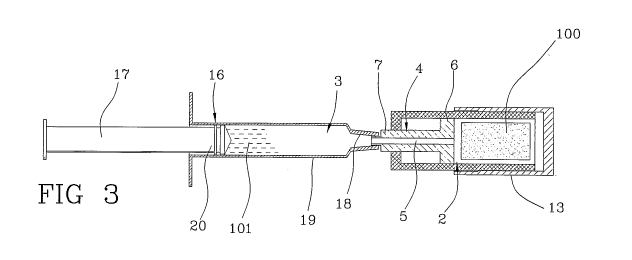
With a particular reference to Figure 1, the kit in-
cludes a perfusion chamber 2 apt to contain a graft
100 to be perfused and a transfer chamber 3 apt to
contain a perfusion liquid 101.
The graft 100 has been outlined in the enclosed fig-
ures by a cylinder, however the graft 100 to be per-
fused can be in any forms and dimensions apt to the
specific implant to which it is intended.
The graft 100 generally includes a biocompatible mate-
rial of a natural or synthetic origin, of organic, in-
organic or composite nature, which is capable of in-
corporating liquids, having a typical porous or fi-
CA 02719534 2010-09-23
WO 2009/125282 PCT/IB2009/005208
9
brous (in woven or non woven form) , preferably hydro-
philic structure.
Examples of biocompatible materials commonly used are:
calcium phosphate-based ceramic materials, for example
hydroxylapatite (HA), tricalcium phosphate (alpha or
beta TCP) or dicalcium phosphate (HA/TCP in different
relative o); bone, osteo-cartilage and cartilage sub-
stitutes of a homologous, heterologous or biopolimeric
origin (for example, hyaluronic acid-based and deriva-
tives); materials of an autologous origin withdrawn by
donor sites.
Such materials can be associated with biopolymers so
as to form composite materials. Examples of biopoly-
mers are: poly-lactic acid (PLA), poly-l-lactic acid
(PLLA), polyglycolic acid (PGA), collagen, alginate,
hyaluronic acid and its derivatives, carboxymethyl
cellulose (CMC) and its derivatives, such as for exam-
ple hydroxypropylmethyl cellulose (HPMC) or hy-
droxyethyl cellulose (HEC).
As for the perfusion liquid, this can consist, for ex-
ample, of biological liquids or aqueous solutions of a
different nature, such as for example: physiological
saline, bone marrow, medullary concentrate, peripheral
blood, platelet concentrate, antibiotic solutions,
stem cell suspensions, growth factors or other bio-
CA 02719534 2010-09-23
WO 2009/125282 PCT/IB2009/005208
logically active elements apt to promote the graft os-
teointegration.
The perfusion chamber 2 and the transfer chamber 3 are
connectable between them in a fluid-tight manner to be
5 able to transfer the liquid contained in the transfer
chamber 3 within the perfusion chamber 2, as it will
be more evident in the following of the present de-
scription.
The kit 1 further includes a connection 4 which can be
10 directly coupled with the transfer chamber 3 and in-
serted within the perfusion chamber 2.
The connection 4 put in fluid communication the trans-
fer chamber 3 with the perfusion chamber 2. For this
end, the connection 4 has a through hole 5 which de-
velops itself starting from a first portion 6 until a
second portion 7, arranged opposite relative to the
first one 6 of the connection 4, therefore passing
through the entire connection 4.
The connection 4 can be slideably introduced within
the perfusion chamber 2. In particular, the first por-
tion 6 of the connection 4 can be slideably coupled
within the perfusion chamber 2 and is countershaped at
the internal walls of the perfusion walls 2.
The movable coupling of the first portion 6 of the
connection 4 with the perfusion chamber 2 is made in a
CA 02719534 2010-09-23
WO 2009/125282 PCT/IB2009/005208
11
fluid-tight manner, in such a way that any fluids,
both gas and liquid, cannot pass between the connec-
tion 4 and the internal walls of the perfusion chamber
2.
The second portion 7 of the connection has an end 8
apt to be restrained to the transfer chamber 3.
The perfusion chamber 2 includes a first 9 and a sec-
ond 10 openings, respectively arranged at a first 11
and a second 12 ends of the perfusion chamber 2. The
first opening 9 is apt to allow the introduction of
the graft 100 within the perfusion chamber 2, while
the second opening 10 is apt to allow the passage of
the second end 7 of the connection 4.
The kit includes a closing element 13 for closing in a
fluid-tight manner the first opening 9 of the perfu-
sion chamber 2 once the graft 100 has been introduced
therein.
In the preferred embodiment of the invention, the per-
fusion chamber 2 is a hollow cylinder, preferably made
of a plastic material.
The closing element 13 can consist, for example, of a
screw stopper having a diameter greater than the di-
ameter of the cylinder constituting the perfusion
chamber 2. On the external wall of the cylinder a
thread 14 engageable by the screw stopper for closing
CA 02719534 2010-09-23
WO 2009/125282 PCT/IB2009/005208
12
the first opening of the hollow cylinder is preferably
provided.
In the proximity of the tread 14 on the cylinder, a
toroidal gasket 15, which ensures the fluid-tight
closing of the screw stopper 13 on the cylinder form-
ing the perfusion chamber 2, is preferably provided.
In this embodiment, the first portion 6 of the connec-
tion 4 has a cylindrical form, whose external wall is
intended for sliding in a fluid-tight manner on the
internal wall of the cylinder which forms the perfu-
sion chamber 2.
The second opening 10 of the perfusion chamber 2 has a
passage section lower than the diameter of the hollow
cylinder, in such a way that the first portion 6 of
the connection 4 is not able, by sliding within the
hollow cylinder, to exit from the perfusion chamber 2
through the second opening 10 thereof.
Also the second portion 7 of the connection 4 has a
cylindrical form having a diameter slightly lower than
the diameter of the second opening 10 of the perfusion
chamber 2, in such a way that the connection 4 can
partly exit from the perfusion chamber 2 through the
second opening 10.
As above shown, the presence of the first portion 6,
however, prevents the connection 4 from completely ex-
CA 02719534 2010-09-23
WO 2009/125282 PCT/IB2009/005208
13
iting through the second opening 10. On the contrary,
the connection 4 can completely exit from the perfu-
sion chamber 4 trough the first opening 9.
The transfer chamber 3 has the feature of being able
to increase or decrease its own volume, due to reasons
that will result apparent below.
In the preferred embodiment of the invention, the
transfer chamber 3 is made by .a syringe 16.
In particular, the transfer chamber 3 is formed by the
volume existing between a plunger 17 of the syringe 16
and the bottom wall 18 of the cylindrical element 19
which constitutes the body of the syringe 16.
More particularly, the cylindrical element 19 of the
syringe 16 includes an opening through which the
plunger 17 is introduced. This latter has a head 20
having a fluid-tight sliding gasket 21 relative to the
internal wall of the cylinder 19, in such a way that
any fluids, both gas and liquid, cannot pass between
the head 20 and the internal wall of the cylinder 19.
The bottom wall 18 of the cylindrical element 19 is
placed opposite relative to the opening within which
the plunger 17 is introduced.
The space between the bottom wall 18 and the head 20
of the plunger defines the transfer chamber 3.
The bottom wall 18 is further equipped with an opening
CA 02719534 2010-09-23
WO 2009/125282 PCT/IB2009/005208
14
22 through which the transfer chamber 3 is placed into
a fluid communication with the perfusion chamber 2. In
particular, the opening 22 can be coupled with the
second portion 7 of the connection 4.
In order to ensure a stable coupling between the
transfer chamber 3 and the connection 4, the opening
22 of the syringe includes an internally threaded cy-
lindrical portion 23 which engages a thread 24 placed
at one end of the second portion 7 of the connection
4. Such thread 24 partly surrounds the hole 5 which
passes through the connection 4.
The kit further includes an extractor 25 to discharge
the graft 100 from the perfusion chamber 2 when the
graft 100 has been perfused of liquid.
The extractor 25 can be coupled to the second end 7 of
the connection 4 for pushing the first end 6 of the
connection 4 towards the first opening 9 of the perfu-
sion chamber 2. In particular, the extractor 25 in-
cludes a first threaded end 26 which can be screwed on
the second end 7 of the connection 4.
The extractor further includes a second end 27, oppo-
site to the first one 26, having cross dimensions
greater than the diameter of the cylinder constituting
the perfusion chamber 2.
A method for perfusing a biocompatible material graft
CA 02719534 2010-09-23
WO 2009/125282 PCT/IB2009/005208
with a perfusion liquid according to the present in-
vention will be now described.
For the sake of explanatory clarity, a method carried
out by the perfusion kit above described will be dis-
5 cussed, which constitutes a preferred but not exclu-
sive implementation means of the method itself.
The method for perfusing a biocompatible material
graft with a perfusion liquid includes the steps of
introducing a graft in a perfusion chamber 2, arrang-
10 ing a transfer chamber 3 partly filled with a perfu-
sion liquid 101 and coupling in a fluid-tight manner
the perfusion chamber 2 and the transfer chamber 3,
for establishing a fluid communication among the two.
The method then provides the lowering of the pressure
15 in the transfer chamber 3 for transferring within the
same part of the air existing in the perfusion chamber
2 and successively the increase of the pressure within
the transfer chamber 3, in order to inject in the per-
fusion chamber 2 the liquid 101 existing in the trans-
fer chamber 3.
It is to be underlined that the steps of decreasing
and increasing the pressure and injecting the fluid
101 are carried out under conditions of fluid isola-
tion between the perfusion 2 and transfer 3 chambers
with respect to the external environment.
CA 02719534 2010-09-23
WO 2009/125282 PCT/IB2009/005208
16
In particular, the graft 100 is first introduced
within the perfusion chamber 2. This operation is car-
ried out by introducing the graft 100 through the
first opening 9 of the perfusion chamber.
Before introducing the graft 100 within the perfusion
chamber 2, the connection 4 is introduced therein. In
particular, the connection 4 is introduced by first
introducing the second portion 7 of the same and suc-
cessively the first portion 6.
In this way, the second portion 7 exits from the sec-
ond opening 10 of the perfusion chamber 2 and the
first portion 9 engages, in a sliding and fluid-tight
manner, the internal wall of the perfusion chamber 2.
Next, the perfusion chamber 2 is closed by the closing
element 13. Note that in this configuration, the per-
fusion chamber 2 has a one-way fluid communication
with the external environment. Such communication way
is due to the hole 5 which passes through the connec-
tion 4.
Advantageously, the connection 4 is retreated towards
the graft 100 introduced within the perfusion chamber
4 (as shown in Figures 2 to 5) , in such a way to de-
crease the volume of the same within which the graft
100 is housed.
In particular, the first portion 6 of the connection 4
CA 02719534 2010-09-23
WO 2009/125282 PCT/IB2009/005208
17
is slid along the internal wall of the perfusion cham-
ber 2 and towards the first opening 9 of the same.
This allows to minimize the volume of fluid 101 re-
quired for perfusing the graft 100, as it will be more
explained hereinbelow.
At this point, the perfusion chamber 2 is tight-
coupled with the transfer chamber 3 (see Figure 2).
This latter is preventively at least partly filled
with the perfusion liquid 101, preferably by directly
sucking-up the liquid from a container with the sy-
ringe 16 or directly withdrawing the same from a pa-
tient through a proper suction kit.
The fluid communication between the perfusion chamber
2 and the transfer chamber 3 is carried out through
the connection 4. In particular, the second portion 7
of this latter is screwed on the threaded cylindrical
element 19 of the bottom wall 18 of the syringe 16.
In this configuration, the plunger 17 is introduced
within the syringe 16 for defining the transfer cham-
ber 3 as already above described.
It is to be underlined that, in this configuration,
the perfusion chamber 2 and the transfer chamber 3 are
in a fluid communication between them but in a fluid
isolation with the external environment.
The perfusion chamber 2 and the graft 100 are placed
CA 02719534 2010-09-23
WO 2009/125282 PCT/IB2009/005208
18
at the ambient pressure existing during the steps of
introduction of the graft 100 within the perfusion
chamber 2. Analogously, the liquid in the transfer
chamber 3 is at ambient pressure.
At this point, the pressure within the transfer cham-
ber 3 is lowered. Such pressure lowering takes places
by increasing the volume of the transfer chamber 3. In
the preferred embodiment, this is obtained by moving
the plunger 17 of the syringe 16 away from the bottom
wall 18 of the syringe 16 (see Figure 3).
The pressure reduction in the transfer chamber 3
causes a pressure lowering in the perfusion chamber 2,
in such a way to balance again the pressures within
the two chambers.
The pressure lowering within the perfusion chamber 2
takes place through an air transfer from the perfusion
chamber 2 to the transfer chamber 3. This inevitably
causes the exit of the air existing within the pores
of the graft 100.
Therefore, as a function of the degree of pressure de-
crease within the transfer chamber 3, namely as a
function of the volume increase of the chamber itself,
a significant portion of air is evacuated from the
pores of the graft 100.
Next, the pressure within the transfer chamber 3 is
CA 02719534 2010-09-23
WO 2009/125282 PCT/IB2009/005208
19
increased for injecting the liquid 101 within the per-
fusion chamber 2.
The liquid 101, while entering the perfusion chamber 2
at a pressure greater than that existing within the
same, permeates the pores of the graft 101 previously
at least partly emptied from the air.
This operation is carried out by decreasing the volume
of the transfer chamber 3. In particular, the opera-
tion is carried out by pushing the plunger 17 towards
the bottom wall 18 of the syringe 16.
In order to avoid that during the injection of liquid
101 within the perfusion chamber 2 also the air exist-
ing in the transfer chamber 3 (previously withdrawn by
the perfusion chamber 3) returns again in the perfu-
sion chamber 2, the transfer chamber 3 and the perfu-
sion chamber 2 are rotated for vertically arranging
themselves with the transfer chamber 3 placed above
the perfusion chamber 2 (see Figure 4).
In order to increase the perfusion effectiveness of
the graft 100, the pressure within the transfer cham-
ber 3 is again lowered, with the consequent withdrawal
from the perfusion chamber 2 of the liquid 101 just
injected and the air, if any, still existing within
the perfusion chamber and the pores of the graft 100.
The pressure decrease within the transfer chamber 3 is
CA 02719534 2010-09-23
WO 2009/125282 PCT/IB2009/005208
obtained in the same way already above described.
At this point, the pressure in the transfer chamber 3
is again increased (in the same way above described)
for transferring again only the liquid 101 in the per-
5 fusion chamber 2 and the pores of the graft 100.
These operations, namely the empting and the filling
with the liquid 101 of the perfusion chamber 2, are
repeated until the liquid 101 is uniformly distributed
within the graft 100.
10 The number of repetitions depends on the type of the
material of the graft 100 to be perfuse, the type of
liquid 101 and the dimensions of the graft 100 itself.
once the perfusion is ended, the pressure within the
perfusion chamber 2 is equal to the one at the begin-
15 ning of the operations, as well as the pressure within
the transfer chamber 3. In fact, during all the opera-
tions of fluid transfers between the two chambers,
these latter are remained isolated from the external
environment.
20 The final effect is that the liquid existing in the
transfer chamber 3 has been transferred in the perfu-
sion chamber 2 and inside the graft 100 and that the
air existing in the perfusion chamber 2 and the graft
100 has been transferred in the transfer chamber 3.
Once the perfusion of the graft is ended, the transfer
CA 02719534 2010-09-23
WO 2009/125282 PCT/IB2009/005208
21
chamber 3 is decoupled from the perfusion chamber 2.
In particular, the syringe 16 is unscrewed from the
connection 4.
On the second portion 7 of the connection 4 is then
mounted the extractor 25, as shown in Figure 5. Pref-
erably, the threaded end 26 of the extractor 25 is
screwed on the second portion 7 of the connection 4.
After or before this latter operation, the perfusion
chamber 2 is opened in correspondence with its first
opening 9. In particular, the closing element 13 is
removed by the first opening of the perfusion chamber
2.
By pushing the extractor towards the first opening 9
of the perfusion chamber 2,_the perfused graft 100 is
pushed by the connection 4 and comes out from the
first opening of the perfusion chamber 2 , as shown in
Figure 6. Note that the use of the extractor prevents
the perfused graft 100 from being directly handled by
the operator.
The invention attains the proposed aims. In fact, the
method and the kit of the present invention allow,
first of all, to optimize the quantity of perfusion
liquid 101 used independently from the dimensions of
the graft 100, as the connection 4, being sliding in a
fluid-tight manner within the perfusion chamber 2, en-
CA 02719534 2010-09-23
WO 2009/125282 PCT/IB2009/005208
22
surer that the perfusion chamber 2 adapts its volume
as a function of the dimensions of the graft 100.
Moreover, the air is effectively evacuated from the
pores of the graft 100 and replaced by the liquid 101,
since the air is evacuated or however the pressure
thereof is remarkably reduced before the perfusion of
the graft 100 by the liquid, by avoiding.that air and
liquid are mixed within the graft when the air has a
high pressure.
Furthermore, it is to be underlined that the perfusion
means of the present invention is capable of optimiz-
ing the quantity of the perfusion liquid independently
from the dimensions and the configuration of the
graft, up to a volume equivalent to that within the
perfusion chamber.