Note: Descriptions are shown in the official language in which they were submitted.
CA 02719769 2010-09-27
WO 2009/118283
PCT/EP2009/053363
1
Methods for producing hair microfollicles and de novo papillae
and their use for in vitro tests and in vivo implantations
The present invention relates to a method for producing hair microfollicles by
co-culturing the
de novo papillae with another cell population of the hair follicle in the
ultra-low attachment
culture vessels. The invention also relates to a method for producing de novo
papillae usable
in a method for producing hair microfollicle. Object of the invention are also
the hair
microfollicles and the de novo papillae produced by the aforementioned
methods. The hair
microfollicles and/or de novo papillae can be used as implants for treating
reduced hair
conditions, and for in vitro testing hair-modulating effects or toxic effects
of substances.
Because of the critical role that hair plays in human non-verbal
communication, an affected
individual invariably demands help when hair growth diminishes. The ultimate
therapy, of
course, is to restore or regenerate new, healthy, cycling hair follicles.
Until very recently,
medicine was unable to offer any valid treatments to these patients. In the
late twentieth
century, several drugs were marketed that, however modestly and
inconsistently, did
stimulate hair growth. Examples are minoxidil, finasteride, and latanoprost.
The complex
timing and myriad gene expression changes required for orchestration of hair
follicle
development and cycling are likely to preclude a simple pharmaceutical
approach to the
treatment of advanced alopecia. Consequently, their effects fall short of the
ultimate goal to
generate new hair follicles in bald scalp. By taking advantage of cell types
that know how to
form a hair follicle, cell-based therapies will arrive in the clinic sooner
than the purely
molecular approach.
One approach to hair follicle cell-based therapy would entail removing a small
number of hair
follicles, isolating competent and/or inductive cells from them, and then
expanding those cells
ex vivo while maintaining their special ability to generate new hair
follicles. Clearly, cell
culture conditions that maintain the inductive ability of dermal follicular
cells and the
competence of hair follicle epithelial cells are necessary before any type of
cell-based
therapy for alopecia can be developed. As early studies showed that the
inductive property of
dermal cells wanes with time in vitro, research has focused on maintaining the
trichogenic
properties of hair follicle cells in culture. Much of the recent progress has
resulted from
CA 02719769 2010-09-27
WO 2009/118283
PCT/EP2009/053363
2
advances in culture methodology for intact hair follicles, and their cellular
components.
Human hair follicle cells grown in culture include follicular papilla
fibroblasts, outer root
sheath (ORS) keratinocytes and germinative epidermal cells from the hair
matrix (Tobin et al.,
J. Invest. Dermatology 104(1), 86-88, 1995).
The goal of current bioengineering efforts is to generate or reconstitute
fully organized and
functional organ systems starting from dissociated cells that have been
propagated under
defined tissue culture conditions. It has long been recognized that the hair
follicle has
profound regenerative ability, in that it cycles over the life-time of the
individual and
reproduces its lower half cycle after cycle. The fibroblasts of the dermal
hair papilla and
connective tissue sheath are stem cell-like in character and have specific
hair growth-
inducing properties. The hair follicle reforms itself by means of interactions
between
competent epithelial stem cells and powerfully inductive dermal cells during
its growth cycle.
It is possible to reconstitute a complete hair follicle from epithelial and
nnesenchymal stem
cells of hair follicles.
Major challenges that need to be addressed with any type of cell-based
treatment for
alopecia include the efficiency of hair follicle formation and the choice of
cell type, which are
summarized by Stenn & Cotsarelis, Curr. Opinion Biotech. 16, 493-497, 2005.
For
bioengineering the hair follicle, one could start with dermal elements from
dissociated follicles
with or without competent cells from the follicle or other epithelial sources.
The number of
dissociated cells would be expanded in culture, and then dermal cells alone,
or in
combination with competent epithelial cells, re-introduced to the alopecic
scalp. Previous
studies have shown that starting with correctly placed inducer dermal cells
will result in new
follicle formation. Moreover, starting with a combination of dissociated, or
aggregated,
trichogenic epithelial and dermal cells has also proven to be an efficient way
of producing
new hair follicles.
First attempts at cell-based approaches for treating alopecia are likely to
use autologous
tissue for bioengineering hair follicles to avoid immune rejection of the
donor cells. However,
the intriguing possibility that heterologous (allogeneic) hair follicle tissue
could be developed
for tissue transplantation exists, based on the concept that the hair follicle
is an immune-
privileged site that does not express MHC (major histoconnpatibility complex)
class I
antigens. Nevertheless, the safety testing and regulatory hurdles for this
type of approach
would require enormous financial resources.
CA 02719769 2010-09-27
WO 2009/118283
PCT/EP2009/053363
3
Another possible approach for bioengineering hair follicles involves actually
forming hair
follicles as mini organs in vitro, and then transplanting the newly generated
follicles back to
the alopecic scalp. It is known from DE 101 62 814 B4 that a skin and hair
equivalent can be
produced by providing a pseudodermis or a pseudodermis preparation, as well as
pseudopapillae comprising cultivated dermal papilla cells on a suitable
carrier or in a suitable
matrix, or pseudopapillae precursors comprising cultivated dermal papilla
cells on a suitable
matrix-forming medium, capable of forming a matrix in situ, and introducing
the
pseudopapillae or the pseudopapillae precursor into the pseudodermis or
pseudodermis
preparation. This sort of approach would require a much more complicated cell
culture
system involving three-dimensional matrices, perhaps embedded with appropriate
growth
factors, to allow both dermal and epidermal cells to differentiate towards the
three-
dimensional structure of a normal hair follicle. Particularly, the papilla
structure is only
obtained by forming cavities, such as by punching or pricking, in said
pseudodermis, and
placing said pseudopapillae therein, which are so shaped that their dimensions
correspond
to the cavities formed in the PD. The approach lacks the voluntary arrangement
of cells in
the approximate physiological papilla structure, as well as direct cell
contacts.
It has been recently shown another method for producing a population of
multipotent stem
cells or progeny thereof, which are originated from a hair follicle or a
dermal papilla-
containing portion thereof. The method of WO 2005/071063 Al comprises the
culture of said
hair follicle or dermal papilla-containing portion in conditions under which
multipotent stem
cells grow and proliferate non-adherently. Dermal papillae are not obtained by
this procedure,
but the isolated multipotent stem cells directly used for inducing hair growth
or regenerating
skin in a mammal. However, it is also recognized in this document that a
certain amount of
adherent cells is formed as confirmed in WO 2005/113747 A2, which discloses a
method for
producing multicellular aggregates from at least two multipotent or
pluripotent adult stem cell
types by cultivation under steric conditions. Presently, the organoid bodies
are restricted to
the aforementioned stem cells gathered from exocrine gland tissue.
Therefore, the technical problem forming the basis of the present invention is
to avoid the
above-mentioned disadvantages of the prior art and to provide a method for
generating re-
constructed hair follicle and papillae that arrange free and easy into the
size and shape of a
physiological DP. Another problem addressed by the present invention is to
find a hair
CA 02719769 2010-09-27
WO 2009/118283
PCT/EP2009/053363
4
equivalent or skin substitute, which would be suitable as an in vitro model,
more particularly
for testing and/or evaluating active substances, most particularly on the hair
follicle.
The present invention solves the problem by providing a method for producing
mammalian
hair microfollicle comprising the steps of:
(a) providing at least one de novo papilla,
(b) providing at least one other cell population selected from the group of
fibroblasts,
keratinocytes and melanocytes, and
(c) co-culturing the de novo papilla with the at least one other cell
population under non-
adherent culture conditions.
The terms "de novo papillae" or "neopapillae" are interchangeably used herein,
and denote
cell aggregates of mammalian dermal hair papilla fibroblasts (DPF), which have
at least half
the size and approximately the shape of a physiological dermal papilla (DP) of
a hair follicle
after isolation. De novo papillae can comprise a coating comprising one or
multiple different
extracellular matrix proteins, preferably collagen IV, fibronectin and/or
laminin. Such coating
can be generated by the DPFs forming the de novo papilla themselves, or can be
added at
any stage before de novo papillae will be co-cultured according to step (c) of
the method for
producing hair microfollicles.
The terms "microfollicle" and "neofollicle" are used interchangeably herein
and refer to an
incomplete mammalian hair follicle structure that is composed of the dermal
cellular scaffold,
but lacking other cell types, such as muscle cells, nerves, blood vessels,
etc., resulting in a
reduced size if compared to the natural follicle. A microfollicle of the
present invention is
composed of a de novo papilla that is stably covered or colonized by cells of
at least one
other cell population selected from the group of fibroblasts, keratinocytes
and/or melanocytes.
The fibroblasts, keratinocytes and/or melanocytes do not have to originate
from a
mammalian hair follicle, but can be derived from other mammalian tissues.
Preferably a
microfollicle of the present invention is composed of a de novo papilla that
is stably covered
or colonized by cells of at least one other cell population, which is
derivable and/or derived
from a mammal hair follicle, e.g. selected from the group of fibroblasts of
the connective
tissue sheath, keratinocytes and/or melanocytes. The three-dimensional form of
said
microfollicle mimics the three-dimensional appearance of a physiological
mammal hair follicle.
The microfollicles can in particular be three-dimensionally formed, optionally
spatially
CA 02719769 2010-09-27
WO 2009/118283
PCT/EP2009/053363
demarcated, reconstructed de novo papillae with follicle-like structures on
the surface of the
de novo papillae, including those reminiscent of the earliest stage of hair
morphogenesis,
and comprising dermal papilla cells.
The invention relates to a method for producing hair microfollicles,
comprising multiple steps.
These steps comprise the provision of de novo papillae. According to the
invention, the de
novo papillae to be provided can be produced by any suitable method,
preferably de novo
papillae are used that have been produced according to one of the methods for
production of
de novo papillae according to the invention disclosed below.
The steps comprise further the provision of other cell populations. The other
cell population
may be derivable from a mammal hair follicle, preferably from the same hair
follicle as used
for DP preparation, and co-culturing the de novo papillae with at least one
other cell
population in non-adherent culture vessels. At least 4 different cell
populations, i.e. DPFs,
fibroblasts of the connective tissue sheath (CTSF), keratinocytes (KC), and
melanocytes
(MC), can be isolated from a mammal hair follicle in a defined manner,
separately cultured
under standard conditions, and subsequently multiplied. It is also possible to
form cell
cultures with long-term viability from these isolates and put them to use,
e.g. for screening
methods.
It is a preferred embodiment of the present invention that the other cell
populations are
selected from the group of fibroblasts, keratinocytes, and melanocytes. The
fibroblasts,
keratinocytes and/or melanocytes do not have to originate from a mammalian
hair follicle, but
can be derived from other mammalian tissues. The fibroblasts originate
preferably from
connective tissue sheath.
Preferably the at least one other cell population is derivable and/or derived
from a mammal
hair follicle, which is selected from the group of fibroblasts of the
connective tissue sheath,
keratinocytes and/or melanocytes.
It is another preferred embodiment of the present invention that the de novo
papillae are co-
cultured with keratinocytes at least. Keratinocytes are supplemented for basic
microfollicle
neogenesis. It is still another preferred embodiment of the invention that
keratinocytes and
melanocytes are simultaneously provided to the papilla culture. The addition
of melanocytes
is especially useful for investigations of hair pigmentation, or for the
purpose of future
implantation with a desired hair color. The other, separately expanded cell
components can
be added to a non-adherent cell culture vessel described below for the method
of producing
CA 02719769 2010-09-27
WO 2009/118283
PCT/EP2009/053363
6
de novo papillae, such as the ultra-low attachment culture vessels, in which
the neopapillae
have already been produced. Keratinocytes, or mixtures of keratinocytes and
melanocytes
are added to the coated neopapillae at a specific mixing ratio and incubated
with a suitable
medium in the non-adherent culture vessels for several days. Preferable ratios
are KC to MC
2:1, 5:1, 10:1 or 20:1. The co-culturing lasts at least 12 hours, preferably
between 1 day to 5
weeks, more preferably 2 days to 3 weeks, most preferably 1 week or
substantially 2 days.
To elaborate a multilayered neofollicle, which even better mimics the hair
follicle structure
and function, it can be obtained by coating the generated neofollicle with
CTSF for a defined
period of time selected from 12 hours to 10 days, preferably 1 to 5 days, most
preferably
substantially 2 days. Under these conditions, the cell mixture develops into a
follicular
structure, forming hair follicle-typical features, such as development of a
hair shaft. The
organ-like structure is referred to as microfollicle from the formation of an
approximate hair
shaft on.
In a preferred embodiment of the method of the invention for producing hair
microfollicle
(a) de nova papillae are provided;
(b) de-novo papillae are contacted with KC, MC or a mixture of KC and MC
with a
predetermined ratio and co-cultured in a non-adherent culture vessel for a
predetermined
amount of time;
(b')
optionally, repeat step (b), preferably wherein cells of a different cell-type
are used for
contacting and co-culturing than used in the first round;
(c) optionally covered de-novo papillae of step (b) or (b') can be coated
with an
extracellular matrix protein, preferably collagen IV, prior to and/or
simultaneously with step
(d);
(d) covered de-novo papillae of step (b), (b') or (c) are co-cultured with
CTSF for a
predetermined amount of time in a non-adherent culture vessel.
The present invention is also directed to providing a method for producing de
nova papillae
comprising the steps of:
(a) providing at least one dermal papilla (DP) from at least one mammal hair
follicle,
(b) isolating dermal hair papilla fibroblasts (DPFs) from the DP by
mechanically fixing
said DP at the surface of a cell culture vessel, whereby the basal lamina is
perforated
to allow said DPFs migrating out,
CA 02719769 2010-09-27
WO 2009/118283
PCT/EP2009/053363
7
(c) expanding the isolated DPFs in monolayer culture without collagen coating,
wherein
said DPFs are passaged at least once,
(d) condensing the expanded DPFs into cell aggregates that exhibit the size
and shape
of the physiological DP, wherein said DPFs are differentiated in non-adherent
culture
vessels in a cell concentration per vessel surface of 1.000 to 100.000
DPFs/cm2,
optionally the expanded DPFs are condensed for at least 48h, and
(e) coating the de novo papillae with extracellular matrix proteins,
preferably collagen IV,
fibronectin and/or laminin.
The present invention is also directed to providing a method for producing de
novo papillae
comprising the steps of:
(a) providing at least one dermal papilla (DP) from at least one mammal
hair follicle,
(b) isolating dermal hair papilla fibroblasts (DPFs) from the DP by
mechanically fixing
said DP at the surface of a cell culture vessel, whereby the basal lamina is
perforated
to allow said DPFs migrating out,
(c) expanding the isolated DPFs in monolayer culture without collagen
coating, wherein
said DPFs are passaged at least once,
(d) condensing the expanded DPFs into cell aggregates that exhibit the size
and shape
of the physiological DP, wherein said DPFs are differentiated in non-adhesive
culture
vessels in a cell concentration per vessel surface of 1.000 to 100.000
DPFs/cm2, and
wherein the expanded DPFs are condensed for at least 48h;
(e) optionally coating the de novo papillae with extracellular matrix
proteins, preferably
collagen IV, fibronectin and/or laminin.
It is particularly the expansion of the isolated DPFs and the physiological
administration form
that represent limiting factors to the use of these cells in hair growth
induction of prior art. It
takes several multiplication cycles to achieve the required quantity of cells,
during which
process the cells cultured in monolayer cultures, especially those of the
dermal papilla
fibroblasts, lose their inductive abilities, namely, after 5-8 multiplication
cycles as shown by
experience. The cells thus treated dedifferentiate and express stem cell
markers, but can no
longer be used to generate hair follicles.
It has been surprisingly demonstrated by the inventors that the cells reach
the level of
differentiation and stabilization or regain their hair growth-inducing ability
after several days
CA 02719769 2010-09-27
WO 2009/118283
PCT/EP2009/053363
8
to several weeks under the specific culture conditions of the invention,
wherein the DPFs
expanded following cell culturing are transferred at well-defined
concentration into special
non-adhesive cell culture vessels. In addition to the regain of inductive
properties, it has been
surprisingly found that as a result of active cell-cell contacts and exchange
of signaling
molecules, the cells subsequently form cell aggregates and differentiate.
Under these
specific conditions, the cells thus treated condense into approximately the
shape and size
corresponding to the physiological shape of a dermal papilla in a hair
follicle following
isolation. To this effect, the ratio of cells used/culture vessel surface is
of crucial importance.
Prior step (a), a hair follicle is taken from a mammal being a patient or
donor. The sample is
especially gathered from a human, rodent, pig, canine, monkey, sheep, cat, or
dog,
preferably a human. It is essentially preferred to gather a tissue sample by
skin biopsy,
especially taken close to the location of ailment. In the present invention,
the sample of the
hair follicle is preferably withdrawn from head hair, beard, eyebrows, genital
hair or other
body hair. The withdrawal of the hair follicle follows good medical practice.
The sample may
be purified to remove disturbing substances.
The provision of DPs from the hair follicles and the isolation of DPFs
according to step (a)
proceeds in a form newly developed from previous standard protocols. Using the
small
pieces of skin biopsies, the epidermis is severed from the underlying dermis
and fatty tissue,
e.g. by using a scalpel. The fatty tissue is slightly compressed, e.g. by
using pincers, so that
the hair bulbs located therein are easily prepared under a dissection
microscope. Such
isolated hair follicles are fixed on the hair shaft, e.g. by means of pincers
again, and the
connective tissue sheath is carefully separated in a diametrical fashion, e.g.
by means of
another pair of pincers, so that the bulb is everted to expose the DPFs and
the hair shaft with
the hair matrix. In this way, the proximal part of the bulb, together with the
connective tissue
sheath fibroblasts, and the dermal papillae are easily separated from the
remaining part of
the hair follicle, such as by using a needle or cannula. Also, the hair shaft,
which includes the
likewise required hair matrix keratinocytes and melanocytes, is optimally
prepared for further
culturing.
Thereafter in step (b), the isolated dermal papillae are transferred into a
cell culture vessel,
and mechanically fixed at the surface of the vessel. Preferably, the DPFs are
obtained by
mechanically arresting the isolated dermal papillae on the culture vessel
bottom using a
pinpoint or scalpel rather than subjecting them to enzymatic separation. It is
preferred to
CA 02719769 2010-09-27
WO 2009/118283
PCT/EP2009/053363
9
incubate 1 to 8 DPs per culture vessel. Particularly, 2 to 4 DPs are
transferred into each well
of 6-well or 12-well cell culture plates, for instance, and fixed on the plate
bottom. As a result,
the basal lamina is slightly perforated, but the papilla morphology is
approximately
maintained, and the DPFs can migrate out of the dermal papillae, and
proliferate.
Culturing of the DPFs in step (c) is effected without collagen coating in
standard medium,
preferably including a reduced amount of fetal calf serum (FCS), e.g. 10 A in
contrast to
conventional 20 % FCS, and additionally supplemented with antibiotics, such as
lx
penicillin/streptomycin. Following isolation, the first medium change is
preferably effected
after 1 week at the earliest, but after 2 weeks at the latest. As soon as
cells have grown out
of the DP, the medium can be changed once to twice a week, depending on the
cell density.
When the cells reach 70-80 A confluence, they are detached from the plate
bottom, for
instance by using trypsin/EDTA at room temperature, and passaged into other
culture
vessels, such as T25 cell culture flasks. In the course of culturing, the
cells can be further
expanded for direct use in experiments, or frozen in liquid nitrogen for
future use. Although
the number of passages is not limited, so that cells can be expanded to any
high density if
viability is given, it is preferred that said DPFs are passaged at least two
times, more
preferably at least five times, most preferably less than nine times. For a
high troughput
procedure eight passages are preferred. Consequently, in another preferred
embodiment of
the present invention, the method is performed by condensing non-inductive
DPFs. It is an
unexpected finding that such DPFs lacking inductive properties can still be
used for tissue
engineering.
Next to follow is the condensation phase of step (d), wherein the separated,
multiplied DPFs
are cultured in non-adherent culture vessels with medium including well-
defined components,
i.e. standard culture medium. In this system, a special vessel's surface, or a
special coating
of the vessel surface, especially the bottom surface, reduces the cell
attachment or even
prevents the cells from attaching to the surface. Preferably, the non-adhesive
culture vessels
are made of glass, polystyrol, and/or a surface treated with an anti-adhesion
layer. More
preferably, a PTFE- or poly-HEMA-coated surface is applied in the vessels. The
use of ultra-
low attachment culture vessels is particularly preferred in the scope of the
invention, which
are e.g. distributed by Corning, Inc., USA. Such non-adherent vessels and
coatings are
known to the skilled artisan, and can be easily purchased, or manufactured.
CA 02719769 2010-09-27
WO 2009/118283
PCT/EP2009/053363
As a result of free contacting, the cells aggregate into a defined size,
forming aggregates
after several days which resemble the original papilla in size and shape. The
expanded
DPFs are preferably condensed for at least 48hours, 2 to 5 days, more
preferably for 2 to 3
days, most preferably substantially 2 days, and optionally further cultivated
for 3 to 15 days,
preferably for 5 to 10 days or 2 to 21 days. Size and shape of the condensates
being formed
likewise depend on the region where skin biopsies have been removed. DPFs,
which are
removed from beard, body hair, eyebrows, genital hair, or head hair, and
subjected to
expansion form cell aggregates of different size, which in turn determine the
respective hair
shape, size and length. Even more crucial is the initial cell number as
inoculated into the
vessel, which has to be in due proportion to the vessel surface. In another
embodiment of the
method, the cell concentration per vessel surface amounts to 2.000 to 50.000
DPFs/cm2 in
step (d), preferably 3.000 to 20.000 DPFs/cm2, more preferably 5.000 to 10.000
DPFs/cm2,
most preferably substantially 6.666 DPFs/cm2.
At this stage, a self-produced matrix has already formed around the
condensate. To
accelerate this process and exert influence thereon in a targeted manner,
physiologic matrix
components are added to the medium at this point to form a capsule mimicking
the
properties of a dermal papilla. In the subsequent process step (e), the de
novo papillae thus
obtained are coated with a composition of extracellular matrix proteins. This
composition is
made so as to mimic physiological ones. It may preferably consist of collagen
IV, fibronectin
and/or laminin. It is particularly preferred that the extracellular matrix
proteins are a mixture of
collagen IV, fibronectin and laminin in shares of 2-6 : 0.5-2 : 0.5-2 parts
per weight, most
preferably having a ratio of substantially 4 : 1 : 1.15 parts per weight. It
is not excluded,
however, that the aforementioned extracellular matrix proteins can also be
used individually
or with varying volume percentages or combined with other matrices. In another
embodiment
of the invention, the extracellular matrix proteins additionally comprise
other collagens (eg. 1,
10 Al, 18 Al), glycosaminoglycans and/or proteoglycans, preferably heparan
sulfate,
decorin, keratan sulfate, biglycan, aggrecan, versican, perlecan, CD44v3,
and/or syndecan.
Again, such coating of the neopapillae is carried out in a minimally adhesive
cell culture
vessel, e.g. ultra-low attachment cell culture vessels. The coating is
performed for 1 to 5
days, preferably for 1 to 2 days.
The present invention also relates to the de novo papillae, and the hair
microfollicles
obtainable by the processes according to the invention. The prior teaching of
the present
specification concerning the methods for producing neopapillae and
microfollicles,
CA 02719769 2010-09-27
WO 2009/118283
PCT/EP2009/053363
11
respectively, is considered as valid and applicable without restrictions to
the products of the
production methods if expedient.
Both, the de novo papillae and/or hair microfollicles of the invention can be
used for the
production of skin equivalents. Preferably, the skin equivalents are
constructed, such as by
using Matriderm (Dr. Suwelack Skin & Health Care AG), according to standard
methods, and
the insertion sites for the microfollicles are cut at regular intervals by
means of a 2-photon
laser, or pre-perforated with a punch. Consequently, skin equivalents
comprising the de novo
papillae and/or hair microfollicles of the invention are another object of the
invention. In
addition to the reconstructed dermal papillae, they comprise one or more
layers of
keratinocytes, which may form themselves into an epidermal structure, or
peridermal
structure, and optionally one or more layers of nnelanocytes, which can be
applied over the
dermal papillae structure.
The de novo papillae and/or the hair microfollicles according to the invention
can also be
used as implants. Therefore, still another object of the invention is an
implant comprising as
active ingredient an effective amount of the de novo papillae of the invention
and/or the hair
microfollicles of the invention, optionally together with pharmaceutically
tolerable adjuvants.
Similarly, the skin equivalent of the invention can be used as transplant. It
is still another
object of the invention to provide a transplant comprising as active
ingredient an effective
amount of the skin equivalent according to the invention, optionally together
with
pharmaceutically tolerable adjuvants.
The term "effective amount" denotes an amount of the implant or transplant,
respectively,
having a prophylactically or therapeutically relevant effect on a disease or
pathological
conditions. A prophylactic effect prevents the outbreak of a disease or even
the infection with
a pathogen after the infiltration of single representatives such that the
subsequent
propagation of the pathogen is strictly diminished, or it is even completely
inactivated. A
therapeutically relevant effect relieves to some extent one or more symptoms
of a disease or
returns to normal either partially or completely one or more physiological or
biochemical
parameters associated with or causative of the disease or pathological
conditions. The
respective amount for administering the implant or transplant, respectively,
is sufficiently high
in order to achieve the desired prophylactic or therapeutic effect of reducing
symptoms of
reduced amount of hair. It will be understood that the specific dose level,
frequency and
CA 02719769 2010-09-27
WO 2009/118283
PCT/EP2009/053363
12
period of administration to any particular mammal will depend upon a variety
of factors
including the activity of the specific components employed, the age, body
weight, general
health, sex, diet time of administration, route of administration, drug
combination, and the
severity of the specific therapy. Using well-known means and methods, the
exact amount
can be determined by one of skill in the art as a matter of routine
experimentation.
The implants or transplants of the invention are produced in a known way using
common
solid or liquid carriers, diluents and/or additives and usual adjuvants for
pharmaceutical
engineering and with an appropriate amount depending on the intended mode of
application.
These pharmaceutically acceptable excipients comprise salts, buffers, fillers,
chelating
agents, antioxidants, solvents, bonding agents, lubricants, coatings,
additives, preservatives,
and suspending agents. In the meaning of the invention, an "adjuvant" denotes
every
substance that enables, intensifies or modifies a specific body response as
result of
implanting or transplanting if administered simultaneously, contemporarily or
sequentially.
The amount of excipient material that is combined with the active ingredient
to produce a
single dosage form varies depending upon the host treated and the particular
mode of
administration.
Depending upon the manner of introduction, the implant or transplant,
respectively, may be
formulated in a variety of ways. The concentration of therapeutically active
ingredients in the
formulation may vary from about 0.1 to 100 wt %. They may be administered
alone or in
combination with other treatments.
The invention also teaches de novo papillae, hair microfollicles and/or skin
equivalents
according to the invention for the prophylactic or therapeutic treatment of a
condition of
reduced amount of hair. The aforementioned products of the inventive methods
are
preferably used for the therapeutic treatment. A therapeutically relevant
effect relieves to
some extent one or more symptoms of a reduced amount of hair, or returns to
normality,
either partially or completely, one or more physiological parameters
associated with or
causative of the pathological conditions. Monitoring is considered as a kind
of treatment
provided that the products of the inventive methods are administered in
distinct intervals, e.g.
in order to booster the proliferation response and eradicate the symptoms of
the condition
completely. Either identical products or different products can be applied. In
the meaning of
the invention, prophylactic treatment is advisable if the subject possesses
any preconditions
CA 02719769 2010-09-27
WO 2009/118283
PCT/EP2009/053363
13
for the beginning of hair loss, such as a familial disposition, a genetic
defect, or a previously
passed disease.
The pathologic conditions of a reduced amount of hair as concerned by the
invention may be
the result of alopecia (e.g. androgenetic alopecia, alopecia areata, etc.),
hereditary baldness,
scarring, burns, radiation therapy, chemotherapy, disease-related loss of
hair, accidencal
injury, damage to hair follicle, surgical trauma, an incisional wound, or a
donor site wound
from a skin transplant.
The invention also relates to the use of the de novo papillae, hair
microfollicles and/or skin
equivalents according to the invention for the production of an implant or
transplant,
respectively, for the prophylactic or therapeutic treatment of a condition of
reduced amount of
hair. The implant and transplant can be either administered to prevent the
initiation of hair
loss of a mammal, preferably a human individual, and the resulting trouble in
advance, or to
treat the arising and continuing symptoms.
It is another object of the invention to provide a method for treating a
condition of reduced
amount of hair, wherein the de novo papillae, hair microfollicles and/or skin
equivalents
according to the invention are incorporated into the skin of a mammal in need
of such
treatment. The microorganoid follicles, especially autologous/allogenic human
hair follicle
precursors, are used for implantation with the aim of inducing hair growth,
whereas the skin
substitutes do regenerate skin, preferably the scalp. The microorganoid
follicles are
incorporated into the openings of previously depilated, miniaturized hair
follicles (isthmus) of
affected skin areas. Preferably, the de novo papillae and hair microfollicles
are injected, more
preferably by means of a specially constructed device of about 150 pm in size.
It is also
preferred that all components are used in an autologous fashion, and treated
under
GLP/GMP conditions. The neopapillae, hair microfollicles, or hair follicle
compartments
stimulate the new development of hair growth, such as in cases of hereditary
baldness,
scarring (bums), disease-related loss of hair, chemotherapy/radiation-induced
loss of hair,
and the like as already described in the course of the present specification.
Maturing of the
microfollicles into hair follicles having long-term viability is fostered by
the micro-medium, i.e.
the permanent, distal hair follicle.
The invention also relates to the use of the aforementioned products for the
direct
pharmacological and cosmetic in vitro testing of substances, which exert a
hair-modulating
CA 02719769 2010-09-27
WO 2009/118283
PCT/EP2009/053363
14
influence. The hair-modulating effects are especially selected from the group
of hair growth,
hair shape, hair structure, hair color, and hair pigmentation. It is preferred
to analyze the
effect of modifying hair growth - with the intention of promoting hair growth
in cases of hair
loss, such as caused by alopecia, as well as inhibiting hair growth in cases
of excessive,
undesirable hair growth, such as caused by hypertrichosis and/or hirsutism, or
female beard
growth, or undesirable body hair. In particular, the use of a high-throughput
method allows
the pharmaceutical and cosmetic industries to effectively test existing or new
substances for
a potential hair growth-modulating effect. The substances comprise
pharmaceutical agents,
cosmetic agents, chemical compounds, polymeric compounds, growth factors,
cellular
products, living cells and/or biomolecules. Furthermore, when adding
melanocytes, i.e. the
pigment-forming cells, to the microfollicles, it is possible to investigate
substance effects on
the pigmentation and/or coloring of the hair shaft being formed. Likewise, the
substance
effect on hair shape and hair structure can be tested, e.g. formation of
curls, etc.
The following end points can be evaluated or measured to obtain information on
the
effectiveness of substances in regard to an improvement in hair structure and
the influencing
of hair growth: analysis of hair shaft formation, length growth and
characteristics of the hair
shaft, hair array analysis, volume and structure of the dermal papilla,
proliferation
measurement (e.g. Ki67 expression, BrdU incorporation, etc.), apoptosis
measurement (e.g.
TUNEL, enzyme assays, annexin measurement, etc.), differential marker analysis
(e.g.
immunhistology, in situ hybridization, RT-PCR, etc.), determination of
alkaline phosphatase
as DPF marker, analysis of certain hair-specific proteins (e.g. hair-specific
keratins, etc.),
analysis of cytokines, growth factors, chemokines and all kinds of messenger
substances
formed inter alia by the dermal papilla (e.g. by BioPlex, ELISA, etc.), and/or
proteome or
expression analysis of matrix proteins, growth factors (e.g. MSP, HGF, CTGF,
etc.),
transcription factors, molecules of the wnt-pathway (e.g. DKK1, BMP2-4, etc.),
interleukins
(e.g. IL-6, etc.) and/or chemokines/chemokine receptors (e.g. CXCR, etc.),
which exhibit an
enhanced appearance, as well as apoptosis-inducing molecules and/or
proliferation- f
stimulating molecules, which exhibit a reduced appearance. The influence on
hair
pigmentation can be measured by means of arrangement/migration of melanocytes,
melanin
granula formation/distribution, and the activity of tyrosinase and/or array
analysis of gene
expression involved in melanin synthesis. Other embodiments, modifications and
variations
of the present invention will be readily apparent to the expert on reading the
specification and
can be put into practice without departing from the scope of the invention.
CA 02719769 2010-09-27
WO 2009/118283
PCT/EP2009/053363
Furthermore, the de novo papillae or microfollicles of the invention can be
used separately,
or in connection with the generation of skin equivalents with hair follicles,
for the
pharmacological and toxicological in vitro testing of substances in medicine,
pharmacy, and
beauty culture. Such use, e.g. performed as high-throughput method, is of
special interest for
the pharmaceutical, chemical and cosmetic industries if obliged to test their
substances and
products for toxic effects. New legal instructions demand replacement of
previous animal
tests by suitable in vitro test methods. Therefore, the microfollicles
themselves, but also,
artificial skin replacement systems with integrated microfollicles can be
employed as ideal
screening systems for toxicological investigations including irritations,
genotoxic effects, etc.
The model structures of the invention may completely replace animal tests, as
well as
substitute less suitable in vitro models being currently available, since the
present models
make the analysis of complex physiological processes possible. Such tests can
be
performed by exposing the model structure to a substance of interest in a
bioreactor.
Following a substance-specific incubation period, which is particularly
between 3 minutes
and 4 hours, the model is washed with medium, and subsequently analyzed by
suitable
assays exemplarily described in the prior course of the specification.
The present invention additionally teaches a method for screening substances,
which
modulate hair properties, comprising the steps of providing a sample of the de
novo papillae,
hair microfollicles and/or skin equivalent according to the invention,
dividing the respective
sample into portions, incubating at least one portion with substances to be
screened, and
comparing parameters of hair properties in the portion with another portion
that is not
incubated with the substances. Briefly, the inventive method makes the
identification and
analysis of substances possible, which exert an influence on hair via the
model structures of
the invention. The sample, which shall be understood to comprise a certain
number of
product subjects according to the invention, is divided into multiple
portions. At least two
subsets are provided; one is used for screening while the other one serves as
negative
control. Preferably, the number of screening parts exceeds the number of
control parts.
Usually, numerous portions are subjected to a high-throughput screening. The
substances to
be screened in the inventive method are not restricted anyway. In an
embodiment of the
invention, the substances are selected from the group of nucleic acids
including RNAi,
rybozymes, aptamers, antibodies, peptides, carbohydrates, polymers, small
molecules
having a molecular weight between 50 and 1.000 Da, and proteins, preferably
antibodies,
cytokines and lipocalins. These substances are often available in libraries.
It is preferred to
incubate a single compound within a distinct portion of the sample. However,
it is also
CA 02719769 2010-09-27
WO 2009/118283
PCT/EP2009/053363
16
possible to investigate the cooperative effect of substances by incubating at
least two
substances within one portion. A further subset of subjects is simultaneously
incubated in the
absence of the substances. The incubation process depends on various
parameters, e.g. the
cell types and the sensitivity of detection, which optimization follows
routine procedures
known to those skilled in the art. The identification of effective substances
in the meaning of
the invention is indirectly performed, preferably by determining the
expression patterns
and/or the cell viability, which are altered. The determination is performed
at a specified
moment and correlated to the signal strength at the beginning of the
experiment and the
negative control. Suitable tests are known to those skilled in the art or can
be easily designed
as a matter of routine.
Further, the invention may be practiced as a kit comprising the de novo
papillae, hair
microfollicles, skin equivalent, implant and/or transplant according to the
invention,
particularly in order to perform the inventive methods of treating a condition
of reduced
amount of hair, or screening substances, respectively. The kit of the
invention may include
an article that comprises written instructions, or directs the user to written
instructions for how
to practice the methods of the invention. For further details, reference may
be made to the
foregoing observations on the treatment method as well as the screening method
according
to the invention, which also apply accordingly to the kit of the invention.
In the scope of the present invention, methods for producing de novo papillae
and hair
microfollicle, which applies the condensing of expanded DPFs into
physiological cell
aggregates by means of three-dimensional cultivation and a certain ratio of
cell concentration
to vessel surface, is provided for the first time. As seen from the formation
of hair shaft-
producing microfollicles, and by means of gene and protein expression
analyses, the DPFs
reassume their original, inductive properties after specific condensation.
Since the inductive
properties of cells can be re-established in the course of the present method,
the number of
passages to expand cells does not matter. Thus, it is advantageously possible
to use
constant and high numbers of starting cells and carry out controlled supply of
media in a
reproducible fashion when using three-dimensional culturing in non-adherent
culture vessels.
Compared to other 3D culturing methods for general differentiation
experiments, such as
micro-mass, pellet culture or the hanging drop method, the cells in the
culturing system of the
invention are not forced into cell-cell contact, but are allowed to associate
individually to form
cell aggregates and, in the event of DPFs, papilla-like condensates.
CA 02719769 2010-09-27
WO 2009/118283
PCT/EP2009/053363
17
The provision of the de novo papillae, hair microfollicles and skin
equivalents as defined by
the production process results in implants or transplants, respectively, for
the prophylactic or
therapeutic treatment of conditions of reduced amount of hair. The present
products of the
invention afford a number of advantages. Both, the hair follicle equivalent
and the skin
substitute are more standardizable than the isolated hair follicle. They
reduce the demand for
hair follicles and are closer to the in vivo situation than any prior art
model. The complex
three-dimensional models of the invention simulate the hair follicle in vivo
in its structure and
histological composition, resulting in a high level of relevance of the
information provided on
the effectiveness and compatibility of active substances. All products of the
invention are
further characterized by a high stability, low manufacturing costs, convenient
handling, and
are available at any time. These features form the basis for a reproducible
action in
standardized protocols, and for a reliable and safe interaction with their
matching effector
molecules. Their use is a promising, novel approach for a broad spectrum of
therapies
causing a direct and immediate reduction of symptoms.
The products according to the invention are suitable for various applications
in the medical,
pharmaceutical and cosmetic fields, such as for the development of cosmetic
products, or for
the discovery of active substances with a biological effect on the hair
follicle by influencing
hair pigmentation, hair growth, hair structure, and the like, which may be
performed in in vitro
test systems or screening processes, thereby providing information on the
effect of
substances on hair follicle cells with in vivo relevance. The provision of the
products thus
formed is of particular benefit for large-scale screening tests. All of them
can be employed in
screening procedures suitable for high throughput to identify active
substances, or active
substance compositions for growth promotion or growth inhibition of hair, and
in toxicological
testing of substances and chemicals. The production methods as well as arising
screening
methods of the invention can be performed in a simple and fast manner. In
addition, the
appropriate kit is cost-efficiently produced.
It is to be understood that this invention is not limited to the particular
methods, products, kit
or uses described herein, as such matter may, of course, vary. It is also to
be understood
that the terminology used herein is for the purpose of describing particular
embodiments only
and is not intended to limit the scope of the present invention, which is only
defined by the
appended claims. As used herein, including the appended claims, singular forms
of words
such as "a," "an," and "the" include their corresponding plural referents
unless the context
clearly dictates otherwise. Thus, e.g., reference to "a vessel" includes a
single or more
CA 02719769 2010-09-27
WO 2009/118283
PCT/EP2009/053363
18
vessels, which can be either identically or differently sized, shaped or
composed, and
reference to "a method" includes reference to equivalent steps and methods
known to a
person of ordinary skill in the art, and so forth. Unless otherwise defined,
all technical and
scientific terms used herein have the same meaning as commonly understood by a
person of
ordinary skill in the art to which this invention belongs.
Although methods and materials similar or equivalent to those described herein
can be used
in the practice or testing of the present invention, suitable examples are
described below.
The following examples are provided by way of illustration and not by way of
limitation.
Within the examples, standard reagents and buffers that are free from
contaminating
activities (whenever practical) are used.
FIGURES:
Fig. I shows:
A) Punch biopsy of a donor skin taken from a lifting surgery. The dashed line
indicates the
cutting line of the dermosubcutaneous border for further hair follicle
isolation.
B) The "amputated" and dissected hair follicle (highlighted box) is
dissociated by a novel
technique: The connected tissue sheath is pulled diametrically over the hair
shaft
resulting in a pure separation of the hair shaft with outer and inner root
sheath
keratinocytes and melanocytes on the on hand and Dermal papilla and connective
tissue
sheath on the other hand.
C) Dissected connective tissue sheath with its fibroblasts plus adjacent
dermal papilla, which
can accurately, been cut off (highlighted box).
D) A dissected dermal papilla taken by electron microscopy.
E) Outgrowth of dermal papilla fibroblast from the slightly scratched and
anchored dermal
papilla, leaving their capsule structure almost intact.
F) Cultured dermal papilla fibroblast of the 3' passage in uncoated culture
75cm2 flasks.
CA 02719769 2010-09-27
WO 2009/118283
PCT/EP2009/053363
19
G) Dermal papilla fibroblast forming dermal papilla-like condensates on ultra
low attachment
75cm2 flasks.
H) Magnification of a dermal papilla fibroblast condensation ready to be
processed to a
Neopapilla with extracellular matrix components in 6well low attachment
plates.
Fig. 2 shows:
A) Neopapilla (white arrow) with extracellular matrix and surrounded
keratinocytes, which
have been added to the ultra low attachment plate forming a multi-cellular
condensate.
B) First signs of the formation of follicular structures after 24h in ultra
low attachment culture.
C) Neofollicle formation after 1 week. Clearly visible is the formation of a
primitive hair shaft.
D) Neofolllicle formation taken by DIC light microscopy illustrating the
intact dermal papilla
structure after 1 week of culture.
E) Neofollicles inserted in a skin equivalent showing defined hair follicle
like structures. The
highlited box shows a down-growing hair follicle. With the inverted microscope
you see
the proximal portion of the follicle/skin equivalent.
F) Further culture of the Neofollicles within the skin equivalent
demonstrating a clear
anchorage of the hair follicle and continued growth.
Fig. 3 shows:
Neofollicle produced using KC and MC derived from sources different than
mammalian hair
follicle durich different stages of development and formation:
A) Stage 1: Neopapilla (without the addition of exogenous extracellular matrix
proteins)
surrounded by Melanocytes and keratinocytes, which have been added to the
ultra low
attachment plate forming a early multi-cellular condensate.
B) Stage 2: First signs of the formation of follicular structures after 24h in
ultra low
attachment culture (Note the protuberance on top).The attached Cells adhere to
the
Neopapilla and adopt a flattened shape
CA 02719769 2010-09-27
WO 2009/118283
PCT/EP2009/053363
C) Stage 3: Beginning Neofollicle formation after 1 week with hair follicle-
like sheath
development.
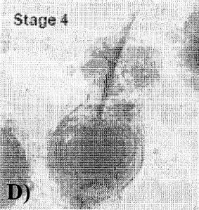
D) Stage 4: Within the Neofolllicle a clearly visible formation of a primitive
hair shaft
becomes visible.
Fig. 4 shows:
Formation of functional neopapillae at two different time points;
A) after 48hours condensation; and
B) after 7 days of condensation; at day 7, the aggregation of cells is much
more dense and
the formation of self derived extracellular matrix becomes visible.
EXAMPLES:
EXAMPLE 1: Isolation and culture of human follicular Dermal Papilla
Fibroblasts (DPF),
Connective Tissue Sheath Fibroblasts (CTSF), Keratinocytes (KC), and
Melanocytes (MC)
Single hair follicles were obtained after micro-dissection of human scalp
samples from
excess of lifting surgeries, received under required regulations. To isolate
matrix KC and MC,
CTSF and fibroblasts of the dermal papilla (DPF), the skin was cut at the
dermosubcutaneous interface with a scalpel and hair follicles at anagen stage
(growing
phase) were pull out with forceps under a dissecting microscope.
In contrast to previously described isolation techniques (e.g. Magerl et al.,
Methods Exp.
Dermat. 11, 381-385, 2002), pure fractions of the desired cell populations,
were obtained by
longitudinally slicing the connective tissue of the upper part of the
dissected hair follicle. By
fixing the hair shaft on the one site, the CTS can be diametrically pulled
over the hair matrix
with forceps towards the lower proximal portion. With this technique DP cells
were
automatically uncovered avoiding damage and mixing cell types. Thus, the CTS
and the
dermal papilla as well as the hair matrix containing KC and MC cells were
clearly separated
and can easily been cut off by a scalpel.
CA 02719769 2010-09-27
WO 2009/118283
PCT/EP2009/053363
21
DP and CTS isolates were separately cultured into 6-well cell culture plates
(2-4 in one well)
by fixing them to the culture plate with a needle. To enable a fast outgrowth
but keeping the
capsule morphology and niche of the fibroblast at the same time, the thin
membrane was
slightly scratched to loosen extracellular matrix components (ECM). The cells
were
submerged with DMEM+ (Gibco/lnvitrogen) plus 10% fetal calf serum (FCS) until
growth of
fibroblast was observed after 1-2 weeks based on donor variations. The cells
were then
moved to a 25cm2 culture flask for another week and were further passaged to a
75 cm2
culture flask. After reaching sub-confluence passage 3 were split into three
75 cm2-flasks
obtaining 1.5 - 2 million DPF or CTSF, respectively.
The KC and amelanotic hair follicle MC were removed from the remaining hair
shaft and the
attached hair matrix by trypsinisation (0.05 % trypsin and 0.53 mM EDTA) and
were
separated by differential trypsinisation and cultured with standard methods as
described in
Tobin et al., J. Invest. Dermatology 104(1), 86-88, 1995.
EXAMPLE 2: Formation of Neopapillae
The optimized count of 500.000 DPF were seeded into a 75cm2 ultra-low
attachment culture
flask (Corning) containing DMEM+ and were allowed to form cell aggregates.
After 48 hours
keeping them unmoved these aggregates shape the size of a native human hair
follicle
dermal papilla. Condensates were then transferred to a 6-well ultra-low
attachment plate and
a mixture of Laminin (final concentration 11.5 pg/rin1), Fibronectin (final
concentration 10
pg/ml) and Collagen IV (final concentration 40 pg/ml) is added to the wells.
After 24-48 hours
in culture, intrinsic ECM secretion and the added proteins built a stable
matrix envelop and
Neopapillae have formed. To facilitate faster ECM accumulation and
differentiation, growth
factors i.e. Hepatocyte Growth Factor (30ng/m1) and/or Connective Tissue
Growth Factor (20
ng/ml) can alternatively been added to the medium. These Neopapillae are ready
to be
implanted into skin in vivo to develop hair inductive properties.
EXAMPLE 3: Formation of Neofollicles
250.000 KC and MC (10:1) were added to the Neopapillae in ultra-low attachment
culture
flask (6 well, Corning) and DMEM+ Medium was changed to Defined Keratinocyte
Serum-
CA 02719769 2010-09-27
WO 2009/118283
PCT/EP2009/053363
22
Free Medium (Gibco). After 1 week the covered Neopapillae form hair follicle
like structures.
These Neofollicles could already been used for testing. To elaborate a
multilayered
Neofollicle, which even better mimics hair follicle structure and function can
be obtained by
coating the generated Neofollicle with a mixture of collagen IV (60pg/m1) with
CTSF (200.000
cells) in ultra low attachment plates with DMEM+ 10% FCS for further 2 days
and change of
medium every further 3rd day.
EXAMPLE 4: Generation of skin equivalents with Neofollicles
Juvenile Human Foreskin Fibroblasts were isolated using previously described
methods
(Toma et al., Stem Cells 23, 727-37, 2005) and cultured in DMEM+10 /0 FCS.
250.000
dermal fibroblast (also DPF or CTSF were used, but foreskin fibroblasts were
preferred for
handling reasons) were mixed to a Fibrinogen solution (Sigma, 3 mg/ml) and 2.0
% (v/v)
Aprotinin (20 pg/ml) and 2.5 % (v/v) Thrombin 1.25 Wm! were added. This
chilled solution
was filled in transwell culture chambers avoiding air bubbles. After adjusting
the temperature
for 2-3 min at room temperature, 30-40 Neofollicles were gently introduced to
the solution
and polymerization to a gel was done for 10 min at 37 C. The composed matrix
was soaked
with DMEM+ medium for 48 h.
Dermal Keratinocytes were isolated from skin biopsies obtained from face lift
surgeries or
from foreskin (Barrandon & Green, Proc Natl. Acad. Sci. 82, 5390-4, 1985).
Briefly, the
underlying fatty tissue and dermis were cut off and the remaining epidermis
was digested
overnight in a Trypsin/EDTA solution (PAA) at 4 C. KC were harvested using a
cell scraper
and after passing through a 70pm cell strainer (Becton Dickinson) they were
seeded onto
collagen I coated culture flasks. Defined Keratinocyte Serum-Free Medium
(Gibco) was
changed twice a week and KC were passaged or harvested at 60-80 % confluence.
KC (250.000) were added on top of the matrix and let them adhere for 24 hours.
Excessive
cells were taken by changing the Defined Keratinocyte Serum-Free Medium
(Gibco) and
after having reached confluence, the transwell chambers were lifted to the air-
liquid
interphase to enable KC differentiation.
=
= CA 02719769 2016-01-18
23
Also cell lines (i.e. HaCat for KC and HS27 for fibroblasts) were tested for
generating a
similar skin model as they are easier to culture and reduce donor variations.
They were
grown in DMEM+ (Gibco) supplemented with 5 % fetal calf serum (FCS, Gibco).
EXAMPLE 5: Formation of Neofollicles comprising cells not derived from
mammalian hair
follicles
Neofollicles have been prepared as described in Example 3 using KC and MC
obtained as
outlined below.
Cell Culture Human Foreskin Keratinocytes and Fibroblasts:
Human Foreskin Fibroblasts and Keratinocytes were isolated from circumcisions
using
previously described methods (Toma et al., Stem Cells 23, 727-37, 2005 ,
Barrandon and
Green, Proc Natl Acad Sci. 1985, 82:5390-4. ). Briefly the underlying fatty
tissue and dermis
were cut off and the remaining epidermis was digested overnight in a DispaseTM
solution (4mg/ml, Sigma ) at 4 C overnight. Keratinocytes were then harvested
using a cell
scraper and after passing through a 70m cell strainer (Becton Dickinson) are
then
seeded into culture flasks. All Keratinocytes were grown in Collagen I coated
cell
culture flasks and Defined Keratinocyte Serum-Free Medium (Gibco). Medium was
changed twice a week and the Keratinocytes were passaged or harvested at 60-
80%
confluence. Fibroblasts were cultured in DMEM+10% FCS.
Cell Culture Human Epidermal Melanocytes:
Starting from a cryovial of Human Adult Epidermal Melanocytes (purchased from
CellMade)
cell culture procedures were done according to the manufacturers protocol.
Briefly, 15 ml
Melanocytes Growth Medium was added to a T75 culture flask. The cells were
thawed
quickly by placing the lower half of the vial in a 37 C water bath for
1minute. The cells were
the resuspended in into the T-75 flask containing Melanocytes Growth medium.
The T-75
flask was placed in a 37 C, 5% CO2 humidified incubator. The Melanocytes
Growth Medium
was changed every 2-3 days. The cells were subcultured when the culture
reached 80%
confluent.
Resulting neofollicles are depicted in Fig. 3. were the different stages of
development of
Neofollicles produced using KC and MC derived from sources different than
mammalian hair
CA 02719769 2010-09-27
WO 2009/118283
PCT/EP2009/053363
24
follicle are shown. In stage 1, Neopapilla (without the addition of exogenous
extracellular
matrix proteins) are surrounded by Melanocytes and keratinocytes, which have
been added
to the ultra low attachment plate forming a early multi-cellular condensate. A
loose aggregate
has been created. In stage 2, first signs of the formation of follicular
structures in ultra low
attachment culture can be seen. The attached cells adhere to the Neopapilla
and adopt a
flattened shape while a protuberance has been built on top of the condensate.
After
approximately 1 week, the Neofollicle begins to form comprising hair follicle-
like sheaths
development. Within the established Neofolllicle, a clearly visible primitive
hair shaft
becomes visible.
EXAMPLE 5: Gene expression analysis of different human dermal papilla derived
cell
samples using Agilent Whole Human Genome Oligo Microarrays (one-color)
1. SuperAmp TM RNA amplification
Two isolated native Dermal Papillae, lx 103 monnolayer-cultured Dermal Papilla
Fibroblasts,
re-condensed Dermal Papilla Fibroblasts after 48 hours and re-condensed Dermal
Papilla
Fibroblasts after 14 days were prepared as described above. The Four human
cell samples
were lysed using SuperAmp TM Lysis Buffer.
Sample no. Cell sample ID
1 DP 1
2 MONO 2
3 KOND 1 3
4 KOND 2 4
Table 2: List of samples
SuperAmp RNA amplification was performed according to Miltenyi Biotec's
procedure.
Briefly, the amplification is based on a global PCR protocol using mRNA-
derived cDNA.
mRNA was isolated via magnetic bead technology. Amplified cDNA samples were
quantified
using the ND-1000 Spectrophotometer (NanoDrop Technologies).
CA 02719769 2010-09-27
WO 2009/118283
PCT/EP2009/053363
Cell Concentration Ratio V olume ( pi) Total amount
sample (ng/pL) (260/280) of cDNA (pg)
1 161.92 1.81 20 3.2
2 143.79 1.86 20 2.9
3 123.06 1_83 20 2_6
4 117.41 1.84 20 2.3
Table 3: Summary of cDNA yields
The integrity of the cDNA was checked via the Agilent 2100 Bioanalyzer
platform (Agilent
Technologies). The results of the Bioanalyzer run have been analysed using a
gel image and
an electropherogram using the Agilent 2100 Bioanalyzer expert software. The
average length
of the highly amplified cDNA products ranged between 200-1,000 bp.
2. Hybridization of Agilent Whole Genome Oligo Microarrays
250 ng of each of the cDNAs were used as template for Cy3 labeling which was
performed
according to Miltenyi Biotec's protocol. The Cy3- labeled cDNAs were
hybridized overnight
(17 hours, 65 C) to an Agilent Whole Human Genome Oligo Microarrays 4 x 44K
(table 4)
using Agilent's recommended hybridization chamber and oven.
Experiment no. Cy3 , , Microarray no.
1 1 251485031842 1 1
2 2 251485031842 1 2
3 3 251485031842 1 3
4 4 251485031842 1 4
Table 4: Hybridisation schedule
Finally, the microarrays were washed once with 6x SSPE buffer containing
0.005% N-
lauroylsarcosine for 1 min at room temperature followed by a second wash with
pre-heated
0.06x SSPE buffer (37 C) containing 0.005% N-lauroylsarcosine for 1 min. The
last washing
step was performed with acetonitrile for 30 sec.
CA 02719769 2010-09-27
WO 2009/118283
PCT/EP2009/053363
26
3. Scanning results
Fluorescence signals of the hybridized Agilent Microarrays were detected using
Agilent's
Microarray Scanner System (Agilent Technologies).
4. Image and data analysis
The Agilent Feature Extraction Software (FES) was used to read out and process
the
microarray image files. The software determines feature intensities (including
background
subtraction), rejects outliers and calculates statistical confidences. For
determination of
differential gene expression FES derived output data files were further
analyzed using the
Rosetta Resolver gene expression data analysis system (Rosetta Biosoftware).
This
software offers ¨ among other features ¨ the possibility to compare two single
intensity
profiles in a ratio experiment. All samples were labeled with Cy3, here, the
ratio experiments
are designated as control versus (vs.) sample experiments (automated data
output of the
Resolver system). Please note, that the ratios are always calculated by
dividing sample
signal intensity through control signal intensity.
5. Gene lists Single-experiment raw data list Single-experiment normalized
data list Gene
ratio list / Pre-selected candidate gene list
The output data of the Agilent Feature Extraction software includes gene lists
with the
complete raw data sets, referred to as single-experiment raw data list.
Furthermore, the
signal intensities from the single-experiment raw data lists are normalized by
dividing the
intensity values by their median. These normalized signal intensities are
joined to a common
table the single-experiment normalized data list. This list comprises in
addition to the
normalized intensity values the feature gIsPosAndSignif which indicates with a
value = 1 that
the signal intensity is positive and significant above the background and a
value = 0 that the
signal intensity is not positive and significant above the background. The
Resolver Software
allows the export of a gene list with all normalized sample/controllog10
ratios and -fold
changes, sequence descriptions, p-values, etc., referred to as gene ratio list
(of all genes).
For example: A "-10 fold change" in the gene ratio lists therefore indicates a
10-fold higher
gene expression in the control compared to the sample. Putative candidate
genes with a fold
change >2 and p-value <0.01 are summarized in a pre-selected candidate gene
list. An
extract of such a list comprising some of the differentially expressed genes
is shown in table
5.
CA 02719769 2010-09-27
WO 2009/118283 PCT/EP2009/053363
27
Gene Name Cellular Function Ratio Mono Ratio 48 h Ratio 14 d Biological
Process
COMP Matrix -26,07 -100 -16 nancollagenous extracellular
matrix glycoprotein, matrix integrity, cell
adhesion
MMP10 Matrix -43,08 -12,25 8,47 matrix metalloproteinase DAMP)
family are involved in the of extracellular
matrix (tissue) remodeling
MOP Matrix -100 -8,25 -3,87 Extracellular matrix strudural
constituent, Multicellular organismal
development, Cell differentiation
CI)K8 Cell Cycle 23,89 16,07 4,47 member of the cydin-
dependent protein kinase (CDK) family, Regulation of
transcription,
CDH3 Adhesion -100 -52,61 -34,49 DI( ion binding, Regulation
of transcription,
PCDHB5 Adhesion -100 -100 -10,12 Protoodherin beta 5, specify
differential cell-cell connections
L1CAM Adhesion 22,49 - 7,95 -1,65 Regulation of actin
cytoskeleton
PCDH20 Adhesion :18,97 -27,7 1,76 Protocadherin 20,
establishment and function of specific cell-cell
connections
PECAM1 Adhesion -71,51 -100 -46,87 Cell to Cell Adhesion
Signaling
lag2 Cytokine -100 -100 -21 Impact in: cell differentiation,
Notch signaling pathway, Regulation of
apoptosis, cell proliferation, Cell communication, Multicellular organismal
development, Cell cycle, Cell fate determination, Morphogenesis of
embryonic epithelium, cell adhesion, cell migration
INfSF10 Cytnkine -75 -100 -10,24 Apoptosis
LEFI Transcription Factor -100 -100 -8,5 LEFI is a nudear protein,
Regulation of Writ receptor signaling pathway
SPRYI Growth Factors -100 -100 -9,25 Sprouty regulation of
tyrosine kinase signals
CTGF Growth Factors 2 -20 4 Connective tissue growth factor,
major connective tissue mitoattractant,
Response to wounding, Proteinaceous extracellular matrix
Table 5: Level (Ratio) of defined genes measured by microarray analysis within
fibroblasts
(monolayered, condensed 48hours, condensed 14 days) compared to native dermal
papilla
fibroblasts.
What can be deduced from table 5 is that with prolonged culture time,-the
expression level of
genes involved in three dimensional .arrangement and tissue formation is
approaching the
expression level observed in native dermal papilla fibroblasts.
CA 02719769 2010-09-27
WO 2009/118283
PCT/EP2009/053363
28
Table 1: DMEM +- Dulbecco's modified Eagle Medium ¨ Composition (Gibco)
COMPONENTS
Molecular Weight Concentration (mg/L) Molarity (mM)
Amino Acids
Glycine 75 37.5 0.500
L-Alanine 8.9
L-Arginine hydrochloride 84
L-Asparagine 13.2
L-Aspartic acid 13.3
L-Cystine 2HCI 63
L-Glutamic Acid 14.7
L-Histidine hydrochloride-H20 42
L-Isoleucine 105
L-Leucine 105
L-Lysine hydrochloride 146
L-Methionine 30
L-Phenylalanine
66
L-Proline 11.5
L-Serine 52.5
L-Threonine 95
L-Tryptophan 16
L-Tyrosine disodium salt dihydrate 104
L-Valine 94
Vitamins
Ascorbic Acid phosphate 2.5
Choline chloride 4
D-Calcium pantothenate 477 4 0.00839
Folic Acid 441 4 0.00907
i-lnositol 7.2
Niacinamide 4
Pyridoxine hydrochloride 4
Riboflavin 0.4
Thiamine hydrochloride 4
Inorganic Salts
CA 02719769 2010-09-27
WO 2009/118283
PCT/EP2009/053363
29
Calcium Chloride (CaCl2) (anhyd.) 111 200 1.801
Ferric Nitrate (Fe(NO3)3"9H20) 0.1
Magnesium Sulfate (MgSO4) (anhyd.) 97.67
Potassium Chloride (KCI) 400
Sodium Bicarbonate (NaHCO3) 3700
Sodium Chloride (NaCl) 6400
Sodium Phosphate dibasic (Na2HPO4-H20) 125
Proteins
AlbuMAX0 II 400
Human Transfenin (Holo) 7.5
[i
Insulin Recombinant Full Chain 10
Trace Elements
Ammonium Metavanadate 0.0003
Cupric Sulfate 0.00125
Manganous Chloride 5
Sodium Selenite 0.005
1
Other Components
D-Glucose (Dextrose) 4500
Ethanolamine 1.9
1
Glutathione (reduced) 307 1 0.00326
Phenol Red 15
Sodium Pyruvate 110
Penicillin I Streptomycin 100u/m1 I 100u/m1
GlutaMAX TM