Note: Descriptions are shown in the official language in which they were submitted.
CA 02719832 2010-11-03
ACCESS DEVICE INCLUDING AN INTEGRATED LIGHT SOURCE
BACKGROUND
1. Technical Field
[00021 The present disclosure relates to medical devices including an
integrated light
source. More particularly, the present disclosure relates to an access device
including an
integrated light source.
2. Background of Related Art
[00031 Minimally invasive procedures are procedures that are performed through
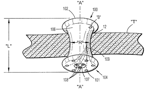
small
incisions in a patient's skin. Such procedures have several advantages over
traditional, open
surgeries, such as reduced trauma to the patient and a faster recovery time.
Generally, such
procedures are referred to as "endoscopic", unless performed on the patient's
abdomen.
Procedures that are performed on the abdomen are referred to as
"laparoscopic". Throughout the
present disclosure, the term "minimally invasive" should be understood to
encompass both
endoscopic and laparoscopic procedures.
[00041 During a typical minimally invasive procedure, surgical objects, such
as surgical
access devices (e.g., trocar and cannula assemblies) or endoscopes, are
inserted into the patient's
body through an incision in tissue or into a naturally occurring orifice
(e.g., mouth, anus, or
vagina). In general, prior to the introduction of the surgical object into the
patient's body,
-1-
CA 02719832 2010-11-03
insufflation gases are used to enlarge the area surrounding the target
surgical site to create a
larger, more accessible work area.
[0005] Minimally invasive procedures are complicated by a number of visual
limitations.
In particular, the surgeon's view is limited to what is directly in front of
and is illuminated by a
scope inserted in the incision. To inhibit damage to internal body structures,
it is desirable to
illuminate the surgeon's viewing field to a greater degree than is currently
possible.
SUMMARY
[0006] Disclosed herein is a seal anchor member that is configured and adapted
for
placement within a tissue tract. The seal anchor member includes a housing
that includes leading
and trailing ends. One or both of the trailing and leading ends may define
surfaces that are
substantially convex or concave to assist in the insertion of seal anchor
member the within tissue
tract. An intermediate portion is positioned between the leading and trailing
ends and is adapted
to be positioned within the tissue tract in a substantially sealed relation
with tissue surfaces that
define the tissue tract. At least one longitudinal port extends between the
leading and trailing
ends, and is adapted to receive an instrument therein in a substantially
sealed relation.
[0007] The seal anchor member may be formed from a compressible material that
is
adapted to transition between a first expanded condition and a second
compressed condition.
The first expanded condition facilitates securing of the seal anchor member
within the tissue
tract. The second compressed condition facilitates at least partial insertion
of the seal anchor
member within the tissue tract.
[0008] At least one light source is operatively associated with the leading
end. The light
source may be operatively coupled to either an external or an internal power
source, e.g., a
battery. The at least one light source may be a light emitting diode (LED) or
may be a fiber optic
-2-
CA 02719832 2010-11-03
cable that is operatively coupled to an external light source. Other light
sources as are known in
the art may be used as well. The light source may be selectively positionable,
i.e.., reoriented
with respect to the leading end, to facilitate the illumination of particular
areas of interest within
the body cavity.
[0009] The light source may emit light having any known wavelength. For
example, the
light source may emit visible, ultraviolet, or near-infrared light. In an
embodiment, the light
source may emit light adapted to polymerize a sealant or an adhesive. In
another embodiment,
the light source may emit light adapted to excite a biomarker or an
immunological agent or dye.
In addition, in certain applications, the light sources may emit more than one
type, i.e,
wavelength of light. Moreover, the intensity of the light emitted from the
light sources may be
adjusted before or during a procedure, i.e., decreased or increased as
desired, or otherwise
regulated. In addition, the intensity and wavelength, as well as the
activation, of the light sources
may be synchronized to the use or activation of other instruments, e.g.,
camera or light detection
systems.
[0010] These and other features of the apparatus disclosed herein will become
more
readily apparent to those skilled in the art from the following detailed
description of various
embodiments of the present disclosure.
BRIEF DESCRIPTION OF THE DRAWINGS
[0011] Various embodiments of the present disclosure are described hereinbelow
with
reference to the drawings, wherein:
[0012] Fig. I is a front perspective view of an embodiment of a seal anchor
member in
accordance with the present disclosure shown positioned within tissue;
[0013] Fig. 2 is a bottom view of the seal anchor member of Fig. 1; and
-3-
CA 02719832 2010-11-03
[0014] Fig. 3 is a front perspective view of an access system including seal
anchor
member and a light source in accordance with the present disclosure.
DETAILED DESCRIPTION OF EMBODIMENTS
[0015] In the drawings and in the description which follows, in which like
references
numerals identify similar or identical elements, the term "proximal" will
refer to the end of the
apparatus which is closest to the clinician during use, while the term
"distal" will refer to the end
which is furthest from the clinician, as is traditional and known in the art.
[0016] A seal anchor member 100 will now be described with reference to Figs.
1-2.
Seal anchor member 100 is configured and adapted to be inserted within a
tissue tract 12 defined
by tissue surfaces formed in tissue "T", e.g., an incision. Although the
presently described seal
anchor member 100 is discussed in connection with minimally invasive
procedures, it is within
the scope of the present disclosure that the seal anchor member may be used
through a naturally
occurring opening (e.g., anus or vagina) or any incision in a patient's skin.
[0017] Seal anchor member 100 defines a longitudinal axis "A" and has
respective
trailing (proximal) and leading (distal) ends 102, 104. Intermediate portion
106 is disposed
between the trailing and leading ends 102, 104. As depicted in Fig. 1,
trailing and leading ends
102, 104 define substantially planar surfaces. However, in other embodiments,
either or both of
the trailing and leading ends 102, 104 define surfaces that are substantially
convex or concave to
assist in the insertion of seal anchor member 100 within tissue tract 12. A
working chamber 107
may include one or more lumens 108 that are longitudinally disposed between
the trailing and
leading ends 102, 104 and are adapted to receive a surgical instrument in a
substantially sealed
relation.
-4-
CA 02719832 2010-11-03
[0018] Intermediate portion 106 defines a radial dimension "R" and extends
longitudinally between the trailing and leading ends 102, 104 to define an
axial dimension or
length "L". As shown in Fig. 1, the radial dimension "R" of intermediate
portion 106 varies
along the length "L", i.e., the cross-sectional dimension may vary along
length "L" and may
have an hour-glass configuration. The hour-glass configuration of the
intermediate portion 106
facilitates anchoring of the seal anchor member 100 within tissue tract 12
defined by tissue
surfaces formed in tissue "T". However, in other embodiments, the radial
dimension "R" may
remain substantially uniform along the length "L".
[0019] The seal anchor members 100 may be formed from a compressible material
and
may be adapted to transition between a first expanded condition to facilitate
securing of the seal
anchor member 100 within the tissue tract 12 and in a substantial sealed
relation with tissue
surfaces "T" defining the tissue tract 12, and a second compressed condition
to facilitate at least
partial insertion of the seal anchor member 100 within the tissue tract 12.
[0020] As shown in Figs. 1 and 2, one or more light sources 101 may be
disposed on the
distal end 104 of the seal anchor member 100. Light sources 101 maybe chosen
from any
known light source, including but not limited to, light emitting diodes (LEDs)
and incandescent
lights.
[0021] As shown in Fig. 2, the light sources 101 are positioned within the
leading end
104. The one or more light sources 101 may be selectively positioned with
respect to the leading
end 104. As such, one or more of the light sources 101 may be rotated or
axially repositioned
with respect to the distal end 104 of the seal anchor member. It is also
contemplated that one or
more of the light sources 101 may be radially repositionable with respect to
the seal anchor
member 100. In this configuration, the light source 101 provides a larger
illumination field
-5-
CA 02719832 2010-11-03
thereby increasing the illumination of the working space beyond the distal end
104 of the seal
anchor member 100. By adjusting the orientation of the light sources 101 with
respect the
leading end 104, the illumination of particular internal body structures is
facilitated.
[0022] An internal battery "B", shown in Fig. 1, may power the light sources
101 and
may be operatively coupled to the light sources through power leads 109. In
other embodiments,
each of the light sources 101 may have its own power source. In still other
embodiments, the
light sources 101 may be powered by a power source that is external to the
seal anchor member
100.
[0023] Light source 101 may be selected to have a desired wavelength depending
on its
intended use. For example, some procedures may involve the use of light
sensitive materials that
are responsive to certain wavelengths of light and the light source 101 may be
selected
accordingly. Some procedures may involve the application of light sensitive
sealants and/or
adhesives. For example, photocurable surgical tissue adhesives that are
composed of
photoreactive gelatin and poly (ethylene glycol) diacrylate may be used in
laparoscopic
procedures to effect prompt and effective hemostasis. For such procedures,
light sources 101
may emit light of any known wavelength of light including, but not limited to,
ultraviolet,
visible, and near-infrared light.
[0024] The light sources 101 are integrated into the seal anchor member 100
and may
emit light having the same, different, or variable wavelengths depending on
the application.
Therefore, two or more light sources 101 having different wavelengths and/or
properties may be
included within a single seal anchor member 100. For example, one light source
101 may be
adapted to emit visible light, e.g., white light, for traditional
illumination, while other light
sources 101 may be adapted to excite biomarkers or immunological agents/dyes
adapted to mark
-6-
CA 02719832 2010-11-03
certain structures, e.g., vasculature, lymph, nerve, and neural networks, or
conditions, e.g.,
tumors or other diseases.
[0025] During a procedure, a clinician may activate, deactivate, and regulate
the light
emitted from light sources 101. For example, the intensity of the light
emitted from the light
sources 101 may be adjusted during the procedure. Some procedures may include
the
application of biologically active compounds that are light-responsive. By
adjusting the light
exposure, the concentration of the active form of such compounds may be
controlled. Some
molecules can either be can either be irreversibly activated with light or
reversibly switched
between active and inactive states.
[00261 In a further embodiment, the light sources 101 may be adapted to be
synchronized
with other instruments such as camera and/or light detection systems (not
shown) to provide on
demand direct local illumination and visualization as needed. For example,
operation of the
camera may be synchronized with the emission of light into the body space on
which the
procedure is being performed. Wireless or other means may be used to
synchronize the light
sources 101 with the other instruments.
[0027] In yet another embodiment, as illustrated in Fig. 3, an access system
200 includes
a seal anchor member 201, a light source 210, and fiber optic cables 211. The
seal anchor
member 201 is substantially similar to the seal anchor member 100 described
above, and is
adapted to be inserted within a tissue tract in a substantially identical
manner as described above
with reference to seal anchor member 100.
[00281 The differences between the seal anchor members 100, 201 will now be
described. In particular, the seal anchor member 201 includes trailing and
leading ends 102, 104,
an intermediate portion 106, a working chamber 107 may include one or more
lumens 108, and
-7-
CA 02719832 2010-11-03
one or more fiber optics disposed within the seal anchor member 201
operatively coupled to an
external light source 210 and terminating in the leading end 104.
[00291 Although light source 210 is shown external to the seal anchor member
201, in
other embodiments the light source to which the fiber optic cables 211 are
connected may be
integral to the seal anchor member 201. Each of the fiber optics 211 may be
adapted to emit
light of a wavelength suitable for its intended use.
[00301 Although the illustrative embodiments of the present disclosure have
been
described herein with reference to the accompanying drawings, the above
description, disclosure,
and figures should not be construed as limiting, but merely as
exemplifications of particular
embodiments. It is to be understood that the disclosure is not limited to
those precise
embodiments, and that various other changes and modifications may be effected
therein by one
skilled in the art without departing from the scope or spirit of the
disclosure.
-8-
