Note: Descriptions are shown in the official language in which they were submitted.
CA 02720184 2016-07-14
PREDICTIVE MARKER FOR TOPOISOMERASE I INHIBITORS
FIELD OF THE INVENTION
[002]The present invention relates generally to the fields of cancer therapy
and cancer prevention.
More particularly, the present invention generally relates to a diagnostic
marker for predicting the
efficacy of topoisomerase I (topo I) inhibitors in the treatment of cancers,
and the level of sensitivity
or resistance of a biological sample to topoisomerase I inhibitors.
BACKGROUND OF THE INVENTION
[003]One of the main problems associated with cancer chemotherapy is that
individual subjects with
the same histology do not respond identically to a given agent or a given
therapeutic protocol. The
response range may vary in large proportions. even in chemosensitive tumors
such as breast cancer.
A number of determinants of drug sensitivity are well known, such as drug
dose, drug combinations
and schedule of administration, subject age and status, tumor localization
etc, but the intrinsic
sensitivity of a given tumor is a major factor in which remains difficult to
evaluate.
[004)One strategy to improve the effectiveness of treatment has been to
individualize drug treatment
as a function of the sensitivity of tumor cells. Methods to predict how
effective a drug may be in a
subject are typically based on in vitro or ex vivo testing of the tumor cells
(taken during a biopsy) to a
battery of drugs and chemotherapy agents. Such strategies have several
limitations: they are often
poor predictors of chemosensitivity in vivo, they are time-consuming, and both
manually and cost
expensive. The identification of novel cancer subtypes promises to provide
more specific, more
effective and less toxic therapies. This tumor subset is refractory to
commonly used chemotherapeutic
agents and therefore is associated with a poor prognosis (Sorlie, et al.,
2001, Proc Natl Acad Sci U S
A 98:10869).. To date little progress has been made in identifying specific
molecular pathways
associated with these refractory cancers that may be effectively targeted for
therapeutic purposes.
[005]Human topoisomerase I (topoI) is an essential and ubiquitous enzyme that
is involved in various
DNA transactions, The identification of topoI as the target of a new class of
anti-neoplastic drug
(camptothecin, also referred herein as "CPT") has led to the rapid development
of topoI structure-
function in the context of cancer therapy. Two CPT analogues, topotecan and
irinotecan, are currently
1
CA 02720184 2010-09-30
WO 2009/124064 PCT/US2009/038981
used in clinics for small cell lung cancer (SCLC), colon and ovarian cancer
and in several refractory
cancers, including breast and cervical. However, like most cancer drugs, not
all patients respond, in
this case only about 30% of patients respond to topoI inhibitors. The topoI
protein level is high in
most solid tumors, and thus topoI levels can not be used as a predictive
marker. Additionally,
although topoI is the specific target of CPT, the expression profile of topoI
does not provide
prognostic index. Based on the preclinical studies, it is likely that clinical
resistance to these drugs
might be the result of (1) inadequate accumulation of the drug in the tumor,
or (2) post-translational
modification of topoI. It has been demonstrated that topoI is ubiquitinated
and degraded in cells in the
response to CPT by ubiquitin proteosomal pathway (UPP). Importantly, the rate
of UPP mediated
degradation varies in different cancer cells and is correlated to the CPT
sensitivity. However the
mechanism of ubiquitination dependent proteosomal degradation of topoI in the
response to CPT is
not understood.
[006]There is a significant need in the art for a satisfactory treatment of
tumor subsets refectory or
non-responsive to commonly used chemotherapeutic agents, (specifically in
epithelial cell cancers
such as breast, lung, ovarian, brain, colon and prostate cancers), which
overcomes the non-
responsiveness exhibited by subjects. Such a treatment could have a dramatic
impact on the health of
individuals, especially older individuals, among whom cancer is especially
common, and females
whom have a high incidence of breast cancer.
SUMMARY OF THE INVENTION
[007]The present invention generally relates to a diagnostic marker for
predicting the efficacy of
topoisomerase I (topo I) inhibitors in the treatment of cancers. In
particular, one aspect of the present
invention relates to methods to determine effectiveness of topoisomerase I
(topo I) inhibitors in the
treatment of cancer. Specifically, the inventors have discovered herein that
topoI is phosphorylated at
S10 by DNA-PK and results in its ubiquitination by BRAC1/BARD1 heterodimer and
its subsequent
degradation. The inventors have discovered that the presence of
phosphorylation of topoI, in
particular the phosphorylation of topoI on serine 10 (S10) indicates rapid
topoI degradation and
resistance to topo I inhibitors such as camptothecin (CPT), or CTP analogues
such as topotecan and
irinotecan and derivatives thereof. Accordingly, one aspect of the present
invention relates to
detection of phosphorylation of topoI on serine 10, i.e. the detection of
phospho-topoI-S10 in a
biological sample from a subject with cancer, as a prognostic determinant for
drug efficacy with topo
I inhibitors such as CTP and its analogues.
[008]One aspect of the present invention relates to methods and compositions
to determine if a
topoisomerase I inhibitor is effective in a subject with cancer. One aspect of
the present invention
relates to a method to determine the presence of phosphorylation of topo I
polypeptide, wherein the
presence of phosphorylation of a topo I polypeptide indicates that the subject
having such cancer will
2
CA 02720184 2010-09-30
WO 2009/124064 PCT/US2009/038981
likely be nonresponsive (or unresponsive) to a topo I inhibitor as compared to
a subject with a cancer
comprising a non-phosphorylated topo I polypeptide. In one embodiment of this
aspect and all other
aspects disclosed herein, the phosphorylation is phosphorylation of a serine
residue of a topo I
polypeptide, wherein the presence of phosphorylation of a serine residue of
the topo I polypeptide
identifies that the cancer will likely be nonresponsive (or unresponsive) to a
topo I inhibitor. In
another embodiment of this aspect and all other aspects disclosed herein, the
phosphorylation is
phosphorylation of the serine 10 (S10) residue of a topo I polypeptide,
wherein the presence of
phosphorylation of the serine 10 reside (S10) of the topo I polypeptide
indicates that a subject with a
cancer will likely be nonresponsive (or unresponsive) to a topo I inhibitor as
compared to a subject
where the corresponding residue is not phosphorylated. Another embodiment of
this aspect and all
other aspects disclosed herein, detection of lack of phosphorylation of the
topoisomerase I
polypeptide, in particular the lack of phosphorylation of a topo I polypeptide
on serine 10 (S10)
indicates such a cancer is likely to be responsive to a topo I inhibitor.
[009] In some embodiments, one presence of phosphorylation of the serine 10
reside (S10) of the topo
I polypeptide indicates that a subject with a cancer will likely be
nonresponsive (or unresponsive) to a
topo I inhibitor as compared to a subject where the corresponding residue is
not phosphorylated. One
aspect of the invention relates to grading the level of phosphorylation of S10
topo I polypeptide in a
subject. For example, the level of phospho-S10 topo I polypeptide as compared
to non-phospho S10
topo I polypeptide can be categorized or graded on 4 levels, where, for
example, level 1 is about a 0%
level phospho-S10 topo I polypeptide and indicates a subject is likely to be
fully responsive to a topo I
inhibitor, level 2 is about a 10-25% level phospho-S10 topo I polypeptide and
indicates a subject is
likely to be partially responsive to a topo I inhibitor; level 3 is a 25-50%
level of level phospho-S10
topo I polypeptide and indicates a subject is likely to be unresponsive to a
topo I inhibitor, and level 4
is any level of level phospho-S10 topo I polypeptide above 50%, for example at
least 50%, or at least
about 60% or at least about 70% or at least about 80% or at least about 90% or
at least about 100%
indicates a subject is likely to be completely non-responsive to a topo I
inhibitor. Stated another way,
a subjects' likelihood of being responsive to a topo I inhibitor can be graded
on the degree of
phospho-S10 topo I polypeptide as compared to degree of non-phospho S10 topo I
polypeptide, which
can be graded on 4 levels; grades 1 (0-10%), 2 (10-25%), 3 (25-50%) or 4
(>50%), where level 1
indicates a subject is likely to be responsive to a topo I inhibitor, where
level 2 indicates a subject is
likely to be partially responsive (i.e. about 50% or less responsive) to a
topo I inhibitor as compared to
level 1, where level 3 indicates a subject is likely to be unresponsive to a
topo I inhibitor as compared
to a subject classified as level 1 (i.e. a subject will likely have about 10%
or less efficacy of a topo I
inhibitor), and where level 4 indicates a subject is likely to be completely
unresponsive to a topo I
inhibitor as compared to a subject classified as a level 1 (i.e. a subject
will likely have about 5% or
about a 2% or less efficacy of a topo I inhibitor). In other embodiments, more
than 4 levels of
3
CA 02720184 2010-09-30
WO 2009/124064 PCT/US2009/038981
classification can be used, for example, at least 5, or at least 6, or at
least 7, or at least 8 or at least 9
levels or more than 9 different classification levels can be used.
[0010] In some embodiments, if a subject is identified as being above a
certain pre-defined threshold
level the subject is likely to be identified to be unresponsive to a topo I
inhibitor. In some
embodiments, a pre-defined threshold level is about level 3, wherein the % of
phospho-S10 topo I
polypeptide (from the total topo I polypeptide) is about 25% or above, a
subject is likely to be
unresponsive to a topo I inhibitor as compared to a subject with a threshold
level below 3 (i.e. less
than 25%). Accordingly, in some embodiments, a pre-defined threshold level to
identify if a subject
is unresponsive to a topo I inhibitor is a 25% or greater, wherein a subject
having a % of phospho-S10
topo I inhibitor to total topo I polypeptide of 25% or greater (i.e. about at
least 30% or at least about
40% or a least about 50% or at least about 60% or more) is identified as being
unresponsive to a topo
I inhibitor, whereas a subject having a % of phospho-S10 topo I inhibitor to
total topo I polypeptide of
less than 25% (i.e. about 20% or about 10% or about 5% or about 2% or less) is
identified as being
responsive, or partially responsive to a topo I inhibitor.
[0011] Another aspect of the present invention relates to a method to treat
cancer in a subject, the
method comprising measuring the level of phosphorylation of a topoisomerase I
polypeptide in a
biological subject comprising cancer cells from a subject; and detecting the
level of phosphorylation
of the topo I polypeptide, in particular detecting the level of
phosphorylation at serine 10 (S10) of the
topo I polypeptide, and if the topo I polypeptide is phosphorylated, for
example at residue S10, the
cancer is identified as being unresponsive to a topoisomerase I inhibitor. The
cancer cells in one
embodiment are taken from a subject and tested using the methods, kits,
machines and computer
systems and computer readable media as described herein.
[0012] A subject identified as being likely to be responsive to a
topoisomerase I inhibitor can be
treated with a therapeutically effective amount of a topo I inhibitor, such
as, but not limited to CPT, or
analogues thereof such as topotecan and irinotecan, either alone or in
combination with other
therapeutic and/or anti-cancer drugs.
[0013] In some embodiments, a topo I inhibitor is a chemotherapy agent, for
example but not limited
to CPT, or analogues thereof such as topotecan and irinotecan and derivatives
as these terms are
defined herein. Another aspect of the present invention provides to methods
for treating and/or
preventing a subject affected with or at risk of developing cancer, the method
comprising determining
the presence of phosphorylation of a topo I polypeptide (phospho-topo I), and
in particular
embodiments, the method comprises determining the presence of phosphorylation
at serine 10 (S10)
residue of a topoI polypeptide (phosphor-S10-topo I). In other embodiments,
the methods, kits,
machines, computer systems and computer readable media are used to determine
the phosphorylation
status of a topo I polypeptide in a biological sample, in particular the
presence of phospho-topo I, and
more specifically, the presence of phospho-S10-topo I polypeptide in a
biological sample.
4
CA 02720184 2010-09-30
WO 2009/124064 PCT/US2009/038981
[0014] In one embodiment, any means known to a skilled artisan can be used to
determine the
phosphorylation status of topo I polypeptide, and in particular the presence
of phospho-S10 topo I
polypeptide in a cancer in a subject. Accordingly, the present invention
encompasses use of any in
vivo detection method, any ex vivo detection method or any in vitro detection
method to determine the
phosphorylation status of topo I polypeptide, and in particular the presence
of phospho-S10 topo I
polypeptide in subject with cancer. In some embodiments, the method is a high
throughput automated
immunohistochemistry method commonly known by a one of ordinary skill in the
art. In another
embodiment, in vivo detection method can determine the phosphorylation status
of topo I polypeptide,
in particular the presence of a phospho-S10 topo I polypeptide in a cancer
present in a subject, by
administering to the subject for example but not limited to, labeled
antibodies such as anti-phospho-
S10 topo I antibodies to the subject, including for example radiolabeled, or
fluorescence-labeled, or
bioluminescence-labeled such as luciferase anti-phospho-S10 topo I antibodies
or alternatively
radiolabeled nucleotides and using a detection module, for example an in vivo
imaging camera or
machine, such as a MRI, CAT scan or other in vivo imaging machines to
determine the presence of
the anti-phospho-S10 topo I antibodies in the subject, where in one
embodiment, the output data from
the detection module is analyzed using a computer system or computer readable
media as disclosed
herein, or in an alternative embodiment, the output data of the detection
module is received by the
storage module which is connected to the comparison module of a machine as
described herein.
[0015] In another embodiment, the phosphorylation status of topo I polypeptide
such as the presence
of a phospho-S10 topo I polypeptide can be determined in a biological sample
taken from a subject,
where a biological sample is placed into a detection module which determines
the phosphorylation
status of topo I polypeptide, such as presence of a phospho-S10 topo I
polypeptide in the biological
sample, where the output data of the detection module is received by the
storage module which is
connected to the comparison module of a machine as described herein, or in an
alternative
embodiment, the output data of the detection module is analyzed by the
computer system and
computer readable media as disclosed herein.
[0016] In some embodiments, a subject identified to be unresponsive to a topo
I inhibitor by the
methods, kits, machines, computer systems and computer readable media as
disclosed herein is
administered a pharmaceutical composition comprising a chemotherapeutic agent
other than a topo I
inhibitor. In an alternative embodiment, a subject can be administered a topo
I inhibitor and an agent
which increases the sensitivity of the subject to a topo I inhibitor (herein
referred to as a "topo I
inhibitor sensitivity agent"), where a topo I inhibitor sensitivity agent can
increase the
dephosphorylation of a topo I polypeptide, in particular, increase the
dephosphorylation of serine 10
(S10) of the topo I polypeptide. In another embodiment, a topo I inhibitor
sensitivity agent can be an
antagonist which inhibits the phosphorylation of topo I polypeptide, such as
for example but not
limited to, an anti-phospho-S10 topo I antibody or an antagonist or inhibitor
of DNA-PK such as, but
not limited to NU7026 also known as 2-morpholin-4-y1)-benzo[h]chromen-4-one,
and derivatives and
CA 02720184 2010-09-30
WO 2009/124064 PCT/US2009/038981
analogues thereof. Accordingly, one aspect of the invention relates to co-
administering a topo I
inhibitor substantially simultaneously with a topo I inhibitor sensitivity
agent to a subject identified to
be unresponsive to a topo I inhibitor. In some embodiments, such a
pharmaceutical composition
comprising a topo I inhibitor can be administered alone or substantially at
the same time, before or
after the administration of a pharmaceutical composition comprising an agent
which increases the
sensitivity of the subject to a topo I inhibitor (i.e. a topo I inhibitor
sensitivity agent such as an agent
which increases the dephosphorylation of S10 on the topo I polypeptide and/or
an agent which
inhibits topo I phosphorylation on S10, such as an anti-phospho-S10 topo I
antibody or an antagonist
of DNA-PK).
[0017] One aspect of the present invention and all other aspect described
herein, a machine can be
used to determine phosphorylation status of topo I polypeptides in a
biological sample, for example, a
machine for obtaining data regarding a biological sample from a subject
comprising: a biological
sample container to hold the biological sample; a determination module
configured to detect the
presence of phosphorylation of a topoisomerase I polypeptide, for example the
detection of phospho-
S10 topo Tin the biological sample which produces output data, in some
embodiments the output data
in a computer readable media format; a storage device configured to store the
output data from the
determination module; a comparison module adapted to compare the output data
from the
determination module with data stored on the storage device, such as stored
reference data and control
data, and a display module for displaying a page of retrieved content for the
user on a client computer,
wherein the retrieved content comprises any one or a combination of the
following; (i) the presence or
absence of phosphorylation of the topoisomerase I polypeptide, for example the
retrieved content is
the presence or absence of phospho-S10 topo I; (ii) the absence of
phosphorylation of topoisomerase
I, for example the absence of phospho-S10 topo I, (iii) the presence of
phosphorylation of
topoisomerase I, such as the presence of phospho-S10 topo I polypeptide, (iv)
a positive test result
(i.e. a positive phosphorylation status such as positive S10 topo I
phosphorylation status) which
indicates that the subject is likely to be more unresponsive to a topoI
inhibitor than a subject having a
cancer with a negative phosphorylation status, (v) a negative test result
(i.e. a negative
phosphorylation status such a negative S10 topo I phosphorylation status)
which indicates that the
subject is likely to be more responsive to a topoI inhibitor than a subject
having a cancer with a
positive phosphorylation.
[0018] One aspect of the present invention is a computer system that can be
used to determine if a
subject is responsive to a topo I inhibitor. In such an embodiment, a computer
system is connected to
a determination module and is configured to obtain output data from a
determination module
regarding a biological specimen, where a determination module is configured to
detect the presence of
phosphorylation of a topoisomerase I polypeptide, for example the presence of
phospho-S10 topo I
polypeptide within a subject or in a biological sample obtained from the
subject; and where the
computer system comprises (a) a storage device configured to store data output
from the
6
CA 02720184 2010-09-30
WO 2009/124064 PCT/US2009/038981
determination module as well as reference data; where the storage device is
connected to (b) a
comparison module which in one embodiment, is adapted to compare the output
data stored on the
storage device with stored reference data, and in alternative embodiments,
adapted to compare the
output data with itself, where the comparison module produces report data and
is connected to (c) a
display module for displaying a page of retrieved content (i.e. report data
from the comparison
module) for the user on a client computer, wherein the retrieved content
comprises any one or a
combination of the following; (i) the presence or absence of phosphorylation
of the topoisomerase I
polypeptide, for example the retrieved content is the presence or absence of
phospho-S10 topo I; (ii)
the absence of phosphorylation of topoisomerase I, for example the absence of
phospho-S10 topo I,
(iii) the presence of phosphorylation of topoisomerase I, such as the presence
of phospho-S10 topo I
polypeptide, (iv) a positive test result (i.e. a positive phosphorylation
status such as positive S10 topo
I phosphorylation status) which indicates that the subject is likely to be
more unresponsive to a topoI
inhibitor than a subject having a cancer with a negative phosphorylation
status, (v) a negative test
result (i.e. a negative phosphorylation status such a negative S10 topo I
phosphorylation status) which
indicates that the subject is likely to be more responsive to a topoI
inhibitor than a subject having a
cancer with a positive phosphorylation.
[0019] In some embodiments the comparison module compares the output data
stored on the storage
device with itself or stored reference data, and calculates a positive S10
topo I phosphorylation status
(i.e. the presence of phospho-S10 topo I polypeptide) which indicates a
positive test result and
generates report data to indicate that the subject is likely to be more
unresponsive to a topoI inhibitor
than a subject having a cancer with a negative phosphorylation status, where
the report data from the
comparison module is retrieved from the display module and displayed on the
display module.
[0020] One aspect of the present invention and all other aspect described
herein, one can use a
computer readable media to determine phosphorylation status of topo I
polypeptides from a subject
having or at risk of having cancer, for example, a computer readable media
having computer readable
instructions recorded thereon to define software modules including a
determination module and a
comparison module for implementing a method on a computer, said method
comprising: a storage
device configured to store data reference data and output data from a
determination module which has
measured the presence or absence of the phosphorylation of topo I polypeptide,
such as the presence
or absence of phospho-S10 topo I polypeptide; a comparison module which
generates report data,
where the comparison module is adapted to compare the data stored on the
storage device, for
example a comparison of output data from the determination module with itself
or alternatively with
reference data, and a display module for displaying a page of retrieved
content which is the report data
from the comparison module for the user on a client computer, wherein the
retrieved content
comprises any one or a combination of the following; (i) the presence or
absence of phosphorylation
of the topoisomerase I polypeptide, for example the retrieved content is the
presence or absence of
phospho-S10 topo I; (ii) the absence of phosphorylation of topoisomerase I,
for example the absence
7
CA 02720184 2010-09-30
WO 2009/124064 PCT/US2009/038981
of phospho-S10 topo I, (iii) the presence of phosphorylation of topoisomerase
I, such as the presence
of phospho-S10 topo I polypeptide, (iv) a positive test result (i.e. a
positive phosphorylation status
such as positive S10 topo I phosphorylation status) which indicates that the
subject is likely to be
more unresponsive to a topoI inhibitor than a subject having a cancer with a
negative phosphorylation
status, (v) a negative test result (i.e. a negative phosphorylation status
such a negative S10 topo I
phosphorylation status) which indicates that the subject is likely to be more
responsive to a topoI
inhibitor than a subject having a cancer with a positive phosphorylation.
[0021] Another aspect of the present invention also relates to a method to
identifying the likelihood
of a cancer to be unresponsive to a topoisomerase I inhibitor, the method
comprising measuring the
level of phosphorylation of topoisomerase I polypeptide in at least one cancer
cell, wherein the
presence of phosphorylation identifies the cancer as being more likely to be
unresponsive to a
topoisomerase inhibitor as compared to a cancer wherein the absence of
phosphorylation of
topoisomerase I is detected.
[0022] Another aspect of the present invention relates to a method for
treating cancer in a subject, the
methods comprising: (i) measuring the level of phosphorylation of
topoisomerase I polypeptide in a
biological sample comprising cancer cells obtained from the subject; (ii)
detecting the level of
topoisomerase I polypeptide, wherein if the topoisomerase I polypeptide is
phosphorylated the cancer
is identified as being unresponsive to a topoisomerase I inhibitor, or wherein
if the topoisomerase I
polypeptide is not phosphorylated the cancer is identified as being likely to
be responsive to a
topoisomerase I inhibitor; (iii) administering to a subject an anti-cancer
agent other than a
topoisomerase I inhibitor where the cancer is identified as being unresponsive
to a topoisomerase I
inhibitor.
[0023] In some embodiments, a topo I inhibitor is an antagonist of a topo I
polypeptide of SEQ ID
NO: 2 or a variant thereof, where an antagonist of a topo I polypeptide is any
agent commonly known
by one of ordinary skill in the art that inhibits the gene expression and/or
the biological activity of a
topo I polypeptide, and includes for example, but are not limited to agents
such as antibodies,
antibody fragments, small molecules, peptides, proteins, antisense nucleic
acids, ribosomes, PNA,
siRNA, oligonucleotides, aptamer, and peptide aptamer and derivatives and
fragments thereof. In
some embodiments, an antagonist of a topo I polypeptide useful in the methods
of the present
invention can be a nucleic acid-based inhibitor, nucleic acid construct, a
peptide-based inhibitor or a
small molecule inhibitor of topo I polypeptide or a polynucleotide encoding
the same. In some
embodiments a nucleic-acid inhibitor may be a RNAi (RNA interference) agent,
such as for example a
siRNA molecule or an antisense construct. Exemplary topo I inhibitors are
disclosed herein, and
include but are not limited to CPT, or analogues thereof such as topotecan and
irinotecan. It is
encompassed that the present invention provides methods, kits, machines,
computer systems and
computer readable media for determining if a subject is responsive to a topo I
inhibitor regardless
what the topo inhibitor is being used for. In some embodiments, the topo I
inhibitor is being used as
8
CA 02720184 2010-09-30
WO 2009/124064 PCT/US2009/038981
an anti-cancer treatment, including therapeutic and prophylactic anti-cancer
treatment, however, a
topo I inhibitor can be used for the treatment of non-cancer diseases or
disorders where use of a topo I
inhibitor is desired, for example any treatment strategy where cell death is
the desired outcome.
[0024] Another aspect of the present invention provides an isolated anti-
phospho-S10 topo I antibody
which binds with specific affinity to the phosphorylated serine residue of SEQ
ID NO: 1 or
phosphorylated serine residue on SEQ ID NO: 4, or to serine 10 (S10) of the
polypeptide of the amino
acid sequence of SEQ ID NO: 2.
[0025] Another aspect of the present invention relates to methods for
increasing the sensitivity of a
subject or a cancer, or cancer cell to a topo I inhibitor, the method
comprising administering to the
subject or a cancer cell a combination of an effective amount of an a topo I
inhibitor and an effective
amount a topo I inhibitor sensitivity agent (i.e. an agent which increases the
sensitivity of a cancer cell
to a topo I inhibitor), where a topo I inhibitor sensitivity agent can include
for example, but is not
limited to, an agent which increases the dephosphorylation of S10 of the topo
I polypeptide and/or an
agent which inhibits topo I phosphorylation on S10, such as an anti-phospho-
S10 topo I antibody or
an antagonist of DNA-PK.
[0026] In some embodiments, the disclosed methods, kits, machines, computer
systems and
computer readable media are useful for determining if a subject is likely to
be responsive to a topo I
inhibitor, and where a topo I inhibitor is used in the treatment of cancer,
the present invention is
useful in the prevention and/or treatment of cancers such as, but are not
limited to, tumors can be
selected from a group of cancers consisting of: SCLC cancer, colon cancer,
ovarian cancer, or a
refractory cancer, for example, breast cancer or cervical cancer. In other
embodiments, a cancer useful
to be treated in the methods as disclosed herein is any cancer which can be
treated, or is desirable to
be treated with a topo I inhibitor and can be selected from any cancer in the
group consisting of:
gastrointestinal cancer, gastric cancer, squamous cell carcinomas (SCC), head
and neck cancer, lung
cancer, non-small cell lung cancer (NSCLC) and small-cell lung cancer (SCLC),
lymphoma, sarcoma,
primary and metastic melanoma, thymoma, non-Hodgkin's lymphoma, Hodgkin's
lymphoma, cancer
of the nervous system, brain cancer, bone-marrow cancer, bone cancer, kidney
cancer, uterine cancer,
cervical cancer, colon cancer, retina cancer, skin cancer, bladder cancer,
colon cancer, esophageal
cancer, testicular cancer, cervical cancer, liver cancer, renal cancer,
pancreatic cancer, genital-urinary
cancer, gastrointestinal, gum cancer, tongue cancer, kidney cancer,
nasopharynx cancer, stomach
cancer, endometrial cancer and bowel tumor cell cancer, adrenocarcinomas such
as prostate cancer,
ovarian cancer, breast cancer, and pancreatic cancer. In particular, the
cancer is breast cancer, for
example the triple-negative subtype of breast cancer. In one embodiment, a
topo I inhibitor is used to
treat cancer. In some embodiment the cancer is epithelial in origin, for
example, the cancer is, but is
not limited to; gastiointestinal cancer, prostate cancer, ovarian cancer,
breast cancer, head and neck
cancer, lung cancer, small-cell lung cancer, cancer of the nervous system,
kidney cancer, retina
cancer, skin cancer, liver cancer, pancreatic cancer, genital-urinary cancer
and bladder cancer.
9
CA 02720184 2016-07-14
=
[0027] In some aspects of the invention, if a subject is identified to be
responsive to a topo I
inhibitor, a pharmaceutical composition comprising a topo I inhibitor as
disclosed herein can be
administered alone or with one or more other therapeutic agents. For example,
in the treatment of
cancer, the pharmaceutical composition can be administered substantially at
the same time as, or
subsequent to administration of an anti-cancer therapy, such as, for example,
chemotherapy,
radiotherapy, hormone therapy, thermal therapy, immunotherapy, surgical
resection and alternative
cancer therapies commonly known by persons of ordinary skill in the art. Such
anti-cancer therapies
can be administered prior to, during or after administration of the
pharmaceutical composition as
disclosed herein. In some embodiments, the anti-cancer therapy is administered
once, or more than
once to the subject.
[0028] It is contemplated that any methods or compositions described herein
can be implemented
with respect of any other methods or compositions. Other objects, features and
advantages will
= become apparent from the following detailed description.
BRIEF DESCRIPTION OF THE FIGURES
[0029] Figures 1A-1E shows Ku 70/80 associates with topo I. Figure IA shows
HeLa cell nuclear
lysates were incubated with GST and GST-topo I bound to glutathione sepharose
beads and after
extensive wash with PBS the adsorbates were eluted with high salt buffer (PBS
with 350 and 500mM
NaC1). The adsorbates were analyzed by SDS-PAGE and silver staining. The
protein bands were cut
and in gel digestion was performed for protein identification by mass
spectrometry. Figure 1B shows
GST and GST- topo I bound to glutathione beads were incubated with purified Ku-
DNA-PK complex
(containing Ku 70/80 heterodimer), the adsorbates were analyzed by
immunoblotting with indicated
antibodies. To determine the protein directly binding to topoI GST, GST-Ku70
and GST-Ku80 bound
to glutathione beads were incubated with topoI protein. Figure 1C shows The
adsorbates were
analyzed by immunoblot analysis with anti-topo I. Figure 1D shows specific
Ku70 binding region of
topo I was determined by incubation of topo I fragments. and Figure lE shows
bound to glutathione
beads with purified Ku-DNA-PK complex, adsorbates were analyzed by
immunoblotting with anti-Ku
70 antibody.
[0030] Figures 2A-2E shows TopoI associates with Ku-DNA-PK complex and with
BRCAL Figure
2A shows HeLa cell lysates were subjected to immunoprecipitation with anti-
Ku70, anti-Ku80. anti-
DNA-PK. Figure 2B shows immunoprecipitation of HeLa cell lysates with anti-
topoI, and
immunoblotting with anti-Ku-80. Figure 2C shows immunoprecipitation of HeLa
cell lysates with
anti-topoI, and immunoblotting with anti-Ku-70. Figure 2D shows
immunoprecipitation of HeLa cell
CA 02720184 2010-09-30
WO 2009/124064 PCT/US2009/038981
lysates with anti-BRCA1 or PIRS (control), and immunoblotting with anti-TopoI.
Figure 2E shows
immunoprecipitation of HA-BRCA1 expressing HeLa cell lysates with anti-HA, and
immunoblotting
with anti-BRCA1. The precipitates were analyzed by immunoblotting with
indicated antibodies.
[0031] Figures 3A-3B shows DNA-PK phosphorylates topoI. Purified GST-topoI
(5pg; left lane)
was incubated with DNA-PK in kinase buffer (20 mM Tris-HC1, pH 7.4, 10 mM
MgC12 and 10 mM
MnC12) containing 117 -32P]ATP or cold ATP for 30 min at 30 C. Figure 3A shows
SDS-PAGE
analysis of the reaction products and autoradiography (left lane). Figure 3A
also shows the
comparison of an identical reaction in the absence of topoI as a control
(right lane). GST-topoI protein
band shown in Fig 3A (left lane) was cut in to small pieces and processed for
trypsin digestion. The
trypsin digested topoI peptides analyzed by mass spectrometry. Figure 3B shows
the phosphopeptides
were enriched by IMAC column and then analyzed by LC-MS-MS (Q-Star, ABI).
00321 Figures 4A-4D shows BRCA1 is the E3 ligase for topoI ubiquitination. 700
ng of topo I was
incubated with 200 nM El-His (El), 5 pM UbcH5c-His (E2) and 200 ng BRCA1-
Flag/BARD1 in
ubiquitination buffer (10 mM HEPES (pH 7.9), 0.5mM EDTA, 5 mM MgC12, 2 mM NaF,
2 mM
ATP, 60 mM KC1, 1 uM ubiquitin) and was incubated at 37 C for 30 minutes.
Figure 4A shows
immunoblot analysis using anti-ubiquitin antibody. GST-topoI bound to
glutathione bead was
phosphorylated by DNA-PK. Kinase reaction was terminated and a part of
phosphorylated TopoI was
dephosphorylated by calf intestine phosphorylase (CIP). Both phosphorylated
and dephosphorylated
TopoI were subjected to ubiquitination using a kit (Boston Biochemical,
Cambridge, MA), and the
reaction was terminated and beads were washed, and topoI was eluted by boiling
the beads in SDS-
PAGE buffer. Figure 4B shows immunoblotting analysis of the eluted topoI with
anti-ubiquitin. GFP-
topoI was expressed in HeLa cells by transient transfection using the
GenePorter2 kit (Genelantis).
BARD1 and BRCA1 were over-expressed using the same method of transient
transfection. Negative
controls were established by expression of GFP-topoI and over-expression of
BARD1 as well as
expression of GFP-topoI alone. Figure 4C shows analysis of cell lysates for
GFP-topoI degradation by
immunoblot using anti-topoI antibody. Figure 4D shows % of topoI in elutant
samples 1, 2 and 3 from
Figure 4C.
[0033] Figures 5A-5B shows BRCA1 dependent ubiquitination and proteosomal
degredation of topo
Tin BT 474 cells. Figure 5A BT474 cells were subjected to silencing of BRCA1
by shRNA
delivered through viral transduction. Figure 5B shows immunoblot analysis with
anti-topoI of
BRCA1-silenced and unsilenced BT474 cells showing treatment with CPT for 2 and
6 hours. Protein
levels in Figs 5A and 5B were determined by immuno blot analysis of cell
lysate with anti-Pactin.
Figure 5C shows quantification of % topoI expression level in siRNA treated
siBRCA1 cells as
compared to control cells following treatment with CPT for 0, 3 and 6 hours
(fig.5B)
[0034] Figures 6A-6B shows SlOA mutant is not phosphorylated by DNA-PK.
Purified GST-topoI
(Sig; left lane) and GST-topoI SlOA (mutant) (right lane) was incubated with
DNA-PK in kinase
11
CA 02720184 2010-09-30
WO 2009/124064 PCT/US2009/038981
buffer (20 mM Tris-HC1, pH 7.4, 10 mM MgC12 and 10 mM MnC12) containing ry -
3211ATP or cold
ATP for 30 min at 30 C. Figure A shows SDS-PAGE analysis of the reaction
products and
autoradiography. Figure 6B shows the comparative expression of wild type and
mutant topoI proteins
used in the reaction for fig 6A. GST-topoI(WT) (left lane) and GST-topoI-S10A
(mutant) (right lane)
were analyzed by SDS-PAGE and coomasie staining.
[0035] Figures 7A-7B shows status of topoI in DNA-PK deficient cells. Figure
7A shows anti-topoI
immunoblot analysis of ScSv3 (lane 1) and ScH8 (lane 2) cells. Figure 7B shows
quantification of
mRNA levels in the ScSv3 and ScH8 cell samples used for Figure 7A.
[0036] Figures 8A-8D shows topoI down regulation in DNA-PK deficient cells
(SvSC3) and
sensitivity to topoisomerase I inhibitor CPT. Figure 8A shows immunoblot with
anti-topoI of ScH-8
(DNA-PK +/+ cells) and ScCv-3 (DNA-PK -/- cells) following 0, 3 or 6 hrs of
CTP treatment.
Protein levels in Fig 8A were determined by immuno blot analysis of cell
lysate with anti-Pactin.
Figure 8B shows % topoI expression in wild type cells (DNA-PK +/+ , white) and
DNA-PK deficient
cells (DNA-PK -/-, shaded bars) following 0, 3 or 6 hrs of CTP treatment.
Figure 8C shows the
higher percentage of cell death in DNA-PK -/- cells (lower right panel)
compared to DNA-PK +/+
cells (upper right panel) in response to CPT, left panel, both upper and lower
are controls. Figure 8D
shows % apoptosis of ScSv-3 and ScH8 cells with and without treatment of CPT.
[0037] Figure 9 shows DNA-PK inhibitor obliterates CTP induced topoI
downregulation by UPP.
Figure 9 shows immunoblot with anti-topoI for HeLa cells following CPT
treatment in the presence or
absence of DNA-PK inhibitor (CTP+DNA-PK inhibitor) (NU7026, 2-morpholin-4-y1)-
benzo[h]chromen-4-one). Protein levels were determined by immuno blot analysis
of cell lysate with
anti-I3 actin.
[0038] Figure 10 shows TopoI-pS10 is a molecular determinant for CPT response.
Figure 10A shows
immunoblot using an anti-pS10 topoI antibody. HeLa cells were treated with CPT
for 0 (lane 1), 1
(lane 2), 2 (lane 3) and 4 hours (Lane 4) and cell lysates were analyzed by
immunoblot analysis with
anti topoI-pS10. Figure 10B shows protein levels of samples in Fig 10A by
immunoblot analysis
with anti-I3-actin.
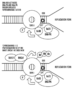
[0039] Figure 11A-11C shows a schematic diagram showing the fate of cellular
topoI in the response
to anti cancer drug (CPT) treatment. It has been established that topoI is
degraded by ubiquitination in
the response to CPT. DNA-TopoI- CPT makes cleavage complex during S phase.
Figure 11A shows
that due to the presence of CPT topoI fails to religate the cleaved DNA during
the religation cycle.
The collision of replication fork with cleaved DNA leads to DNA-double strand
breaks (DNA-DSB).
To repair the DNA-DSB topoI is removed by ubiquitination proteosomal pathway
(UPP). However,
the mechanism of UPP mediated topoI degradation is not understood. The
inventors have
demonstrated, as shown by the schematic in Figure 11B, that topoI associates
with Ku-DNA-PK
complex, and that DNA-DSB mediated activation of DNA-PK phosphorylates topoI
at S10. Figure
11C is a schematic demonstrating the phosphorylation of topoI at S10 ensures
the ubiquitination of
12
CA 02720184 2010-09-30
WO 2009/124064 PCT/US2009/038981
topoI by BRCA 1/BARD1 heterodimer, and subsequent degradation of the
ubiquitinated topoI by
ubiquitin-proteosomal pathway.
[0040] Figure 12, shows a simplified block diagram of an embodiment of the
present invention
which relates to a machine for determining if a subject is responsive to a
topo I inhibitor.
[0041] Figure 13 of a machine 10 for determining if a subject is responsive to
a topo I inhibitor
according to an embodiment of the invention.
[0042] Figure 14 depicts an exemplary block diagram of a computer system 151
that may be
configured to execute the prognostic application 155 illustrated in FIG. 13.
[0043] Figure 15 shows a flow chart of a instructions for analyzing if a
subject is responsive to a
topo I inhibitor.
[0044] Figure 16 shows a higher level of S10 topoI phosphorylation in CPT
resistant cells. Figure 16
shows three colon cancer cell lines; HCT15 (lane 1), Co1o205 (lane 2) and
Co1o320 cells (lane 3)
which were lysed and a total of 50 lug protein was analyzed by SDS-PAGE and
immunoblotting with
topoI S10 Phosphospecific antibody (anti-phospho-S10 topo I antibody; upper
panel). The membrane
was re-probed with anti 13-actin (lower panel) to control for total protein
levels.
DETAILED DESCRIPTION OF THE INVENTION
[0045] The present invention generally relates to a diagnostic marker for
predicting the efficacy of
topoisomerase I (topo I) inhibitors in the treatment of cancers. In
particular, one aspect of the present
invention relates to methods to determine effectiveness of topoisomerase I
(topo I) inhibitors in the
treatment of cancer. Specifically, the present invention is based on the
discovery that the topo I
polypeptide is phosphorylated at serine 10 (S10) by the kinase DNA-PK and
results in its
ubiquitination by BRAC1/BARD1 heterodimer and its subsequent degradation. The
inventors have
discovered that the presence of phosphorylation of a topo I polypeptide
(herein referred to as
"phospho-topo I"), in particular the phosphorylation at the serine 10 (S10)
amino acid residue of a
topo I polypeptide (herein referred to as "phospho-S10 topo I") in a
biological sample indicates the
resistance and/or unresponsiveness to a topo I inhibitor, such as camptothecin
(CPT), or CTP
analogues such as topotecan and irinotecan and derivatives thereof.
Accordingly, one aspect of the
present invention relates to detection of phospho-topo I, and in particular
the detection of phospho-
S10 topo I in a subject or in a biological sample taken from a subject having,
or likely having cancer,
as a prognostic determinant for drug efficacy with a topo I inhibitor such as
CTP and analogues
thereof.
[0046] Accordingly, one aspect of the present invention relates to methods,
kits, machines, computer
systems and computer readable media to detect and analyze the presence phospho-
topo I, and in
particular the presence of phospho-S10 topo I in a subject having or likely
having cancer, or a
biological sample taken from such a subject, and if the subject, or biological
sample is determined to
comprise phospho-topo I, in particular phosphorylation at the serine 10 (S10)
residue of the topo I
13
CA 02720184 2010-09-30
WO 2009/124064 PCT/US2009/038981
polypeptide (i.e. the presence of phospho-S10 topo I) then the subject, or the
subject from which the
biological sample was obtained is identified as being likely to be
unresponsive to a topo I inhibitor
such as camptothecin (CPT), or CTP analogues such as topotecan and irinotecan
and derivatives
thereof, as compared to a subject with cancer where the corresponding S10
residue on the topo I
polypeptide is not phosphorylated.
[0047] In all aspects of the inventions as disclosed herein, any means to
determine and measure the
phosphorylation status of topo I polypeptide are encompassed for use in the
present invention. In
some embodiments, the phosphorylation status of topo I polypeptide is detected
by any means known
by a skilled artisan, and can be detected in vivo, in vitro or ex vivo.
[0048] In another embodiment, a cancer or cancer cell is identified as likely
being responsive to a
topo I inhibitor if the cancer cell comprises minimal, or substantially lack
of, or the absence of
phosphorylated topo I polypeptide, and in particular the absence or
substantial lack of
phosphorylation on the serine 10 (S10) reside of topo I polypeptide (i.e. the
absence of phospho-S10
topo I), where the cancer cell can be present in the subject (for example as
detected using in vivo
detection method) or in alternative embodiments, the cancer cell can be in a
biological sample taken
from a subject (for example as detected via in vitro or ex vivo detection
methods).
[0049] Accordingly, another aspect of the present invention provides a method
to identify a subject
which is responsive to a topo I inhibitor, such as for example where the
subject is currently
undergoing or has been selected to be treated for cancer with a topo I
inhibitor, for example but not
limited to CTP or analogues such as topotecan and irinotecan and other
analogues or derivatives
thereof.
[0050] One aspect of the present invention relates to a method for identifying
a subject responsive to
a topo I inhibitor, the method comprising measuring the level of
phosphorylation of a topoisomerase I
polypeptide in a subject, or a biological subject comprising cancer cells
taken from a subject; and
detecting the level of phosphorylation of the topo I polypeptide, in
particular detecting the level of
phosphorylation at serine 10 (S10) of the topo I polypeptide, and if the topo
I polypeptide is
phosphorylated, for example at residue 510, it indicates that the subject has
a cancer which is likely to
be more unresponsive to a topoisomerase I inhibitor as compared to a subject
with cancer where that
lacks phosphorylated topo I polypeptide and in particular lacks phospho-S10
topo I polypeptide.
[0051] Another aspect of the present invention also relates to a method to
identifying the likelihood
of a cancer to be unresponsive to a topoisomerase I inhibitor, the method
comprising measuring the
level of phosphorylation of topoisomerase I polypeptide (i.e. determining the
phosphorylation status
of topo I polypeptide) in at least one cancer cell, wherein the presence of
phosphorylation identifies
the cancer as being more likely to be unresponsive to a topoisomerase
inhibitor as compared to a
cancer wherein the absence of phosphorylation of topoisomerase I is detected.
In some embodiments,
the cancer cell is present in a subject. In some embodiments, the cancer cell
is present in a biological
sample. In other embodiments, a cancer cell is a cancer stem cell.
14
CA 02720184 2010-09-30
WO 2009/124064 PCT/US2009/038981
[0052] Another aspect of the present invention relates to a method for
treating cancer in a subject, the
methods comprising: (i) measuring the level of phosphorylation of
topoisomerase I polypeptide in a
biological sample comprising cancer cells obtained from the subject; (ii)
detecting the level of
topoisomerase I polypeptide, wherein if the topoisomerase I polypeptide is
phosphorylated the cancer
is identified as being unresponsive to a topoisomerase I inhibitor, or wherein
if the topoisomerase I
polypeptide is not phosphorylated the cancer is identified as being likely to
be responsive to a
topoisomerase I inhibitor; (iii) administering to a subject an anti-cancer
agent other than a
topoisomerase I inhibitor where the cancer is identified as being unresponsive
to a topoisomerase I
inhibitor. In some embodiments, subjects identified to be responsive to a topo
I inhibitor can then be
treated with a topo I inhibitor as that term is described herein, whereas a
subject identified to be non-
responsive to a topo I inhibitor can be treated with any anti-cancer treatment
known to one of ordinary
skill in the art which is not a topo I inhibitor, or alternatively, such a
subject identified to be non-
responsive to a topo I inhibitor can be administered a combination of a topo I
inhibitor with (i.e. in
conjunction with) an agent which increases a cell's sensitivity to a topo I
inhibitor (i.e. a topo I
inhibitor sensitivity agent). In some embodiments for example, a topo I
inhibitor can be co-
administered with an agent which dephosphorylates S10 residue of topo I
polypeptide or alternatively
with an anent which inhibits the phosphorylation of the S10 residue on topo I,
such as an anti-
phospho-S10 topo I antibody or an antagonist to DNA-PK.
[0053] One aspect of the present invention and all other aspect described
herein, a machine can be
used to determine phosphorylation status of topo I polypeptides in a
biological sample, for example, a
machine for obtaining data regarding a biological sample from a subject
comprising: a biological
sample container to hold the biological sample; a determination module
configured to detect the
presence of phosphorylation of a topoisomerase I polypeptide, for example the
detection of phospho-
S10 topo Tin the biological sample which produces output data, in some
embodiments the output data
in a computer readable media format; a storage device configured to store the
output data from the
determination module; a comparison module adapted to compare the output data
from the
determination module with data stored on the storage device, such as stored
reference data and control
data, and a display module for displaying a page of retrieved content for the
user on a client computer,
wherein (i) the retrieved content is the presence of topoisomerase I
polypeptide, and/or (ii) the
retrieved content is the presence or absence of phosphorylation of the
topoisomerase I polypeptide, for
example the retrieved content is the presence or absence of phospho-S10 topo I
and/or (iii) the
retrieved content is the absence of phosphorylation of topoisomerase I, for
example the absence of
phospho-S10 topo I, which is a signal that the subject likely to be responsive
to topoisomerase I
inhibitor; and/or (iv) the retrieved content is the presence of
phosphorylation of topoisomerase I, such
as the presence of phospho-S10 topo I polypeptide which is a signal that the
subject likely to be
unresponsive to topoisomerase I inhibitor.
CA 02720184 2010-09-30
WO 2009/124064 PCT/US2009/038981
[0054] One aspect of the present invention is a computer system that can be
used to determine if a
subject is responsive to a topo I inhibitor. In such an embodiment, a computer
system is connected to
a determination module and is configured to obtain output data from a
determination module
regarding a biological specimen, where a determination module is configured to
detect the presence of
phosphorylation of a topoisomerase I polypeptide, for example the presence of
phospho-S10 topo I
polypeptide within a subject or in a biological sample obtained from the
subject; and where the
computer system comprises (a) a storage device configured to store data output
from the
determination module as well as reference data; where the storage device is
connected to (b) a
comparison module which in one embodiment, is adapted to compare the output
data stored on the
storage device with stored reference data, and in alternative embodiments,
adapted to compare the
output data with itself, where the comparison module produces report data and
is connected to (c) a
display module for displaying a page of retrieved content (i.e. report data
from the comparison
module) for the user on a client computer, wherein the retrieved content
comprises any one or a
combination of the following; (i) the presence or absence of phosphorylation
of the topoisomerase I
polypeptide, for example the retrieved content is the presence or absence of
phospho-S10 topo I; (ii)
the absence of phosphorylation of topoisomerase I, for example the absence of
phospho-S10 topo I,
(iii) the presence of phosphorylation of topoisomerase I, such as the presence
of phospho-S10 topo I
polypeptide, (iv) a positive test result (i.e. a positive phosphorylation
status such as positive S10 topo
I phosphorylation status) which indicates that the subject is likely to be
more unresponsive to a topoI
inhibitor than a subject having a cancer with a negative phosphorylation
status, (v) a negative test
result (i.e. a negative phosphorylation status such a negative S10 topo I
phosphorylation status) which
indicates that the subject is likely to be more responsive to a topoI
inhibitor than a subject having a
cancer with a positive phosphorylation.
[0055] In some embodiments the comparison module compares the output data
stored on the storage
device with itself or stored reference data, and calculates a positive S10
topo I phosphorylation status
(i.e. the presence of phospho-S10 topo I polypeptide) which indicates a
positive test result and
generates report data to indicate that the subject is likely to be more
unresponsive to a topoI inhibitor
than a subject having a cancer with a negative phosphorylation status, where
the report data from the
comparison module is retrieved from the display module and displayed on the
display module.
[0056] One aspect of the present invention and all other aspect described
herein, one can use a
computer readable media to determine phosphorylation status of topo I
polypeptides from a subject
having or at risk of having cancer, for example, a computer readable media
having computer readable
instructions recorded thereon to define software modules including a
determination module and a
comparison module for implementing a method on a computer, said method
comprising: a storage
device configured to store data reference data and output data from a
determination module which has
measured the presence or absence of the phosphorylation of topo I polypeptide,
such as the presence
or absence of phospho-S10 topo I polypeptide; a comparison module which
generates report data,
16
CA 02720184 2010-09-30
WO 2009/124064 PCT/US2009/038981
where the comparison module is adapted to compare the data stored on the
storage device, for
example a comparison of output data from the determination module with itself
or alternatively with
reference data, and a display module for displaying a page of retrieved
content which is the report data
from the comparison module for the user on a client computer, wherein the
retrieved content
comprises any one or a combination of the following; (i) the presence or
absence of phosphorylation
of the topoisomerase I polypeptide, for example the retrieved content is the
presence or absence of
phospho-S10 topo I; (ii) the absence of phosphorylation of topoisomerase I,
for example the absence
of phospho-S10 topo I, (iii) the presence of phosphorylation of topoisomerase
I, such as the presence
of phospho-S10 topo I polypeptide, (iv) a positive test result (i.e. a
positive phosphorylation status
such as positive S10 topo I phosphorylation status) which indicates that the
subject is likely to be
more unresponsive to a topoI inhibitor than a subject having a cancer with a
negative phosphorylation
status, (v) a negative test result (i.e. a negative phosphorylation status
such a negative S10 topo I
phosphorylation status) which indicates that the subject is likely to be more
responsive to a topoI
inhibitor than a subject having a cancer with a positive phosphorylation.
[0057] In some embodiments, a topo I inhibitor useful in the methods as
disclosed herein is any agent
or entity which inhibits the biological activity, such as protein activity of
topoisomerase, including but
not limited to camptothecin (CPT), or CTP analogues such as topotecan and
irinotecan and derivatives
thereof, CTP compounds, CTP metabolites, CTP derivatives and mimetics thereof.
[0058] In an alternative embodiment, a subject identified to be responsive to
a topo I inhibitor can be
administered a topo I inhibitor, such as for example but not limited to CTP or
analogues thereof.
[0059] In one embodiment the cancer is SCLC, colon or ovarian cancer, or a
refractory cancer, for
example, breast cancer or cervical cancer. In other embodiments, a cancer
useful to be treated in the
methods as disclosed herein is any cancer which can be treated, or is
desirable to be treated with a
topo I inhibitor and includes, for example but are not limited to cancers
comprising those of epithelial
origin, including, but are not limited to, gastrointestinal cancer, prostate
cancer, ovarian cancer, breast
cancer, head and neck cancer, lung cancer, non-small cell lung cancer, cancer
of the nervous system,
kidney cancer, retina cancer, skin cancer, liver cancer, pancreatic cancer,
genital-urinary cancer and
bladder cancer. In one embodiment, the cancer is non-small cell lung cancer.
In another embodiment,
the cancer is triple-negative subtype of cancer, which lacks the expression of
the progesterone
receptor (PR), the estrogen receptor (ER) and also lacks Her-2 amplification.
[0060] Tumor cell types can also be selected from a group comprising of
gastrointestinal cancer,
gastric cancer, squamous cell carcinomas (SCC), head and neck cancer, lung
cancer, non-small cell
lung cancer (NSCLC) and small-cell lung cancer (SCLC), lymphoma, sarcoma,
primary and metastic
melanoma, thymoma, non-Hodgkin's lymphoma, Hodgkin's lymphoma, cancer of the
nervous
system, brain cancer, bone-marrow cancer, bone cancer, kidney cancer, uterine
cancer, cervical
cancer, colon cancer, retina cancer, skin cancer, bladder cancer, colon
cancer, esophageal cancer,
testicular cancer, cervical cancer, liver cancer, renal cancer, pancreatic
cancer, genital-urinary cancer,
17
CA 02720184 2010-09-30
WO 2009/124064 PCT/US2009/038981
gastrointestinal, gum cancer, tongue cancer, kidney cancer, nasopharynx
cancer, stomach cancer,
endometrial cancer and bowel tumor cell cancer, adrenocarcinomas such as
prostate cancer, ovarian
cancer, breast cancer, and pancreatic cancer.
Definitions
[108] For convenience, certain terms employed in the entire application
(including the specification,
examples, and appended claims) are collected here. Unless defined otherwise,
all technical and
scientific terms used herein have the same meaning as commonly understood by
one of ordinary skill
in the art to which this invention belongs.
[0061] The term "topoisomerase I" is used interchangeably herein with "topo I"
and refers to the
polypeptide encoded by SEQ ID NO: 2 and variants and homologues thereof,
including conservative
substitutions, additions, deletions therein not adversely affecting the
structure of function.
Topoisomerase I is referred in the art as TOPI or TOP1. Human topo I is
encoded by nucleic acid
corresponding to GenBank Accession No: BC136297.1 (SEQ ID NO: 3) or
GI:223460079 and the
human topo I polypeptide corresponds to protein sequence corresponding to
RefSeq ID No:
NM_003286 or NP_003277.1. DNA topoisomerase is an enzyme that controls and
alters the
topologic states of DNA during transcription, and catalyzes the transient
breaking and rejoining of a
single strand of DNA which allows the strands to pass through one another,
thus altering the topology
of DNA. The gene for TOP1 is localized to chromosome 20 and has pseudogenes
which reside on
chromosomes 1 and 22. The biological activity of topo I polypeptide refers to
the polypeptides
enzymatic activity to catalyze the transient breaking and rejoining of a
single strand of DNA, where
one strand pass through one another, thus altering the topology of DNA.
[0062] The term "inhibitor" as used herein refers to any agent or entity which
results in the inhibition
of a proteins biological activity. By a "decrease" or "inhibition" used in the
context of the level of
activity of a gene refers to a reduction in protein or nucleic acid level or
biological activity in a cell, a
cell extract, or a cell supernatant. For example, such inhibition may be due
to decreased binding of
the polypeptide to its endogenous ligand, or by non-completive binding of an
inhibitor to a
polypeptide to reduce catalytic activity or affinity for target ligand etc, or
alternatively to reduced
RNA stability, transcription, or translation, increased protein degradation,
or RNA interference.
Preferably, a decrease is at least about 5%, at least about 10%, at least
about 25%, at least about 50%,
at least about 75%, at least about 80%, or even at least about 90% of the
level of expression or activity
under control conditions.
[0063] By an "increase" in the expression or activity of a gene or protein is
meant a positive change
in protein or polypeptide or nucleic acid level or activity in a cell, a cell
extract, or a cell supernatant.
For example, such a increase may be due to increased RNA stability,
transcription, or translation, or
decreased protein degradation. Preferably, this increase is at least 5%, at
least about 10%, at least
18
CA 02720184 2010-09-30
WO 2009/124064 PCT/US2009/038981
about 25%, at least about 50%, at least about 75%, at least about 80%, at
least about 100%, at least
about 200%, or even about 500% or more over the level of expression or
activity under control
conditions.
[0064] The term "topo I inhibitor" as used herein refers to any entity which
mediates some, all or part
of its biological function, through acting directly or indirectly on the gene
product or polynucleotide
of topoisomerase I polypeptide. A topo I inhibitor can directly or indirectly
inactivate topo I
polypeptide.
[0065] Exemplarily examples of topo I inhibitor include for example but not
limited to, camptothecin
(CTP) and analogues thereof including but not limited to irinotecan and
topotecan, and derivatives
thereof, as these terms are described herein.
[0066] The term "CTP" can include a "mimetic" of CTP or a derivative or
analogue thereof, which
includes compounds which may not be structurally similar to CTP but mimic the
therapeutic activity
or therapeutic mechanism of CTP or structurally similar CTP compound in vitro
and in vivo.
[0067] As used herein, the terms "effective" and "effectiveness" or
"responsive" includes both
pharmacological effectiveness and physiological safety of an agent, such as a
topo I inhibitor.
"Pharmacological effectiveness" refers to the ability of the treatment to
result in a desired biological
effect in the subject. "Physiological safety" refers to the level of toxicity,
or other adverse
physiological effects at the cellular, organ and/or organism level (often
referred to as side-effects)
resulting from administration of the treatment, "less effective" means that
the treatment results in a
therapeutically significant lower level of pharmacological effectiveness
and/or a therapeutically
greater level of adverse physiological effects.
[0068] The term "lack of effectiveness", "non-responsiveness" , "refractory"
or "unresponsiveness"
are used interchangeably herein, and refer to the inability of an agent or
treatment to result in a desired
biological effect in the subject.
[0069] The term "phosphorylation status" as used herein comprises the absolute
or relative degree of
phosphorylation of proteins and/or reagents. The term phosphorylation status
of topo I is the degree of
phosphorylation on all phosphorylation sites on the topo I polypeptide, and
includes the degree (or
level) of phosphorylation on the serine 10 (S10) amino acid residue on the
topo I polypeptide.
[0070] The term "activity" when used in reference to the activity of a protein
as used herein,
comprises the enzymatic activity, binding affinity and/or posttranslational
activity, in particular
phosphorylation.
[0071] The term "target" as used herein may mean a polynucleotide that may be
bound by one or
more probes under stringent hybridization conditions.
[0072] The term "entity" refers to any structural molecule or combination of
molecules.
[0073] The term "drug", "agent" or "compound" as used herein refers to a
chemical entity or
biological product, or combination of chemical entities or biological
products, administered to a
person to treat or prevent or control a disease or condition. The chemical
entity or biological product
19
CA 02720184 2010-09-30
WO 2009/124064 PCT/US2009/038981
is preferably, but not necessarily a low molecular weight compound, but may
also be a larger
compound, for example, an oligomer of nucleic acids, amino acids, or
carbohydrates including
without limitation proteins, oligonucleotides, ribozymes, DNAzymes,
glycoproteins, siRNAs,
lipoproteins, aptamers, and modifications and combinations thereof.
[0074] The term "agent" refers to any entity which is normally absent or not
present at the levels
being administered, in the cell. Agent may be selected from a group
comprising; chemicals; small
molecules; nucleic acid sequences; nucleic acid analogues; proteins; peptides;
aptamers; antibodies; or
fragments thereof. A nucleic acid sequence may be RNA or DNA, and may be
single or double
stranded, and can be selected from a group comprising; nucleic acid encoding a
protein of interest,
oligonucleotides, nucleic acid analogues, for example peptide-nucleic acid
(PNA), pseudo-
complementary PNA (pc-PNA), locked nucleic acid (LNA), etc.. Such nucleic acid
sequences
include, for example, but not limited to, nucleic acid sequence encoding
proteins, for example that act
as transcriptional repressors, antisense molecules, ribozymes, small
inhibitory nucleic acid sequences,
for example but not limited to RNAi, shRNAi, siRNA, micro RNAi (mRNAi),
antisense
oligonucleotides etc. A protein and/or peptide or fragment thereof can be any
protein of interest, for
example, but not limited to; mutated proteins; therapeutic proteins; truncated
proteins, wherein the
protein is normally absent or expressed at lower levels in the cell. Proteins
can also be selected from a
group comprising; mutated proteins, genetically engineered proteins, peptides,
synthetic peptides,
recombinant proteins, chimeric proteins, antibodies, midibodies, tribodies,
humanized proteins,
humanized antibodies, chimeric antibodies, modified proteins and fragments
thereof. The agent may
be applied to the media, where it contacts the cell and induces its effects.
Alternatively, the agent may
be intracellular within the cell as a result of introduction of the nucleic
acid sequence into the cell and
its transcription resulting in the production of the nucleic acid and/or
protein environmental stimuli
within the cell. In some embodiments, the agent is any chemical, entity or
moiety, including without
limitation synthetic and naturally-occurring non-proteinaceous entities. In
certain embodiments the
agent is a small molecule having a chemical moiety. For example, chemical
moieties included
unsubstituted or substituted alkyl, aromatic, or heterocyclyl moieties
including macrolides,
leptomycins and related natural products or analogues thereof. Agents can be
known to have a
desired activity and/or property, or can be selected from a library of diverse
compounds.
[0075] The term "antagonist" refers to any agent or entity capable of
inhibiting the expression or
activity of a protein, polypeptide portion thereof, or polynucleotide. Thus,
the antagonist may operate
to prevent transcription, translation, post-transcriptional or post-
translational processing or otherwise
inhibit the activity of the protein, polypeptide or polynucleotide in any way,
via either direct of
indirect action. The antagonist may for example be a nucleic acid, peptide, or
any other suitable
chemical compound or molecule or any combination of these. Additionally, it
will be understood that
in indirectly impairing the activity of a protein, polypeptide of
polynucleotide, the antagonist may
affect the activity of the cellular molecules which may in turn act as
regulators or the protein,
CA 02720184 2010-09-30
WO 2009/124064 PCT/US2009/038981
polypeptide or polynucleotide itself. Similarly, the antagonist may affect the
activity of molecules
which are themselves subject to the regulation or modulation by the protein,
polypeptide of
polynucleotide.
[0076] The term "inhibiting" as used herein as it pertains to the expression
or activity of the protein
or polypeptide of topoisomerase I or variants thereof does not necessarily
mean complete inhibition of
expression and/or activity. Rather, expression or activity of the protein,
polypeptide or polynucleotide
is inhibited to an extent, and/or for a time, sufficient to produce the
desired effect.
[0077] The term "protein binding moiety" is used interchangeably herein with
"protein binding
molecule" or protein binding entity" and refers to any entity which has
specific affinity for a protein.
The term "protein-binding molecule" also includes antibody-based binding
moieties and antibodies
and includes immunoglobulin molecules and immunologically active determinants
of
immunoglobulin molecules, e.g., molecules that contain an antigen binding site
which specifically
binds (immunoreacts with) to the Psap proteins. The term "antibody-based
binding moiety" is
intended to include whole antibodies, e.g., of any isotype (IgG, IgA, IgM,
IgE, etc), and includes
fragments thereof which are also specifically reactive with the Psap proteins.
Antibodies can be
fragmented using conventional techniques. Thus, the term includes segments of
proteolytically-
cleaved or recombinantly-prepared portions of an antibody molecule that are
capable of selectively
reacting with a certain protein. Non limiting examples of such proteolytic
and/or recombinant
fragments include Fab, F(ab')2, Fab' , Fv, dAbs and single chain antibodies
(scFv) containing a VL
and VH domain joined by a peptide linker. The scFv's can be covalently or non-
covalently linked to
form antibodies having two or more binding sites. Thus, "antibody-base binding
moiety" includes
polyclonal, monoclonal, or other purified preparations of antibodies and
recombinant antibodies. The
term "antibody-base binding moiety" is further intended to include humanized
antibodies, bispecific
antibodies, and chimeric molecules having at least one antigen binding
determinant derived from an
antibody molecule. In a preferred embodiment, the antibody-based binding
moiety detectably labeled.
In some embodiments, a "protein-binding molecule" is a co-factor or binding
protein that interacts
with the protein to be measured, for example a co-factor or binding protein to
a topo I polypeptide
protein.
[0078] The term "labeled antibody", as used herein, includes antibodies that
are labeled by a
detectable means and include, but are not limited to, antibodies that are
enzymatically, radioactively,
fluorescently, and chemiluminescently labeled. Antibodies can also be labeled
with a detectable tag,
such as c-Myc, HA, VSV-G, HSV, FLAG, V5, or HIS. The detection and
quantification of Psap or
Tsp-1 present in the tissue samples correlate to the intensity of the signal
emitted from the detectably
labeled antibody.
[0079] The term "specific affinity" or "specifically binds" or "specific
binding" are used
interchangeably herein refers to an entity such as a protein-binding molecule
or antibody that
recognizes and binds a desired polypeptide but that does not substantially
recognize and bind other
21
CA 02720184 2010-09-30
WO 2009/124064 PCT/US2009/038981
molecules in a sample, for example, a biological sample, which naturally
includes a polypeptide of the
invention, for example phospho-S10 topo I polypeptide.
[0080] The term "antibody" is meant to be an immunoglobulin protein that is
capable of binding an
antigen. Antibody as used herein is meant to include antibody fragments, e.g.
F(ab')2, Fab', Fab,
capable of binding the antigen or antigenic fragment of interest.
[0081] The term "humanized antibody" is used herein to describe complete
antibody molecules, i.e.
composed of two complete light chains and two complete heavy chains, as well
as antibodies
consisting only of antibody fragments, e.g. Fab, Fab', F(ab')2, and Fv,
wherein the CDRs are derived
from a non-human source and the remaining portion of the Ig molecule or
fragment thereof is derived
from a human antibody, preferably produced from a nucleic acid sequence
encoding a human
antibody.
[0082] The terms "human antibody" and "humanized antibody" are used herein to
describe an
antibody of which all portions of the antibody molecule are derived from a
nucleic acid sequence
encoding a human antibody. Such human antibodies are most desirable for use in
antibody therapies,
as such antibodies would elicit little or no immune response in the human
subject.
[0083] The term "chimeric antibody" is used herein to describe an antibody
molecule as well as
antibody fragments, as described above in the definition of the term
"humanized antibody." The term
"chimeric antibody" encompasses humanized antibodies. Chimeric antibodies have
at least one
portion of a heavy or light chain amino acid sequence derived from a first
mammalian species and
another portion of the heavy or light chain amino acid sequence derived from a
second, different
mammalian species. In some embodiments, a variable region is derived from a
non-human
mammalian species and the constant region is derived from a human species.
Specifically, the
chimeric antibody is preferably produced from a 9 nucleotide sequence from a
non-human mammal
encoding a variable region and a nucleotide sequence from a human encoding a
constant region of an
antibody.
[0084] In the context of this invention, the term "probe" refers to a molecule
which can detectably
distinguish between target molecules differing in structure. Detection can be
accomplished in a
variety of different ways depending on the type of probe used and the type of
target molecule, thus,
for example, detection may be based on discrimination of activity levels of
the target molecule, but
preferably is based on detection of specific binding. Examples of such
specific binding include
antibody binding and nucleic acid probe hybridization. Thus, for example,
probes can include enzyme
substrates, antibodies and antibody fragments, and preferably nucleic acid
hybridization probes.
[0085] The term "label" refers to a composition capable of producing a
detectable signal indicative of
the presence of the target polynucleotide in an assay sample. Suitable labels
include radioisotopes,
nucleotide chromophores, enzymes, substrates, fluorescent molecules,
chemiluminescent moieties,
magnetic particles, bioluminescent moieties, and the like. As such, a label is
any composition
22
CA 02720184 2010-09-30
WO 2009/124064 PCT/US2009/038981
detectable by spectroscopic, photochemical, biochemical, immunochemical,
electrical, optical or
chemical means.
[0086] As used herein the term "reference data" when used in the context of
the reference level of
phosphorylation refers to the level of phosphorylation of topo I, in
particular the level of
phosphorylation on serine 10 of topo Tin at least one reference biological
sample, or a group of
biological samples from at least subject or a group of subjects which have
been identified to be non-
responsive or responsive to a topo I inhibitor. By way of example, a positive
reference level is a level
of the degree of phosphorylation, in particular the level of phosphorylation
at S10 of topo I which
indicates the sample is non-responsive to a topo I inhibitor, whereas a
negative reference level is a
level of the degree of phosphorylation, in particular the level of
phosphorylation at S10 of topo I
which indicates the sample is responsive to a topo I inhibitor. In some
embodiments, a positive
reference level is normalized to 100%, where 100% of the total topo I
polypeptide is phosphorylated,
for example 100% of topo I exists as phospho-S10-topoI, and in some
embodiments a negative
reference level is normalized to 0%, where 0% of the total topo I polypeptide
is phosphorylated, for
example 0% of topo I exists as phospho-S10-topoI. Thus, so an increase in the
level of a
phosphorylation of topo I, such as an increase of at least 1% to 100% of the
level of phosphorylation
at S10 of topo Tin a biological sample as compared with a negative reference
level, including all
percentages between 1% and 100%, i.e. at least 1%, 2%, 3%, 4%, 5%, 10%, 15%,
20%, 25%, 30%,
35%, 40%, 45%, 50%, 60%, 70%, 80%, 90%, 95%, 100% indicates the degree of
likelihood (i.e. %)
the biological sample is likely to be unresponsive to a topo I inhibitor.
Stated another way, if 50% of
the total available topo I protein is detected to be phospho-S10 topo I, then
the efficacy of the topo I
inhibitor is likely to be reduced by 50% as compared to the efficacy of the
topo I inhibitor if 0% of the
total available topo I protein exists as phospho-S10 topo I (i.e. topo I is
not phosphorylated at S10).
[0087] The term "support" refers to conventional supports such as beads,
particles, dipsticks, fibers,
filters, membranes and silane or silicate supports such as glass slides.
[0088] The term "cancer", as used herein refers to a cellular proliferative
disease in a human or
animal subject.
[0089] The terms "tumor" or "tumor cell" or "cancer cell" are used
interchangeably herein refers to
the tissue mass or tissue type or cell type that is undergoing uncontrolled
proliferation.
[0090] The term "tissue" is intended to include intact cells, blood, blood
preparations such as plasma
and serum, bones, joints, muscles, smooth muscles, and organs.
[0091] The term "Triple-negative subtype" used herein refers to any subtype of
cancer, particularly
breast cancer, which lacks the expression of the progesterone receptor (PR),
lacks the estrogen
receptor (ER) and also lacks Her-2 amplification.
[0092] As used herein, the term "biological sample" also refers to a cell or
population of cells or a
quantity of tissue or fluid from a subject. Most often, the sample has been
removed from a subject, but
the term "biological sample" can also refer to cells or tissue analyzed in
vivo, i.e. without removal
23
CA 02720184 2010-09-30
WO 2009/124064 PCT/US2009/038981
from the subject. Often, a "biological sample" will contain cells from a
subject, but the term can also
refer to non-cellular biological material, such as non-cellular fractions of
blood, saliva, or urine, that
can be used to measure protein phosphorylation levels. As used herein, a
"biological sample" or
"tissue sample" refers to a sample of tissue or fluid isolated from an
individual, including but not
limited to, for example, blood, plasma, serum, tumor biopsy, urine, stool,
sputum, spinal fluid, pleural
fluid, nipple aspirates, lymph fluid, the external sections of the skin,
respiratory, intestinal, and
genitourinary tracts, tears, saliva, milk, cells (including but not limited to
blood cells), tumors, organs,
and also samples of in vitro cell culture constituent. In some embodiments, a
biological sample is
from a resection, bronchoscopic biopsy, or core needle biopsy of a primary,
secondary or metastatic
tumor, or a cellblock from pleural fluid. In addition, fine needle aspirate
biological samples are also
useful. In some embodiments, a biological sample is primary ascite cells.
Samples can be fresh,
frozen, fixed or optionally paraffin-embedded, frozen or subjected to other
tissue preservation
methods, including for example methods to preserve the phosphorylation status
of polypeptides in the
biological sample. A biological sample can also mean a sample of biological
tissue or fluid that
comprises protein or cells. Such samples include, but are not limited to,
tissue isolated from subjects
or animals. Biological samples may also include sections of tissues such as
biopsy and autopsy
samples, frozen sections taken for histological purposes, blood, plasma,
serum, sputum, stool, tears,
mucus, hair, and skin. Biological samples also include explants and primary
and/or transformed cell
cultures derived from patient tissues. A biological sample may be provided by
removing a sample of
cells from subject, but can also be accomplished by using previously isolated
cells (e.g., isolated by
another person, at another time, and/or for another purpose), or by performing
the methods of the
invention in vivo. Archival tissues, such as those having treatment or outcome
history may also be
used. Biological samples include, but are not limited to, tissue biopsies,
scrapes (e.g. buccal scrapes),
whole blood, plasma, serum, urine, saliva, cell culture, or cerebrospinal
fluid. Biological samples also
include tissue biopsies, cell culture. The sample can be obtained by removing
a sample of cells from a
subject, but can also be accomplished by using previously isolated cells (e.g.
isolated by another
person), or by performing the methods of the invention in vivo.
[0093] The term 'malignancy' and 'cancer' are used interchangeably herein,
refers to diseases that
are characterized by uncontrolled, abnormal growth of cells. Cancer cells can
spread locally or
through the bloodstream and lymphatic system to other parts of the body. The
term "malignancy" or
"cancer" are used interchangeably herein and refers to any disease of an organ
or tissue in mammals
characterized by poorly controlled or uncontrolled multiplication of normal or
abnormal cells in that
tissue and its effect on the body as a whole. Cancer diseases within the scope
of the definition
comprise benign neoplasms, dysplasias, hyperplasias as well as neoplasms
showing metastatic growth
or any other transformations like e.g. leukoplakias which often precede a
breakout of cancer.
[0094] The terms "patient", "subject" and "individual" are used
interchangeably herein, and refer to
an animal, particularly a human, to whom treatment including prophylaxic
treatment is provided. The
24
CA 02720184 2010-09-30
WO 2009/124064 PCT/US2009/038981
term "subject" as used herein refers to human and non-human animals. The term
"non-human
animals" and "non-human mammals" are used interchangeably herein includes all
vertebrates, e.g.,
mammals, such as non-human primates, (particularly higher primates), sheep,
dog, rodent (e.g. mouse
or rat), guinea pig, goat, pig, cat, rabbits, cows, and non-mammals such as
chickens, amphibians,
reptiles etc. In one embodiment, the subject is human. In another embodiment,
the subject is an
experimental animal or animal substitute as a disease model.
[0095] The term "effective amount" includes within its meaning a sufficient
amount of a
pharmacological composition to provide the desired effect. The exact amount
required will vary
depending on factors such as the level of phosphorylation (i.e. presence or
absence of phosphorylation
of topo I, such as phosphorylation of S10 of topo I), the type of tumor to be
treated, the severity of the
tumor, the drug resistance level of the tumor, the species being treated, the
age and general condition
of the subject, the particular topo I inhibitor being used as a treatment, the
mode of administration and
so forth. Thus, it is not possible to specify the exact "effective amount".
However, for any given case,
an appropriate "effective amount" may be determined by one of ordinary skill
in the art using only
routine experimentation. As used herein, the effective amount is the amount of
an agent or treatment
to reduce a symptom of the disease, for example, but not limited to, to reduce
the size of a tumor, for
example to reduce the size by about 10%, to attenuate the growth rate of the
tumor, for example to
reduce the rate at which a tumor grows by 10%. For example, an effective
amount using the methods
as disclosed herein would be considered as the amount sufficient to reduce a
symptom of the cancer,
for example at least one symptom of a cancer or malignancy by at least 10%.
Further, an effective
amount as used herein would also include an amount sufficient to prevent or
delay the development of
a symptom of the disease, alter the course of a symptom disease (for example
but not limited to, slow
the progression of a symptom of the disease), or reverse a symptom of the
disease.
[0096] As used herein, the term "treating" includes reducing or alleviating at
least one adverse effect
or symptom of a condition, disease or disorder associated with cancer. As used
herein, the term
treating is used to refer to the reduction of a symptom and/or a biochemical
marker of cancer, for
example a reduction in at least one biochemical marker of cancer by at least
10%. For example but are
not limited to, a reduction in a biochemical marker of cancer, for example a
reduction in, as an
illustrative example only, at least one of the following biomarkers; CD44,
telomerase, TGF-a, TGF-I3,
erbB-2, erbB-3, MUC1, MUC2, CK20, PSA, CA125, FOBT, by 10%, or a reduction in
the rate of
proliferation of the cancer cells by 10%, would be considered effective
treatments by the methods as
disclosed herein. As alternative examples, a reduction in a symptom of cancer,
for example, a slowing
of the rate of growth of the cancer by 10% or a cessation of the increase in
tumor size, or a reduction
in the size of a tumor by 10% or a reduction in the tumor spread (i.e. tumor
metastasis) by 10% would
also be considered as affective treatments by the methods as disclosed herein.
[0097] The term "polynucleotide" as used herein, refers to single- or double-
stranded polymer of
deoxyribonucleotide, ribonucleotide bases or known analogies of natural
nucleotides, or mixtures
CA 02720184 2010-09-30
WO 2009/124064 PCT/US2009/038981
thereof. The term includes reference to the specified sequence as well as to
the sequence
complementary thereto, unless otherwise indicated.
[0098] The term "polypeptide" means a polymer made up of amino acids linked
together by peptide
bonds. The terms "polypeptide" and "protein" are used interchangeably herein,
although for the
purposes for the present invention, a polypeptide may constitute a portion or
the full length protein.
[0099] The term "expression" as used herein refers to interchangeably to the
expression of a
polypeptide or protein and expression of a polynucleotide or gene. Expression
of a polynucleotide
may be determined, for example, by measuring the production of messenger RNA
(mRNA) transcript
levels. Expression of a protein or polypeptide may be determined, for example,
by immunoassay
using an antibody(ies) that bind with the polypeptide.
[00100] The term "endogenously expressed" or "endogenous expression" as used
herein, refers to the
expression of a gene product at normal levels and under normal regulation for
that cell type.
[00101] In the context of this specification, the term "activity" as it
pertains to a protein, polypeptide
or polynucleotide means any cellular function, action, effect of influence
exerted by the protein,
polypeptide or polynucleotide, either by nucleic acid sequence or fragment
thereof, or by the protein
or polypeptide itself or any fragment thereof.
[00102] The term "nucleic acid" or "oligonucleotide" or "polynucleotide" used
herein can mean at
least two nucleotides covalently linked together. As will be appreciated by
those in the art, the
depiction of a single strand also defines the sequence of the complementary
strand. Thus, a nucleic
acid also encompasses the complementary strand of a depicted single strand. As
will also be
appreciated by those in the art, many variants of a nucleic acid can be used
for the same purpose as a
given nucleic acid. Thus, a nucleic acid also encompasses substantially
identical nucleic acids and
complements thereof. As will also be appreciated by those in the art, a single
strand provides a probe
for a probe that can hybridize to the target sequence under stringent
hybridization conditions. Thus, a
nucleic acid also encompasses a probe that hybridizes under stringent
hybridization conditions.
[00103] The term "RNAi" as used herein refers to RNA interference (RNAi) a RNA-
based molecule
that inhibits gene expression. RNAi refers to a means of selective post-
transcriptional gene silencing
by destruction of specific mRNA by small interfering RNA molecules (siRNA).
The siRNA is
typically generated by cleavage of double stranded RNA, where one strand is
identical to the message
to be inactivated. As used herein, the term "RNAi" refers to any type of
interfering RNA, including
but are not limited to, siRNAi, shRNAi, endogenous microRNA and artificial
microRNA. For
instance, it includes sequences previously identified as siRNA, regardless of
the mechanism of down-
stream processing of the RNA (i.e. although siRNAs are believed to have a
specific method of in vivo
processing resulting in the cleavage of mRNA, such sequences can be
incorporated into the vectors in
the context of the flanking sequences described herein).
[00104] As used herein an "siRNA" refers to a nucleic acid that forms a double
stranded RNA, which
double stranded RNA has the ability to reduce or inhibit expression of a gene
or target gene when the
26
CA 02720184 2010-09-30
WO 2009/124064 PCT/US2009/038981
siRNA is present or expressed in the same cell as the target gene, for example
where a target gene is
for example DNA-PK . The double stranded RNA siRNA can be formed by the
complementary
strands. In one embodiment, a siRNA refers to a nucleic acid that can form a
double stranded siRNA.
The sequence of the siRNA can correspond to the full length target gene, or a
subsequence thereof.
Typically, the siRNA is at least about 15-50 nucleotides in length (e.g., each
complementary sequence
of the double stranded siRNA is about 15-50 nucleotides in length, and the
double stranded siRNA is
about 15-50 base pairs in length, preferably about 19-30 base nucleotides,
preferably about 20-25
nucleotides in length, e.g., 20, 21, 22, 23, 24, 25, 26, 27, 28, 29, or 30
nucleotides in length).
[00105] As used herein "shRNA" or "small hairpin RNA" (also called stem loop)
is a type of siRNA.
In one embodiment, these shRNAs are composed of a short, e.g. about 19 to
about 25 nucleotide,
antisense strand, followed by a nucleotide loop of about 5 to about 9
nucleotides, and the analogous
sense strand. Alternatively, the sense strand can precede the nucleotide loop
structure and the
antisense strand can follow.
[00106] The term "anti-cancer agent" or "anti-cancer drug" as used herein
refers to any agent,
compound or entity that would be capably of negatively affecting the cancer in
the subject, for
example killing cancer cells, inducing apoptosis in cancer cells, reducing the
growth rate of cancer
cells, reducing the number of mestatic cells, reducing tumor size, inhibiting
tumor growth, reducing
blood supply to a tumor or cancer cells, promoting an immune response against
cancer cells or a
tumor, preventing or inhibiting the progression of cancer, or increasing the
lifespan of the subject with
cancer. An anti-cancer therapy encompasses any immunotherapy or biological
agent (biotherapy),
chemotherapy agents, and radiotherapy agents. The combination of chemotherapy
with biological
therapy is known in the art as biochemotherapy.
[00107] The term "computer" can refer to any apparatus that is capable of
accepting a structured input,
processing the structured input according to prescribed rules, and producing
results of the processing
as output. Examples of a computer include: a computer; a general purpose
computer; a
supercomputer; a mainframe; a super mini-computer; a mini-computer; a
workstation; a micro-
computer; a server; an interactive television; a hybrid combination of a
computer and an interactive
television; and application-specific hardware to emulate a computer and/or
software. A computer can
have a single processor or multiple processors, which can operate in parallel
and/or not in parallel. A
computer also refers to two or more computers connected together via a network
for transmitting or
receiving information between the computers. An example of such a computer
includes a distributed
computer system for processing information via computers linked by a network.
[00108] The term "computer-readable medium" may refer to any storage device
used for storing data
accessible by a computer, as well as any other means for providing access to
data by a computer.
Examples of a storage-device-type computer-readable medium include: a magnetic
hard disk; a floppy
disk; an optical disk, such as a CD-ROM and a DVD; a magnetic tape; a memory
chip.
27
CA 02720184 2016-07-14
[00109] The term "software" can refer to prescribed rules to operate a
computer. Examples of
software include: software; code segments; instructions; computer programs;
and programmed logic.
[00110] The term a "computer system" may refer to a system having a computer,
where the computer
comprises a computer-readable medium embodying software to operate the
computer.
[00111] The term "proteomics" may refer to the study of the expression,
structure, and function of
proteins within cells, including the way they work and interact with each
other, providing different
information than genomic analysis of gene expression.
[00112] Compositions or methods "comprising" one or more recited elements may
include other
elements not specifically recited. For example, a composition that comprises a
fibril component
peptide encompasses both the isolated peptide and the peptide as a component
of a larger polypeptide
sequence. By way of further example, a composition that comprises elements A
and B also
encompasses a composition consisting of A, B and C. The terms "comprising"
means "including
principally, but not necessary solely". Furthermore, variation of the word
"comprising", such as
"comprise" and "comprises", have correspondingly varied meanings. The term
"consisting
essentially" means "including principally, but not necessary solely at least
one", and as such, is
intended to mean a "selection of one or more, and in any combination." In the
context of the
specification, the term "comprising" means "including principally, but not
necessary solely".
Furthermore, variation of the word "comprising", such as "comprise" and
"comprises", have
correspondingly varied meanings.
[00113] The use of the word "a" or "an" when used in conjunction with the term
"comprising" in the
claims and/or the specification may mean "one" but is also consistent with the
meaning of "one or
more", "at least one" and "one or more than one."
[00114] Other than in the operating examples, or where otherwise indicated,
all numbers expressing
quantities of ingredients or reaction conditions used herein should be
understood as modified in all
instances by the term "about." The term "about" when used in connection with
percentages can mean
+1%.
[00115] This invention is further illustrated by the following examples which
should not be construed
as limiting.
[00116] It should be understood that this invention is not limited to the
particular methodology,
protocols, and reagents, etc., described herein and as such can vary. The
terminology used herein is
for the purpose of describing particular embodiments only, and is not intended
to limit the scope of
the present invention, which is defined solely by the claims.
28
CA 02720184 2010-09-30
WO 2009/124064 PCT/US2009/038981
Methods to identify a cancer responsive to a topo I inhibitor
[00117] In some embodiments, the methods disclosed herein encompass
determining the
phosphorylation status of topo I polypeptide (i.e. the presence of
phosphorylation of topo I), for
example the phosphorylation at the serine 10 residue of the topo I polypeptide
in a biological sample,
for example a biological sample comprising cancer cells from a subject with or
at risk for developing
a cancer.
[00118] One aspect of the present invention relates to a method to determine
the likelihood of a topo I
inhibitor being effective in a subject with, or at risk of developing cancer.
In one embodiment the
cancer is SCLC, colon or ovarian cancer, or a refractory cancer, for example,
breast cancer or cervical
cancer. In one embodiment, the method comprises detecting the presence of
phosphorylation, in
particular the presence of phosphorylation at the serine 10 (S10) amino acid
residue on topoisomerase
polypeptide in a biological subject, and in some embodiments, the biological
subject is obtained from
a subject. In one embodiment, a method to determine the likelihood of a topo I
inhibitor being
effective in a subject affected with, or at risk of developing cancer
comprises determining the
phosphorylation status of the topoisomerase I polypeptide in a biological
sample. In particular
embodiments of all aspects described herein, a method to determine the
likelihood of a topo I inhibitor
being effective in a subject affected with, or at risk of developing cancer
comprises determining the
presence of phosphorylation at serine 10 (S10) amino acid residue of the
topoisomerase I polypeptide
in a biological sample obtained from a subject. In particular, the presence of
phosphorylation at
serine 10 (S10) amino acid residue of the topoisomerase I polypeptide
identifies that a topo I inhibitor
is likely not to be effective in a subject, whereas the absence of a phosphate
group (i.e. lack of
phosphorylation at the serine 10 (S10) amino acid residue of the topoisomerase
I polypeptide
indicates that a topo I inhibitor is likely to be effective in a subject.
[00119] Another aspect of the invention relates to the use of methods, kits,
machines, computer
systems and computer readable media as disclosed herein to identify subjects
that have cancers which
are likely to be responsive to a topo I inhibitor, the methods, kits,
machines, computer systems and
computer readable media employing detection of phosphorylation at S10 of
topoisomerase I
polypeptide in a biological sample from a subject with cancer, where the
absence of phosphorylation
at S10 of topo Tin the biological sample identifies a subject with cancer
which is likely to be
responsive to a topo I inhibitor. In some embodiments, a cancer comprises at
least one cancer cell.
[00120] In all aspects of the invention as described herein, a topo I
inhibitor is likely to be not to
effective (i.e. to lack efficacy) in a subject with cancer if a biological
subject obtained from the
subject comprises a phosphorylated form of the topo I polypeptide (herein
referred to as "phospho-
topo I"). In a particular aspect of the invention, a topo I inhibitor is
likely to not be effective (i.e. to
lack efficacy) in the subject with cancer if a biological subject obtained
from the subject comprises
phosphorylation at the serine 10 (S10) amino acid residue of the topo I
polypeptide (herein referred to
29
CA 02720184 2010-09-30
WO 2009/124064 PCT/US2009/038981
as "phospho-S10 topo I"). The presence of phospho-topo I and/or phospho-S10
topoI in a biological
sample indicates that the subject from which the biological sample was
obtained is unresponsive or
non-responsive to a topo I inhibitor. Stated another way for example, if there
is lack of
phosphorylation, in particular lack of phosphorylation at S10 of the topo I
polypeptide in a biological
sample indicates that the subject from which the biological sample was
obtained is likely to be
responsive to a topo I inhibitor.
Methods to identify a cancer unresponsive to a topo I inhibitor
[00121] In another embodiment, the methods, kits, machines, computer systems
and computer
readable media as disclosed herein can be used to determine if a cancer in a
subject is unresponsive to
a topo I inhibitor such as CPT or analogues thereof where the methods, kits,
machines, computer
systems or computer readable media assess a biological sample from a subject
with cancer, and if a
biological sample is determined to have a phosphorylation of topo I, in
particular the phosphorylation
of serine 10 (S10) on topo I (i.e. phospho-510-topoI), it identifies a subject
having or likely having a
cancer which is unresponsive to a topo I inhibitor, such as CPT and
derivatives and analogues thereof.
[00122] In some embodiments of this aspect and all aspect described herein,
the presence of phospho-
510-topoI can be determined by a machine, computer system or computer readable
media as
described herein, wherein the presence of phosphorylation of topo I, in
particular the presence of
phospho-S10-topoI is determined by a determination module, followed by
comparative analysis with
a reference sample comparison with stored data, for instance in stored
reference data in a comparison
module and displaying the retrieved data with a display module method.
[00123] The term "biological sample" is intended to include tissues, cells and
biological fluids
isolated from a subject, as well as tissues, cells and fluids present within a
subject. In some
embodiments, the methods as disclosed herein provide for the detection of the
phosphorylation status
of topo I using commonly known methods of ordinary skill in the art, which
include for example but
are not limited to; enzyme linked immunosorbent assays (ELISAs), Western
blots,
immunoprecipitations, immunofluorescence etc.
[00124] In some embodiments, the method also encompasses techniques for
detecting the
phosphorylation status of topo I polypeptide, in particular the phospho-S10
topo Tin cancer cell in
vivo. As a non-limiting example, the methods as disclosed here encompass
introducing into a subject a
labeled antibody or protein-binding molecule specific for phospho-S10 topo I.
In some embodiments,
the antibodies can be labeled with markers whose presence and location in a
subject can be detected
by standard imaging techniques, for example such markers, include but are not
limited to radioactive,
fluorescence and bioluminescence markers etc. In one embodiment, the
biological sample contains
cancer cells from a subject.
CA 02720184 2010-09-30
WO 2009/124064 PCT/US2009/038981
[00125] In some embodiments, the level of phosphorylation of topo I of a
biological sample is
compared against a reference level of phosphorylation of topo I using the
methods, kits, machines,
computer systems and computer readable media as disclosed herein. A reference
level of
phosphorylation of topo I is also referred to herein as "reference data". In
some embodiments, the
reference level of phospho-topo I is obtained from a control biological sample
or a reference
biological sample. In some embodiments, a reference biological sample
comprises cells taken from
the same subject or a different subject, for example a biological sample of a
physiologically matched
tissue can comprise non-cancerous cells. In some embodiments, a reference
biological sample
comprises cells taken from the same subject at an earlier timepoint (i.e. at a
time point 0 or "to") and
as such, a reference sample can be used as reference data which can be
compared with the
phosphorylation status of topo Tin a biological sample taken from the same
subject at one or more
later timepoint (i.e. t1, t2, t3, t4 etc). In some embodiments, a reference
biological sample comprises a
physiologically matched tissue, or can comprise non-cancerous cells.
Alternatively, a reference
biological sample can be obtained from a control subject. In some embodiments,
a reference
biological sample is contacted with an protein-binding molecule capable of
interactions with the topo
I polypeptide to determine the phosphorylation status of topo I, such that the
phosphorylation status of
topo Tin the reference biological sample can be compared with the levels of
phosphorylation of topo I
polypeptide in a biological sample from the subject using the kits, methods,
machines, computer
systems and media as disclosed herein. In some embodiments, where a biological
sample from a
subject has a greater level of phospho-topo I, such as a greater level of
phospho-S10-topo I as
compared to a reference biological sample, for example, if the level of
phospho-topo I, such as
phospho-S10-topo Tin the subjects' biological sample is greater, for example
at least about 0.5-fold,
or at least about 1.0-fold, or 1.2-fold greater or at least 1.5 fold greater
or at least two-fold more than
the level of phospho-topo I, such as phospho-S10-topo Tin the reference
biological sample, the cancer
is identified to be likely to be unresponsive to a topo I inhibitor.
[00126] In some embodiments, the methods as disclosed herein provide a
diagnostic test for the
activity of topo I inhibitors in the treatment of cancer, i.e. efficacy of a
topo I inhibitor such as CPT to
reduce cell viability. In one embodiment, a diagnostic test useful in the
methods as disclosed herein
detects the phosphorylation status of topo I, such as the presence of phospho-
S10 topo I, and the
presence of phospho-S10 topo I indicates that a topo I inhibitor is likely to
be less effective in the
treatment of cancer in a subject when compared to if there is lack of, or
minimal presence of phospho-
S10 topo I. Thus, in some embodiments, detection of high levels of phospho-S10
topo Tin a
biological sample comprising cancer cells from a subject can be used as a
diagnostic to identify
cancers which are likely to be non-responsive to a topo I inhibitor treatment.
In related embodiments,
the comparison of the phosphorylation status of topo I protein, such as
phospho-S10 topo I to non-
phosphorylated topo I or topo I polypeptide which is not phosphorylated on S10
amino acid residue
can be determined between treated and untreated biopsy samples, cell lines,
transgenic animals, or
31
CA 02720184 2010-09-30
WO 2009/124064 PCT/US2009/038981
extracts from any of these, to determine the effect of a given treatment topo
I inhibitor as compared to
an untreated control.
Methods to detect phosphorylation of a topo I polypeptide
[00127] In all aspects of the present invention, the phosphorylation status
(i.e. degree of
phosphorylation) of a topo I polypeptide can be determined by any means known
by a person of
ordinary skill in the art. In all aspects of the invention, the degree of
phosphorylation can be
determined using a protein-binding molecule or protein binding entity using
standard methods known
by one of ordinary skill in the art. One can use any protein-binding molecule,
including for example
but not limited to, antibody-based binding moieties and antibodies and
fragment thereof, to determine
the phosphorylation status of the topo I polypeptide, such as the presence of
phosphorylated topo I
(i.e. phospho-topo I) and/or the presence of phospho-S10 topo I polypeptide.
Examples of protein-
binding molecules include, but are not limited to immunoglobulin molecules and
immunologically
active determinants of immunoglobulin molecules, e.g., molecules that contain
an antigen binding site
which specifically binds (immunoreacts with) to the phosphorylated form of
topo I, in particular the
phospho-S10 topo I polypeptide. One can detect the presence of phospho-topo I,
and in particular the
presence of phospho-S10 topo I polypeptide by detecting a signal from the
binding of a labeled
protein-binding molecule specific for phospho-S10 topo I polypeptide.
[00128] In one embodiment, a protein-binding molecule or antibody-based
binding moiety is
detectably labeled by linking the antibody to an enzyme. The enzyme, in turn,
when exposed to it's
substrate, will react with the substrate in such a manner as to produce a
chemical moiety which can be
detected, for example, by spectrophotometric, fluorometric or by visual means.
Enzymes which can
be used to detectably label the antibodies of the present invention include,
but are not limited to,
malate dehydrogenase, staphylococcal nuclease, delta-V-steroid isomerase,
yeast alcohol
dehydrogenase, alpha-glycerophosphate dehydrogenase, triose phosphate
isomerase, horseradish
peroxidase, alkaline phosphatase, asparaginase, glucose oxidase, beta-
galactosidase, ribonuclease,
urease, catalase, glucose-VI-phosphate dehydrogenase, glucoamylase and
acetylcholinesterase.
[00129]Detection can also be accomplished using any of a variety of other
immunoassays. For
example, by radioactively labeling an antibody, it is possible to detect the
antibody through the use of
radioimmune assays. The radioactive isotope can be detected by such means as
the use of a gamma
counter or a scintillation counter or by audioradiography. Isotopes which are
particularly useful for
the purpose of the present invention are 3H, 1311, 35s, 14C, ,
32¨r and preferably all isotopes of atoms in a
phosphate group.
[00130] It is also possible to label an antibody with a fluorescent compound.
When the fluorescently
labeled antibody is exposed to light of the proper wavelength, its presence
can then be detected due to
fluorescence. Among the most commonly used fluorescent labeling compounds are
CYE dyes,
32
CA 02720184 2016-07-14
fluorescein isothiocyanate, rhodamine, phycoelytherin, phycocyanin,
allophycocyanin, o-
phthaldehyde and fluorescamine.
[00131] An antibody or protein-binding molecule can also be detectably labeled
using fluorescence
emitting metals such as 152Eu, or others of the lanthanide series. These
metals can be attached to the
antibody using such metal cbelating groups as diethylenetriaminepentaacetic
acid (DTPA) or
ethylenediaminetetraacetic acid (EDTA). An antibody or protein-binding
molecule also can be
detectably labeled by coupling it to a chemilumineseent compound. The presence
of the
chemiluminescent-antibody is then determined by detecting the presence of
luminescence that arises
during the course of a chemical reaction. Examples of particularly useful
chemiluminescent labeling
compounds are lurninol, luciferin, isoluminol, theromatic acridinium ester,
imidazole, acridinium salt
and oxalate ester.
[00132] Accordingly, in all aspects of the present invention, the
phosphorylation status (i.e. degree of
phosphorylation) of a topo I polypeptide can be determined can be using an to
a protein-binding
agent, also referred to herein as "protein-binding entity" or an "affinity
reagent" can be used, in
particular, antibodies. For instance, the affinity reagents, in particular,
antibodies such as anti-
phospho-S10 topo I antibodies can be used in an immunoassay, particularly in
an ELISA (Enzyme
Linked Irnmunosorbent Assay). In embodiments where the level of phospho topo
I. such as level of
phospho-S10 topo I can be measured in a biological sample using methods
commonly known in the
art, and include, for example but not limited to; isoform-specific chemical or
enzymatic cleavage of
isoform proteins, irrununobloting, immunohistochemical analysis, ELISA, and
mass spectrometry.
[00133] As mentioned above, levels of phospho topo I. such as level of phospho-
S10 topo I can be
detected by immunoassays, such as enzyme linked immunoabsorbant assay (ELISA),
radioimmunoassay (RIA), Immunoradiometric assay (IRMA), Western blotting,
immunocytochemistry or immunohistochemistry, each of which are described in
more detail below.
Immunoassays such as ELISA or RLA, which can be extremely rapid, are more
generally preferred.
Antibody arrays or protein chips can also be employed, see for example U.S.
Patent Application Nos:
20030013208A1; 20020155493A1; 20030017515 and U.S. Patent Nos: 6.329,209;
6,365,418,
[00134] Immunoassays
[00135] The most common enzyme immunoassay is the "Enzyme-Linked Immunosorbent
Assay
(ELISA)." ELISA is a technique for detecting and measuring the concentration
of an antigen using a
labeled (e.g. enzyme linked) form of the antibody. There are different forms
of ELISA, which are
well known to those skilled in the art. The standard techniques known in the
art for ELISA are
described in "Methods in Immunodia2nosis", 2nd Edition, Rose and Bigazzi, eds.
John Wiley & Sons,
1980; Campbell et al., "Methods and Immunology", W. A. Benjamin, Inc., 1964;
and Oellerich, M.
1984, J. Gin. Chem. Clin. Biochem., 22:895-904.
33
CA 02720184 2010-09-30
WO 2009/124064 PCT/US2009/038981
[00136] In a "sandwich ELISA", an antibody (e.g. anti-enzyme) is linked to a
solid phase (i.e. a
microtiter plate) and exposed to a biological sample containing antigen (e.g.
enzyme). The solid phase
is then washed to remove unbound antigen. A labeled antibody (e.g. enzyme
linked) is then bound to
the bound-antigen (if present) forming an antibody-antigen-antibody sandwich.
Examples of enzymes
that can be linked to the antibody are alkaline phosphatase, horseradish
peroxidase, luciferase, urease,
and B-galactosidase. The enzyme linked antibody reacts with a substrate to
generate a colored
reaction product that can be measured.
[00137] In a "competitive ELISA", antibody is incubated with a sample
containing antigen (i.e.
enzyme). The antigen-antibody mixture is then contacted with a solid phase
(e.g. a microtiter plate)
that is coated with antigen (i.e., enzyme). The more antigen present in the
sample, the less free
antibody that will be available to bind to the solid phase. A labeled (e.g.,
enzyme linked) secondary
antibody is then added to the solid phase to determine the amount of primary
antibody bound to the
solid phase.
[00138] In an "immunohistochemistry assay" a section of tissue is tested for
specific proteins by
exposing the tissue to antibodies that are specific for the protein that is
being assayed. The antibodies
are then visualized by any of a number of methods to determine the presence
and amount of the
protein present. Examples of methods used to visualize antibodies are, for
example, through enzymes
linked to the antibodies (e.g., luciferase, alkaline phosphatase, horseradish
peroxidase, or beta-
galactosidase), or chemical methods (e.g., DAB/Substrate chromagen). The
sample is then analysed
microscopically, most preferably by light microscopy of a sample stained with
a stain that is detected
in the visible spectrum, using any of a variety of such staining methods and
reagents known to those
skilled in the art.
[00139] Alternatively, "Radioimmunoassays" can be employed. A radioimmunoassay
is a technique
for detecting and measuring the concentration of an antigen using a labeled
(e.g.. radioactively or
fluorescently labeled) form of the antigen. Examples of radioactive labels for
antigens include 3H,
14C, and 1251. The concentration of antigen enzyme in a biological sample is
measured by having
the antigen in the biological sample compete with the labeled (e.g.
radioactively) antigen for binding
to an antibody to the antigen. To ensure competitive binding between the
labeled antigen and the
unlabeled antigen, the labeled antigen is present in a concentration
sufficient to saturate the binding
sites of the antibody. The higher the concentration of antigen in the sample,
the lower the
concentration of labeled antigen that will bind to the antibody.
[00140] In a radioimmunoassay, to determine the concentration of labeled
antigen bound to antibody,
the antigen-antibody complex must be separated from the free antigen. One
method for separating the
antigen-antibody complex from the free antigen is by precipitating the antigen-
antibody complex with
an anti-isotype antiserum. Another method for separating the antigen-antibody
complex from the free
antigen is by precipitating the antigen-antibody complex with formalin-killed
S. aureus. Yet another
method for separating the antigen-antibody complex from the free antigen is by
performing a "solid-
34
CA 02720184 2016-07-14
phase radioimmunoassay" where the antibody is linked (e.g., covalently) to
Sepharose beads,
polystyrene wells, polyyinylchloride wells, or microtiter wells. By comparing
the concentration of
labeled antigen bound to antibody to a standard curve based on samples having
a known concentration
of antigen, the concentration of antigen in the biological sample can be
determined.
t00141] An "Imxnunoradiometric assay" (IRMA) is an immunoassay in which the
antibody reagent is
radioactively labeled. An IRMA requires the production of a multivalent
antigen conjugate, by
techniques such as conjugation to a protein e.g., rabbit serum albumin (RSA).
The multivalent antigen
conjugate must have at least 2 antigen residues per molecule and the antigen
residues must be of
sufficient distance apart to allow binding by at least two antibodies to the
antigen. For example, in an
IRMA the multivalent antigen conjugate can be attached to a solid surface such
as a plastic sphere.
Unlabeled "sample" antigen and antibody to antigen which is radioactively
labeled are added to a test
tube containing the multivalent antigen conjugate coated sphere. The antigen
in the sample competes
with the multivalent antigen conjugate for antigen antibody binding sites.
After an appropriate
incubation period, the unbound reactants are removed by washing and the amount
of radioactivity on
the solid phase is determined. The amount of bound radioactive antibody is
inversely proportional to
the concentration of antigen in the sample.
[00142]Other techniques can be used to detect the phosphorylation status of
topo I, in particular to
detect the presence of phospho-S10 topo I polypeptide in a biological sample
can be performed
according to a practitioner's preference, and based upon the present
disclosure and the type of
biological sample (i.e. plasma, urine, tissue sample etc). One such technique
is Western blotting
(Towbin et at., Proc. Nat, Acad. Sci. 76:4350 (1979)), wherein a suitably
treated sample is run on an
SDS-PAGE gel before being transferred to a solid support, such as a
nitrocellulose filter. Detectably
labeled anti-phospho S10 antibodies or protein binding molecules can then be
used to assess the
phosphorylation status of topo I, where the intensity of the signal from the
detectable label
corresponds to the amount of phosphorylation of topo I. Levels of the amount
of phosphorylation of
topo I and the amount of the total topo I polypeptide present can also be
quantified, for example by
densitometry.
[00143]In one embodiment, the phosphorylation status of topo I, in particular
the phosphorylation
status of S10 residue on the topo I polypeptide in a biological sample can be
determined by mass
spectrometry such as MALDITTOF (time-of-flight), SELDI/TOF, liquid
chromatography-mass
spectrometry (LC-MS), gas chromatography-mass spectrometry (GC-MS), high
performance liquid
chromatography-mass spectrometry (HPLC-MS), capillary electrophoresis-mass
spectrometry,
nuclear magnetic resonance spectrometry, or tandem mass spectrometry (e.g.,
MS/MS, MS/MS/MS,
ESI-MS/MS, etc.). See for example, U.S. Patent Application Nos: 20030199001,
20030134304,
20030077616,
[00144]In particular embodiments, these methodologies can be combined with the
machines,
computer systems and media to produce an automated system for determining the
presence of
CA 02720184 2016-07-14
phospho-topo I, and in particular phosplio-S10 topo I in a biological sample
and analysis to produce a
printable report which identifies at least one of the following: (i) the level
of phospho-S10 topo I in a
biological sample (ii) the presence of phospho-S10 topo I in a biological
sample, (iii) the % or ratio
of phospho-S10 topo I: non-phosphorylated S10 topo I, (iv) the % chance a
subject is likely to be
non-responsive to treatment with a topo I inhibitor, (v) the % chance a
subject is likely to be
responsive to treatment with a topo I inhibitor, (vi) a positive indication a
subject is likely to be
responsive to a topo I inhibitor, or (vii) an indication a subject is likely
to be unresponsive to a topo I
inhibitor.
[00145] Mass spectrometry methods are well known in the art and have been used
to quantify and/or
identify biomolecules, such as proteins (see, e.g., Li et al. (2000) Tibtech
18:151-160; Rowley et al.
(2000) Methods 20: 383-397; and Kuster and Mann (1998) Cum Opin. Structural
Biol. 8: 393-400).
Further, mass spectrometric techniques have been developed that permit at
least partial de novo
sequencing of isolated proteins. Chait et al., Science 262:89-92 (1993);
Keough et al., Proc. Natl.
Acad. Sci. USA. 96:7131-6 (1999); reviewed in Bergman, EXS 88:133-44 (2000).
[00146] In certain embodiments, a gas phase ion spectrophotometer is used. In
other embodiments,
laser-desorption/ionization mass spectrometry is used to analyze the sample.
Modern laser
desorption/ionization mass spectrometry ("LDI-MS") can be practiced in two
main variations: matrix
assisted laser desorption/ionization ("MALDI") mass spectrometry and surface-
enhanced laser
desorption/ionization ("SELDI"). In MALDI, the analyte is mixed with a
solution containing a
matrix, and a drop of the liquid is placed on the surface of a substrate. The
matrix solution then co-
crystallizes with the biological molecules. The substrate is inserted into the
mass spectrometer. Laser
energy is directed to the substrate surface where it desorbs and ionizes the
biological molecules
without significantly fragmenting them. See. e.g., U.S. Pat. No. 5.118,937
(Hillenkamp et al.), and
U.S. Pat. No. 5,045,694 (Beavis & Chait)õ
[00147] In SELDI, the substrate surface is modified so that it is an active
participant in the desorption
process. In one variant, the surface is derivatized with adsorbent and/or
capture reagents that
selectively bind the protein of interest. In another variant, the surface is
derivatized with energy
absorbing molecules that are not desorbed when struck with the laser. In
another variant, the surface
is derivatized with molecules that bind the protein of interest and that
contain a photolytic bond that is
broken upon application of the laser. In each of these methods, the
derivatizing agent generally is
localized to a specific location on the substrate surface where the sample is
applied. See, e.g., U.S.
Pat. No. 5.719,060 and WO 98/59361, The two
methods
can be combined by, for example, using a SELDI affinity surface to capture an
analyte and adding
matrix-containing liquid to the captured analyte to provide the energy
absorbing material.
[00148] For additional information regarding mass spectrometers, see, e.g.,
Principles of Instrumental
Analysis, 3rd edition., Skoog, Saunders College Publishing, Philadelphia,
1985; and Kirk-Othmer
36
CA 02720184 2010-09-30
WO 2009/124064 PCT/US2009/038981
Encyclopedia of Chemical Technology, 4th ed. Vol. 15 (John Wiley & Sons,
New York 1995),
pp. 1071-1094.
[00149] Detection of the presence or level of phospho-topo I, and in
particular phospho-S10 topo I
will typically depend on the detection of signal intensity. This, in turn, can
reflect the quantity and
character of a polypeptide bound to the substrate. For example, in certain
embodiments, the signal
strength of peak values from spectra of a first sample and a second sample can
be compared (e.g.,
visually, by computer analysis etc.), to determine the relative amounts of
particular biomolecules.
Software programs such as the Biomarker Wizard program (Ciphergen Biosystems,
Inc., Fremont,
Calif.) can be used to aid in analyzing mass spectra. The mass spectrometers
and their techniques are
well known to those of skill in the art.
[00150] In some embodiment of this aspect and all aspects disclosed herein, a
biological sample can
be monitored using radioactive labeling, in particular, to an inverse
radioactive labeling, preferably
with iodine isotopes. Preferably, an inverse radioactive labeling is performed
using 1251 and 1311
isotopes. In another embodiment, a subject, for example a human subject can be
subjected to a
radioactive labeling, in particular, to an inverse radioactive labeling,
preferably with iodine isotopes,
such as but not limited to 1251 and 1311 isotopes.
[00151] In all aspects of the present invention, the phosphorylation status
(i.e. degree of
phosphorylation) of a topo I polypeptide can be determined based on gel
electrophoresis techniques,
in particular SDS-PAGE (Sodium Dodecylsulfate Polyacrylamide Gel
Elektrophoresis), especially
two dimensional PAGE (2D-PAGE), preferably two dimensional SDS-PAGE (2D-SDS-
PAGE).
According to a particular example, the assay is based on 2D-PAGE, in
particular, using immobilized
pH gradients (IPGs) with a pH range preferably over pH 4-9.
[00152] In all aspects of the present invention, the phosphorylation status
(i.e. degree of
phosphorylation) of a topo I polypeptide can be determined can be using gel
electrophoresis
techniques, in particular, the above mentioned techniques may be combined with
other protein
separation methods, particularly methods known to those skilled in the art, in
particular,
chromatography and/or size exclusion. In all aspects of the present invention,
the phosphorylation
status (i.e. degree of phosphorylation) of a topo I polypeptide can be
determined, if appropriate, using
a combination of any of the above mentioned methods with a combination of
detection methods
which are well known to those skilled in the art, such as, but not limited to
antibody detection and/or
mass spectrometry.
[00153] Since the difference between non-responsiveness and responsiveness to
topoisomerase
inhibitors is due to the phosphorylation or non-phosphorylation of topo I
polypeptide respectively,
thus all methods enabling the detection of subtle or extreme differences in
the stoichiometry of
phosphate or oxygen atoms in proteins are within the scope of this disclosure.
In this regard, in some
aspects of the present invention, methods of elemental analysis, measurement
of the state of ionization
or differential electrical conductivity are useful and are encompassed within
the scope of this
37
CA 02720184 2010-09-30
WO 2009/124064 PCT/US2009/038981
disclosure. According to a further example, methods enabling the measurement
of differences in the
degradation rate of a topoisomerase I polypeptide, as well as stable isotope
content of proteins, in
particular, of chemically modified proteins, or degradation products thereof
are also encompassed as
part of this disclosure.
[00154] In a further embodiment of all aspects of the present invention, the
phosphorylation status (i.e.
degree of phosphorylation) of a topo I polypeptide can be determined can be
using mass spectrometry
as disclose herein in the Examples, and in particular, MALDI (Matrix Assisted
Laser
Desorption/Ionization) and/or SELDI (Surface enhanced Laser
Desorption/Ionization). In an
alternative embodiment, resonance techniques, in particular, plasma surface
resonance, can be used.
[0122]In some cases, it may be advantageous to achieve a separation of a
topoisomerase I proteins
from a heterogeneous population of proteins in a biological sample for example
using a means of one
of the above outlined methods before cleaving the proteins. Such a cleavage
step can be performed by
applying enzymes, chemicals or other suitable reagents which are known to
those skilled in the art. In
an alternative embodiment, one may perform a cleavage step and subsequent
separation of the cleaved
topoisomerase I polypeptide fragments, in particular, followed by, for
example, measurements of topo
I polypeptide concerning its abundance and/or degree of phosphorylation using
any one of the
methods, kits, machines, computer systems or media as disclosed herein. In
some embodiments of this
aspect of the invention, the cleaved topoisomerase I polypeptide fragments can
be labeled and,
optionally separated where the protein spots which correspond to cleaved
topoisomerase I polypeptide
fragments can be visualized by imaging techniques, for instance using the
PROTEP TOPOO imaging
technique.
[00155] In some embodiments, a protein-binding agents or antibodies or useful
in the methods as
disclosed herein bind or have affinity for phosphorylated topo I polypeptide,
in particular to
phosphorylation on serine 10 residue of topo I polypeptide.
[00156] In some embodiments, protein-binding moieties such as antibodies can
be utilized to detect
the presence of phosphorylation of topo I polypeptide by itself (i.e.
individually), or when the topo I
polypeptide exists in complex with other polypeptides, for example when topo I
is complexed with
any, or a combination of polypeptides which triggers its degradation by the
ubiquitination pathway,
for example, DNA-PK, BRAC1 and/or BRAC1/BARD1 complex. Additionally, in other
embodiments, protein-binding moieties such as antibodies can be utilized to
detect the presence of
phosphorylation of topo I polypeptide when it is post-translationally
modified, for example when topo
I polypeptide is ubiquitiniated. In some embodiments, protein binding moieties
such as antibodies can
bind to topo I proteins individually or in a complex, and in some embodiments
a protein-binding
moiety such as an antibody can be labeled with a detectable label.
[00157] In some embodiments, antibodies and protein-binding molecules are
labeled. The term
"labeled", with regard to the probe or antibody, is intended to encompass
direct labeling of the probe
or antibody by coupling (i.e., physically linking) a detectable substance to
the probe or antibody, as
38
CA 02720184 2010-09-30
WO 2009/124064 PCT/US2009/038981
well as indirect labeling of the probe or antibody by reactivity with another
reagent that is directly
labeled. Examples of indirect labeling include detection of a primary antibody
using a fluorescently-
labeled secondary antibody and end-labeling of a DNA probe with biotin such
that it can be detected
with fluorescently-labeled streptavidin.
[00158] In all aspects of the present invention, the phosphorylation status of
topo I polypeptide can be
determined by using immunological techniques using a phospho-topo I antibody,
such as an anti-
phospho-topoI-S10 antibody using common methods known by a person of ordinary
skill in the art,
e.g., antibody techniques such as immunohistochemistry, immunocytochemistry,
FACS scanning,
immunoblotting, radioimmunoassays, western blotting, immunoprecipitation,
enzyme-linked
immunosorbant assays (ELISA), and derivative techniques that make use of
antibodies directed
against phosphorylated topo I, such as a phospho-topo I antibody or anti-
phospho-topoI-S10 antibody.
[00159] Any method to detect topo I protein phosphorylation known by a person
of ordinary skill in
the art are useful in the methods, kits, machines and computer systems and
media as disclosed herein
to detect the level of phosphorylation, such as the level of phosphorylation
of serine 10 of topo I
protein. For example, immunohistochemistry ("IHC") and immunocytochernistry
("ICC") techniques
can be used. IHC is the application of immunochemistry to tissue sections,
whereas ICC is the
application of immunochemistry to cells or tissue imprints after they have
undergone specific
cytological preparations such as, for example, liquid-based preparations.
Immunochemistry is a
family of techniques based on the use of a specific antibody, wherein
antibodies are used to
specifically target molecules inside or on the surface of cells. The antibody
typically contains a
marker that will undergo a biochemical reaction, and thereby experience a
change color, upon
encountering the targeted molecules. In some instances, signal amplification
may be integrated into
the particular protocol, wherein a secondary antibody, that includes the
marker stain, follows the
application of a primary specific antibody.
[00160] Immunohistochemical assays are well known to those of skill in the art
(e.g., see Jalkanen, et
al., J. Cell. Biol. 101:976-985 (1985); Jalkanen, et al., J. Cell. Biol.
105:3087-3096 (1987).
[00161] In some embodiments, antibodies, polyclonal, monoclonal and chimeric
antibodies useful in
the methods as disclosed herein can be purchased from a variety of commercial
suppliers, or may be
manufactured using well-known methods, e. g., as described in Harlow et al.,
Antibodies: A
Laboratory Manual, 2nd Ed; Cold. Spring Harbor Laboratory Press, Cold Spring
Harbor, N.Y. (1988).
In general, examples of antibodies useful in the present invention include
anti-serine antibodies. Such
antibodies can be purchased, for example, from Sigma-Aldrich, CalBiochem,
Abcam, Santa-Cruz
Biotechnology, novus Bio, U.S. biologicals, Millipore, LifeSpan, Abnova,
CellSignalling etc.
[00162] Typically, for immunohistochemistry, tissue obtained from a subject
and fixed by a suitable
fixing agent such as alcohol, acetone, and paraformaldehyde, is sectioned and
reacted with an
antibody. Conventional methods for immunohistochemistry are described in
Harlow and Lane (Eds)
(1988) In "Antibodies A Laboratory Manual", Cold Spring Harbor Press, Cold
Spring Harbor, New
39
CA 02720184 2010-09-30
WO 2009/124064 PCT/US2009/038981
York; Ausbel et al (Eds) (1987), in Current Protocols In Molecular Biology,
John Wiley and Sons
(New York, NY). Biological samples appropriate for such detection assays
include, but are not limited
to, cells, tissue biopsy, whole blood, plasma, serum, sputum, cerebrospinal
fluid, breast aspirates,
pleural fluid, urine and the like.
[00163] In some embodiments, direct labeling techniques can be used, where a
labeled antibody is
utilized. For indirect labeling techniques, the sample is further reacted with
a labeled substance.
[00164] In some embodiments, immunocytochemistry may be utilized where, in
general, tissue or
cells are obtained from a subject are fixed by a suitable fixing agent such as
alcohol, acetone, and
paraformaldehyde, to which is reacted an antibody. Methods of
immunocytological staining of human
samples is known to those of skill in the art and described, for example, in
Brauer et al., 2001
(FASEB J, 15, 2689- 2701), Smith Swintosky et al., 1997.
[00165] Immunological methods are particularly useful in the methods as
disclosed herein, because
they require only small quantities of biological material, and are easily
performed and at multiple
different locations. In some embodiments, such an immunological method useful
in the methods as
disclosed herein uses a "lab-on-a-chip" device, involving a single device to
run a single or multiple
biological samples and requires minimal reagents and apparatus and is easily
performed, making the
"lab-on-a-chip" devices which detect the phosphorylation status, in particular
the phosphorylation
status of S10 residue of a topo I protein is ideal for rapid, on-site
diagnostic tests to identify if a
biological sample obtained from a subject is likely to be responsive to a topo
I inhibitor. In some
embodiments, the immunological methods can be done at the cellular level and
thereby necessitate a
minimum of one cell. Preferably, several cells are obtained from a subject
affected with or at risk for
developing cancer and assayed using the methods, kits, machines, computer
systems and media as
disclosed herein.
[00166] Antibodies useful in the methods as disclosed herein to detect the
level of phosphorylation of
topo I, such as phosphorylation at S10 of topo I (herein referred to as an
"anti-phospho-topo I S10
antibody") can be polyclonal, monoclonal, chimeric antibodies, humanized
antibodies, tribodies,
midibodies, recombinant antibodies and any antibody, or fragment thereof,
commonly known by
persons of ordinary skill in the art. In some embodiments, an intact antibody,
or a fragment thereof
(e.g., Fab or F(ab)2) can be used. Antibodies reactive to, or bind
specifically to phosphorylated topo I,
in particular the topo I protein which is phosphorylated on serine S10 residue
be readily raised in
animals such as rabbits or mice by immunization with an antigenic fragment of
topo I as discussed in
more detail below. In some embodiments, an antigenic fragment of topo I useful
to generate phospho-
topo I antibodies and/or phospho-topo I S10 antibodies comprise the peptide of
SEQ ID NO: 1.
Immunized mice are particularly useful for providing sources of B cells for
the manufacture of
hybridomas, which in turn are cultured to produce large quantities of
monoclonal antibodies.
[00167] In one embodiment of this invention, the inhibitor to the gene
products identified herein can
be an antibody molecule or the epitope-binding moiety of an antibody molecule
and the like.
CA 02720184 2016-07-14
Antibodies provide high binding avidity and unique specificity to a wide range
of target antigens and
haptens. Monoclonal antibodies useful in the practice of the present invention
include whole antibody
and fragments thereof and are generated in accordance with conventional
techniques, such as
hybridoma synthesis, recombinant DNA techniques and protein synthesis.
[00168] Useful monoclonal antibodies and fragments can be derived from any
species (including
humans) or can be formed as chimeric proteins which employ sequences from more
than one species.
Human monoclonal antibodies or "humanized" murine antibody are also used in
accordance with the
present invention. For example, murine monoclonal antibody can be "humanized"
by genetically
recombining the nucleotide sequence encoding the murine Fv region (i.e.,
containing the antigen
binding sites) or the complementarily determining regions thereof with the
nucleotide sequence
encoding a human constant domain region and an Fe region. Humanized targeting
moieties are
recognized to decrease the immunoreactivity of the antibody or polypeptide in
the host recipient,
permitting an increase in the half-life and a reduction the possibly of
adverse immune reactions in a
manner similar to that disclosed in European Patent Application No, 0,411,893
A2. The murine
monoclonal antibodies should preferably be employed in humanized form. Antigen
binding activity is
determined by the sequences and conformation of the amino acids of the six
complementarily
determining regions (CDRs) that are located (three each) on the light and
heavy chains of the variable
portion (Fv) of the antibody. The 25-1(Da single-chain Fv (scFv) molecule,
composed of a variable
region (VL) of the light chain and a variable region (VH) of the heavy chain
joined via a short peptide
spacer sequence, is the smallest antibody fragment developed to date.
Techniques have been
developed to display scFv molecules on the surface of filamentous phage that
contain the gene for the
scFv. scFv molecules with a broad range of antigenic-specificities can be
present in a single large pool
of scFv-phage library. Some examples of high affinity monoclonal antibodies
and chimeric
derivatives thereof, useful in the methods of the present invention, are
described in the European
Patent Application EP 186,833; PCT Patent Application WO 92/16553; and US
Patent No. 6,090,923.
[00169] Chimeric antibodies are immunoglobin molecules characterized by two or
more segments or
portions derived from different animal species. Generally, the variable region
of the chimeric
antibody is derived from a non-human mammalian antibody, such as murine
monoclonal antibody,
and the immunoglobin constant region is derived from a human immunoglobin
molecule. Preferably,
both regions and the combination have low immunogenicity as routinely
determined.
[00170] One limitation of scFv molecules is their monovalent interaction with
target antigen. One of
the easiest methods of improving the binding of a scFv to its target antigen
is to increase its functional
affinity through the creation of a multimer. Association of identical scFv
molecules to form diabodies,
triabodies and tetrabodies can comprise a number of identical Fr modules.
These reagents are
therefore multivalent, but monospecific. The association of two different scFv
molecules, each
comprising a VH and VL domain derived from different parent Ig will form a
fully functional
41
CA 02720184 2010-09-30
WO 2009/124064 PCT/US2009/038981
bispecific diabody. A unique application of bispecific scFvs is to bind two
sites simultaneously on the
same target molecule via two (adjacent) surface epitopes. These reagents gain
a significant avidity
advantage over a single scFv or Fab fragments. A number of multivalent scFv-
based structures has
been engineered, including for example, miniantibodies, dimeric
miniantibodies, minibodies, (scFv)2,
diabodies and triabodies. These molecules span a range of valence (two to four
binding sites), size (50
to 120 kDa), flexibility and ease of production. Single chain Fv antibody
fragments (scFvs) are
predominantly monomeric when the VH and VL domains are joined by, polypeptide
linkers of at least
12 residues. The monomer scFv is thermodynamically stable with linkers of 12
and 25 amino acids
length under all conditions. The noncovalent diabody and triabody molecules
are easy to engineer and
are produced by shortening the peptide linker that connects the variable heavy
and variable light
chains of a single scFv molecule. The scFv dimers are joined by amphipathic
helices that offer a high
degree of flexibility and the miniantibody structure can be modified to create
a dimeric bispecific
(DiBi) miniantibody that contains two miniantibodies (four scFv molecules)
connected via a double
helix. Gene-fused or disulfide bonded scFv dimers provide an intermediate
degree of flexibility and
are generated by straightforward cloning techniques adding a C-terminal
Gly4Cys sequence. scFv-
CH3 minibodies are comprised of two scFv molecules joined to an IgG CH3 domain
either directly
(LD minibody) or via a very flexible hinge region (Flex minibody). With a
molecular weight of
approximately 80 kDa, these divalent constructs are capable of significant
binding to antigens. The
Flex minibody exhibits impressive tumor localization in mice. Bi- and tri-
specific multimers can be
formed by association of different scFv molecules. Increase in functional
affinity can be reached when
Fab or single chain Fv antibody fragments (scFv) fragments are complexed into
dimers, trimers or
larger aggregates. The most important advantage of multivalent scFvs over
monovalent scFv and Fab
fragments is the gain in functional binding affinity (avidity) to target
antigens. High avidity requires
that scFv multimers are capable of binding simultaneously to separate target
antigens. The gain in
functional affinity for scFv diabodies compared to scFv monomers is
significant and is seen primarily
in reduced off-rates, which result from multiple binding to two or more target
antigens and to
rebinding when one Fv dissociates. When such scFv molecules associate into
multimers, they can be
designed with either high avidity to a single target antigen or with multiple
specificities to different
target antigens. Multiple binding to antigens is dependent on correct
alignment and orientation in the
Fv modules. For full avidity in multivalent scFvs target, the antigen binding
sites must point towards
the same direction. If multiple binding is not sterically possible then
apparent gains in functional
affinity are likely to be due the effect of increased rebinding, which is
dependent on diffusion rates
and antigen concentration. Antibodies conjugated with moieties that improve
their properties are also
contemplated for the instant invention. For example, antibody conjugates with
PEG that increases
their half-life in vivo can be used for the present invention. Immune
libraries are prepared by
subjecting the genes encoding variable antibody fragments from the B
lymphocytes of naive or
immunized animals or patients to PCR amplification. Combinations of
oligonucleotides which are
42
CA 02720184 2010-09-30
WO 2009/124064 PCT/US2009/038981
specific for immunoglobulin genes or for the immunoglobulin gene families are
used.
Immunoglobulin germ line genes can be used to prepare semisynthetic antibody
repertoires, with the
complementarity-determining region of the variable fragments being amplified
by PCR using
degenerate primers. These single-pot libraries have the advantage that
antibody fragments against a
large number of antigens can be isolated from one single library. The phage-
display technique can be
used to increase the affinity of antibody fragments, with new libraries being
prepared from already
existing antibody fragments by random, codon-based or site-directed
mutagenesis, by shuffling the
chains of individual domains with those of fragments from naive repertoires or
by using bacterial
mutator strains.
[00171] Alternatively, a SCID-hu mouse, for example the model developed by
Genpharm, can be used
to produce antibodies, or fragments thereof. In one embodiment, a new type of
high avidity binding
molecule, termed peptabody, created by harnessing the effect of multivalent
interaction is
contemplated. A short peptide ligand was fused via a semirigid hinge region
with the coiled-coil
assembly domain of the cartilage oligomeric matrix protein, resulting in a
pentameric multivalent
binding molecule. In some embodiments, proteins-binding agents can be targeted
to tissue- or tumor-
specific targets by using bispecific antibodies, for example produced by
chemical linkage of an anti-
ligand antibody (Ab) and an Ab directed toward a specific target. To avoid the
limitations of chemical
conjugates, molecular conjugates of antibodies can be used for production of
recombinant bispecific
single-chain Abs directing ligands and/or chimeric inhibitors at cell surface
molecules. Alternatively
in some embodiments, two or more protein-binding molecules can be
administered, for example in
some embodiments a protein binding molecule can be an antibody that is
conjugated to another a
different antibody. Each antibody is reactive with a different target site
epitope (associated with the
same or a different target site antigen). The different antibodies with the
agents attached accumulate
additively at the desired target site. Antibody-based or non- antibody-based
targeting moieties can be
employed to deliver a ligand or the inhibitor to a target site. Preferably, a
natural binding agent for an
unregulated or disease associated antigen is used for this purpose.
Anti-phospho-topo I S10 antibodies.
[00172] In all aspects of the present invention, the methods, kits, machines
and computer systems and
media can use anti-phospho-S10-topo I antibodies to detect the phosphorylation
status of topo I, in
particular to detect the presence of phospho-S10 topo I polypeptide. Suitable
anti-phospho-S10-topo I
antibodies, include, but are not limited to polyclonal antibodies, monoclonal
antibodies, chimeric
antibodies, humanized antibodies, human antibodies, single chain antibodies
and Fab fragments.
[00173] Anti-phospho-topo I S10 antibodies can be prepared from antigenic
fragments of the topo I
polypeptide comprising the serine 10 residue. Typically an antigenic
polypeptide contains at least
about 5, and preferably at least about 10 or more amino acids. In one
embodiment, an antigenic
polypeptide useful in the generation of an anti-phospho-S10 topo I antibody is
43
CA 02720184 2016-07-14
MSGDIILHND(pS)QIEADER (SEQ ID NO: 1), where the Serine is phosphorylated (pS).
In another
embodiment, an antigenic polypeptide useful in the generation of an anti-
phospho-S10 topo I antibody
is ND(pS)Q1EADERLNDC (SEQ ID NO: 4), where the Serine is phosphorylated (pS).
The anti-
phospho-S10 topo I antibody used in the examples was generated using the
phosphorylated peptide of
SEQ ID NO: 4.
[00174] Methods for the generation of suitable antibodies will be readily
appreciated by those skilled
in the art. For example, a suitable monoclonal antibody, typically containing
Fab portions, may be
prepared using the hybridoma technology described in Antibodies - A Laboratory
Manual Harlow and
Lane, Eds. Cold Spring Harbor Laboratory. N.Y. (1988).
[00175] Similarly, there are various procedures known in the art which may be
used for the production
of polyclonal antibodies to polypeptides of interest as disclosed herein. For
the production of
polyclonal antibodies, various host animals, including but not limited to
rabbit mice, rats, sheep,
goats, etc, can be immunized by injection with a polypeptide, or fragment or
analogue thereof.
Further, the polypeptide or fragment or analogue thereof can be conjugated to
an immunogenic
carrier, e.g., bovine serum albumin (BSA) or keyhole limpet hemocyanin (KLH).
Also, various
adjuvants may be used to increase the immunological response, including but
not limited to Freund's
(complete and incomplete), mineral gels such as aluminum hydroxide, surface
active substances such
as lysolecithin, pluronic polyols, polyanions. peptides, oil emulsions,
keyhole limpet hemocyanins,
dinitrophenol, and potentially useful human adjuvants such as BCG (Bacillus
Calmette-Guerin) and
Corynebacterium parvum.
[00176] Screening of a candidate anti-phospho-S10-topo I antibody which binds
to with specific
affinity to topo I polypeptide that is phosphorylated on the serine 10 residue
can also be accomplished
by a variety of techniques known in the art. For example, such assays for
inununospecific binding of
antibodies include, but are not limited to, radioimmunoassays, ELISAs (enzyme-
linked
irrununosorbent assay), sandwich immunoassays, irnmunoradiometric assays, gel
diffusion
precipitation reactions, immunodiffusion assays, in situ immunoassays, Western
blots, precipitation
reactions, agglutination assays, complement fixation assays,
immunofluorescence assays, protein A
assays, and Immunoelectrophoresis assays, and the like (see, for example,
Ausubel et al., eds, 1994,
Current Protocols in Molecular Biology, Vol. 1, John Wiley & Sons, Inc., New
York). Antibody
binding of a 'candidate anti-phospho-S 0-topo I antibody can be detected by
virtue of a detectable
label on the primary antibody. Alternatively, an anti-phospho-S10-topo I
primary antibody can be
detected by virtue of its binding with a secondary antibody or reagent which
is appropriately labeled.
Numerous methods are known by persons of ordinary skill in the art to
detecting binding in an
immunoassay and are within the scope of the present invention.
/1/1
CA 02720184 2010-09-30
WO 2009/124064 PCT/US2009/038981
Biological Sample
[00177] Accordingly in one embodiment of this aspect and all other aspects
described herein, a
biological sample as defined herein can include a human biological sample,
preferably a
microdissected human samples, are derived from a small tissue fraction,
particularly from a tumor
tissue fraction. In some embodiments, the tissue tumor fraction is from SCLC,
colon or ovarian
cancer, or from a refractory cancer, including but not limited to a breast or
cervical cancer tissue
fraction. In some embodiments, the human samples are preferably harvested by
biopsy and/or surgical
extraction, and in some embodiments, the human sample can be stored, for
example as frozen
biological sample prior to subjecting to the detection of phosphorylation
status of topo I polypeptide
using the methods, kits, machines, computer systems and media as disclosed
herein.
[00178] In some embodiments, the biological sample is treated after it is
obtained from a subject to
"fix" the phosphorylation status of phospho-S10 topo I polypeptide, such that
there is not a change in
phosphorylation status (i.e. a dephosphorylation or increase in
phosphorylation) of topo I polypeptude
from the time the tissue (i.e. biological sample) was harvested from the
subject and the time it is
analysed by the methods, kits, machines, computer systems and computer
readable media as disclosed
herein. In some embodiments, this is important, because phosphorylation status
can rapidly alter (i.e.
dephosphoryle or phosphorylate) if the phosphorylation status is not stable
after removal of a
biological sample or tissue from a subject, thus dephosphorylation may
increase or phosphorylation
may increase leading to inaccuracies (i.e. false positives or false negatives)
in the detection and
analysis as disclosed herein. Accordingly, in some embodiments, a biological
sample, such as tissue
biopsy is optionally treated to "fix" the phosphorylation status of topoI
polypeptide before the
biological sample is subject to analysis by the methods, kits, machines,
computer systems and
computer readable media as disclosed herein. Methods to fix a biological
sample, such as a tissue
biopsy are well known by a skilled artisan, and include formaldehyde,
formalin, FAA fixative and
other fixatives and methods commonly known by a skilled artisan.
Topoisomerase I Inhibitors
[00179] In some embodiments of all aspect of the invention described herein, a
topo I inhibitor is any
agent which substantially decreases the biological activity of the topo I
polypeptide in vitro, in vivo or
ex vivo. Exemplary topo I inhibitors are, for example but not limited to
camptothecin (CTP) and
analogues thereof including but not limited to irinotecan and topotecan, and
derivatives thereof.
[00180] In one aspect of the present invention, topoisomerase I inhibitors can
be any topoisomerase I
inhibitor commonly known by persons of ordinary skill in the art. For example,
Camptothecin
(CPT)represents the most extensively studied mammalian topoisomerase I
inhibitor. See R. C. Gallo
et al., J. Natl. Cancer Inst., 46, 789 (1971) and B. C. Giovanella et al.,
Cancer Res., 51 3052 (1991).
CA 02720184 2016-07-14
The broad spectrum of potent antineoplastic activity observed for camptothecin
has prompted further
efforts to identify other agents which can effectively poison mammalian
topoisomerase I. For instance
camptothecin analogues are disclosed in U.S. Patents 5,364,858, 5,106,742,
5,468,754; 5,604,233;
5.674,873,
[001811 Camptothecin is a pentacyclic alkaloid initially isolated from the
wood and bark of
C.amptotheca acuminata by Wall et al (M.E. Wall, M.C. Wani. C.E. Cook, K.H.
Palmer, A.T.
McPhail, and G.A. Sim, J. Am. Chem. Soc., 94, 388 (1966). Camptothecin is
highly biologically
active and displays strong inhibitory activity toward the biosynthesis of
nucleic acids. Additionally,
camptothecin exhibits potent anti-tumor activity against experimentally
transplanted carcinoma such
as leukemia L-1210 in mice or Walker 256 tumor in rats. Several methods for
the synthesis of
camptothecin and camptothecin analogs are known. These synthetic methods
include (i) methods in
which naturally occurring camptothecin is synthetically modified to produce a
number of analogs and
(ii) totally synthetic methods. U.S. Pat. Nos. 4,604,463; 4.545,880; and
4,473.692.
as well as European Patent Application 0074256 are examples of the
former type of synthetic strategy. Additional examples of this strategy can be
found in Japanese
Patents 84/46,284; 84/51,287; and 82/116,015. These methods required naturally
occurring
camptothecin which is difficult to isolate and hence these methods are not
suitable for the production
of large quantities of camptothecin or analogs.
[00182] Examples of a variety of totally synthetic routes to camptothecin and
camptothecin analogs
can be found in the following references: Sci. Sin. (Engl. Ed), 21(1), 87-98
(1978); Fitoterpapia,
45(3). 87-101 (1974); Yakugaku Zashi, 92(6), 743-6 (1972); J. Org. Chem.,
40(14), 2140-1 (1975);
Hua Hsueh Hsueh Pao, 39(2), 171-8 (1981); J. Chem. Soc., Perkin Trans 1, (5),
1563-8 (1981);
Heterocycles, 14(7). 951-3 (1980); J. Amer. Chem. Soc., 94(10), 3631-2 (1972);
J. Chem. Soc. D, (7),
404 (1970) and U.S. Pat, No. 4,031,098.
Wani et al, J. Med. Chem., 23. 554 (1980) discloses a synthesis of
camptothecin and camptothecin
analogs which involves the reaction of a tricyclic compound with a suitably
substituted
orthoaminoaldehyde to yield desoxycamptothecin. Desoxycamptothecin is then
treated with oxygen to
give camptothecin analogs.
[00183] Camptothecin and camptothecin analogs are agents that target and
inhibit the intranuclear
enzyme topoisomerase I . Camptothecin includes but is not limited to 20 (S)-
camptothecin, an analog
of 20 (S)-camptothecin, a derivative of 20 (S)-camptothecin, a predrug of 20
(S)-camptothecin or
pharmaceutical active metabolites thereof, are collectively referred to herein
as CPT.
[00184] According to any one of the aspects of the invention as disclosed
herein, CPT may be 20 (S)-
camptothecin or any analog or derivative of 20 (S)-camptothecin. Examples of
20 (S)-camptothecin
analogs include, but are not limited to 9-nitro-20 (S)- camptothecin and 9-
amino-20 (5)-camptothecin.
Examples of 20 (S)-camptothecin derivatives include, but are not limited to 9-
methyl- camptothecin,
9-chloro-camptothecin, 9-flouro-camptothecin. 7-ethyl camptothecin, 10-methyl-
camptothecin, 10-
46
CA 02720184 2016-07-14
chloro-camptothecin, 10- bromo-camptothecin, 10-fluoro-camptothecin, 9-methoxy-
camptothecin,
11-fluoro-camptothecin, 7-ethyl-10-hydroxy camptothecin, 10,11- methylenedioxy
camptothecin,
10,11-ethylenedioxy camptothecin, 7- (4- methylpiperazinomethylene)-10, 11-
methylenedioxy
camptothecin, camptothecin 20-0-propionate, camptothecin 20-0-butyrate,
camptothecin 20-0-
valerate, camptothecin 20-0-heptanoate. camptothecin 20-0-nonanoate,
camptothecin 20-0-
crotonate, camptothecin 20-0-2', 3'-epoxy-butyrate, nitrocamptothecin 20-0-
acetate, nitrocamptothecin
20-0-propionate, and nitrocamptothecin 20-0-butyrate.
[00185] "Camptothecin", as it is referred to in the present invention,
includes the plant alkaloid 20 (S)-
camptothecin, water insoluble or soluble analogs and derivatives of 20 (S)-
camptothecin, prodrugs of
camptothecin, and metabolites of 20 (S)-camptothecin. Examples of camptothecin
derivatives include,
but are not limited to. 9-nitro-20 (S)- camptothecin, 9-amino-20 (S)-
camptothecin, 9-methyl-
camptothecin, 9- chloro-camptothecin, 9-flouro-camptothecin, 7-ethyl
camptothecin. 10- methyl-
camptothecin, 10-chloro--camptothecin, 10-bromo-camptothecin, 10-fluoro-
camptothecin, 9-
methoxy-camptothecin. 11-fluoro- camptothecin, 7-ethyl-10-hydroxy
camptothecin, 10,11-
methylenedioxy camptothecin. and 10,11-ethylenedioxy camptothecin. and 7- (4-
methylpiperazinomethylene)-10, 11-methylenedioxy camptothecin.
[00186] Prodrugs of camptothecin include, but are not limited to, esterified
camptothecin derivatives
as decribed in US Patent No. 5,731,316, such as camptothecin 20-0-propionate,
camptothecin 20-0-
butyrate, camptothecin 20-0-valerate, camptothecin 20-0-heptanoate,
camptothecin 20-0-nonanoate,
camptothecin 20-0-crotonate, camptothecin 20-0-2'. 3'-epoxy-butyrate,
nitrocamptothecin 20-0-
acetate, nitrocamptothecin 20-0-propionate, and nitrocamptothecin 20- 0-
butyrate.
[00187] Native. unsubstituted, camptothecin can be obtained by purification of
the natural extract, or
may be obtained from the Stehlin Foundation for Cancer Research (Houston,
Texas). Substituted
camptothecins can be obtained using methods known in the literature, or can be
obtained from
commercial suppliers. For example, 9-nitro- camptothecin may be obtained from
SuperGen, Inc. (San
Ramon, California), and 9-amino-camptothecin may be obtained from Idec
Pharmaceuticals (San
Diego, California). Camptothecin and various of its analogs and derivatives
may also be obtained
from standard fine chemical supply houses, such as Sigma Chemicals.
[00188] Camptothecin and camptothecin derivatives useful in all aspect
described herein can be
modified for optimal delivery. For instance, for optimal delivery methods
camptothecin and
camptothecin derivatives can be conjugated to any molecule, for example, IT
101 is a conjugate of
CYCLOSERTTm, and the potent anti-cancer compound camptothecin, which is
disclosed in U.S.
Patent 7.270,808, TOCOSOL Camptothecin
is a camptothecin compound that is a conjugate of SN-38. SN-38 is the active
ingredient in irinotecan,
a camptothecin analog. Preclinical data suggest that TOCOSOL Camptothecin may
be more effective
and better tolerated than irinotecan, and will be easier and more convenient
to administer. TOCOSOL
47
CA 02720184 2016-07-14
camptothecin is disclosed in U.S Patent 7,223,770.
[00189] Second-generation camptothecin derivatives have been optimized for
improved water
solubility to facilitate intravenous drug administration. Highlights resulting
from various programs at
different companies and institutions are irinotccan 2 and topotecan 3, two
compounds which are
successfully used in clinical practice, and SN-38 4, exatecan 5, liposomal
lurtotecan 6 (OSI-211) and
CKD-602 7, which are in advanced stages of clinical development. The chemical
structures of these
compounds are shown in FIGS. lA and 1B of U.S. Patent Application
US20080280935,
[00190] SN-38 is a camptothecin derivative that contains a hydroxyl group at
the C10 position and an
ethyl group at the C7 position. Irinotecan is a camptothecin derivative (it
may also be viewed as a
derivative of SN-38) that contains a sidechain at the C10 position and an
ethyl group at the C7
position. Irinotecan was discovered at Yakult Honsha and was first approved in
Japan in 1994
(CAMPTOTESINO) for lung, cervical and ovarian cancer, Today it is marketed in
the U.S. by
Pharmacia (CAMPTOSARO) and by Aventis in Europe (CAMPT00). Irinotecan is a
prodrug which
is cleaved in vivo by carboxylic esterases, particularly by hCE-2, to release
the active metabolite SN-
38.
[00191] The synthesis of irinotecan has been described in the chemical
literature and in patents. A
common approach to the synthesis of irinotecan is to form SN-38 and then add a
sidechain to the C10
position of SN-38, to thereby form irinotecan. U.S. Pat, No, 4.604.463
is one example of a patent that describes this approach, wherein either an
activated form of the sidechain is separately formed and then reacted with SN-
38. or the C10 hydroxyl
group is activated and then in a separate reaction the sidechain is added.
Another method to synthesis
of irinotecan has been described in U.S. Patent Application US20080280935.
[00192] Topotecan topoisomerase I inhibitor can be produced as described in US
Patent 6,660,861.
Other topoisomerase I inhibitors can be used, for example.
Hoechst 33342 (1), 2'-(4-ethoxypheny1)-5-(4-methyl-1-piperazinyl)-2,5'-bi-1H-
benzimidazole is an
inhibitor of topoisomerase I. and is disclosed in U.S. Patent 5,807,874.
L001931Topotecan, a semisynthetic analog of camptotheein was shown to inhibit
both acute and
chronic HIV-1 infections in vitro. J. L. Zhang, et al."Topoisomerase inhibits
human
immunodeficiency virus type 1 infection through a topisomerase-independent
mechanism in a cell
line with altered topoisomerase I"Antimicrob. Agents Chemother. 41: 977- 981
(1997). The antiviral
effects of topotecan were observed not only in the topoisomerase-mutated CPT-
K5 cell line but also
in peripheral blood mononuclear cells (PBMC) acutely infected with clinical
isolates and in OW 0.1
cells latently infected with HIV and activated by tumor necrosis factor alpha
(TNT-a). It was again
48
CA 02720184 2010-09-30
WO 2009/124064 PCT/US2009/038981
hypothesized that this camptothecin targets factors in virus replication other
than cellular
topoisomerase I and inhibits cytokine-mediated activation in latently infected
cells by means other
than cytotoxicity.
[00194] Other topo I inhibitors include, for example, but not limited to,
antibodies (polyclonal or
monoclonal), neutralizing antibodies, antibody fragments, peptides, proteins,
peptide-mimetics,
aptamers, oligonucleotides, hormones, small molecules, nucleic acids, nucleic
acid analogues,
carbohydrates or variants thereof that function to inactivate the nucleic acid
and/or protein of the gene
products identified herein, and those as yet unidentified. Nucleic acids
include, for example but not
limited to, DNA, RNA, oligonucleotides, peptide nucleic acid (PNA), pseudo-
complementary-PNA
(pcPNA), locked nucleic acid (LNA), RNAi, microRNAi, siRNA, shRNA etc. Topo I
inhibitors can
also be chemicals, small molecules, chemical entities, nucleic acid sequences,
nucleic acid analogues
or protein or polypeptide or analogue or fragment thereof. In some
embodiments, a nucleic acid topo I
inhibitor is DNA or RNA, and nucleic acid analogues, for example can be PNA,
pcPNA and LNA. A
nucleic acid topo I inhibitor can be single or double stranded, and can be
selected from a group
comprising; nucleic acid encoding a protein of interest, oligonucleotides,
PNA, etc. Such nucleic acid
sequences include, for example, but not limited to, nucleic acid sequence
encoding proteins that act as
transcriptional repressors, antisense molecules, ribozymes, small inhibitory
nucleic acid sequences,
for example but not limited to RNAi, shRNAi, siRNA, micro RNAi (mRNAi),
antisense
oligonucleotides etc.
[00195] Alternatively, a topo I inhibitor can be a protein and/or peptide topo
I inhibitor or fragment
thereof. For example, topo I inhibitor can be, for example but not limited to
mutated proteins;
therapeutic proteins and recombinant proteins. Proteins and peptides
inhibitors can also include for
example; mutated proteins, genetically modified proteins, peptides, synthetic
peptides, recombinant
proteins, chimeric proteins, antibodies, humanized proteins, humanized
antibodies, human antibodies,
single chain antibodies and Fab fragments, chimeric antibodies, modified
proteins and fragments
thereof.
[00196] In some embodiments, a topo I inhibitor is a nucleic acid, nucleic
acid analogues, peptides,
phage, phagemids, polypeptides, peptidomimetics, ribosomes, aptamers,
antibodies, small or large
organic or inorganic molecules, or any combination thereof.
Methods to treat a subject identified to be responsive to a topoI inhibitor.
[00197] Exemplarily examples of topo I inhibitors are for example but not
limited to, camptothecin
(CTP) and analogues thereof including but not limited to irinotecan and
topotecan, and derivatives
thereof disclosed in the section entitled "topo I inhibitors" herein.
[00198] Another aspect of the present invention relates to increasing the
efficacy of topoisomerase I
inhibitors. For example, in one such an aspect and all other aspects described
herein, where a
49
CA 02720184 2016-07-14
biological sample has been determined to have a positive phosphorylation
status of the topo I
polypeptide (i.e. the biological sample comprises phosphorylation of
topoisomerase I protein), in
particular where the biological sample has been determined to have
phosphorylation of S10 residue of
topo I polypeptide using the methods, kits, machines, computer systems and
media as disclosed
herein, a subject from which the biological sample was obtained can be
administered an agent which
decreases the phosphorylation of topo I, for example the subject can be
administered an agent which
dephosphorylates S10 of the topo I polypeptide concurrently with the
administration of a topo I
inhibitor. In some embodiments, an agent which dephosphorylates topo I, such
as an agent which
dephosphorylates S10 of topo I can be administered prior to, concurrent with,
or subsequent to the
administration of a topo I inhibitor as that term is defined herein.
[00199] Accordingly, another aspect of the present invention relates to
administering to a subject an
agent which increases the sensitivity (i.e. decreases the non-responsiveness)
of a tumor cell to a
topoisomerase I inhibitor, where an agent which increases the sensitivity to a
topo I inhibitor (i.e. a
topo I inhibitor sensitivity agent) is for example an agent which results in
the dephosphorylation of
topo I, preferably the dephosphorylation of S10 of topo I. Such methods are
particularly useful when
a subject has been identified to likely to be non-responsive to a
topoisomerase I inhibitor using the
methods, kits, machines, computer systems and media as disclosed herein. As
discussed above, a topo
I inhibitor which can be administered subsequent to, concurrent with or prior
to administration of an
agent which results in the dephosphorylation of topo I. preferably the
dephosphorylation of SIO of
topo I is camptothecin (CPT), or CTP analogues such as topotecan and
irinotecan and derivatives
thereof, including but not limited CPT compounds. CPT metabolites, derivatives
or analogues thereof
having a skeleton similar to CPT.
[00200] Those skilled in the art will appreciate that a topo I inhibitor to
which the present invention
refers arc not limited to the above-mentioned specific agents but include any
compound or entity that
functions as a topo I inhibitor, i.e. any agent which decreases the biological
activity of the topo I
polypeptide.
[00201]Antagonists or inhibitor agents of DNA-PK
[00202]hi some embodiments of this aspect of the invention which relates to
increasing the efficacy
of topoisomerase I inhibitors, an agent which increases the sensitivity (i.e.
decreases the non-
responsiveness and is referred to herein as a topo I inhibitor sensitivity
agent) of a tumor cell to a
topoisomerase I inhibitor is an anti-phospho-S 10 topo I antibody. In an
alternative embodiment, a
topo I inhibitor sensitivity agent is an inhibitor of the kinase DNA-PK. In
some embodiments, an
inhibitor of the kinase DNA-PK is NU7026, 2-morpholin-4-y1)-benzo[h]chromen-4-
one, or
derivatives or analogues thereof, as disclosed herein in the examples. Other
examples of such
inhibitors of DNA-PK include those disclosed in US Patent 7,402,607, US Patent
7,226,918, and in
U.S. Applications US2007/238729, US2004/192687, US2008,0242664,
US2008/0038277,
US2007/0167441 and US2009/0035394.
CA 02720184 2016-07-14
Other examples of such inhibitors of DNA-PK include, for example, but not
limited to,
antibodies (polyclonal or monoclonal), neutralizing antibodies, antibody
fragments, peptides, proteins,
peptide-mimetics, aptamers, oligonucleotides, hormones, small molecules,
nucleic acids, nucleic acid
analogues, carbohydrates or variants thereof that function to inactivate the
nucleic acid and/or protein
of the gene products identified herein, and those as yet unidentified. Nucleic
acids include, for
example but not limited to, DNA, RNA, oligonucleotides, peptide nucleic acid
(PNA), pseudo-
complementary-PNA (pcPNA), locked nucleic acid (LNA), RNAi, microRNAi, siRNA,
shRNA etc.
The inhibitors can be selected from a group of a chemical, small molecule,
chemical entity, nucleic
acid sequences, nucleic acid analogues or protein or polypeptide or analogue
or fragment thereof. In
some embodiments, the nucleic acid is DNA or RNA, and nucleic acid analogues,
for example can be
PNA, pcPNA and LNA. A nucleic acid may be single or double stranded, and can
be selected from a
group comprising; nucleic acid encoding a protein of interest,
oligonucleotides, PNA, etc. Such
nucleic acid sequences include, for example, but not limited to, nucleic acid
sequence encoding
proteins that act as transcriptional repressors, antisense molecules,
ribozymes, small inhibitory nucleic
acid sequences, for example but not limited to RNAi, shRNAi, siRNA, micro RNAi
(mRNAi),
antisense oligonucleotides etc. A protein and/or peptide inhibitor or fragment
thereof, can be, for
example, but not limited to mutated proteins; therapeutic proteins and
recombinant proteins. Proteins
and peptides inhibitors can also include for example; mutated proteins,
genetically modified proteins,
peptides, synthetic peptides, recombinant proteins, chimeric proteins,
antibodies, humanized proteins,
humanized antibodies, chimeric antibodies, modified proteins and fragments
thereof.
[00203] Accordingly, in some embodiments of this aspect and all other aspects
described herein, a
topo I inhibitor is administered in combination with an agent which increases
the sensitivity of a cell
to said topo I inhibitor, wherein in one embodiment an agent is an agent which
increases
dephosphorylation of topo I polypeptide, such as decreases phosphorylation at
serine 10 of topo I
polypeptide, and in an alternative embodiment, the agent is an agent which
inhibits the biological
activity of DNA-PK, thus inhibiting phosphorylation of topo I by DNA-PK. In
some embodiments,
inhibition of DNA-PK can occur via inhibition of nucleic acid transcripts
encoding DNA-PK, for
example inhibition of messenger RNA (mRNA). In alternative embodiments,
inhibition of DNA-PK
is inhibition of the expression and/or inhibition of activity of the gene
product of DNA-PK, for
example the polypeptide or protein of DNA-PK. As used herein, the term "gene
product" refers to
RNA transcribed from a gene, or a polypeptide encoded by a gene or translated
from RNA.
[00204] In some embodiments, inhibition of DNA-PK is by an agent. One can use
any agent, for
example but are not limited to nucleic acids, nucleic acid analogues,
peptides, phage, phagemids,
polypeptides, peptidomimetics, ribosomes, aptamers, antibodies, small or large
organic or inorganic
molecules, or any combination thereof. In some embodiments, agents useful in
methods of the
present invention include agents that function as inhibitors of DNA-PK, for
example inhibitors of
mRNA encoding DNA-PK.
51
CA 02720184 2010-09-30
WO 2009/124064 PCT/US2009/038981
[00205] Agents useful in the methods as disclosed herein can also inhibit gene
expression (i.e.
suppress and/or repress the expression of the gene). Such agents are referred
to in the art as "gene
silencers" and are commonly known to those of ordinary skill in the art.
Examples include, but are not
limited to a nucleic acid sequence, for an RNA, DNA or nucleic acid analogue,
and can be single or
double stranded, and can be selected from a group comprising nucleic acid
encoding a protein of
interest, oligonucleotides, nucleic acids, nucleic acid analogues, for example
but are not limited to
peptide nucleic acid (PNA), pseudo-complementary PNA (pc-PNA), locked nucleic
acids (LNA) and
derivatives thereof etc. Nucleic acid agents also include, for example, but
are not limited to nucleic
acid sequences encoding proteins that act as transcriptional repressors,
antisense molecules,
ribozymes, small inhibitory nucleic acid sequences, for example but are not
limited to RNAi, shRNAi,
siRNA, micro RNAi (miRNA), antisense oligonucleotides,etc.
[00206] As used herein, agents useful in the method as inhibitors of DNA-PK
can be any type of
entity, for example but are not limited to chemicals, nucleic acid sequences,
nucleic acid analogues,
proteins, peptides or fragments thereof. In some embodiments, the agent is any
chemical, entity or
moiety, including without limitation, synthetic and naturally-occurring non-
proteinaceous entities. In
certain embodiments the agent is a small molecule having a chemical moiety.
For example, in some
embodiments, the chemical moiety is a pyrimidione-based compound as disclosed
herein.
[00207] In alternative embodiments, agents useful in the methods as disclosed
herein are proteins
and/or peptides or fragment thereof, which inhibit the gene expression of DNA-
PK or the function of
the DNA-PK protein. Such agents include, for example but are not limited to
protein variants, mutated
proteins, therapeutic proteins, truncated proteins and protein fragments.
Protein agents can also be
selected from a group comprising mutated proteins, genetically engineered
proteins, peptides,
synthetic peptides, recombinant proteins, chimeric proteins, antibodies,
midibodies, minibodies,
triabodies, humanized proteins, humanized antibodies, chimeric antibodies,
modified proteins and
fragments thereof.
[00208] Alternatively, agents useful in the methods as disclosed herein as
inhibitors of DNA-PK can
be a chemicals, small molecule, large molecule or entity or moiety, including
without limitation
synthetic and naturally-occurring non-proteinaceous entities. In certain
embodiments the agent is a
small molecule having the chemical moieties as disclosed herein.
[00209] In particular embodiments the antagonist is a nucleic-acid based
inhibitor of expression of
polynucleotide encoding DNA-PK or fragments thereof. Suitable molecules
include small interfering
RNA (siRNA) species, antisense constructs, such as antisense oligonucleotides,
and catalytic
antisense nucleic acid constructs. Suitable molecules can be manufactured by
chemical synthesis,
recombinant DNA procedures or, in the case of antisense RNAi by transcription
in vitro or in vivo
when linked to a promoter, by methods known to those skilled in the art.
[00210] One suitable technology for inhibiting gene expression, known as RNA
interference (RNAi),
(see, e.g. Chuang et al. (2000) PNAS USA 97: 4985) may be used for the
purposes of the present
52
CA 02720184 2016-07-14
invention, according to known methods in the art (for example Fire et al.
(1998) Nature 391: 806-811;
Hammond, et al. (2001) Nature Rev, Genet. 2: 110-1119; Hammond et al. (2000)
Nature 404:293-
296; Bernstein et al. (2001) Nature 409: 363-366; Elbashir et al (2001) Nature
411: 494-498; WO
99/49029 and WO 01/70949), , to
inhibit
the expression of DNA-PK. RNAi refers to a means of selective post-
transcriptional gene silencing by
destruction of specific mRNA by small interfering RNA molecules (siRNA). The
siRNA is typically
generated by cleavage of double stranded RNA, where one strand is identical to
the message to be
inactivated. Double-stranded RNA molecules may be synthesized in which one
strand is identical to a
specific region of the mRNA transcript of DNA-PK and introduced directly.
Alternatively
corresponding double stranded DNA (dsDNA) can be employed, which can be
converted into dsRNA.
Methods for the synthesis of suitable siRNA molecules for use in RNAi and for
achieving post-
transcriptional gene silencing are known to those of skill in the art. Those
skilled in the art will also
appreciate that a range of suitable siRNA constructs capable of inhibiting the
expression of DNA-PK
can be identified and generated based on knowledge of the sequence of the gene
in question using
routine procedures known to those skilled in the art without undue
experimentation.
[00211] The isolated inhibitory nucleic acid construct comprising a nucleic
acid sequence specific to a
least a portion of the polynucleotide encoding DNA-PK, wherein the nucleic
acid construct
substantially inhibits the expression of DNA-PK in tumor cells. Alternatively,
inhibitory nucleic acid
constructs may comprise of a nucleic acid sequences specific to at least a
portion of a polynucleotide
encoding one or more genes which regulate the expression of DNA-PK. Genes that
regulate the
expression of DNA-PK comprise, for example, but not limited to, transcription
factors, co-activators,
activators, enhancers and cofactors of DNA-PK.
[00212] Those skilled in the art will appreciate that there need not
necessarily be 100% nucleotide
sequence match between the target sequence and the siRNA sequence. The
capacity for mismatch
there between is dependent largely on the location of the mismatch within the
sequences.
[00213] In particular embodiments of the invention suitable inhibitory nucleic
acid molecules may be
administered to the tumor cells in a vector. The vector may be a plasmid
vector, a viral vector, or any
other suitable vehicle adapted for the insertion and foreign sequence and for
the introduction into
eukaryotic cells. The vector can be an expression vector capable of directing
the transcription of the
DNA sequence of inhibitory nucleic acid molecules into RNA. Viral expression
vectors can be
selected from a group comprising. for example, reteroviruses, lentiviruses,
Epstein Ban- virus-, bovine
papilloma virus, adenovirus- and adeno-associated-based vectors or hybrid
virus of any of the above.
In one embodiment, the vector is episomal. The use of a suitable episomal
vector provides a means of
maintaining the inhibitory nucleic add molecule in the tumor cells in high
copy number extra
chromosomal DNA thereby eliminating potential effects of chromosomal
integration.
[00214] A further means of substantially inhibiting the expression of DNA-PK
may be achieved by
introducing catalytic antisense nucleic acid constructs, such as ribozymes,
which are capable of
53
CA 02720184 2016-07-14
cleaving RNA transcripts and thereby preventing the production of wildtype
protein. Ribozymcs are
targeted to and anneal with a particular sequence by virtue of two regions of
sequence complementary
to the target flanking the ribozyme catalytic site. After binding the ribozyme
cleaves the target in a
site specific manner. The design and testing of ribozymes which specifically
recognize and cleave
sequences of isoforms of DNA-PK can be achieved by techniques well known to
those in the art (for
example Lieber and Strauss, (1995) Mol Cell Biol 15:540.551).
[00215] Alterative antagonists of DNA-PK may include antibodies. Suitable
antibodies include, but
are not limited to polyclonal antibodies, monoclonal antibodies, chimeric
antibodies, humanized
antibodies, human antibodies, single chain antibodies and Fab fragments.
[00216] Antibodies may be prepared from discrete regions of fragments of the
polypeptide of interest.
An antigenic polypeptide contains at least about 5, and preferably at least
about 10 amino acids.
[00217] Methods for the generation of suitable antibodies will be readily
appreciated by those skilled
in the art. For example, a suitable monoclonal antibody, typically containing
Fab portions, may be
prepared using the hybridoma technology described in Antibodies - A Laboratory
Manual Harlow and
Lane. Eds. Cold Spring Harbor Laboratory, N.Y. (1988),
[00218] Similarly, there are various procedures known in the art which may be
used for the production
of polyclonal antibodies to polypeptides of interest as disclosed herein. For
the production of
polyclonal antibodies, various host animals, including but not limited to
rabbit mice, rats, sheep,
goats, etc, can be immunized by injection with a polypeptide, or fragment or
analogue thereof.
Further, the polypeptide or fragment or analogue thereof can be conjugated to
an immunogenic
carrier, e.g., bovine serum albumin (BSA) or keyhole limpet hemocyanin (KLH).
Also, various
adjuvants may be used to increase the immunological response, including but
not limited to Freund's
(complete and incomplete), mineral gels such as aluminum hydroxide, surface
active substances such
as lysolecithin, pluronic polyols, polyanions, peptides, oil emulsions,
keyhole limpet hemoeyanins,
dinitrophenol, and potentially useful human adjuvants such as BCG (Bacillus
Calmette-Guerin) and
Corynebacterium parvum.
[00219] Screening for the desired antibody can also be accomplished by a
variety of techniques known
in the art. Assays for immunospecific binding of antibodies may include, but
are not limited to,
radioimmunoassays, EL1SAs (enzyme-linked immunosorbent assay), sandwich
immunoassays,
immunoradiometric assays, gel diffusion precipitation reactions,
immunodiffusion assays, in situ
immunoassays. Western blots, precipitation reactions, agglutination assays,
complement fixation
assays, immunofluorescence assays, protein A assays, and Immunoelectrophoresis
assays, and the like
(see, for example. Ausubel et al., eds, 1994, Current Protocols in Molecular
Biology, Vol. I, John
Wiley & Sons, Inc., New York). Antibody binding may be detected by virtue of a
detectable label on
the primary antibody. Alternatively, the primary antibody may be detected by
virtue of its binding
54
CA 02720184 2010-09-30
WO 2009/124064 PCT/US2009/038981
with a secondary antibody or reagent which is appropriately labeled. Numerous
methods are known
by persons of ordinary skill in the art to detecting binding in an immunoassay
and are within the scope
of the present invention.
[00220] Also included within the scope of the present invention are
alternative forms to inhibit the
expression of DNA-PK, including, for example, small molecule or other non-
nucleic acid or non-
proteinaceous inhibitors. Such inhibitors may be identified by those skilled
in the art by screening
using routine techniques.
Automated methods to determine of the phosphorylation status of topo I using
machines, computer
systems and computer readable media.
[00221] In all aspects of the invention, methods to determine the
phosphorylation status of topo I, in
particular the presence of phospho-S10-topo I polypeptide can be performed
using an automated
machine or system. Such machines and systems generate a report, such as
displaying a report on a
visible screen or a printable report which indicates the phosphorylation
status of topo I, such as the
presence or absence of phospho-S10-topo I polypeptide in a biological sample
and report of the
biological sample is likely to be unresponsive or responsive to a topo I
inhibitor respectively.
[00222] Accordingly, some embodiments of the invention also provide for a
machine, computer
systems and computer readable media for performing the steps of (i)
determining the phosphorylation
status of topo I, in particular the presence of phospho-S10-topo I polypeptide
and (ii) indicating or
reporting whether a subject has a likelihood of being responsive to a topo I
inhibitor, and thus
prognostic indicator if a topo I inhibitor is likely to be effective the
treatment of cancer in the subject.
[00223] Embodiments of this aspect of the present invention are described
through functional
modules, which are defined by computer executable instructions recorded on
computer readable
media and which cause a computer to perform method steps when executed. The
modules have been
segregated by function for the sake of clarity. However, it should be
understood that the modules
need not correspond to discreet blocks of code and the described functions can
be carried out by the
execution of various code portions stored on various media and executed at
various times.
Furthermore, it should be appreciated that the modules may perform other
functions, thus the modules
are not limited to having any particular functions or set of functions.
[00224] Data processing systems:
[00225] One aspect of the present invention and all other aspect described
herein, a machine can be
used to determine phosphorylation status of topo I polypeptides in a
biological sample, for example, a
machine for obtaining data regarding a biological sample from a subject
comprising: a biological
sample container to hold the biological sample; a determination module
configured to detect the
presence of phosphorylation of a topoisomerase I polypeptide, for example the
detection of phospho-
S10 topo Tin the biological sample which produces output data, in some
embodiments the output data
in a computer readable media format; a storage device configured to store the
output data from the
CA 02720184 2010-09-30
WO 2009/124064 PCT/US2009/038981
determination module; a comparison module adapted to compare the output data
from the
determination module with data stored on the storage device, such as stored
reference data and control
data, and a display module for displaying a page of retrieved content for the
user on a client computer,
wherein (i) the retrieved content is the presence of topoisomerase I
polypeptide, and/or (ii) the
retrieved content is the presence or absence of phosphorylation of the
topoisomerase I polypeptide, for
example the retrieved content is the presence or absence of phospho-S10 topo I
and/or (iii) the
retrieved content is the absence of phosphorylation of topoisomerase I, for
example the absence of
phospho-S10 topo I, which is a signal that the subject likely to be responsive
to topoisomerase I
inhibitor; and/or (iv) the retrieved content is the presence of
phosphorylation of topoisomerase I, such
as the presence of phospho-S10 topo I polypeptide which is a signal that the
subject likely to be
unresponsive to topoisomerase I inhibitor.
[00226] Computer systems:
[00227] One aspect of the present invention is a computer system that can be
used to determine if a
subject is responsive to a topo I inhibitor. In such an embodiment, a computer
system is connected to
a determination module and is configured to obtain output data from a
determination module
regarding a biological specimen, where a determination module is configured to
detect the presence of
phosphorylation of a topoisomerase I polypeptide, for example the presence of
phospho-S10 topo I
polypeptide within a subject or in a biological sample obtained from the
subject; and where the
computer system comprises (a) a storage device configured to store data output
from the
determination module as well as reference data; where the storage device is
connected to (b) a
comparison module which in one embodiment, is adapted to compare the output
data stored on the
storage device with stored reference data, and in alternative embodiments,
adapted to compare the
output data with itself, where the comparison module produces report data and
is connected to (c) a
display module for displaying a page of retrieved content (i.e. report data
from the comparison
module) for the user on a client computer, wherein the retrieved content
comprises any one or a
combination of the following; (i) the presence or absence of phosphorylation
of the topoisomerase I
polypeptide, for example the retrieved content is the presence or absence of
phospho-S10 topo I; (ii)
the absence of phosphorylation of topoisomerase I, for example the absence of
phospho-S10 topo I,
(iii) the presence of phosphorylation of topoisomerase I, such as the presence
of phospho-S10 topo I
polypeptide, (iv) a positive test result (i.e. a positive phosphorylation
status such as positive S10 topo
I phosphorylation status) which indicates that the subject is likely to be
more unresponsive to a topoI
inhibitor than a subject having a cancer with a negative phosphorylation
status, (v) a negative test
result (i.e. a negative phosphorylation status such a negative S10 topo I
phosphorylation status) which
indicates that the subject is likely to be more responsive to a topoI
inhibitor than a subject having a
cancer with a positive phosphorylation.
[00228] In some embodiments the comparison module compares the output data
stored on the storage
device with itself or stored reference data, and calculates a positive S10
topo I phosphorylation status
56
CA 02720184 2010-09-30
WO 2009/124064 PCT/US2009/038981
(i.e. the presence of phospho-S10 topo I polypeptide) which indicates a
positive test result and
generates report data to indicate that the subject is likely to be more
unresponsive to a topoI inhibitor
than a subject having a cancer with a negative phosphorylation status, where
the report data from the
comparison module is retrieved from the display module and displayed on the
display module.
[00229] One aspect of the present invention and all other aspect described
herein, one can use a
computer readable media to determine phosphorylation status of topo I
polypeptides from a subject
having or at risk of having cancer, for example, a computer readable media
having computer readable
instructions recorded thereon to define software modules including a
determination module and a
comparison module for implementing a method on a computer, said method
comprising: a storage
device configured to store data reference data and output data from a
determination module which has
measured the presence or absence of the phosphorylation of topo I polypeptide,
such as the presence
or absence of phospho-S10 topo I polypeptide; a comparison module which
generates report data,
where the comparison module is adapted to compare the data stored on the
storage device, for
example a comparison of output data from the determination module with itself
or alternatively with
reference data, and a display module for displaying a page of retrieved
content which is the report data
from the comparison module for the user on a client computer, wherein the
retrieved content
comprises any one or a combination of the following; (i) the presence or
absence of phosphorylation
of the topoisomerase I polypeptide, for example the retrieved content is the
presence or absence of
phospho-S10 topo I; (ii) the absence of phosphorylation of topoisomerase I,
for example the absence
of phospho-S10 topo I, (iii) the presence of phosphorylation of topoisomerase
I, such as the presence
of phospho-S10 topo I polypeptide, (iv) a positive test result (i.e. a
positive phosphorylation status
such as positive S10 topo I phosphorylation status) which indicates that the
subject is likely to be
more unresponsive to a topoI inhibitor than a subject having a cancer with a
negative phosphorylation
status, (v) a negative test result (i.e. a negative phosphorylation status
such a negative S10 topo I
phosphorylation status) which indicates that the subject is likely to be more
responsive to a topoI
inhibitor than a subject having a cancer with a positive phosphorylation.
[00230] In some embodiments of this aspect and all other aspects of the
present invention, a function
module of embodiments of the invention include a determination module, a
storage device, a
comparison module and a display module. The modules can be executed on one, or
multiple,
computers, or by using one, or multiple, computer networks.
[00231] In some embodiments of this aspect and all other aspects of the
present invention, the present
invention therefore provides for automated machines, computer systems and
computer readable media
for performing methods of determining whether a subject is likely to be
responsive to an topo I
inhibitor based on the phosphorylation status of topo I, in particular the
presence of phospho-S10 topo
I polypeptide which would indicate that a topo I inhibitor is likely to lack
efficacy in a subject (i.e. the
subject is likely unresponsive to a topo I inhibitor) expression profiles or
sequence information.
57
CA 02720184 2010-09-30
WO 2009/124064 PCT/US2009/038981
[00232]Computer system 150, and computer readable medium shown in Figure 15,
are merely an
illustrative embodiments of the invention for performing methods of
determining whether a subject
will likely be responsive to a topo I inhibitor based on the protein
phosphorylation profile of topo I, in
particular the % of phospho-S10 topo I protein as compared to total topo I
protein level, and is not
intended to limit the scope of the invention. Variations of computer systems,
and computer readable
medium, are possible and are intended to fall within the scope of the
invention.
[00233] The modules of the machine, or used in the computer readable medium,
may assume
numerous configurations. For example, function may be provided on a single
machine or distributed
over multiple machines.
[00234]Referring now to the drawings, Figure 12 depicts a block diagram of a
machine 10 for
determining if a subject is responsive to a topo I inhibitor according to an
embodiment of the
invention. The biological sample 20 is obtained from the subject, and placed
into a biological sample
container 30. The container 30 is placed in the determination module 40, where
the determination
module is a means for measuring and determining the phosphorylation status of
topo I, for example
but not limited to using mass spectrometry 50 or another means such as an
immuno-detection means,
for example using a protein binding molecule such as, but not limited to an
anti-phospho-S10 topo I
antibody. In some embodiments, a means to determine the phosphorylation status
of topo I, for
example the presence or level of phospho-S10 topo I is by an antibody array
comprising a protein-
binding molecules such as but not limited to anti-topo I antibodies and anti-
phospho-S10 topo I
antibodies. In some embodiments, the determination module 40 also determines
the total level of topo
I polypeptide (i.e. the combined total of phosphorylated and non-
phosphorylated topo I protein level).
The output data 60 is stored in the storage module 70, which can also comprise
reference data. A
comparison module 80 comprises computer readable media 90 such as a comparison
software
program or algorithm to compare the level of phosphorylation of S10 of topo I
with the stored
reference data, and in some embodiments the comparison module 80 calculates
the % of phospho-S10
topo I protein in the biological sample from the total amount of topo I
protein. The comparison
module 80 produces report data 105, which is retrieved as retrieved content
100 which is displayed by
a display module 110, which is a means to display the retrieved content 100
from the comparison
module 80. In some embodiments, the display module 110 can be a computer
screen 120, or a report
140 printed from a printer 130. The display module 110 can also be include
displays 120, printers
130, speakers, cathode ray tubes (CRTs), plasma displays, light-emitting diode
(LED) displays, liquid
crystal displays (LCDs), printers, vacuum florescent displays (VFDs), surface-
conduction electron-
emitter displays (SEDs), field emission displays (FEDs), etc. In one
embodiment, the displayed
content is a positive indication that the subject is not responsive to a topo
I inhibitor. In some
embodiments the displayed information is a score of the % likely efficacy of
the topo I inhibitor being
effective. As an illustrative example only, a 60 % score is displayed where
the comparison module
calculates that approximately 60% of the total topo I polypeptide is not
phosphorylated on the S10
58
CA 02720184 2016-07-14
residue and approximately 40% of the total topo I polypcptide exists as
phospho-S10 topo I, and
where a 60% score indicates that a topo I inhibitor is likely to be about 60%
responsive in the subject
with cancer as compared to a subject which has a cancer where there the serine
10 on topo I
polypeptide is non-phosphorylated (i.e. such a subject where absence of
phospho-S10-topo I is
detected indicates a that a topo I inhibitor is likely to be 100% responsive).
[00235] Figure 13 shows a flow diagram of an automated computer system 150 for
determining if a
subject is responsive to a topo I inhibitor an embodiment of the invention.
Illustratively, a system 150
may be adapted to be accessed by a physicians. medical professionals, and/or
their assistants using a
stand alone computer (not shown). or one or more plurality of networked
computers 160 acting as
clients. Such clients 160. in turn, may include one or more conventional
personal computers and
workstations, operating either as a "fat" client or a "thin" client. It should
be understood, nevertheless,
that other clients, such as Web-enabled hand-held devices (e.g., the Palm V
organizer
manufactured by Palm, Inc., Santa Clara, Calif. U.S.A., Windows CE devices,
and "smart" phones),
which use the wireless access protocol (i.e., WAP), and Internet appliances
fall within the spirit and
scope of the present invention.
[00236] Clients 160 of the above types may access system 150 by way of a
network 170. Network 170
may include a number of computers and associated devices that are connected by
communication
facilities. A network may involve permanent connections such as cables, or
temporary connections
such as those made through telephone or other communication links. Examples of
a network include:
an internet. such as the Internet; an intranet; a local area network (LAN); a
wide area network (WAN);
and a combination of networks, such as an internet and an intranet. By use of
the term "network", it
should be understood that the foregoing is not intended to limit the present
invention to any particular
wireline or wireless network, such as local area networks (i.e.. LANs),
metropolitan area networks
(i.e.. MANs), or wide area networks (i.e., WANs). Network 170 may include the
Internet (also known
as the "World Wide Web"), but it may similarly include intranets, extranets,
and virtual private
networks (i.e., VPNs) and the like.
[00237] In accordance with an embodiment of the invention, system 150 may
include a user interface
180. a database 210, a machine 10, comprising a determination module 40, a
storage module 30, a
comparison module 80 and a display module 110; a therapy sequencer 190, and a
diagnostic tracker
200. Collectively, user interface 180, a machine 10, therapy sequencer 190,
and diagnostic tracker 200
may comprise a topo I inhibitor prognostic application 155.
[00238] User interface 180 may be used to interact with system 150, including
viewing data and data
comparisons graphically. User interface 180 may permit a user to specify, for
example, which subject
to analyze. which subject or timepoint to collect data for, which data display
to use, etc.
[00239] Database 210 may include data collected from patients. Database 210
may include aggregated
or statistically processed data. The data may be collected from healthy
patients and/or from diseased
patients. The data may be classified according to disease type, for example
what type of cancer, as
59
CA 02720184 2016-07-14
well as clinical trial information, topo I inhibitor responsiveness,
biographic and demographic subject
information. Database 210 may also include data correlating specific topo I
inhibitor efficacy with the
subjects genetic background and other relevant subject information.
[00240]Machine 10 may analyze patient data and/or data from the database.
Machine 10 may
compare a patient sample to statistically processed data from database 210 to
assist in diagnosis and
prognosis of a subject cancer and responsiveness to topo I inhibitors. For
example, machine 10 may
compare a protein topo I phosphorylation profile from a patient with breast
cancer to statistically
processed data of topo I phosphorylation profiles from other breast cancer
patients to determine which
types of breast cancer are responsive to topo I inhibitors, and may suggest
one or more anti-cancer
therapies, including topo I inhibitors which can be used to treat the cancer.
[00241] Therapy sequencer 190 may suggest and sequence the course of
treatment. Such treatment
suggestion could be a single treatment with a topo I inhibitor where the
subject is identified to be
responsive to a topo I inhibitor, or a combination of agents, for example a
combination of a topo I
inhibitor and a topo I inhibitor sensitivity agent where a subject is
identified to be unresponsive to a
topo I inhibitor. In alternative embodiments, the suggested treatment with an
anti-cancer agent which
is not a topo I inhibitor where the subject is identified to be unresponsive
to a topo I inhibitor.
Alternatively, the suggested therapy could be multiple doses of topo I
inhibitor where the subject has
a some. but not all the topo I polypeptide phosphorylated on serine 10.
[002421Diagnostic tracker 200 may track progress of a patient within the
course of treatment.
Figure 14 depicts an exemplary block diagram of a computer system 151 that may
be
configured to execute the prognostic application 155 illustrated in FIG. 13.
Computer 160 may
include one or more components that may include a bus 220, a comparison module
80, a memory 240,
a read only memory (ROM) 230, a storage device :30a, an display data, an
determination module
40, and a communication interface 250. Bus 220 may include one or more
interconnects that permit
communication among the components of computer 160, a comparison module 80, a
memory 240, a
read only memory (ROM) 230, a storage device 30, an display data, an
determination module 40,
and a communication interface 250.
[00243] A comparison module 80 can be any type of processor, microprocessor.
or processing logic
that may interpret and execute instructions (e.g., a field programmable gate
array (FPGA)). A
comparison module 80 may comprise a single device (e.g., a single core) and/or
a group of devices
(e.g., multi-core). A comparison module 80 may include logic configured to
execute computer-
executable instructions configured to implement one or more embodiments. The
instructions may
reside in the memory 240 or ROM 230, and may include instructions associated
with the computer
readable media 260.
.Memory 240 may be a computer-readable medium that may be configured to
store
instructions configured to implement one or more embodiments. The memory 240
may be a primary
storage accessible to the comparison module 240 and may comprise a random-
access memory (RAM)
CA 02720184 2016-07-14
that may include RAM devices, such as Dynamic RAM (DRAM) devices, flash memory
devices,
Static RAM (SRAM) devices, etc. [0076]ROM 230 may include a non-volatile
storage that may store
information and computer-executable instructions or computer readable media
for the comparison
module 80. The computer-executable instructions may include instructions
executed by the
comparison module 80.
[00244] Storage device 30a may be configured to store information and
instructions for the comparison
module 80. Examples of storage device 30a may include a magnetic disk, optical
disk, flash drive, etc. The
information and computer-executable instructions and information may be stored
on a medium contained in
the storage device 30a. Examples of media may include a magnetic disk, optical
disk, flash memory, etc.
Storage device 30a may include a single storage device or multiple storage
devices. Moreover, storage device
30a may attach directly to computer 160 and/or may be remote with respect to
computer 160 and connected
thereto via a network 170 and/or another type of connection, such as a
dedicated link or channel.
[00245]Determination module 40 may include any mechanism or combination of
mechanisms that
may permit information to be input into computer 160 from, e.g., a user.
Determination module 40
may include logic configured to receive information for computer 160 from,
e.g. a user. Examples of
methods to input data into the determination module 40 may include a keyboard,
mouse, touch
sensitive display device. microphone, pen-based pointing device, and/or
biometric input device, etc.
[00246]Display module 110 may include any mechanism or combination of
mechanisms that may
output information from computer 160. Display module 110 may include logic
configured to output
information from computer 160. Embodiments of display module 110 may include
displays, printers,
speakers, cathode ray tubes (CRIs), plasma displays, light-emitting diode
(LED) displays, liquid
crystal displays (LCDs), printers, vacuum florescent displays (VEDs), surface-
conduction electron-
emitter displays (SEDs), field emission displays (FEDs), etc.
[00247]Communication interface 250 may include logic configured to interface
computer 160 with
network 170 and enable computer 160 to exchange information with other
entities connected to
network 170. Communication interface 250 may include any transceiver-like
mechanism that enables
computer 160 to communicate with other devices and/or systems, such as a
client, a server, a license
manager, a vendor. etc. The communications may occur over a communication
medium, such as a
data network. Communication interface 250 may include one or more interfaces
that are connected to
the communication medium. The communication medium may be wired or wireless.
Communication
interface 250 may be implemented as a built-in network adapter, network
interface card (NIC),
Personal Computer Memory Card International Association (PCMCIA) network card,
card bus
network adapter, wireless network adapter, Universal Serial Bus (USB) network
adapter, modem or
any other device suitable for interfacing computer 160 to any type of network.
[00248] It should be noted that embodiments may be implemented using some
combination of
hardware and/or software. It should be further noted that a computer-readable
medium that comprises
61
CA 02720184 2016-07-14
computer-executable instructions for execution in a processor may be
configured to store various
embodiments. The computer-readable medium may include volatile memories, non-
volatile
memories, flash memories, removable discs, non-removable discs and so on. In
addition, it should be
noted that various electromagnetic signals such as wireless signals,
electrical signals carried over a
wire, optical signals carried over optical fiber and the like may be encoded
to carry computer-
executable instructions and/or computer data that embodiments of the invention
on e.g., a
communication network.
[0082]Embodiments may be embodied in many different ways as a software
component. For example,
it may be a stand-alone software package, or it may be a software package
incorporated as a "tool" in
a larger software product, such as, for example, a medical diagnostic product.
It may be downloadable
from a network, for example, a website, as a stand-alone product or as an add-
in package for
installation in an existing software application. It may also be available as
a client-server software
application, or as a web-enabled software application.
[00249]Figure 15 shows a flow chart of a instructions for analyzing if a
subject is responsive to a
topo I inhibitor. In block 260, a biological sample is placed in a biological
sample container.
Examples of biological samples may be biopsy tissue, samples grown in culture,
laser microdissected
cells. FACs sorted cells, blood cells, fine needle aspirant, core biopsy. non-
cellular bodily fluid etc. In
block 270, the phosphorylation status is determined by the determination
module 40 using any
analysis technique of a skilled artisan as disclosed herein, including for
example, RPMA. ELISA,
suspension bead array (e.g. Lumuinex), surface Plasmon resonance, evanescent
wave, cantilever
based nano-sensors. immunofluorescence, immunohistochernistry, MRI etc. The
output data 60 from
the determination module 40 is stored on the storge device 30. In block 280
the output data 60 is
stored on the storage module 40, and in block 290, the comparison module
compares the output data
60 with the stored reference data on the storage device 40. In block 300, the
analysis performed by
the comparison module determines if there is presence of phospho-topo I in the
biological sample as
compared to reference data, and if the analysis determines the presence of
phospho-topo I, the
analysis progresses of block 310 to determine if there is the presence of
phospho-S10 topo I, whereas
if the analysis determines the absence of phospho-topo I, the display module
110 indicates the subject
is more responsive to a topo I inhibitor as compared to a subject with cancer
that has the presence of
phospho-S10 topo I (330). In block 320, if the analysis determines the absence
of phospho-S10-topo I,
the display module 110 indicates the subject is more responsive to a topo I
inhibitor as compared to a
subject with cancer that has the presence of phospho-S10 topo 1(330). If the
analysis determines the
presence of phospho-S10 topo I, the analysis progresses to block 320 to
calculate the % of phospho-
S10 topo I of total topo I polypeptide amount, and if the % of phospho-S10
topo I is above a pre-
determined threshold level, the display module 110 indicates the subject is
less likely to be responsive
(i.e. subject is likely to be responsive) to a topo I inhibitor as compared to
a subject with cancer that
has absence of phospho-S10 topo 1(340). If the % of phospho-S10 topo I is
below a pre-determined
62
CA 02720184 2016-07-14
threshold level, the display module 110 indicates the subject is likely to be
responsive to a topo I
inhibitor as compared to a subject with cancer that has absence of phospho-S10
topo I, shown in block
350. In block 360, the result data or display data is optionally transmitted
to a patient or physician.
Block 370 is program end point. Illustratively, a computer may take the form
of a computer
workstation such as SGI Octane R1000, and IBM compatible PC (both windows and
Linux platforms)
and other similar computer systems.
[00250] Accordingly, in some embodiments the system, or comparison module can
determine if a
subject is above a certain pre-defined threshold level, and produce output
data indicate that the
subject is likely to be identified to be unresponsive to a topo I inhibitor.
In some embodiments, a pre-
defined threshold level is level 3, wherein the % of phospho-S10 topo I
polypeptide (from the total
topo I polypeptide) is about 25% or above, a subject is likely to be
unresponsive to a topo I inhibitor
as compared to a subject with a threshold level below 3 (i.e. less than 25%).
Accordingly, in some
embodiments, a pre-defined threshold level to identify if a subject is
unresponsive to a topo I inhibitor
is a 25% or greater, wherein a subject having a % of phospho-S10 topo I
inhibitor to total topo I
polypeptide of 25% or greater (i.e. about at least 30% or at least about 40%
or a least about 50% or at
least about 60% or more) is identified as being unresponsive to a topo I
inhibitor, whereas a subject
having a ck of phospho-S10 topo I inhibitor to total topo I polypeptide of
less than 25% (i.e. about
20% or about 10% or about 5% or about 2% or less) is identified as being
responsive, or partially
responsive to a topo I inhibitor.
[00251] In some embodiments, the analysis (block 300) performed by the
comparison module
determines the % of phospho-S10 topo I polypeptide of total topo I polypeptide
and the output data
60 from the determination module 40 classifies the % of phospho-S10 topo I
polypeptide of total
topo I polypeptide in the biological sample into specific classifications, for
example, grades 1, 2, 3
and 4 as disclosed herein. For example, the output data 60 can grade (i.e. the
level of phospho-S10
topo I polypeptide as compared to non-phospho SIO topo I polypeptide) which
can be categorized on
4 levels. For example but not limited to, level 1 is about a 0% to 10% level
phospho-S10 topo I
polypeptide and indicates a subject is likely to be fully responsive to a topo
I inhibitor, level 2 is about
a 10-25% level phospho-S10 topo I polypeptide and indicates a subject is
likely to he partially
responsive to a topo I inhibitor; level 3 is a 25-50% level of level phospho-
S10 topo I polypeptide and
indicates a subject is likely to be unresponsive to a topo I inhibitor, and
level 4 is any level of level
phospho-S10 topo 1 polypeptide above 50%, for example at least 50%, or at
least about 60% or at
least about 70% or at least about 80% or at least about 90% or at least about
100% indicates a subject
is likely to be completely non-responsive to a topo I inhibitor. In some
embodiments, the output data
60 is a grade of 1 to 4, where the grade represents the degree of phospho-SIO
topo I polypeptide as
compared to degree of non-phospho S10 topo I polypeptide in the biological
sample. In some
embodiments, the output data is a grade of 1 of 4 grades; grades 1(0-10%),
2(10-25%), 3 (25-50%)
or 4 (>50%), where level 1 indicates a subject is likely to be responsive to a
topo I inhibitor, where
63
CA 02720184 2016-07-14
level 2 indicates a subject is likely to be partially responsive (i.e. about
50% or less responsive) to a
topo I inhibitor as compared to level 1, where level 3 indicates a subject is
likely to be unresponsive to
a topo I inhibitor as compared to a subject classified as level 1 (i.e. a
subject will likely have about
10% or less efficacy of a topo I inhibitor), and where level 4 indicates a
subject is likely to be
completely unresponsive to a topo I inhibitor as compared to a subject
classified as a level 1 (i.e. a
subject will likely have about 5% or about a 2% or less efficacy of a topo I
inhibitor).
[00252] A.s discussed herein, the machine and computer system and computer
readable media
comprises various modules as discussed in detail below. As can be appreciated
by one of ordinary
skill in the art, each of the modules comprise various sub-routines,
procedures, definitional
statements, and macros. Each of the modules are typically separately compiled
and linked into a
single executable program. Therefore, the following description of each of the
modules is used for
convenience to describe the functionality of the system. Thus, the processes
that are undergone by
each of the modules may be arbitrarily redistributed to one of the other
modules, combine together in
a single module, or made available in. for example, a shareable dynamic link
library.
[00253] Determination Module.
[00254] The determination module has computer executable instructions to
provide sequence
information in computer readable form.
[00255] As an example, determination modes for determining the binding of a
protein-binding
molecule to a protein, for example but not limited to the binding of an anti-
phospho-S10 antibody to a
topo I polypeptide which is phosphorylated at serine 10 (S10) include for
example but are not limited
to automated immunohistochemistry apparatus, for example, robotically
automated
immunohistochemistry apparatus which in an automated system section the tissue
or biological
sample specimen, prepare slides, perform immunhistochemistry procedure and
detect intensity of
immunostainin2., such as intensity of anti-phospho-S10 topo I antibody
staining in the biological
sample or tissue and produce output data. Examples of such automated
immunohistochemistry
apparatus are commercially available, for example such Autostainers 360, 480,
720 and Labvision PT
module machines from LabVision Corporation, which are disclosed in U.S.
Patents 7,435,383;
6,998,270; 6,746,851, 6,735,531; 6,349,264; and 5,839; 091.
. Other commercially available automated immunohistochemistry instruments
are also encompassed for use in the present invention, for example, but not
are limited BONDTM
Automated Immunohistochemistry & In Situ Hybridization System, Automate slide
loader from GTI
vision. Automated analysis of immunohistochemistry can be performed by
commercially available
systems such as, for example, IHC Scorer and Path EX, which can be combined
with the Applied
spectral Images (ASI) CytoLab view, also available from GTI vision or Applied
Spectral Imaging
(ASI) which can all be integrated into data sharing systems such as, for
example, Laboratory
Information System (US), which incorporates Picture Archive Communication
System (PACS), also
available from Applied Spectral Imaging (ASI) (see world-wide-web: spectral-
imaging.com). Other a
64
CA 02720184 2010-09-30
WO 2009/124064 PCT/US2009/038981
determination module can be an automated immunohistochemistry systems such as
NexESO
automated immunohistochemistry (IHC) slide staining system or BenchMark LT
automated IHC
instrument from Ventana Discovery SA, which can be combined with VIASTM image
analysis system
also available Ventana Discovery. BioGenex Super Sensitive MultiLink
Detection Systems, in
either manual or automated protocols can also be used as the detection module,
preferably using the
BioGenex Automated Staining Systems. Such systems can be combined with a
BioGenex automated
staining systems, the i6000TM (and its predecessor, the OptiMax Plus), which
is geared for the
Clinical Diagnostics lab, and the GenoMx 6000TM, for Drug Discovery labs. Both
systems BioGenex
systems perform "All-in-One, All-at-Once" functions for cell and tissue
testing, such as
Immunohistochemistry (IHC) and In Situ Hybridization (ISH).
[00256] As used herein, "sequence information" refers to any nucleotide and/or
amino acid sequence,
including but not limited to full-length nucleotide and/or amino acid
sequences, partial nucleotide
and/or amino acid sequences, or mutated sequences. Moreover, information
"related to the sequence
information includes detection of the presence or absence of a sequence (e.g.,
detection of a mutation
or deletion), determination of the concentration of a sequence in the sample
(e.g. amino acid sequence
expression levels, or nucleotide (RNA or DNA) expression levels), and the
like.
[00257] As an example, determination modules for determining phosphorylation
status of topo I
polypeptide, for instance the detecting the total level of topo I polypeptide
amount and/or the level of
phospho-S10 topo I polypeptide may include known systems for automated protein
expression
analysis including but not limited Mass Spectrometry systems including MALDI-
TOF, or Matrix
Assisted Laser Desorption Ionization ¨ Time of Flight systems; SELDI-TOF-MS
ProteinChip array
profiling systems, e.g. Machines with Ciphergen Protein Biology System JJTM
software; systems for
analyzing gene expression data (see for example U.S. 2003/0194711); systems
for array based
expression analysis, for example HT array systems and cartridge array systems
available from
Affymetrix (Santa Clara, CA 95051) AutoLoader, Complete GeneChip Instrument
System, Fluidics
Station 450, Hybridization Oven 645, QC Toolbox Software Kit, Scanner 3000 7G,
Scanner 3000 7G
plus Targeted Genotyping System, Scanner 3000 7G Whole-Genome Association
System,
GeneTitanTm Instrument , GeneChip0 Array Station, HT Array; an automated ELISA
system (e.g.
DSXO or D52 form Dynax, Chantilly, VA or the ENEASYSTEM III , Triturus0, The
Mago0
Plus); Densitometers (e.g. X-Rite-508-Spectro Densitometer0, The HYRYSTM 2
densitometer);
automated Fluorescence insitu hybridization systems (see for example, United
States Patent
6,136,540); 2D gel imaging systems coupled with 2-D imaging software;
microplate readers;
Fluorescence activated cell sorters (FACS) (e.g. Flow Cytometer FACS Vantage
SE, Becton
Dickinson); radio isotope analyzers (e.g. scintillation counters).
[00258] Algorithms for identifying protein phosphorylation profiles, such as
the total amount of
phospho-S10 topo I polypeptide or alternative the % of phospho-S10 topo I
polypeptide of the total
CA 02720184 2010-09-30
WO 2009/124064 PCT/US2009/038981
topo I polypeptide available in a biological sample can include the use of
optimization algorithms
such as the mean variance algorithm, e.g. J MP Genomics algorithm available
from JMP Software.
[00259] As used herein, "expression level information" refers to any
nucleotide and/or amino acid
expression level information, including but not limited to full-length
nucleotide and/or amino acid
sequences, partial nucleotide and/or amino acid sequences, or mutated
sequences. Moreover,
information "related to the expression level information includes detection of
the presence or absence
of a sequence (e.g., presence or absence of an amino acid sequence or
nucleotide sequence),
determination of the concentration of a sequence in the sample (e.g. amino
acid sequence levels, or
nucleotide (RNA or DNA) expression levels), and the like
[00260] Storage Module
[00261] In some embodiments, the topo I phosphorylation information determined
in the
determination module can be read by the storage device. As used herein the
"storage device" is
intended to include any suitable computing or processing apparatus or other
device configured or
adapted for storing data or information. Examples of electronic apparatus
suitable for use with the
present invention include stand-alone computing apparatus; communications
networks, including
local area networks (LAN), wide area networks (WAN), Internet, Intranet, and
Extranet; and local and
distributed processing systems. Storage devices also include, but are not
limited to: magnetic storage
media, such as floppy discs, hard disc storage medium, and magnetic tape;
optical storage media such
as compact disc; electronic storage media such as RAM, ROM, EPROM, EEPROM and
the like;
general hard disks and hybrids of these categories such as magnetic/optical
storage media. The
medium is adapted or configured for having recorded thereon sequence
information or expression
level information. The data are typically provided in digital form that can be
transmitted and read
electronically, e.g., via the Internet, on diskette, or any other mode of
electronic or non-electronic
communication.
[00262] Computer storage media includes volatile and nonvolatile, removable
and non-removable
media implemented in any method or technology for storage of information such
as computer
readable instructions, data structures, program modules or other data.
Computer storage media
includes, but is not limited to, RAM, ROM, EEPROM, flash memory or other
memory technology,
CD-ROM, digital versatile disks (DVD) or other optical storage, magnetic
cassettes, magnetic tape,
magnetic disk storage or other magnetic storage devices, other types of
volatile and non-volatile
memory, any other medium which can be used to store the desired information
and which can
accessed by a computer, and any suitable combination of the foregoing. The
computer readable media
does not encompass a data signal or a carrier wave, preferably the computer
readable medium is of
physical form.
[00263] In some embodiments of this aspect and all other aspects of the
present invention, a computer
readable media can be any available media that can be accessed by a computer.
By way of example,
66
CA 02720184 2010-09-30
WO 2009/124064 PCT/US2009/038981
and not a limitation, computer readable media may comprise computer storage
media and
communication media.
[00264] As used herein, "stored" refers to a process for encoding information
on the storage device.
Those skilled in the art can readily adopt any of the presently known methods
for recording
information on known media to generate manufactures comprising the sequence
information or
expression level information.
[00265] In some embodiments of this aspect and all other aspects of the
present invention a variety of
software programs and formats can be used to store the phosphorylation
information or expression
level information on the storage device. Any number of data processor
structuring formats (e.g., text
file or database) can be employed to obtain or create a medium having recorded
thereon the sequence
information or expression level information.
[00266] In some embodiments of this aspect and all other aspects of the
present invention, the
reference data stored in the storage device to be read by the comparison
module is sequence
information data obtained from a control biological sample of the same type as
the biological sample
to be tested. Alternatively, the reference data are a database, e.g., a part
of the entire genome sequence
of an organism, or a protein family of sequences, or an expression level
profile (RNA, protein or
peptide). In one embodiment the reference data are sequence information or
expression level profiles
that are indicative of a specific disease or disorder.
[00267] In one embodiment, the reference data is the reference phosphorylation
status of topo I
polypeptide of SEQ ID NO: 2.
[00268] In some embodiments of this aspect and all other aspects of the
present invention, the
reference data are electronically or digitally recorded and annotated from
databases including, but not
limited to GenBank (NCBI) protein and DNA databases such as genome, ESTs,
SNPS, Traces,
Celara, Ventor Reads, Watson reads, HGTS, etc.; Swiss Institute of
Bioinformatics databases, such as
ENZYME, PROSITE, SWISS-2DPAGE, Swiss-Prot and TrEMBL databases; the Melanie
software
package or the ExPASy WWVV server, etc., the SWISS-MODEL, Swiss-Shop and other
network-
based computational tools; the Comprehensive Microbial Resource database (The
institute of
Genomic Research). The resulting information can be stored in a relational
data base that may be
employed to determine homologies between the reference data or genes or
proteins within and among
genomes.
[00269] Comparison Module
[00270] By providing phosphorylation information or expression level
information in computer-
readable form, one can use the phosphorylation information or expression level
information in
readable form in the comparison module to compare a specific phosphorylation
or expression profile
with the reference data within the storage device. For example, search
programs can be used to
identify relevant reference data the the phosphorylation status or expression
level information that
67
CA 02720184 2016-07-14
match a particular subject (reference data, e.g. data obtained from a control
reference biological
sample from the same subject, for example at an earlier timepoint, i.e. t1,
t2. t3 when comparing against
a timepoint of t4 or above) or direct comparison of the determined
phosphorylation and/or topo I
polypeptide expression level can be compared to the reference data phospho-S10
topo I level or total
topo I protein expression level (e.g. data obtained from a control sample).
The comparison made in
computer-readable form provides computer readable content which can be
processed by a variety of
means. The content can be retrieved from the comparison module, the retrieved
content.
[00271] In some embodiments of this aspect and all other aspects of the
present invention, the
"comparison module" can use a variety of available software programs and
formats for the
comparison operative to compare sequence information determined in the
determination module to
reference data. In one embodiment, the comparison module is configured to use
pattern recognition
techniques to compare sequence information from one or more entries to one or
more reference data
patterns. The comparison module may be configured using existing commercially-
available or freely-
available software for comparing patterns, and may be optimized for particular
data comparisons that
are conducted. The comparison module provides computer readable information
related to the
sequence information that can include, for example, detection of the presence
or absence of a
sequence (e.g., detection of a mutation or deletion (protein or DNA),
information regarding distinct
alleles, or omission or repetition of sequences); determination of the
concentration of a sequence in
the sample (e.g. amino acid sequence/protein expression levels, or nucleotide
(RNA or DNA)
expression levels), or determination of an expression profile.
[00272] In one embodiment, the comparison module permits the comparison of
information of the
protein phosphorylation of topo I polypeptide from the output data of the
determination module, in
particular the information of phospho-S10 topo 1 polypeptide with reference
data.
[00273] In one embodiment, the comparison module performs comparisons with
mass-spectrometry
spectra. for example comparisons of peptide fragment sequence information can
be carried out using
spectra processed in MATLB with script called "Qcealign" (see for example
W02007/022248),
and "Qpeaks" (Spectrum Square Associates, Ithaca, NY), or Ciphergen
Peaks 2.1TM software. The processed spectra can then be aliened using
alignment algorithms that
align sample data to the control data using minimum entropy algorithm by
taking baseline corrected
data (see for example W02007/022248). The
retrieved content can
be further processed by calculating ratios. For example, the retrieved content
can be processed by a
simple algorithm to calculate the % phosphorylated protein over the total
protein present. In one
example, the % of phospho-S10 topo I is calculated as follows: the amount of
phospho-S 1 0-topo
polypeptide (molar value) / the amount of total topo I polypeptide (molar
value) x 100. Thus, the
comparison module allows protein expression profiles can be discerned and
protein phosphorylation
profiles can be discerned.
68
CA 02720184 2010-09-30
WO 2009/124064 PCT/US2009/038981
[00274] In one embodiment, the comparison module compares the protein
phosphorylation profiles. In
one embodiment, the comparison module compares gene expression profiles. For
example, detection
of gene expression profiles can be determined using Affymetrix Microarray
Suite software version 5.0
(MAS 5.0) to analyze the relative abundance of a gene or genes on the basis of
the intensity of the
signal from probe sets and the MAS 5.0 data files can be transferred into a
database and analyzed
with Microsoft Excel and GeneSpring 6.0 software (Silicon genetics). The
detection algorithm of
MAS 5.0 software can be used to obtain a comprehensive overview of how many
transcripts are
detected in given samples and allows a comparative analysis of 2 or more
microarray data sets.
[00275] In some embodiments of this aspect and all other aspects of the
present invention, the
comparison module compares the phosphorylation status of topo I, or protein
phosphorylation profiles
for instance the phospho-S10 topo I protein phosphorylation profiles. Any
available comparison
software can be used, including but not limited to, the Ciphergen Express (CE)
and Biomarker
Patterns Software (BPS) package, Ciphergen Biosystems, Inc., CA, USA.
Comparative analysis can
be done with protein chip system software (e.g. The Proteinchip suite for Bio-
Rad Laboratories).
[00276] In one embodiment, computational algorithms such as expectation-
maximization (EM),
subtraction and PHASE are used in methods for statistical estimation of
haplotypes (see, e.g., Clark,
A.G. Inference of haplotypes from PCR-amplified samples of diploid
populations. Mol Biol Evol 7,
111-22. (1990); Stephens, M., Smith, N.J. & Donnelly, P. A new statistical
method for haplotype
reconstruction from population data. Am J Hum Genet 68, 978-89. (2001);
Templeton, A.R., Sing,
C.F., Kessling, A. & Humphries, S. A cladistic analysis of phenotype
associations with haplotypes
inferred from restriction endonuclease mapping. II. The analysis of natural
populations. Genetics 120,
1145-54. (1988)).
[00277] In some embodiments of this aspect and all other aspects of the
present invention, the
comparison module, or any other module of the invention, may include an
operating system (e.g.,
UNIX) on which runs a relational database management system, a World Wide Web
application, and
a World Wide Web server. World Wide Web application includes the executable
code necessary for
generation of database language statements [e.g., Standard Query Language
(SQL) statements].
Generally, the executables will include embedded SQL statements. In addition,
the World Wide Web
application may include a configuration file which contains pointers and
addresses to the various
software entities that comprise the server as well as the various external and
internal databases which
must be accessed to service user requests. The Configuration file also directs
requests for server
resources to the appropriate hardware--as may be necessary should the server
be distributed over two
or more separate computers. In one embodiment, the World Wide Web server
supports a TCP/IP
protocol. Local networks such as this are sometimes referred to as
"Intranets." An advantage of such
Intranets is that they allow easy communication with public domain databases
residing on the World
Wide Web (e.g., the GenBank or Swiss Pro World Wide Web site). Thus, in a
particular preferred
69
CA 02720184 2010-09-30
WO 2009/124064 PCT/US2009/038981
embodiment of the present invention, users can directly access data (via
Hypertext links for example)
residing on Internet databases using a HTML interface provided by Web browsers
and Web servers.
[00278]
[00279] In some embodiments of this aspect and all other aspects of the
present invention, a computer-
readable data embodied on one or more computer-readable media may define
instructions, for
example, as part of one or more programs, that, as a result of being executed
by a computer, instruct
the computer to perform one or more of the functions described herein (e.g.,
in relation to computer
system 150, or computer readable medium 260), and/or various embodiments,
variations and
combinations thereof. Such instructions may be written in any of a plurality
of programming
languages, for example, Java, J, Visual Basic, C, C#, or C++, Fortran, Pascal,
Eiffel, Basic, COBOL,
etc., or any of a variety of combinations thereof. The computer-readable media
on which such
instructions are embodied may reside on one or more of the components of
either of computer system
150 [machine 10], or computer readable medium 260 described herein, may be
distributed across one
or more of such components, and may be in transition there between.
[00280] In some embodiments of this aspect and all other aspects of the
present invention, a computer-
readable media may be transportable such that the instructions stored thereon
can be loaded onto any
computer resource to implement the aspects of the present invention discussed
herein. In addition, it
should be appreciated that the instructions stored on the computer-readable
medium, described above,
are not limited to instructions embodied as part of an application program
running on a host computer.
Rather, the instructions may be embodied as any type of computer code (e.g.,
software or microcode)
that can be employed to program a processor to implement aspects of the
present invention. The
computer executable instructions may be written in a suitable computer
language or combination of
several languages. Basic computational biology methods are described in, e.g.
Setubal and Meidanis
et al., Introduction to Computational Biology Methods (PWS Publishing Company,
Boston, 1997);
Salzberg, Searles, Kasif, (Ed.), Computational Methods in Molecular Biology,
(Elsevier, Amsterdam,
1998); Rashidi and Buehler, Bioinformatics Basics: Application in Biological
Science and Medicine
(CRC Press, London, 2000) and Ouelette and Bzevanis Bioinformatics: A
Practical Guide for
Analysis of Gene and Proteins (Wiley & Sons, Inc., 2nd ed., 2001).
[00281] instructions can be provided to the computer systems 150 which refers
to a number of
computer-implemented steps for processing information in the system.
Instructions can be
implemented in software, firmware or hardware and include any type of
programmed step undertaken
by modules of the electronic financing system. The computer system 150 can be
connected to a local
network. One example of the Local Area Network may be a corporate computing
network, including
access to the Internet, to which computers and computing devices comprising
the financing system are
connected. In one embodiment, the LAN conforms to the Transmission Control
Protocol/Internet
Protocol (TCP/IP) industry standard. Transmission Control Protocol
Transmission Control Protocol
(TCP) is a transport layer protocol used to provide a reliable, connection-
oriented, transport layer link
CA 02720184 2010-09-30
WO 2009/124064 PCT/US2009/038981
among computer systems, The network layer provides services to the transport
layer. Using a two--
way handshaking scheme, "ECT provides the mechanism. for establishing,
maintaining, and
terminating logical connections among computer systems. TCP transport layer
uses IP as its network
layer protocol. Additionally, TCP provides protocol ports to distinguish
multiple programs executing
on a single device by including the destination and source port number with
each message. TCP
performs functions such as transmission of byte streams, data flow
definitions, data
acknowledgments, lost or conrupt data re-transmissions, and multiplexing
multiple connections
through a single network connection, Finally, TCP is responsible for
encapsulating information into a
datagram structure.
[00282] in alternative embodiments, the LAN may conform to other network
standards, including, but
not limited to, the International Standards Organization's Open Systems
Interconnection, IBM's SNA,
Novell's Netware, and Banyan VINES, The computer system may comprise a
microprocessor. A
microprocessor may be any conventional general purpose single-or multi-chip
microprocessor such as
a Pentiumw processor, a PentiumX Pro processor, a 8051 processor, a MISS,
processor, a Power
PC'processor, or an ALPHAZ processor. In addition, the microprocessor may be
any conventional
special purpose microprocessor such as a digital signal processor or a
graphics processor, The
microprocessor typically has conventional address lines, conventional data
lines, and one or more
conventional control lines.
[00283] in some embodiments, the computer system 150 as described herein can
include any type of
electronically connected group of computers including, for instance, the
following networks: Internet,
Intranet, Local Area Networks (LAN) or Wide Area Networks l',WAN), In
addition, the connectivity
to the network may be, for example, remote modem, Ethernet (IEEE 802.3), Token
Ring (IEEE
802.5), Fiber Distributed Datalink Interface (FDDI) or Asynchronous Transfer
Mode (ATM). Note
that computing devices may be desktop, server, portable, hand-held, set-top,
or any other desired type
of configuration. As used herein, an Internet includes network variations such
as public internet, a
private internet, a secure hamlet, a private network, a public network, a
value-added network, an
intranet, and the like.
[00284] The computer systems and comparison module can use a variety of
operating Systems. For
example the computer system 150 can be used in connection with various
operating systems such as:
UNIX, Disk Operating System (DOS), OS/2, Windows 3. X, Windows 95, Windows 98,
and
Windows NT. The computer system 150 as described herein can be programmed in
any programming
language, for example the system may be written in any programming language
such as C, C.++,
BASIC, Pascal, Java, and FORTRAN and ran under the well-known operating
system. C. C++,
BASIC, Pascal, Java, and FORTRAN are industry standard programming languages
for which many
commercial compilers can be used to create executable code.
[00285] In one embodiment of the invention, the computer system can comprise a
pattern comparison
software can be used to determine whether patterns of protein phosphorylation
profiles are indicative
71
CA 02720184 2010-09-30
WO 2009/124064 PCT/US2009/038981
of a subject being responsive to a topo I inhibitor, or the likelihood of
efficacy of a topo I inhibitor in
the treatment of a cancer.
[00286] In some embodiments of this aspect and all other aspects of the
present invention, a
comparison module provides computer readable data that can be processed in
computer readable form
by predefined criteria, or criteria defined by a user, to provide a retrieved
content that may be stored
and output as requested by a user using a display module.
[00287] In some embodiments of this aspect and all other aspects of the
present invention, the
retrieved content can be an expression profile, and/or a protein
phosphorylation profile of one or more
proteins. In one embodiment the retrieved content is the presence of phospho-
S10-topo I protein, and
in another embodiment, the retrieved content is the level of phospho-S10-topo
I, for example the level
(i.e. %) of topo I protein which exists as phospho-S10-topo I as compared to
the total amount of topo I
polypeptide. In one embodiment, the retrieved content is a positive or
negative regarding the presence
or absence of phospho-S10-topo I protein. In anther embodiment, the retrieved
content is a positive
indicator that the biological sample is unresponsive to a topo I inhibitor and
in another embodiment
the retrieved content is an indicator that the biological sample is likely to
be responsive to a topo I
inhibitor.
[00288] Display Module
[00289] In some embodiments of this aspect and all other aspects of the
present invention, a page of
the retrieved content which is the report data from the comparison module is
displayed on a computer
monitor 120. In one embodiment of the invention, a page of the retrieved
content is displayed through
printable media 130 and 140. The display module 120 can be any computer
adapted for display of
computer readable information to a user, non limiting examples include, for
example, general-purpose
computers such as those based on Intel PENTIUM-type processor, Motorola
PowerPC, Sun
UltraSPARC, Hewlett-Packard PA-RISC processors, any of a variety of processors
available from
Advanced Micro Devices (AMD), or any other type of processor. Other displays
modules include;
speakers, cathode ray tubes (CRTs), plasma displays, light-emitting diode
(LED) displays, liquid
crystal displays (LCDs), printers, vacuum florescent displays (VFDs), surface-
conduction electron-
emitter displays (SEDs), field emission displays (FEDs), etc
[00290] In some embodiments of this aspect and all other aspects of the
present invention, a World
Wide Web browser is used for providing a user interface for display of the
retrieved content. It should
be understood that other modules of the invention can be adapted to have a web
browser interface.
Through the Web browser, a user may construct requests for retrieving data
from the comparison
module. Thus, the user will typically point and click to user interface
elements such as buttons, pull
down menus, scroll bars, etc. conventionally employed in graphical user
interfaces. The requests so
formulated with the user's Web browser are transmitted to a Web application
which formats them to
produce a query that can be employed to extract the pertinent information
related to the sequence
72
CA 02720184 2010-09-30
WO 2009/124064 PCT/US2009/038981
information, the retrieved content, e.g. display of an indication of the
presence or absence of mutation
or deletion (DNA or protein); display of expression levels of an amino acid
sequence (protein);
display of nucleotide (RNA or DNA) expression levels; or display of
expression, SNP, or mutation
profiles, or haplotypes. In one embodiment, the sequence information of the
reference sample data is
also displayed.
[00291] The display module 110 also displays whether the retrieved content is
indicative of the subject
being responsive or nonresponsive to a topo I inhibitor, e.g. whether the
subject has increased
phospho-S10 topo I inhibitor as compared to a reference control subject which
does not have
phospho-S10 topo I polypeptide indicates the subject is more likely to be
unresponsive to a topo I
inhibitor compared the control subject. In one embodiment, the retrieved
content is a positive or
negative regarding the presence or absence of a phospho-S10 topo I polypeptide
is displayed, where a
positive indication indicates the subject is likely to be more unresponsive to
a topo I inhibitor, where a
negative indication indicates the subject is likely to be more responsive to a
topo I inhibitor.
Selection of subjects amenable to determining their responsiveness to a topo I
inhibitor treatment.
[00292] Embodiments of the invention provide methods for the determination of
the likelihood of a
topo I inhibitor treatment to be ineffective, predicted on the inventor's
finding of the presence of
phospho-S10 topo Tin a biological sample from a subject. Subjects amenable to
testing the
phosphorylation status of topo I polypeptide, for example the levels of
phospho-S10 topo using the
methods, kits, machines, computer systems and media as disclosed herein
include subjects at risk of a
cancer, as well as subjects at risk of developing cancer.
[00293] In one embodiment, the cancer tissue is breast cancer of the triple-
negative subtype.
Embodiments of the invention also provide methods for altering the sensitivity
(i.e. increasing the
sensitivity) of a tumor cell to a topo I inhibitor treatments, in particular
CPT or analogues thereof, by
co-administering an agent which dephosphorylates S10 on the topo I polypeptide
and/or inhibits
phosphorylation at S10 of topo I, for example an antagonists to DNA-PK.
[00294] Accordingly, the methods of the invention relate to the analysis and
treatment of a variety of
tumor cell types, to a topo I inhibitor treatment. For example, the tumor cell
types can be selected
from a group comprising of gastrointestinal cancer, gastric cancer, squamous
cell carcinomas (SCC),
head and neck cancer, lung cancer, non-small cell lung cancer (NSCLC) and
small-cell lung cancer
(SCLC), lymphoma, sarcoma, primary and metastic melanoma, thymoma, non-
Hodgkin's lymphoma,
Hodgkin's lymphoma, cancer of the nervous system, brain cancer, bone-marrow
cancer, bone cancer,
kidney cancer, uterine cancer, cervical cancer, colon cancer, retina cancer,
skin cancer, bladder
cancer, colon cancer, esophageal cancer, testicular cancer, cervical cancer,
liver cancer, renal cancer,
pancreatic cancer, genital-urinary cancer, gastrointestinal, gum cancer,
tongue cancer, kidney cancer,
nasopharynx cancer, stomach cancer, endometrial cancer and bowel tumor cell
cancer,
adrenocarcinomas such as prostate cancer, ovarian cancer, breast cancer, and
pancreatic cancer.
73
CA 02720184 2010-09-30
WO 2009/124064 PCT/US2009/038981
[00295] In some embodiments, subjects amenable to testing for the
phosphorylation status of topo I
protein, and in particular the level of phospho-S10 topo I polypeptide using
the methods, kits,
machines, computer systems and media as disclosed herein include subjects with
breast cancer, in
particular the triple negative subtype breast cancer, which is characterized
by ER/PR-negative also
lacking HER2 expression. In alternative embodiments, subjects amenable to
testing using the methods
as disclosed herein are subjects with squamous cell carcinomas (SCC) or
prostate cancer.
[00296] In some embodiments, subjects amenable to testing for the
phosphorylation status of topo I
protein, and in particular the level of phospho-S10 topo I polypeptide using
the methods, kits,
machines, computer systems and media as disclosed herein include any subject
currently being
administered or about to be administered a topo I inhibitor based treatment,
such as such as CPT or an
analogue, mimetic or derivatives thereof. In alternative embodiments, subjects
amenable to the
diagnostic tests as disclosed herein to determine the phosphorylation status
of topo I protein, and in
particular the level of phospho-S10 topo I polypeptide using the methods,
kits, machines, computer
systems and media as disclosed herein, include any subject that has been
administered a topo I
inhibitor, such as CPT or analogues or derivatives thereof, in the past and
was found that such
treatment was not effective, or the subject is, or has had cancer remission.
Testing of such subjects
using the methods, kits, machines, computer systems and media as disclosed
herein is useful to
determine if the failure of the prior administration of a topo I inhibitor
treatment was due to the
phosphorylation status of topo I protein, and in particular the presence of
phospho-S10 topo I
polypeptide, and thus identifies a subject not likely to be responsive to such
a topo I inhibitor
treatment. Accordingly, a physician can direct such subjects to be
administered an alternative
treatment regime not involving a topo I inhibitor in future cancer treatments
or prophylactic cancer
treatments.
[00297] In some embodiments, subjects amenable to testing for the
phosphorylation status of topo I
protein, and in particular the level of phospho-S10 topo I polypeptide using
the methods, kits,
machines, computer systems and media as disclosed herein include are adult and
pediatric oncology
subjects which have cancers such as solid phase tumors/malignancies, locally
advanced tumors,
human soft tissue sarcomas, metastatic cancer, including lymphatic metastases,
blood cell
malignancies including multiple myeloma, acute and chronic leukemias, and
lymphomas, head and
neck cancers including mouth cancer, larynx cancer and thyroid cancer, lung
cancers including small
cell carcinoma and non-small cell cancers, breast cancers including small cell
carcinoma and ductal
carcinoma, gastrointestinal cancers including esophageal cancer, stomach
cancer, colon cancer,
colorectal cancer and polyps associated with colorectal neoplasia, pancreatic
cancers, liver cancer,
urologic cancers including bladder cancer and prostate cancer, malignancies of
the female genital tract
including ovarian carcinoma, uterine (including endometrial) cancers, and
solid tumor in the ovarian
follicle, kidney cancers including renal cell carcinoma, brain cancers
including intrinsic brain tumors,
neuroblastoma, askocytic brain tumors, gliomas, metastatic tumor cell invasion
in the central nervous
74
CA 02720184 2010-09-30
WO 2009/124064 PCT/US2009/038981
system, bone cancers including osteomas, skin cancers including malignant
melanoma, tumor
progression of human skin keratinocytes, squamous cell carcinoma, basal cell
carcinoma,
hemangiopericytoma and Kaposi's sarcoma.
[00298] In some embodiments, subjects amenable to testing for the
phosphorylation status of topo I
protein, and in particular the level of phospho-S10 topo I polypeptide using
the methods, kits,
machines, computer systems and media as disclosed herein include subjects with
cancers such as, but
are not limited to, bladder cancer; breast cancer; brain cancer including
glioblastomas and
medulloblastomas; cervical cancer; choriocarcinoma; colon cancer including
colorectal carcinomas;
endometrial cancer; esophageal cancer; gastric cancer; head and neck cancer;
hematological
neoplasms including acute lymphocytic and myelogenous leukemia, multiple
myeloma, AIDS
associated leukemias and adult T-cell leukemia lymphoma; intraepithelial
neoplasms including
Bowen's disease and Paget's disease, liver cancer; lung cancer including small
cell lung cancer and
non-small cell lung cancer; lymphomas including Hodgkin's disease and
lymphocytic lymphomas;
neuroblastomas; oral cancer including squamous cell carcinoma; osteosarcomas;
ovarian cancer
including those arising from epithelial cells, stromal cells, germ cells and
mesenchymal cells;
pancreatic cancer; prostate cancer; rectal cancer; sarcomas including
leiomyosarcoma,
rhabdomyosarcoma, liposarcoma, fibrosarcoma, synovial sarcoma and
osteosarcoma; skin cancer
including melanomas, Kaposi's sarcoma, basocellular cancer, and squamous cell
cancer; testicular
cancer including germinal tumors such as seminoma, non-seminoma (teratomas,
choriocarcinomas),
stromal tumors, and germ cell tumors; thyroid cancer including thyroid
adenocarcinoma and medullar
carcinoma; transitional cancer and renal cancer including adenocarcinoma and
Wilm's tumor.
[00299] In some embodiments, subjects amenable to testing for the
phosphorylation status of topo I
protein, and in particular the level of phospho-S10 topo I polypeptide using
the methods, kits,
machines, computer systems and media as disclosed herein include subjects
identified with or having
increased risk of cancer, for example subjects identified to carry a genetic
mutation or polymorphism
associated with an increase risk of developing cancer. Such mutations and
genetic susceptibility genes
and loci are commonly known by persons skilled in the art, for example some of
the more commonly
known genes where a mutation is associated with increase in cancer include,
but are not limited to;
BRAC1, BRAC2, EGFR, EIF4A2, ERBB2, RB 1, CDKN2A., P53, INK4a, APC, MLH1, MSH2,
MSH6, WTI, NF1, NF2, and VHL (see
http://www.cancer.org/docroot/ETO/content/ET0_1_4x_oncogenes_and_tumor_suppress
or_genes.as
13).
[00300] In some embodiments, subjects amenable to determination of the
phosphorylation status of
topo I protein, and in particular the level of phospho-S10 topo I polypeptide
using the methods, kits,
machines, computer systems and media as disclosed have been identified to have
cancer as
determined by a number of cancer screens commonly known by persons of ordinary
skill in the art,
for example a number of biochemical and genetic markers or other biomarkers.
Biomarkers are
CA 02720184 2010-09-30
WO 2009/124064 PCT/US2009/038981
defined as cellular, biochemical, molecular or genetic alterations by which a
normal, abnormal or
simply biologic process can be recognized or monitored. Biomarkers are
measurable in biological
media, such as human tissues, cells or fluids. Biomarkers could be used to
identify pathological
processes before individuals become symptomatic or to identify individuals who
are responsive to
cancer.
[00301] Several classes of biomarkers in cancer cells and bodily fluids have
been studied, mostly in
laboratories examining specific observations but also in limited clinical
settings. Several biomarkers
have shown only limited utility: e.g., CD44, telomerase, transforming growth
factor-a (TGF-a)3,
transforming growth factor-I3 (TGF-I3), epidermal growth factor receptor erbB-
2 (erbB-2), epidermal
growth factor receptor erbB-3 (erbB-3), mucin 1 (MUC1), mucin 2 (MUC2) and
cytokeratin 20
(CK20). Other biomarkers are used in clinical practice and include, for
example Prostate specific
antigen (PSA) and cancer antibody or tumor marker 125 (CA125). Several protein
markers can be
used as cancer biomarkers, for example but not limited to, Fecal occult blood
test (FOBT), which is a
protein biomarker shown to decrease cause-specific mortality in cancer
screens.
[00302] In some embodiments, the biological sample obtained from the subject
is from a biopsy tissue
sample, and in some embodiments, the sample is from a tumor or cancer tissue
sample. The testing for
the phosphorylation status of topo I protein, and in particular the level of
phospho-S10 topo I
polypeptide can be determined using the methods, kits, machines, computer
systems and media as
disclosed herein and include, without limitation known, any automated method
operated by the skilled
artisan, for example by automated immunohistochemical methods, or machines
such as mass
spectrometry.
Application of the methods, kits, machines, computer systems, computer
readable media:
[00303] In the research context, embodiments of the invention may provide a
method for drug
screening and reporting of drug effects in preclinical and clinical trials.
The inventive methods can be
used to identify which subjects are likely to be responsive to a topo I
inhibitor, assess the
effectiveness of topo I inhibitors in a population of subjects alone or in
combination with other
anticancer drugs and other therapeutic agents, improve the quality and reduce
costs of clinical trials,
discover the subset of positive responders to a particular class of
topoisomerase I inhibitor (i.e.
stratifying patient populations), improve therapeutic success rates, and/or
reduce sample sizes, trial
duration and costs of clinical trials.
[00304] In the health care context, embodiments of the invention may provide a
service to physicians
that will enable the physicians to tailor optimal personalized patient
therapies. For example, a
biological sample taken from a subject can be sent by the pathologist and/or
clinical oncologist to a
laboratory facility, for example, one such lab is operated by Theranostics
Health, LLC. The laboratory
may analyze the phosphorylation status of topo Tin the biological sample and
provide a report to the
76
CA 02720184 2010-09-30
WO 2009/124064 PCT/US2009/038981
physician or health care provider. The laboratory may provide the treating
pathologist or clinical
oncologist with a report indicating if the subject from which the biological
sample was taken is
responsive or unresponsive to a topo I inhibitor and optionally provide a
listing the topo I inhibitors
which can be used should the subject be identified as being responsive, or
alternative anti-cancer
agents which are not topo I inhibitors, or a list of topo I inhibitor
sensitivity agents to be used in
combination with a topo I inhibitor should the subject be identified to be
unresponsive to a topo I
inhibitor. This may enable a physician to tailor therapy to the individual
subject's tumor or other
disorder, prescribe the right therapy to the right patient at right time,
provide a higher treatment
success rate, spare the patient unnecessary toxicity and side effects, reduce
the cost to patients and
insurers of unnecessary or dangerous ineffective medication, and improve
patient quality of life,
eventually making cancer a managed disease, with follow up assays as
appropriate. Physicians can
use the reported information to tailor optimal personalized patient therapies
instead of the current
"trial and error" or one size fits all methods used to prescribe chemotherapy
under current systems.
The inventive methods may establish a system of personalized medicine.
[00305]In some embodiments of the present invention may be defined in any of
the following
numbered paragraphs:
1. A machine for obtaining data regarding a biological sample from a
subject comprising:
a. a biological sample container to hold the biological sample;
b. a determination module configured to detect the presence of
phosphorylation of a
topoisomerase I polypeptide in the biological sample;
c. a storage device configured to store data output from the determination
module;
d. a comparison module adapted to compare the data stored on the storage
device with a
control data, and
e. a display module for displaying a page of retrieved content for the user
on a client
computer, wherein
(i) the retrieved content is the presence of topoisomerase I polypeptide,
and/or
(ii) the retrieved content is the presence or absence of phosphorylation of
the
topoisomerase I polypeptide, and/or
(iii) the retrieved content is the absence of phosphorylation of topoisomerase
I
and a signal that the subject likely to be responsive to topoisomerase I
inhibitor; and/or
(iv) the retrieved content is the presence of phosphorylation of topoisomerase
I
and a signal that the subject likely to be unresponsive to topoisomerase I
inhibitor.
77
CA 02720184 2010-09-30
WO 2009/124064 PCT/US2009/038981
2. The machine of paragraph 1, wherein the determination module measures
the level of
phosphorylation of topoisomerase I polypeptide.
3. The machine of paragraph 1, wherein the determination module measures
the level of
phosphorylation of serine 10 (S10) of the topoisomerase I polypeptide.
4. The machine of paragraphs 2 of 3, wherein the level of phosphorylation
is measured using a
protein-binding moiety.
5. The machine of paragraph 4, wherein the determination module contacts
the biological
sample with at least one protein binding moiety.
6. The machine of paragraph 5, wherein the protein binding moiety is
selected from the group
consisting of; antibodies; recombinant antibodies, chimeric antibodies,
tribodies, midibodies,
protein-binding agents, small molecule, recombinant protein, peptides,
aptamers, avimers and
derivatives or fragments thereof.
7. The machine of paragraph 6, wherein the determination module detects the
presence of
phosphorylation of topoisomerase I using a method selected from the group
consisting of;
immunoblot analysis, immunohistochemical analysis; ELISA, isoform-specific
chemical or
enzymatic cleavage, protein array or mass spectrometry.
8. The machine of paragraph 1, wherein the biological sample comprises a
cancer or at least one
cancer cell.
9. The machine of paragraph 8, wherein the cancer cell is a cancer stem
cell.
10. The machine of paragraph 1, wherein the biological sample is selected from
the group
consisting of: a tissue sample, a tumor sample, a biopsy sample, an ex vivo
cultivated sample,
a ex vivo cultivated tumor sample, a surgically dissected tissue sample, a
blood sample,
plasma sample, a cancer sample, a lymph fluid sample, a primary ascite sample.
11. The machine of paragraph 8, wherein the cancer is selected from the group
consisting of:
small cell lung cancer (SCLC), colon cancer, ovarian cancer, breast cancer and
cervical
cancer.
12. The machine of paragraph 8, wherein the cancer is a refractory cancer.
13. The machine of paragraph 8, wherein the cancer is selected from the group
of: gastrointestinal
cancer, prostate cancer, ovarian cancer, breast cancer, squamous cell
carcinomas (SCC),
squamous cell carcinomas (SCC) of the head, neck lung and esophagus, head and
neck
cancer, lung cancer, non-small cell lung cancer, cancer of the nervous system,
brain cancer,
bone-marrow cancer, bone cancer, kidney cancer, retina cancer, skin cancer,
bladder cancer,
colon cancer, esophageal cancer, testicular cancer, cervical cancer, liver
cancer, renal cancer,
pancreatic cancer, genital-urinary cancer, gastrointestinal, gum cancer,
tongue cancer, kidney
cancer, nasopharynx cancer, stomach cancer, endometrial cancer and bowel tumor
cell cancer.
78
CA 02720184 2010-09-30
WO 2009/124064 PCT/US2009/038981
14. The machine of paragraph 11, wherein the breast cancer is a triple-
negative subtype breast
cancer; or a cancer which lacks the expression of estrogen receptor (ER), the
progesterone
receptor (PR) and lacks Her-2 expression.
15. The machine of paragraph 1, wherein a topoisomerase I inhibitor is
camptothecin (CPT) or an
analogue or mimetic thereof.
16. The machine of paragraph 1, wherein an analogue of CPT consist of the
group consisting of:
topotecan and irinotecan.
17. The machine of paragraph 1, wherein the subject is a subject identified to
have, or likely to
have cancer.
18. The machine of paragraph 1, where in the subject is a human subject.
19. A computer system for obtaining data regarding a biological specimen
comprising:
(a) a determination module configured to receive phosphorylation information,
wherein the
phosphorylation information comprises:
(i) the level of phosphorylation of topoisomerase I polypeptide; or
(ii) whether there is phosphorylation on serine 10 (S10) of topoisomerase I;
(b) a storage device configured to store data output from the determination
module;
(c) a comparison module adapted to compare the data stored on the storage
device with
reference data, and to provide a retrieved content, and
(d) a display module for displaying a page of the retrieved content for the
user, wherein
(i) the retrieved content is the presence of topoisomerase I polypeptide,
and/or
(ii) the retrieved content is the presence or absence of phosphorylation of
the
topoisomerase I polypeptide, and/or
(iii) the retrieved content is the absence of phosphorylation of topoisomerase
I
and a signal that the subject is likely to be responsive to topoisomerase I
inhibitor; and/or
(iv) the retrieved content is the presence of phosphorylation of topoisomerase
I
and a signal that the subject is likely to be unresponsive to topoisomerase I
inhibitor.
20. The computer system of paragraph 19, wherein the determination module
measures the level
of phosphorylation of topoisomerase I polypeptide.
21. The computer system of paragraph 19, wherein the determination module
measures the level
of phosphorylation of serine 10 (S10) of the topoisomerase I polypeptide.
22. The computer system of paragraphs 20 of 21, wherein the level of
phosphorylation is
measured using a protein-binding moiety.
79
CA 02720184 2010-09-30
WO 2009/124064 PCT/US2009/038981
23. The computer system of paragraph 22, wherein the determination module
contacts the
biological sample with at least one protein binding moiety.
24. The computer system of paragraph 23, wherein the protein binding moiety is
selected from
the group consisting of; antibodies; recombinant antibodies, chimeric
antibodies, tribodies,
midibodies, protein-binding agents, small molecule, recombinant protein,
peptides, aptamers,
avimers and derivatives or fragments thereof.
25. The computer system of paragraph 24, wherein the determination module
detects the presence
of phosphorylation of topoisomerase I using a method selected from the group
consisting of;
immunoblot analysis, immunohistochemical analysis; ELISA, isoform-specific
chemical or
enzymatic cleavage, protein array or mass spectrometry.
26. The computer system of paragraph 19, wherein the biological sample
comprises a cancer or at
least one cancer cell.
27. The computer system of paragraph 19, wherein a topoisomerase I inhibitor
is camptothecin
(CPT) or an analogue or mimetic thereof.
28. The computer system of paragraph 27, wherein an analogue of CPT consist of
the group
consisting of: topotecan and irinotecan.
29. The computer system of paragraph 19, wherein the subject is a subject
identified to have, or
likely to have cancer.
30. The computer system of paragraph 19, where in the subject is a human
subject.
31. A computer readable medium having computer readable instructions recorded
thereon to
define software modules including a determination module and a comparison
module for
implementing a method on a computer, said method comprising:
(a) storing data about phosphorylation information of a topoisomerase I
polypeptide,
wherein the phosphorylation information comprises
(i) the level of phosphorylation of topoisomerase I polypeptide; or
(ii) whether there is phosphorylation on serine 10 (S10) of topoisomerase I;
output from the determination module
(b) comparing with the comparison module the data stored on the storage device
with
reference data, and to provide a retrieved content, and
(c) displaying the retrieved content for the user, wherein the phosphorylation
level of a
topoisomerase I polypeptide is a profile, wherein the profile is indicative of
the
responsiveness of a cancer to a topoisomerase I inhibitor; wherein the absence
of detection of
phosphorylation of a topoisomerase I polypeptide is indicative of a cancer
likely to be
responsive to a topoisomerase I inhibitor, and the presence of more than 10%
phosphorylation
is indicative that the cancer is likely to be non-responsive to a
topoisomerase I inhibitor.
CA 02720184 2010-09-30
WO 2009/124064 PCT/US2009/038981
32. The computer readable medium of paragraph 31, wherein the determination
module measures
the level of phosphorylation of topoisomerase I polypeptide.
33. The computer readable medium of paragraph 31, wherein the determination
module measures
the level of phosphorylation of serine 10 (S10) of the topoisomerase I
polypeptide.
34. The computer readable medium of paragraphs 32 and 33, wherein the level of
phosphorylation is measured using a protein-binding moiety.
35. The computer readable medium of paragraph 34, wherein the determination
module contacts
the biological sample with at least one protein binding moiety.
36. The computer readable medium of paragraph 35, wherein the protein binding
moiety is
selected from the group consisting of; antibodies; recombinant antibodies,
chimeric
antibodies, tribodies, midibodies, protein-binding agents, small molecule,
recombinant
protein, peptides, aptamers, avimers and derivatives or fragments thereof.
37. The computer readable medium of paragraph 31, wherein the determination
module detects
the presence of phosphorylation of topoisomerase I using a method selected
from the group
consisting of; immunoblot analysis, immunohistochemical analysis; ELISA,
isoform-specific
chemical or enzymatic cleavage, protein array or mass spectrometry.
38. The computer readable medium of paragraph 31, wherein the biological
sample comprises a
cancer or at least one cancer cell.
39. The computer readable medium of paragraph 31, wherein a topoisomerase I
inhibitor is
camptothecin (CPT) or an analogue or mimetic thereof.
40. The computer readable medium of paragraph 39, wherein an analogue of CPT
consist of the
group consisting of: topotecan and irinotecan.
41. The computer readable medium of paragraph 31, wherein the subject is a
subject identified to
have, or likely to have cancer.
42. The computer readable medium of paragraph 31, where in the subject is a
human subject.
43. The use of the machine of paragraph 1 to identify the likelihood of a
cancer to be responsive
to a topoisomerase I inhibitor, wherein a cancer is likely to be responsive to
a topoisomerase I
inhibitor when the machine displays from its display module the absence of
phosphorylation
of a topoisomerase I polypeptide.
44. The use of the machine of paragraph 1 to identify the likelihood of a
cancer to be responsive
to a topoisomerase I inhibitor, wherein a cancer is likely to be responsive to
a topoisomerase I
inhibitor when the machine displays from its display module the absence of
phosphorylation
of serine 10 (S10) on a topoisomerase I polypeptide.
45. The use of the computer system of paragraph 19 to identify the likelihood
of a cancer to be
responsive to a topoisomerase I inhibitor, wherein a cancer is likely to be
responsive to a
81
CA 02720184 2010-09-30
WO 2009/124064 PCT/US2009/038981
topoisomerase I inhibitor when the machine displays from its display module
the absence of
phosphorylation of a topoisomerase I polypeptide.
46. The use of the computer system of paragraph 19 to identify the likelihood
of a cancer to be
responsive to a topoisomerase I inhibitor, wherein a cancer is likely to be
responsive to a
topoisomerase I inhibitor when the machine displays from its display module
the absence of
phosphorylation of serine 10 (S10) on a topoisomerase I polypeptide.
47. The use of the computer readable medium of paragraph 31 to identify the
likelihood of a
cancer to be responsive to a topoisomerase I inhibitor, wherein a cancer is
likely to be
responsive to a topoisomerase I inhibitor when the machine displays from its
display module
the absence of phosphorylation of a topoisomerase I polypeptide.
48. The use of the computer readable medium of paragraph 31 to identify the
likelihood of a
cancer to be responsive to a topoisomerase I inhibitor, wherein a cancer is
likely to be
responsive to a topoisomerase I inhibitor when the machine displays from its
display module
the absence of phosphorylation of serine 10 (S10) on a topoisomerase I
polypeptide.
49. A method of identifying the likelihood of a cancer to be unresponsive to a
topoisomerase I
inhibitor, the method comprising measuring the level of phosphorylation of
topoisomerase I
polypeptide in at least one cancer cell, wherein the presence of
phosphorylation identifies the
cancer as being more likely to be unresponsive to a topoisomerase inhibitor as
compared to a
cancer wherein the absence of phosphorylation of topoisomerase I is detected.
50. A method for treating cancer in a subject, the methods comprising:
(i) measuring the level of phosphorylation of topoisomerase I polypeptide
in a
biological sample comprising cancer cells obtained from the subject;
(ii) detecting the level of topoisomerase I polypeptide, wherein if the
topoisomerase I
polypeptide is phosphorylated the cancer is identified as being unresponsive
to a
topoisomerase I inhibitor, or wherein if the topoisomerase I polypeptide is
not
phosphorylated the cancer is identified as being likely to be responsive to a
topoisomerase I inhibitor;
(iii) administering to a subject an anti-cancer agent other than a
topoisomerase I inhibitor
where the cancer is identified as being unresponsive to a topoisomerase I
inhibitor.
51. The method of paragraphs 49 or 50, wherein the phosphorylation of the
topoisomerase I
polypeptide is phosphorylation of serine 10 (S10) of topoisomerase I
polypeptide.
52. The method of paragraphs 49 or 50, wherein the topoisomerase I inhibitor
is camptothecin
(CPT) or an analogue or mimetic thereof.
53. The method of paragraph 52, wherein an analogue of CPT consist of the
group consisting of:
topotecan and irinotecan.
82
CA 02720184 2016-07-14
54. The method of paragraphs 49 or 50, wherein the subject is human.
55. A protein binding moiety with specific affinity for phosphorylated
topoisomerase I, wherein
the phosphorylated topoisomerase I comprises a phosphate group at the serine
10 (S10) amino
acid residue.
56. The protein binding moiety of paragraph 55, wherein the protein binding
moiety is an
antibody or a fragment thereof.
57. The protein binding moiety of paragraph 55, wherein the protein binding
moiety is selected
from the group consisting of: antibodies; recombinant antibodies, chimeric
antibodies,
midibodies, protein-binding agents, small molecule, recombinant protein,
peptides,
aptamers, avimers and derivatives or fragments thereof.
58. Use of a protein binding moiety of paragraph 55 to identify the likelihood
of a cancer to be
unresponsive to a topoisomerase I inhibitor according to the method of
paragraph 49 or 50.
59. A kit comprising the protein binding moiety of paragraph 55.
EXAMPLES
[00306] The examples presented herein relate to the methods, kits, machines
and computer systems
and media to identify the presence of phosphorylation of a topoisomerse I
polypeptide in a biological
sample, in particular the presence of phosphorylation at the serine 10 (S10)
residue of topo
polypeptide, and determination of responsiveness or efficacy to topoI
inhibitors such as, for example
but not limited to camptothecin (CPT), or CTP analogues such as topotecan and
irinotecan and
derivatives thereof. Throughout this application, various publications are
referenced.
The following examples are not intended to limit the scope of the
claims to the invention, but are rather intended to be exemplary of certain
embodiments. Any
variations in the exemplified methods which occur to the skilled artisan are
intended to fall within the
scope of the present invention.
[00307] Methods.
[00308] Tissue Culture: Human cervical carcinoma (HeLa) cells, MCF-7 breast
cancer cells, BT-474
breast cancer cells and Mouse embryo fibroblasts (MEF) reconstituted for
stable expression of DNA-
PK (ScH8) and wild type MEF (ScSv3, DNA-PK -/-) were grown and maintained in
Dulbecco's
Modification of Eagle's Medium containing 10% fetal bovine serum, 2rnM L-
Glutamine 100
units/mL of Streptomycin and 100 units/mL of Penicillin.
83
CA 02720184 2010-09-30
WO 2009/124064 PCT/US2009/038981
[00309] Cell Synchronization and Drug Treatment. MEF cells were phase-
synchronized by serum
starvation. Cells were incubated for 30 hours in Dulbecco's Modification of
Eagle Medium containing
0.1% fetal bovine serum followed by 16 hour incubation in Medium containing
10% fetal bovine
serum. Topo I inhibitor treatment was performed using a 25 ILEM concentration
of Irinotecan (Sigma).
Transient transfection of cell lines for BRCA1 and BARD1 expression was
performed using the
Geneporter 2 transfection kit (GenLantis).
[00310] BRCA1 Silencing: Virus Production. BRCA1 shRNA and control plasmids
were purchased
from Open Biosystems (Huntsville, AL). All pLK0.1 plasmids were developed by
the RNAi
consortium (Boston, MA). Packaging and envelope plasmids were obtained from
Addgene
(Cambridge, MA). To produce virus, pLK0.1 plasmids, the packaging plasmid
p5PAX2 and the
envelope plasmid pCMV-VSVg were simultaneously transfected into 293FT cells
(Invitrogen). 18
hours after transfection cells were refed DMEM + 30% FBS. Virus containing
media was
subsequently removed in 2 aliquots at 24 and 48 hours and frozen at -80 C.
[00311] Viral Transduction. Virus transduction was completed by adding the
appropriate amount of
virus-containing media and 8 g/mL Hexadimethrine Bromide to growth media
containing non-
attached cells. Upon completion of transduction and attachment 24 hours later,
virus-containing
media was removed and replaced with fresh media containing 2.5 lug/mL
puromycin. Transduced
cells were selected for at least 2 days before use. Transduction efficiencies
typically exceeded 95% in
most cell types. The following TRC designated plasmids were tested:
TRCN0000039833,
TRCN0000039834, TRCN0000039835, TRCN0000039836, and TRCN0000039837. It was
determined that TRCN0000039834 resulted in the greatest knockdown.
[00312] Immunoprecipitation and immunoblot analysis: Cell lysates were
prepared for
immunoprecipitation and soluble proteins were incubated with anti-BRCA1 or
anti-topoI (Topogen,
Inc.). The immunoprecipitates were subjected to immunoblotting with anti-BRCA1
or anti-topoI.
Antigen-antibody complexes were visualized by enhanced chemiluminescence (ECL
detection
system, GE, Piscataway,NJ).
[00313] Isolation of topoI associated proteins: GST Pull down: GST bead
preparation. Purified GST
(control) and GST-topo I were eluted from glutathione sepharose beads by
elution buffer (10 mM
reduced glutathione, 150mM NaC1, 50 mM Tris, pH 8.0). The eluted protein was
dialyzed in PBS for
twelve hours at 4 C. Protein concentration of dialyzed samples was determined
by modified Bradford
reagent (Bio-Rad). The eluted and dialyzed GST and GST-topo I protein was
incubated with
glutathione sepharose beads. For 100 ial of GS beads 150 lig of protein were
incubated at 4 C for two
hours in PBS. A total of 1 nil beads were incubated for one pull down
experiments. In our
experiments 1.5 gm of GST and GST-topo I were reattached to 1m1 of GS beads.
The reattached PBS
equilibrated GST and GST-topo I beads were poured into two empty columns, the
columns were
washed with PBS and used for pull down experiments.
84
CA 02720184 2010-09-30
WO 2009/124064 PCT/US2009/038981
[00314] Nuclear isolation. 4X10 8 cells were harvested. The cell pallet was
resuspended in buffer A
(10 mM Hepes pH7.8, 10mM KC1 , 0.1mM EDTA, 1mM DTT) with protease inhibitor
cocktail
(Roche). Cells were incubated for 10 minutes on ice and centrifuged for 5
minutes at 1500 g. The
cells were resuspended in buffer A and homogenized (10-12 strokes) with type A
hand held
homogenizer. The homogenate was centrifuged at 1500g for five minutes; nuclear
pallet was collected
for protein extraction.
[00315] Nuclear extract. The nuclei was resuspended in buffer B ( 50 mM Hepes
pH 7.8, 420mM
KC1, 0.1mM EDTA, 5mM MgC12 , 1mM DTT and 2% glycerol with protease inhibitor
cocktail). The
resuspended nuclei were rotated for 30 minutes at 4 C and the nuclear extract
was collected after
centrifugation at 24,000g for 30 minutes. The nuclear extract was dialyzed in
buffer C (30mM Tris
pH 7.4, 5mM MgC12, 1mM EDTA and 1mM DTT) for 20 hours with two buffer changes.
[00316] Pull Down Experiment. Equal volume of the dialyzed nuclear extract was
passed through
GST and GST-topo I columns after equilibrating them with buffer C. The step
was repeated three
times to provide sufficient time for proteins in the extract to bind with GST
and GST-topo I. The
column was washed with ten volume of buffer C. After the completion of the
wash, 1 nil of buffer D
(buffer C+150mM NaC1) was added to elute the interacting proteins. The eluted
proteins were
collected in five fractions of 200 .1 each. The proteins were further eluted
and fractionated with 1 ml
of buffer E (buffer C+500mM NaC1). The fractions containing interacting
proteins were analyzed by
SDS-PAGE and silver staining.
[00317] Sample Preparation for Analysis by MALDI-TOF-MS: Proteins from
coomasie /silver stained
gels were excised and processed for in gel trypsin digestion. Briefly, the
gels were cut into small but
uniform pieces. The gel pieces were dehydrated by acetonitrile and then
rehydrated with 100 mM
ammonium bicarbonate. To protect peptides from oxidation, 100mM Dithiothreitol
was added to the
ammonium bicarbonate and incubated at 56 C for one hour. Protecting the amino
terminus protection
was accomplished by blocking the gel with 10mM iodoacetamide in 100 mM
ammonium
bicarbonate. The gel pieces were washed with ammonium bicarbonate and
dehydrated using
acetonitrile. Blocking and washing were repeated twice each. After complete
dehydration with
acetonitrile, gel pieces were suspended in 12.5 ng/uL trypsin in 50 mM
ammonium bicarbonate. The
in-gel digestion was carried at 37 C for 10-12 hours. Peptides were extracted
from gel pieces in 50%
acetonitrile and 5% formic acid. The extract was concentrated under reduced
pressure and finally
desalted by C-18 containing Zip-Tip (Millipore, MA). Samples were suspended in
an oi-cyanol matrix
and analyzed by matrix assisted laser desorption/ionization-time of flight-
mass spectroscopy
(MALDI-TOF-MS) using a Voyager DE-PRO (Perceptive Biosystem Inc, Framingham,
MA). The
proteins were identified by mass fingerprinting using database analysis.
Selected peptides were
further analyzed by sequencing using PSD.
[00318] Identification of topoI interacting proteins: The proteins
specifically associated with topoI
were identified by mass spectrometry. Analysis of these proteins by mass
spectrometry led us to
CA 02720184 2010-09-30
WO 2009/124064 PCT/US2009/038981
identify more than nine proteins. Prominent among these proteins were
Ku70/Ku80 heterodimer and
BRCT domain of BRCA 1. The protein identification was based on the percentage
of representative
peptide coverage and also sequence of peptides.
[00319] In Vitro Phosphorylation: Purified GST-topoI (55) was incubated with
DNA-PK in kinase
32
buffer (20 mM Tris-HC1, pH 7.4, 10 mM MgC12 and 10 mM MnC12)containing II-y-
P]ATP or cold
ATP for 30 min at 30 C. The reaction products were analyzed by SDS-PAGE and
autoradiography.
[00320] Identification of the DNA-PK mediated topoI phosphorylation sites: To
identify the in vitro
phosphorylation site, GST-topoI was incubated with purified Ku-DNA-PK in DNA-
PK kinase buffer
in the presence of ATP. The kinase reaction was carried out at 30 C for 30
min. The reaction product
was analyzed by SDS-PAGE and coomassie staining. GST-topoI protein band
identified by staining
was cut in to small pieces and processed for trypsin digestion. The trypsin
digested topoI peptides
were analyzed by MS. The phosphopeptides were enriched by IMAC column and then
analyzed by
LC-MS-MS (Q-Star, ABI).
[00321] In Vitro Ubiquitination: Reactions on 700 ng of topo I were carried
out in 10 mM HEPES
(pH 7.9), 0.5mM EDTA, 5 mM MgC12, 2 mM NaF, 2 mM ATP, 60 mM KC1, 1 IuM
ubiquitin, 200
nM El-His, 5 IuM UbcH5c-His (E2) with 200 to 400 ng of BRCA 1-Flag/BARD1 (E3).
Negative
controls were also established in the absence of BRCA 1-Flag/BARD1. Reactions
were resolved by
SDS-PAGE and observed by autoradiography.
EXAMPLE 1
[00322] Human topoisomerase I (topoI) is an essential and ubiquitous enzyme
that is involved in
various DNA transactions, and is a target of a class of anti-neoplastic drugs
such as camptothecin
(CPT) and CPT analogues such as topotecan and irinotecan, which are used in
the clinic for the
treatment of SCLC, colon and ovarian cancer and in several refractory cancers,
including breast and
cervical. However, like most cancer drugs, only 30% of patients respond to
topo I inhibitors. Because
the level of the topoI protein is high in most solid tumors, topo I levels can
not be used as a predictive
marker to determine efficacy of topo I inhibitors. The inventors have
demonstrated herein that DNA-
PK dependent phosphorylation of topoI initiates the ubiquitination of topo I
and also BRCA1 is the
E3 ligase. The inventors have discovered that the presence of phosphorylation
at the serine 10 amino
acid residue of topoI determines the rate of ubiquitination and degradation of
topoI in the response to
CPT. Accordingly, the inventors have discovered that the presence of
phosphorylation at the serine 10
(S10) residue of topo I polypeptide results in degradation of topo I and thus
a topo I inhibitor is likely
to be ineffective. Thus, the inventors have identified a prognostic marker to
identify if a topo I
inhibitor is likely to be effective, where the absence of phosphorylation at
the serine 10 (S10) amino
acid residue identifies that a topo I inhibitor is likely to be effective in a
biological sample (i.e. the
topo I inhibitor is likely to result to cell death) whereas the presence of
phosphorylation at serine 10
86
CA 02720184 2010-09-30
WO 2009/124064 PCT/US2009/038981
(S10) identifies that a topo I inhibitor is likely not to be effective in a
biological sample (i.e. a topo I
inhibitor is likely not to result in cell death).
[00323]Association of TopoI with DNA-PK-Ku complex and BR CA]. Proteins
specifically associated
with topoI were identified by mass spectrometry. Analysis of these proteins by
mass spectrometry led
us to identify more than nine proteins. Prominent among these proteins were
Ku70/Ku80 heterodimer
and BRCT domain of BRCA 1. Isolation of nucleolin as one of the topoI
interacting proteins validated
our technique as it confirmed our earlier finding (Bharti et al 1995). The
inventors discovered that
TopoI associates with Ku 70/80 (Figs 1A-1E) and associates with the Ku-DNA-PK
complex as
demonstrated by pull down experiments and immunoprecipitation analysis
(Figures 2A-2C).
EXAMPLE 2
[00324] Topo I is Phosphorylated by DNA-PK at site S10. Analysis of the
reaction product of topoI
phosphorylation with DNA-PK by SDS-PAGE and autoradiography demonstrated that
DNA-PK
phosphorylates topoI in vitro. GST was not a substrate of DNA-PK. In the
absence of topoI
significant higher level of auto-phosphorylation of DNA-PK was also observed.
Further analysis of
phosphorylated topoI by mass spectrometry revealed that S10 of topoI is
phosphorylated by DNA-PK.
Figure 3 shows that TopoI is phosphorylated on S10.
[00325] BRCA1 acts as an E3 Ubiquitin Ligase for TopoI, for which
Phosphorylation is Required. In
vitro experiments shown in Figure 4 demonstrate that phosphorylation of topo I
by DNA-PK up-
regulates ubiquitination. The inventors investigated if BRCA1, a known E3
ubiquitin ligase could act
as the E3 ubiquitin ligase for topo I. Immunoblott analysis of the product of
in vitro topoI
ubiquitination reaction showed ubiquitination of topo I by BRCA 1. The
inventors confirmed that
ubiqutination was the result of the E3 ligase activity of the BARD1/BRCA1
heterodimer, as
determined by the enhanced rate of down-regulation of GFP-topoI in cells with
the over expression of
BARD1/BRCA1.
EXAMPLE 3
[00326]Down-regulation of TopoI in Response to CPT is Mediated by DNA-PK and
BRCAl.
Response to CPT has been shown to be linked to the ubiquitination and
consequent down regulation
of topoI (Desai et al., J Biol Chem.;272(39):24159-64. 1997). The inventors
demonstrated by the
analysis of topo I levels of DNA-PK deficient and efficient cells in response
to CPT, a significantly
higher rate of topoI down-regulation in DNA-PK +/+ cells. ScH8 cells treated
with CPT showed a
complete degradation of topoI at six hours however more than 50% topoI
remained intact in ScSV3
cells, actin level remain similar in these cells. Using DNA relaxation assays
of nuclear extract from
these cells, the inventors demonstrated that there was no appreciable
difference in topoI activity in
these two cell lines (data not shown). The inventors also demonstrated that
there was a similar level of
the relative total topo I protein versus topo I mRNA levels in these cells.
The inventors demonstrated
a decrease in the rate of topoI down-regulation in BRCA1-silenced Hela cells.
To further observe the
87
CA 02720184 2016-07-14
interaction between topoI and BRCA1 in response to CPT treatment, the
inventors performed co-
immunostaining with GFP-topoI and BRCA1 and determined that GFP-topol and
BRCA1 colocalized
in DNA repair foci in response to treatment with CPT.
REFERENCES
Bharti AK, Olson MO, Kufc DW, Rubin EH. J Biol Chem. 271(4):1993-7.1996
Yu D, Khan E, Khaleque MA, Lee J. Laco G. Kohlhagen G. Kharbanda S, Cheng YC,
Ponunier Y.
Bharti A. J Biol Chem.: 279(50):51851-61. 2004
Desai SD. Liu LF, Vazquez-Abad D. D'Arpa P. J Biol Chem.;272(39):24159-64.
1997
88
CA 02720184 2010-09-30
WO 2009/124064 PCT/US2009/038981
SEQUENCE LISTING:
SEQ ID NO: 1
MSGDHLHNDpSQIEADFR
SEQ ID NO: 2
MSGDHLHNDSQIEADFRLNDSHKHKDKHKDREHRHKEHKKEKDR
EKSKHSNSEHKDSEKKHKEKEKTKHKDGSSEKHKDKHKDRDKEKRKEEKVRASGDAKI
KKEKENGFSSPPQIKDEPEDDGYFVPPKEDIKPLKRPRDEDDADYKPKKIKTEDTKKE
KKRKLEEEEDGKLKKPKNKDKDKKVPEPDNKKKKPKKEEEQKWKWWEEERYPEGIKWK
FLEHKGPVFAPPYEPLPENVKFYYDGKVMKLSPKAEEVATFFAKMLDHEYTTKEIFRK
NFFKDWRKEMTNEEKNIITNLSKCDFTQMSQYFKAQTEARKQMSKEEKLKIKEENEKL
LKEYGFCIMDNHKERIANFKIEPPGLFRGRGNHPKMGMLKRRIMPEDIIINCSKDAKV
PSPPPGHKWKEVRHDNKVTWLVSWTENIQGSIKYIMLNPSSRIKGEKDWQKYETARRL
KKCVDKIRNQYREDWKSKEMKVRQRAVALYFIDKLALRAGNEKEEGETADTVGCCSLR
VEHINLHPELDGQEYVVEFDFLGKDSIRYYNKVPVEKRVFKNLQLFMENKQPEDDLFD
RLNTGILNKHLQDLMEGLTAKVFRTYNASITLQQQLKELTAPDENIPAKILSYNRANR
AVAILCNHQRAPPKTFEKSMMNLQTKIDAKKEQLADARRDLKSAKADAKVMKDAKTKK
VVESKKKAVQRLEEQLMKLEVQATDREENKQIALGTSKLNYLDPRITVAWCKKWGVPI
EKIYNKTQREKFAWAIDMADEDYEF
SEQ ID NO: 3
1 caaatgcgaa cttaggctgt tacacaactg ctggggtctg ttctcgccgc ccgccoggca
61 gtcaggcagc gtcgccgccg tggtagcagc ctcagccgtt totggagtot cgggcccaca
121 gtcaccgccg cttacctgcg cctcctcgag cctccggagt ccccgtccgc ccgcacaggc
181 cggttcgccg tctgcgtctc ccccacgccg cctcgcctgc cgccgcgctc gtccctccgg
241 gccgacatga gtggggacca cctccacaac gattcccaga tcgaagcgga tttccgattg
301 aatgattctc ataaacacaa agataaacac aaagatcgag aacaccggca caaagaacac
361 aagaaggaga aggaccggga aaagtccaag catagcaaca gtgaacataa agattctgaa
421 aagaaacaca aagagaagga gaagaccaaa cacaaagatg gaagctcaga aaagcataaa
481 gacaaacata aagacagaga caaggaaaaa cgaaaagagg aaaaggttcg agcctctggg
541 gatgcaaaaa taaagaagga gaaggaaaat ggcttctcta gtccaccaca aattaaagat
601 gaacctgaag atgatggcta ttttgttcct cctaaagagg atataaagcc attaaagaga
661 cctcgagatg aggatgatgc tgattataaa cctaagaaaa ttaaaacaga agataccaag
721 aaggagaaga aaagaaaact agaagaagaa gaggatggta aattgaaaaa acccaagaat
781 aaagataaag ataaaaaagt tcctgagcca gataacaaga aaaagaagcc gaagaaagaa
841 gaggaacaga agtggaaatg gtgggaagaa gagcgctatc ctgaaggcat caagtggaaa
901 ttcctagaac ataaaggtcc agtatttgcc ccaccatatg agcctcttcc agagaatgtc
961 aagttttatt atgatggtaa agtcatgaag ctgagcccca aagcagagga agtagctacg
1021 ttctttgcaa aaatgctcga ccatgaatat actaccaagg aaatatttag gaaaaatttc
1081 tttaaagact ggagaaagga aatgactaat gaagagaaga atattatcac caacctaagc
1141 aaatgtgatt ttacccagat gagccagtat ttcaaagccc agacggaagc tcggaaacag
1201 atgagcaagg aagagaaact gaaaatcaaa gaggagaatg aaaaattact gaaagaatat
1261 ggattctgta ttatggataa ccacaaagag aggattgcta acttcaagat agagcctcct
1321 ggacttttcc gtggccgcgg caaccacccc aagatgggca tgctgaagag acgaatcatg
1381 cccgaggata taatcatcaa ctgtagcaaa gatgccaagg ttccttctcc tcctccagga
1441 cataagtgga aagaagtccg gcatgataac aaggttactt ggctggtttc ctggacagag
1501 aacatccaag gttccattaa atacatcatg cttaacccta gttcacgaat caagggtgag
1561 aaggactggc agaaatacga gactgctcgg cggctgaaaa aatgtgtgga caagatccgg
1621 aaccagtatc gagaagactg gaagtccaaa gagatgaaag tccggcagag agctgtagcc
1681 ctgtacttca tcgacaagct tgctctgaga gcaggcaatg aaaaggagga aggagaaaca
1741 gcggacactg tgggctgctg ctcacttcgt gtggagcaca tcaatctaca cccagagttg
1801 gatggtcagg aatatgtggt agagtttgac ttcctcggga aggactccat cagatactat
1861 aacaaggtcc ctgttgagaa acgagttttt aagaacctac aactatttat ggagaacaag
1921 cagcccgagg atgatctttt tgatagactc aatactggta ttctgaataa gcatcttcag
1981 gatctcatgg agggcttgac agccaaggta ttccgtacat acaatgcctc catcacgcta
2041 cagcagcagc taaaagaact gacagccccg gatgagaaca tcccagcgaa gatcctttct
2101 tataaccgtg ccaatcgagc tgttgcaatt ctttgtaacc atcagagggc accaccaaaa
89
CA 02720184 2010-09-30
VIM) 2009/124064 PCT/US2009/038981
2161 acttttgaga agtctatgat gaacttgcaa actaagattg atgccaagaa ggaacagcta
2221 gcagatgccc ggagagacct gaaaagtgct aaggctgatg ccaaggtcat gaaggatgca
2281 aagacgaaga aggtagtaga gtcaaagaag aaggctgttc agagactgga ggaacagttg
2341 atgaagctgg aagttcaagc cacagaccga gaggaaaata aacagattgc cctgggaacc
2401 tccaaactca attatctgga ccctaggatc acagtggctt ggtgcaagaa gtggggtgtc
2461 ccaattgaga agatttacaa caaaacccag cgggagaagt ttgcctgggc cattgacatg
2521 gctgatgaag actatgagtt ttagccagtc tcaagaggca gagttctgtg aagaggaaca
2581 gtgtggtttg ggaaagatgg ataaactgag cctcacttgc cctcgtgcct gggggagaga
2641 ggcagcaagt cttaacaaac caacatcttt gcgaaaagat aaacctggag atattataag
2701 ggagagctga gccagttgtc ctatggacaa cttatttaaa aatatttcag atatcaaaat
2761 tctagctgta tgatttgttt tgaattttgt ttttattttc aagagggcaa gtggatggga
2821 atttgtcagc gttctaccag gcaaattcac tgtttcactg aaatgtttgg attctcttag
2881 ctactgtatg caaagtccga ttatattggt gcgtttttac agttagggtt ttgcaataac
2941 ttctatattt taatagaaat aaattcctaa actcccttcc ctctctccca tttcaggaat
3001 ttaaaattaa gtagaacaaa aaacccagcg cacctgttag agtcgtcact ctctattgtc
3061 atggggatca attttcatta aacttgaagc agtcgtggct ttggcagtgt tttggttcag
3121 acacctgttc acagaaaaag catgatggga aaatatttcc tgacttgagt gttccttttt
3181 aaatgtgaat ttttatttct ttttaattat tttaaaatat ttaaaccttt ttcttgatct
3241 taaagatcgt gtagattggg gttggggagg gatgaagggc gagtgaatct aaggataatg
3301 aaataatcag tgactgaaac cattttccca tcatcctttg ttctgagcat tcgctgtacc
3361 ctttaagata tccatctttt tctttttaac cctaatcttt cacttgaaag attttattgt
3421 ataaaaagtt tcacaggtca ataaacttag aggaaaatga gtatttggtc caaaaaaagg
3481 aaaaataatc aagattttag ggcttttatt ttttcttttg taattgtgta aaaaatggaa
3541 aaaaacataa aaagcagaat tttaatgtga agacattttt tgctataatc attagtttta
3601 gaggcattgt tagtttagtg tgtgtgcaga gtccatttcc cacatctttc ctcaagtatc
3661 ttctattttt atcatgaatt cccttttaat caactgtagg ttatttaaaa taaattccta
3721 caacttaatg gaaa
SEQ ID NO: 4
ND(SP)QIEADFRLNDC