Note: Descriptions are shown in the official language in which they were submitted.
CA 02720206 2015-11-18
CA2720206
IMPLANTABLE FISTULA CLOSURE DEVICE
CROSS-REFERENCE TO RELATED APPLICATIONS
[001] <DELETED>
[002] The present patent application is related to co-pending U.S.
Nonprovisional
Patent Application 12/416,788, which is entitled "Implantable Fistula Closure
Device",
filed April 1,2009 and co-pending U.S. Nonprovisional Patent Application
12/416,851,
which is entitled "Implantable Fistula Closure Device", filed April 1, 2009.
FIELD OF THE INVENTION
[003] The present invention relates to medical apparatus and methods. More
specifically, the present invention relates to implantable devices for closing
fistulas and
methods of using such devices.
BACKGROUND OF THE INVENTION
[004] Fistulas are a major cause of morbidity and mortality, as there are over
one
hundred thousand cases of pathologic fistulas a year, which account for over
ten
thousand deaths. They cost the healthcare system billions of dollars each year
to treat.
[005] Fistulas are tissue-lined connections between body cavities and hollow
organs
or between such cavities or organs and the surface of the body. The fistula
tract
includes a void in the soft tissues extending from a primary fistula opening
to a blind
ending or leading to one or more secondary fistula opening. Fistulas
frequently
develop as a consequence of infections or accompany abscess formations.
Although
some fistulas are purposely created for therapeutic purposes such as
tracheostomy
tracts, gastric feeding tube tracts, or arterio-venous fistulas for dialysis
access,
pathological fistulas are abnormal tracts that typically occur either
congenitally or
1
CA 02720206 2010-09-30
WO 2009/124148 PCT/US2009/039209
form after surgery, surgery-related complications, or trauma. They are most
often
open tracts that have epithelialized, endothelialized, or mucosalized.
[006] Fistulas can form between almost any two-organ systems. For example,
they
may occur between internal organs and skin (enterocutaneous fistulas,
gastrocutaneous fistulas, anal fistulas, rectovaginal fistulas, colocutaneous
fistulas,
vesiclocutaneous fistulas, intestinocutanous fistulas, tracheocutaneous
fistulas,
brochocutaneous fistulas, etc.) or between internal organs themselves
(tracheal-
esophogeal fistulas, gastrointestinal fistulas, colovesicular fistulas,
palatal fistulas,
etc.). Fistulas may also form between blood vessels such as arterial-venous
fistulas.
[007] Although fistulas may form in many locations in the body, they are
almost
universally highly morbid to patients and difficult for clinicians to treat.
For example,
enterocutaneous fistulas are one of the most feared complications of abdominal
surgery. Enterocutaneous fistulas are abnormal connections that form between
the
bowel and skin and can occur after abdominal surgery, after trauma, or as a
complication of Crohn's disease. Some reports estimate that enterocutaneous
fistulas may form in as many as 1% of patients that undergo major abdominal
surgery. They often require months of supportive care and/or major abdominal
surgery. The overall mortality rate for patients that develop enterocutaneous
fistulas
remains high at around 20%.
[008] Current options for treatment of enterocutaneous fistulas include long-
term
conservative management or major surgery. In a first option, the patients are
placed
on restricted enteric intake and managed with parenteral nutritional support.
The
fistula leakage is controlled using a stoma bag. If the fistula output is
high, drains are
sometimes placed to try and control the fistula output. Spontaneous closure is
relatively low at around 25%. If fistulas fail to spontaneously close with
current
management after 5 weeks of bowel rest, then many surgeons advocate surgical
treatment at this point, though supportive care could continue indefinitely.
Patients
with open fistula tracts often have ongoing associated malnutrition and
electrolyte
imbalance issues as well as chronic non-healing abdominal wounds.
[009] A second option is a major surgery, which has a mortality rate near 30%.
The
surgery involves resection of the diseased intestinal segment, extirpation of
the
fistula, and debridement of the fistulous tract through the abdominal wall and
subcutaneous tissue. This major abdominal surgery often requires blood
transfusion
and post-operative ICU admissions. As a result of chronic inflammation and
having
2
CA 02720206 2010-09-30
WO 2009/124148 PCT/US2009/039209
previously operated on abdomens, these patients typically form dense adhesions
and have highly friable tissues. In addition, these patients can be severely
malnourished. These conditions make operations on enterocutaneous fistulas
extremely difficult and dangerous. After the surgery the patient is put on
total
parenteral nutrition ("TPN") for several more days before the patient can be
weaned
off TPN and slowly introduced to normal foods.
[010] Other treatment options may include implantable devices designed to aid
in
the closure of the fistula. These devices, however, may cause adverse
immunological reactions in patients, may allow leakage of fluid around the
device, or
the device may migrate or become dislodged when the patient exerts himself,
such
as during exercise. There is a need in the art for an implantable device for
closing a
fistula that reduces the chance of adverse immunological reactions, reduces
the
leakage of fluid through the fistula tract and reduces the chance of migration
or
dislodgement of the device.
SUMMARY
[011] Disclosed herein is an implantable device for the treatment of a
fistula. In one
embodiment, the device includes a distal end, a proximal end and a member near
the distal end. The member can be caused to assume a radially expanded state
when the device is located in a fistula and caused to transition from the
radially
expanded state to a radially retracted state, thereby allowing the withdrawal
of the
device from the fistula.
[012] Disclosed herein is an implantable device for the treatment of a
fistula. In one
embodiment, the device includes a distal end, a proximal end and an inflatable
member near the distal end.
[013] Disclosed herein is an implantable device for the treatment of a
fistula. In one
embodiment, the device includes a distal end, a proximal end and a radially
expandable member including a body formed of at least one of a gel, a porous
material, and a resilient outer skin enclosing a fluid.
[014] Disclosed herein is an implantable fistula closure device. In one
embodiment,
the device includes a distal end, a proximal end and an expandable member at
the
distal end, wherein application of a first force to the member causes the
member to
expand from a non-expanded state, and application of a second force causes the
member to generally revert to the non-expanded state.
3
CA 02720206 2015-11-18
CA2720206
[014a] Various embodiments of the claimed invention relate to an
implantable
device for the treatment of a fistula, the device comprising: a resorbable
connecting member having a distal end and a proximal end; a plurality of
individual
porous bodies operably connected via the connecting member and configured to
expand from a non-expanded state to an expanded state; an expandable distal
anchor at the distal end of the connecting member, configured to assume a
radially
expanded state when the device is located in the fistula, to occlude and seal
a
distal opening of the fistula, and to transition from the radially expanded
state to a
radially retracted state, thereby allowing the withdrawal of the device from
the
fistula, wherein the distal anchor comprises two discs, and wherein the distal
anchor is configured to fall away from the distal opening of the fistula and
extrude
through the gastrointestinal tract; an actuator arrangement comprising a
thread
extending along the connecting member and coupled with the two discs to cause
the discs to converge towards each other and eventually engage each other to
become fixed in a converged state; and a proximal anchor at the proximal end
of
the connecting member, configured to occlude but not seal a proximal opening
of
the fistula.
3a
CA 02720206 2010-09-30
WO 2009/124148 PCT/US2009/039209
[015] While multiple embodiments are disclosed, still other embodiments of the
present invention will become apparent to those skilled in the art from the
following
Detailed Description, which shows and describes illustrative embodiments of
the
invention. As will be realized, the invention is capable of modifications in
various
aspects, all without departing from the spirit and scope of the present
invention.
Accordingly, the drawings and detailed description are to be regarded as
illustrative
in nature and not restrictive.
BRIEF DESCRIPTION OF THE DRAWINGS
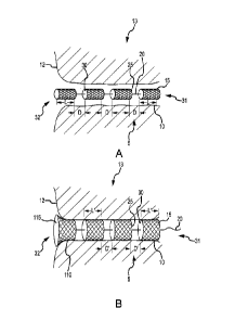
[016] FIG. 1A is an isometric view of an implantable fistula closure device
having a
segmented body and located in a fistula tract in a compressed or non-expanded
state.
[017] FIG. 1B is the same view as FIG. 1A, except the implantable fistula
closure
device is in a non-compressed or expanded state within the fistula tract.
[018] FIG. 2A is an isometric view of the implantable fistula closure device
located
in a fistula tract in a compressed or non-expanded state, wherein the distal
most
body of the device body has a conical shape, as opposed to a cylindrical
shape.
[019] FIG. 2B is the same view as FIG. 2A, except the implantable fistula
closure
device is in a non-compressed or expanded state within the fistula tract.
[020] FIG. 3A is an isometric view of an implantable fistula closure device
having a
non-segmented body and located in a fistula tract in a compressed or non-
expanded
state.
[021] FIG. 3B is the same view as FIG. 3A, except the implantable fistula
closure
device is in a non-compressed or expanded state within the fistula tract.
[022] FIG. 4A is an isometric view of the implantable fistula closure device
located
in a fistula tract in a compressed or non-expanded state, wherein the distal
end of
the device includes an expanding feature in the form of a gel-filled
expandable
member sandwiched between discs.
[023] FIG. 4B is the same view as FIG. 4A, except the implantable fistula
closure
device and its expanding feature are in a non-compressed or expanded state.
[024] FIG. 5A is the same view as FIG. 4A, except the expanding feature
includes a
porous expandable member sandwiched between discs.
4
CA 02720206 2010-09-30
WO 2009/124148
PCT/US2009/039209
[025] FIG. 5B is the same view as FIG. 5A, except the implantable fistula
closure
device and its expanding feature are in a non-compressed or expanded state.
[026] FIG. 6A is an isometric view of the implantable fistula closure device
located
in a fistula tract in a compressed or non-expanded state, wherein the distal
end of
the device includes an expanding feature in the form of an expandable member
having a dual conical configuration.
[027] FIG. 6B is the same view as FIG. 6A, except the implantable fistula
closure
device and its expanding feature are in a non-compressed or expanded state.
[028] FIG. 7A is an isometric view of the implantable fistula closure device
located
in a fistula tract in a compressed or non-expanded state, wherein the distal
end of
the device includes an expanding feature in the form of an expandable balloon.
[029] FIG. 7B is the same view as FIG. 7A, except the implantable fistula
closure
device and its expanding feature are in a non-compressed or expanded state.
[030] FIG. 8A is an isometric view of an expanding feature in a slightly
expanded
state and similar to the balloon-type expanding feature of of FIG. 7A, except
the
balloon is expanded via a jack-like feature.
[031] FIG. 8B is an isometric view of the expanding feature of FIG. 8B,
wherein the
expanding feature is more fully expanded.
[032] FIG. 9A is a side view of one embodiment of a delivery device for the
implantable fistula closure device disclosed herein, wherein a portion of the
delivery
device is inserted into a fistula tract.
[033] FIG. 9B is the same view as FIG. 9A, except the entire delivery device
is
shown inserted into the fistula tract.
[034] FIG. 9C is the same view as FIG. 9A, except the delivery device is
withdrawn
from about the device body and the device body is fully expanded.
[035] FIG. 9D is an end isometric of one embodiment of the delivery device of
FIG.
9A.
[036] FIG. 9E is an end isometric view of an alternative embodiment of the
delivery
device of FIG. 9A.
[037] FIG. 9F is an end isometric view of another alternative embodiment of
the
delivery device of FIG. 9A.
[038] FIG. 10 is a front view of a proximal clip.
[039] FIG. 11 is a side view of the clip of FIG. 10.
CA 02720206 2010-09-30
WO 2009/124148 PCT/US2009/039209
[040] FIGS. 12A-12F are isometric views of the fistula closure device
illustrating one
embodiment of a method of treating a fistula.
DETAILED DESCRIPTION
[041] Fistula tracts 10 can be nonlinear or curvilinear and contain cavities
of varying
sizes at different intervals within the tract. An implantable fistula closure
device 5
disclosed herein employs advantageous design, configuration techniques and
attributes to accommodate such constraints. For example, in one embodiment,
the
device 5 may have a segmented expandable body 13 formed of a plurality of
individual expandable bodies or members 15 coupled together in an immediately
adjacent abutting fashion or in a spaced-apart fashion. Upon being inserted
into the
fistula tract 10 with its expandable members 15 in a collapsed or compressed
state,
which allows for convenient insertion of the device 5 into the fistula tract
10, the
expandable members 15 are allowed to expand to fill the portion of the fistula
tract
in which each expandable member 15 is located. The segmented nature of the
body 13 of the device 5 or, more specifically, the fact the device's body 13
is formed
of a plurality of individual members 15 allows the body 13 to be more easily
placed in
and more readily conform to the tortuous and diametrically varying
configuration of a
fistula tract 10 when expanded within the fistula tract. Thus, once the body
13 is
allowed to expand within the fistula tract, the device generally completely
fills the
fistula tract. In one embodiment, when the body 13 expands to fill the fistula
tract,
the device may generally stop fluid flow from the bowel from running out
through the
fistula tract by occluding the distal end of the tract via a distal end of the
device body
13 that is generally non-porous or has an ability to seal the distal end of
the tract.
However, generally speaking, a fistula tract will leak fluid from within the
tissue walls
surrounding the fistula tract and some of this fluid will be absorbed by the
device and
the remaining fluid will drain out of the proximal end of the tract,
potentially through
the proximal end of the device body 13, which is generally porous or has the
ability
to allow the passage of fluids while generally occluding or filling the tract.
[042] Preventing bodily fluids that originate at the distal end of the tract
(e.g., bowel
fluids) from passing through a fistula tract 10 and, in some embodiments, also
reducing the amount or rate of flow through the fistula tract for body fluids
originating
6
CA 02720206 2010-09-30
WO 2009/124148 PCT/US2009/039209
in the tract itself may significantly reduce the time to closure and reduce
the
necessity for surgery. In one embodiment, the device 5 disclosed herein may
reduce
or eliminate the passage of fluids through the tract 10 as well as providing a
matrix
that promotes tissue growth. This device 5 may be utilized to treat a variety
of
clinically significant fistulas 10, including enterocutaneous fistulas, anal
fistulas,
bronchopleural fistulas, non-healing g-tube tracts, tracheal-esophogeal
fistulas, and
others.
[043] For a discussion of an embodiment of the implantable fistula closure
device 5,
reference is made to FIGS. 1A and 1B. FIG. 1A is an isometric view of the
device 5
located in a fistula tract 10 in a compressed or non-expanded state, and FIG.
1B is
the same view as FIG. 1A, except the device 5 is in a non-compressed or
expanded
state. As shown in FIGS. 1A and 1B, the implantable fistula closure device 5
includes a proximal end 31, a distal end 32, and an expandable body 13 formed
of a
plurality of individual porous bodies 15 operably connected via a connecting
member
20. Each porous body 15 includes a proximal end 25 and a distal end 30. Each
porous body 15 is adapted to expand from a compressed or non-expanded state
(FIG. 1A) to a non-compressed or expanded state (FIG. 1B) after insertion into
the
tract 10, thereby filling any cavities within the tract 10 and approximating
the fistula
tract walls.
[044] As can be understood from FIG. 1A, in some embodiments, when the bodies
15 are in a compressed or non-expanded state, the bodies 15 will be spaced-
apart
from each other along the length of the device 5 to form a segmented
configuration
for the device body 13. In some embodiments, the spaced-apart distances D
between adjacent proximal and distal ends 25, 30 of the bodies 15 in a
compressed
or non-expanded state is between approximately zero mm and approximately five
mm. In one embodiment, the space apart distance D between adjacent proximal
and distal ends 25, 30 of the bodies 15 in a compressed or non-expanded state
are
between approximately zero mm and approximately 25 mm. Where the distance D
between immediately adjacent bodies 15 is approximately zero mm when the
bodies
15 are in a non-expanded state, the bodies 15 will be said to be in an
abutting or
touching configuration, as opposed to a spaced-apart condition. Regardless,
the
device body 13 will still be considered to be segmented on account of the
device
body 13 being formed of a plurality of individual porous bodies 15.
7
CA 02720206 2010-09-30
WO 2009/124148 PCT/US2009/039209
[045] In some embodiments, the spaced-apart distances D between adjacent
proximal and distal ends 25, 30 of the bodies 15 in a compressed or non-
expanded
state are between approximately zero percent and approximately two and one-
half
percent of the overall non-expanded length L of a body 15. Where the distance
D
between immediately adjacent bodies 15 is approximately zero percent of the
length
L of a body 15 when the bodies 15 are in a non-expanded state, the bodies 15
will
be said to be in an abutting or touching configuration, as opposed to a spaced-
apart
condition. Regardless, the device body 13 will still be considered to be
segmented
on account of the device body 13 being formed of a plurality of individual
porous
bodies 15.
[046] Regardless of whether the bodies are in a spaced-apart configuration or
an
abutting or touching configuration when the bodies 15 are in the compressed
state
depicted in FIG. 1A, the segmented configuration of the device body 13
facilitates
the device body 13 being inserted in and conforming to the tortuous
diametrically
varied route formed by the tract 10.
[047] As can be understood from FIG. 1B, when the bodies 15 are fully expanded
within the tract 10, the spaced-apart distances D' between adjacent proximal
and
distal ends 25, 30 of bodies 15 in a non-compressed or expanded state is
between
approximately zero mm and approximately five mm. In some embodiments, the
spaced-apart distances D' between adjacent proximal and distal ends 25, 30 of
the
bodies 15 in a non-compressed or expanded state is between approximately zero
percent and approximately two and one-half percent of the overall expanded
length
L' of a body 15. The expansion of the bodies 15 after insertion into the
fistula tract
allows the device body 13 to approximate the walls of the fistula tract, as
well as
fill open cavities. Because the segmented configuration of the device body 13
allows
the device to closely conform to the tortuous and diametrically varied route
formed
by the tract 10, the bodies 15, when in an expanded state within the tract 10
generally fill the tract 10 in a manner that minimizes voids and dead space.
Minimizing voids and dead space lowers the chance of sepsis and other
complications.
[048] While multiple bodies 15 are used for a segmented body 13 and such a
segmented body 13 is contemplated for the various embodiments disclosed
herein, a
non-segmented body (i.e., a body 13 that is a continuous, single-piece body 13
as
opposed to being formed from multiple bodies 15) is also contemplated for
most, if
8
CA 02720206 2010-09-30
WO 2009/124148 PCT/US2009/039209
not all of the embodiments disclosed herein pertaining to various distal
and/or
proximal anchors such as, for example, those similar to the proximal and
distal
anchors depicted in the various figures as 50 and 900. An example of a non-
segmented body 15 is depicted in FIGS. 3A and 3B. Such embodiments may have a
single porous body 15 forming the porous non-segmented body 13.
[049] In one embodiment, one or more of the porous bodies 15 of the device 5
may
be a compressed open cell polymer and may be made of any synthetic or natural
biodegradable, resorbable, biocompatible polymer, such as collagen, hyaluronic
acid
and polyglycolic acid ("PGA"). The biodegradability allows for degradation at
a
specified rate that matches the rate of tissue ingrowth and fistula tract
healing, such
that by the time the fistula tract is healed, the material is completely
absorbed by the
body. It should be noted that the fistula tract may heal before the material
is
completely absorbed by the body. That is, the degradation rate of the device
does
not match, or is slower than, the rate of tissue ingrowth and fistula tract
healing. It
should also be noted that a mixture of different biodegradable polymers may
also be
utilized.
[050] Expansion of the bodies 15 within the tract 10 provides a porous
scaffold to
the fistula tract and may partially or entirely stop the flow of bodily fluids
through the
tract. The scaffold provides a matrix that may promote tissue in-growth
allowing the
fistula to close. The incorporation of an antimicrobial agent, such as silver,
in the
porous bodies 15 or in the insertion methodology may also be incorporated to
actively prevent infection and/or sepsis formation and aid in the healing of
the tract.
The porous bodies 15 may include wound-healing agents, such as growth factors.
In
some embodiments, the porous bodies include fibrosis-promoting agents.
[051] The porous body may be adapted and configured to expand after placement
in the fistula tract and absorb fluid thereby approximating closely the tract
intra-
lumina! walls. The porous body may include a porous resorbable open cell
polymer
foam adapted to expand and serve as a scaffold for tissue growth and closure
of the
fistula tract.
[052] In one embodiment, the porous body comprises collapsed or compressed
pores, adapted and configured to increase in size after placement in a fistula
tract,
thus filling the fistula tract. In some embodiments, the pores of the bodies
are of a
reduced size, which is advantageous. For example, the pore size may vary from
5 to
1000 microns in size with an overall porosity of 25-95%. In one embodiment,
bodies
9
CA 02720206 2010-09-30
WO 2009/124148 PCT/US2009/039209
with a controlled pore size of between approximately 50 microns and
approximately
100 microns may be used. A body with a controlled pore size, that is, a body
without
a broad distribution of pore sizes, may promote greater angiogenesis, which,
in turn,
may promote better wound-healing. Examples of materials that may provide some
or all of the controlled pore size and porosities include various biomaterials
manufactured by Kensey Nash Corporation, CollaPlug or other collagen products
as
manufactured by Integra Corporation, and STAR materials as manufactured by
Healionics Corporation.
[053] As mentioned above with respect to FIG. 1A, the porous bodies 15 of the
device 5 may be operably connected by a connecting member 20. The connecting
member 20 may be a bioresorbable and biocompatible filament or string. In some
embodiments, the connecting member 20 may also be a filamentous string, which
enables the decoupling of the plurality of porous bodies 15 from the
connecting
member subsequent to implantation of the device 5 in the tract 10.
[054] As mentioned above with respect to FIGS. 1A and 1B, in one embodiment,
the device 5 includes at least two porous bodies 15 which are adapted and
configured to work together to form the device's overall body 13 and
separately to
allow the device body 13 to conform to the tract 10 and fill all of the tract
voids. In
other words, the bodies 15 are separate individual bodies joined together via
the
connecting member 20 along the length of the device 5 such that the resulting
device
body 13 has a segmented configuration. In one embodiment, when the bodies 15
are in an expanded state or even in a non-expanded state, the spaced-apart
distances D, D' may be zero such that the proximal and distal ends 25, 30 of
adjacent bodies 15 abut. In such an embodiment, the bodies 15 appear to form a
generally continuous porous device body 13 that is segmented by the interfaces
of
the adjacent proximal and distal ends 25, 30 of adjacent bodies 15. Thus,
regardless of the magnitude of the spaced-apart distances D, D', in one
embodiment, the device body 13 can be considered to be a chain or series of
individual porous bodies 15 configured to work together and separately,
resulting in
an overall body 13 of the device 5 that is segmented and capable of conforming
to
the tract 10. It should be noted that the device 5 does not stent open the
tract 10,
but rather, the device 5, when in an expanded or non-compressed state, is
capable
of conforming to the tract 10
CA 02720206 2010-09-30
WO 2009/124148 PCT/US2009/039209
[055] In some embodiments, the device 5 will be configured to fill multi-tract
fistulas.
For example, the device 5 may have multiple device bodies 13 joined together
at a
common point of the device 5. In other words, the device may have at least two
chains of porous bodies 15 joined together to allow a segmented device body 13
to
be inserted into each of the tracts 10 of a multi-tract fistula.
Alternatively, at least two
chains of porous bodies 15 may be joined together to create a device 5 with at
least
two segmented device bodies 13.
[056] As can be understood from FIG. 9B, in one embodiment, the device 5 may
be
deployed from the lumen of a delivery sheath 600 via a long, flexible rod or a
"pusher" 603. The pusher 603 may be inserted through the delivery device 600
and
may enable the clinician to push or otherwise direct the segmented device body
13
into the tract 10, thereby minimizing the dead space or void that may be left
between
the individual segments of the device body 13 or between the body 13 and tract
10.
In some embodiments, the porous bodies 15 may not be connected via a
connecting
member 20, but instead may be multiple free bodies 15 that are inserted into
the
lumen of the sheath 600 for delivery into the tract. Thus, a pusher may enable
the
clinician to push or otherwise direct the unconnected bodies 15 into the
fistula tract
10.
[057] In one embodiment, as illustrated in FIGS. 12A-12G, the device 5 is
loaded in
a lumen of a catheter, sheath or guidewire. As can be understood from FIGS.
12A-
12B, the loaded catheter, sheath or guidewire 600, 601 is then inserted into
the tract
and then, as shown in FIG. 12C, withdrawn from about the device body 13 to
leave the device body 13 within the tract 10. As indicated in FIGS. 12C-12F,
the
device body 13 then expands to fill and occlude the tract 10. As illustrated
in FIG.
12F, and as described in more detail below, the proximal end of the tract 10
may
include a proximal clip 900 to further secure the device 5 in the tract 10.
[058] In another embodiment, as shown in FIGS. 9A-9F, the catheter or sheath
may
be a dual lumen catheter 600, where one lumen contains the device 5 and the
other
lumen contains a guidewire 601. In one embodiment, the catheter may be a multi-
lumen catheter where at least one lumen is shaped like a "D". As can be
understood
from FIGS. 9A-9B, the guidewire 601 is inserted into the fistula tract 10 and
the
catheter 600 is tracked over the guidewire 601. As shown in FIG. 9C, the
device 5 is
deployed and the catheter 600 is withdrawn from about the device body 13 to
leave
11
CA 02720206 2010-09-30
WO 2009/124148 PCT/US2009/039209
the device body within the tract 10. The device body 13 then expands to fill
and
occlude the tract 10.
[059] As illustrated in FIGS. 9D-9E, which show various embodiments of the
delivery device of FIG. 9A, the catheter 600 may be a peel away sheath. For
example, a skive, score, partial cut, mechanical joint or formed groove may
create a
longitudinally extending stress concentration 334 for causing the catheter to
peal
along the stress concentration 334. As indicated in FIG. 9E, the stress
concentration
334, which may be a mechanical joint, may include a grasping member 337 that
may
be used to exert the necessary force on the stress concentration to bring
about its
separation.
[060] The delivery devices depicted in FIGS. 9D-9F may include a central or
main
lumen 335 through which the fistula closure device 5 may pass and a secondary
lumen 336 through which the guidewire 601 may pass.
[061] As can be understood from FIGS. 9D-9F, the delivery device 600 may be
tracked over a guidewire 601 with the fistula occlusion device 5 residing in
the main
lumen 335. Once properly positioned in the fistula tract, the delivery device
600 can
be removed from about the closure device 5. The removal of the delivery device
600
from about the closure device 5 may be accomplished by grasping an exposed
portion of the delivery device 5 or a grasping member 337 (see FIG. 9E) and
then
pulling or pushing the delivery device relative to the closure device 5.
Alternatively, a
hooked member 340 having a hook or other engagement feature 341 that engages
an end of the delivery device 600 may be employed where the hooked member 340
can be used to pull the delivery device 600 from about the closure device 5,
as can
be understood from FIGS. 9D and 9F.
[062] Regardless of whether a catheter, sheath, guidewire or stylet or
combination
thereof is used to deploy the device 5 in the tract 10, once located within
the tract 10,
the device body 13 will begin to expand and fill the voids of the tract 10.
Expansion
of the bodies 15 may be a result of being free of the constraints of the lumen
of the
sheath, catheter or guidewire used to deliver the device 5. Expansion of the
bodies
15 may be a result of being free of the constraints of a restraining mechanism
such
as a biodegradable ring, sheath, member, etc. extending about the bodies 15
when
first deployed in the tract 10. Expansion may be a result of being exposed to
body
fluids or temperature within the tract 10. Expansion may be a result of any
one or
more of these aforementioned expansion methods.
12
CA 02720206 2010-09-30
WO 2009/124148 PCT/US2009/039209
[063] As can be understood from FIG. 1B, the porous bodies 15 at the proximal
and/or distal ends 31, 32 of the device 5 may be configured to protrude from
the
distal and/or proximal fistula openings when implanted in the fistula tract
10. As
depicted in FIG. 1B, the protruding end 115 of the most distal body 110, or
the
entirety of the most distal body 110, may be configured to expand more than
the rest
of the porous bodies 15. Such an over-expanding capability at the distal ends
32 of
the device 5 when within the fistula tract may produce an occluding and
anchoring
effect. Additionally or alternatively, the same concept may be applied to the
most
proximal body 15 at the device proximal end 31. Such embodiments can be
considered to have at least one body 15 with a magnitude of expansion that is
different from (i.e., exceeds) the magnitude of expansion of the other bodies
15. In
one embodiment, a device 5 with a distal most body 110 that is configured to
have
increased expansion as compared to its fellow bodies 15 will be positioned in
the
tract 10 such that the most distal body 110 is partially within the tract 10
and partially
extending from the distal opening 12 into, for example, the bowel lumen. Thus,
as
illustrated in FIG. 1B, once the distal portion of the device 5 is in place,
the distal
most body 110 of the device 5 expands to contact the edges of distal opening
12 of
the fistula tract 10, thereby occluding the distal opening 12 of the fistula
tract 10.
The device 5 also expands to fill the rest of the fistula tract 10. To
facilitate a
generally complete sealing of the distal opening 12, the distal most body 110
of the
device 5 may include an impermeable coating.
[064] In a manner similar to that discussed above with respect to the distal
most
body 110, the proximal most body at the proximal end 31 of the device 5 may be
adapted and configured to anchor or otherwise hold the device 5 in place
within the
fistula tract. Where both the distal and proximal most bodies are so
configured, the
distal and proximal most bodies will provide a counter force or counter
balance to
each other through the connecting member 20. In some embodiments, the proximal
most and/or distal most bodies may be or include an adhesive layer to further
strengthen the seal around the respective fistula tract openings.
[065] For a discussion of distal most or proximal most bodies 15 having shapes
other than generally cylindrical, reference is made to FIGS. 2A and 2B, which
are
respectively the same as FIGS. 1A and 1B, except illustrating the differently
shaped
bodies 15. As shown in FIGS. 2A and 2B, the distal most body 120 may have a
shape that is non-cylindrical and, more specifically, conical. The proximal
most body
13
CA 02720206 2010-09-30
WO 2009/124148 PCT/US2009/039209
15 at the proximal end 31 of the device 5 may also have a conical shape as
opposed
to a cylindrical shape.
[066] In some embodiments, the conically shaped most distal body 120 is
generally
shaped such that its distal end 125 is generally greater in diameter than on
its
proximal end. The distal end 32 of the device 5 may be advanced into the
distal
opening 12 of the fistula tract 10 such that a distal portion 125 of the body
120
extends from the tract opening 12 into, for example, the bowel lumen. As
illustrated
in FIG. 1B, once the distal end of the device 5 is in place, the distal end
125 of the
body 120 expands to contact the edges of the distal opening 12 of the fistula
tract
10, thereby occluding the distal opening 12 of the fistula tract 10. The rest
of the
device body 13 also expands to generally fill the rest of the fistula tract 10
as
described above. In some embodiments, the proximal end 31 of the device 5 does
not extend beyond the edge of the fistula tract, while in other embodiments it
does.
[067] In some embodiments, the difference in diameter of the distal end 125
could
be a result of a difference in the distance by which the different parts of
the distal
body 120 can expand. For example, the diameter of the cylinder in the
compressed
or non-expanded state is uniform, however when the cylinder expands, the
proximal
end of the cylinder may reach the wall of the fistula tract 10, but the distal
end may
have a greater distance to expand before reaching the wall of the fistula
tract 10
which corresponds to its target area of expansion. In this case, the diameter
of the
cylinder in a non-expanded state is uniform, but the diameter of the cylinder
in the
expanded state forms a conical shape.
[068] In some embodiments of the device, as can be understood from FIGS. 10
and
11, the proximal end 31 may be adapted and configured to receive a proximal
clip
900 that secures the device 5 in place. As shown in FIG. 10, which illustrates
a front
view of one embodiment of such a clip 900, the clip 900 may include an outer
ring
902 and a mesh-like membrane 904 that extends across the clip 900. In one
embodiment, as illustrated in FIG. 11, which is a side view of the clip, the
clip 900 is
disc-shaped. In alternative embodiments, the clip 900 is a shape other than a
disc,
such as a polygon. The clip 900 may be made of any biocompatible material,
such
as PGLA, PVA or PVC or other suitable biocompatible plastic. The material may
also be resorbable.
[069] As can be understood from FIG. 11, the clip 900 extends across the
proximal
end of the fistula tract 10 and is generally flush or slightly raised relative
to the
14
CA 02720206 2010-09-30
WO 2009/124148 PCT/US2009/039209
proximal end of the fistula tract 10. The clip 900 helps to maintain tension
on the
connecting member 20 that couples the expanding member 50 with the clip 900
thus
helping to maintain or anchor the device 5 in the tract 10. The clip 900 may
be
coupled to the connecting member 20 via friction, pinching, suturing or other
suitable
method.
[070] Features of the clip 900 and/or proximal end 31 of the device 5 may be
transparent to allow visual inspection of the tract. In some embodiments, the
clip
900 and/or proximal end of the device may be adapted to cover the proximal end
of
the fistula tract without completely sealing the proximal end of the tract,
thereby
allowing accumulating fluids to drain or escape from the proximal end of the
tract. In
addition, the mesh-like membrane 904 permits drainage of accumulating fluids
from
the proximal end of the tract. After the tract 10 heals, the proximal clip 900
will
resorb or otherwise be removed.
[071] In some embodiments, the distal end of the device body 13 may include an
expandable feature 50 that may serve to anchor the device distal end in place
at the
fistula distal opening 12 and/or seal the fistula distal opening 12. For a
discussion of
a first embodiment of such an expandable feature 50, reference is made to
FIGS. 4A
and 4B, which are respective isometric views of the device 5 located in the
fistula
tract 10 and the expandable feature 50 in a non-expanded state and an expanded
state.
[072] As shown in FIGS. 4A and 4B, the device body 13 is generally the same as
discussed above with respect to the embodiments depicted in FIGS. 1A and 1B
such
that the device body 13 includes individual porous bodies 15 coupled together
via a
connecting member 20. However, as indicated in FIGS. 4A and 4B, the distal end
32 of the device 5 terminates in the expandable feature 50, which is coupled
to the
distal end of the connector member 20. The expandable feature 50 may include a
gel-filled or otherwise readily deformable member 85 sandwiched between a pair
of
generally rigid discs 90. An actuation mechanism 95 extends along the
connector
member 20 to couple with the feature 50. The actuation mechanism 95 may be
filamentous or bioresorbable thread. Alternatively or additionally, the
actuation
mechanism may include a catheter 52 and one or more wires 51 longitudinally
displaceable within lumens of the catheter 52. The catheter 52 may extend
through
the bodies 15 the entire length of the device 5 and terminate at or near the
expandable feature 50. In some embodiments, the expandable feature 50 may
CA 02720206 2010-09-30
WO 2009/124148 PCT/US2009/039209
expand without an actuation mechanism 95, e.g., the expandable feature expands
upon exposure to body fluids or a temperature differential within the tract 10
or via its
own biased nature.
[073] The proximal end of the actuation mechanism 95 may be pulled or
otherwise
displaced relative to the rest of the actuation mechanism such that the
actuation
mechanism may cause the feature 50 to expand. For example, in one embodiment,
the feature 50 is biased in a non-expanded state and pulling on the mechanism
95,
as indicated by arrow A in FIG. 4A, causes the discs 90 to converge towards
each
other, eventually engaging each other to become fixed in the converged state,
as
depicted in FIG. 4B. The discs 90 converging causes the deformable member 85
to
squish or deflect outward, as illustrated in FIG. 4B, thereby serving as an
anchor
and/or sealing the tract opening 12. The device body 13 expands to generally
fill the
rest of the fistula tract 10 as described above.
[074] In another embodiment, the feature 50 is biased in an expanded state and
operating the mechanism 95 forces the discs 90 away from each other to cause
the
feature 50 to assume the generally cylindrical configuration depicted in FIG.
4A as
the device 5 is being negotiated through the tract 10. Once the feature 50
passes
through the tract opening 12, the mechanism 95 can be released to allow the
feature
50 to bias into the expanded state depicted in FIG. 4B. The feature 50 may
then
serve as an anchor and/or seal for the tract opening 12. The device body 13
expands to generally fill the rest of the fistula tract 10 as described above.
[075] As indicated in FIGS. 5A and 5B, which are the same respective views as
FIGS. 4A and 4B, in another embodiment, the feature 50 may have the same
configuration and operation as discussed above with respect to FIGS. 4A and
4B.
However, the readily expandable member 85 depicted in FIGS. 4A and 4B does not
have a gel-filled member 85 but instead has a porous member 85 formed from a
material similar to that employed for the various bodies 15. In one
embodiment, the
expandable member 85 may be a super compressed collagen. Like the member 85
depicted in FIGS. 4A and 4B, the member 85 depicted in FIGS. 5A and 5B may be
caused or allowed to expand laterally to serve as an anchor and/or seal, as
can be
understood from FIG. 5B. Expansion in the lateral direction may be
advantageous in
that it reduces the profile of the distal portion of the device 5 in the bowel
lumen.
The device body 13 expands to fill the remainder of the fistula tract 10 as
described
above.
16
CA 02720206 2010-09-30
WO 2009/124148 PCT/US2009/039209
[076] In an alternative to the embodiments discussed above with respect to
FIGS.
4A-5B, the expanding feature 50 may be biased to assume the biased
configuration
of FIGS. 4B and 5B. However, the device 5 will not employ an actuation
mechanism
95 to retain the feature 50 in a non-expanded state until properly located in
the fistula
tract 10. Instead, the feature 50 will be maintained in the non-expanded state
via the
lumen walls of a catheter, sheath or guidewire employed to deliver the device
5.
Once the device 5 is properly located within the tract 10, the catheter,
sheath or
guidewire can be withdrawn from about the device 5 to allow the feature 50 to
bias
into its expanded state.
[077] For a discussion of another embodiment of an expandable feature 50,
reference is made to FIGS. 6A-6B, which are respective isometric views of the
device 5 located in the fistula tract 10 and the expandable feature 50 is in
non-
expanded and expanded states. As shown in FIGS. 6A and 6B, the device body 13
is generally the same as discussed above with respect to the embodiments
depicted
in FIGS. 1A and 1B such that the device body 13 includes individual porous
bodies
15 coupled together via a connecting member 20. However, as indicated in FIGS.
6A and 6B, the distal end 32 of the device 5 terminates in the expandable
feature 50,
which is coupled to the distal end of the connector member 20 and has a dual-
conical configuration when in a non-expanded state.
[078] As depicted in FIG. 6A, in one embodiment, the expandable feature 50
when
in its dual-conical non-expanded state has a tip 101 of a first conical
section 50a
pointing distally, a tip 103 of a second conical section 50b pointing
proximally, and
the wide bases of each conical section 50a, 50b joined together. The tips 101,
103
may terminate in discs 90, the proximal of which may be connected to the
connection member 20. As shown in FIG. 6B, when the expandable feature 50 is
in
an expanded state, the feature 50 mushrooms laterally.
[079] In one embodiment, the conical sections 50a, 50b may be a gel-filled or
otherwise readily deformable member sandwiched between the pair of generally
rigid
discs 90. The conical sections 50a, 50b may be a porous member formed from a
material similar to that employed for the various bodies 15. The conical
sections
50a, 50b may be a super compressed collagen. The conical sections 50a, 50b may
be balloon-like in that the conical sections 50a, 50b have a resilient outer
surface or
skin enclosing a fluid, such as air, carbon dioxide, nitrogen, saline,
silicone rubber
gel, etc.
17
CA 02720206 2010-09-30
WO 2009/124148 PCT/US2009/039209
[080] Similar to the embodiment discussed with respect to FIGS. 4A-5B, in some
embodiments, an actuation mechanism may extend along the connector member 20
to couple with the feature 50. The actuation mechanism may be filamentous or
bioresorbable thread. Alternatively or additionally, the actuation mechanism
may
include a catheter and one or more wires longitudinally displaceable within
lumens of
the catheter. The catheter may extend through the bodies 15 the entire length
of the
device 5 and terminate at or near the expandable feature 50.
[081] The proximal end of the actuation mechanism may be pulled or otherwise
displaced relative to the rest of the actuation mechanism such that the
actuation
mechanism may cause the feature 50 to expand. For example, in one embodiment,
the feature 50 is biased in a non-expanded state and pulling on the mechanism
causes the discs 90 to converge towards each other, eventually engaging each
other
to become fixed in the converged state, as depicted in FIG. 6B. The discs 90
converging causes the deformable member 50a, 50b to squish or deflect outward,
as
illustrated in FIG. 6B, thereby serving as an anchor and/or sealing the tract
opening
12. Expansion in the lateral direction may be advantageous in that it reduces
the
profile of the distal portion of the device 5 in the bowel lumen. The device
body 13
expands to generally fill the rest of the fistula tract 10 as described above.
[082] In another embodiment, the feature 50 is biased in an expanded state and
operating the mechanism forces the discs 90 away from each other to cause the
feature 50 to assume the dual-conical configuration depicted in FIG. 6A as the
device 5 is being negotiated through the tract 10. Once the feature 50 passes
through the tract opening 12, the mechanism can be released to allow the
feature 50
to bias into the expanded state depicted in FIG. 6B. The feature 50 may then
serve
as an anchor and/or seal for the tract opening 12. The device body 13 expands
to
generally fill the rest of the fistula tract 10 as described above.
[083] In an alternative to the embodiments discussed above with respect to
FIGS.
6A-6B, the expanding feature 50 may be biased to assume the biased
configuration
of FIG. 6B. However, the device 5 will not employ an actuation mechanism to
retain
the feature 50 in a non-expanded state until properly located in the fistula
tract 10.
Instead, the feature 50 will be maintained in the non-expanded state via the
lumen
walls of a catheter, sheath or guidewire employed to deliver the device 5.
Once the
device 5 is properly located within the tract 10, the catheter, sheath or
guidewire can
18
CA 02720206 2010-09-30
WO 2009/124148 PCT/US2009/039209
be withdrawn from about the device 5 to allow the feature 50 to bias into its
expanded state.
[084] In one embodiment, the dual-conical configured expandable feature 50 may
be formed of a sheet or membrane extended over a collapsible and expandable
framework similar in configuration, operation and material to those discussed
with
respect to FIGS. 8A-8B. In such an embodiment, the device 5 may include an
actuation mechanism similar to that discussed with respect to FIG. 8A-8B.
[085] For a discussion of another embodiment of an expandable feature 50,
reference is made to FIGS. 7A-7B, which are respective isometric views of the
device 5 located in the fistula tract 10 and the expandable feature 50 in non-
expanded and expanded states. As shown in FIGS. 7A and 7B, the device body 13
is generally the same as discussed above with respect to the embodiments
depicted
in FIGS. 1A and 1B such that the device body 13 includes individual porous
bodies
15 coupled together via a connecting member 20. However, as indicated in FIGS.
7A and 7B, the distal end 32 of the device 5 terminates in the expandable
feature 50,
which is coupled to the distal end of the connector member 20 and is in the
form of
an inflatable balloon 50.
[086] As depicted in FIGS. 7A and 7B, the balloon 50 may be coupled to the
connector member 20. The connector member 20 may be a lumen 20 through which
an inflation fluid may be transferred to the balloon 50 for its inflation.
Alternatively,
the lumen may be a separate structure that extends along or near to the
connector
member 20.
[087] As indicated in FIG. 7A, the expandable feature or, more specifically,
balloon
member 50 of the device 5 is advanced in a non-inflated state through the
distal
opening 12 of the fistula tract 10. As can be understood from FIG. 7B, once
the
balloon 50 of the device 5 is in position, the balloon 50 may be inflated via
the lumen
20 with a material such as air, saline or other biocompatible fluid or
solidifying gel.
Tension may then be applied to the device 5 via the connector member 20, which
causes the balloon member 50 to occlude the distal opening 12 of the fistula
tract 10.
In some embodiments, tension may be applied to the device 5 via the connector
member 20 where the connector member 20 is only connected to the balloon
member 50 and is not otherwise connected to the device body 13. The balloon
member 50 may also be retracted back against the distal opening 12 of the
tract 10.
19
CA 02720206 2010-09-30
WO 2009/124148 PCT/US2009/039209
The device body 13 expands to generally fill the rest of the fistula tract 10
as
described above.
[088] In one embodiment, the balloon 50 may include an adhesive coating
adapted
to adhere to the tissue surface of the region adjacent the distal opening 12
of the
fistula tract 10. The balloon 50 may include micropores on the side of the
balloon 50
intended to face towards the tissue to be contacted by the balloon 50. The
micropores may allow any inflating fluid to leak out of said pores, thereby
allowing
the delivery of an adhesive/sealant to the distal opening 12.
[089] Depending on the embodiment, the balloon 50 may be a fluid inflatable or
expandable disc-shaped balloon adapted to occlude the distal tract opening.
Alternatively, the balloon 50 may be a fluid inflatable or expandable flat
cone-shaped
balloon adapted to occlude the distal tract opening. The balloon 50 may be
formed
of a bioconipatible polymer. Alternatively, the balloon 50 may be formed of a
biodegradable or bioabsorbable material.
[090] In one embodiment, the balloon 50 may be injected with a time curing
liquid
material, e.g., a silicone material such as that manufactured by Nusil
Silicone
Technology. Once the liquid material starts to cure, the clinician may force
the
balloon against the pen-opening area at the distal opening of the fistula
tract, thereby
causing the balloon and the liquid material contained therein to assume the
shape of
the pen-opening area. Once the liquid material is substantially cured, the
balloon 50
will retain the shape it assumed, resulting in a balloon that is custom shaped
for the
distal tract opening and creating a seal of the distal tract opening that is
potentially
more likely to be fluid-tight as compared to other distal anchor
configurations.
[091] Alternatively, the balloon 50 may be mechanically inflated or expanded,
as
can be understood from FIGS. 8A and 8B, which show side views of such a device
5. The mechanically inflatable or expandable balloon 50 includes a jack-like
feature
800 and a radio-opaque marker band 801 on a first central axis point 802 of
the jack-
like feature 800. In one embodiment, the jack-like feature also includes a
connecting
member 20 to connect the jack-like feature 800 to porous bodies 15 of the
device 5.
In one embodiment, the jack-like feature 800 includes four arms 810 with weak
points 805 which aid in the transition between non-expanded and expanded
states.
In other embodiments, the jack-like feature 800 may have more than four arms
or
less than four arms. The arms 810 are joined at at least one of a first or
second
central axis point 802.
CA 02720206 2010-09-30
WO 2009/124148 PCT/US2009/039209
[092] The balloon 50 generally conforms to the jack-like feature 800. That is,
when
the jack-like feature 800 is in a non-expanded state, the balloon 50 is not
inflated.
When the jack-like feature 800 is in an expanded state, the balloon 50 is
inflated
and, when in the appropriate position, occludes the distal tract opening.
Following
installation of the balloon 50 at the distal end 12 of the tract 10, the jack-
like feature
800 may be collapsed and removed from the fistula closure device 5 via a
recoil
member 815, which may be a filamentous string or suture line.
[093] Regardless of whether the balloon 50 is expanded via injection of a
fluid or via
an expanding mechanical framework 800, the material forming the balloon 50 may
provide a resilient distal anchor 50 that may readily conform to irregular
distal tract
openings. As a result, the balloon 50 may be able to readily seal an irregular
distal
tract opening.
[094] In some embodiments of each of the fistula closure devices 5 equipped
with
an expandable feature 50, as discussed above, the device 5 and its expandable
feature 50 in a non-expanded state are configured to pass through a lumen of
catheter size of nine French or smaller, and in some embodiments, twenty
French or
smaller. The expandable feature 50 or portions thereof may be adapted to
adhere to
the tissue surface area forming a distal tract opening 12. For example, the
expandable feature 50 may include a biocompatible adhesive surface of the
feature
50 intended to contact the tissue surface area forming the opening 12. The
adhesive
may activate after exposure to a fluid (e.g., body fluid) or body temperature.
The
adhesive may initially strengthen the bond of the feature 50 to the tissue and
then
gradually degrade in strength as fistula tract healing occurs or after fistula
tract
healing. Depending on the embodiment, the adhesive may create a fluid
impermeable seal for at least 7, 14, 21, 28, 35, 60 or any other number of
days.
[095] In some embodiments of each of the expandable features 50 discussed
above, the expandable feature 50 may include attachment members 45 such as
micro hooks or tines. Such attachment members 45 may be located on a surface
of
the feature 50 intended to contact the tissue surface area forming the opening
12,
thereby facilitating the adherence of the feature to the tissue surface
bordering the
distal tract opening 10 and the occlusion thereof.
[096] In some embodiments of each of the expandable features 50 discussed
above, the expandable feature 50 or various components thereof may be
resorbable
and adapted to occlude the fistula tract and then resorb after the tract 10
has closed
21
CA 02720206 2010-09-30
WO 2009/124148 PCT/US2009/039209
at least 45%, 55%, 65%, 75%, 85%, 95%, 100% or any other percentage. The
feature 50 or various components thereof may be biodegradable and/or adapted
to
fall away from the distal fistula opening 12 and be extruded through the
gastrointestinal tract. For example, the feature 50 or various components
thereof
may be secreted from the body after the tract 10 has progressed towards
closure
(e.g., after at least 7, 14, 21, 28, 35 or any other number of days adequate
to
achieve sufficient closure.
[097] In some embodiments of the devices 5 employing each of the expandable
features 50 discussed above, the connecting member 20 may be a biocompatible
polymer string extending through the tract from the expanding feature 50. The
connecting member 20 may be formed of a resorbable material and may resorb
after
the tract 10 has closed at least 45%, 55%, 65%, 75%, 85%, 95%, 100% or any
other
percentage. The member 20 may provide tensile force substantially
perpendicularly
to the feature 50, thereby pulling the feature 50 against the tract's distal
opening 12
and anchoring the feature 50 in place to occlude the distal tract opening. As
explained above with respect to FIGS. 10 and 11, the device 5 may include a
clip
900 at the proximal end, which may generally occlude, but not seal, the
proximal end
of the tract and allow tension in the member 20, which extends between the
clip 900
and feature 50.
[098] The fistula closure devices 10 as described herein may be implanted into
a
fistula tract 10 via various methods. For example, the fistula tract 10 may be
visualized via direct visual inspection or medical imaging methods (e.g.,
Fluoroscopy, CT scan, MRI, etc.). A guidewire may be negotiated through the
tract
10. The tract 10 may then be de-epithelializing irrigated. The device 5 may
then be
threaded over the guidewire and pushed into the tract 10. The distal fistula
opening
12 may be occluded via elements of the device 5 (e.g., the most distal body
110
and/or expanding feature 50). The device 5 may be trimmed to the length of the
tract 10, after which the guidewire is removed. The device 5 and, more
specifically,
the device body 13 may be irrigated to cause expansion of the body 13. The
device
may be anchored at the proximal fistula opening with a proximal end piece. For
example, a retaining member may be connected to the distal end of the device 5
and
secured to the region surround the proximal end opening of the tract 10,
thereby
creating tension in the device 5. The proximal fistula opening may then be
covered
with a dressing.
22
CA 02720206 2010-09-30
WO 2009/124148 PCT/US2009/039209
[099] In another method of implanting the fistula closure device 5 in a
fistula tract
10, a compressed porous scaffold 13 is placed in the fistula tract 10, wherein
the
scaffold 13 is at least partially inserted into the tract 10. The porous
scaffold may be
filled with an injectable polymer fluid 100, which may form an occlusive plug
and may
promote tissue growth and hence healing of the fistula tract. The method may
further include fixating the device 5 in the tract 10 using a biocompatible
connecting
member 20, such as a string, which is attached to the device 5. The polymer
100
injected into the tract 10 may be in a form that allows the foam to
approximate the
walls of the fistula tract 10 and fill any voids in the tract.
[0100] In another method of implanting the fistula closure device 5 in a
fistula tract
10, a distal end 32 of the device 5 may be placed in such a way as to protect
and
occlude the distal end 12 of a fistula tract 10. The body 13 of the device 5
may be
inserted into the fistula tract 10 in such a way as to at least partially fill
the fistula
tract 10. The surface load or point load dependant expansion of porous bodies
15
may then be activated within the fistula tract and the device 5 can be
anchored in
place at the distal and/or proximal ends 32, 31 as discussed above. For
purposes of
this disclosure, surface load or point load dependent expansion refers to the
expansion of the porous bodies where, upon contact between the fistula tract
wall
(the "load") and a point on the porous body, that point of the porous body
will stop
expanding. The points on any or all of the rest of the porous body will
continue to
expand until the remaining points also make contact with the fistula tract
wall. Thus,
unlike the occluding bodies of fistula closure devices known in the art, the
surface
load or point load dependant expansion of the bodies 13 of the device 5
disclosed
herein allows the body 13 to generally fill and conform to the tract 10
without
distorting the tract 10 or causing the tract to conform or deform due to the
expansion
of the body 13 in the tract. This ability of the body 13 can be a result of
pre-
compression of the body 13 and/or the nature of the material used. Examples of
materials from which to form the bodies 15 of the device 5 include: AngioSeal-
like
products, collagen sponge or other biomaterial materials as manufactured by
Kensey
Nash Corporation of 735 Pennsylvania Drive, Exton, PA 19341; CollaPlug or
other
collagen products as manufactured by Integra Corporation of 311 Enterprise
Drive,
Plainsboro, New Jersey 08536; and STAR materials as manufactured by Healionics
Corporation of 14787 NE 95th Street, Redmond, WA 98052.
23
CA 02720206 2010-09-30
WO 2009/124148 PCT/US2009/039209
[0101] With respect to the CollaPlug material, in some embodiments, the
CollaPlug
material is compressed prior to delivery into the tract 10, the CollaPlug
material
being approximately 90% porous.
[0102] With respect to the STAR materials, some such materials are know to
have a
specific pore size that promotes better angiogenesis. The STAR materials and
some
of the materials and products discussed above are capable of achieving the
controlled pore size and overall porosity discussed earlier in this Detailed
Discussion.
[0103] In another method of implanting the fistula closure device 5 in a
fistula tract
10, the tract is visualized and a guidewire is routed into the tract 10. The
tract 10 is
de-epthialized and irrigated to remove any unwanted internal matter. The
fistula
closure device 5 may be tracked over the guidewire and the device 5 may then
be
received into the fistula tract until the distal end of the device 5 extends
beyond the
distal fistula opening 12. The device 5 may be expanded by irrigation so as to
approximate the fistula tract 10. The device 5 may be trimmed if required. The
method may include clipping or otherwise securing the proximal end of the
device 10
at the proximal tract opening to provide a secure anchor. The proximal opening
may
then be coved with a dressing. In one embodiment, the segmented body 13 of the
device 5, when in an expanded state, generally approximates the volume of the
fistula tract with minimal distortion of the fistula tract.
[0104] In some embodiments, the bodies 15 of the fistula closure device 5 are
formed from materials other than a graft, wherein graft is defined as a
transplant
from animal or human tissue.
[0105] In some embodiment, the bodies 15 of the fistula closure device 5 are
formed
from materials other than an extracellular matrix ("ECM") material, wherein
ECM
material is defined as decellularized organic tissue of human or animal
origin.
Furthermore, in some such embodiments, the bodies 15 of the fistula closure
device
are formed from materials other than those that are remodelable, wherein
remodelable is defined as the ability of the material to become a part of the
tissue.
Instead, in some embodiments, the bodies 15 of the fistula closure device 5
may rely
heavily on the amount of induced cross-linking that allows control of the
resorbtion
rate. Cross-linking essentially destroys the remodelable properties of a
material.
While remodelable may not exclude resorbable material completely, in some
embodiments, the bodies 15 of the fistula closure device 5 may be formed of
24
CA 02720206 2010-09-30
WO 2009/124148 PCT/US2009/039209
material that is completely resorbable and has no remodelable requirements or
capabilities.
[0106] In some embodiments of the fistula closure device 5, the device body 13
is
formed of multiple bodies 15 to form a segmented body 13. The body 13 may
include a distal occlusion member 50 (e.g., an umbrella-like member), the
member
50 acting as an occlusion mechanism that is more of an occlusive cover rather
than
a plug or sealing member.
[0107] In one embodiment, the body 13, whether a segmented body 13 formed of a
series of individual bodies 15 or a non-segmented body 13 formed of a single
continuous body, may have a hole extending longitudinally through the body 13.
The
hole may be centrally located or at any other location on the body 13 so long
as the
body runs generally longitudinally through the body 13 and substantially the
full
length of the body 13. In one embodiment, the hole may be the hole through
which
the connecting member 20 extends. In other embodiments, the hole may be a hole
other than the hole through which the connecting member 20 extends.
[0108] Subsequent to the implantation of the device 5 within the fistula
tract, a
fluoroscopic material (e.g., a radiopaque fluid) may be delivered (e.g.,
injected) into
the hole. The fluoroscopic material will then disperse throughout the fistula
tract.
The fistula tract may then be fluoroscopically visualized to determine the
state of
healing within fistula tract and the extent to which the device 5 has begun to
biodegrade.
[0109] In one embodiment, the distal end of the body 13 may be impregnated or
loaded with medical compounds that will cause tissue inflammation when eluded
from the body 13 to the surrounding tissue of the fistula tract. For example,
a distal
anchor 50, a distal most body 15 of a segmented body 13, and/or a distal most
portion of non-segmented body 13 may be impregnated with the inflammatory
compound such that the surrounding fistula tract tissue will be caused to have
inflammation and swell. Thus, as the feature responsible for sealing the
distal
opening of the fistula tract (e.g., the distal anchor 50 and/or distal most
portion of the
body 13) begins to degrade, the inflammatory compound will cause the
surrounding
tissue to swell so as to maintain the seal at the distal fistula opening or
pen-opening
despite the reduction in size caused by the degradation of the sealing
feature. The
device 5 may have medical compounds tailored to take advantage of inflammatory
responses and environments specific to a specific type of fistula in a
specific location
CA 02720206 2010-09-30
WO 2009/124148 PCT/US2009/039209
in the body (e.g., enterocutaneous fistulas, gastrocutaneous fistulas, anal
fistulas,
rectovaginal fistulas, colocutaneous fistulas, vesiclocutaneous fistulas,
intestinocutanous fistulas, tracheocutaneous fistulas, brochocutaneous
fistulas,
tracheal-esophogeal fistulas, gastrointestinal fistulas, colovesicular
fistulas, palatal
fistulas, etc.
[0110]As can be understood from the preceding discussion, in some embodiments,
the device 5 when deployed in a fistula tact 10 may eliminate or greatly
reduce fluid
egress through the fistula tract 10. More specifically, the device 5 when
deployed in
a fistula tract 10 may divert or redirect at least some of the fluid egress
away from
the fistula tract 10. For example, as can be understood from FIG. 12F, in one
embodiment, the device 5 may be include a distal anchor 50 configured to
provide a
generally fluid tight diversion or redirection mechanism in the tract 10 in
the vicinity of
the distal opening 12, the distal anchor 50 generally preventing proximal
displacement of the device 5 within the tract 10. The device 5 may further
include a
proximal anchor 900 configured to allow fluid migration from the fistula tract
10 that is
at least one of through and past the proximal anchor 900 when the proximal
anchor
900 is deployed in the vicinity of the proximal opening of the fistula tract
10. With
such a device 5 deployed in the tract 10 in such a manner, intestinal fluid
may be
diverted or redirected away from entering the distal opening 12 of the fistula
tract 10,
greatly reducing, if not totally eliminating, the amount of intestinal fluid
that would
otherwise enter the fistula tract 10 via the distal opening 50 where the
barrier
provided by the distal anchor 50 not otherwise present. The barrier 50 to the
egress
of the intestinal fluid from the intestinal tract into the fistula tract 10
substantially
reduces, if not totally eliminates, one of the major conditions impairing the
healing of
the fistula tract 10. As the proximal anchor 900 may be configured to allow
fluids
generated within the fistula tract 10 to exit the fistula tract 10, conditions
needed for
the healing of the fistula tract 10 are substantially facilitated for the
deploying of the
device 5 within the tract 10.
[0111]While preferred embodiments of the present invention have been shown and
described herein, it will be obvious to those skilled in the art that those
examples are
brought by way of example only. Numerous changes, variations, and
substitutions
will now occur to those skilled in the art without departing from the
invention. It
should be understood that various alternatives to the embodiments of the
invention
described herein may be employed in practicing the invention. It is intended
that the
26
CA 02720206 2010-09-30
WO 2009/124148
PCT/US2009/039209
following claims define the scope of the invention and that the methods and
structures within the scope of these claims will be covered thereby.
27