Note: Descriptions are shown in the official language in which they were submitted.
CA 02720269 2010-10-01
WO 2008/128342 PCT/CA2008/000733
COMPOSITION FOR ENHANCING BONE FORMATION
FIELD OF THE INVENTION
The present invention concerns compositions for enhancing bone formation, bone
and/or
bone graft remodelling and more particularly to compositions for causing
osteoclast
differentiation in vitro or in vivo.
BACKGROUND OF THE INVENTION
Current clinical approaches for the treatment of osteoporosis are based on
inhibition of
osteoclast action using, for example, bisphosphonates such as Fosamax or
Boniva, or
hormone replacement therapy, which at best preserve bone volume. Furthermore,
long
term use of the aforesaid treatment is thought to have, in some cases,
resulted in adverse
effects such as osteonecrosis and delayed fracture healing.
Various preclinical studies with bone morphogenic proteins (BMP), which are
inductive
factors, have shown that bone may be induced, however their use is limited.
During the
bone turnover process, bone formation is coupled to bone resorption, although
molecular
mediators of this process have not been identified. It was previously
suggested that
factors released from bone by resorbing osteoclasts might be responsible for
the coupling.
In addition, there is evidence which suggests that the osteoclasts may release
soluble
factors, which then affect osteoblasts.
It was recently demonstrated that a key osteoclastogenic factor Receptor
Activator for
Nuclear Factor K B Ligand (RANKL) together with angiogenic factor VEGF
(vascular
endothelial cell growth factor) are required for efficient remodelling of
devitalized autograft
(Ito H, Koefoed M, Tiyapatanaputi P, et al. Remodeling of cortical bone
allografts
mediated by adherent rAAV-RANKL and VEGF gene therapy. Nature Medicine 11(3):
291-297(2005)). Deficiency in RANKL has also been shown to prevent allograft
healing,
suggesting the important role of RANKL in fracture healing.
Calcium phosphate cements are well known and have been used for numerous
orthopedic
and dental applications. For various reasons, such as impaired healing due to
a diseased
state (e.g. diabetes), micromovement, too large a defect, non physiological
loading
1
CA 02720269 2010-10-01
WO 2008/128342 PCT/CA2008/000733
patterns, and age, bone healing in and around a graft or prosthesis may not
proceed fully
or at all. This often leads to additional surgeries and additional associated
costs. Several
attempts have been made by others to produce cements for use as implants to
stimulate
bone growth. Constantz et al. in United States patent application no.
2005/0106260 Al,
published on May 19, 2005 for: "Calcium phosphate cements comprising an
osteoclastogenic agent" disclose methods of producing flowable or paste
compositions
using calcium phosphate including RANKL and a setting agent. Zheng et al. in
United
States patent application no. 2003/0144197 Al, published July 31, 2003 for:
Autologous
growth factor cocktail composition, method of production and use" disclose at
least one
extracted growth factor, including RANKL, suitable for treating osteogenesis
or
tenogenesis in a cement or calcium phosphate composition. Disadvantageously,
RANKL's
stability is unknown and it is thought to be unstable, requiring storage at -
80 C, and it is
typically added to culture medium twice in order to induce osteoclastogenesis.
("The
calcium sensing receptor is directly involved in both osteoclast
differentiation and
apoptosis " Mentaverri R (Mentaverri, R.), Yano S (Yano, S.), Chattopadhyay N
(Chattopadhyay, N.), Petit L (Petit, L.), Kifor 0 (Kifor, 0.), Kamel S (Kamel,
S.), Terwilliger
EF (Terwilliger, E. F.), Brazier M (Brazier, M.), Brown EM (Brown, E. M.)
FASEB 20 14 2562-+ 2006).
Thus there is a need for an improved composition, which is stable and which
can be used
to enhance bone formation.
SUMMARY OF THE INVENTION
We have made the unexpected discovery that a matrix made from certain
biomaterials,
when loaded with osteoclastogenic agents (pro-osteoclastogenesis molecules),
can
induce osteoclastogenesis for at least up to 38 days when osteoclast
precursors are within
close proximity to the matrix. Advantageously, this will provide long term
release of the
osteoclastogenic agents from a variety of biomaterial matrices to enhance bone
remodeling. This will lead to improved bone healing after surgery, accelerated
implant
fixation by bone ingrowth. Moreover, the matrices will provide simpler and
less expensive
methods to induce osteoclast differentiation in vitro, when compared to
currently available
methods. We have also unexpectedly discovered that the use of RANKL in
combination
with either sodium pyruvate or ascorbic acid with or without a matrix can
enhance the
osteoclastogenesis activity of RANKL
2
CA 02720269 2010-10-01
WO 2008/128342 PCT/CA2008/000733
Accordingly, in one embodiment of the present invention there is provided a
matrix for
inducing or enhancing osteoclast differentiation, the matrix comprising: a
material having
an osteoclastogenic agent associated therewith, the agent being releasable
from the
material in an amount which is sufficient to induce or enhance osteoclast
differentiation.
Typically, the material is a biomaterial. The material is microcrystalline.
The material
comprises crystals less than 500 pm in linear dimension. The material is
porous. The
material is porous at greater than 5 % relative porosity. The material has a
surface area
of greater than 0.5 m2 g"1. The material is a ceramic or a non-metallic
coating.
The material is a calcium phosphate-containing cement. The material is a
cement reactant
or a mixture of cement reactants. The material is a cement reactant in a
slurry. The
cement is a cement product formed by the reaction of the cement reactants. The
material
is a diphosphate, or a triphosphate, or a tetraphosphate or a polyphosphate.
The material
is hydroxyapatite. The material is brushite. The material is a hydrogel. In
one example,
the osteoclastogenic agent is adsorbed onto the surface of the material at a
concentration
of 1 ng or more and 1000000 ng or less of agent per mg of material. The
osteoclastogenic agent is adsorbed onto the surface of the material at a
concentration of
10 ng or more and 500 ng or less of agent per mg of material. The
osteoclastogenic
agent is adsorbed onto the surface of the biomaterial at a concentration of
15ng or more
and 25 ng or less of agent per mg of material. In one example, the
osteoclastogenic agent
is RANKL. The RANKL is in combination with an antioxidant. The RANKL is in
combination with an antioxidant and brushite. The RANKL is in combination with
an
antioxidant and hydroxyapatite. The antioxidant is ascorbic acid or
dehydroascorbic acid
or salts thereof. The ascorbic acid is L-ascorbic acid, D-ascorbic acid or DL-
ascorbic acid,
or salts thereof. The RANKL is in combination with pyruvate salts or pyruvic
acid. The
ascorbic acid is in combination with pyruvate salts or pyruvic acid. The
ascorbic acid and
the RANKL are located separately in the matrix and released simultaneously
therefrom.
The pyruvate salts or pyruvic acid and the RANKL are located separately in the
matrix and
released simultaneously therefrom. The RANKL is stable at ambient temperature.
The
RANKL is stable at 37 C. The osteoclastogenic agent is dried on the surface of
the matrix.
The osteoclastogenic agent is in an amount sufficient to cause or enhance
implant
osteointegration. The osteoclastogenic agent is in an amount sufficient to
cause or
enhance fractured bone to remodel. RANKL is in combination with pyruvate
salts, pyruvic
acid, ascorbic acid, or trehalose.
3
CA 02720269 2010-10-01
WO 2008/128342 PCT/CA2008/000733
Accordingly, in another embodiment of the present invention, there is provided
a
composition for promoting osteoclastogenesis in vitro or in vivo, the
composition
comprising: a combination of RANKL and an antioxidant in amounts sufficient to
promote
osteoclastogenesis in vitro or in vivo.
Typically, the ratio of the RANKL to the antioxidant is 0.0001 to 100000. The
antioxidant
is ascorbic acid or dehydroascorbic acid or salts thereof. The ascorbic acid
is L-ascorbic
acid, D-ascorbic acid or DL-ascorbic acid, or salts thereof.
Accordingly, in another embodiment of the present invention, there is provided
a
composition for promoting osteoclastogenesis in vitro or in vivo, the
composition
comprising: a combination of an osteoclastogenic agent, a protein stabilizing
agent and a
cement, in amounts that are sufficient to promote osteoclastogenesis.
Typically, the osteoclastogenic agent is RANKL. The protein stabilizing agent
is trehalose.
Accordingly, in yet another embodiment of the present invention, there is
provided a
composition for inducing differentiation or tissue repair in vitro or in vivo,
the composition
comprising: an anabolic compound for enhancing the bioactivity of an inductive
protein.
Typically, the anabolic compound is a pyruvate. The inductive protein is
RANKL. The
pyruvate and the RANKL are in a matrix. The pyruvate and the RANKL are in
solution.
The pyruvate and the RANKL are in combination with ascorbic acid.
Accordingly, in yet another embodiment of the present invention there is
provided a
method of promoting osteoclastogenesis in vitro or in vivo, the method
comprising:
differentiating progenitor cells into osteoclasts in contact with or localized
near the
composition, as described above.
Typically, the progenitor cells are osteoclast precursor cells.
Accordingly in yet another embodiment of the present invention, there is
provided a
method of producing a dehydrated matrix suitable for storage, the method
comprising: a)
adding an aqueous solution of RANKL to a material, the aqueous solution
containing a
4
CA 02720269 2010-10-01
WO 2008/128342 PCT/CA2008/000733
protein stabilizing agent and b) dehydrating the mixture of step a) so as to
produce a
dehydrated matrix.
Typically, the method further comprising combining in solution an antioxidant
and RANKL.
The method further comprising combining in solution pyruvate salt or pyruvic
acid with
RANKL. The method further comprising combining in solution pyruvate salt or
pyruvic acid
with an antioxidant. The material is brushite cement. The dehydrated RANKL can
induce
differentiation of primary or cell line osteoclast precursors upon
rehydration. The
antioxidant is ascorbic acid. The protein stabilizing agent is trehalose.
According to another embodiment of the present invention, there is provided a
method of
treating bone trauma in a subject, the method, comprising: implanting a
matrix, as
described above, adjacent to a site of the trauma so as to enhance bone
formation
adjacent to or within the implant.
Typically, the bone trauma results from a bone degenerative disease. The bone
trauma
results from surgery. The bone degenerative disease is osteoporosis,
osteoarthritis,
rheumatoid arthritis, or periodontitis. The bone trauma is a bone fracture.
Accordingly, in another embodiment of the present invention, there is provided
use of a
matrix, as described above. to enhance bone formation.
Accordingly, in another embodiment of the present invention, there is provided
use of a
matrix coated onto or within a metallic implant, as described above, to
enhance bone
formation.
Accordingly, in another embodiment of the present invention, there is provided
use of the
matrix, as described above, as a bone graft for osteoclast resorption of the
material
Accordingly, in another embodiment of the present invention there is provided
a stabilized
RANKL composition suitable for storage, the composition comprising a
dehydrated
mixture of RANKL and a protein stabilizing agent.
5
CA 02720269 2010-10-01
WO 2008/128342 PCT/CA2008/000733
Accordingly, in another embodiment of the present invention there is provided
a stabilized
RANKL composition suitable for storage, the composition comprising a
dehydrated
mixture of RANKL and an antioxidant.
BRIEF DESCRIPTION OF THE DRAWINGS
Further aspects and advantages of the present invention will become better
understood
with reference to the description in association with the following Figures,
wherein:
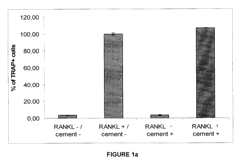
Figure 1a is a graph illustrating percentage of TRAP positives cells
normalized to positive
control (RANKL + / cement -);
Figure 1b, c are photographs comparing TRAP staining of RAW264.7 cells (b) and
TRAP+ multinucleated osteoclasts formed from RAW264.7 cells in the presence of
cement and addition of RANKL in the medium (c);
Figure Id is a photograph illustrating actin (red) and nuclei (blue)
visualization inside
osteoclastic cells formed on the surface of brushite cement from RAW264.7
cells cultured
with RANKL;
Figure 2 are photographs of cylinders of brushite cement before in vitro
experiments;
Figure 3a is a graph illustrating the ability of RANKL loaded (800 ng) cement
cylinders (40
mg) to differentiate 10x104 RAW264.7 cells into osteoclasts after six
successive 7 or 5 day
culture periods. (Culture medium (1 ml) changed daily; cement cylinders
transferred to
fresh undifferentiated monocyte cultures after each datum point shown in the
figure)
(n=3, mean +/- sd)
Figure 3b is a graph illustrating the ability of RANKL loaded (800 ng) cement
(19 mg) set
within the pores of sintered titanium beads to differentiate 10x104 RAW264.7
cells into
osteoclasts after one 7 day and two five day successive cultures with the same
matrices
(Culture medium (1 ml) changed daily; cement cylinders transferred to fresh
undifferentiated monocyte cultures after each datum point shown in the
figure.)
Figure 3c are a series of photographs of sintered titanium beads on one half
of a
cylindrical sample (8mm diameter) before (top) and after (bottom) impregnation
with
brushite (arrows indicate the brushite);
Figure 3d is a graph illustrating the percentage of TRAP positives cells
compared with
positive control formed after 5 days of culture in the presence of calcium
cross linked
alginate (1 ml 3 wt% aqueous solution) and RANKL (800 ng) incorporated into
alginate.
(n=3, mean +/- sd);
6
CA 02720269 2010-10-01
WO 2008/128342 PCT/CA2008/000733
Figure 4 is a series of photographs showing TRAP+ multinucleated osteoclasts
formed
from RAW264.7 cells in the presence of (a,b) of daily addition of 50 ng RANKL,
(c,d) in the
presence of brushite after 33 days of culture coated with 800 ng of RANKL and
(e,f) in the
presence of metallic implant with brushite inside his porosity coated with 800
ng of RANKL
after 7 days of culture;
Figure 5 is a graph illustrating the percentage of TRAP positives cells after
5 days of
culture in presence of RANKL stored at 37 C for 7, 14, 21 and 35 days prior to
use in cell
culture experiments. (n=4, mean +/- sd), when compared with control not stored
prior to
use;
Figure 6a is a graph illustrating percentage of TRAP positive cells formed
after 5 days of
culture in the presence of brushite cement with two different ways of
associating RANKL;
mixing RANKL during cement setting and topical adsorption after setting;
Figure 6b is a graph illustrating percentage of TRAP positive cells formed
after 5 days of
culture in the presence of hydroxyapatite cement with two different ways of
associating
RANKL;
Figure 7 is a graph illustrating the Effect of ascorbic acid on the efficacy
of RANKL in
differentiating primary bone marrow cells. Number of TRAP positive
multinucleated cells
(Small <100 pm, Big >100 pm) formed after 5 and 7 days of culture of primary
mouse
bone marrow cells in the presence of RANKL (50 ng/ml) and ascorbic acid (50
ng/ml) in
the culture medium. (n=3, mean +/- sd);
Figure 8 is a graph illustrating the effect of ascorbic acid addition to the
cell culture
medium on the efficacy of RANKL in differentiating RAW264.7 monocyte cell
line.
Percentage of TRAP positive cells formed after 5 days of culture of osteoclast
precursors
in the presence of RANKL (50 ng/ml) and ascorbic acid (50 ng/ml) in the
culture medium;
Figure 9 is a series of photographs showing TRAP+ multinucleated osteoclasts
formed
after 5 days of culturing of RAW264.7 monocyte cell line with RANKL alone (a,
b) or in
combination with ascorbic acid (c, d);
Figures 10a-f are a series of photographs showing the characteristics of
osteoclastogenesis in the presence ofcalcium phosphate cement and trehalose;
Figure 10g is a graph illustrating the percentage of TRAP positive cells
formed after 7
days of RAW 264.7 cell culture in the presence or absence brushite cement
cylinder
coated or not coated with RANKL (800 ng), after treatment or not with RANKL
(50 ng/ml),
after addition to the culture medium or not of trehalose (300 mM) and
normalized to the
positive control (cement - / RANKL + / Trehalose -). Data are mean SEM, n =
3
independent experiments. (c) indicates that RANKL was coated onto brushite
cement
cylinder;
7
CA 02720269 2010-10-01
WO 2008/128342 PCT/CA2008/000733
Figure 11a is a graph illustrating the evaluation of the stability of RANKL
solution stored
for 7, 14, 21 and 28 days at room temperature (RT)., as determined by capacity
for
osteoclastogenesis;
Figure 11 b illustrates the percentage of TRAP positive cells formed after 7
days in RAW
264.7 cell culture in the presence either of RANKL, loaded brushite (RaB) or
RANKL and
trehalose loaded brushite (RaTB) cement cylinder and normalized to the
positive control
(culture medium + fresh RANKL). These two formulations were stored at RT
during either
30 minutes (sa) or 1 day (1d). Data are mean SD, n = 3 replicates. Thus for
RaTB, the
brushite cement (40 mg) includes the following solution soaked onto it :800ng
RANKL in
16 pL phosphate buffered saline to which 1.8 mg of trehalose was added;
Figure 11c is a representation of the culture processes used to determine the
stability the
different formulations during 4 successive 7 days monocyte cell cultures. RaB
and RaTB
formulations were stored at RT during either 30 minutes (sa) or 1 day (1d).
Figure 11d illustrates the percentage TRAP positive cells normalized to the
positive
control (culture medium + fresh RANKL) and formed after 4 successive 7 days
culture
periods of RAW 264.7 cell in presence of (RaB)sa, (RaB)ld, (RaTB)ld, (RaTB)sa.
Data
are mean SD, n = 3 replicates. The differences were evaluated by analysis of
variance
(ANOVA) with Fisher's probability least significant difference (PLSD) post hoc
test and
considered to be significant at p < 0.05. RaB : 40mg brushite cement + 800ng
RANKL in
16pL phosphate buffered saline. RaTB: 40mg brushite cement + 800ng RANKL + 1.8
mg
Trehalose in 16pL phosphate buffered saline sa: left 30 minutes at room
temperature prior
to use. 1d: left 1 day at room temperature prior to use;
Figure 12a and b are graphs showing an evaluation of the stability of RANKL-
trehalose-
coated brushite cement (RaTB) under different storage conditions. RaTB
formulation (as
above) was stored either fort day (1 d), 3 weeks (3w) or 5 weeks (5w) at
ambient
conditions (a), ambient conditions with light excluded (Fig 8 bi), at 4 C or -
20 C with light
excluded (Figure 12 bii), room temperature light excluded in air (A) or
nitrogen (N) (Figure
12b iii), compared with positive control after 7 days culture. Data are mean
SD, n = 3
replicates;
Figurel3 is a graph illustrating the number of TRAP positives cells formed
after 7 days of
mouse bone marrow cell culture in the presence of RANKL-coated brushite cement
(800
ng) (Culture medium (0,5 ml) changed every two days; addition of ascorbic acid
(50 pg/ml)
(AA) was required for this culture to form osteoclasts) (n=3, mean +/- sd);
8
CA 02720269 2010-10-01
WO 2008/128342 PCT/CA2008/000733
Figurel4 illustrates the effect of sodium pyruvate concentration and on
osteoclastic
potential of RANKL and in combination with ascorbic acid after 7 days culture
with
RAW.264.7 cell line; and
Figure 15 is a graph illustrating the number of TRAP positives cells formed
after 7 days of
RAW 264.7 cell culture in the presence of cylinder of brushite cement loaded
either with
RANKL alone (800 ng) or RANKL (800 ng) and ascorbic acid (pg). (Culture medium
(1 ml)
changed every two days) (n=3, mean +/- sd).
DETAILED DESCRIPTION OF THE INVENTION
Definitions
Unless otherwise stated, the following terms apply:
The singular forms "a", "an" and "the" include corresponding plural references
unless the
context clearly dictates otherwise.
As used herein, the term "comprising" is intended to mean that the list of
elements
following the word "comprising" are required or mandatory but that other
elements are
optional and may or may not be present.
As used herein, the term "consisting of is intended to mean including and
limited to
whatever follows the phrase "consisting or. Thus the phrase "consisting of
indicates that
the listed elements are required or mandatory and that no other elements may
be present.
As used herein, the term "pro-osteoclastogenic" or "osteoclastogenesis" is
intended to
mean induced formation of osteoclasts.
As used herein, the term "osteoclastogenic agent" is intended to mean an agent
or agents,
which when used either singly or in combination can induce the formation of
osteoclasts
(also described herein as an inductive protein). One example of such as agent
is
Receptor Activator for Nuclear Factor K B Ligand (RANKL). RANKL may be used in
combination with, ascorbic acid, sodium pyruvate, to induce the formation of
osteoclasts.
As used herein, the term "an anabolic compound" is intended to mean a compound
that is
capable of enhancing the metabolic activity of a cell. Example of such
compounds include
pyruvate salts (sodium pyruvate), pyruvic acid and the like.
9
CA 02720269 2010-10-01
WO 2008/128342 PCT/CA2008/000733
As used herein the term "induce or enhance osteoclast formation" is intended
to mean to
cause osetoclast precursors a) to fuse forming multinucleated cells, b) to
express specific
proteins, such as tartrate-resistant acid phosphatase (TRAP), c) and following
a) and b)
to acquire the ability to become functional osteoclasts.
As used herein, the term "osteoconductive" is intended to mean promotion of
bone
apposition onto the surface of a graft or implant, thereby functioning as
receptive scaffold.
As used herein, the term "bone apposition" is intended to mean the formation
of new bone
on the bone surface.
As used herein, the term "matrix" is intended to mean a biomaterial capable of
a) storing
osteoclastic agent and b) releasing it in active form and quantity to induce
osteoclastogenesis.
As used herein, the term "matrix osteointegration" is intended to mean bone
ingrowth into
the porous surface of a matrix, or bone bonding to the surface of a matrix
causing
anchorage of the implant in the bone.
As used herein, the term "implantation site" is intended to mean a location in
a subject's
body where prosthesis, endoprosthesis, bone graft substitute, bone graft, soft
tissue graft,
are located to accelerate healing or to restore function to the
musculoskeletal system
including bone.
As used herein, the term "material" is intended to include natural and man-
made
materials, which are generally classed as metals, polymers, ceramics or
composites
thereof, and which are compatible for use in medical applications. The term
"biomaterial"
when used in conjunction with a matrix refers to a material that does not
degrade the
osteoclastogenic agent and is thus capable of releasing the agent in an active
form in vitro
or in vivo without adverse tissue or cell response.
As used herein the term "associated with" when referring to the relationship
between the
osteoclastogenic agent and the material, is intended to mean that the agent
can be
impregnated and/or coated onto and/or within the matrix. The agent is in an
amount
CA 02720269 2010-10-01
WO 2008/128342 PCT/CA2008/000733
which when it is released from the matrix is in an amount which is sufficient
to cause
osteoclastogenesis.
As used herein, the term "metals" is intended to include biocompatible metals
including,
but not limited to, stainless steel, titanium, tantalum and nitinol.
As used herein, the term "polymer biomaterial" is intended to include two
subclasses,
namely polymers and hydrogels. Hydrogels are swollen polymer networks
containing
significant (>50%) quantities of water, (more typically > 85%). Examples of
hydrogels
include crosslinked alginates, non-fibrillar collagens, PEG (polyethylene
glycol), PAA
(polyacrylic acid), HEMA (hydroxy ethyl methacrylate), and chitosan. Polymers
include PE
(polyethylene), PGA (polyglycolic acid), PLA (poly lactic acid), PU
(polyurathanes), PHB
(polyhydroxybutyrate), and PTFE (polytetrafluoroethylene), PVA (poly(vinyl
alcohol)),
cellulose.
As used herein, the term "ceramic" or "bioceramic" is intended to include
hydroxyapatite,
calcium phosphate, calcium hydrogen phosphate, calcium carbonate, calcium
silicates,
zeolites, artificial apatite, brushite, calcite, gypsum, phosphate calcium or,
a and or R
tricalcium phosphate, octocalcium phosphate, calcium pyrophosphate (anhydrous
or
hydrated), calcium polyphosphates (n?3) dicalcium phosphate dihydrate or
anhydrous,
iron oxides, calcium carbonate, calcium sulphate, magnesium phosphate, calcium
deficient apatites, amorphous calcium phosphates, crystalline or amorphous
calcium
carbonates, pyrophosphates and polyphosphates. Ceramics may contain one or
more of
titanium, zinc, aluminium, zirconium, magnesium, potassium, calcium, iron, and
sodium
ions or atoms in addition to one or more of an oxide; carbonates, carbides,
nitrides,
titanates, zirconates, phosphonates, sulphides, sulphates, selenides,
selanates,
phosphate, such as orthophosphate, pyrophosphate, di-phosphate, tri-phosphate,
tetra-
phosphate, penta-phosphate, meta-phosphate, poly-phosphate; a silicate, .
As used herein, the terms "ceramic" or "bioceramic" are used interchangeably
throughout
and are intended to include all ceramics which may be formed from oxides,
selenites, of
calcium, sodium, potassium, aluminium, magnesium, zinc, silicon, strontium,
barium, or
transition metals. Bioceramics further include composites thereof with
metallic, ceramic
and polymeric phases that can be used for example as bone replacement.. Any
gel, such
as a sol gel, xerogel, alcogel or aerogels and the like are also contemplated.
11
CA 02720269 2010-10-01
WO 2008/128342 PCT/CA2008/000733
As used herein, the term "metallic implant" is intended to include an implant
made from an
elemental metal or alloys thereof, such as for example titanium and alloys
nitinol thereof.
As used herein, the term "non-hydrogel polymer" is intended to mean
polyurethane,
polyester, polytetrafluoroethylene, polyethylene, polymethylmethacrylate,
polysiloxanes,
and all poly hydroxyacids. Examples of non-hydrogel polymers include, but are
not limited
to, the following synthetic and natural polymers:
Synthetic polymers Natural polymers
Poly lactic acid Fibrin
- Poly-L-lactic acid Albumin
- Poly-D,L-lactic acid Casein
Poly glycolic acid Keratin
Poly-e-caprolactone Fibrillar Collagen
Poly-p-dioxanon silk fibroin
Tri-methylen carbonate lipids
Poly anhydrides phospholipids
Poly ortho ester Amphiphiles
Poly urethanes Polyhydroxybutyric acid
Poly amino acids
Poly hydroxy alcanoates
Poly phosphazenes
Polystyrenes e.g. Poly(styrene-co-
chloromethylsytrene)
lipids (e.g. monoolein)
phospholipids
Polyphosphoesters
Polyphosphazenes
Aliphatic Polyesters e.g. PCL PGA
PLA & Copolymers
PHB PHV & Copolymers
Poly(1,4-butylene succinate),
Nylons
Non Hydrogel Polysaccharides, e.g.
cellulose acetates
PEG Based Polymers
Poly(ethylene oxide) average
Polyanhydrides
Poly(butylene Terephthalate)
Amphiphiles
12
CA 02720269 2010-10-01
WO 2008/128342 PCT/CA2008/000733
As used herein, the term "treating bone trauma" is intended to mean treatment
of a
trauma associated with bone, as disclosed herein, in a subject, and includes
the
implantation of a matrix as described herein, adjacent to a site of the trauma
so as to
enhance bone formation adjacent to the implant to a subject.
As used herein, the term "subject" or "patient" is intended to mean humans and
non-
human mammals such as primates, cats, dogs, swine, cattle, sheep, goats,
horses,
rabbits, rats, mice and the like. In one example, the subject is a human.
I. Matrix and composition
Our discovery concerns a matrix that is useful for inducing or enhancing
osteoclast
differentiation. The matrix comprises a material with an osteoclastogenic
agent which is
associated with the material. The osteoclastogenic agent is located in an
amount which is
sufficient to induce or enhance osteoclast differentiation.
Current clinical approaches for the treatment of osteoporosis are based on
inhibition of
osteoclast action, but this do not improve the quantity of bone. Without
wishing to be
bound by theory, we posit that localised induction of bone resorption by the
release of pro-
osteoclastogenesis molecules (osteoclastogenic agents) from the surface of the
of the
biomaterial or within the biomaterial may lead to a compensatory increase in
bone
formation, resulting in faster and better osteointeg ration of the matrix.
Generally speaking, we use matrices that are made of a set biomaterials. The
biomaterial
is typically microcrystalline and comprises crystals < 500 pm in linear
dimension. In order
for the osteoclastogenic agent to be retained on and/or within the matrix.
With the
exception of hydrogels, the biomaterial generally has a porosity at greater
than 5 %
relative porosity and a surface area of greater than 0.5 M2 g-1, which is
sufficient for the
osteoclastogenic agent to move out via the pores, by diffusion through the
polymer
network or dissolution of the biomaterial.
There are many examples of biomaterial which are contemplated for use as a
matrix. In
one example, the biomaterial is a ceramic or a non-metallic coating, such as a
polymer
coating carbon and the like. In one example used herein, the biomaterial is a
calcium
phosphate-based cement, however it is to be understood that the phosphate
containing
cement can be an orthophosphate, a pyrophosphate, a di-phosphate, a tri-
phosphate, a
13
CA 02720269 2010-10-01
WO 2008/128342 PCT/CA2008/000733
tetra-phosphate, a penta-phosphate, a meta-phosphate, or a poly-phosphate,
plaster,
calcium silicate, calcium sulphate etc. In examples described herein, the
cement sets to
form mainly either brushite or hydroxyapatite.
The biomaterials comprise the osteoclastogenic agent, for example RANKL, which
is
impregnated onto and/or within the biomaterial.
The osteoclastogenic agent is combined with the biomaterial at a concentration
of 1 ng or
more and 1000000 ng or less of agent per mg of biomaterial. Typically, the
osteoclastogenic agent is impregnated onto and/or within the biomaterial at a
concentration of 10 ng or more and 500 ng or less of agent per mg of
biomaterial. In one
example, the osteoclastogenic agent is impregnated onto and/or within the
biomaterial at
a concentration of 15ng or more and 25 ng or less of agent per mg of
biomaterial.
Cements are porous,, which allows the RANKL to be absorbed into the cement
The combination may also be used with either brushite or hydroxyapatite as the
biomaterial, although it is contemplated that other biomaterials as defined
herein may also
be used. In the examples described herein, the antioxidant is ascorbic acid.
It is to be
understood that ascorbic acid may be in either of its isomeric forms such as L-
ascorbic
acid, D-ascorbic acid or DL-ascorbic acid, or salts thereof. Furthermore,
dehydroascorbic
acid or salts thereof, can also be used.
The combination may also include pyruvates, such as sodium pyruvate and
pyruvic acid,
either singly or in combination with ascorbic acid. Without wishing to be
bound by theory,
we believe that the pyruvates provide energy to cells for cellular respiration
and that
compounds such as glucose, glucose phosphate and the like will also work.
Generally speaking, impregnated onto and/or within a material, the
osteoclastogenic agent
is in an amount that is sufficient to cause implant osteointegration, matrix
remodelling, and
osteoclast differentiation.
The aforesaid composition may be used to promote osteoclastogenesis in vitro
or in vivo,
in which the combination of RANKL ascorbic acid or sodium pyruvate are in
amounts,
which are sufficient to promote osteoclastogenesis in vitro or in vivo, when
progenitor
cells, generally osteoclast precursor cells, are contacted with the
composition .
14
CA 02720269 2010-10-01
WO 2008/128342 PCT/CA2008/000733
Ascorbic acid and sodium pyruvate are in the medium. It is contemplated that
these
compounds can be combined with the matrix with the RANKL
2. RANKL stability
In vitro assays for osteoclast growth and resorption are useful for the
screening of
potential therapeutic agents for conditions such as osteoporosis. These assays
rely on the
use of RANKL that needs to be added, typically at least every 48 hours, to
cultures in
order for differentiation to occur and is typically stored at -80 C prior to
use. It is to be
noted that RANKL's stability is rate determined. Thus it will degrade slowly
over time.
Typically, the stock is stored at -80 C and solution are stored short term at -
20 C.
Our discovery is that a matrix comprising a single bioceramic pellet with
RANKL adsorbed
onto its surface causes osteoclastogenesis. However, RANKL is expensive and is
unstable above -80 C.
In a clinical setting, degradable biomaterials are currently removed from the
implantation
site by virtue of being soluble in vivo or by being rendered soluble through
hydrolysis or by
enzymatic action. Bone autograft is a patient's own bone used as a graft. The
graft is
remodeled, that is, the graft recruits osteoclasts that erode the bone. The
osteoclasts
recruit osteoblasts that form new bone and gradually the graft is remodeled to
become
entirely new bone. By recruiting osteoclasts in vivo using our matrices coated
with
RANKL, may create a more autograft-like response.
To achieve this, we have demonstrated that RANKL, which is adsorbed in the
pores of
porous metals, is stable at ambient temperatures, specifically at 37 C.
Specifically, the
pores in porous metal are filled with calcium phosphate cement. RANKLis then
soaked
into that cement without a protein stabilizing agent.
This is achieved by drying the RANKL in the matrix. Furthermore the stability
of RANKL in
a dehydrated matrix could be enhanced using a sugar such as trehalose.
Thus, the aforesaid methods may also be adapted to include dehydrating an
aqueous
solution of RANKL and a protein stabilizing agent such as, for example,
trehalose, so as to
produce a dehydrated mixture of RANKL and the protein stabilizing agent. The
trehalose
is used to stabilise proteins during dehydration, thus the trehalose improves
stability of
RANKL loaded in brushite cement during storage. The ability of the dehydrated
RANKL to
CA 02720269 2010-10-01
WO 2008/128342 PCT/CA2008/000733
induce differentiation of primary or cell line osteoclast precursors at any
concentration can
be measured using assays described herein.
3. Therapeutic applications
Various models and studies have indicated that a short burst of osteoclastic
activity is
followed by a corresponding burst of osteoblast activity that results in a net
gain in bone
volume. Implants, which can induce the aforesaid osteoclast and osteoblast
activity are
likely to be highly valuable in accelerating implant fixation. Our matrix, in
the form of an
implant, may be useful to treat osteoporotic patients who often lack
sufficient bone stock in
which implants may be fixed.
As described below, we now believe that the initial stimulation of osteoclasts
by RANKL
leads to faster recruitment of more functionally active osteoblasts, resulting
in better matrix
integration and increase in bone mass. The combination of material and
osteoclastogenic
agent improves the recruitment of osteoclast at, adjacent to, or a distance
away from the
implantation site, and increases the quantity of surrounding bone. The
combination might
be very useful to treat numerous pathological diseases, which result in bone
trauma, such
as for example osteoporosis, osteoarthritis, rheumatoid arthritis, and
periodontitis. The
combination might also be useful to treat bone fractures, as well as to
accelerate healing,
or improve the quality of the bone formed, thereby increasing the success of
surgeries,
such as orthopaedic and maxillofacial surgery. Further uses include
osteointegration,
which is the induction of bone ingrowth into prosthesis stems, such as
artificial joints and
also artificial tooth roots. Bone formation, such as for example, bone
grafting following
non-union, treatment of osteoporotic fracture, trauma, or reconstruction after
void filling
following treatment of osteolysis, and spinal fusion, are also contemplated
uses for the
matrix. Thus, it is contemplated that the matrix may be used in a method of
treating bone
trauma in a subject, the method comprising: implanting the matrix adjacent to
a site of the
trauma so as to enhance bone formation adjacent to the implant. The bone
trauma may
result from a bone degenerative disease, such as osteoporosis, osteoarthritis,
rheumatoid
arthritis, or periodontitis, or the bone trauma may be a bone fracture or
other injury.
4. in vitro Applications
In vitro models of osteoclast formation are highly valuable for studying the
cellular and
molecular regulation of osteoclast differentiation and activation as well as
pharmaceutical
strategies to inhibit their action for treatment of osteoporosis. Principally
these are cell
16
CA 02720269 2010-10-01
WO 2008/128342 PCT/CA2008/000733
lines based (often RAW 264.7) or primary cells treated with RANKL. A typical
protocol
involves the repeated addition of growth medium containing fresh 50 ng/ml
soluble
RANKL on the first, the third and the fifth day.
Our combination of RANKL with a biomaterial provides a new tool to simplify
the current
protocol by avoiding repeated addition of RANKL. Moreover, this material
combined with
RANKL is also of interest to induce and study osteoclastogenesis during a long
term.
Materials and Methods
The following examples are offered by way of illustration, not by way of
limitation. While
specific examples have been provided, the above description is illustrative
and not
restrictive.
1. Preparation of calcium phosphate cement matrix.
1.1. Brushite cement
Brushite cement powder was prepared from an equimolar amounts of calcium
phosphate
monohydrate (Mallinckrodt Baker, Germany) and (3-TCP as described previously
(Ionic
modification of calcium phosphate cement viscosity. Part II: hypodermic
injection and
strength improvement of brushite cement: Barralet JE, Grover LM, Gbureck U,
Biomaterials Volume: 25 Issue: 11 pages: 2197-2203; May 2004). The resulting
powder was mixed with 0.8 M citric acid solution with a powder/liquid ratio
3.5 g/ml. The
cement setting reaction is given by:
Ca3(PO4)2 + Ca(H2P04)2=H20 + 7 H2O -> 4CaHP04.2H2O
For cell culture experiments brushite cement was set at room temperature in
cylindrical
molds to form 3 mm diameter and 3 mm height cylinders. The phase purity, the
density
and the specific surface area of brushite set cement were determined by X-ray
diffraction
by using a Siemens D5005 diffractometer (Siemens, Karlsruhe, Germany) with
monochromated Cu Ka radiation, by using a helium pycnometer (AccPycl330 ,
Micromeritics) and by using the Brunauer-Emmett-Teller (BET) method with
helium
adsorption-desorption (Tristar3000 , Micromeritics), respectively.
1.2 Alternative cement preparation
17
CA 02720269 2010-10-01
WO 2008/128342 PCT/CA2008/000733
Either brushite or hydroxyapatite cements or brushite cement have been
prepared. The
brushite cement consisted of an equimolar mixture of R-TCP (tricalcium
phosphate) and
monocalcium phosphate hydrate. Powder and liquid was combined with a
powder/liquid
ratio 3.5 g/ml and was been moulded into cylinders 3 mm in diameter, 5 mm in
length with
an average weight of 40 mg. Hydroxyapatite cement consisted of an equimolar
mixture of
tetracalcium phosphate and dicalcium phosphate. Materials were washed 3 times
in 70 %
alcohol and left under UV light for 12 hours. 16 pl (or 800 ng) of RANKL (50
pg/ml) was
adsorbed onto these materials for 20 minutes.
1.3 incorporation of RANKL loaded cement into porous metal
Porous titanium metallic implants 4 mm in diameter, 5 mm in length (Figure
3c), had
brushite cement paste infiltrated into the pores such that an average brushite
weight of
11.2 mg was deposited in the pores. Once set, 800ng of RANKL was soaked into
the
cement. 1.4 A 3 wt.% sodium alginate solution was prepared by mixing sodium
alginate
powder (MVG , Pronova Biomedical a.s.) with double distilled water. The sodium
alginate
solution was sterilised by autoclaving (45 minutes at 121 C) and cross-linked
for 12 hours
with 0.1 M CaCl2 solution in the form of cylinders of 3 mm diameter and 3 mm
height.
Once the gels had 'set' 800 ng RANKL was injected into the gel using a
hypodermic
needle.
2a: Osteoclast differentiation from cell line
RAW264.7 monocyte cell line were seeded at 2.5x10⁵ cells/cm2, at day 0, and
cultured for
5 to 7 days in 1 ml Dulbecco's Modified Eagle's Medium (DMEM) with 10% fetal
bovine
serum (FBS), 1 % antibiotics and 1 % sodium pyruvate at 37 C, 5% CO2.
To induce osteoclast differentiation, either 1 pl of a pro-resorptive cytokine
(RANKL (50
pg/mL)) was added to each ml of fresh medium every day following day 1, or
materials
combined with RANKL were added to cell culture at day 1. On day 5, cells were
fixed
using 4% paraformaldehyde during 10 minutes, washed 3 times with 1x PBS and
stained
for osteoclast marker TRAP, and the numbers of multinucleated, TRAP positive
cells were
assessed and cell number with matrices were compared with those in the
positive control.
Osteoclast resorption requires formation of specialized cytoskeletal
structure, actin ring.
To characterize actin organization in osteoclasts, we used BODIPY 581/591-
conjugated
phalloidin and DAPI to visualize F-actin and nuclei using fluorescence
microscopy.
Materials were used in subsequent experiments in the same conditions (without
another
18
CA 02720269 2010-10-01
WO 2008/128342 PCT/CA2008/000733
addition of RANKL) to determine how long biologically active levels of RANKL
can be
release from these material. Stability of RANKL at 37 C has also been tested.
2b: Osteoclast differentiation from bone marrow
Mouse bone marrow cells were collected from mouse tibia and femora. Inbred
mice
(C57BL/6J, female, 6 weeks old) (Charles River Co., Wilmington, Massachusetts,
USA)..
Both femurs and tibia were dissected from dead mice under sterile conditions
and
immediately placed in sterile PBS solution. Under a laminar flow cabinet, the
surrounding
muscles were detached from the bones. Tibia and femurs were then cut in half
and each
part was placed in a different eppendorf tube and centrifuges (3 times, 5
seconds, 12,000
rpm). Bone pieces were removed and bone marrow was resuspended with 300 ml of
medium (MEM) and collected. The bone marrow was then flushed with a 10 ml
syringue
with a 22-gauge needle in order to remove bone debris and blood clots.
Nucleated cells
were counted on Malassez haemocytometer slides. Cell viability was greater
than 90% as
determined by the trypan-blue dye exclusion test. The number of nucleated
cells was
around 80 x 106 per ml of medium. Bone marrow cells were seeded at a final
density of 10
x 106 cells per cm2 in 48 wells plate and cultured for 7 days at 37 C, 5 %
CO2 with MEM
supplemented with 10 % FCS, 1% L-glutamine and 1% antibiotics. Medium was
changed
on day 1, 3 and 5 and 500 pl of fresh medium (MEM) added alone or supplemented
with
AA (50 pg/ml) and or sodium pyruvate (1-3%).
After 7 days of culture, mouse bone marrow cells were fixed with using 4%
paraformaldehyde for 15 minutes, washed with phosphate buffered saline (PBS)
and
stained for osteoclast marker Tartrate-resistant acid phosphatase (TRAP). The
cultured
cells were stained for 10 to 20 minutes at 37 C and the numbers of
multinucleated TRAP
positive cells were assessed using a light microscope (Eclipse TS100, Nikon,
USA).
3. Incorporation of RANKL and RANKL-trehalose solutions onto cement matrix and
storage conditions.
A recombinant glutathione S-transferase-soluble RANKL solution (50 pg/ml) and
a D-(+)-
trehalose dehydrate powder (Sigma-Aldrich, USA) were used in this study.
3.1. Stability of Receptor Activator for Nuclear Factor K B Ligand (RANKL)
solution to
induce osteoclast formation.
RANKL solution (50 pg/ml) was stored at room temperature (RT) for up to 5
weeks. After
1, 2, 3 and 5 weeks, osteoclast formation induced by stored RANKL solution was
19
CA 02720269 2010-10-01
WO 2008/128342 PCT/CA2008/000733
assessed and compared to osteoclast formation induced by a fresh RANKL
solution (as
seen in Figure 5).
3.2. RANKL and RANKL-trehalose incorporation onto materials.
Brushite cement cylinders (B) (40 mg, 3 mm diameter and 3 mm height) were
combined
either with RANKL solution (50 pg/ml) alone (Ra) or with a RANKL-trehalose
solution
(RaT). The RaT solution was prepared by adding trehalose powder to 800 ng of
RANKL
solution to a final concentration of trehalose of 300 mM. Then Ra and RaT
solutions were
adsorbed onto the surface of set calcium phosphate cement. RaB and RaTB
formulations
were obtained and were stored at RT either for a short period of time of 30
minutes (sa) or
over a long period time of 1 day, 3 weeks or 5 weeks, as seen in Figures 11
and 12)
3.3. Storage conditions for the long adsorption period.
To test the influence of different storage parameters on the stability of
RANKL-trehalose
solution loaded onto brushite cement cylinders, formulations were stored under
specific
conditions over a long period of time. Briefly, different times conditions (1
day (1d), 3
weeks (3w) or 5 weeks(5w)), two different temperature conditions (4 C (4) and
-20 C (-
20C)), two conditions of light exposure (protected from light (d) or not), and
storage with
dried air condition in the presence of silicate gel beads (A) or with pure
nitrogen air
condition (N) were compared, as seen in Figure 12.
To assess the effects of trehalose addition and storage conditions on RANKL-
Brushite,
different formulations were prepared, added to the monocyte cell culture at
day 1 and
medium was changed at day 1, 3 and 5. In control cultures, osteoclastogenesis
was
induced addition of soluble RANKL (50 ng/ml) to fresh medium at day 1, 3 and
5. RAW
264.7 cells cultured without RANKL or biomaterial addition were used as the
negative
control.
On day 7, cells were fixed using 4 % paraformaldehyde for 15 minutes, washed
with
phosphate buffered saline (PBS) and stained for osteoclast marker Tartrate-
resistant acid
phosphatase (TRAP). TRAP staining solution (4 % solution of 2.5 M acetate
buffer (pH =
5.2), 12.5 mg/ml naphthol AS-BI phosphoric acid, 0.67 M tartrate buffer (pH =
5.2), and 15
mg of fast Garnet salt) was freshly prepared and filtered before use. The
cultured cells
were stained for 10 to 20 minutes at 37 C and the numbers of multinucleated
TRAP
positive cells were assessed using a light microscope (Eclipse TS100, Nikon,
USA).
CA 02720269 2010-10-01
WO 2008/128342 PCT/CA2008/000733
3.4. Time stability of RANKL and RANKL-trehalose -coated brushite cement
Cylinders of brushite cement coated either with 800 ng of RANKL only (RaB) or
with 800
ng of RANKL mixed with 300 mM trehalose (RaTB) were used during four
consecutive 7
days RAW264.7 cell cultures for a total of 28 days. Before using these two
formulations
both RaB and RaTB were stored either for a short period of time of 30 minutes
(sa) or for
a period of time of 1 day (1 d). RAW 264.7 cells at a density of 2.5 x 10⁵
cells/cm2 with 1 ml
of medium were cultured. The four different formulations, (RaB)sa, (RaB)ld,
(RaTB)sa
and (RaTB)1d were added to the cell culture at day 1. Cells were cultured
during 7 days at
37 C and medium was changed at day 1, 3 and 5. At the end of this culture
period, we
transferred the cylinders of brushite cement to freshly plated monocyte cell
cultures and
we assessed the number of TRAP positive cells. This process was repeated four
times.
The results are shown in Figure 7d.
4. Statistical analysis.
All data were expressed as mean standard deviation or standard error of the
mean. The
differences were evaluated by analysis of variance (ANOVA) with Fisher's
probability least
significant difference (PLSD) post hoc test and considered to be significant
at p < 0.05.
Results
As illustrated in Figure 1a, there is differentiation of RAW264.7 cells toward
osteoclast-like
cells on cement with addition of RANKL in the medium. Since the brushite
cement is
opaque, we assessed the numbers of osteoclasts formed on the plastic
surrounding and
underlying the cement and compared to the numbers of osteoclasts formed in the
absence of the cement. RANKL induced osteoclast formation to a similar extent
was
independent of the presence of cement in the well. Osteoclast differentiation
in the
presence or absence of brushite cement was observed only in the presence of
RANKL as
illustrated in Figures 1 b and 1 c. These data indicate that the brushite
cement exhibits
neither a detrimental nor stimulatory effects of on osteoclast formation.
As illustrated in Figure 1d, fluorescently labeled osteoclasts are formed
directly on the
cement. In the presence of RANKL, osteoclasts formed exhibited an actin ring
surrounding
numerous nuclei. Without RANKL, no actin ring formation was observed. These
data
indicate that cement supports adhesion and formation of functional
osteoclasts.
21
CA 02720269 2010-10-01
WO 2008/128342 PCT/CA2008/000733
Table 1 below shows different tests performed by coating increasing amount of
RANKL
onto small (40mg) cylinders of brushite as illustrated in Figure 2. After 7
days of culture in
presence of RANKL coated brushite cylinders, the number of wells in which
RAW264.7
were differentiated toward osteoclastic-like cells, were noted as "positive".
As culture in
the presence of RANKL in the medium, brushite material coated with at least
600 ng of
RANKL induced differentiation of RAW264.7 cells. No significant difference was
observed
between materials coated with 800 ng or 1000 ng of RANKL. The following
experiments
were realized with an amount of RANKL of 800 ng to obtain the optimal
response.
Table 1: Effect brushite materials coated with increasing coating amount of
RANKL
onto RAW264.7 cells differentiation.
Amount of RANKL coated (ng)
50 100 150 200 250 300 450 600 800 1000
number of 0/2 0/2 0/2 0/2 0/2 0/2 0/2 1/2 2/2 2/2
positive wells
Thus the matrix comprises the osteoclastogenic agent which is adsorbed onto
the surface
of the biomaterial at a concentration of 1 ng or more and 1000000 ng or less
of agent per
mg of biomaterial. In one example, the agent is at a concentration of 10 ng or
more and
500 ng or less of agent per mg of biomaterial. In another example, the agent
is at a
concentration of 15ng or more and 25 ng or less of agent per mg of
biomaterial.
Figure 3a shows the number of osteoclastic-like cells found after 7, 14, 21,
28, 33 and 38
days of culture of RAW264.7 cells in presence of brushite cement cylinder
impregnated
with 800ng of RANKL. Figure 3b shows the number of osteoclastic-like cells,
which were
found after 7, 12 and 17 days of culture of RAW264.7 cells in presence of a
macroporous
titanium sample with brushite cement loaded within the macroporosity. 19.2 mg
amounts
of brushite were incorporated inside the porosity of the metallic material.
RANKL (800 ng)
was coated onto the metallic/brushite material as shown in Figure 3c. Figure
3d shows the
number of osteoclastic-like cells found after 5 days of culture of RAW264.7
cells in
presence of calcium cross linked sodium alginate and alginate with RANKL
incorporated
within it (800 ng).
For all these conditions and materials, the numbers of osteoclasts formed on
the plastic
surrounding and underlying brushite based materials were assessed.
22
CA 02720269 2010-10-01
WO 2008/128342 PCT/CA2008/000733
These data indicate that the brushite based materials continuously release
amounts of
RANKL in the medium to induce differentiation of RAW264.7 cell toward
osteoclastic-like
cells. Even after 38 days of culture, the materials still have the capacity to
release enough
RANKL to induce formation of osteoclast-like cells. Nevertheless, Alginate, a
hydrogel
polysaccharide material, with the same amount of RANKL incorporated within it,
did not
induce a significant differentiation of RAW264.7 toward osteoclastic cells.
Osteoclast differentiation was observed in all conditions, with brushite
cement cylinder
coated with 800 ng of RANKL (see Figures 4c and 4d) and with the
metallic/brushite
material coated with 800 ng of RANKL (see Figures 4e and 4f). Osteoclast
formation was
compared with osteoclast formation observed after culture in a fresh medium
daily
supplemented with 50 ng/ml of RANKL (see Figures 4a and 4b).
TRAP-positive multinucleated osteoclasts formed from RAW264.7 cells in the
presence of
daily addition of 50 ng/ml RANKL (as illustrated in Figures 4a and 4b), in the
presence of
brushite after 33 days of culture coated with 800 ng of RANKL (as illustrated
in Figures 4c
and 4d) and in the presence of a macroporous metallic implant with RANKL
loaded
brushite inside the macropores (800 ng of RANKL) after 7 days of culture (as
illustrated in
Figures 4e and 4f).
As illustrated in Figure 5, RANKL stock solution (50 pg/ml in 1x PBS) is
stable after 7, 14,
21 and 35 days of storage at 37 C compared to fresh RANKL solution. These data
indicate that even after 35 days of storage at 37 C, RANKL is still an active
molecule and
able to induce RAW264.7 cell differentiation toward osteoclast-like cells.
These data also
suggest that RANKL coated onto and released from our materials is stable for a
long time
at ambient temperature and could induce osteoclastic differentiation in a long
succession
of in vitro experiments.
Overall data suggest that microcrystalline bioceramics such as brushite or
apatite based
materials are suitable as a matrix for the release of RANKL and induce
differentiation of
RAW264.7 cells toward osteoclastic cells. This release is continuous with time
and the
amount of RANKL released is in amounts which are sufficient to induce
osteoclast
formation, even after 33 days of culture.
23
CA 02720269 2010-10-01
WO 2008/128342 PCT/CA2008/000733
Brushite cement is composed of powder (tricalcium phosphate) and a liquid
component
(phosphoric acid solution and a retardant). After mixing, a paste is obtained
and set in
about 10 minutes. Besides incorporating the RANKL solution within the set
brushite
cement, another way of incorporating RANKL into brushite cement was to mix
RANKL
(800 ng) to the liquid component (20 pL 3 M H3PO4 + 500 mM citric acid) and
mix this
solution with 40 mg powder to obtain a setting cement. These two ways of
incorporation of
RANKL (namely by mixing during the preparation of the cement or by coating
after the
setting) were tested. No TRAP positive cell was observed for the culture when
brushite
cement made with a liquid component containing RANKL was added to cultures.
TRAP
positive cells were only observed when preset brushite cement was coated with
RANKL
(see Figure 6a).
Hydroxyapatite cement is composed of powder (e.g. equimolar mixture of
tetracalcium
phosphate and dicalcium phosphate anhydrous) and a liquid component (water and
an
accelerator). After mixing, a paste is obtained and set in about 10 minutes.
Besides
incorporating the RANKL solution within the set hydroxapatite cement, another
way of
incorporating RANKL into hydroxyapatite cement was to mix RANKL (800 ng) to
the liquid
component (18 pL sodium phosphate solution) and mix this solution with 40 mg
powder to
obtain a setting cement. As illustrated in Figure 6b, these two ways of
incorporation of
RANKL (namely by mixing during the preparation of the cement or by coating
after the
setting) were tested. One third of the percentage of TRAP positive cells was
observed
when hydroxyapatite cement made with a liquid component containing RANKL
compared
with set hydroxyapatite cement which was coated with RANKL. Moreover, much
less
differentiation was observed compared with a culture to which 50ng of
RANKLsolution
was added to the cell culture medium (positive control)
Any microporous material or material capable of storing and releasing protein
without
significant loss of protein biological activity is likely to work similarly
provided the specific
surface area is adequate RANKL adsorption and/or the relative porosity is high
enough
that the pores may act as storage depots. Typically, crystals would have to be
less than
1000pm in linear dimension for an adequate specific surface area and relative
porosity
would need to be greater than 5% ion order for sufficient open porosity to
exist in the
material. The porosity is at > 5 % relative porosity, which is relative to
full density (i.e.
zero porosity).
24
CA 02720269 2010-10-01
WO 2008/128342 PCT/CA2008/000733
In addition to RANKL (1 pl of a 50 pg/ml RANKL stock solution) in fresh
culture medium (1
ml), ascorbic acid (1 pl of a 50 pg/ml ascorbic acid stock solution) improves
osteoclastogenesis from both primary mouse bone marrow cells (Figure 7) and
RAW264.7
cells (Figure 8). This combination decreased the time to form osteoclasts and
increased
both the number and the size of osteoclasts (Figure 9).
The ascorbic acid used may be L-ascorbic acid, D-ascorbic acid or DL-ascorbic
acid, or
salts thereof. Additionally, dehydroascorbic acid or salts thereof is also
contemplated.
Examples of other antioxidants are also contemplated such as, but not limited
to, thiols,
phenols, glutathione, vitamin E, catalase, super oxide dismutase, peroxidases
and
cofactors thereof, lipoic acid, uric acid, carotenes, lipid (3-carotene,
retinol, or ubiquinone.
Data indicate that when RANKL and ascorbic acid were coated onto two different
brushite
cement cylinders, differentiation of RAW264.7 cell toward osteoclastic-like
cells after 5 to
7 days of culture. RANKL (800 ng) and RANKL (800 ng)/ascorbic acid (800 ng)
loaded
brushite cement were tested to reproduce previous data. Addition of both RANKL
coated
brushite cement and ascorbic acid coated brushite cement induced much greater
formation of osteoclasts after 5 to 7 days of culture than the RANKL coated
brushite
cement alone (see Figure 11).
Long term conservation at room temperature (24 hours at least) of RANKL: (800
ng) or
RANKL (800 ng) loaded cement (40 mg) was tested by addition or not of
trehalose (300
mM) to the pro-osletoclastogenic molecules before coating. Incorporation of
RANKL with
or without addition of trehalose was tested. TRAP positive cells were observed
in all
RANKL culture conditions with addition of trehalose.
The effects of brushite cement and trehalose on osteoclast formation from
monocyte cell
culture were observed. The morphology and the number of osteoclast formed
after 7 days
of RAW 264.7 cell culture in the presence of loaded or unloaded material with
RANKL and
in the presence or not of trehalose were analyzed. Monocyte cell culture
treated with
soluble RANKL (50 ng/ml) or in the presence of RANKL-coated brushite cement
formed
multinucleated TRAP positive osteoclastic cells at the same level (Figure 10a,
b and c).
Addition of trehalose (300 mM) to the different culture conditions did not
affect either
positively or negatively the osteoclastogenic process induced by soluble RANKL
or
RANKL-coated brushite material (see Figures 10d to g)
CA 02720269 2010-10-01
WO 2008/128342 PCT/CA2008/000733
Stability of RANKL solution alone or coated onto brushite cement and effects
of trehalose
addition to the coated RANKL solution were evaluated. We first studied the
stability of
RANKL to induce osteoclast formation (Figure 11a). After 7, 14, 21 and 35 days
of storage
at RT, RANKL solution induced a percentage of osteoclastogenesis of 85.90
8.65 %,
72.12 7.98 %, 82.93 6.29 % and 106.71 8.26 % compared to the positive
control
(fresh RANKL solution) respectively. Thus, RANKL solution retained a high
osteoclastogenic activity comparable to the positive control even after 35
days of storage
at RT.
We next investigated the stability of RANKL (800 ng) coated onto the surface
of cement
cylinders and the effect of trehalose addition (300 mM) to the coated RANKL
formulation.
RANKL-Brushite cement (RaB) and RANKL-trehalose-Brushite cement (RaTB) were
stored during 30 minutes (sa) or 1 day (1 d) prior to use in monocyte cell
culture (Figure
11 b). After 7 days of culture, (RaB)sa induced in this study a percentage of
osteoclast
formation of 161.50 3.79 % of positive control. In contrast, the same
formulation stored
during 1 day induced a percentage of osteoclast formation significantly lower
with 22.54
1.00 % of positive control. Addition of trehalose to RaB formulation stored
either during 30
minutes ((RaTB)sa) or during 1 day ((RaTB)ld) significantly increased the
osteoclastogenic processes to 173.48 1.53 % and 101.41 2.00 % of positive
control
respectively
Then we determined how long trehalose could preserve the bioactivity of RANKL
in
(RaB)sa, (RaB)ld, (RaTB)sa and (RaTB)ld formulations. We compared the
stability of
these four formulations to induce osteoclast formation during four consecutive
monocyte
cell cultures (Figure 11 c and d). The percentage of TRAP positive cells
induced by
(RaB)sa formulation continuously decreased from 161.50 3.79 % after the
first 7 days
culture period to 15.18 2.08 % of positive control after 28 cumulative days
of culture.
After the first culture period, (RaB)ld formulation induced an osteoclast
significantly lower
with 22.54 1,00 % of positive control. During the next three cell cultures,
the percentage
of TRAP positive cells decreased from 8.81 1.15 % after 14 days to 6.50
1.00 % of the
positive control at the end of the fourth culture period. Addition of
trehalose to (RaB)sa
formulation significantly increased the osteoclastic formation induced at the
end of each
culture period compared to (RaB)sa with percentages of TRAP positive cells
from 173.48
1.53 % to 48.48 0.58 % of positive control. In contrast addition of
trehalose did not
26
CA 02720269 2010-10-01
WO 2008/128342 PCT/CA2008/000733
change the decreasing trend of osteoclastogenic activity observed during these
four
culture periods with (RaB)sa. In the same manner the osteoclast formation
induced at the
end of each culture period by (RaB)ld was significantly increased by the
addition of
trehalose to the formulation with percentages from 101.41 2.00 % at the end
of the first
culture period to 74.80 2.00 % after 28 cumulative days of culture.
Interestingly, after the
third culture period, the percentage of TRAP positive cells induce by (RaTB)ld
and
(RaTB)sa were non significantly different. Moreover, at the end of last
culture period, the
percentage of TRAP positive cells induce by (RaTB)ld was significantly higher
than the
percentage of TRAP positive cells induced by (RaTB)sa. Thus, RANKL-Brushite
cement
cylinder retained a higher osteoclastogenic activity during 4 weeks with
addition of
trehalose to (RaB) formulations.
We investigated the effects of different parameters of storage on the
osteoclastogenic
activity of RANKL-trehalose-loaded brushite cement. First the stability RANKL-
trehalose-
coated brushite cement formulation (RaTB) stored during increased time period
(1
day(1 d), 3 weeks (3w) and 5 weeks (5w)) to induce osteoclast formation was
assessed
(Figure 12a). After 7 days of monocyte cell culture, (RaTB)1d formulation
induced an
osteoclastogenic process comparable to the positive control. The percentage of
osteoclast
formation induced by (RaTB) stored during 3 weeks (RaTB)3w and 5 weeks
(RaTB)5w
significantly decreased compared to (RaTB)1d with 65.92 3.77 % and 59.35
2.87 % of
positive control respectively. After 3 weeks and 5 weeks of storage RaTB
formulations
retained significantly the same osteoclastogenic activities. There was no
further
deterioration of the osteoclastogenesis after 3 weeks.
To determine the parameters responsible of the decrease of efficiency of RaTB
formulation to induce osteoclast differentiation, we first tested the effect
of light exposure
during the storage. We assessed the osteoclastogenic activity of both (RaTB)3w
and
(RaTB)5w formulations stored protected from light (d) after 7 days of RAW
264.7 cell
culture (Figure 12bi). The percentage of osteoclast formation induced by
(RaTB)3w d was
higher but not significantly different form the percentage induced by (RaTB)5w
d
formulations with 68.18 2.38 % and 59.74 4.97 % of positive control
respectively.
Moreover, the osteoclastogenic activities of (RaTB)3w and (RaTB)5w
formulations stored
protected from light or not were not significantly different (Figure 12a).
Thus, light
exposure during the storage did not significantly influence the
osteoclastogenic stability of
the RaTB formulation.
27
CA 02720269 2010-10-01
WO 2008/128342 PCT/CA2008/000733
Then we investigated the effect of temperature on the stability of RaTB
formulation to
induce osteoclast formation. (RaTB) formulation was stored protected from
light during 3
weeks either at 4 C ((RaTB)3w_d4C) or at -20 C ((RaTB)3w d 20C) (Figure 12
bii). After
7 days of monocyte cell culture, the percentage of TRAP positive cells induced
by
(RaTB)3w formulation stored at 4 C was 63.64 0.82 % of positive control.
The
percentage of TRAP positive cells induced by the same formulation stored at -
20 C
(81.82 0.82 % of positive control) was significantly higher from the
percentage of TRAP
positive cells formed both by (RaTB)3w d4C formulation and by (RaTB)3w
formulation
(Figure 12a). Thus, the stability of (RaTB)3w formulation to induce osteoclast
formation
was increased by a storage temperature condition of -20 C.
Next, we tested the effects of atmosphere composition on the stability of our
formulation
during the storage (Figure 12 biii). (RaTB was stored protected from light
during 3 weeks
and 5 weeks either in the presence of dried (with silica gel) air (A) or in
the presence of
nitrogen gas only (N). After 3 weeks of storage, RaTB formulation induced an
osteoclast
formation significantly higher in the presence of nitrogen gas only than in
the presence of
dried air with percentages of osteoclast formation of 84.27 2.08 % and 71.28
2.52 % of
positive respectively. The same trend was observed with RaTB formulation
stored during
5 weeks and after 7 days of monocyte cell culture (RaTB)5w dN induced a
percentage of
TRAP positive cells significantly higher than the percentage of TRAP positive
cells formed
by (RaTB)5w dA with 77.63 2.08 % and 63.20 6.24 % of positive control
respectively.
The osteoclastogenic process induced by (RaTB)3w dN was significantly higher
than the
osteoclastogenic process induced by (RaTB)5w dN but percentages of osteoclast
formation induced both by (RaTB)3w_dN and by (RaTB)5w dN were significantly
higher
than the percentages of osteoclast formation induced by (RaTB)3w and (RaTB)5w
formulations respectively (Figure 12a). Thus, absence of oxygen during the
storage of
RANKL-trehalose-brushite formulation for 3 weeks and 5 weeks significantly
increased the
osteoclastogenic activity of these formulations.
We tested the ability of brushite cement cylinder loaded with RANKL to induce
osteoclast
formation from a primary culture of mouse bone marrow cells. Mouse bone mouse
cells
with RANKL in solution or combined with a cement matrix for 7 days and
formation of
osteoclastic cells was only observed either in the presence of addition of
RANKL and
ascorbic acid in the medium or in the presence of RANKL coated brushite cement
and
28
CA 02720269 2010-10-01
WO 2008/128342 PCT/CA2008/000733
addition of fresh ascorbic acid to the medium (Figure 13). For these two
conditions, the
number of TRAP positive cells was not statically different.
We investigated the effect of sodium pyruvate on RANKL osteoclastogenesis from
monocyte cell line with and without ascorbic acid. RAW 24.7 cells were culture
for 7 days
in different conditions in the presence of RANKL, ascorbic acid, sodium
pyruvate and
various combinations of these factors (Figure 14). Addition of sodium pyruvate
(1%, 2%
and 3%) to RAW cell cultured in the presence of RANKL increased the number of
osteoclast up to 1500% with 2% of sodium pyruvate and compared to the positive
control
(culture medium with RANKL). RAW cells cultures for 7 days in the presence of
a
combination of RANKL, 50pg/ml ascorbic acid and 1 %sodium pyruvate formed
2400% of
TRAP positive cells compared to the positive control (culture medium with
RANKL).
We investigated the capability of brushite cement cylinder(s) loaded with
RANKL (800 ng)
and ascorbic acid (800 pg) to induce osteoclastogenesis. RAW 264.7 cells were
cultured
during 7 days in the presence of brushite cement cylinders either loaded with
RANKL (800
ng) alone or with RANKL (800 ng) and ascorbic acid (800 pg). Osteoclast
formation was
increased by 60% and 40% compared to positive control (culture medium with
RANKL)
and compared to RAW cell cultured in the presence of RANKL-coated brushite
cement,
respectively (Figure 15).
Other Embodiments
While specific embodiments have been described, those skilled in the art will
recognize
many alterations that could be made within the spirit of the invention, which
is defined
solely according to the following claims:
29