Note: Descriptions are shown in the official language in which they were submitted.
CA 02720324 2015-09-25
EX-VIVO MULTI-DIMENSIONAL SYSTEM FOR THE SEPARATION
AND ISOLATION OF CELLS, VESICLES, NANOPARTICLES AND
BIOMARKERS
STATEMENT AS TO RIGHTS TO INVENTIONS MADE UNDER
FEDERALLY SPONSORED RESEARCH AND DEVELOPMENT
[0002] This work was supported by NIH Grant/Contract CA119335. The Government
of
the United States of America may have certain rights in this invention.
BACKGROUND
[0003] In biomolecular research and clinical diagnostics it is both important
and a
challenge to separate and identify rare cells, bacteria, virus, and biomarkers
(e.g. DNA,
RNA, antibodies, other proteins, etc.) in complex fluid samples like blood,
plasma, serum,
saliva, and urine. Additionally, the advent of bio-nanotechnology has led to
numerous drug
delivery approaches that involve encapsulation of drugs and imaging agents
within
nanovesicles and nanoparticles. Such approaches mean it will now also be
important to
identify and separate residual nanovesicles and nanoparticles that remain in
the blood
stream. A variety of physical, electronic, and biological techniques and
mechanisms can
be used for sample preparation and isolation of specific cells, nanovesides,
and
biomolecules from complex samples like blood. These techniques and mechanisms
include centrifugation, gel filtration, affinity binding, DC electrophoresis,
and various
combinations incorporated into lab-on-a-chip, microfluidic devices, and sample-
to-answer
systems.
[0004] Many of these conventional techniques (or combinations) are relatively
time
consuming processes that are not without problems and limitations. In
particular, the
isolation of rare cells (cancer cells, fetal cells, and stem cells), low
numbers of bacteria and
virus or very low numbers of specific antibodies, proteins, enzymes, DNA, and
RNA
molecules, still remains difficult. In the case of clinical diagnostics, rare
cell and biomarker
detection may also be limited by sample size; i.e., only a relatively small
amount of blood
CA 02720324 2010-10-01
WO 2009/146143 PCT/US2009/039565
may be drawn from very ill patients, the elderly and infants. Thus, sample
preparation
processes that are inefficient or require high dilution of the original sample
often fail or are
unreliable for isolating cells and other disease-related markers at lower
concentration
ranges. This is in particular a problem for early detection of cancer,
residual disease, fetal
cells/DNA/RNA in maternal blood, bacteria and virus in blood (septic
infection), and the
detection of low numbers of pathogens (e.g. bacteria, virus, etc.) and
bioterror agents in
large volumes of air, water, or in food stuffs.
[0005] Alternating current electrokinetic techniques that involve the use of
AC fields to
manipulate cells and nanoparticles offers some particularly attractive
mechanisms for the
separation of cells [see References 2-5], biomarkers (DNA [Ref. 5-8], proteins
[Ref. 9],
etc.), and ultimately drug delivery nanovesicles [Ref. 10]. These techniques
can be broken
down into three distinct phenomena: (1) AC electroosmosis, which is surface
fluid flow due
to the surface charge on an electrode; (2) electrothermal flow, which is bulk
flow in solution
due to thermal gradients produced by the electric fields; and (3)
dielectrophoresis (DEP),
which is an induced motion of particles produced by the dielectric differences
between the
particles and media in an AC electric field [Ref. 10]. Unfortunately, most
conventional
forms of DEP and related electrokinetic effects have problems that limit the
usefulness of
these technologies for clinically relevant sample preparation and diagnostics.
[0006] First, efficient DEP separations in terms of speed and control of
selectivity usually
have to be carried out at relatively low conductance on the order of <10-100
mS/m [Ref.
11]. Additionally, the ability to isolate the desired entities/analytes such
as nanoparticles or
DNA biomarkers in the positive or DEP high field regions (usually around or on
the
electrodes) becomes more difficult as the solution ionic strength increases
and the
conductance becomes greater than 10 mS/m. Thus, biological samples such as
blood or
plasma that have ionic strengths in the 100-200 mM range (conductance ¨ 500-
1000
mS/m) must be significantly diluted and/or processed before DEP separations
can be
carried out [Ref. 13, 14]. This alone often limits the usefulness of DEP for
clinical
diagnostics involving the detection of rare cells or low numbers of
biomarkers. In cases
where a sample (one ml blood) has to be diluted 100 to 1000-fold, now means
that a very
large sample volume must be processed, which can be prohibitively time
consuming. If
cells are first concentrated by physical mechanisms such as centrifugation or
filtration and
then are diluted into low conductance buffers, these processes are not only
time
consuming, but also costly and cause considerable perturbation to the sample.
In the case
where DEP might be used for stem cell separations, dilution into low ionic
strength, less
physiological-type buffers may result in perturbation of sensitive stem cells
and may affect
2
CA 02720324 2010-10-01
WO 2009/146143 PCT/US2009/039565
their further differentiation. The isolation of DNA, RNA, and protein
biomarkers from blood
is also important for future clinical diagnostics, in particular for
monitoring cancer
chemotherapy [Ref. 15], residual disease [Ref. 16], and early detection of
cancer [Ref. 17].
[0007] While DEP has been used for the isolation of DNA and proteins, problems
and
limitations do exist in using DEP to carry out the detection of DNA in blood.
The first
problem again is the need to dilute and/or process the blood sample before DEP
analysis.
In the case of clinically relevant cell-free circulating DNA and RNA
biomarkers in blood,
finding and measuring the amount of DNA/RNA, its size and base composition
(mutations
and polymorphisms) is important [Ref. 17-19]. Sample processing that involves
or requires
centrifugation, filtration, and washing procedures can cause the release of
DNA molecules
by normal cells that are damaged or lysed in the process, as well as shear the
clinically
relevant DNA into smaller fragments. The release of extraneous DNA fragments
and
processing damage to the clinically relevant DNA greatly compromises and
limits the
diagnostic value of using such procedures. Such sample processing is also
highly
inefficient, and up to 60% of the DNA and over 90% of the RNA in the blood can
be lost
during the procedure [17].
[0008] A second problem area is that most DEP separation devices that have
been used
for DNA, protein, and nanoparticle separations use either polynomial gold
microelectrodes
created with a very small separation (6pm or less) between them to serve as
particle traps;
or use castellated gold microelectrode arrays with 6-8 microns or less
separation between
them [Ref. 18-19]. These gold microelectrode array devices are usually
fabricated by
sputtering gold unto a glass substrate material. There are also a number of
DEP
approaches involving the use of nanoelectrodes [20]. The problem with these
approaches
are that the arrays have intrinsically low throughput, since the actual space
to capture DNA
or other biomolecules is relatively small and the electric field effect is
significantly reduced
when distance from the nanoelectrode increases (e.g. >10nm). If this type of
device is
scaled for sample preparation (e.g., to process 1-10 ml of blood), the actual
sample area
that can be interrogated by the limited DEP field near the electrodes means
that most of
the DNA will be missed, or an extremely long sample processing time would be
required. If
the device is designed to constrict the liquid flow so as to pass within ten's
of nanometers
of the nanoelectrodes, then the processing time is again extremely long or a
massively
large (x-y dimension) device would be required. A variety of other problems
exist including
uncontrolled fluidic eddy currents due to other electrokinetic effects and
osmotic forces. In
other DEP applications, arrays that utilize circular platinum microelectrodes
(50pm to-80pm
diameter) with about 200um spacing and over-coated with a porous hydrogel have
also
3
CA 02720324 2010-10-01
WO 2009/146143 PCT/US2009/039565
been used to carry out the DEP separation of bacteria from blood, and for the
separation of
cancer cells [Ref. 13, 14]. Again, for these DEP separations, the blood sample
was
centrifuged and a small fraction of the cells were re-suspended in a low ionic
strength
buffer [Ref. 13, 14, 24-26].
[0009] A third general problem for AC electrokinetic techniques is often that
the resulting
sensitivity versus specificity ratios are not sufficiently high for carrying
out important or
clinically relevant separations. For cell separations using dielectrophoresis
(DEP), carrying
out efficient rare cell separations with ratios of one in a million is
difficult. Because many
early disease diagnostics require rare cell or low level biomarker detection,
it is important to
be able to improve sensitivity versus specificity ratios as much as possible.
In general,
most DEP devices are not scaled properly to deal with the clinical reality of
rare cell or low
level biomarker isolation and detection, where a relatively large sample of
from 1-10m1 of
blood might be necessary for simple statistical reasons. When DEP device are
designed
for large samples, they are usually inefficient and unable to operate at high
conductance
conditions, and thus require further sample dilution.
[0010] A fourth problem for AC electrokinetic techniques is carrying out
efficient (low
loss) and highly selective separation processes in complex biological samples
(e.g. blood,
plasma, serum, etc.) for analytes and biomarkers which include; rare cells,
bacteria, virus,
DNA, RNA and proteins where all the entities might have 2-3 orders of
magnitude
difference in size range, and it is still necessary to achieve an efficient
separation between
entities that are more similar in size and composition. One important example
is the
separation of DNA nanoparticulates (20-50kb), high molecular weight DNA (5-
20kb),
intermediate molecular weight DNA (1-5kb), and lower molecular weight DNA (0.1-
1kb).
[0011] The final and most serious problem for AC electrokinetic (DEP) devices
and
techniques is the introduction of electrochemistry that becomes more
pronounced in higher
conductance solutions (>100 mS/m), at lower AC frequencies (< 20 kHz) and at
higher
voltages (>20 volts pt-pt). As will be shown in the Detailed Description
section of this
document, such electrochemistry can cause a number of adverse effects
including
bubbling, heating, fluidic turbulence, electrode degradation, and destruction
of labile
analytes. These adverse effects greatly limit the overall DEP device
performance, prevent
the accumulation, isolation, and detection of specific entities (cells,
nanoparticles, DNA and
proteins) from occurring in the DEP high field regions, and interfere with the
isolation of
cells and analytes into the DEP low field regions.
[0012] Other types of AC electrokinetic devices have been used to separate
cells and
nanoparticles, but have not proved viable in high conductance solutions. One
of the most
4
CA 02720324 2010-10-01
WO 2009/146143
PCT/US2009/039565
convincing arguments for the non-viability of AC electrokinetic and DEP
devices is the fact
that unlike DC electrophoresis, which has widespread use in biological
research and
clinical diagnostics, DEP has not been used for any practical applications. It
would be
desirable to perform dielectrophoresis with high performance characteristics
that allow
separations in high conductance biological samples and buffers.
SUMMARY
[0013] Embodiments of the present invention relate to novel sample
preparation,
sample-to-answer and point-of-care systems, devices, methods, and techniques
that
involve unique combinations of multidimensional AC electrokinetic and
dielectrophoretic
(DEP), DC electrophoretic, on-device microelectrophoresis and fluidic
techniques for
separating and identifying rare cells, bacteria, virus, drug delivery
nanovesicles and
nanoparticles, cellular organelles and structures (nuclei, mitochondria,
vacuoles,
chloroplasts, cylomicrons, etc.), cell-free circulating DNA/RNA biomarkers and
other
disease-related cellular nanoparticulates (e.g. partially degraded cellular
components
which are released into the blood, lymph or organs by cancerous, diseased or
damaged
cells), antibodies, antibody complexes, proteins, enzymes, and drugs and
therapeutics
directly in blood or other biological samples or buffers. In the disclosed
embodiments, a
combination of continuous and/or pulsed electrokinetic/dielectrophoretic (DEP)
forces,
continuous and/or pulsed field DC electrophoretic forces, on-device
microelectrophoresis
size separation, and controlled fluid flow (externally pumped and/or DC/AC
electrokinetic
driven) are utilized via novel chambered devices and other devices that
incorporate arrays
of robust electrodes (micro and/or macro sized) with over-layered porous
structures which
are used to carry out complex sample preparation, biomolecule separations, and
diagnostic
analyses.
[0014] This specification first discloses novel electrokinetic DEP devices and
systems in
which the electrodes are placed into separate chambers and positive DEP
regions and
negative DEP regions are created within an inner chamber by passage of the AC
DEP field
through pore or hole structures. Various geometries can be used to form the
desired
positive DEP (high field) regions and DEP negative (low field) regions for
carrying cell,
nanoparticle, and biomarker separations. Such pore or hole structures can
contain (or be
filled with) porous material (hydrogels) or can be covered with porous
membrane type
structures. By segregating the electrodes into separate chambers, such
pore/hole
structure DEP devices basically eliminate any electrochemistry effects,
heating, or chaotic
5
CA 02720324 2010-10-01
WO 2009/146143
PCT/US2009/039565
fluidic movement from occurring in the inner separation chamber during the DEP
process
(see Figure 1 and Figure 2).
[0015] The specification also discloses the use of scaled sectioned (x-y
dimensional)
arrays of robust electrodes and strategically placed (x-y-z dimensional)
arrangements of
auxiliary electrodes that combine DEP, electrophoretic, and fluidic forces so
that clinically
relevant volumes of blood, serum, plasma, or other samples may be more
directly analyzed
under higher ionic strength/conductance conditions. This specification
discloses the
overlaying of robust electrode structures (e.g. platinum, palladium, gold,
etc.) with one or
more porous layers of materials (natural or synthetic porous hydrogels,
membranes,
controlled nanopore materials, and thin dielectric layered materials) to
reduce the effects of
any electrochemistry (electrolysis) reactions, heating, and chaotic fluid
movement that
occur on or near the electrodes, and still allow the effective separation of
cells, bacteria,
virus, nanoparticles, DNA, and other biomolecules to be carried out (Figures 3-
8). In
addition to using AC frequency cross-over points to achieve higher resolution
separations,
on-device (on-array) DC microelectrophoresis can also be used for the
secondary
separations. For example, the separation of DNA nanoparticulates (20-50 kb),
high
molecular weight DNA (5-20 kb), intermediate molecular weight DNA (1-5 kb),
and lower
molecular weight DNA (0.1-1kb) fragments (Figures 9-12). The fact that the
device can be
sub-sectioned means concurrent separations of different blood cells, bacteria
and virus,
and DNA can be carried out simultaneously on such a device (Figures 13-16).
[0016] Embodiments of the present invention also relate to the use of
temperature
control to provide more selective and efficient cell separations (e.g. of
cancer and stem
cells). Embodiments of the invention thus relate in one aspect to ex-vivo
sample
preparation, seamless sample-to-answer, lab-on-a chip and point of care (POC)
diagnostic
systems that can be used to monitor and/or analyze blood for cancer cells,
bacteria, virus,
nanovesicles (drug delivery), nanoparticles, high molecular weight DNA
nanoparticulates,
cellular organelles, proteins, antibodies and antibody complexes, and a
variety of other
clinically relevant biomarkers of disease and metabolic state. Such ex-vivo
systems and
devices can monitor or scan the blood by AC electric fields, separating,
isolating, highly
concentrating, and detecting and analytes and clinically relevant entities.
Systems can be
used to selectively collect such entities for more complex analysis including
but not limited
to immunochemistry; DNA/RNA probe hybridization; polymerase chain reaction
(PCR),
rolling circle amplification (RCA), strand displacement amplification (SDA)
and other
techniques for genotyping, sequence analysis, gene expression all within the
same sample
chamber (seamless sample to answer), or via associated analytical devices
and/or
6
CA 02720324 2010-10-01
WO 2009/146143 PCT/US2009/039565
collection systems. A novel device constructed in accordance with the
invention could be a
point-of-care (POC) seamless sample-to-answer system that allows rapid
molecular
diagnostics to be rapidly carried out on an undiluted blood sample. Another
novel device in
accordance with the invention could be an ex-vivo cancer chemotherapy
monitoring system
that would allow blood to be shunted from the patient, rapidly analyzed
(measure
biomarker DNA, drug or drug delivery nanovesicle levels and isolate cancer
cells), and
then returned to the patient (via closed loop) with minimal dilution or
physical/chemical
perturbation to the sample. Such ex-vivo systems could also be used for
monitoring other
therapeutics, diseases, and patient dispositions, particularly in critical
care situations.
[0017] The disclosed systems, devices, methods, and techniques embodying the
invention allow the separation of cells, nanoparticles, and biomarker entities
to now be
carried out under higher conductance (>100mS/m) ionic strength conditions, at
lower AC
(DEP) frequencies (< 20 kHz), and at higher field strengths (> 20 voltages pk-
pk) than
those used for most previous DEP separations. More specifically, DEP
separations can be
carried out not only under higher ionic strength conditions, but also directly
in complex
biological samples including blood, plasma, serum, and undiluted buffers where
now
nanoscale (500nm to 5nm) analytes and entities can be isolated in the DEP high
field
regions, while the larger entities (cells, micron particles, etc.) can
isolated in the DEP low-
field regions between the electrodes.
[0018] The new devices ameliorate the electrochemistry, heating, and chaotic
fluidic
effects that occur with the use of castellated DEP electrode arrays, which are
currently a
preferred method to separate nanoparticles and biomolecules. In another
aspect, devices
and processes can use more macroscopically scaled arrangements of robust
multiple
electrodes in sectioned arrays, which not only allows larger sample volumes to
be more
rapidly and efficiently interrogated, but essentially allows a very small
number of cancer
cells, bacteria, virus, nanoparticles, and nanoparticulates and very low
concentrations of
DNA, RNA biomarkers, and antibody complexes to be isolated from complex
samples
containing very large numbers of normal cells, i.e. blood. Essentially, the
use of a "properly
scaled" macroscopic system of electrodes changes the processes of finding one
specific
cell (or other entity) in a million, to finding one specific cell in one
thousand, i.e., the sample
is spread out over many subgroups of electrodes, creating a parallel
hierarchical sorting
mechanism. This separation process can be applied to treat blood, and will
remove
smaller-size DNA, RNA, and higher molecular weight DNA from proteins as well
as cells.
As a result of the size of the electrodes (10-100 micron diameter, with 20-100
micron
separation) and ability to use less diluted samples, the separation process
can now be
7
CA 02720324 2010-10-01
WO 2009/146143 PCT/US2009/039565
completed in a rapid and high-throughput manner on scaled array devices, which
have
from 2-100 array sections, each section of which might contain from 100-1000
individual
electrodes. The device would also incorporate strategically placed auxiliary
electrodes in
the x-y-z dimensions.
[0019] Embodiments described herein show that the disclosed array devices and
systems can be used to separate out nanoparticles and cellular
nanoparticulates in lower
frequency ranges (10-50 kHz) from entities of larger sizes (cells and micron-
size particles)
based off of the Clausius Mossotti factor effects (along with other AC
Electrokinetic
phenomena) inherent in every nanoparticulate less than or equal to about 500nm
in
diameter. This specification also discloses that when AC electrokinetics
effects are used in
conjunction fluid flow, the process will relieve excess heat build-up. This
specification
further discloses that when fluid flow and DC electrophoresis are combined
with AC
electrokinetics effects, both cells and proteins can be effectively moved
downstream to the
lower array section of the illustrated devices, while the highly negatively
charged DNA
nanoparticulates and DNA molecules can be concentrated upstream in the upper
array
section of the devices. Thus, the different array sections of the illustrated
devices can now
be used to carry a more selective separation process such as: multiplexing
with red blood
cell, white blood cell, cancer cell separations, and protein removal on the
lower array
section; bacteria, virus, nanoparticles and nanovesicles in the middle array
section; and
DNA nanoparticulates and DNA molecules on the upper array section of the
devices.
[0020] Finally, this specification also discloses devices with separate
electrode chambers
and pore/hole structures leading to an isolated separation chamber, as well as
robust
electrode array devices that are over-layered with nanoporous materials (from
one
nanometer to one millimeter in thickness) that can be used to carry out
simultaneous or
subsequent secondary size-separation processes. For example, if the upper
array section
of an illustrated device can be used to concentrate a complex mixture of DNA
components,
then a combination of AC electrokinetics effects and DC electrophoretic forces
can be used
to achieve the secondary separation of DNA nanoparticulates from high
molecular weight
DNA (5-50kb), intermediate molecular weight DNA (1-5kb), and lower molecular
weight
DNA (0.1-1kb). In addition, the illustrated embodiments permit DC
microelectrophoresis
within the nanoporous layers to be used to carry out the size separation of
the various DNA
fragments.
8
CA 02720324 2010-10-01
WO 2009/146143
PCT/US2009/039565
BRIEF DESCRIPTION OF THE DRAWINGS
[0021] Figure 1 shows a new electrokinetic DEP device in which electrodes have
been
placed into separate chambers and DEP fields are created within an inner
chamber by
passage through pore structures.
[0022] Figure 2 shows surface pore/hole geometry for new electrokinetic DEP
device
shown in Figure 1.
[0023] Figure 3 shows an electrode arrangement constructed in accordance with
the
invention, with an exemplar fluid flow and sample indicated.
[0024] Figure 4 illustrates the electrode arrangement of Figure 1 with
electrode pulsing in
accordance with the invention.
[0025] Figure 5 shows the electrode arrangement of Figure 1 with selective
activation of
electrodes to achieve to achieve better separation results.
[0026] Figure 6 shows more detailed scheme of blood sample separation process,
before combined pulsed AC DEP/DC electrophoresis/controlled fluidic flow are
applied.
[0027] Figure 7 shows blood sample at initial stage of combined pulsed AC
DEP/DC
electrophoresis/controlled fluidic flow.
[0028] Figure 8 shows blood sample at final stage of combined pulsed AC DEP/DC
electrophoresis/controlled fluidic flow
[0029] Figure 9 shows the combined pulsed AC DEP and DC electrophoresis of
Fluorescent stained DNA nanopariculates, very high molecular weight DNA and
intermediate-lower molecular weight DNA selection and separation on the upper
array
section.
[0030] Figure 10 shows the initial combined pulsed AC DEP and DC
electrophoresis of
fluorescent stained DNA nanoparticulates, vh MW DNA and intermediate-lower MW
DNA
selection and separation on upper array section.
[0031] Figure 11 shows the final combined pulsed AC DEP and DC electrophoresis
of
fluorescent stained DNAnanopariculates,vh-MW-DNA-and-intermediate-10werMWDNA -
selection and separation on upper array sections
[0032] Figure 12 shows the removal of DNA nanopariculates and very high MW DNA
and on-array DC electrophoretic size separation of the intermediate and low MW
DNA
fragments.
9
CA 02720324 2010-10-01
WO 2009/146143
PCT/US2009/039565
[0033] Figure 13 shows the initial pulsed AC DEP applied to red and white
blood cells on
lower array section of device.
[0034] Figure 14 shows the final pulsed AC DEP applied to red and white blood
cells on
the lower array section of device.
[0035] Figure 15 shows the initial pulsed AC DEP for separation of bacteria,
virus and
nanovesicles on the middle array section of the device.
[0036] Figure 16 shows the final pulsed AC DEP for separation of bacteria,
virus and
nanovesicles on the middle array section of the device.
[0037] Figure 17A-H shows DEP separation of 60nm and 200nm nanoparticles under
intermediate and high conductance conditions.
[0038] Figure 18A-D shows DEP separation of 200nm nanoparticles under
intermediate
and high conductance conditions.
[0039] Figure 19A-H shows 3D fluorescent intensity images for the DEP
separation of
60nm and 200nm nanoparticles under intermediate and high conductance
conditions.
[0040] Figure 20A-D shows real image and 3D intensity images for the DEP
separation
of 60nm nanoparticles high conductance conditions.
[0041] Figure 21A-B shows graphs of nanoparticle fluorescent intensity
increase versus
increasing conductance for 60nm and 200nm nanoparticles.
[0042] Figure 22 shows graph of the experimental results versus theoretical
DEP
crossover curves for 60nm and 200nm nanoparticles as function of conductance.
[0043] Figure 23A-H shows both real images and 3D intensity images for the DEP
separation of 200nm nanoparticles on un-coated and hydrogel over-coated
platinum
electrodes at increasing conductances (shows electrode darkening).
[0044] Figure 24A-F shows light microscope and SEM images of electrode damage
following high conductance DEP without nanoparticles present.
[0045] Figure 25A-H shows SEM images of electrode damage and fusion of 200nm
nanoparticles following high conductance DEP.
[0046] Figure 26A-C shows fluorescent and SEM images of 60nm nanoparticles and
electrode damage following high conductance DEP.
CA 02720324 2010-10-01
WO 2009/146143
PCT/US2009/039565
[0047] Figure 27 shows a seamless sample-to-answer hmw-DNA process in blood
comprising step 1.
[0048] Figure 28 shows a seamless sample-to-answer hmw-DNA process in blood
comprising step 2.
[0049] Figure 29 shows a seamless sample-to-answer hmw-DNA process in blood
comprising step 3.
[0050] Figure 30 shows a seamless sample-to-answer hmw-DNA process in blood
comprising step 4.
[0051] Figure 31 shows a seamless sample-to-answer hmw-DNA process in blood
comprising step 5.
[0052] Figure 32 shows a seamless sample-to-answer process with complex
sample.
[0053] Figure 33 shows a seamless sample-to-answer complex sample process with
PCR and immunoassay analyses.
[0054] Figure 34 shows a seamless sample-to-answer complex sample process with
PCR and immunoassay analyses and detection.
DETAILED DESCRIPTION
[0055] This document teaches novel sample separation and sample-to-answer
systems,
devices, methods, and techniques that combine multi-dimensional AC
electrokinetics,
including dielectrophoresis (DEP), DC electrophoretics, microelectrophoresis,
and fluidics
in unique ways that can be used to separate and identify cells, nanovesicles
and
nanoparticulates, bacteria and/or viruses, as well as a host of other
clinically relevant
biomarkers of disease from relevant volumes of high conductance (ionic
strength)
biological and clinical samples and buffers including but not limited to
blood, plasma,
serum, urine, lymph fluid, saliva, biopsied samples, cell cultures (stem
cells), bacterial, and
fermentation cultures. While disclosed embodiments of the invention enable the
DEP
separation of cells, nanoparticles, and other analytes to be carried out
directly in undiluted
samples (blood, plasma, biological buffers), the embodiments do not preclude
the use of
the disclosed devices and methods for partially diluted samples or buffers, or
for samples
that have gone through other sample preparation procedures.
[0056] Using novel multi-chambered devices and electrode array devices with
robust
electrodes of defined diameter and separation distances allows viable DEP to
be carried
11
CA 02720324 2010-10-01
WO 2009/146143 PCT/US2009/039565
out in high conductance (ionic strength) solutions. These novel DEP devices
are designed
in such a manner that bubbling, heating, and other adverse effects due to the
increased
electrochemistry that occurs under high conductance conditions does not reduce
the
efficiency or prevent the separation, concentration, and detection of specific
analytes or
entities from complex biological samples and high ionic strength buffers.
Carrying out DEP
separations under high conductance conditions has been a major problem and
limitation
for most problematic AC electrokinetic and dielectrophoretic separation
devices [Ref. 1-28].
Even when some degree of high conductance DEP separations could be achieved
for a
short period of time using microarray devices with hydrogel over-coating the
electrodes,
such devices were not viable as a sample separation tool and diagnostic device
[Ref. 13,
14, 24-281.
[0057] In order to better demonstrate this DEP conductance limitation, an
initial first
example described herein shows the DEP separation of nanoparticles in a low
conductivity
buffer. This example involves separating 60nm DNA derivatized nanoparticles
from 10pm
particles in MilliQ water (5.5 pS/m). The separation was carried out at 10 kHz
AC at 10
volts peak to peak (pk-pk). Figure 17a shows the initial conditions under
white light before
the AC electric field is applied with a random distribution of the 10pm
particles over the
microelectrode array. The initial conditions under red fluorescence detection
show a red
fluorescent haze across the microarray as would be expected from the 60nm DNA
derivatized fluorescent nanoparticles (see Figure 17b). After the AC DEP field
was applied
for only 30 seconds, most of the 10pm particles have concentrated in very
orderly
arrangements into the negative DEP low field regions (see Figure 17c). After a
1-minute
application of the AC field, the 60nm DNA derivatized nanoparticles have
concentrated
onto the positive DEP high field regions over the microelectrodes (see Figure
17d). The
high fluorescent intensity on the microelectrodes together with the decrease
of fluorescent
intensity in the surrounding areas indicates that most of the nanoparticles
have
concentrated into the high field regions. The next example shows the DEP
separation of
200nm nanoparticles mixed with 10 pm particles in 0.01x TBE (1.81 mS/m)
carried out at 3
kHz AC at 10 volts pk-pk. The initial white light view shows a random
distribution of the
10pm particles before the field is applied (Figure 17e), and the green
fluorescence view
shows no accumulation of the 200nm nanoparticles in the high field regions
(Figure 17f).
In less than 10 minutes, the 10pm particles are concentrated into the low
field regions
(Figure 17g), and the 200nm nanoparticles are highly concentrated into the
positive DEP
high field regions (Figure 17h). These low conductivity DEP results are
generally
consistent with other low conductivity DEP nanoparticle separations cited in
the literature,
and expected from classical DEP theory [Ref. 11-14].
12
CA 02720324 2010-10-01
WO 2009/146143 PCT/US2009/039565
[0058] The next set of DEP examples shows the separations of 60nm DNA
derivatized
nanoparticles, 200nm nanoparticles, and 10pm particles in buffer solutions
with
conductivities greater than 100 mS/m. For lx TBE (0.109 S/m), after the AC
field was
applied for 20 minutes, the separation between 200nm nanoparticles and 10pm
particles in
under white light conditions showed the 10pm particles concentrated in the low
field
regions (Figure 18a). Under green fluorescence, the 200nm nanoparticles were
concentrated in the positive DEP high field regions on top of the
microelectrodes (Figure
18b). For DEP experiments carried out in lx PBS (1.68 S/m), after 20 minutes
the 10pm
particles are concentrated into the low field regions (Figure 18c). The green
fluorescence
20 minute image for the high conductance lx PBS buffer experiment was taken
after
removal of some small bubbles and at an increased gain (Figure 18d). The image
shows
that the 200 nm nanoparticles have concentrated into the positive DEP high
field regions of
four microelectrodes. The microelectrodes, however, now show significant
darkening and
two of the microelectrodes had bubbled. The observation that the 200nm
nanoparticles
have predominantly concentrated on these four microelectrodes is consistent
with the fact
that they produce slightly higher fields.
[0059] The high conductance experiments in lx PBS buffer that were carried out
using
60nm DNA derivatized nanoparticles also yielded similar results, i.e., in that
the 60nm
nanoparticles were still observed to concentrate in the positive DEP high
field regions over
three of the microelectrodes. Further analysis of the fluorescence images was
performed
in a mathematical model using MATLAB to produce three-dimensional peaks, which
better
demonstrate the concentration of the fluorescent nanoparticles over the high
field regions.
For the lx TBE experiments with 60nm DNA derivatized nanoparticles, the 3D
fluorescent
data showed a significant increase from time points 0 minutes (Figure 19a), 2
minutes
(Figure 19b), 8 minutes(Figure 19c), and 16 minutes (Figure 19d). Similarly,
the 3D
fluorescent data for the 200nm nanoparticles in lx PBS also shows an increase
from time
points at 0 minutes (Figure 19e), 8 minutes (Figure 191), 16 minutes (Figure
19g), and after
20 minutes (Figure 19h).
[0060] For the 60nm DNA derivatized nanoparticles in lx PBS, there is still
concentration
as is seen in as seen in the fluorescent image (Figure 20a). The 3D
fluorescent image
data also shows a similar fluorescence increase from 0 minutes (Figure 20b),
to 8 minutes
(Figure 20c) and to 20 minutes (Figure 20d). Due to the in-activation of one
of the
microelectrodes (third row, second column) shown in Figure 20a, the electric
field pattern is
slightly altered. The overall fluorescence data was compiled using MATLAB for
experiments in buffers of 1x TBE, 0.1x PBS (0.177 S/m) and lx PBS at the time
points of
13
CA 02720324 2010-10-01
WO 2009/146143
PCT/US2009/039565
0, 0.5, 1, 2, 4, 8, 16, and 20 minutes. The results for the 60nm DNA
derivatized
nanoparticles are shown in graph (Figure 21a), and the results for the 200nm
nanoparticles
are shown in graph (Figure 21 b). These examples show an increase in
concentration of
the fluorescent nanoparticles over time. More importantly, these examples also
show a
significant decrease in overall concentration of the fluorescent nanoparticles
as the
conductivity of the buffers increases, i.e., a much longer time is needed to
concentrate
entities at the higher conductance conditions.
[0061] Figure 22 now shows the theoretical curves and the ranges for the
experimental
results for the real part of the Clausius-Mossotti factor (Re(K(w))) versus
conductivity for
the 60nm DNA derivatized nanoparticles and the 200nm nanoparticles. The graph
indicates that the theoretical Re(K(w)) values should be negative for the
conductivities
used in these examples, and therefore the nanoparticles should have
accumulated in the
low field regions. Nevertheless, the actual results show that accumulation of
nanoparticles
continues in the high field region. Unfortunately, under these high
conductance conditions,
(>100 mS/M) bubbles, electrode darkening, and electrode failures occur, and
much longer
DEP times are required which produce relatively inefficient separations.
[0062] It has been discovered that these DEP-related adverse effects are due
to the
increased electrochemical activity that occurs when using higher ionic
strength buffers that
contained sodium (Nat) and chloride (Cr) electrolytes [Ref. 29-301. A better
understanding
of these effects was necessary to develop more viable and robust DEP devices
for
molecular diagnostic applications. Further examples that clearly demonstrate
the
microelectrode/nanoparticle/electrolyte adverse interactions under high
conductance
conditions are now shown in the examples described herein. These examples
involved
carrying out the separation and detection of 200nm yellow-green fluorescent
polystyrene
nanoparticles from 10 micron spheres under different conductance (ionic
strength)
conditions, on platinum microelectrode structures with hydrogels (Figure 23 A-
F), and
without a hydrogel layers (Figure 23 G-H). The results for all buffers (0.01x
TBE, lx TBE,
lx PBS) show the separation and concentration of the green fluorescent 200nm
nanoparticles into the DEP high field regions over the microelectrodes, and
the
concentration of the lOpm spheres into the low field regions between the
microelectrodes.
Again, the concentration of 200nm nanoparticles appears highest for 0.01x TBE,
and
decreases as the buffer ionic strength increase to lx PBS (see Figure 23B,
23D, 23F, and
23H). The concentration of nanoparticles occurs more at the center of
microelectrodes
with hydrogels, and at the outside perimeter for the uncoated microelectrodes
(Figure 23A,
23C, 23E, 230). At the highest buffer conductance (lx PBS), both the hydrogel
over-
14
CA 02720324 2010-10-01
WO 2009/146143 PCT/US2009/039565
coated microelectrodes (Figure 23E) and the uncoated microelectrodes show
significant
darkening of the electrodes (Figure 23G).
[0063] While not shown in these drawing figures, increased micro-bubbling was
also
observed in lx PBS buffer on both the hydrogel over-coated and the uncoated
microelectrodes after four minutes of DEP. Nevertheless, the micro-bubbling
appeared
more pronounced on the uncoated microelectrodes. Also, in a 1X PBS buffer,
chaotic
bubbling occurs over almost all the electrodes when the AC voltage is
increased above 20
volts pt-pt. While nanoparticle concentration and darkening could be observed
on both the
hydrogel overcoated and the uncoated platinum microelectrodes, the uncoated
microelectrodes provided an opportunity to use scanning electron microscopy
(SEM) to
analyze the electrochemical effects and to verify nanoparticle concentration
and adhesion.
[0064] In the next set of examples, DEP was carried out in high conductivity
1x PBS
buffer on uncoated microelectrodes with no nanoparticles present. The
microelectrode
array was washed, dried, and then imaged using SEM. Figure 24A first shows the
light
microscope images of an un-activated control microelectrode, and an activated
microelectrode (Figure 24 B) after 10 minutes of DEP at 3000Hz, 10 volts pk-
pk. Significant
darkening of the activated microelectrode is clearly observed. Figure 24C and
24D now
show the SEM images of the same un-activated and activated microelectrodes.
Significant
damage and degradation of the activated microelectrode is clearly observed in
the SEM
image. Figures 24E and 24F are higher magnification SEM images of the
microelectrodes,
and show even more clearly the degradation of the platinum layer that has
occurred around
the microelectrode perimeter (Figure 24F).
[0065] Similar DEP examples were carried out in high conductance lx PBS buffer
with
the 200nm nanoparticles present. Figure 25A first shows SEM images of the un-
activated
control microelectrode after two minutes of DEP at 3000Hz, 10 volts pk-pk in
high
conductivity lx PBS buffer. The control microelectrode with no activation
shows only a few
200nm nanoparticles randomly distributed over the structure. Figure 25B shows
a higher
magnification SEM image of the edge of a control microelectrode, where some
nanoparticles appear randomly trapped in the area between the edge of the
platinum
microelectrode. Figure 250 shows the SEM image of a microelectrode, which was
activated for 2 minutes with 200nm nanoparticles present. A large number of
nanoparticles
have concentrated and adhered to the microelectrode, especially at the edges.
The close-
up image (Figure 25D) shows much better the concentrated clusters of
nanoparticles and
indicates some degradation of the platinum at the edge of the microelectrode.
Figure 25E
and 25F now show images of an activated microelectrode after 5 minutes of DEP
with
CA 02720324 2010-10-01
WO 2009/146143
PCT/US2009/039565
200nm nanoparticles. Again concentration and clustering of the 200nm
nanoparticles is
clearly observed, but the platinum microelectrode structure now appears more
severely
damaged and degraded. Figure 25G is a higher magnification SEM image of the
edge of
the microelectrode from Figure 25D, again showing clustering of the
nanoparticles. Finally,
Figure 25H is a higher magnification image of the degraded microelectrode
(seen in Figure
25F), showing the nanoparticle clusters interspersed with fused or melted
clusters of
nanoparticles. These fused nanoparticle clusters are the results from the
aggressive
electrochemical activity (heat, 1-1+ and OH-) at the longer DEP activation
times.
[0066] Another set of examples involved carrying out the DEP separation and
detection
of 40nm red fluorescent nanoparticles from 10 micron spheres in high
conductance lx PBS
buffer on microelectrode structures without a hydrogel. Figure 26A is a red
fluorescent
image of the microelectrode before DEP activation showing no concentration of
the 40nm
nanoparticles. Figure 26B is the red fluorescent image of microelectrode after
DEP
activation for 4 minutes at 10,000 Hz, 10 volts pk-pk, which now clearly shows
the
concentration of 40nm nanoparticles on the perimeter of the microelectrodes.
Figure 26C
is an SEM high magnification image showing the damaged microelectrode and
clustering
of the 40nm nanoparticles.
[0067] These examples clearly show that increased electrochemical activity is
occurring
when DEP is carried out under high conductance conditions (> 100mS/m). This
very
aggressive electrochemistry causes micro-bubbling and darkening of the
microelectrodes.
More importantly, it shows that significant microelectrode degradation is
occurring, which
ultimately leads to electrode failure, and it shows that this microelectrode
destruction
increases as DEP activation time increases. The fact that fusion of the
polystyrene
nanoparticles was observed on the degraded microelectrode structures suggests
that
significant heating is occurring, in spite of DEP being an AC electrokinetic
process. These
results can be attributed to DC electrolysis reactions which would produce 02,
H2, H+, OH",
heat and bubbles. The presence of sodium (Nat), potassium (K+), and chloride
(Cr)
electrolytes in the lx PBS buffer may also contribute to overall corrosive
conditions present
on the microelectrode surfaces during DEP. In addition to high conductance,
most
biological and clinical samples and buffers have relatively high
concentrations of sodium
(Na), potassium (Kr), and chloride (Cr). These results immediately make it
clear as to why
classical DEP, which utilizes less robust sputtered gold electrodes, could
only be carried
out at low conductance conditions (Ref. 29, 30), i.e., the electrodes would be
destroyed in
seconds.
16
CA 02720324 2010-10-01
WO 2009/146143
PCT/US2009/039565
[0068] While the hydrogel overcoated platinum microelectrodes do allow
separation of
nanoparticles at high conductance conditions, they are nevertheless still
unsuitable for any
practical applications for the following reasons. First, pronounced random
bubbling and
electrode failure would make the device itself unreliable for any type sample
to answer
molecular diagnostics using blood. In further experiments involving the
separation of
nanoparticles from buffy coat and whole blood, bubbling, electrode darkening,
and
electrode failure were observed. While nanoparticles could be isolated into
the high field
regions, they were difficult to remove by fluidic washing, indicating adverse
heating may
have fused them to the array surface. Second, for biological sample
separations and
subsequent molecular analyses (e.g. PCR, immunoassay, etc.) this heating and
aggressive
electrochemistry would be severely damaging to cells, DNA, proteins, and most
other
analytes. Third, in order to improve the separation efficiency (increase the
total amount of
analyte concentrated), longer DEP times would be required, which would produce
even
more adverse effects. Fourth, if higher AC voltages (e.g. 20 volts pt-pt) are
used to
increase concentration speed, this would also cause even more bubbling and
electrochemistry effects. This discovery of the underlying reasons for
classical DEP device
and conductance limitations now provides the opportunity to create more viable
DEP
sample preparation devices and novel "seamless" sample-to-answer diagnostic
systems.
These novel DEP devices will allow rare cells, nanoparticles, and a variety
important
disease biomarkers to be directly isolated, concentrated, and detected in
blood, plasma,
serum, and most other biological samples and buffers.
[0069] This description next discloses a combination of continuous and/or
pulsed
electrokinetic/dielectrophoretic (DEP) forces, continuous and/or pulsed DC
electrophoretic
forces, on-device (on-array) microelectrophoretic size separation, and
controlled fluid flow
(externally pumped and/or DC/AC electrokinetic driven) together with the novel
devices of
this invention that can be used to carry out complex sample preparation,
leading to specific
analyte separation and concentration, and subsequent molecular diagnostic
analyses and
detection. This can include but is not limited to (1) both the DEP separation
and detection
of labeled analytes and/or the subsequent detection of unlabeled analytes
after DEP
separation, using immunochemistry and ligand binding techniques that include
fluorescent
antibodies, non-fluorescent antibodies, antibody derivatized nanoparticles,
antibody
derivatized microspheres, antibody derivatized surfaces (specific sites on the
DEP device),
biotin/strepavidin, and various lectins; (2) the pre-DEP or post-DEP use of
general and/or
specific color stains, fluorescent dyes, fluorescent nanoparticles, quantum
dots for
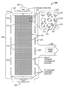
detecting specific cells, bacteria, virus, DNA, RNA, nuclei, membranes,
cellular organelles,
and cellular nanoparticulates (it is important to keep in mind that DEP is
intrinsically a "label
17
CA 02720324 2010-10-01
WO 2009/146143 PCT/US2009/039565
less" technique and that cells, nanoparticles, and other analytes can be
identified by their
cross-over frequencies; labeling is used to increase detection sensitivity,
identify individual
entities, and carry out more detailed analysis); and (3) the post analysis of
cells, nuclei,
DNA, and RNA by fluorescent probe in-situ hybridization (FISH, etc.); and (4)
use of well-
known molecular analysis methods for cells, nuclei, DNA, and RNA including but
not
limited to PCR, RCA, SDA, and other genotyping, sequencing, and gene
expression
techniques--all of which can be carried out in the same chambered compartment
in which
the DEP separation has occurred.
[0070] The above examples do not exclude carrying out subsequent analyses in
another
separate chamber of the device or moving the analytes to a sample collection
tube(s) for
off-device analyses, storage, or archiving of samples. Additionally, other
types of detection
techniques that can be used for analysis include, but are not limited to,
radioisotopes,
colorometric, chemiluminescence, electrochemical, or other methods for
biosensing or
nanosensing of the analytes, biomolecules, and cells once they have been
isolated. The
devices and processes described herein can be considered a truly "seamless"
sample-to-
answer diagnostic system that can be used directly with undiluted blood or
other complex
clinical or biological samples. The seamless sample-to-answer process using
the
exemplary DEP devices herein are described below in more detail.
[0071] Figure 27 shows the first step in seamless sample-to-answer diagnostics
where a
blood sample is applied directly to the device and DEP is used to carry out,
in this case, the
separation of a very low concentration of high molecular weight (hmw) DNA
and/or RNA
from the un-diluted whole blood sample. It should be noted, however, that
almost any
analyte including but not limited to rare cells, nanoparticles, cellular
nanoparticulates,
antibodies, immunocomplexes, proteins, and RNA could be separated,
concentrated, and
detected; and samples could include but are not limited to plasma, serum,
urine, and
saliva.
[0072] Figure 28 shows the second step in a sample-to-answer diagnostics
process
where the DEP field is now applied at the proper AC frequency and voltage that
causes the
blood cells (red and white) to move to negative (DEP) low field regions, and
the hmw DNA
(RNA) to concentrate into the positive (DEP) high field regions (in the
drawing, dome
structures represent the DEP high field strength areas).
[0073] Figure 29 shows the third step, where a simple fluidic wash is used to
remove the
blood cells from the DEP array device, while the hmw-DNA (RNA) remains highly
concentrated in the DEP high field regions. It is also within the scope of
this disclosure to
use a continuous, pulsed, or intermittent fluidic flow across the DEP device
in order to
18
CA 02720324 2010-10-01
WO 2009/146143
PCT/US2009/039565
process a larger sample volume, and as a mechanism to reduce heating, which is
more
pronounced in higher conductance solutions, at lower AC frequencies (< 20
kHz), and at
high voltages (>20 volts pt-pt).
[0074] Figure 30 shows the next step in the process, which involves the in-
situ labeling
of the DNA (RNA) by addition of a DNA (RNA) specific fluorescent dye (e.g.
CyberGreen,
OliGreen, ethiduim bromide, TOTO, YOYO, acridine orange, etc.). In this
process, a
solution of the appropriate fluorescent dye is flushed across the DEP device
to stain the
DNA or RNA. The DNA/RNA can be held in place by maintaining the DEP field
while
staining is in progress. The fluorescent stained DNA/RNA can now be detected
and
quantified by using an epifluorescent detection system (Figure 31).
Fluorescent detection
systems and devices are well-known in the art of molecular biology and
clinical diagnostics
for analysis of microarray devices, and a variety of systems are commercially
available.
[0075] It is also within the scope of this disclosure to: (1) use other
molecular analysis
detection methods and techniques including but not limited to PCR, real time
PCR with
molecular beacons, RCA, and SDA; (2) to hybridize the sample DNA/RNA during
the DEP
separation process to capture probes (DNA, RNA, pNA, etc) immobilized to
specific sites
on the DEP device for subsequent analysis/detection using fluorescent reporter
probes; (3)
to release the DNA/RNA from the DEP concentration sites, amplify it using PCR,
RT-PCR,
RCA, or SDA, denature and then use site-selective DC electrophoresis to re-
hybridize the
amplicons to capture probes immobilized on the DEP device for subsequent
analysis/detection using fluorescent reporter probes; (4) to use fluorescent
in-situ
hybridization with sequence specific DNA/RNA/pNA probes; (5) to release the
DNA/RNA
and transport it either by DEP or electrophoretically (DC fields) to another
specific location
on the device; and (6) to release the DNA/RNA and move it (by fluid flow) to
another
chamber of the device or to a sample collection tube for further analysis or
storage.
[0076] Figure 32 shows how the sample-to-answer device can be used to carry
out more
multiplex DEP separation of rare cells, bacteria, virus, cellular
nanoparticulates, or GNPs
(cellular membrane, nuclei, hmw-DNA, hmw-RNA, vacuoles, endoplasmic reticulum,
mitochondria, etc.), proteins, antibody complexes, and other biomarkers from
whole blood.
Figure 33 shows the molecular analyses methods that can now be used to
identify the
specific analytes that have been concentrated onto the device; these methods
include but
are not limited fluorescent staining, fluorescent immunoassay, FISH, and PCR,
RCA, and
SDA procedures. Finally, Figure 34 shows the final detection of cells,
bacteria, virus,
CNPs, and antibody complexes using well-known fluorescent and other detection
techniques.
19
CA 02720324 2010-10-01
WO 2009/146143
PCT/US2009/039565
[0077] This disclosure further describes unique methods that can be used to
enhance
the analytical and diagnostic capability of the sample-to-answer devices and
systems
described herein. In the case of DNA and RNA isolation and detection, while
DEP can be
used to efficiently isolate and concentrate hmw-DNA/RNA, lower molecular
weight DNA
and RNA (< 10 kb) are more difficult to isolate by DEP. In this case, double-
stranded ds-
DNA specific antibodies and single-stranded ss-DNA specific antibodies are
available that
can be used to label the lower molecular weight DNA and RNA, creating larger
nanostructures (> 5nm). These larger DNA-antibody complexes can be more
efficiently
isolated and concentrated by DEP.
[0078] Additionally, a variety of new antibody tests can be enabled using the
devices
described herein. More specifically, the ability of DEP to separate single
antibodies form
larger antibody complexes means that numerous single and double antibody
assays can
be developed in which the formation of the larger antibody-antigen complex can
be
separated from the clinical sample by DEP. In these cases, fluorescent
antibodies and/or
secondary antibodies could be added directly to the sample, DEP is applied,
and only the
fluorescent labeled antibody-antigen complexes would be concentrated into the
DEP high
field regions for subsequent detection. Such DEP based antibody assays can be
used for
small molecule antigens including but not limited to drugs, hormones,
metabolites, and
peptides; as well as for larger antigens including but not limited to
proteins, enzymes, and
other antibodies. It is also in the scope of this description to enable many
other similar
DEP assays that are based on the formation of larger complexes, including but
not limited
to detection of bacteria, virus, bacteriophage, nanoparticles, CNP's using
selective ligand
binding with antibodies, biotin/streptavidin, lectins, proteins, enzymes,
peptides,
dendrimers, apatamers, quantum dots, fluorescent nanoparticles, carbon
nanotubes, and
other nanoentities designed for selective labeling an detection purposes.
Finally, In
addition to attaching or immobilizing DNA/RNA/pNA capture probes on the DEP
device, a
variety of other binding entities can be also be attached to the DEP device,
including but
not limited to antibodies, biotin/streptavidin, lectins, proteins, enzymes,
peptides,
dendrimers, and apatamers. Such immobilized ligands will provide for selective
binding of
analytes to the DEP device after the DEP field has been turned off.
[0079] It should be noted that the novel DEP devices described herein now
enable all
these methods by the fact that these new DEP devices eliminate or greatly
reduce the
adverse bubbling, heating, and electrochemistry effects that would otherwise
damage or
destroy most of the biomolecules (e.g. DNA, RNA, antibodies, proteins, etc)
that are used
CA 02720324 2010-10-01
WO 2009/146143
PCT/US2009/039565
for immobilization, as well as the analytes and biomarkers being isolated and
concentrated
on specific DEP high field sites on the device for detection and analyses.
[0080] This first specification discloses in more detail novel electrokinetic
DEP devices
and systems in which the electrodes are placed into separate chambers and
positive DEP
regions and negative DEP regions are created within an inner sample chamber by
passage
of the AC DEP field through pore or hole structures. Various geometries can be
used to
form the desired positive DEP (high field) regions and DEP negative (low
field) regions for
carrying cell, nanoparticle and biomarker separations with the sample chamber.
Such pore
or hole structures can be filled with a porous material (agarose or
polyacrylamiide
hydrogels) or be covered with porous membrane type structures (paper,
cellulose, nylon,
etc). Such porous membrane overlaying structures can have thicknesses from one
micron
to one millimeter, but more preferably form 10 microns to 100 microns; and
pore sizes that
range from one nanometer to 100 microns, but more preferably from 10
nanometers to one
micron. By segregating the electrodes into separate chambers, these unique DEP
devices
basically eliminate any electrochemistry effects, heating or chaotic fluidic
movement from
influencing the analyte separations that are occurring in the inner sample
chamber during
the DEP process. These chambered devices can be operated at very high AC
voltages (>
100 volts pt-pt), and in addition to DEP they could also be used to carry out
DC
electrophoretic transport and electrophoresis in sample chamber. In general
these devices
and systems can be operated in the AC frequency range of from 1000 Hz to 100
mHz, at
voltages which could range from 1 volt to 2000 volts pt-pt; and DC voltages
from 1 volt to
1000 volts, at flow rates of from 10 microliters per minute to 10 milliliter
per minute and in
temperature ranges from 1 C to 100 C . The chambered devices are shown in
Figure 1
and Figure 2. Such devices can be created with a variety of pore and/or hole
structures
(nanoscale, microscale and even macroscale) and may contain membranes, gels or
filtering materials which can control, confine or prevent cells, nanoparticles
or other entities
from diffusing or being transported into the inner chambers. However, the
AC/DC electric
fields, solute molecules, buffer and other small molecules can pass through
the chambers.
[0081] Figure 1 and 2 represents a most basic version of the chambered devices
that
can be constructed in accordance with the invention. A variety of
configurations are
envisioned for the devices in accordance with the invention. Such devices
include, but are
not limited to, multiplexed electrode and chambered devices, devices that
allow
reconfigurable electric field patterns to be created, devices that combine DC
electrophoretic and fluidic processes; sample preparation devices, sample
preparation and
diagnostic devices that include subsequent detection and analysis, lab-on-chip
devices,
21
CA 02720324 2010-10-01
WO 2009/146143
PCT/US2009/039565
point-of-care and other clinical diagnostic systems or versions. Figure 1 is a
schematic
diagram of a sample processing device constructed in accordance with the
teachings
herein, and shows that the device 100 includes a plurality of electrodes 102
and electrode-
containing chambers 104 within a housing 106. A controller 108 of the device
independently controls the electrodes 102, as described further herein.
[0082] Figure 2 shows a top view of a the device 100 which is illustrated with
six
electrode chambers 104, each of which has at least one robust platinum
electrode. Figure
2 shows the device configured with one main central separation chamber 110,
which has
an arrangement of eighteen pore/hole structures 112 of varying size that are
filled with a
hydro-gel (the inner chamber could also have a porous membrane covering the
pores or
holes). The pore/hole structures are arranged in three groups of six pore/hole
structures.
While the upper part of the separation chamber 110 has no physical
separations, the lower
portion is divided into nine separate compartments (indicated by the light
dashed line).
Each of these compartments is in fluidic contact with an electrode chamber,
but not with
each other. When an AC DEP field is applied to the electrodes, the field
passes through
the pores 112, creating positive DEP high field regions on top of the pore
structures and
negative DEP low field regions between the pore structures. Samples can be
added and
removed from the device via the inlet 220 and outlet 222. The device may
additional inlets
224 and outlets 226. The device shown in Figure 1 and 2 represents just one
form of a
high conductance DEP chambered device; it should be understood that a large
number of
different types of devices with larger numbers of pores/holes and different
geometries can
be created.
[0083] Another device embodiment involves using electrode arrays with robust
electrodes of defined diameter and separation distances that will allow for
less
electrochemical effects and heating, which is a problem in current
electrokinetic and
dielectrophoretic separation devices. Proper construction and overcoating of
robust
electrodes (e.g. platinum, palladium and gold) can reduce adverse effects of
electrochemistry products on the separation process, and allow much higher
voltages to be
applied, which can greatly improve separation times. Also, current devices are
relatively
low throughput and this embodiments described herein have overcome that
problem by
providing a system that uses multiplexed parallel section arrays and that
allows the device
to be used as one large separation zone and then switched to separately
controlled
separations zones, resulting in increased sensitivity and selectivity of the
overall system. A
third problem seen in other conventional systems is the inability to separate
sample
components that are relatively similar in size and composition. This problem
is overcome
22
CA 02720324 2010-10-01
WO 2009/146143 PCT/US2009/039565
in accordance with the description herein by providing a device that can carry
out
secondary separation processes, such microelectrophoresis, directly on the DEP
array
device itself.
[0084] Negative dielectrophoretic (DEP) forces are relatively weaker than
positive DEP
forces; thus entities that experience negative DEP can be moved by fluid flow,
while
positive DEP experiencing entities will remain in place. In the presently
described
embodiments, by using both fluid and DC electrophoretic forces in opposite
directions,
DNA fragments and highly charged DNA nanoparticulates can be separated from
cells and
proteins in blood and other samples. In this way, using multiple AC
frequencies, pulsed
DC electrophoresis, and micro-electrophoresis, a more complete size separation
of DNA
nanoparticulates and DNA fragments can be accomplished.
[0085] Commercial uses of such novel systems and devices that now allow DEP to
be
carried out under high conductance conditions (blood, plasma, serum, etc.)
will likely
include numerous research and clinical diagnostic applications, such as point
of care
diagnostics, therapeutics and drug monitoring, environmental and water supply
monitoring,
and bioterror agent detection. Numerous analytes and entities such as rare
cells (cancer
cells, fetal cells, hematopoietic stem cells), bacteria, virus, DNA/RNA, and
DNA
nanoparticulate biomarkers, drug delivery nanovesicles, as well as normal or
aberrant
proteins, might be detected using such a system.
[0086] An experimental AC DEP and DC electrophoretic separation system (a
laboratory
bench-top version described further in the Experimental Section below) has
been built and
experiments were conducted to refine the new prototype devices. The results
obtained on
these devices (which are described above) lead to the important discovery as
to why
classical DEP has been limited to low conductance solutions.
[0087] Now new devices that use planar, parallel, and robust platinum
electrode arrays
with electrodes of roughly about 1-1000 micron in diameters with 10 to 5000
micron
separation distance and overcoated with a 5-100 micron thick hydrogel
(agarose,
polyacylamide) or porous membrane layer(s), allows for less heating and
electrochemistry
issues, as the electric field lines are not as highly concentrated as they are
in other
classical conventional DEP systems, and more importantly the DEP high field
accumulation
region is actually now some distance from the actual electrode surface. One
significant
difference from previous electrode designs is not using sputtered platinum or
gold
electrodes, which are easily degraded and destroyed by electrochemistry,
particularly at
higher field strengths and high solution conductance. The electrodes for the
new devices
will be constructed from solid platinum or gold materials, including wires or
rods. A second
23
CA 02720324 2010-10-01
WO 2009/146143 PCT/US2009/039565
difference is that the separation efficiency for isolating one unique entity
in a million
relatively similar entities (cells, nanoparticles, biomarkers) can be improved
by changing
the problem of one large separation to that of many separate separations which
are much
more controllable. The devices described herein accomplish this by using
multiplexed
sectioned arrays and a controlled parallel sorting process. This is achieved
by using
individually controlled array subsets of 10 to 100 or more electrodes in a
large array device
that allows a complex biological sample (blood) to be distributed across the
array device,
separating the components into smaller separation sections (areas) for further
separation
and isolation of the desired analytes or entities. Breaking the complex sample
separation
problem down into smaller parts holds the most promise for solving the issue
of sensitivity
versus specificity, i.e., the process allows both rapid and higher overall
sample throughput,
as well as relatively longer interrogation (separation) times for isolating
and identifying
unique cells or other entities in the sample. Finally, the last problem can be
overcome by
creating a multi-dimensional hierarchical sorting device. This solution relies
on the fact that
negative DEP is a weaker force than positive DEP and cells or other entities
experiencing
negative DEP can be moved by controlled fluid flow, whereas the positive DEP
experiencing analytes or entities will stay concentrated in the DEP high field
areas.
Through the use of controlled fluid flow and pulsed DC electrophoresis in
opposite
directions, DNA/RNA and charged nanoparticulates can be separated from cells
and
proteins in a complex biological sample (this is in addition to the intrinsic
ability of DEP to
separate cells and DNA).
[0088] Combining controlled fluid flow and pulsed DC electrophoresis with
using multiple
AC frequencies, i.e. low frequency to trap the CNPs and hmw-DNA/RNA
nanoparticulates
in on the initial electrodes array subset, and higher AC frequencies on other
electrode array
subsets to trap cells progressively larger particles (bacteria and virus) a
complete
separation of most of the cells and entities by size can be obtained. If
desired the
electrodes can be switched to different frequencies for finer separation to
occur locally
while globally the overall size separation is maintained.
[0089] We describe a separation system involving a device with planar, robust,
platinum
electrode array structures and auxiliary electrodes, into which a complex
biological sample
(blood, plasma, serum) is directly applied, such that controlled AC signals
from one or
more function generators produce dielectrophoretic forces, and a controlled DC
power
supply produces electrophoretic forces. The inlet and outlets of the device
also allow for
the controlled passage of fluids (water, buffers, etc.) through the system at
a controlled flow
rate. The system also includes an optical/epifluorescent microscope and
digital camera for
24
CA 02720324 2010-10-01
WO 2009/146143 PCT/US2009/039565
monitoring, detecting, quantifying, and recording the separation processes
that are
occurring on the device (visual and fluorescent). The device is ultimately a
multiplexing,
parallel hierarchical sorting system that is enabled by controlling
electrokinetic effects,
dielectrophoretic forces, electrophoretic forces, microelectrophoresis, and
fluid flow. It
should be noted that such novel multiplex sample-to-answer processes are made
possible
by the fact that the new DEP devices eliminate or greatly reduce the adverse
bubbling,
heating, and electrochemistry effects experienced by conventional devices.
[0090] Figure 3 shows just one version of a planar platinum electrode array
device 300
comprising a housing 302 through which a sample fluid can flow. The fluid flow
pattern
through the device is indicated by the large arrows, representing flow of an
idealized
sample, from an inlet end 304 at the top of the drawing to an outlet end 306
at the bottom,
and a lateral analyte outlet 308. The device includes multiple AC electrodes
310. Only a
few of the electrodes 310 are identified in Figure 3, for simplicity of
illustration, but it should
be understood that all the small open circles in the drawing figure represent
electrodes of
similar construction. One enlarged 3x3 array 312 of the electrodes is
illustrated on the right
side of the drawing figure to show a sample fluid in the device 300. The
sample consists of
a combination of micron-sized entities or cells 314 (the largest filled-in
circles shown in the
enlarged view), larger nanoparticulates 316 (the intermediate-sized filled-in
circles) and
smaller nanoparticulates or biomolecules 318 (the smallest-sized circles). The
larger
nanoparticulates 316 could represent high molecular weight DNA, nucleosomes,
or CNPs
or cellular debris dispersed in the sample. The smaller nanoparticulates 318
could
represent proteins, smaller DNA, RNA and cellular fragments. The planar
electrode array
device 300 in the figure is a 60x20 electrode array that can be sectioned into
three 20x20
arrays that can be separately controlled but operated simultaneously. The
auxiliary DC
electrodes 320 at the top of the figure can be switched on to positive charge,
while the DC
electrodes 322 at the bottom of the figure are switched on to negative charge
for
electrophoretic purposes. Each of the controlled AC and DC systems can be used
in both a
continuous and/or pulsed manner (e.g., each can be pulsed on and off at
relatively short
time intervals). The planar electrode arrays 324 along the sides of the sample
flow, when
over-layered with nanoporous materials, can be used to generate DC
electrophoretic
forces as well as AC DEP. Additionally, microelectrophoretic separation
processes can be
cared out within the nanopore layers using planar electrodes in the array
and/or auxiliary
electrodes in the x-y-z dimensions. In general these devices and systems can
be operated
in the AC frequency range of from 1000 Hz to 100 mHz, at voltages which could
range
from approximately 1 volt to 2000 volts pk-pk; at DC voltages from 1 volt to
1000 volts, at
flow rates of from 10 microliters per minute to 10 milliliter per minute, and
in temperature
CA 02720324 2010-10-01
WO 2009/146143
PCT/US2009/039565
ranges from 1 C to 100 C. The controller 108 (Figure 1) independently
controls each of
the electrodes 310, 320, 322, 324. The controller may be externally connected
to the
device 100 such as by a socket and plug connection (not illustrated), or can
be integrated
with the device housing. Electrical lead lines for the electrodes are not
shown in the
drawings, for simplicity of illustration.
[0091] It can be assumed that the cells and particles and other entities in
the sample are
evenly distributed throughout the electrode array, though only the enlarged
3x3 electrode
section 312 is shown in the drawing figure. The fluid flow rate is such that
it exerts a force
stronger than the negative DEP that the larger particles experience, but
weaker than the
positive DEP that the larger particles experience.
[0092] Figure 4 shows the top 320 and bottom 322 DC electrodes being pulsed on
and
off (one second on followed by one second off), thereby providing a brief
electrophoretic
pulse pushing the DNA, RNA, and small nanoparticulates toward the positive DC
electrode
320, which is located at the top of the drawing figure. The 60x20 electrode
array is
visualized as broken into three distinct sections or sub-arrays that are
independently
controlled. The top twenty AC electrode array rows 402 are tuned to a lower
frequency AC
field to ensure that the smaller entities, which generally move toward the
electrodes, due to
positive DEP and AC electrokinetic phenomena at lower frequencies, will be
trapped at
those electrodes while the larger cells and entities experience negative DEP
at these
frequencies, and are therefore moved to the lower section of the device by the
constant
fluid flow. The middle twenty rows 404 of AC electrodes will hold the large
sub-micron
particles (e.g. virus) while allowing the micron-sized particles and cells to
flow though.
Finally, the last twenty rows 406 of AC electrodes can be attuned, if desired,
to a high AC
frequency, which can then be used to capture desired cells and micron-sized
particles.
[0093] Figure 5 shows the separation mechanism for isolating "one cell in a
million", i.e.,
rare cell detection. By using the complete electrode array, it is possible to
multiplex and
parallelize the problem of separation to make it simpler. This can be achieved
by merely
activating as much of the electrode array as necessary to achieve better
separation. By
effectively splitting the array into specific separation areas that can be
analyzed by optical
detection (i.e., epifluorescence), it should be possible to separate out one
specific cell
experiencing positive DEP from all the cells around it, once all the cells are
evenly
distributed. In Figure 5, the intermediate-sized filled-in circles 502
represent 10 pm cells of
one specific kind, such as lymphocytes, red blood cells, and the like, and the
single filled-in
circle 504 shown on the AC electrode 506 of the third section 406 of AC
electrodes
represents the lone "one cell in a million" of a type different from the
others 502 in the
26
CA 02720324 2010-10-01
WO 2009/146143
PCT/US2009/039565
sample, which is also the only cell that experiences positive
dielectrophoresis and is
therefore easily distinguishable from the other cells. Using only
dielectrophoresis, it should
be possible to separate out cells of the lone "one cell in a million" 504 type
from the
undifferentiated 502 cell types. This is more easily accomplished if there are
a sufficieint
number of AC electrodes to spread the separation problem into smaller, more
easily
separable and analyzable chunks. Once the cell-type separation problem is
spread out in
such a manner, if only certain sections of the electrode array are analyzed at
a time, such
as the 3x3 array shown in Figure 5, it should be possible to find the lone
particle 504 of
interest. Additionally, temperature control can be effective in allowing more
selective and
efficient separation of cells (e.g., separation of cancer and stem cells).
[0094] Figure 6 shows a more detailed scheme of a blood sample separation
process,
before the application of combined pulsed AC DEP/DC electrophoresis/controlled
fluidic
flow. The Figure 6 diagram shows some of a wide variety of potential
diagnostic and
biomarker entities that would be found in a complex sample such as blood,
which entities
may include: red and white blood cells, bacteria, virus, nanovesicles, DNA/RNA
nanoparticulates, an assortment of DNA and RNA fragments, and proteins. The
Figure 6
diagram also shows the planar platinum array electrodes 310 covered with an
intermediate
density nanopore layer 604, a low density nanopore layer 606, and a high
density
nanopore layer 608 directly over the AC electrodes 310.
[0095] Figure 7 shows the blood sample in the initial stages of combined
pulsed AC
DEP/DC electrophoresis/controlled fluidic flow. In Figure 7, the whole array
device 300 is
utilized to carry out an overall separation process that begins to concentrate
different
classes of entities into each of the electrode sub-array sections 402, 404,
406 (upper,
middle, lower, respectively).
[0096] Figure 8 shows the blood sample now in final stages of combined pulsed
AC
DEP, DC electrophoresis, and controlled fluidic flow. In Figure 8, the
different entities have
been concentrated into their appropriate electrode array sections 402, 404,
406. In this
example, DNA nanoparticulates and smaller DNA fragments are shown in the upper
array
section 402; bacteria, virus, and nanovesicles are shown in the middle array
section 404;
and cells and proteins are shown in the lower array section 408.
[0097] Figure 9 shows an enlarged view of the AC electrode array in which the
combined
pulsed AC DEP and DC electrophoresis of fluorescent-stained DNA
nanoparticulates, very
high molecular weight DNA and intermediate lower molecular weight DNA
selection and
separation on the upper array section 402. Because these entities have now
been
concentrated and isolated in the upper array section they can be selectively
stained with
27
CA 02720324 2010-10-01
WO 2009/146143 PCT/US2009/039565
appropriate DNA fluorescent dye reagents, and the secondary separation process
can now
be cared out.
[0098] Figure 10 shows the enlarged view of the AC electrode array with
initial combined
pulsed AC DEP and DC electrophoresis of fluorescent stained DNA
nanoparticulates, very
high MW DNA and intermediate-lower MW DNA selection and separation on the
upper
array section 402. This initial process will cause the DNA nanoparticulates to
begin to
concentrate onto the top of the intermediate nanopore layer which has a pore
size that
excludes these very large DNA entities; while the more intermediate and lower
molecular
weight DNA fragments are transported into lower nanopore density layer.
[0099] Figure 11 shows the enlarged view of the AC electrode array with final
combined
pulsed AC DEP and DC electrophoresis of fluorescent stained DNA
nanoparticulates, very
high MW DNA and intermediate-lower MW DNA selection, and microelectrophoresis
separation on the upper array section 402. At this point in the operation of
the device 300,
the DNA nanoparticulates and very high molecular weight DNA are fully
concentrated and
isolated on the top of the intermediate density nanopore layer 604 and the
more
intermediate and lower molecular weight DNA fragments are concentrated within
the inner
lower density nanopore layer 606.
[0100] Figure 12 shows the enlarged view of the AC electrode array 402 after
removal of
DNA nanoparticulates and very high MW DNA and on-array DC electrophoretic size
separation of the intermediate and low MW DNA fragments. The DNA
nanoparticulates
and very high molecular weight DNA can be further analyzed on another part of
the device
300, while the more intermediate and lower molecular weight DNA fragments can
be size-
separated by microelectrophoresis within the nanopore layers 604, 606, 608.
Figure 12
shows that some of the AC electrodes 310a are positively charged and other AC
electrodes 310b are negatively charged.
[0101] Figure 13 shows an enlarged view of the AC electrode array with the
initial pulsed
AC DEP applied to red and white blood cells on lower array section 406 of the
device 300.
In this process, the proteins in the sample can be removed and/or analyzed on
another
component of the device, while the cells and other micron-sized entities can
be further
separated and differentiated by AC DEP on the lower array section 406.
[0102] Figure 14 shows the enlarged view of the electrode array with the final
pulsed AC
DEP applied to red and white blood cells on the lower array section 406 of the
device 300.
At this point of the device operation, the red and white blood cells have been
separated in
28
CA 02720324 2010-10-01
WO 2009/146143
PCT/US2009/039565
the DEP high and low field regions, subsequently the red cells can be removed
and the
white cells further differentiated; i.e., begin the process of isolating
cancer cells.
[0103] Figure 15 shows an enlarged view of the AC electrode array with the
initial pulsed
AC DEP for separation of bacteria, virus and nanovesicles on the middle array
section 404
of the device 300. The Figure 15 drawing is an example of how sub-sections of
the array
device 300 that can be independently controlled can be used to carry out
additional
important separation processes.
[0104] Figure 16 shows the enlarged view of the electrode array with the final
pulsed AC
DEP for separation of bacteria, virus and nanovesicles on the middle array
section 404 of
the device 300. Again, it should be kept in mind that this separation process
on the middle
array section can be run concurrently with, and independently of, other
separation
processes that are occurring on the other array sub-sections; e.g., DNA
fragment
separations can take place on the upper array section and cell separations can
take place
simultaneously on the other (lower) array sections.
[0105] Lastly, the parallel multiplexed electrode array can be used in
conjunction with
hierarchical cell sorting to create defined areas within the rows of
electrodes where specific
particles that are similar in size but have different dielectric properties
can be trapped. A
variety of diagnostic and therapeutic applications which can utilize
electrokinetic,
dielectrophoretic, electrophoretic and fluidic forces and effects all in
conjunction with each
other to increase separation sensitivity and efficiency in a device. Most
importantly, these
high performance and clinically useful separation processes are achieved only
when the
electrokinetic, electrophoretic and fluidic forces and effects are uniquely
combined on a
properly scaled and controlled electrode array device. In addition to this
type of separation,
dielectrophoresis, which is a lossless, potentially label less, parallel
separation method, can
be used in conjunction with more traditional separation methods which have far
more
sample preparation involved as well as greater loss of the sample, such as
field flow
fractionation, fluorescence assisted cell sorting (FACS) or magnetic assisted
cell sorting, to
achieve even greater levels of cell and nanoparticle separation for use in
applications.
[0106] With regard to other aspects in the illustrated embodiments, it should
be pointed
out that when labeling (optical fluorescent, luminescent, electrochemical,
magnetic, etc.) is
added to the cells, nanoparticles, and biomarkers to be separated, the
multiplexing
described herein would likely be even more effective due to the labels helping
to affect the
size, conductivity and detectability of the entities.
29
CA 02720324 2010-10-01
WO 2009/146143
PCT/US2009/039565
[0107] Presently, the DEP separation mechanisms described above are in an
early
experimental data stage. A prototype system has been constructed, utilizing an
electrode
array structure as described above that receives biological materials for
separation, that
include cells, nanoparticles and hmw-DNA. Selective energizing of subsets of
electrodes in
the array structure has now been achieved with a function generator operating
under
control of suitable software programming, which can execute on conventional
computers
such as desktop or workstation computers. Individual electrodes can be
controlled using
this apparatus. The prototype system has an associated epifluorescent
microscope for
monitoring and recording the separation experiments (see Experimental Section
below for
additional description).
[0108] A variety of separation and isolation applications which include rare
cell detection
for adult stem cell isolation from blood, other bodily fluids or any buffers,
e.g. hematopoietic
progenitors; gross separation between cells, proteins and DNA/RNA fragments in
blood,
other bodily fluids or other buffers for the purposes of cancer detection and
other
diagnostics; cancer cell isolation from blood, other bodily fluids or other
buffers for
research, diagnostic as well as therapeutic purposes. Also envisioned are the
uses for
environmental monitoring and for the rapid detection of pathogens and
bioterror agents.
Finally, also envisioned are systems, devices and techniques described in this
invention
can be used to separate, isolate and purify a variety of non-biological
entities that include in
addition to drug nanoparticles and nanovesicles; quantum dots, metallic
nanoparticles,
carbon nanotubes (CNTs), nanowires, and even micron and submicron CMOS devices
and
components; basically any macromolecule or nanocomponent that can be suspended
or
solubilized in an aqueous or mixed solvent system can be processed in the
embodiments
illustrated and using the techniques described herein. We also envision that
these new
devices will now allow directed self-assembly of DNA and other bioderivatized
nanoparticles, nanocomponents, and mesoscale objects to be carried out. This
can lead to
new DNA genotyping and sequencing technologies ($1000 genome) and nano/micro
bio/chemsensor applications, including highly integrated cell-sized "Fantastic
Voyage"
devices (inspired by the movie of the same name) that could be placed in the
blood stream
to carry out diagnostics, therapeutic delivery in-vitro microsurgery, i.e.,
remove clots and
plaques, repair atherosclerotic arteries, etc.; as well as nanoelectronic,
nanophotonic,
photovoltaic, fuel cell, batteries, nanomaterials, and numerous other
heterogeneous
integration applications.
[0109] Using the devices and techniques described herein, a "sample to answer"
result
can be provided, wherein the separation operation results in holding at least
one type of
CA 02720324 2010-10-01
WO 2009/146143
PCT/US2009/039565
biological material at one of the electrode subsections, while the remainder
of the sample
fluid is washed from the device, so that a reagent into the sample processing
device,
followed by reacting the introduced reagent with the held type of biological
material in the
sample processing device. As noted above, the reagent may comprise a
fluorescent die,
antibodies, or the like. The sample-to-answer process may be used to perform a
variety of
tasks as described above, including PCR operations and the like.
[0110] The invention has been described above in terms of presently preferred
embodiments so that an understanding of the present invention can be conveyed.
There
are, however, many configurations and permutations of the devices, system and
separation
mechanisms not specifically described herein, but to which the present
invention is
applicable. The present invention should therefore not be seen as limited to
the particular
embodiments described herein, but rather, it should be understood that the
present
invention has wide applicability with respect to biological separation systems
generally. All
modifications, variations, or equivalent arrangements and implementations that
are within
the scope of the attached claims should therefore be considered within the
scope of the
invention.
[0111] Experimental Section
[0112] Buffers and Conductivity Measurements
[0113] Concentrated 5x Tris Borate EDTA (TBE) buffer solution was obtained
from USB
Corporation (USB, Cleveland, Ohio, USA), and was diluted using deionized Milli-
Q
Ultrapure water (55 nS/cm) to the following concentrations: 0.01x TBE, 0.1x
TBE and lx
TBE. Dulbecco's Phosphate Buffer Saline (lx PBS) solution was obtained from
Invitrogen
(Invitrogen, Carlsbad, CA, USA) and was diluted using Milli-Q water to 0.1x
PBS.
Conductivity measurements were made with an Accumet Research AR-50
Conductivity
meter (Fisher Scientific, Fair Lawn, NJ, USA) using a 2 cell (range: 10-2000
PS) and a 4
cell (range: 1-200 mS) electrode and was adjusted with proper conductivity
standards. The
following buffer conductivities were measured: 0.01x TBE ¨ 18.1 pS/cm; 0.1x
TBE ¨ 125
p5/cm; lx TBE ¨ 1.09 mS/cm; 0.1x PBS ¨ 1.77 mS/cm; and lx PBS ¨ 16.8 mS/cm.
[0114] Particles, Nanoparticles and DNA Derivatization
[0115] Fluorescent polystyrene nanoparticles (FluoSpheres) with NeutrAvidin
coated
surfaces were purchased from Invitrogen (Invitrogen, San Diego, CA, USA). The
nanoparticle diameters were 0.04pm (40nm) and 0.2pm (200nm). The 40nm
polystyrene
nanoparticles were red fluorescent (ex:585/em:605) and the 200nm polystyrene
nanoparticles were yellow-green fluorescent (ex:505/em:515). Larger 10.14pm
31
CA 02720324 2010-10-01
WO 2009/146143
PCT/US2009/039565
carboxylated polystyrene particles were obtained from Bangs Labs (Bangs Labs,
Fishers,
IN, USA). Biotinylated DNA oligonucleotide sequences were obtained from
Trilink Bio
Technologies (Trilink, San Diego, CA, USA). The single- stranded 51mer DNA
oligonucleotide used to derivatize the 40nm nanoparticles had the sequence ¨
[5]'-Biotin-
TCA GGG COT CAC CAC CTA CTT CAT CCA CGT TCA CTC AGO GCC TCA CCA COT
[3]'. A second single-stranded 23mer DNA oligonucleotide used had the sequence
¨ [5]'-
Biotin- GTA CGG CTG TCA TCA OTT AGA CC [3]'. The derivatization of the 40nm
NeutrAvidin nanoparticles with the biotinylated DNA oligonucleotides was
carried out by
first suspending the nanoparticles in different concentrations of Iris Borate
EDTA (0.01x,
0.1x, lx TBE) or Phosphate Buffered Saline (0.1x, lx PBS) buffers. The ss-DNA
oligonucleotide was added to the mixtures in the amounts of 400:1 (DNA:40nm
nanoparticles) ratio for the 51mer ss-DNA sequence, and 6500:1 (DNA:40 nm
nanoparticle) ratio for the 23mer ss-DNA sequence. Once the DNA was added, the
solution
was vortexed at high speed for 20 seconds and then allowed to react for about
20 minutes.
For the 40nm DNA derivatized nanoparticle experiments, the DNA nanoparticle
mixture
was made by adding 0.5pL of the stock solution into 299pL of the appropriate
buffer. For
the 200nm nanoparticle experiments, 0.5pL of the stock solution was added to
299pL of
the appropriate buffer. Finally, 1pL of the 10.14pm polystyrene particle stock
solution was
added to the samples, the samples were then slowly mixed for about 10 seconds.
The
samples were now ready to be applied to the microarray cartridge device.
[0116] DEP Microelectrode Array Device
[0117] The microelectrode array devices used for these studies were obtained
from
Nanogen (San Diego, CA, USA, NanoChip 100 Cartridges). The circular
microelectrodes
on the array are 80pm in diameter and made of platinum. The microarray is over-
coated
with 10pm thick porous polyacrylamide hydrogel layer. The microarrays are
enclosed in a
microfluidic cartridge which forms a 20pL sample chamber over the array that
is covered
with a glass window. Electrical connections to each individual microelectrode
are pinned
out to the bottom of the cartridge. Only a 3x3 subset of nine microelectrodes
was used to
carry out DEP. Alternating current (AC) electric fields were applied to the
nine
microelectrodes in a checkerboard addressing pattern. In this checkerboard
pattern of
addressing, each microelectrode has the opposite bias of its nearest neighbor.
The
corresponding computer model for the asymmetric electric field distribution
created by this
pattern has been discussed previously [27]. This model indicates that the
positive DEP field
maxima (high field regions) exist at (on) the microelectrodes and the negative
DEP field
minima (low field regions) exist in the areas between the electrodes. In
general, for DEP in
32
CA 02720324 2010-10-01
WO 2009/146143
PCT/US2009/039565
low conductance solutions the 60nm DNA and 200nm nanoparticles are expected to
concentrate in the positive or high field regions over the microelectrodes
[28] and the 10
micron particles concentrate in the negative or low field DEP regions [29]
between the
microelectrodes. The computations from the previous model were performed for a
5x5
microelectrode set [27]. Before each experiment, the microarray cartridge is
flushed 10
times with 200pL of the appropriate buffer, over a span of 5 minutes. The
cartridge is
allowed to sit for 5 minutes, and is then washed two more times with 200pL of
buffer. A
total of 150 pL of the sample solution containing the nanoparticle mixture is
then slowly
injected into the cartridge, a final sample volume of about 20 pL remains in
the cartridge.
[0118] Experimental Setup, Measurements, Fluorescence and SEM Analysis
[0119] The microarray devices were controlled using a custom made switching
system
(designed and constructed in our lab) that allows for individual control over
the voltage
being applied to each of the 100 microelectrodes. The microelectrodes were set
to the
proper AC frequency and voltages using an Agilent 33120A Arbitrary Function
Generator
(Agilent, Santa Clara, CA, USA). AC frequencies ranged from 1000Hz to
10,000Hz, at 10
volts peak to peak (pk-pk). The wave form used for all experiments was
sinusoidal. The
experiments were visualized using a 10x PL Fluotar objective in a JenaLumar
epifluorescent microscope (Zeiss, Jena, Germany) employing the appropriate
excitation
and emission filters (green fluorescence Ex 505nm, Em 515nm; red fluorescence
Ex
585nm, Em 605nm. Both back lighted and the fluorescent images were captured
using an
Optronics 24-bit RGB CCD camera (Optronics, Goleta, CA, USA). The image data
was
processed using a Canopus ADVC-55 video capture card (Canopus, San Jose, CA,
USA)
connected to a laptop computer using either Adobe Premiere Pro (Adobe Systems
Inc, San
Jose, CA, USA) or Windows Movie Maker. The final fluorescence data was
analyzed by
inputting individual fluorescent image frames of the video into MATLAB
(Mathworks,
Natick, MA, USA) at 0 minutes, 30 seconds, 1 minute, 2 minutes, 4 minutes, 8
minutes, 16
minutes, and 20 minutes time points. The graphs were created using Excel from
data
gathered through MATLAB analysis of fluorescence intensity readings across the
microelectrode. was created using MATLAB. The following data was used to
create the
graph: op (for 200nm)= 18mS, op (for 40nm+DNA)= 50mS K5=0.9nS, Ep=2.55E0. r =
30nm &
r = 100nm. f=3kHz. . After the conclusion of the DEP experiments the FCOS
microarrays
had all the fluid removed from their surface and were visualized using a
Phillips XL30
scanning electron microscope (SEM). The SEM was used to image the final
nanoparticle
layers on the surface of the microarray.
33
CA 02720324 2010-10-01
WO 2009/146143 PCT/US2009/039565
[0120] References cited in this description comprise the following:
1. Tong, Y-K; Lo, Y. M. D. Diagnostic developments involving cell-free
(circulating) nucleic
acids. Clinica Chimica Acta. 363:187-96; 2006
2. Becker, F.F.; Wang, X-B.; Huang, Y.; Pethig, R.; Vykoukalt, Y.; Gascoyne,
P.R.C. The
removal of human leukemia cells from blood using interdigitated
microelectrodes. J Phys.
D: Appl. Phys. 27:2659-2662; 1994.
3. Becker, F.F.; Wang, X-B.; Huang, Y.; Pethig, R.; Vykoukalt, Y.; Gascoyne,
P.R.C.
Separation of Human Breast Cancer Cells From Blood by Differential Dielectric
Affinity.
Proceedings of the National Academy of Sciences, 92:860-864; 1995.
4. Stephens M, Talary MS, Pethig R, Burett AK, Mils KI. The dielectrophoresis
enrichment
of CD34+ cells from peripheral blood stem cell harvests. Bone Marrow
Transplant. 18:777-
82. 1996
5. Washizu, M.; Kurosawa, 0. Electrostatic manipulation of DNA in
microfabricated
structures. Industry Applications, IEEE Transactions on. 26:1165-1172; 1990.
6. Washizu, M.; Kurosawa, 0.; Arai, I.; Suzuki, S.; Shimamoto, N Applications
of
electrostatic stretch-and-positioning of DNA. Industry Applications, IEEE
Transactions on.
31:447-456; 1995.
7. Asbury, C.L; Van Den Engh, G. Trapping of DNA in Nonuniform Oscillating
Electric
Fields. Biophys J. 74:1024-1030; 1998.
8. Asbury, C.L; Diercks, A.H.; Van Den Engh, G. Trapping of DNA by
dielectrophoresis.
Electrophoresis. 23:2658 - 2666; 2002.
9. Holzel, R.; Calander, N.; Chiragwandi, Z; Wil ander, M.; Bier, F.F.
Trapping Single
Molecules by Dielectrophoresis. Phys. Rev. Left. 95:128102 (2005)
10. Ramos, A.; Morgan, H.; Green, N. G.; Castellanos, A. Ac electrokinetics: a
review of
forces in microelectrode structures. J Phys. D: Appl. Phys. 31:2338-2353;
1998.
11. Goodard W.A., Brenner, D.W.; Lyshevski, S.B.; lafrate, G.J.; Handbook
ofNanoscience,
2nd edition, ch 16, p5-8
12. HiguGhi Yo, GhrQ1DOsomaIDNA fragmentation inapoptosis and necrosis induced
by
oxidative stress. Biochem Pharacol. 66:1527-35. 2003
13. Ziegler A, Zangemeister-Wittke D, Stahel RA. Circulating DNA: a new
diagnostic gold
mine? Cancer Treat Rev. 5:255-71. 2002
14. Wu TL, Zhang D, Chia JH, Tsao KH, Sun CF, Wu JT. Cell-free DNA:
measurement in
varous carcinomas and establishment of normal reference range. Clin ChimActa.
21:77-87.
2002
15. Higuchi Y.; Matsukawa S. Appearance of 1-2 Mbp giant DNA fragments as an
early
34
CA 02720324 2010-10-01
WO 2009/146143
PCT/US2009/039565
common response leading to cell death induced by various substances that cause
oxidative stress. Free Radical Biology & Medicine, 23:90-99, 1997 23
16. Gautschi 0, Bigosch C, Huegli B, Jerman M, Mar A, Chasse E, Ratschiler D,
Weder W,
Joerger M, Betticher DC, Stahel RA, Ziegler A. Circulating deoxyrbonucleic
Acid as
prognostic marker in non-small-cell lung cancer patients undergoing
chemotherapy. J Clin
Oneol. 22:4157- 64. 2004
17. Stroun M, Aner P, Lyautey J, et aL. Isolation and characterization of DNA
from the
plasma of cancer patients. Eur J Cancer Clin Oneol 23:707-712. 1987.
18. Morgan, H.; Hughes, M.P.; Green, N.G. "Separation of Sub micron
Bioparticles by
Dielectrophoresis" Biophysical Journal.77:516-525. 1999
19. Green, N. G.; Ramos, A.; Morgan, H. Ac electrokinetics: a survey of sub-
micrometre
paricle dynamics. J. Phys. D: Appl. Phys. 33:632-641; 2000.
20. Tuukanen, S.; Toppar, J. J.; Kuzyk, A.; Hirviniemi, L.; Hytonen, V. P.;
Ilalainen T.;
Torma, P. Carbon nanotubes as electrodes for dielectrophoresis of DNA. Nano
Letters.
6:1339-1343; 2006.
21. Ching 1. et al., "Isolation of Cultured Cerivcal Carcinoma Cells Mixed
with Peripheral
Blood Cells on a Bioelectronic Chip", Analytical Chemistry, VoL. 70, #11, pp.
2321-2326,
1998.
22. Cheng, J. et al., "Preparation and Hybridization Analysis ofDNA/A from E.
coli on
15 Microfabricated Bioelectronic Chips", Nature Biotechnology, VoL. 16, pp.
541-546,
1998.
23. Huang Y, Ewalt KL, Tirado M, Haigis R, Forster A, Ackley D, Heller MJ,
O'Connell JP,
and Krhak M, "Electric Manipulation ofBioparic1 es and Macromolecules on
Microfabricated
Electrodes", Analytical Chemistry 2001, (73):1549-59.
24. Huang Y, Joo S, Duhon M, Heller MJ, Wallace B and Xu, "Dielectrophoretic
Cell
Separation 20 and Gene Expression Profiling on Microelectronic Chip Arrays",
Analytical
Chern. 2002, 74, 3362-71.
25. U.S. Patent No. 5,632,957, May 27, 1997, Heller et al., Molecular
Biological System
Including Electrodes.
26. U.S. Patent No. 6,289,590 BI, August 28,2001, Cheng et al., Chanell-Less
Separation
of 25 Bioparicles on a Bioelectronic Chip by Dielectrophoresis.
27. U.S. Patent NQ. 6,403,367,367 BI, June 11,2002, Cheng et al., Integrated
Portable
Biological System. 24.