Note: Descriptions are shown in the official language in which they were submitted.
CA 02720637 2010-10-01
WO 2009/124242 PCT/US2009/039449
1
TITLE OF THE INVENTION
System and Method for Intravascular Structural Analysis Compensation of
Chemical Analysis
Modality
BACKGROUND OF THE INVENTION
[0001] Intravascular ultrasound (IVUS) is a medical imaging technology. It
uses a
specially designed catheter that includes an ultrasound transducer. In the
typical application,
the catheter is inserted into the vascular system of a patient and moved to an
artery or vein of
interest. It allows the doctor to obtain an image of the inner walls of the
blood vessels, even
through intervening blood. Specifically, it allows visualization of the
endothelium (inner wall)
of blood vessels, and structures within the vessels walls.
[00021 In its typical application, IVUS is used in coronary arteries of the
heart to locate,
identify and characterize atherosclerotic plaques in patients. It can be used
both to determine
the plaque volume in the blood vessel wall and also the degree of stenosis
(narrowing) of the
blood vessels. In this way, IVUS is an important technology for the structural
analysis of
blood vessels.
[00031 Optical coherence tomography (OCT) is an emerging technology that also
provides
structural information similar to IVUS. OCT also uses a catheter that is moved
through the
blood vessels to regions of interest. An optical signal is emitted from the
catheter head and the
returning signal is analyzed for phase or coherence in a Michelson
interferometer, usually.
[00041 OCT has potential advantages over IVUS. Generally, OCT provides the
opportunity for much higher spatial resolution, but the optical signals have
limited penetration
through blood and attenuate very quickly when propagating through the walls of
the blood
7se s.
[00051 An objective to using systems based on OCT and IVUS structural imaging
technologies is the early identification of vulnerable plaques since
disruption or rupture of
atherosclerotic plaques appears to be the major cause of heart attacks and
strokes. After the
CA 02720637 2010-10-01
WO 2009/124242 PCT/US2009/039449
2
plaques rupture, local obstructive thromboses form within the blood vessels.
Both venous and
arterial thrombosis can occur. A coronary thrombus often initially forms at
the site of rupture
of a vulnerable plaque; i.e. at the location of a plaque with a lipid-rich
core and a thin fibrous
cap (thin-cap fibroatheroma or TCFA).
[ 0 0 0 61 Another class of intravascular analysis systems directed to the
diagnosis and
analysis of atherosclerosis uses chemical analysis modalities. These
approaches generally rely
on optical analysis including near infrared (NIR), Raman, and fluorescence
spectral analysis.
[ 0007 Probably the most common and well developed of these chemical analysis
modalities is NIR analysis of the blood vessel walls. Similar to OCT, NIR
analysis utilizes an
intravascular optical catheter. In a typical application, the catheter is
driven by a pullback and
rotation unit that simultaneously rotates the catheter head around its
longitudinal axis while
withdrawing the catheter head through the region of the blood vessel of
interest.
[ 0 0 0 8 During this pullback operation, the spectral response of the inner
vessel walls is
acquired in a raster scan operation. This provides a spatially-resolved
spectroscopic analysis
of the region of interest. The strategy is that by determining the
spectroscopic response of
blood vessel walls, the chemical constituents of those blood vessel walls can
be determined by
application of chemometric analysis for example. In this way, potentially
vulnerable plaques
are identified so that, for example, stents can be deployed in order reduce
the risk of
myocardial infarction.
[0009] In Raman spectral analysis, the inner walls of the blood vessel are
illuminated by a
narrow band, such as laser, signal. The Raman spectral response is then
detected. This
response is generated by the inelastic collisions betweens photons and the
chemical
constituents in the blood vessel walls. This similarly produces chemical
information for the
vessel walls.
[0010] Problems associated with Raman analysis are, however, that the Raman
process is a
very weak and requires the use of high power optical signals in order to
generate an adequate
Raman response. Fluorescence has some advantages in that the fluorescence
response is
CA 02720637 2010-10-01
WO 2009/124242 PCT/US2009/039449
3
sometimes much larger than the Raman response. Generally, however,
fluorescence analysis
does not yield as much information as Raman or NIR analysis.
[0011] Another advantage of NIR analysis is that the blood flow does not
necessarily have
to be occluded during the analysis. The judicious selection of the wavelengths
of the optical
signals allows adequate penetration through intervening blood to the vessels
walls and back to
the catheter head.
[00121 In an effort to obtain the valuable information from both the chemical
and
structural analysis modalities, hybrid IVUS/optical catheters have been
proposed. For
example, in U.S. Patent No. 6,949,072, a "device for vulnerable plaque
detection" is disclosed.
Specifically, this patent is directed to intravascular probe that includes
optical waveguides and
ports for the near infrared analysis of the blood vessel walls while
simultaneously including an
ultrasound transducer in the probe in order to enable IVUS analysis of the
blood vessel walls.
SUMMARY OF THE INVENTION
[00131 The present invention concerns multimodal intravascular analysis. It
uses a
structural intravascular analysis modality to compensate for a chemical
analysis modality.
Examples of structural analysis are IVUS, OCT, including optical coherence
domain
Reflectometry (OCDR) and optical frequency domain imaging (OFDI), and/or sonar
rangefinding. Examples of chemical or functional analysis are optical, NIR,
Raman,
fluorescence and spectroscopy, thermography and reflectometry. In one example,
the
structural analysis is used to characterize the environment, such as catheter
head-vessel wall
distance. This information is then used to select from two or more algorithms
that are depth
specific (e.g. shallow vs. deep), to achieve improved accuracy in the chemical
or functional
analysis.
[00141 In general, according to one aspect, the invention features a method
for analyzing
blood vessel walls. This method comprises advancing a catheter through blood
vessels to
regions of interest of blood vessel walls. A first form of energy is
transmitted from the head of
the catheter and detected after interaction with the blood vessel walls. A
second form of
CA 02720637 2010-10-01
WO 2009/124242 PCT/US2009/039449
4
energy is also transmitted and detected from the blood vessel walls. The first
form of energy is
used to determine a structural measure associated with the blood vessel walls.
Then the blood
vessel walls are analyzed using the second form of energy compensated by the
determined
structural measure based on the detected first form of energy.
[00151 In this way, the present invention is directed to a hybrid system that
combines the
use of two different analysis modalities: a first modality associated with a
more structural
analysis; combined with a second modality that is largely a chemical analysis
modality. In this
way, the structural analysis information is used to compensate or improve the
information
from the chemical analysis, which has the potential of providing better direct
information
concerning the regions of interest and whether a specific vulnerable plaque
lesion is present, or
not.
[00161 In one embodiment, the first form of energy is ultrasonic energy. In
this way, the
system has an IVUS capability. In some examples, this ultrasound signal is
generated photo
acoustically. In other examples, the ultrasonic energy is used in a simpler
sonar rangefinding
implementation. In still other examples, the first form of energy is an
optical signal as used in
OCT analysis.
[0017] In the preferred embodiment, the second form of energy is optical
energy.
Specifically, analyzing the blood vessel walls comprises using the detected
optical energy to
resolve the spectral response of the blood vessel walls. In examples, the NIR,
fluorescence or
Raman response of the blood vessels walls is obtained.
[00181 In still further examples, simply the reflectances of the blood vessel
walls are
detected using the second form of energy.
[00191 In one example, the first form of energy is used to select a prediction
model for
analyzing the detected second form of energy.
[00201 In other examples, the first form of energy is used to select
thresholds for analyzing
the detected second form of energy.
CA 02720637 2010-10-01
WO 2009/124242 PCT/US2009/039449
[00211 In implementations, the structural measure includes a physical
relationship between
the head of the catheter and the blood vessel walls. In other cases, it
includes the thickness of
a plaque of the blood vessel walls or the thickness of the blood vessel walls
themselves. In
this way, by determining the distance between the catheter head and the blood
vessel walls
using the structural analysis modality on a point-by-point basis, the
chemometric analysis
generated by the NIR analysis of the blood vessel walls can be compensated
with this
information to thereby improve the accuracy of this chemometric analysis.
[00221 Depending on the various implementations, the first form of energy and
the second
form of energy are transmitted simultaneously while withdrawing the catheter
head through the
blood vessels. In other examples, the first form of energy and the second form
of energy are
generated and detected during successive of pullback and rotation operations
of the catheter
head.
[00231 In general, according to another aspect, the invention features a
system for
analyzing blood vessel walls. This system comprises a catheter that is
advanced through blood
vessels to regions of interest of the blood vessel walls. The catheter
comprises a catheter head.
It houses a first energy form system that transmits a first form of energy
from the head of the
catheter and detects the first form of energy from the blood vessel walls and
a second energy
form system that transits a second form of energy from the catheter and
receives the second
form of energy from the blood vessel walls. A pullback and rotation system is
used to
simultaneously withdraw the catheter head through the blood vessels while
rotating the head
around a longitudinal axis. Finally, an analyzer combines the information from
each of the
first and second form analyses in order to improve the analysis of the blood
vessel walls.
Specifically, the analyzer determines a structural measure using the first
form of energy and
then analyzes the blood vessel walls using the detected second form of energy
after
compensation by the determined structural measure.
[00241 The above and other features of the invention including various novel
details of
construction and combinations of parts, and other advantages, will now be more
particularly
described with reference to the accompanying drawings and pointed out in the
claims. It will
CA 02720637 2010-10-01
WO 2009/124242 PCT/US2009/039449
6
be understood that the particular method and device embodying the invention
are shown by
way of illustration and not as a limitation of the invention. The principles
and features of this
invention may be employed in various and numerous embodiments without
departing from the
scope of the invention.
BRIEF DESCRIPTION OF THE DRAWINGS
[00251 In the accompanying drawings, reference characters refer to the same
parts
throughout the different views. The drawings are not necessarily to scale;
emphasis has
instead been placed upon illustrating the principles of the invention. Of the
drawings:
[00261 Fig. 1 is a cross-sectional view of an intravascular probe with a
guidewire in a
distal end of a catheter;
[00271 Fig. 2 is a schematic diagram illustrating the use of the catheter
system and a
system controller, according to the invention;
[00281 Fig. 3 is a flow diagram illustrating a method for using information
from a
structural analysis modality to compensate information from a chemical
analysis modality,
according to the invention;
[00291 Fig. 4 is a flow diagram illustrating another method for using
information from a
structural analysis modality to compensate information from a chemical
analysis modality,
according to the invention; and
[00301 Fig. 5 is a schematic diagram illustrating the point by point method
for
chemometric model compensation according to the invention.
DETAILED DESCRIPTION OF THE PREFERRED EMBODIMENTS
[0031] Fig. 1 shows an embodiment of an intravascular catheter system 100 that
combines
two analysis modalities based on two forms of energy: a first form of energy
that yields
spatially resolved structural information or even an image and a second form
of energy that
yields spatially resolved chemical information. Information from both sources
is used to
CA 02720637 2010-10-01
WO 2009/124242 PCT/US2009/039449
7
identify vulnerable plaques 102 in an arterial wall 104 of a patient. The
combination of both:
1) chemical analysis modalities, using infrared spectroscopy to detect lipid
content, and 2)
morphometric analysis modalities, using IVUS to detect cap thickness or
distance to vessel
wall, enables greater selectivity in identifying potentially vulnerable
plaques than either
detection modality alone. These two detection modalities can achieve high
sensitivity even in
an environment containing blood.
[00321 In more detail, the intravascular catheter system 100 includes a
guidewire lumen
110 at a distal end of the catheter system 100. In typical operation, the
intravascular catheter
100 is advanced into a blood vessel 18 using guidewire 108 that is threaded
through the
guidewire lumen 110.
[00331 The catheter system 100 further comprises an inner scanning catheter
head 112 and
a sheath 114. The combination of the scanning catheter head 112 and sheath 114
enables the
inner scanning catheter head 112 to perform longitudinal translation and
rotation while the
sheath 114 prevents this movement from damaging the vessel 18 and specifically
walls 104.
[00341 At least the distal end of the sheath 114 is composed of materials that
are
transparent to infrared light (e.g., a polymer). The head of the scanning
catheter 112 is located
at the distal end of the catheter 100 and includes an optical bench 118 to
transmit and receive
infrared light and an ultrasound transducer 120 to transmit and receive
ultrasound energy.
[00351 The optical bench 118 contains the terminations of a delivery fiber 122
and a
collection fiber 123, which extend between the proximal and distal ends of the
catheter 100. A
light source couples light into a proximal end of the delivery fiber 122, and
a delivery mirror
124 redirects light 125 emitted from a distal end of the delivery fiber 122
towards the arterial
wall 104. A collection mirror 126 redirects light 127 scattered from various
depths of the
arterial wall 104 into a distal end of the collection fiber 123.
[00361 The ultrasound transducer system 120, which is longitudinally adjacent
to the
optical bench 118, includes one or more transducers that direct ultrasound
energy 130 towards
the arterial wall 104 and receive ultrasound energy 132 reflected from the
arterial wall 104.
CA 02720637 2010-10-01
WO 2009/124242 PCT/US2009/039449
8
Using time multiplexing in one implementation, a single ultrasound transducer
both generates
the transmitted energy 130 and transduces received energy 132 into an
electrical signal carried
on wires 128. For example, during a first time interval, an electrical signal
carried on wires
128 actuates the ultrasound transducer 120 to emit a corresponding ultrasound
signal 130.
Then during a second time interval, after the ultrasound signal 130 has
reflected from the
arterial wall 104, the ultrasound transducer 120 produces an electrical signal
carried on wires
128. This electrical signal corresponds to the received ultrasound signal 132.
The received
electrical signal 132 is used to reconstruct the shape of the arterial wall,
including cap
thickness tc of any plaque 102 and/or a distance D(wall) between the head or
distal end of the
scanning catheter 112 and the vessel wall 104, for example, for each spatially
resolved point
along the wall 104 as the head is scanned through the vessel 18.
[00371 In other embodiments, the ultrasound signal is generated photo-
acoustically by
sending a light pulse through optical fiber with enough energy to create an
acoustic event that
is detected by the IVUS transducer system 120.
[00381 Inside the sheath 114 is a transmission medium 134, such as saline or
other fluid,
surrounding the ultrasound transducer 120 for improved acoustic transmission.
The
transmission medium 134 is also selected to be transparent to the infrared
light emitted from
and received by the optical bench 118.
[00391 A torque cable 136 is attached to a scanning catheter housing 116 and
surrounds the
optical fibers 122, 123 and the wires 128. This cable 136 transmits the torque
from a pullback
and rotation system through to the scanning catheter head 112. This feature
enables the
scanning catheter head 112 to rotate within sheath 114 to circumferentially
scan the arterial
wall 104 with light 125 and ultrasound energy 130.
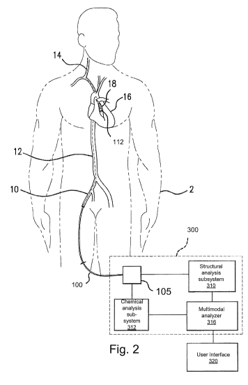
[00401 Fig. 2 illustrates an exemplary system for detecting and analyzing the
spectral
responses in two energy-form scanning.
[00411 The system generally comprises the catheter 100, a controller 3 00, and
a user
interface 320.
CA 02720637 2010-10-01
WO 2009/124242 PCT/US2009/039449
9
[00421 In operation, first the guide wire and then the catheter 100 are
inserted into the
patient 2 via a peripheral vessel, such as the femoral artery 10. The catheter
head 112 is then
moved to a desired target region, such as a coronary artery 18 of the heart 16
or the carotid
artery 14. This is achieved by moving the catheter head 112 up through the
aorta 12, riding on
the guidewire.
[00431 When at the desired site, NIR radiation is generated, in one
embodiment. In
preferred embodiment, a tunable laser in the chemical analysis subsystem 312
generates a
narrowband optical signal that is wavelength scanned over a scan band in the
NIR, covering
one or more spectral bands of interest. In other embodiments, one or more
broadband sources
are used to access the spectral bands of interest. In either case, the optical
signals are coupled
into the single mode delivery fiber 122 of the catheter 100 to be transmitted
to the optical
bench 118.
[00441 In other examples, reflectances are measured. This is based on the
discovery that
lipid-rich plaques are "brighter" than other plaques, and blood is typically
"darker" than tissue
in the NIR. So, just a brightness measurement, corrected for blood depth,
sometimes yields
adequate accuracy for detection.
[00451 In the current embodiment, optical radiation in the near infrared (NIR)
spectral
regions is used for spectroscopy. Exemplary scan bands include 1000 to 1450
nanometers
(nm) generally, or 1000 nm to 1350 nm, 1150 nm to 1250 nm, 1175 nm to 1280 nm,
and 1190
nm to 1250 nm, more specifically. Other exemplary scan bands include 1660 nm
to 1740 nm,
and 1630 nm to 1800 run.
[00461 However, in other optical implementations, broad band signals, other
scan bands,
or single frequency excitation signals appropriate for fluorescence and/or
Raman spectroscopy
are generated by the chemical analysis subsystem 312. In still other
implementations, scan
bands in the visible or ultraviolet regions are used.
[00471 In the current embodiment, the returning light is transmitted back down
multimode
collection fiber 123 of the catheter 100. The returning radiation is provided
to the chemical
CA 02720637 2010-10-01
WO 2009/124242 PCT/US2009/039449
analysis subsystem 312, which can comprise one or multiple optical detectors
or
spectrometers.
[ 0 0 4 8 ] The chemical analysis subsystem 312 monitors the response of the
detector, while
controlling the source or tunable laser in order to resolve the spectral
response of vessel walls
104 including a target area, typically on an inner wall of a blood vessel 18
and through the
intervening blood or other unwanted signal sources. This spectral response is
further spatially
resolved as the catheter head is rotated and pulled back through the vessel
18.
[00491 As a result, the chemical analysis subsystem 312 is able to collect
spectra. When
the acquisition of the spectra is complete, chemical analysis subsystem 312
then provides the
data to the multimodal analyzer 316.
[00501 The structural analysis subsystem 310 uses the information from the
ultrasound
transducer 120, in one embodiment, to generate one or more structural
measures. In other
examples, these structural measures are generated by an OCT, sonar
rangefinding, or other
structural analysis subsystem 310. The structural analysis subsystem 310
produces structural
information, such as structural measures, which are also spatially-resolved
with respect to the
vessels as the head 112 is scanned through the vessels 18. This structural
information, such as
structural measures, is provided to the multi modal analyzer 316.
[00511 In more detail, the structural analysis subsystem 310 comprises the
drive electronics
for driving the ultrasound transducer 120 and analyzing the response of the
transducer 120 to
determine the structural measure of interest in a IVUS-type system. In other
examples, where
the second energy source is an OCT system, the structural analysis subsystem
310 is often an
interferometer that resolves the phase or coherence of the light returning
from the scanning
catheter 112.
[00521 Generally, the analyzer 316 makes an assessment of the state of the
blood vessel
walls 104, which is presented to the operator via interface 320. The collected
spectral
response is used to determine whether each region of interest of the blood
vessel wall 104
CA 02720637 2010-10-01
WO 2009/124242 PCT/US2009/039449
11
comprises a lipid pool or lipid-rich atheroma, a disrupted plaque, a
vulnerable plaque or thin-
cap fibroatheroma (TCFA), a fibrotic lesion, a calcific lesion, and/or normal
tissue.
[00531 In should be noted that the apparent separation between the structural
analysis
subsystem 310, chemical analysis subsystem 312, multimodal analyzer 316, and
the user
interface 320 is provided to describe the various processing performed in the
preferred
embodiment and is thus only a notional separation in some implementations.
That is, the data
processing function of structural analysis subsystem 310, chemical analysis
subsystem 312,
multimodal analyzer 316 and the user interface 320 are performed by one a
single or one or
more computer systems in different implementations.
[00541 The analyzer 316 uses the structural analysis information from the
structural
analysis subsystem 310 to compensate information from the chemical analysis
subsystem 312.
Specifically, the structural analysis system produces a structural measure
that is used by the
multimode analyzer 316. Examples of structural measures include the
instantaneous distance
between the head of the catheter 112 and the blood vessels walls 104 (D(wall))
and/or the
thickness of the blood vessel walls. Another structural measure is the cap
thickness (ta) of the
lesion 102. This information is used to compensate information from the
chemical analysis
subsystem 312 such as serving as an input to a chemometric algorithm that has
dependencies
on the instantaneous or average distance between the catheter head 112 and the
blood vessels
walls 104. Still another structural measure is the lateral extents of plaques
in the blood vessel
walls.
[00551 The pullback and rotation and rotation unit 105 is used both for the
mechanical
drive to the scanning catheter 112 and also to couple the information or
optical signals from
both the IVUS and the NIR analysis portions of the catheter. Specifically, the
pullback and
rotation unit 105 drives the scanning catheter 112 to rotate and withdraw
through the outer
sheath 114.
[00561 Fig. 3 is a flow diagram illustrating the operation of the multimodal
analyzer 316 in
one embodiment.
CA 02720637 2010-10-01
WO 2009/124242 PCT/US2009/039449
12
[ 0 0 5 71 Specifically, the NIR spectral response 410 is produced by the
chemical analysis
subsystem 312. Structural information 413 is further obtained from the
structural analysis
subsystem 310.
[00581 Depending on the implementation, the structural analysis information
413 and the
chemical analysis information 410 are produced during the same or different
scans of the
scanning catheter 112. For example, in one implementation, the chemical
analysis information
410 produced by the NIR analysis and structural information 413 produced by
the IVUS
analysis are captured simultaneously while withdrawing and rotating the
scanning catheter 112
through the blood vessels 104. In other implementations, the chemical analysis
information
410 produced by the NIR analysis and structural information 413 produced by
the IVUS
analysis are captured during different pullback and rotation operations of the
scanning catheter
112. Then the chemical analysis information 410 data set produced by the NIR
analysis and
structural information 413 data set are spatially aligned with respect to each
other. This
alignment includes compensation for the offset distance D(offset) between the
IVUS
transducer 120 and the optical bench 118, see Fig. 1.
[ 0 0 5 91 This structural information is used in step 412 to determine
whether or not the
instantaneous, i.e., spatially resolved, NIR spectral signal was obtained from
a distance of
greater than 3 millimeters between the head of the scanning catheter 112 and
the blood vessel
wall 104.
[ 0 0 6 0 If the distance was greater than 3 millimeters, then a real time
update is performed
on preprocessing algorithms. In one example, such preprocessing algorithms are
described in
U.S. Patent Publication Number is US 2004/0024298-Al, Publication Date
February 5, 2004,
entitled Spectroscopic Unwanted Signal Filters for Discrimination of
Vulnerable Plaque and
Method Therefor. This application is incorporated herein by this reference in
its entirety.
Specifically, these preprocessing algorithms process the near infrared
information differently
depending upon the distance between the catheter head 112 and the blood vessel
wall 104
when the information was obtained.
CA 02720637 2010-10-01
WO 2009/124242 PCT/US2009/039449
13
[00611 In step 416, a discrimination model is selected based upon the 0 to 2
millimeters
distance. The prior preprocessing step corrects dataset generated at greater
than 3.0 mm such
that they can not be analyzed with a discrimination model based on 0-2 mm
distances.
[00621 In more detail, one of five thresholds 422, 426, 430, 434, 438 is
applied based upon
a more the precise determination of the distance between the catheter head 112
and the blood
vessel walls 104 produced by the structural analysis 413. That is, for each
location along the
vessel wall, the corresponding NIR data are processes according to the
distance between the
catheter head 112 and the wall when the data were obtained by reference to the
structural
analysis information 413. In the examples, the granularity for the different
thresholds is less
than 0.5 mm (step 420), 0.5-1.0 mm (step 424), 1.0-1.5 mm (step 428), 1.5-2.0
mm (step 432),
and 2.0-2.5 mm (step 436). The data at each location along the wall is then
processed using a
separate one of the one of five thresholds 422, 426, 430, 434, 438.
[00631 Thus, based upon the distance between the catheter head 112 and the
vessel wall
104 when each NIR spectral signal is obtained, a different threshold is
applied. The
application of the threshold is used to determine whether or not there is a
high probability of a
thin cap atheroma or not, in one example in step 440.
[00641 Fig. 4 shows an alternative embodiment. This similarly uses
preprocessing if the
blood distance is greater than 3 millimeters in step 414. Then based upon the
distance between
the catheter head and the vessel walls when the data were obtained, different
local models are
applied in steps 510, 512, 514, 516, 518. These are chemometric models that
are used to
assess the NIR spectral signal 410.
[00651 Here, IVUS blood depth information is used to improve prediction
accuracy.
Different chemometric prediction models 510, 512, 514, 516, 518 are built for
different blood
depths: less than 0.5 mm (step 420), 0.5-1.0 mm (step 424), 1.0-1.5 mm (step
428), 1.5-2.0
mm (step 432), and 2.0-2.5 mm (step 436).
[00661 In some examples, the blood depths are determined "manually". The user
inputs
the blood depth after measuring the IVUS image.
CA 02720637 2010-10-01
WO 2009/124242 PCT/US2009/039449
14
[00671 In other examples, NIR prediction models are augmented with the IVUS
blood
depth information.
[00681 Fig 5 illustrates still another embodiment of the invention.
Specifically, this
illustrates that the point-by-point NIR analysis (Analysis (Pn)) of the blood
vessels walls is
compensated in each case by the instantaneous information from the IVUS or
first energy form
(distance Pn). In this way, adjacent points in the scan of the inner walls and
their different NIR
responses, (response of P 1) and (response of P2), are combined with the
instantaneous distance
to the vessel walls, (distance P 1) and (distance P2) when the NIR signal data
310 was obtained
to obtain distance compensated analyses Analysis (P 1) and Analysis (P2). In
this way, the first
energy form information is used at a very high level of granularity in order
to compensate the
NIR spectral signal information at the spatial resolution of the chemical
and/or structural
analysis modality.
[00691 While this invention has been particularly shown and described with
references to
preferred embodiments thereof, it will be understood by those skilled in the
art that various
changes in form and details may be made therein without departing from the
scope of the
invention encompassed by the appended claims.