Note: Descriptions are shown in the official language in which they were submitted.
CA 02720639 2010-10-01
WO 2009/124245 PCT/US2009/039452
SYSTEM AND METHOD FOR DESIGNING AND
FORMING A SURGICAL IMPLANT
Cross-reference to Related Application
This application is a continuation-in-part of co-pending U.S. utility
application Serial No. 12/246,581, filed on October 7, 2008, which is a
continuation-in-part of U.S. utility application Serial No. 12/098,375, filed
on
April 4, 2008, the disclosure of which is incorporated herein by reference.
Background
The present invention is directed to systems and devices related to the
design and formation of surgical implants such as surgical linking devices.
More particularly the present invention provides a system and devices for
forming or shaping a surgical implant to conform to two or more selected
attachment points (including surface anatomy) in a six degree of freedom
method for attachment.
Fixation systems for aligning, adjusting and or fixing, either partially or
rigidly, portions of a patient's bony anatomy in a desired spatial
relationship
relative to each other are frequently used in orthopedic surgery. For example,
in spinal surgery for repair or positional adjustment of the vertebrae, it is
often
necessary that multiple vertebrae are surgically manipulated. As spinal
surgery often requires the instrumentation of more bony elements than other
areas of orthopedic surgery, the linkage devices can be extremely challenging
to design and implant. Treatment for conditions such as scoliosis, spinal
injury, disk problems and the like often make use of spinal rod fixation
systems for positioning the vertebrae and supporting the spinal motion
segments.
A spinal rod needs to be oriented in six degrees of freedom to
compensate for the anatomical structure of the particular patient's spine and
the particular attachment points or methods for attaching the rods to the
vertebrae. In addition, the physiological problem being treated as well as
physician's preferences will determine the exact configuration necessary.
SUBSTITUTE SHEET (RULE 26)
CA 02720639 2010-10-01
WO 2009/124245 2 PCT/US2009/039452
on the size, number and position of each vertebra to be constrained, their
spatial relationship as well as the fixating means, such as pedicle screws,
used to hold the rods attached to each vertebra. The relationship of the
vertebrae will be different for each patient and the positioning of the
patient at
the point of installation of the rods. During surgery, the orientation of the
spine and vertebrae can be very different than the corresponding position of a
patient's upright posture. Rods are bent in one or more anatomic planes
measured by distance from each bend, angle of the bend and rotation in
relationship to other bend points in order to fit into two or more vertebral
anchors.
The bending of a spinal rod can be accomplished by a number of
methods. The oldest and most widely used method for bending rods
manually during surgery is a three-point bender called a French Bender in
which a bending pliers type device is manually operated to place one or more
bends in a rod. The French Bender requires both hands to operate and
provides leverage based on the length of the handle. While the device can
make it relatively easy to bend a spinal rod, the determination of the
location,
angle and rotation of bends using such a device is often arbitrary. Problems
can thus occur from bending a device and then rebending to fix mistakes
which impose metal fatigue or stress risers into a rod thus increasing the
risk
of a mechanical failure. Increased time in the operating room (OR) to achieve
optimum bending of the rod can increase the chance of morbidity.
Spinal rods are usually formed of stainless steel, titanium or other
similarly hard metal, and as such are difficult to bend without some sort of
leverage-based bender. In addition, since several spatial relationships have
to be maintained in using a French Bender, the process can take an
extremely long time and its use requires a great degree of physician skill to
accomplish an accurate final product. Even still it is difficult to achieve a
well-
shaped rod using the French Bender. Accordingly, various ways have been
attempted to overcome the limitations of the current technology.
A number of manual benders are described in the art. In US patent
5,113,685 issued May 19, 1992 to Asher et al, there is described an
apparatus for use in bending rods and plates to the spinal column comprising
CA 02720639 2010-10-01
WO 2009/124245 3 PCT/US2009/039452
an elongated bar with a variety of bending angles for bending more angles
than the French Bender. However, this device is hard to use and provides no
means for determining the six degrees of spatial relationship that each bend
must make. In US patent application 2006/0150699 published July 13, 2006
to Garner, et al, there is an instrument and method for bending rod using a
lever pliers type device having bearing surfaces. In addition, the angle of
bend can be determined by use of a gauge that indicates angle bend by
degree of grip movement. While this device may be easier to use, it does not
aid in determination of the other degrees of freedom either in calculating
them
or in making the final bends.
An automatic method designed for pre-surgical formation of spinal rods
is disclosed in US patent application 2005/0262911 published December 1,
2005 to Dankowicz, et al. An automatic series of shaping steps is "imposed"
on a rod from an input mechanism for producing the desired multi-dimensional
bent shape. One problem with this device is that it relies on a pre-surgical
determination of the points at which bends occur to determine the final shape
of the rod. While it is possible to anticipate where the anchors might ideally
end up and occasionally be correct, surgical implantation of attachment points
is as much art as science so a preformed rod may not be accurately produced
when compared to the anchor means as they are actually installed in the
spine. This can lead to a highly problematic circumstance in which the
surgical site has been opened and the surgeon has a rod that does not fit the
attachment points. Further disadvantages are that the device is large and that
some surgeons still would prefer a manual means of producing a rod during
surgery because of the ability to make minute adjustments based on feedback
during surgery.
Effort has been directed to computer-aided design or shaping of spinal rods,
but these efforts have been largely unsuccessful due to the lack of bending
devices as well as a lack of understanding of all the issues involved in
bending surgical devices. For example, an article entitled "A pilot study on
computer-assisted optimal contouring of orthopedic fixation devices,"
Computer Aided Surgery, 1999; 4 (6):305-13, indicated that overcoming these
CA 02720639 2010-10-01
WO 2009/124245 4 PCT/US2009/039452
problems would be difficult if not impossible.
Image guided surgical systems, for example, devices produced by
BrainLAB, as well as three dimensional digitizers are already in the art and
some are already FDA approved for use during surgery. These devices are
fairly commonly used by some physicians in the operating environment. By
moving the digitizer through space or inputting a particular point in space, a
map can be produced of spatial relationships. In U.S. Patent No. 6,400,131,
issued on December 31,2002 to Leitner et al., there is described a contour
mapping system applicable as a spine analyzer and probe. The device is
disclosed as being used to determine the curvature of the spine while
standing and contour mapping of the spine in the intact (non-surgical)
patient.
Accordingly, a means for designing and forming a surgical linking
device, especially for linking bony parts of the body, for use in a surgical
orthopedic procedure such as the attachment of a spinal rod, that is accurate,
quick and takes the various input characteristics into account for the
specific
implanted device as actually needed would be of great value during an
orthopedic implant surgery such as spinal surgery.
CA 02720639 2010-10-01
WO 2009/124245 5 PCT/US2009/039452
Summary
In one embodiment there is a system for shaping a surgical linking
device for attachment to a selected bony body structure having at least two
linking device attachment elements comprising:
a) a means for determining the relative spatial location of at least
one of the attachment elements and the bony structure;
b) a means for converting the relative spatial location into a digital
format;
c) a computer capable of receiving this digital format and using the
relative spatial location to determine one or more shape locations in the
surgical linking device, each shape location having one or more of a shape
angle and shape rotation at each one or more shape locations such that
shaping of the surgical linking device will enable the surgical linking device
to
attach to the bony body structure using the attachment elements; and
d) a means for delivering the determined shape information to a
computer output.
In yet another embodiment there is a surgical linking device on a
selected bony body structure comprising:
a) placing at least two linking device attachment elements on the
bony body structure at desired locations;
b) digitally determining the relative spatial location of at least one of
the bony structure and the attachment elements;
c) transferring the digitized information to a computer which
determines information of one or more of:
i) one or more of the location, angle and rotation of shapes
in a selected surgical linking device that could be made in order for the
linking
device to be attached to the bony structure using the attachment elements;
ii) one or more adjustments to the position of or addition to
the attachment elements that could be made so that a selected preformed,
partially preformed or a minimally shaped surgical linking device can be
attached to the bony structure with the attachment elements;
iii) one or more mathematical adjustments to the digitally
rendered position of the attachment elements so that the final shaped surgical
CA 02720639 2010-10-01
WO 2009/124245 6 PCT/US2009/039452
linking device, once attached to the bony structure, will correct or alter the
shape of the bony structure(s);
d) delivering the computer determined information to a computer
output;
e) using the information from the computer output to perform one
or more of:
i) selecting a preformed or partially preformed surgical
linking device;
ii) shaping a surgical linking device with a device that
measures one or more of the shape location, shape angle and shape rotation;
and
iii) adjusting the position of or adding to the attachment
elements; and
f) attaching the surgical linking device to the attachment elements.
Yet another embodiment includes a device for bending a surgical
linking device, in which the device is particularly suited for manual
operation,
comprising:
a) a lever for bending the linking device; and
b) at least two bend measuring means selected from the group
comprising: bend position measuring means, bend angle measuring means
and bend rotation measuring means.
Another embodiment of the invention includes a device for determining
the rotation for placing a bend in a surgical linking device comprising:
a) a circular gauge indicating the degrees of rotation; and
b) a means for positioning the device on the surgical linking device
or on a means for bending the linking device such that the gauge aligns with
any bends in the linking device.
Yet still another embodiment is a means for determining the selection
of a preformed surgical linking device for use in attaching to a selected bony
body structure having at least two linking device attachment elements
comprising:
a) a means for determining the relative spatial location of each
attachment elements;
CA 02720639 2010-10-01
WO 2009/124245 7 PCT/US2009/039452
b) a means for converting the relative spatial location into a digital
format;
c) a plurality of preformed surgical linking devices;
d) a computer having selected spatial information about the
preformed linking devices wherein the computer is capable of receiving the
digital format in b) and using the digital format to determine if one of the
preformed surgical linking devices fits the attachment elements and if there
is
none that fit, if one or more attachment elements could be adjusted in
relative
location such that one of the preformed surgical linking devices could be
selected and fit the attachment elements; and
e) a means for delivering the determined attachment elements
adjustments and selected preformed linking device to a computer output.
A further embodiment contemplates a method for placing multiple
bends with 6 degrees of freedom in a surgical linking device comprising:
a) establishing a starting point on the device;
b) holding the device relative to the starting point;
c) moving the device and measuring away from the starting point
to establish a second point on the device for placing a bend with 6 degrees of
freedom; and
d) repeating steps b) and c) using either the starting point or the
second point to hold from until the multiple bends are completed.
Another embodiment of the present invention is a process for producing one
or more shapes in a surgical linking device comprising:
a) a digital process for determining the desired spatial parameters
of the shapes to be produced; and
b) a shaping process linked to the digital process wherein the
shaping process applies the spatial parameters to the surgical attachment
device, in which the shaping process is particularly suited for manual
implementation in the surgical operating room.
In yet another embodiment, a method is provided for shaping a surgical
linking device for engagement to at plurality of attachment elements engaged
within selected bony body structure, each of the attachment elements having
CA 02720639 2010-10-01
WO 2009/124245 8 PCT/US2009/039452
an engagement portion for engagement with the shaped linking device, in
which the method comprises:
(a) providing digitized data for the location of the plurality of
attachment elements;
(b) determining a tolerance range corresponding to an acceptable
distance that the shaped linking device is from the engagement portion of
each attachment elements;
(c) developing a curve function to approximate the location of each
of the plurality of attachment elements;
(d) calculating the location of the linking device shaped according to
the curve function at the location of each of the plurality of attachment
elements;
(e) calculating an error based on the difference in the calculated
location of the linking device and the location of each of the plurality of
attachment elements;
(f) determining if the error exceeds the tolerance range and if so
determining a higher order curve function;
(g) when the error falls within the tolerance range, generating a
bend curve having a discrete plurality of bend points using the curve
function,
the discrete plurality of bend points being distributed at a predetermined
distance;
(h) reducing the number of bend points by eliminating certain bend
points and replacing the removed bend points with a straight line between the
next immediately adjacent remaining bend points;
(i) generating a revised bend curve with the remaining bend points;
and
(j) generating bending instructions to be performed on the linking
device by a bending tool at each of the remaining bend points.
In another aspect of the invention, a digitizer probe is provided that is
configured to temporarily mate with the head of an implant. The probe
includes a shaft accessible beyond the implant that can be used to fix the
location of the implant when determining the bending protocol for a rod, plate
or elongate member to engage the implant.
CA 02720639 2010-10-01
WO 2009/124245 9 PCT/US2009/039452
CA 02720639 2010-10-01
WO 2009/124245 10 PCT/US2009/039452
Brief Description of the Drawings
FIGS. 1a through 1d depict a surgical rod and various bends with 6
degrees of freedom.
FIG. 2 depicts three vertebrae each with a surgical rod attachment
screw.
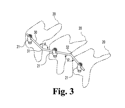
FIG. 3 depicts three vertebrae with a bent surgical rod attached to the
three rod attachment screws.
FIG. 4 depicts a front view of a rotation gauge for attaching to a
surgical rod.
FIGS. 5a and 5b depict surgical rods with ruled markings.
FIG. 6 depicts a small hand device for bending a surgical rod and
having a means for measuring location, rotation and angle bend.
FIG. 7 is a perspective view of a dual lever surgical rod bending device.
FIG. 8 is a side view of a dual lever surgical rod bending device.
FIG. 9 is a view of a dual lever surgical rod bending device with the
levers in the open position.
FIG. 10 is an end on perspective which allows view of the fulcrum
means.
FIG. 11 is a flow diagram of an embodiment for determining bend
information.
FIGS. 12a-h show a comparison between the IdealScrewPositions in
the XY (coronal) plane for an exemplary implant and the calculated positions
according to one example of the curve fitting approach of the present
invention.
FIGS. 13a-f show a comparison between the IdealScrewPositions in
the XZ (sagittal) plane for an exemplary implant and the calculated positions
according to one example of the curve fitting approach of the present
invention
FIG. 14 shows a comparison between a calculated bend curve and a
the curve after "smoothing" according to one aspect of the present invention.
FIGS. 15a-k shows a sequence of bend curves in the XY and XZ
planes with successive bend points eliminated to simplify the bend curve.
CA 02720639 2010-10-01
WO 2009/124245 11 PCT/US2009/039452
FIG. 16 is a representation of a graphical user interface (GUI) for
permitting user input and displaying information to the user during the
operation of the system of the present invention.
FIG. 17 shows the GUI of FIG. 16 after a bend curve has been
calculated for a particular spinal construct.
FIGS. 18a-d show a particular bend instruction as implemented using
the bending tool shown in FIG. 7.
FIG. 19 shows a rod bent according to the bending instructions
displayed on the GUI shown in FIG. 16.
FIG. 20 is a side view of a poly-axial implant with a digitizer probe
according to one embodiment of the invention engaged thereto.
FIG. 21 is a top view of the interface of the digitizer probe with the head
of the implant shown in FIG. 20.
FIG. 22 is a top view of an alternative interface of the digitizer probe
with the head of the implant shown in FIG. 20.
FIG. 23 is a side view of the interface of the digitizer probe with an
alternative implant.
FIG. 24 is a side view of a digitizer probe according to another
embodiment engaged to an implant.
CA 02720639 2010-10-01
WO 2009/124245 12 PCT/US2009/039452
Detailed Description
The present invention refers to a method for improving the shaping of a
surgical linking device, for example, by bending. First, by digitally
calculating
appropriate shapes such as bends in 6 degrees of freedom (three
dimensional) and then outputting that information to the surgeon or other
medical personnel or to a bending device, a linking device can be easily and
quickly shaped by casting, bending or the like. Second, a device is disclosed
for quickly and easily taking the input from a digitally calculated means, or
other similar means, and manually shaping a precisely bent or shaped linking
device. Accordingly, the time spent in surgery bending linking devices can be
greatly reduced thus improving the chances of a successful operation without
complications as well as reduce the cost of such an operation, for example,
from rebending or bending a second device. Since a significant portion of
time is spent in bending and in some cases rebending such devices, taking
minutes to an hour or more off the time to bend a linking device correctly is
an
important advance in the art.
While this invention is susceptible of embodiment in many different
forms, there is shown in the drawings and will herein be described in detail
specific embodiments, with the understanding that the present disclosure of
such embodiments is to be considered as an example of the principles and
not intended to limit the invention to the specific embodiments shown and
described. In the description below, like reference numerals are used to
describe the same, similar or corresponding parts in the several views of the
drawings.
It is understood that the term "coupled", as used herein, is defined as
connected, although not necessarily directly, and not necessarily
mechanically. The term "bending" refers to the act of forcing, or the like, a
linking device from a first position at a particular point to a second angular
or
curved position at that point in three dimensional space. Six degrees of
freedom are considered in bending a particular device once the location of the
bend is determined. In general, once the position of the placement of a bend
is determined, then the angle of the bend and in many cases the rotation
about a central axis may also be determined. In many cases a simple angular
CA 02720639 2010-10-01
WO 2009/124245 13 PCT/US2009/039452
shaping is sufficient while in others, such as is often the case for surgical
rods, a rotation off axis is necessary.
The bending is exemplified in the drawings which explanation follows.
As used herein "shaping" refers to not only bending but other methods of
taking the 6 degrees of freedom information generated with the present
invention and producing a shaped device. In addition to bending, the use of
extrusion, casting, deformation, molding and the like could be considered a
means of shaping a particular device with the information generated herein.
See U.S. Patent 6,749,614 issued June 15, 2004 to Teitelbaum, et al., for an
example of such material which could be used to shape a linking device with
the present invention methods.
A "surgical linking device" as used herein refers to those devices used
during surgery to use to bind to a selected bony body structure to mend,
stabilize, move, reshape, correct deformities or strengthen such as
attachments made to bones. For example, surgical rods, surgical plates,
surgical transverse connecting rods, surgical wire or surgical cable and the
like are used in surgery to mend, stabilize or correct breaks, correct
deformities and the like in selected bones by attachment to two or more
attachment points. Such plates and rods usually are supplied straight in a
number of lengths or preformed arcs and must be bent to fit their intended
use. (See v2-Evren 2008 online catalog, www.v2evren.com.tr for examples of
vertebral rods and connectors as well as other orthopedic devices of a
surgical nature). Typically, these devices are made of titanium or other
extremely durable, stiff and difficult to bend material. Rigid materials such
as
titanium, commercially pure titanium, stainless steel, cobalt chrome and the
like could be used. Other materials include flexible materials such as made of
PEEK or other appropriate plastics, graphite or the like, bumpered systems
and devices in both mono and multi diameter versions. Where casting or
other shaping means are used, any rigid material suitable for surgical use in
these conditions can be used.
Additionally, useful are shape memory alloys, shape altering devices,
materials with varying stiffness, biological materials and any synthetic
material
with bioactive properties. In particular, the benefits of shape memory
CA 02720639 2010-10-01
WO 2009/124245 14 PCT/US2009/039452
materials could be magnified by the processes described herein, especially
when such processes are applied more than once on the same linking device.
The shape memory materials allow an initial shape based on the location of
the fixation points or facilitate rod implantation and final shape
determination
from the altered position. The linking device can then be used to alter the
orientation of the bony structure(s) to help achieve the results of surgery.
Other surgical linking devices could include plates attached to specific body
parts, both in the apendicular and axial skeleton, as well as cables and rigid
clamps used to affix to and alter teeth and their alignment.
The French Bender is the surgical instrument of choice today to bend
these materials but it does so without regard to being able to measure the 6
degrees of freedom of movement in any manner. Accordingly, the process of
bending a surgical rod with a French Bender is laborious and demanding,
requires some degree of artistry and frequently requires starting over.
A "linking device attachment elements" refers to a means attached to a
body structure designed to received the surgical linking device and hold it in
place. Surgical clamps and screws are common examples of these devices.
In the case of a surgical rod, a variety of surgical screws, bolts, and hooks
are
available to screw into the bone and or to hold the rods in place. These
include polyaxial screws, mono axial screws, fixed angle screws, iliac screws,
sacral screws, lateral mass screws, bolts, laminar hooks and pedicle hooks.
In additions, items such as staples, or plates that serve to hold one body
part,
can serve as an anchor to which a linking device can be affixed onto the spine
especially with anterior plating systems. All these systems can be used
together and further connect up to similar anchoring plates.
Connectors such as axial, lateral and transverse connectors are used
while locking screws are often used to hold the linking device in place. Even
further, the attachment elements could be devices added to the means to
change the attachment position. A screw attachment or "offset", for example,
could be used. In the practice of this invention, the devices and methods of
this invention anticipate use when there are at least two and frequently three
or more attachment elements corresponding to each surgical linking device.
Multiple differing types of attachment elements could be used in a single
CA 02720639 2010-10-01
WO 2009/124245 15 PCT/US2009/039452
installation. In addition, in the case of plates, the attachment elements may
be installed after the shaping of the plate based on the shape of the plate
rather than the other way around.
"Determining the relative location of each attachment elements and
bony structure" refers to understanding the spatial relationship between the
bony structure and any points of attachment so that a linking device such as a
surgical rod can connect between the attachment points given the proper
shape of the device. The relative location can be obtained with currently
available image guidance devices such as three dimensional digitizers (such
as the Polhemus Patriot) which can be used simply by engaging the device at
several attachment points or along the bony structure and letting a computer
in the device or elsewhere digitize the information. A partially manual method
could be done, for example, by photographic means such as x-ray or regular
photography and the spatial relationship determined away from the patient.
Such a method might need a plurality of photographs but given this
explanation is well within the skill in the art.
From the determination of the relative spatial location, the information
can easily be digitized either automatically, as is the case with the three
dimensional digitizer, or by entering hand calculated information into a
computer or the like which then stores the information digitally. Either way,
the information is converted into a digital format which a computer is capable
of manipulating. Other devices could be optical, EM, image guidance
tm
systems, Shape Tape , ultrasound, cat scans, and other radiographic
devices. The key is that information needs to be gathered about spatial
relationships and that information is capable of being obtained in a variety
of
ways. It is clear that the enumerated means or any other means which
achieves the determination of the spatial relationship can be used by one
skilled in the art. In some embodiments the expression "determining the
relative location of each attachment elements and bony structure" may also
refer to making multiple determinations after adjustments to the installation
or
attachments means are made. One skilled in the art will know when and how
to make such multiple determinations.
Since structures such as the shape of the patient's anatomy, bone
CA 02720639 2010-10-01
WO 2009/124245 16 PCT/US2009/039452
structure, other devices in the area and the like may also need to be
considered when determining the bend profile, the invention further
contemplates that other structural information may also be created in a
digital
format for transfer or use by a computer. In one embodiment, the contour or
structure can serve as the input by itself, such as with any plating system,
where the input is the topography of the surface of the body part, with this
input being used to guide shaping the implant. The attachment points are
then driven through the plate after the plate is shaped, not prior, in as much
as the information could be determined solely from surface anatomy and not
the attachment points.
A computer such as a laptop, hand held device, desktop or other
computer device can receive the relative location of the attachment elements
and/or the bony structure in a digital format. The computer then programmed
with the spatial information can determine the best way to shape, bend or the
like, the linking device in order to fit the attachment elements. This
determination of bends also takes into consideration the fact that other
structures or the shape of the structure being attached to may be in the way.
For example, in a spinal procedure, the shape of the vertebrae bones must
also be considered.
The computer can be programmed to accommodate any number of
parameters in determining the output or the final shape of the linking device.
In this way, the goals of surgery can be assisted through the alteration of
the
shape of the linking device. Whereas in one embodiment, the shape dictated
by the information above and not altered further could be used to create the
linking device, further alterations in the device's shape can help to address,
straighten, or alter abnormalities in alignment of the body part(s), create
lessen or eliminate deformities, reduce or impose changes in alignment or the
addition or elimination of stresses. It is possible to couple the changes in
different planes or simply apply correction in one plane, rather than in
another
orthogonal plane. These modifications of the shaping information that is
outputted can be obtained through various means -visual, anatomic, guided
by radiographs (intraoperative, preoperative, positioning films, etc.), guided
by
CA 02720639 2010-10-01
WO 2009/124245 17 PCT/US2009/039452
the material properties of the linking device and the plasticity and/or
relative
location of the body part(s) being altered.
The computer need not have direct interaction with the device used for
bending, in one embodiment. In other embodiments, it could input the
information directly to the shaping device such as to a screen or other means
such as to set the dials prior to shaping. The computer defines
mathematically from the spatial location of the attachment elements and the
bony structure of the body, the heads of screws, surface of that bony body
part and the like, a curve which approaches these points in three dimensional
space within the requirements and capabilities of the selected surgical
linking
device. The determined information can be used to select a specific device,
to place bends in an unbent or pre-bent device (or shape as needed) or to
adjust the attachment elements as desired. In addition, a number of different
shape solutions could be accommodated such that the surgeon can use
personal judgment in selecting the best shape solution.
The computer could further customize the output of the bend
information. It could minimize the number of bends if desired (for example,
with a quicker zigzag type design with greater bend angles at fewer bend
points but with potentially greater stress risers). In other embodiments it
could
increase the number of bend locations to create a smoother design, since the
more bend points the smoother the bend. One could limit the program or the
device to specific angles so that all angles would be above, at or below a
particular value. It could also limit the choices to incremental choices such
as
every 5 degrees of bend or rotation or distances to a few millimeters. A
simple design connecting points could be achieved as could a more complex
design as desired. The computer could determine the size of the device, can
determine if the attachments means can be adjusted or added to with
offsetting devices (and therefore increase or decrease the number of bends to
attach the points). In one embodiment, the program can be used to see if the
attachment points can be used with a pre-bent device either without
modification or with adjustment of the attachment elements or the addition of
spatial offsetting devices. The computer could also pick shapes that simplify
the shape of the linking device or improve its biomechanics.
CA 02720639 2010-10-01
WO 2009/124245 18 PCT/US2009/039452
A first step in bending a linking device is to determine a bend location.
The bend location is a point on the linking device where the bend will occur.
It
can be measured from a starting point, for example, 1.5 cm from the distal
end of a surgical rod, or it can be determined by selecting from a set of
fixed
points on the device. For example, ruled markings every centimeter on a rod
or other device could be marked as point 1, 2; or as 1 cm, 2cm, etc., and the
output of the computer deliver the fixed point. In another embodiment, the
device is held in place and moved a given distance from the point held as a
reference starting point.
The bend angle is the degrees that the device is bent away from a
particular axis or plane. The bend can be accomplished as a single bend or it
can be a multiplicity of bends as described above. In general, the bends will
be from just greater than zero to 180 degrees off of straight. In many
embodiments the bend angle is 90 degrees or less. In general, the maximum
bend angle will be determined by a number of factors including the particular
use, the surgeon's typical practice, the materials employed and the like. In
addition, the angle of rotation off of the direction the device was going
could
be determined. So, for example, a surgical rod could be angled from zero to
360 degrees off of the zero axis of the original direction of the rod in
addition
to the bend. Thus a bend of, for example, 45 degrees with a rotation of 15
degrees, 2 centimeters from a starting point could define a particular bend
output. The distance rotation and bend angle after determination is then
delivered to a computer output. The output can be a paper output, a GUI
(Graphic User Interface) or the like, such that a user can read the
information
and begin the process of bending a device. In one embodiment, the
information is delivered directly to the bending device.
The means for placing a bend in the surgical linking device can, in one
embodiment, be accomplished by one or more manual devices. Hand
measuring distance, a rotation disk (as shown in FIG. 4), and then a bending
device for bending to an angle could allow the bending with three interactive
devices. Likewise, the device shown in FIG. 7 could be used to set all three
parameters on one device. For a device that only needs 4 degrees of
freedom, the computer needs only produce distance and bend angle and the
CA 02720639 2010-10-01
WO 2009/124245 19 PCT/US2009/039452
various devices above either singly or one single device could be used.
Rotation in this case could be set at zero. Further, such as in the case
wherein the output of the system determines that a pre-bent rod could be
used, the output of all of the parameters except distance could be zero. The
system could simply determine which linking device that should be chosen,
with or without the need to further manipulate the screw locations or add
additional offsetting devices. In this case, no bends may need to be made.
Surgically, the method for installing the surgical linking device on a
body bony structure using the present invention, in one embodiment, could be
started by placing at least two linking device attachment elements on the body
structure at desired locations. Then the spatial relationship of the
attachment
elements could be determined in a digital manner. The digitized information
would be transferred to (including calculated by) a computer which determines
one or more of the following: one or more of the bend location, bend angle
and bend rotation such that upon making the bends the device will fit the
installed attachment elements; it could also determine that one or more
adjustments or additions to the position of the attachment elements could be
made so that one could select a preformed or partially preformed device or
that a device could be bent with fewer bends or no bends at all to fit the
attachment elements. The computer calculates and delivers the information
to a computer output. The output could be used to perform one or more
functions during surgery, namely selecting a preformed or partially preformed
surgical linking device; placing one or more bends as described above in the
device or adjusting the position of the attachment elements or placing an
addition to the attachment elements. After the proper selection and bending
the surgical linking device is attached to the attachment elements.
The advantages and uses of the computerized means for determining
the shape of a surgical linking device are several. It allows for the
facilitated
implantation of preformed whole rods or segments, and the ability to define
the size and shape of the component pieces of a multi-component linking
device. The linking device can aid a surgeon in the formation of the desired
end result rather than the situation as confronted. The linking device can be
designed and formed based on the intersection of this desired end result, the
CA 02720639 2010-10-01
WO 2009/124245 20 PCT/US2009/039452
current position of the anatomy, and the location of the affixing points. This
can be used to control the reduction of fractures and deformities by defining
the amount to translation, rotation and or angular correction and altering the
shape of a linking device to achieve the result. Further, it can be used to
correct spondylolisthesis.
In another embodiment, this method could be used to define the
resultant rod and thus help form, obtain and/or hold the correction required
in
performing an osteotomy or other type of corrective technique used in
surgery. The linkage device can be implanted without any static load
imparted to the body, or with a predefined load which can aid in adjusting
deformities or set the location of a flexible system. One could determine how
the anatomy moves or has moved or changed, and one can determine the
amount of implant manipulation needed to gain the anatomical change
desired. (For example, using x-rays in the OR and comparing them to images
taken prior to surgery it is possible to figure out how much to alter the
shape
of the linkage device in order to achieve the straightness the patient can
physically achieve by bending). In one embodiment, one linkage device could
be made which would result in completely obtaining the desired end result. In
another embodiment, successive intervening steps could be made (i.e. -
multiple linkage devices incorporating successively greater amounts of
deformity correction) to allow a slower, more gradual correction of the
deformity. As all people's anatomy changes to some degree when lying in an
OR table versus the upright position, the present invention could be used to
account for this change.
Although in one embodiment, the rod can be formed quickly at the time
of surgery, this is not required. One could immediately implant or defer the
linking device implantation such as to let ongrowth or ingrowth occur then
implant the formed rod in a delayed fashion. Further, this system is ideal to
custom design large percutaneous implants. As well, it could be used to
design a transverse connector that joins two or more linking devices or any
other type of implant that could benefit from linking. Further, it can be used
to
accommodate an easy way to extend the linking device should this be
required in the future, as the end configuration and angle of one embodiment
CA 02720639 2010-10-01
WO 2009/124245 21 PCT/US2009/039452
of this device is know at the time of production and therefore this additional
step (which is useful typically in a delayed fashion months to years later)
could further be incorporated.
Bending is preferably accomplished manually at the surgical site by
known means but in the alternative can be accomplished with novel devices
of the present invention. Novel bending devices all comprise at least one
lever, namely in the form of a bar or long arm that can be used to bend an
object around a particular pivot point. With one lever the object to be bent
is
forced with the aid of the lever. In other embodiments, there is a pair of
levers
that can bend around a fulcrum that is a point or device that will aid in
bending
the device around.
Devices such as the French Bender have no means for determining
any of the bend parameters discussed above when bending a surgical linking
device. The present bending device includes means for determining at least
two of those parameters. In one embodiment, the two parameters are
location and bend angle. In another embodiment, the device measures
location, bend angle and bend rotation. Each lever can have a handle
disposed at a distal end to aid in grabbing the lever and leveraging it during
use.
The means to measure the spatial parameters can measure a
continuous location or angle or in other embodiments the measurement
means can measure incrementally (i.e.,non-continuously). So, for example,
the location can measure in half centimeter, one centimeter or other
increments, while the angle of bend or rotation could be measured in five
degree increments or the like. Continuous measurement or click stop
measurement could be used with each measuring means individually or
mixed as desired. Greater accuracy may be obtained by continuous rather
than incremental movement, but the choice would be up to the user and type
of bender device employed.
In addition, the device may be capable of fixedly holding the linking
device. In this manner the bending device can use another means to
advance the linking device to the next bending location based on the
continuous or click stopped measuring means. By fixedly holding the linking
CA 02720639 2010-10-01
WO 2009/124245 22 PCT/US2009/039452
device, the measurements can be made accurately from a specific starting
point adding a new starting point after each bend or using the original
starting
point. For example, a bend could be put at one centimeter and three
centimeters from a starting point. In another embodiment, a bend is at the
starting point and the next bend a fixed distance from the starting point. In
another embodiment, by holding the linking device the linking device could be
advanced based on ruled markings on the linking device instead of ruled
markings on the bending device. Where on the bending device, there could
be regular stop positions that are fixed or in the alternative, continuous
adjustment of distance.
In general, one of the embodiments of the present invention is the
process for producing bends in a surgical linking device which is comprised of
two separate processes linked to each other. The first process is the digital
process for determining the spatial parameters of one or more bends. The
second process is the manual process of shaping a surgical linking device
that applies the location, angle and rotation parameters determined in the
first
process. The complete linking of these two processes is facilitated by the
introduction of the novel device of the present invention. The link can be the
surgeon or other individual who takes the computer output and applies the
result to the linking device, whether manually or by an automated bender or
contouring device. For example, in another embodiment, the process or
method for the determination of the linkage device, including selection,
alteration of fixation points or location, etc., could be applied to any of a
host
of novel devices which would be necessary to help in the formation of the
actual device. This would be ideal as materials used in orthopedic surgery
change over time, such as described in Patent 6,749,614 issued June 15,
2004 to Teitelbaum, et al..
Now referring to the figures, FIGS. 1a through 1d depict various bends
in a surgical rod linking device. FIG. 1a depicts a rod with a first bend 11
and
second bend 12. This depiction has the rod 10 lying in one plane and the
distance between bend 11 and bend 12 is shown as D. By indicating a
distance D from bend 11, one can obtain the location of the second bend 12.
The starting point for measurement could be either from point 13, the first
rod
CA 02720639 2010-10-01
WO 2009/124245 23 PCT/US2009/039452
end or bend 11. The starting point for bend location can stay with the
original
point for subsequent bend location determinations or can move with each
bend location determination. So for example, bend 12 could be the starting
point for the next bend location determination. In FIG. 1b, a single bend 15
is
shown with an angle A. The angle A is the second determined parameter of
the present invention. FIGS. 1c and 1d depict a bent rod with at least one
bend that has been rotated R degrees from the initial plane of the rod.
Second end 19 is also depicted and in FIG. l d the view is head on to the
middle section of the bent rod 10. While a surgical rod 10 is depicted for
clarity, a surgical plate or other surgical linking device could also be
oriented
and bent or shaped in a similar manner.
FIG. 2 depicts body structure vertebrae 20 laid out in perspective view.
Each vertebrae 20 has had attachment elements, vertebral screw 21 installed
for the purpose of installing a surgical rod. Note that while normally rods
are
installed in pairs one set of screws 21 is shown for simplicity's sake.
FIG. 3 depicts a bent surgical rod 30 which has been attached to the
attachment elements 21. Also depicted is bend angle A and rotation angle R
at which the rod has been bent to accommodate the positions of the
attachment screws 21.
FIG. 4 is a rotation gauge 40 which may be fitted on the end of or
around a surgical linking device, for example, the rod 10 depicted in FIG. 1.
The rod 10 fits into hole 41 and then if the rod is rotated to the degree
markings 42, a rotational bend of a selected angle can be achieved. This
device could be fixedly attached to a bending device as further taught herein.
In FIG. 5a and 5b surgical linking rods 50 are shown. These rods are
normally cylindrical but first end 51 is squared off to accommodate a tool or
grabbing means or the like. Any number of other end configurations could, in
addition, be used that can be firmly held or gripped. The gauge 40 from FIG.
4 could also be attached to this end. These surgical linking rods 50 also show
either distance markings 55 to indicate the distance for a bend location. In
the
case of FIG. 5b rotational markings 56 are available not only for distance
measurements but for rotational measurements as well.
CA 02720639 2010-10-01
WO 2009/124245 24 PCT/US2009/039452
FIG. 6 depicts a simple hand bending device 60. By squeezing
handles 61 and 62, rod 10 can be bent around a fulcrum (not seen). The rod
is not held in place but the rod 10 is moved and by matching distance
markings 63 on device 60 with rod distance markings 55 a clear location on
the rod 10 can be determined. Rotational gauge 40 is installed and by
manually rotating the rod 10 one can obtain a desired rotation. While the
rotation is marked in intervals, this embodiment allows free rotation of rod
10
thus infinite rotational angle. The bend angle is measured by angle gauge 65.
Angle gauge 65 measures the angle based on how close the handles 61 and
62 approach each other during the operation of bending rod 10.
FIG. 7 is a perspective view of a more detailed bending device 70 with
less manual manipulation of the rod 10. A first lever 71 is shown as is lever
handle 73 designed for grabbing the lever 71 manually. Likewise, lever 72 is
shown with handle grip 74. Grip 74 has rod pass through 78 so that an
infinitely long rod 10 can be used with this particular handle as well as
steady
the rod during the bending process with bender 70. The user of the device
grabs both handles and opens the handles to bend the particular surgical rod
10 by picking an angle on the angle gauge and closing the handles 71 and 72
together. The device in other embodiments could be produced to bend the
rod during the handle opening movement as well. The rod 10 moves through
mandrel 80 and in between moving die 81 and fixed die 82. A better view of
the dies is in FIG. 10.
The surgical rod is bent between the two dies 81 and 82. Gauges on
the device allow the user to manipulate the surgical 10 rod in order to
determine bend position, bend angle and bend rotation. The surgical rod 10
is held in place by a clamping element, such as collet 75. By sliding slide
block 76, along handle 72, the surgical rod 10 can be moved proximally and
distally in the bending device 70. Position is measured by a series of ratchet
or click stops 77 at regular intervals along handle 72. Each click stop 77 is
a
measured distance along the handle 72 and thus moving a specific number of
click stops 77 gives one a precise location for the location of a surgical rod
10
bend.
The bend angle is measured by using angle gauge 85. Gauge 85 has
CA 02720639 2010-10-01
WO 2009/124245 25 PCT/US2009/039452
ratchet teeth 86 spaced at regular intervals. Each ratchet stop represents
five
degrees of bend angle. Thus the user can bend a surgical rod 10 in five
degree increments with the particular bend angle gauge 85 as the handles 71
and 72 are opened and closed. The bend rotation is controlled by a dial in the
form of collet knob 90. By rotating collet knob 90 either clockwise or
counterclockwise the user can set a particular rotation angle. The collet knob
90 is marked with regular interval notches 91 but this particular embodiment
is
continuously turn able and thus has infinite settings. Once a user turns knob
90 the user can set the knob 90 at a particular marking 91 or in between or
the like to determine a particular angle rotation to a high degree of
accuracy.
In this particular embodiment, once the rod 10 is locked in place with
collet 75 if there is enough room on the lever 72 to move the slider 76
distally
or proximally then the rod 10 can remain fixedly attached to collet 75. Should
a longer area need to be bent, then the rod 10 can be unlocked moved and
relocked and measurements start from the new position. Merely adding the
positions together using the information supplied by the computer output
would be an easy task with the present invention.
FIG. 8 depicts the bending device 70 in a side view. In this view one
can clearly see the rod 10 has bend 92. FIG. 9 shows a side view wherein
handle 71 is open in preparation of making a second bend in rod 10. Bend
gauge window 96 shows bend angle pin 97 which has engaged 2 teeth 86 in
preparation for placing the second bend. As can be seen in this view the rod
10 has moved distally since slider 76 is in a more distal position than shown
in
FIGS. 7 and 8. First bend 92 has moved distally as well and upon closing of
levers 71 and 72 a second bend will be placed in rod 10.
FIG. 10 shows a head on view of the device 70. In this view, the rod
10 can clearly be seen in bent position between moving die 81 and fixed die
82. The moving die 81 allows for free movement of rod 10 and the fixed die
82 allows for relatively easy bending of rod 10.
FIG. 11 depicts a flow chart of a particular embodiment of the operation
of the computer means in combination with the device of the present
invention. The first step 110 in the process is the installation of a linking
device attachment elements to a body structure. In other embodiments, for
CA 02720639 2010-10-01
WO 2009/124245 26 PCT/US2009/039452
example for use with a surgical plate, the first step is to determine the
surface
spatial relationship of the bony structure and then using that spatial
information to determine the shape of the surgical plate. Once the plate is
placed on the bony structure attachment elements are positioned through the
plate and into the bony structure. The linking device such as screws for use
with surgical rods, which to some degree adjustable then determines where
the linking device will be positioned.
The next step 111 is the determination of the spatial relationship of the
attachment elements into a digital format. This is done not only taking into
account the position of attachment, but also taking into consideration any
body structures which may intervene in the process. It would not be useful if
a part of the vertebrae were in the way of a particular bend solution because
the resulting bent rod would not fit the attachment points because of body
structure interference. One skilled in the art could easily make the
appropriate adjustments to the computer calculation based on the disclosure
herein.
Next, the computer with the possession of the digital format determines
the bend parameters and or the device attachment elements adjustments in
step 112. This step may also include the selection of a particular linking
device, the size it needs to be, or to select from a list of pre-bent linking
devices. Once a linking device is selected from the computer output
parameters, the linking device is then, if necessary, bent or shaped and or
the
attachment elements adjusted in step 113. After the appropriate bends have
been made, the linking device is attached to the attachment elements in step
114.
Step 112 of the flowchart of FIG. 11 entails first determining a
mathematic representation of a linking device (such as a rod or a plate) that
will fit each of the attachment elements in situ. Thus, according to one
embodiment, a software program implements a curve fitting algorithm that is
adapted to approximate a smooth curve spanning between the attachment
points, with the curve falling within an acceptable error at the location of
each
attachment point. The software program starts with the digitized data
establishing the three-dimensional position of each attachment point. In order
CA 02720639 2010-10-01
WO 2009/124245 27 PCT/US2009/039452
to simplify the curve fitting protocol, the present invention contemplates
that
the three-dimensional data are used to establish the attachment points in two
orthogonal planes - the sagittal or XZ plane, and the coronal or XY plane. As
is known in this field, the sagittal plane corresponds to a vertical plane
passing through the spine from the front to the back of the patient. The
coronal plane is perpendicular to the sagittal plane and extends side to side
through the patient. The division of the 3D coordinate system into two 2D
planes may be used in one embodiment as described in detail herein, but
would not be required for the functioning of the system. In another
embodiment, a single 3D curve fitting program is employed.
It is thus an object of the software program to derive a curve in each
plane that fits the actual position of the attachment points in situ. In most
cases, the curves in the sagittal and coronal planes are complex, meaning
that the curves will typically incorporate multiple inflection points. Thus,
it
should be understood that a straight line or even an arcuate line will usually
be inadequate to fit the true position of the attachment points, especially as
the number of attachment points increase. It can be appreciated then that a
first or second order polynomial expression for a curve in either plane will
rarely be sufficient to model the three-dimensional representation. It can be
further appreciated that an exact curve fit is unlikely, even if the
polynomial is
extended to a very high order.
The present invention accounts for these difficulties by incorporating an
acceptable error between the actual three-dimensional location of an
attachment point and its mathematical representation. This error is
acceptable if kept within certain constraints because of the ability of the
surgeon to manipulate the linking device, inherent characteristics of the
attachment points and linkage device and even the spine when completing
the spinal construct. For instance, where the attachment point is a bi-axial
or
multi-axial bone screw, the head of the screw can be toggled or pivoted so
that the rod-receiving channel of the screw can be oriented to receive a
linking
device, such as a spinal rod. In addition, in many attachment devices, the
interface point is the channel of a yoke or saddle-shaped structure, which
provides variability to the angle between the rod and the head of the bone
CA 02720639 2010-10-01
WO 2009/124245 28 PCT/US2009/039452
screw. In some devices the screw permits variation in vertical orientation
relative to the bone. Each of these factors contributes to an acceptable error
or tolerance. Other factors that may be considered in deriving the acceptable
error include the material of the bone screw and the liking device, the cross-
sectional shape of the linking member, the distance between attachment
points, and the size of the bone screw and the linking member.
In accordance with the dual plane approach of the present
embodiment, the error, or more appropriately tolerance, is established in each
plane. For illustration purposes these tolerances can be designated
ToleranceXY and Tolerance XZ. The ToleranceXY in one embodiment can
be given by the expression:
ToleranceXY = tan(0.5 * MaxHeadAngle) * (distance from the end of
the fixed shaft of the screw to the ideal rod location).
The term "MaxHeadAngle" corresponds to the maximum angle through
which the head of a bone screw can toggle or pivot. For a typical polyaxial
screw, that angle can be in the range of 50 . The last term in the expression
essentially corresponds to one side of a triangle indicating the depth of the
saddle of the screw that can accommodate the linkage device.
The ToleranceXZ corresponds to the inherent amount of vertical
translation that the screw head can accommodate. For example, a screw
head having a deep yoke channel, the vertical translation may be as much as
several millimeters and in some systems, especially in those in which the
heads resemble posts, the number can be much more.
The two tolerance values, ToleranceXY and ToleranceXZ, are
determined for each attachment point - i.e., for each bone screw. The
tolerance values provide the measure for determining whether a particular
polynomial representation of the rod shape is sufficient. Again, these
tolerance values may take into account all or some of the factors discussed
above.
In accordance with one aspect, the curve approximation for each plane
starts with a first degree polynomial, the lowest order possible, and proceeds
to increase in order until a curve is developed that satisfies all of the
tolerance
values. Thus, an initial approximation for a curve fit starts with the
equation:
CA 02720639 2010-10-01
WO 2009/124245 29 PCT/US2009/039452
y = P1*x1 + P2 , where x and y define the location of the curve in the
coronal or lateral plane with y representing the left to right location of the
curve function and x corresponding to the head to foot location along the
length of the spine, and P; are coefficients.
A similar relationship is applied to find the value of z, namely the
location of the curve in the sagittal, or front-to-back- plane. Further, in
other
embodiments of the present invention, other curve fitting algorithms are used
to establish an equation for the linking device, all increasing in their
complexity as they minimize the error between the determined curve and the
attachment points.
Invariably, this first order polynomial will be insufficient to provide a
curve linking all of the attachment points. The order of the polynomial is
then
successively increased according to the following equation:
y = P1*XN + P2XN-1 + ... + PNX + PN+1.
For each N-th order polynomial, the error in the two planes (XY and XZ) is
calculated using a least squares approach. To determine this error, an
IdealScrewPosition value is obtained for each attachment point, namely
(xscrew, )/screw, Zscrew), which corresponds to the ideal location of the
spinal rod
within the head of the screw. In one embodiment, the Ideal Screw Position
values may correspond to the digitized data for each screw in situ, in a
manner as described above. In accordance with one aspect of the invention,
the two errors ErrorXY and ErrorXZ are defined by the distanced from the
calculated curve function to the IdealScrewPosition at two points on either
side of the Ideal Screw Position according to the following relationships:
ErrorXY=((ycurvel-ycurve2)*xscrew+(xcurve2-xcurvel)*yscrew+(ycurve2*xcurve1-
xcurve2*ycurvel ))
((xcurve2 -xcurve1)2 + (ycurve2 - ycurvel )2)112
E rrorXZ=((Zcurvel-Zcurve2)*xscrew+(xcurve2 -xcurvel)*Zscrew +
(Zcurve2*xcurve1-xcurve2*Zcurvel))
((xcurve2 -xcurvel)2 + (Zcurve2 - Zcurvel )2)112
If any of these error values exceeds the tolerance values
(ImplantToleranceXY or lmplantToleranceXZ), then the order of the
polynomial is increased and the least mean square function and error
calculations are re-run. In some embodiments, the tolerance values are
augmented by some small dimension, for instance 1 mm, to help simplify the
CA 02720639 2010-10-01
WO 2009/124245 30 PCT/US2009/039452
curve function and therefore eliminate bend points when the final bend curve
is created. In other words, it is typically desirable to reduce the number of
bends that are needed to fit the implant locations, especially when performed
manually. When applied otherwise, for example in embodiments using non-
manual bending devices or alternative materials that benefit from, reducing
the number of bends may not be required or considered as advantageous to
achieve a very smooth result. The augmented tolerance values can eliminate
some bends that might otherwise arise with a tightly toleranced curve
calculation.
In another aspect of the inventive procedure, steps may be taken to
ensure that the rod interaction with the head of the screw falls within a
predetermined angle. This predetermined angle is based on the value
MaxHeadAngle, which is described above as the maximum permissible angle
through which the head of the fastener/screw may toggle. It can be
determined that the angle at which the rod crosses the head of the screw is
defined by:
Rod2ScrewAngle = arcsin (Vscrew = Vrod), where the operator "=" signifies
the dot product of the two vectors corresponding to the orientation of the
screw and the approach angle of the rod.
In circumstances in which the rod crosses the head of the screw too acutely
(i.e., outside the boundaries set by MaxHeadAngle), the generated curve is
altered in the immediate region of the screw using a segmental rod
morphology which crosses within the correct range. Specifically, the curve
function is altered by an amount R so that the Rod2ScrewAngle function is
equal to half the value of MaxHeadAngle. In other words,
Vrequired rod = Vrod + (R X Vscrew), and
(1/2 x MaxHeadAngle) = arcsin (Vscrew ' Vrequired rod).
The curve is then altered across the small segment adjacent the
particular screw as follows:
D = Vrequired rod X (X2 - X1);
ylnew = y1 + D
y2new = y2 - D.
It is also contemplated that in using the least mean square approach
described above to determine the rod curve, some regions of the resultant rod
may conflict with the surrounding anatomy. In extreme situations, this
competition can be eliminated in part by simply defining any anatomical points
CA 02720639 2010-10-01
WO 2009/124245 31 PCT/US2009/039452
of concern at the same time that screw positions are determined. In other
words, the anatomy that must be avoided can be defined ab initio along with
the locations of the several bone fasteners/screws.
However, in the typical case, no strange anatomy is encountered. In
this instance, it is common for the bone fastener head to project from the
bony
anatomy in to which it is anchored by a certain distance, usually about 1 cm.
When the bend curve is defined, all that is required is that the resulting rod
position fall within a "safe region" which can be pre-defined as a few
millimeters above and below a straight line connecting successive
IdealScrewPositions. When the curve function would result in the calculated
rod position falling outside this "safe region", the curve is altered towards
the
straight line. In one specific embodiment, if any point along the calculated
curve that extends above a line defined by the slope M of a line connecting
screws at positions (xscrewl, yscrewl, Zscrewl) and (xscrew2, yscrew2,
Zscrew2) exceeds
a specified amount, the whole section of the curve (xcurvel, ycurvel, Zcurvel)
to
(xcurveN, ycurveN, ZcurveN) between the two screw positions is brought closer
the
straight line. Thus,
M = (zscrew2 - zscrewl) / (xscrew2 - xscrewl); and
Zcurve (1 to N) = 1/2 x (zcurve (1 to N) + zcurvel + [ 0 M 2M ... (N-
1)M]).
In yet another embodiment, additional smoothing functions may be
applied to further smooth the overall shape of the resultant linkage device.
By way of example, FIGS. 12a-h show the sequence of curve fitting
polynomials according to one embodiment of the invention, compared to the
ideal screw positions of a desired implant construct. The curve fitting for
the
coronal or XY plane follows the equation: y = P1*xN + P2*xN 1 ... PN*x + PN+1
According to the specific embodiment, the coefficients for each successive
order of the polynomial are:
CA 02720639 2010-10-01
WO 2009/124245 32 PCT/US2009/039452
1St Order 0.0692 1.5576
2nd Order -.0315 0.5285 0.9663
3rd Order 0.0003 -.0374 0.5573 0.9579
4th Order 0.0007 -.0199 0.1381 0.1302 0.9334
5th Order 0.0000 0.0021 -.0374 0.2179 0.0385 0.8989
6th Order 0.0000 -.0008 0.0133 -.1119 0.4007 0.0014 0.8104
7th Order 0.0000 0.0001 -.0024 0.0282 -.1744 0.4974 0.0174 0.7641
8th Order 0.0000 0.0001 -.0018 0.0214 -.1312 0.3593 -.1697 -.1664 1.0883
The polynomial expression for the curve fitting in the sagittal or XZ
plane is the same as that given above the XY plane, with the substitution of
the variable z in lieu of the variable y. For the specific example, the
comparison of the calculated curve to the ideal screw position is shown in
FIGS. 13a-f and incorporates the following coefficients for the XZ curve
polynomials:
1 st Order -0.0979 0.3041
2nd Order 0.0395 -0.6738 1.0455
3rd Order -0.0010 0.0603 -0.7752 1.0749
4th Order -0.0011 0.0295 -0.2050 -0.1293 1.1120
5th Order 0.0001 -0.0049 0.0769 -0.4205 0.1183 1.2052
6th Order 0.0000 -0.0012 0.0148 -0.0548 -0.0972 0.0526 1.0486
It can be noted that the curve approximation for the coronal XY plane
required an 8th order polynomial, while the approximation for the sagittal XZ
plane only required a 6th order polynomial. It should be understood that the
order of the polynomial necessary to closely approximate the ideal screw
positions in the two planes will frequently be different.
For the XZ curve, FIG. 14 illustrates the effect of curve smoothing
described above. In particular, in the region of the curve between the screw
location 9.5 and last screw location 15.0, the calculated curve provides a
suitable contour to fit the four screws in that region. However, the curve
segment between the screw locations 9.5 and 13.0 are more exaggerated
than necessary - i.e., the calculated curve falls well outside the "safe
region"
around a straight line through the four locations. Similarly, the curve
segment
between the penultimate and last screw locations is slightly more exaggerated
than necessary.
Thus, using the curve smoothing approach described above, the curve
between the first two screw locations is flattened significantly, while the
curve
between the last two screw locations is flattened slightly. In both cases, the
CA 02720639 2010-10-01
WO 2009/124245 33 PCT/US2009/039452
resulting smoothed curve more closely follows the slope M of a line segment
passing through the four screw locations. It is contemplated that other curve
fitting and curve smoothing approaches may be implemented by the present
invention. Thus, while a least mean square approach has been described, a
spline function technique or other interpolation techniques may be utilized to
produce a curve that closely approximates the actual position of the plurality
of attachment elements.
With the smoothed curve approximations for the XY and XZ planes, the
next step is to determine where and how to bend a straight rod to achieve the
desired shape. According to one aspect of the present invention, a computer-
based system is provided that generates a sequence of bend instructions. In
the preferred embodiment, these instructions are adapted to the particular
bending tool, such as the tool 70 described above in connection with FIGS. 7-
9. Thus, in the illustrated embodiment, the system of the present invention
produces a list of bends identified by axial location along the rod, the
amount
of rotation about the axis of the rod, and the magnitude of the bend.
In order to accommodate the bending tool, the system of the present
invention seeks to break down the curve function generated above into
manageable line segments that can be readily handled by the bender. Of
course, each bending tool has its own inherent tolerances regarding the
nature of the bends that it is capable of making. For instance, some bending
tools can only make bends in a rod that are separated by 1-2 cm. In
accordance with the present illustrate embodiment, the bending tool can
accept bends in 1/2 cm increments. Thus, the software of the present system
can determine the necessary bend angles at these 1/2 cm increments. In
accordance with one embodiment, the software does generate bend data for
the minimum permissible increment, in this case 1/2 cm. However, it is
expected that the making a bend every 1/2 cm is too cumbersome and time
consuming, and generally not necessary to produce a well-contoured rod for
implantation. In many cases, the surgeon will prefer a "simple" bend - i.e.,
one with the fewest number of bend points - versus a "smooth" bend - i.e.,
one that produces a smoothly contoured rod and that necessarily requires
more bend points. In one feature of the invention, a GUI allows the surgeon
CA 02720639 2010-10-01
WO 2009/124245 34 PCT/US2009/039452
to determine the bend type - simple or smooth - and in some embodiments to
select a sliding scale between simplest and smoothest bend type.
In determining the "simplest" bend, the object is to eliminate as many
bend points as possible without compromising the overall shape of the rod
and the ability of the contoured rod to mate with the implanted bone screws.
In a first step, the bend point with the smallest bend angle is eliminated. In
alternative embodiments, other bend points are chosen first, either
arbitrarily,
to spread the bend points apart or to limit the number and size of bends at or
near an attachment point and the like. Regardless, the remaining adjacent
bend points are then connected with a straight line. However, not all small
bend angle points can be eliminated. The present system thus discriminates
in identifying small bend angle points that cannot be eliminated where
eliminating the particular point would:
1) pull the rod away from any of the screws by an amount
exceeding the values ImplantTolerancesXY or ImplantTolerancesXZ.
This determination is made using the ErrorXZ and ErrorXY equations
above using the closest remaining bend points (xi, y1, z1) and (x2, y2, z2)
on either side of the screw position (xscrew, yscrew, Zscrew);
2) cause any of the bend angles to exceed the maximum desired
bend angle. A bend angle is determined by the arc-cosine of the dot
product of the vectors V12 and V23 formed between adjacent bend points
(x1, y1, Z1) - (x2, y2, z2) and (x2, y2, Z2) - (x3, y3, Z3).
3) cause the rod to screw interaction to exceed that allowed by the
MaxHeadAngle value, calculated using the equation set forth above for
calculating Rod2ScrewAngle.
It can be appreciated that for the "simplest" bend case, the maximum
permitted bend angle may be larger than for the "smoothest" bend case.
Conversely, the smoother bend case will necessarily include more intervening
bend points along the length of the rod.
An exemplary bend reduction process is depicted in the sequence of
FIGS. 15a-15j. The XZ and XY plane calculated rod contour is illustrated in
FIG. 15a with bends every 1/2 cm. In FIG. 15b, one bend point at the 9.5 cm
location has been eliminated. In FIG. 15c, the immediately adjacent bend
CA 02720639 2010-10-01
WO 2009/124245 35 PCT/US2009/039452
point at location 9.0 mm has been eliminated. It can be easily appreciated
that the elimination of these two bend points does not significantly alter the
overall contour of the rod. In FIG. 15d, the bend at location 8.5 cm has also
been eliminated, again with no significant impact on the overall contour.
As also shown in FIG. 15d, the bend at location 1.5 cm has been
eliminated. In successive steps, bends at points 2.0, 2.5, 3.0 and 3.5 are
eliminated and replaced with straight line segments, as reflected in FIG. 15e.
As the process continues, additional bend points are eliminated and replaced
by straight line segments between the remaining adjacent points. Thus, the
present system is operable to produce modified rod bend contours shown in
FIGS. 15f-15j. A comparison between the bend map shown in FIG. 15a and
that shown in FIG. 15j reveals that the number of bends has been significantly
reduced - from 32 bends to 7 bends. While every nuance of the calculated
contour is not present in the final reduced configuration, the overall shape
of
the rod follows the calculated design and is certainly sufficiently close to
the
optimum design to easily mate with the implanted screws.
As explained above, the process of reducing the number of bends is
based in part on the maximum desired bend angle. In the final version shown
in FIG. 15j, the maximum bend angle was 38 degrees. For a smaller
maximum bend angle, 22 degrees, the contour will require a greater number
of bends (12).
It can also be appreciated that the present system generates the series
of bend point modifications depicted in the sequence of FIGS. 15a-15j. If the
surgeon selects the simplest bend, the system will output bend data
corresponding to FIG. 15j. If the surgeon selects the smoothest bend type,
the output data will correspond to the initial bend curve shown in FIG. 15a.
However, the surgeon may make the bend type selection on a continuum
incorporating aspects of both simple and smooth bends. More particularly,
any one of the modified bend configurations in FIGS. 15b-15i may be
selected as corresponding to a ratio of simple and smooth, as would a host of
other bend point location options. Ultimately, the size of the maximum
permissible bend angle chosen will cause the elimination of certain bend
locations and not others, with greater number of bends associated with
CA 02720639 2010-10-01
WO 2009/124245 36 PCT/US2009/039452
smaller permissible maximum bend angles and overall smoother resultant
shaped outputs.
In accordance with one embodiment of the invention, a GUI is provided
for the surgeon to input data and make selections to produce bend data. It is
understood that the surgical objects to be achieved by the bent linking device
or rod may determine the eventual nature of the bend data. Such surgical
objectives include to address, straighten, or alter abnormalities in alignment
of
the body part(s) of the patient; create, lessen or eliminate deformities;
reduce
or impose changes in alignment; or the addition or elimination of stresses.
The GUI in one specific embodiment is illustrated in FIG. 16. The GUI
may incorporate pull-down menus for entry of case-specific information such
as rod type and size, case type, implant system, the range of instrumented
levels and the amount of overhang of the rod beyond the upper and lower
levels. The GUI may also include a sliding scale for selection of bend type,
as
discussed above. A message panel indicates the action to be taken on the
GUI, such as "Press the 'Start' button to begin", identifying the orientation
of
the screw location data during digitizing and calculating the bend profile.
The x,y,z location for the implanted screws is input through the GUI,
resulting in the screw location data shown in the data box adjacent the "Get
Point" button shown in FIG. 17. This screw location information may be
obtained in a conventional manner, as described above, such as using known
3D digitizers. In some cases it is difficult to obtain accurate data with the
stylus provided with typical 3D digitizer systems. It is especially difficult
to
obtain accurate indications of the angle of the head of the fastener to which
the rod or plate is to be engaged after contouring. Accordingly, one aspect of
the invention contemplates a digitizer probe that can be integrated with the
digitizer instrument of these prior systems. In one embodiment, the digitizer
probe 200, shown in FIG. 20, includes an elongated body 201 terminating in a
tip 202. The body preferably tapers along portion 206 to the tip 202. The
proximal portion of the body is in the form of a shaft 204 that is configured
at
its proximal end 205 to mate with the digitizer instrument. Alternatively, the
probe 200 can be formed as an integral part of the digitizer instrument.
CA 02720639 2010-10-01
WO 2009/124245 37 PCT/US2009/039452
The probe 200 is configured to mate with the head 192 of a fastener,
such as the poly-axial fastener 190 shown in FIG. 20, or head 292 of the
fastener 290 shown in FIG. 23. The fastener head includes a tool recess 194
that is configured to engage a driving tool. In a typical fastener, the recess
is
configured as a hex socket or a TORX socket. The tip 202 of the probe is
configured to fit snugly within the recess 194. In one embodiment, shown in
FIG. 21, the tip 202 is circular in cross-section with the radius of the tip
slightly
smaller than the flat dimension of the recess 194. In an alternative
embodiment, the tip 202' may be complementary configured with the recess,
as shown in FIG. 22. In this embodiment the tip 202' has a hex configuration
to mate with the hex socket 194. The probe 200 with the tip 202' may be
disengaged from the digitizer instrument once the fastener locations have
been determined and engaged to a driving tool.
The tip 202 has a length sufficient to be fully seated within the recess
194 (or the recess 294 of the screw 290 shown in FIG. 23). This interface
helps ensure that the probe 200 is aligned with the fastener 190 so that the
angular orientation of the fastener can be accurately determined. In some
cases the fastener includes a yoke 195 for a poly-axial connection to the
fixation rod. The arms 196 of the yoke form a U-shape to receive the fixation
rod. The arms 196 may also provide a guide for alignment of the probe 200,
particularly by contact with the tapered portion 206. The tapered portion thus
ensures that the probe is in stable engagement with the fastener 194 even
when the tip 202 is not fully seated within the recess 194.
A probe 210 is shown in FIG. 24 that is configured to engage the poly-
axial fastener 190. The probe 210 includes a body 211 defining a central hub
212 and outer wings 214. The body further includes a shaft 216 that is sized
and configured to integrate with the digitizer instrument. The hub and wings
are configured to be juxtaposed with the opposite faces of the arms 196 of the
yoke 195. The central hub 212 can be configured as a generally rectangular
body that extends along the U-shaped opening of the yoke. Alternatively,
where the yoke 195 defines a cylindrical cavity between the arms 196, such
as to engage a set screw, the central hub 212 may be circular in cross-section
to mate with the cavity. Likewise, the wings 214 are configured
CA 02720639 2010-10-01
WO 2009/124245 38 PCT/US2009/039452
complementary to the outer surface of the arms 196 of the yoke. In a typical
case, the arms of the yoke have a cylindrical outer surface, so the interior
surface of the wings 214 are similarly cylindrical. This configuration allows
the probe 210 to be used as a tool to re-orient or rotate the yoke 195
relative
to the fastener 190.
The distal end 213 of the central hub 212 may be configured to engage
the upper surface of the head 192 of the bone screw. Alternatively, the hub
and wings may define a perimeter channel 218 that is configured to contact
the top of the arms 196 of the yoke 195. In either case, the distal end 213 or
the channel 218 stabilize the probe 210 when it engages the fastener 190 to
ensure an accurate angular orientation of the probe. It can be appreciated
that in this embodiment, the probe 210 may be keyed off the position and
orientation of the yoke 195, rather than the screw head 192. In this case, the
distal end 213 of the central hub is sized to provide clearance from the upper
surface of the head.
It is contemplated that the probes 200 and 210 can be provided in
configurations for mating with specific fastener types. Moreover, the length
of
the probe from the tip 202 to the proximal end of the shaft 206 of the probe
200 (or from the distal end 213 or channel 218 to the end of the shaft 216 in
the embodiment of FIG. 24) is precisely known. This length can be calibrated
into the digitizing routine to yield accurate data about the fastener position
in
six degrees of freedom, including the angular orientation of the mating
features of the fastener. It is further contemplated that the probe 200/210
itself may be used to identify the angular orientation of the attachment
element relative to the spine. In this approach, the digitizing instrument can
contact the probe at its proximal end and at a known position adjacent the
interface of the probe with the attachment element. The three-dimensional
positional data for these two points can then be used to calculate the spatial
angle of the attachment element. This spatial angle can be used particularly
to determine whether the yoke 195 of certain attachment elements are
properly oriented to accept a linking element, such as a spinal rod.
The probes 200, 210 may be formed of any biocompatible material that
is sufficiently rigid to resist bending during the digitization process. Where
the
CA 02720639 2010-10-01
WO 2009/124245 39 PCT/US2009/039452
probe incorporates a "tool" feature, such as the tip configuration shown in
FIG.
22, the probe must be able to transmit sufficient torque to the fastener.
Returning to FIGS. 16 et seq., the present invention contemplates that
a surgeon may desire to achieve a predetermined deformity correction.
However, the digitized data corresponds to the actual position of the mating
elements of the fasteners. In certain cases, this data is desirable since the
object is to mate a bent rod to engage the fasteners in those very positions.
However, in some cases, a surgeon may find it desirable to impart a
predetermined correction to the existing curvature of the spine. For instance,
in the case of a scoliosis condition it may be desirable to shift certain
vertebrae in the transverse direction to reduce the scoliotic curvature. The
GUI of the present system allows the surgeon to modify the fastener position
data from the original digitized positions. In the subsequent steps of the
procedure, the bent configuration of the spinal rod is determined and the
surgeon can evaluate the resultant predicted curvature or shape to determine
whether the desired correction has been obtained. If necessary the surgeon
can repeat the initial step of establishing the fastener location and adjust
the
amount of modification to achieve the desired resultant shape.
After all of the screw location data has been entered, the system
calculates the bend data based upon the algorithms described above and the
surgeon's selection of bend type. The output on the GUI is a sequence of
bend data, as shown in FIG. 17. In the illustrated embodiment, the bend data
is tailored to the bending tool 70 described herein. The magnitude of the
bend in this embodiment is represented by letters, in this case "F" thru "I",
that
correspond to specific bend angles that are predefined on the bending tool 70.
For example, an "I" bend is greater than an "H" bend, and so on.
Once the bend points are established, the present system translates
the bend point data into the instructions for the bending tool. As indicated
above, for the tool 70 described herein, only three data points are necessary -
all derived from the distance from the last bend, the rotation of the rod
compared to the bend angle of the last bend and the amount of the bend.
These values can be obtained from the relationships described below.
CA 02720639 2010-10-01
WO 2009/124245 40 PCT/US2009/039452
The distance between bends is given by the expression ((x2-x1 )2 + (y2-
y1)2 + (z2-z2)2)'2. The location of the bends is a cumulative summation of the
distances between bend points.
The rotation between bends can be determined by the angle between
the normal to planes containing successive bend points. For instance, for the
rotation between bend 2 and bend 3, a determination is made of the angle
between the normal to the plane N123 containing the three bend points x1y1z1,
x2y2z2, and x3y3z3 and the plane N234 containing the three bend points x2y2z2,
x3y3z3, and x4y4z4. The rotation between these bends is then represented by
the arc-cosine of the dot product of N123 and N234.
The amount of the bend is the angle between the vectors containing
the bend points. Thus for the example with bend 2 and bend 3, the amount of
the bend is given by the arc-cosine of the dot product of the vectors V12 and
V23.
It is contemplated that the amount of the bend at each bend point may
be altered to account for springback of the material. For a typical case the
springback will be derived from a linear function, approximated as 14 degrees
for a 5.5mm stainless steel rod, or 13 degrees for a 5.5mm titanium rod,
based on the elasticity of the two materials. Thus, the present system
increases a calculated bend by an appropriate amount to account for the
springback effect.
The manner in which the bend data is implemented using the bending
tool 70 is depicted in FIGS. 18a-d. In FIG. 18a, the first bend is made using
the tool. With the rod 10 held in place by the collet 75 (see FIG. 7), the
slide
block 76 is moved to the axial location "24" along the handle 72 identified in
the bend data. In addition to the click stops 77 described above, the handle
72 may also incorporate numerical indicia 77a that corresponds to the axial
position number in the bend data shown on the GUI.
The bend rotation value of "300" in the bend data is implemented by
rotating the collet knob 90 to the appropriate indicia 90a. Rotating the
collet
knob rotates the rod 10 relative to the bending dies 81, 82, as described in
more detail above. Finally, the bend magnitude or angle corresponding to the
value "I" in the bend data, is set using the angle gauge 85. In addition to
the
CA 02720639 2010-10-01
WO 2009/124245 41 PCT/US2009/039452
ratchet teeth 86 used to establish the 5 degree angle increments, the angle
gauge 85 may incorporate indicia 86a corresponding to the bend values "F" -
"I" in the bend data of the present illustration. The gauge may include many
more incremental bend angle indicia, ranging from "A" to "N" in the
embodiment illustrated in FIG. 18a, thereby providing 14 discrete bend
angles. In another embodiment, nondiscrete or continuous bend angles could
be employed as could either smaller or larger steps between angle choices.
Once the components of the bending tool have been set according to the
calculated bend data, the bend is made, as shown in the figure.
The second bend is accomplished as shown in FIG. 18b. In this case,
the slide block 76 is advanced to the axial location "35", the collet knob 90
is
rotated to the 20 degree position, and the bend angle "H" is selected on the
angle gauge 85. The second bend is then made. The effect of the third and
fourth bends are shown in FIG. 18c, with the understanding that the bending
tool 70 is manipulated according to the bend data, as described above. The
final bend is made as shown in FIG. 18d, resulting in a rod 10 that is bent to
follow a three-dimensional contour calculated to mate with an array of screws
implanted in a patient's spine, as shown in FIG. 19.
In some procedures the spine is instrumented with multiple linking
devices. For instance, attachment elements and linking elongated rods may
be positioned on either side of the spinous processes. The two rods are
typically interconnected using transverse connectors to provide a rigid
"scaffold" for supporting the spine. The method described above can be used
to generate appropriately shaped rods to be positioned on either side of the
spinal midline. The bend curves calculated for each rod may be used to
determine the size of any transverse connectors or linking devices that may
be utilized.
The above examples and particular embodiments are not intend to limit
the claims which follow. A variety of changes to the gauges, levers and the
device and method of determining the shaping parameters is within the scope
of the present invention.