Note: Descriptions are shown in the official language in which they were submitted.
CA 02720747 2010-10-05
WO 2009/105670 PCT/US2009/034711
SUBSTRATES FOR MULTIPLEXED ASSAYS AND USES THEREOF
CROSS REFERENCE TO RELATED APPLICATIONS
This application claims the benefit of U.S. Prov. Appl. 61/030,368 filed
February
21, 2008, the entire contents of which are herein incorporated by reference.
FIELD OF THE INVENTION
The present invention relates to novel methodologies for performing
multiplexed
assays. In particular, the present invention provides multiplexed assays using
precipitating reagents and optically clear nitrocellulose-coated solid
supports.
BACKGROUND OF THE INVENTION
Immunoassays are commonly used biochemical tests that measure the
concentration of a target molecule in a biological or other sample.
Immunoassays take
advantage of the specific binding of an antibody or antibodies to a specific
antigen and
are used as a research tool in life sciences, as a diagnostic, and for quality
control in
various industries.
One common immunoassay is the Enzyme-Linked ImmunoSorbent Assay, or
ELISA. In one example of a typical ELISA (also called a sandwich assay), a
probe
molecule is first immobilized on a polystyrene microplate or other surface.
Next a
blocking agent such as BSA is applied and incubated. A biological or other
sample
containing a specific target molecule (often a protein) of unknown
concentration is made
to come into contact with the immobilized probe molecule. If present, the
target
molecule is captured by the probe proportionally to the concentration of the
target
molecule. Next, the surface is typically washed with a mild detergent solution
to remove
any molecules that are not specifically bound. Next, an additional molecule,
such as a
second antibody, is applied to form a "sandwich" complex with the capture
probe, target
molecule, and labeled detector probe. The second molecule is often referred to
as a
detector probe or detector antibody, and is commonly covalently linked to an
enzyme,
hapten, or other labeling molecule.
1
CA 02720747 2010-10-05
WO 2009/105670 PCT/US2009/034711
After a final wash step the plate is developed by adding a conjugate that
binds to
the labeled detector antibody and contains an enzymatic substrate,
fluorescently labeled
detection reagent, or a variety of other reporters. The reporter produces a
detectable
signal proportional to the quantity of target antigen in the sample.
Typically, ELISAs are
read using a colorimetric or fluorescent plate reader and result in a single
target analyte
measurement per well.
In many cases, ELISAs are performed in microplates made to match a
standardized format that enables processing via an automated instrument. These
standards are established by the Society of Biomolecular Sciences (SBS) and
are known
as SBS standards. According to SBS standards, the "footprint" for a multiwell
plate is
approximately 85 mm x 125 mm with wells located in a specified positions
format
depending upon the total number of wells. The American National Standards
Institute
(ANSI) has published the SBS Standards for microplates as: "Footprint
Dimensions"
(ANSUSBS 1-2004), "Height Dimensions" (ANSUSBS 2-2004), "Bottom Outside Flange
Dimensions" (ANSUSBS 3-2004) and "Well Positions" (ANSUSBS 4-2004). Most
commonly, ELISA users employ 96-wells in a single plate. Alternately, when
less than
96-wells are needed in an assay, up to twelve 8-well "strips" can be employed
such that
only a portion of the 96-wells are used at a time.
Multiplexed immunoassays enable the simultaneous measurement of multiple
proteins in a single test well. There are many advantages to performing
multiplexed
immunoassays, not the least of which is the conservation of sample, reagents,
and cost,
when measurements of multiple targets are required. There are a variety of
approaches to
multiplexing immunoassays, but most follow the general design and concept of
immunoassays such as the ELISA. Bead-based systems are one example of a
technology
that enables the user to perform a multiplexed immunoassay. Bead-based systems
employ
color- or size-differentiated microspheres conjugated to different capture
probes (such as
antibodies) to capture multiple analytes of unknown concentration. To do this,
conjugated
beads are combined with sample to enable capture of the analyte of interest.
Like an
ELISA, detection occurs using a detector molecule such as a labeled antibody
followed
by detection reagent, such as fluorescently-labeled streptavidin. Also like an
ELISA, a
number of wash steps are performed during the procedure to remove non-
specifically
2
CA 02720747 2010-10-05
WO 2009/105670 PCT/US2009/034711
bound proteins. Readout is completed using a flow cytometry system that
associates each
probe molecule with a specific color or size of microsphere.
Planar arrays (also called microarrays, biochips, or chips) can also be used
to
generate multiplexed immunoassay data. Planar arrays generally comprise a
collection of
spatially addressable spots immobilized on a rigid solid support. Each spot
generally
contains a unique probe molecule (often capture antibodies) specific for a
unique target
analyte in a biological or other sample.
In many cases, the ability to perform multiplexed protein measurements on
biological samples is useful for identifying and evaluating proteins with
potential disease
relevance and enabling critical decision making. Multiplexed assays can be
important
tools in the search for predictive protein biomarkers because identifying
and/or validating
these markers often requires analyzing multiple proteins in a large number of
patient
samples. Multiplexed protein measurement technology is particularly useful
because it
often can supply equivalent or superior precision, accuracy, and sensitivity
than single-
plex ELISA measurements in saliva, blood, plasma, serum, urine, or other
biological
fluids.
Multiplexed protein assays can also benefit diagnostics. One particularly
useful
aspect of the multiplexed assay is that can help reduce sample chain-of-
custody concerns.
This is because multiplexed assays consolidate multiple required tests into a
single well
performed at the same time. This can be particularly helpful for diagnosis of
allergy,
where hundreds of allergens can be immobilized to test for IgE and/or IgG
reactivity in a
patient serum sample. Other particularly useful applications include testing
for the
presence of autoimmune disease. Additionally, suspected cancer antigens can be
immobilized to testing for the presence of cancer autoantibodies that might
indicate
presence of disease at an early stage.
Unfortunately, planar array technology requires very expensive and sensitive
instrumentation generally based on confocal laser microarray scanners to
achieve
required sensitivity and reproducibility. Such scanners comprise a laser
scanner for
excitation of the fluorescent molecules, a pinhole for decreasing the noise
fluorescent
background, and a photomultiplier for increasing the sensitivity of the
detection.
3
CA 02720747 2010-10-05
WO 2009/105670 PCT/US2009/034711
Expectations for the validation of biomarkers for use in a clinical or drug
development setting are very high. Many of these expectations are outlined in
documents
developed in cooperation with the FDA (e.g., Drug-Diagnostic Co-Development
Concept
Paper, Department of Health and Human Services (HHS), Food and Drug
Administration, April 2005; Guidance for Industry Bioanalytical Method
Validation, U.S.
Department of Health and Human Services, Food and Drug Administration, Center
for
Drug Evaluation and Research (CDER), Center for Veterinary Medicine (CVM), May
2001) and Evaluation Methods and Analytical Performance Characteristics of
Immunological Assays for Human Immunoglobulin E (IgE) Antibodies of Defined
Allergen Specificities; Approved Guideline, NCCLS Documentl/LA20-A Vol 17 No
24.
Dec 1997). As outlined in these and other documents, any assay that is to be
considered
for use in a drug development or clinical setting must be successfully
validated for the
fundamental parameters of accuracy, precision, selectivity, sensitivity,
reproducibility,
and stability. Among the most important performance attributes, an assay
should meet
minimal performance criteria based on accuracy, precision, and analyte
recovery.
Since many potential protein biomarkers are often found at very low
concentrations, any practical multiplex protein assay system must be sensitive
enough to
accurately and reproducibly quantify important proteins at physiologically
relevant
concentration in plasma serum, and other patient samples. Spot-to-spot, well-
to-well,
slide-to-slide, and run-to-run variation must be minimized in any practical
system. The
conjugation of protein probes to surfaces should be simple and the variation
in surface
chemistry within a slide, between slides, or within or between beads must be
kept to a
minimum. Assay variation due to detection instruments must also be kept to a
minimum.
Additionally, methods for manufacture, processing, and analysis of protein
microarray slides are arduous, labor intensive, and not compatible with the
expectations
of a typical ELISA user. These complicating factors make multiplexed
immunoassays
inaccessible to typical researchers, who merely want access to high-quality
data at a
reasonable cost.
Thus, there is a critical need for an affordable instrumentation and
microarray
surface chemistry combination that can generate sensitive and reproducible
multiplexed
immunoassay measurements. Ideally, this instrumentation would be coupled to a
4
CA 02720747 2010-10-05
WO 2009/105670 PCT/US2009/034711
multiplexing system, software and ready-made multiplex immunoassay kits, and
compatible with commercially available liquid handling and automation
instrumentation.
SUMMARY OF THE INVENTION
The present invention relates to novel methodologies for performing
multiplexed
assays. In particular, the present invention provides multiplexed assays using
precipitating reagents and optically clear nitrocellulose-coated solid
supports.
For example, in some embodiments, the present invention provides a method for
performing a multiplexed assay, comprising: contacting a substrate with a
sample
comprising a target molecule under conditions such that the target molecule
binds to a
capture molecule, wherein the substrate comprises an array of the capture
molecules
affixed to an optically clear coating of nitrocellulose on the substrate to
generate sample
bound arrays; and contacting the sample bound arrays with reagents under
conditions
such that a precipitate is formed where the target molecule is bound to the
capture
molecule. In some embodiments, the method further comprises the step of
determining
the presence of the precipitate in discrete regions on the array, wherein the
presence of
the precipitate is indicative of the presence of the target molecule in the
sample. In some
embodiments, the method further comprises the step of quantifying the level of
the target
molecule in the sample. In some embodiments, the substrate is plastic or
glass. In some
embodiments, the precipitate is formed from the precipitate of a metallic
compound (e.g.
magnetic metallic compound) upon the complex of the target molecule and the
capture
molecule. In some embodiments, the precipitate is formed via a chemical
reduction of
silver in the presence of colloidal gold particles coupled to the bound target
compound.
In other embodiments, the precipitate is formed enzymatically, using
horseradish
peroxidase or Alkaline Phosphatase. Other examples of precipitating reactions
include
tyramide signal amplification. In some embodiments, determining the presence
of the
precipitate comprises the use of a colorimetric reader (e.g., a CCD, flatbed
scanner, or
CMOS based reader). In some embodiments, the array is selected from a 3"x 1"
slide, a
96-well array plate, or a 384-well plate. In some embodiments, determining the
presence
5
CA 02720747 2010-10-05
WO 2009/105670 PCT/US2009/034711
of the precipitate comprises a detection assay selected from gold particle
catalyzed silver
deposition, horseradish peroxidase, AP, or tyramide signal amplification. In
some
embodiments, the capture molecule is selected from a nucleic acid, a protein
(e.g., an
antibody), and a small molecule. In some embodiments, the methods of the
present
invention provide a signal-to-noise of greater than 100, 200, 300, 400, 500,
600, 700,
800, 900, or 100, or from about 100 to 1000, 100 to 500, 200 to 500, 300 to
500. In some
embodiments, these signal-to-noise ratios are achieved with a target molecule
(e.g.,
antigen) concentration of from about 50 to 1000, 50 to 800, or 50 to 500
pg/ml, or from
about 80 to 100, 80 to 800, or 80 to 500 pg/ml.
The present invention further provides a substrate comprising an array of
capture
molecules affixed to an optically clear coating of nitrocellulose on the
substrate. In some
embodiments, the substrate is plastic or glass. In some embodiments, the
capture
molecule is selected from a nucleic acid, a protein (e.g., an antibody), and a
small
molecule. In some embodiments, the substrates of the present invention provide
a signal-
to-noise of greater than 100, 200, 300, 400, 500, 600, 700, 800, 900, or 100,
or from
about 100 to 1000, 100 to 500, 200 to 500, 300 to 500. In some embodiments,
these
signal-to-noise ratios are achieved with a target molecule (e.g., antigen)
concentration of
from about 50 to 1000, 50 to 800, or 50 to 500 pg/ml, or from about 80 to 100,
80 to 800,
or 80 to 500 pg/ml.
The present invention additionally provide systems and kits, comprising, for
example: a substrate comprising an array of capture molecules affixed to an
optically
clear coating of nitrocellulose on the substrate; and a device for detection
of target
molecules bound to the capture molecules. In some embodiments, the system
comprises
reagents that form a precipitate where the target molecule is bound to the
capture
molecule. In some embodiments, the device detects the presence of a
precipitate of the
capture molecule and the target molecule on the array. In some embodiments,
the device
quantifies the level of the target molecule. In some embodiments, the
substrate is plastic
or glass. In some embodiments, the capture molecule is selected from a nucleic
acid, a
protein (e.g., an antibody), and a small molecule. In some embodiments, the
device is a
colorimetric reader (e.g., a CCD or CMOS based reader). In some embodiments,
the
array is selected from a 3"x 1" slide, a 96-well array plate, or a 384-well
plate. In some
6
CA 02720747 2010-10-05
WO 2009/105670 PCT/US2009/034711
embodiments, the systems and kits further comprise additional reagents or
components
useful, necessary, or sufficient for performing multiplexed assays. In some
embodiments, the systems and kits of the present invention provide a signal-to-
noise of
greater than 100, 200, 300, 400, 500, 600, 700, 800, 900, or 100, or from
about 100 to
1000, 100 to 500, 200 to 500, 300 to 500. In some embodiments, these signal-to-
noise
ratios are achieved with a target molecule (e.g., antigen) concentration of
from about 50
to 1000, 50 to 800, or 50 to 500 pg/ml, or from about 80 to 100, 80 to 800, or
80 to 500
pg/ml.
DESCRIPTION OF THE FIGURES
Figure 1 shows a cartoon schematic of the components of a typical biochip.
Figure 2 shows a cartoon schematic of various colorimetric assay schemes: (a)
gold particle catalyzed silver deposition, (b) latex microparticles, and (c)
enzyme-
catalyzed precipitation.
Figure 3 shows a cartoon schematic of the human cytokine array layout on the
slides.
Figure 4 shows pictures of representative human cytokine arrays on (a) plastic
slide with colorimetric assay, and (b) PATH slide with fluorescence assay.
Figure 5 shows standard curves for the human cytokine assay run on plastic
slides
utilizing gold particle catalyzed silver deposition detection scheme.
Figure 6 shows standard curves for the cytokine assay run on PATHTM slides
utilizing the fluorescence detection scheme.
Figure 7 shows standard curves for the human allergy (Der p 2) assay using (a)
PATHTM slides with fluorescence detection and (b) plastic slides with
colorimetric
detection.
Figure 8 shows the signal-to-noise for the detection of PiGF
(phosphatidylinositol
glycan anchor biosynthesis, class F) using an array-based multiplexed
immunoassay and
colorimetric detection reagents in a standard dilution curve. Sixteen arrays
were printed
on a single clear plastic slide coated with optically clear nitrocellulose and
blocked using
Gentel Block buffer. The array also contained VEGF (Vascular endothelial
growth
factor), PDGF (platelet derived growth factor), and FGF (Fibroblast Growth
Factor).
7
CA 02720747 2010-10-05
WO 2009/105670 PCT/US2009/034711
Noise is calculated using the signal generated at a blank spot on the array. A
SIMplexl6
multiplexing device (Gentel Biosciences) was used to separate the sixteen
individual
arrays.
DEFINITIONS
"Purified polypeptide" or "purified protein" or "purified nucleic acid" means
a
polypeptide or nucleic acid of interest or fragment thereof which is
essentially free of,
e.g., contains less than about 50%, preferably less than about 70%, and more
preferably
less than about 90%, cellular components with which the polypeptide or
polynucleotide
of interest is naturally associated.
The term "isolated" means that the material is removed from its original
environment (e.g., the natural environment if it is naturally occurring). For
example, a
naturally-occurring polynucleotide or polypeptide present in a living animal
is not
isolated, but the same polynucleotide or DNA or polypeptide, which is
separated from
some or all of the coexisting materials in the natural system, is isolated.
Such
polynucleotide could be part of a vector and/or such polynucleotide or
polypeptide could
be part of a composition, and still be isolated in that the vector or
composition is not part
of its natural environment.
"Polypeptide" and "protein" are used interchangeably herein and include all
polypeptides as described below. The basic structure of polypeptides is well
known and
has been described in innumerable textbooks and other publications in the art.
In this
context, the term is used herein to refer to any peptide or protein comprising
two or more
amino acids joined to each other in a linear chain by peptide bonds. As used
herein, the
term refers to both short chains, which also commonly are referred to in the
art as
peptides, oligopeptides and oligomers, for example, and to longer chains,
which generally
are referred to in the art as proteins, of which there are many types.
It will be appreciated that polypeptides often contain amino acids other than
the
20 amino acids commonly referred to as the 20 naturally occurring amino acids,
and that
many amino acids, including the terminal amino acids, may be modified in a
given
polypeptide, either by natural processes, such as processing and other post-
translational
modifications, but also by chemical modification techniques which are well
known to the
8
CA 02720747 2010-10-05
WO 2009/105670 PCT/US2009/034711
art. Even the common modifications that occur naturally in polypeptides are
too
numerous to list exhaustively here, but they are well described in basic texts
and in more
detailed monographs, as well as in a voluminous research literature, and they
are well
known to those of skill in the art. Among the known modifications which may be
present
in polypeptides of the present are, to name an illustrative few, acetylation,
acylation,
ADP-ribosylation, amidation, covalent attachment of flavin, covalent
attachment of a
heme moiety, covalent attachment of a nucleotide or nucleotide derivative,
covalent
attachment of a lipid of lipid derivative, covalent attachment of
phosphatidylinositol,
cross-linking, cyclization, disulfide bond formation, demethylation, formation
of covalent
cross-links, formation of cystine, formation of pyroglutamate, formylation,
gamma-
carboxylation, glycosylation, GPI anchor formation, hydroxylation, iodination,
methylation, myrisoylation, oxidation, proteolytic processing,
phosphorylation,
prenylation, racemization, selenoylation, sulfation, transfer-RNA mediated
addition of
amino acids to proteins such as arginylation, and ubiquitination.
Such modifications are well known to those of skill and have been described in
great detail in the scientific literature. Several particularly common
modifications,
glycosylation, lipid attachment, sulfation, gamma-carboxylation of glutamic
acid
residues, hydroxylation and ADP-ribosylation, for instance, are described in
most basic
texts, such as for instance Proteins--Structure and Molecular Properties,
2nd Ed., T.
E. Creighton, W. H. Freeman and Company, New York (1993). Many detailed
reviews
are available on this subject, such as, for example, those provided by Wold,
F.,
Posttranslational Protein Modifications: Perspectives and Prospects, pg. 1-12
in
Posttranslational Covalent Modification of Proteins, B. C. Johnson, Ed.,
Academic Press,
New York (1983); Seifter et al., Analysis for protein modifications and
nonprotein
cofactors, Meth. Enzymol. 182: 626-646 (1990) and Rattan et al., Protein
synthesis:
Posttranslational Modifications and Aging, Ann N.Y. Acad. Sci. 663: 48-
62(1992).
It will be appreciated, as is well known and as noted above, that polypeptides
are
not always entirely linear. For instance, polypeptides may be branched as a
result of
ubiquitination, and they may be circular, with or without branching, generally
as a result
of posttranslational events, including natural processing events and events
brought about
by human manipulation which do not occur naturally. Circular, branched, and
branched
9
CA 02720747 2010-10-05
WO 2009/105670 PCT/US2009/034711
circular polypeptides may be synthesized by non-translational natural process
and by
entirely synthetic methods as well.
Modifications can occur anywhere in a polypeptide, including the peptide
backbone, the amino acid side-chains and the amino or carboxyl termini. In
fact,
blockage of the amino or carboxyl group in a polypeptide, or both, by a
covalent
modification, is common in naturally occurring and synthetic polypeptides. For
instance,
the amino terminal residue of polypeptides made in E. coli, prior to
proteolytic
processing, almost invariably will be N-formylmethionine.
The modifications that occur in a polypeptide often will be a function of how
it is
made. For polypeptides made by expressing a cloned gene in a host, for
instance, the
nature and extent of the modifications in large part will be determined by the
host cell
posttranslational modification capacity and the modification signals present
in the
polypeptide amino acid sequence. For instance, as is well known, glycosylation
often
does not occur in bacterial hosts such as E. coli. Accordingly, when
glycosylation is
desired, a polypeptide should be expressed in a glycosylating host, generally
a eukaryotic
cell. Insect cells often carry out the same posttranslational glycosylations
as mammalian
cells, and, for this reason, insect cell expression systems have been
developed to express
efficiently mammalian proteins having native patterns of glycosylation.
Similar
considerations apply to other modifications.
It will be appreciated that the same type of modification may be present in
the
same or varying degree at several sites in a given polypeptide. Also, a given
polypeptide
may contain many types of modifications.
In general, as used herein, the term polypeptide encompasses all such
modifications, particularly those that are present in polypeptides synthesized
by
expressing a polynucleotide in a host cell.
The term "mature" polypeptide refers to a polypeptide which has undergone a
complete, post-translational modification appropriate for the subject
polypeptide and the
cell of origin.
A "fragment" of a specified polypeptide refers to an amino acid sequence which
comprises at least about 3-5 amino acids, more preferably at least about 8-10
amino
CA 02720747 2010-10-05
WO 2009/105670 PCT/US2009/034711
acids, and even more preferably at least about 15-20 amino acids derived from
the
specified polypeptide.
The term "immunologically identifiable with/as" refers to the presence of
epitope(s) and polypeptide(s) which also are present in and are unique to the
designated
polypeptide(s). Immunological identity may be determined by antibody binding
and/or
competition in binding. The uniqueness of an epitope also can be determined by
computer searches of known data banks, such as GenBank, for the polynucleotide
sequence which encodes the epitope and by amino acid sequence comparisons with
other
known proteins.
As used herein, "epitope" means an antigenic determinant of a polypeptide or
protein. Conceivably, an epitope can comprise three amino acids in a spatial
conformation which is unique to the epitope. Generally, an epitope consists of
at least
five such amino acids and more usually, it consists of at least eight to ten
amino acids.
Methods of examining spatial conformation are known in the art and include,
for
example, x-ray crystallography and two-dimensional nuclear magnetic resonance.
A "conformational epitope" is an epitope that is comprised of a specific
juxtaposition of amino acids in an immunologically recognizable structure,
such amino
acids being present on the same polypeptide in a contiguous or non-contiguous
order or
present on different polypeptides.
A polypeptide is "immunologically reactive" with an antibody when it binds to
an
antibody due to antibody recognition of a specific epitope contained within
the
polypeptide. Immunological reactivity may be determined by antibody binding,
more
particularly, by the kinetics of antibody binding, and/or by competition in
binding using
as competitor(s) a known polypeptide(s) containing an epitope against which
the
antibody is directed. The methods for determining whether a polypeptide is
immunologically reactive with an antibody are known in the art.
As used herein, the term "immunogenic polypeptide containing an epitope of
interest" means naturally occurring polypeptides of interest or fragments
thereof, as well
as polypeptides prepared by other means, for example, by chemical synthesis or
the
expression of the polypeptide in a recombinant organism.
11
CA 02720747 2010-10-05
WO 2009/105670 PCT/US2009/034711
"Purified product" refers to a preparation of the product which has been
isolated
from the cellular constituents with which the product is normally associated
and from
other types of cells which may be present in the sample of interest.
"Analyte," as used herein, is the substance to be detected which may be
present in
the test sample, including, biological molecules of interest, small molecules,
pathogens,
and the like. The analyte can include a protein, a polypeptide, an amino acid,
a
nucleotide target and the like. The analyte can be soluble in a body fluid
such as blood,
blood plasma or serum, urine or the like. The analyte can be in a tissue,
either on a cell
surface or within a cell. The analyte can be on or in a cell dispersed in a
body fluid such
as blood, urine, breast aspirate, or obtained as a biopsy sample.
As used herein, the term "probe" refers to the entity in a biochemical assay
that
binds the "target" or "analyte" contained in the sample being tested.
As used herein, the term "target" refers to the entity that is being detected
in an
assay. In some embodiments, the term "target" is equivalent to "analyte".
As used herein, the term "detector" refers to a reagent that binds
specifically to
the "target" or "analyte" and contains a moiety that allows that target to be
measured. In
some embodiments, detectors include, but are not limited to, a "labeling
molecule", an
enzyme, a fluorescent dye, etc.
A "specific binding member," as used herein, is a member of a specific binding
pair. That is, two different molecules where one of the molecules, through
chemical or
physical means, specifically binds to the second molecule. Therefore, in
addition to
antigen and antibody specific binding pairs of common immunoassays, other
specific
binding pairs can include biotin and avidin, carbohydrates and lectins,
complementary
nucleotide sequences, effector and receptor molecules, cofactors and enzymes,
enzyme
inhibitors, and enzymes and the like. Furthermore, specific binding pairs can
include
members that are analogs of the original specific binding members, for
example, an
analyte-analog. Immunoreactive specific binding members include antigens,
antigen
fragments, antibodies and antibody fragments, both monoclonal and polyclonal
and
complexes thereof, including those formed by recombinant DNA molecules.
Specific binding members include "specific binding molecules." A "specific
binding molecule" intends any specific binding member, particularly an
immunoreactive
12
CA 02720747 2010-10-05
WO 2009/105670 PCT/US2009/034711
specific binding member. As such, the term "specific binding molecule"
encompasses
antibody molecules (obtained from both polyclonal and monoclonal
preparations), as
well as, the following: hybrid (chimeric) antibody molecules (see, for
example, Winter, et
al., Nature 349: 293-299 (1991), and U.S. Pat. No. 4,816,567); F(ab')2
and F(ab)
fragments; Fv molecules (non-covalent heterodimers, see, for example, Inbar,
et al., Proc.
Natl. Acad. Sci. USA 69: 2659-2662 (1972), and Ehrlich, et al., Biochem. 19:
4091-4096
(1980)); single chain Fv molecules (sFv) (see, for example, Huston, et al.,
Proc. Natl.
Acad. Sci. USA 85: 5879-5883 (1988)); humanized antibody molecules (see, for
example, Riechmann, et al., Nature 332: 323-327 (1988), Verhoeyan, et al.,
Science 239:
1534-1536 (1988), and UK Patent Publication No. GB 2,276,169, published 21
Sep.
1994); and, any functional fragments obtained from such molecules, wherein
such
fragments retain immunological binding properties of the parent antibody
molecule.
The term "hapten," as used herein, refers to a partial antigen or non-protein
binding member which is capable of binding to an antibody, but which is not
capable of
eliciting antibody formation unless coupled to a carrier protein.
A "capture reagent," as used herein, refers to an unlabeled specific binding
member which is specific either for the analyte as in a sandwich assay, for
the indicator
reagent or analyte as in a competitive assay, or for an ancillary specific
binding member,
which itself is specific for the analyte, as in an indirect assay. The capture
reagent can be
directly or indirectly bound to a solid phase material before the performance
of the assay
or during the performance of the assay, thereby enabling the separation of
immobilized
complexes from the test sample.
The "indicator reagent" comprises a "signal-generating compound" ("label")
which is capable of generating and generates a measurable signal detectable by
external
means. In some embodiments, the indicator reagent is conjugated ("attached")
to a
specific binding member. In addition to being an antibody member of a specific
binding
pair, the indicator reagent also can be a member of any specific binding pair,
including
either hapten-anti-hapten systems such as biotin or anti-biotin, avidin or
biotin, a
carbohydrate or a lectin, a complementary nucleotide sequence, an effector or
a receptor
molecule, an enzyme cofactor and an enzyme, an enzyme inhibitor or an enzyme
and the
like. An immunoreactive specific binding member can be an antibody, an
antigen, or an
13
CA 02720747 2010-10-05
WO 2009/105670 PCT/US2009/034711
antibody/antigen complex that is capable of binding either to the polypeptide
of interest
as in a sandwich assay, to the capture reagent as in a competitive assay, or
to the ancillary
specific binding member as in an indirect assay. When describing probes and
probe
assays, the term "reporter molecule" may be used. A reporter molecule
comprises a signal
generating compound as described hereinabove conjugated to a specific binding
member
of a specific binding pair, such as carbazole or adamantane.
The various "signal-generating compounds" (labels) contemplated include
chromagens, catalysts such as enzymes, luminescent compounds such as
fluorescein and
rhodamine, chemiluminescent compounds such as dioxetanes, acridiniums,
phenanthridiniums and luminol, radioactive elements and direct visual labels.
Examples
of enzymes include alkaline phosphatase, horseradish peroxidase, beta-
galactosidase and
the like. The selection of a particular label is not critical, but it should
be capable of
producing a signal either by itself or in conjunction with one or more
additional
substances.
"Solid phases" ("solid supports") are known to those in the art and include
the
walls of wells of a reaction tray, test tubes, polystyrene beads, magnetic or
non-magnetic
beads, nitrocellulose strips, membranes, microparticles such as latex
particles, and others.
The "solid phase" is not critical and can be selected by one skilled in the
art. Thus, latex
particles, microparticles, magnetic or non-magnetic beads, membranes, plastic
tubes,
walls of microtiter wells, glass or silicon chips, are all suitable examples.
It is
contemplated and within the scope of the present invention that the solid
phase also can
comprise any suitable porous material.
As used herein, the terms "detect", "detecting", or "detection" may describe
either
the general act of discovering or discerning or the specific observation of a
detectably
labeled composition.
The term "polynucleotide" refers to a polymer of ribonucleic acid (RNA),
deoxyribonucleic acid (DNA), modified RNA or DNA, or RNA or DNA mimetics. This
term, therefore, includes polynucleotides composed of naturally-occurring
nucleobases,
sugars and covalent internucleoside (backbone) linkages as well as
polynucleotides
having non-naturally-occurring portions which function similarly. Such
modified or
14
CA 02720747 2010-10-05
WO 2009/105670 PCT/US2009/034711
substituted polynucleotides are well-known in the art and for the purposes of
the present
invention, are referred to as "analogues."
As used herein, the term "nucleic acid molecule" refers to any nucleic acid
containing molecule, including but not limited to, DNA or RNA. The term
encompasses
sequences that include any of the known base analogs of DNA and RNA including,
but
not limited to, 4-acetylcytosine, 8-hydroxy-N6-methyladenosine,
aziridinylcytosine,
pseudoisocytosine, 5-(carboxyhydroxylmethyl) uracil, 5-fluorouracil, 5-
bromouracil, 5-
carboxymethylaminomethyl-2-thiouracil, 5-carboxymethylaminomethyluracil,
dihydrouracil, inosine, N6-isopentenyladenine, 1-methyladenine, 1-
methylpseudouracil,
1-methylguanine, 1-methylinosine, 2,2-dimethylguanine, 2-methyladenine,
2-methylguanine, 3-methylcytosine, 5-methylcytosine, N6-methyl adenine,
7-methylguanine, 5-methylaminomethyluracil, 5-methoxyaminomethyl-2-thiouracil,
beta-D-mannosylqueosine, 5'-methoxycarbonylmethyluracil, 5-methoxyuracil,
2-methylthio-N6-isopentenyladenine, uracil-5-oxyacetic acid methylester,
uracil-5-oxyacetic acid, oxybutoxosine, pseudouracil, queosine, 2-
thiocytosine, 5-methyl-
2-thiouracil, 2-thiouracil, 4-thiouracil, 5-methyluracil, N-uracil-5-oxyacetic
acid
methylester, uracil-5-oxyacetic acid, pseudouracil, queosine, 2-thiocytosine,
and
2,6-diaminopurine.
The term "gene" refers to a nucleic acid (e.g., DNA) sequence that comprises
coding sequences necessary for the production of a polypeptide, precursor, or
RNA (e.g.,
rRNA, tRNA). The polypeptide can be encoded by a full length coding sequence
or by
any portion of the coding sequence so long as the desired activity or
functional properties
(e.g., enzymatic activity, ligand binding, signal transduction,
immunogenicity, etc.) of the
full-length or fragment are retained. The term also encompasses the coding
region of a
structural gene and the sequences located adjacent to the coding region on
both the 5' and
3' ends for a distance of about 1 kb or more on either end such that the gene
corresponds
to the length of the full-length mRNA. Sequences located 5' of the coding
region and
present on the mRNA are referred to as 5' non-translated sequences. Sequences
located 3'
or downstream of the coding region and present on the mRNA are referred to as
3' non-
translated sequences. The term "gene" encompasses both cDNA and genomic forms
of a
gene. A genomic form or clone of a gene contains the coding region interrupted
with
CA 02720747 2010-10-05
WO 2009/105670 PCT/US2009/034711
non-coding sequences termed "introns" or "intervening regions" or "intervening
sequences." Introns are segments of a gene that are transcribed into nuclear
RNA
(hnRNA); introns may contain regulatory elements such as enhancers. Introns
are
removed or "spliced out" from the nuclear or primary transcript; introns
therefore are
absent in the messenger RNA (mRNA) transcript. The mRNA functions during
translation to specify the sequence or order of amino acids in a nascent
polypeptide.
The term "nucleic acid amplification reagents" includes conventional reagents
employed in amplification reactions and includes, but is not limited to, one
or more
enzymes having polymerase activity, enzyme cofactors (such as magnesium or
nicotinamide adenine dinucleotide (NAD)), salts, buffers, deoxynucleotide
triphosphates
(dNTPs; for example, deoxyadenosine triphosphate, deoxyguanosine triphosphate,
deoxycytidine triphosphate and deoxythymidine triphosphate) and other reagents
that
modulate the activity of the polymerase enzyme or the specificity of the
primers.
As used herein, the terms "complementary" or "complementarity" are used in
reference to polynucleotides (i.e., a sequence of nucleotides such as an
oligonucleotide or
a target nucleic acid) related by the base-pairing rules. Complementarity may
be "partial,"
in which only some of the nucleic acids' bases are matched according to the
base pairing
rules. Or, there may be "complete" or "total" complementarity between the
nucleic acids.
The degree of complementarity between nucleic acid strands has significant
effects on the
efficiency and strength of hybridization between nucleic acid strands. This is
of particular
importance in amplification reactions, as well as detection methods which
depend upon
binding between nucleic acids.
The term "homology" refers to a degree of identity. There may be partial
homology or complete homology. A partially identical sequence is one that is
less than
100% identical to another sequence.
As used herein, the term "hybridization" is used in reference to the pairing
of
complementary nucleic acids. Hybridization and the strength of hybridization
(i.e., the
strength of the association between the nucleic acids) is impacted by such
factors as the
degree of complementary between the nucleic acids, stringency of the
conditions
involved, the Tm of the formed hybrid, and the G:C ratio within the nucleic
acids.
16
CA 02720747 2010-10-05
WO 2009/105670 PCT/US2009/034711
As used herein, the term "Tin" is used in reference to the "melting
temperature."
The melting temperature is the temperature at which a population of double-
stranded
nucleic acid molecules becomes half dissociated into single strands. The
equation for
calculating the Tm of nucleic acids is well known in the art. As indicated by
standard
references, a simple estimate of the Tm value may be calculated by the
equation:
Tm=81.5+0.41(% G+C), when a nucleic acid is in aqueous solution at 1 M NaCl
(see
e.g., Anderson and Young, Quantitative Filter Hybridization, in Nucleic Acid
Hybridization (1985). Other references include more sophisticated computations
which
take structural as well as sequence characteristics into account for the
calculation of Tm.
As used herein the term "stringency" is used in reference to the conditions of
temperature, ionic strength, and the presence of other compounds, under which
nucleic
acid hybridizations are conducted. With "high stringency" conditions, nucleic
acid base
pairing will occur only between nucleic acid fragments that have a high
frequency of
complementary base sequences. Thus, conditions of "weak" or "low" stringency
are often
required when it is desired that nucleic acids which are not completely
complementary to
one another be hybridized or annealed together.
The term "wild-type" refers to a gene or gene product which has the
characteristics of that gene or gene product when isolated from a naturally
occurring
source. A wild-type gene is that which is most frequently observed in a
population and is
thus arbitrarily designed the "normal" or "wild-type" form of the gene. In
contrast, the
term "modified" or "mutant" refers to a gene or gene product which displays
modifications in sequence and or functional properties (i.e., altered
characteristics) when
compared to the wild-type gene or gene product. It is noted that naturally-
occurring
mutants can be isolated; these are identified by the fact that they have
altered
characteristics when compared to the wild-type gene or gene product.
The term "oligonucleotide" as used herein is defined as a molecule comprised
of
two or more deoxyribonucleotides or ribonucleotides, preferably at least 5
nucleotides,
more preferably at least about 10-15 nucleotides and more preferably at least
about 15 to
nucleotides, or longer. The exact size will depend on many factors, which in
turn
30 depends on the ultimate function or use of the oligonucleotide. The
oligonucleotide may
17
CA 02720747 2010-10-05
WO 2009/105670 PCT/US2009/034711
be generated in any manner, including chemical synthesis, DNA replication,
reverse
transcription, or a combination thereof.
Because mononucleotides are reacted to make oligonucleotides in a manner such
that the 5' phosphate of one mononucleotide pentose ring is attached to the 3'
oxygen of
its neighbor in one direction via a phosphodiester linkage, an end of an
oligonucleotide is
referred to as the "5' end" if its 5' phosphate is not linked to the 3' oxygen
of a
mononucleotide pentose ring and as the "3' end" if its 3' oxygen is not linked
to a 5'
phosphate of a subsequent mononucleotide pentose ring. As used herein, a
nucleic acid
sequence, even if internal to a larger oligonucleotide, also may be said to
have 5' and 3'
ends. A first region along a nucleic acid strand is said to be upstream of
another region if
the 3' end of the first region is before the 5' end of the second region when
moving along
a strand of nucleic acid in a 5' to 3' direction.
When two different, non-overlapping oligonucleotides anneal to different
regions
of the same linear complementary nucleic acid sequence, and the 3' end of one
oligonucleotide points towards the 5' end of the other, the former may be
called the
"upstream" oligonucleotide and the latter the "downstream" oligonucleotide.
The term "primer" refers to an oligonucleotide which is capable of acting as a
point of
initiation of synthesis when placed under conditions in which primer extension
is
initiated. An oligonucleotide "primer" may occur naturally, as in a purified
restriction
digest or may be produced synthetically.
As used herein, the term "quantitative" refers to an assay system that
produces a
numerical measure of the concentration of an analyte (e.g., protein), in the
test specimen.
In some embodiments, quantitative measurements are accurate and reproducible.
In
some embodiments, quantitative are analyzed using homologous or heterologous
interpolation from a calibration curve, which is referenced to a readily
available standard
reference preparation. In some embodiments, the result of a quantitative assay
for a
particular analyte is reported in gravimetric units (e.g. 15 ng/mL) or
international units
(e.g. 73.5 IU).
As used herein, the term "semi-quantitative" refers to an assay system that
defines
the magnitude of a response. The variations in positive can be measured and
assigned a
range or category. For example, in some embodiments, a semi-quantitative assay
states
18
CA 02720747 2010-10-05
WO 2009/105670 PCT/US2009/034711
that an analyte concentration is "high", "medium", "low" or "absent", but does
not assign
a specific value to that concentration.
As used herein, the term "qualitative" refers to an assay system that produces
an
indication of the presence or absence of an analyte but does not provide a
numerical
measure of the concentration of that analyte. For example, a positive test
results indicates
that the assay signal exceeds the analytical threshold or positive cutoff
point that has been
set to an arbitrary combination of diagnostic sensitivity and specificity.
As used herein, the term "solid support" refers to a rigid, non-reactive
material
that is used as a foundation for, but doesn't participate in, a biological
assay. Examples
include, but are not limited to, glass microscope slides.
As used herein, the term "biological process" refers to processes that occur
in
biological systems. Examples include, but are not limited to, transcription,
recombination, and DNA repair.
As used herein, the term "array" refers to a grouping of multiple entities
(e.g.,
biomolecules) that are spatially separated in two dimensions on a surface in a
square or
rectangular arrangement. Arrays are defined by the number of rows and columns
of these
entities.
As used herein, the term "labeling molecule" refers to a molecule that is
chemically bound to another molecule to enable sensitive and specific
recognition by
another molecule. One example of a labeling molecule is biotin, which binds to
streptavidin, labeled streptavidin, anti-biotin, and fluorescently-labeled
anti-biotin.
Another example of a labeling molecule is fluorescein, which binds to anti-
fluorescein,
and fluorescently labeled anti-fluorescein antibodies.
As used herein, the term "biological entity" refers to any molecular
arrangement
that contains physical forces, such as hydrogen bonding, ionic bonding,
covalent
bonding, polar attractions and van der Waals forces that interact with
molecules in a
biological system or any molecular arrangement derived from a biological
system in
whole or in part including, but not limited to, nucleotides, proteins,
inhibitors, receptors,
and molecular arrangements fabricated to interact with or be a part of
biological
molecules including known naturally and non-naturally occurring therapeutic
agonist and
antagonists.
19
CA 02720747 2010-10-05
WO 2009/105670 PCT/US2009/034711
As used herein, the term "blocking agent" refers to a molecular arrangement
that
will absorb to a surface with probe molecules attached. The absorption can
result
because of non-covalent bonding attractive forces or because the blocking
agent contains
a reactive group. For example, a polyethylene glycol group can act as a
blocking agent
when it is covalently bonded to the surface or the protein bovine serum
albumin can act
as a blocking agent when it is non-covalently attached to the surface.
As used herein, the term "fusion protein" refers to a protein that contains
additional molecular arrangements from those found in nature including, but
not limited
to, naturally or non-naturally amino acids. Fusion proteins are generally the
result of
producing the protein by manipulating biological processes.
As used herein, the term "linker" refers to a molecular arrangement with a
reactive group that binds a biological entity by exposure to the reactive
group resulting in
a biological entity linked to the molecular arrangement. The linker includes
the
molecular arrangements before and after the reactive group binds to the
biological entity.
As used herein, the term "sample" is used in its broadest sense. In one sense,
it is
meant to include a specimen or culture obtained from any source, as well as
biological
and environmental samples. Biological samples may be obtained from animals
(including humans) and encompass fluids, solids, tissues, and gases.
Biological samples
include blood products, such as plasma, serum and the like. Environmental
samples
include environmental material such as surface matter, soil, water, crystals
and industrial
samples. Such examples are not however to be construed as limiting the sample
types
applicable to the present invention.
As used herein, the term signal-to-noise or signal-to-noise ratio refer to the
ratio
of signal strength (e.g., colorimetric signal due to a binding event on a
predetermined
area on a substrate surface as quantified, for example, by a plate reader or
other scanning
device) compared to noise for the same area (e.g., as determined from a
predetermined
blank area of the substrate surface by the plate reader or scanning device).
DETAILED DESCRIPTION OF THE INVENTION
The present invention relates to novel methodologies for performing
multiplexed
assays. In particular, the present invention provides multiplexed assays using
CA 02720747 2010-10-05
WO 2009/105670 PCT/US2009/034711
precipitating reagents and optically clear nitrocellulose-coated solid
supports, preferably
polymeric (e.g., plastic) supports.
The present invention relates to novel methodologies for performing
multiplexed
assays with high sensitivity using low-cost materials. In some embodiments,
probe
arrays on solid supports coated with nitrocellulose-containing materials are
combined
with detection methods that form a precipitate at discrete regions to enable
identification
and/or a quantification of target compounds. The amount of the precipitate(s)
at specific
region(s) can be detected and used to quantify the concentration of target
analytes in a
test solution.
1. Systems
In some embodiments, the present invention provides devices (e.g., arrays and
array detectors) and systems for performing biological assays. Exemplary
systems are
described below.
A. Arrays
In some embodiments, the present invention provides arrays of biological
molecules for diagnostic and research applications. In some embodiments,
arrays are
fabricated by the immobilization of biomolecules at discrete sites on a
functionalized
surface.
i. Solid Supports
In some embodiments, the biochip surface includes a solid support. A number of
materials can be used as solid supports including, but not limiting to,
silicon rubber,
glass, organic polymer, inorganic polymer, and combinations thereof. In some
embodiments, optically clear plastics, such as polystyrene, polycarbonate,
poly (methyl
methacrylate), polyurethane or polyamide are utilized. In some embodiments,
the solid
support is made of high-density polyethylene, low-density polyethylene,
polypropylene,
cellulose acetate, vinyl, plasticized vinyl, cellulose acetate butyrate,
melamine-
formaldehyde, polyester, or nylon. In some embodiments, materials are
injection molded
to match the dimensions of a standard microscope slide.
21
CA 02720747 2010-10-05
WO 2009/105670 PCT/US2009/034711
In some embodiments, solid supports are planar surfaces (e.g., microscope
slides).
In other embodiments, solid supports are non-planar surfaces. Such non-planar
carrier
surfaces include, but are not limited to, a microplate well or a microfluidics
device. For
example, in some embodiments, the array is selected from a 3"x 1" slide, a 96-
well array
plate, or a 384-well plate.
In some embodiments, the slide is proportioned so that after microarray
printing,
the slide can be joined with a bottomless multiwell structure configured such
that when
joined, a multiwell plate that matches SBS standards is formed to enable
processing of
microarrays using automated liquid handling systems.
ii. Coatings
In some embodiments, solid supports include a coating or multiple coatings
including, but not limited to, diamond, gold, DLC, silicon nitride, or others.
An array surface can also include surface chemistry, including, but not
limited to,
surface attachment chemistry (e.g. alkanethiols on gold, silanes on glass, or
(0-modified
alkenes on silicon or diamond surfaces) and/or bifunctional linker chemistry.
The immobilizing film can be comprised of, but is not limited to,
nitrocellulose,
polymer hydrogels, PVDF, nylon, silanes, alkane-thiols, nitrocellulose,
ethylene glycols,
biopolymers, gold, silver, Ti02, silicon nitride, polymer, and/or chromium.
The coating
may also include multiple layers or combinations of these materials.
In some embodiments, solid supports are coated with a nitrocellulose solution
(See e.g., US Patent 6,861,251 (Green, et al.), herein incorporated by
reference).
Preferably, both the nitrocellulose and the coated solid support are optically
clear to
enable the use of a wider range of optical detection configurations. Detection
configurations that are particularly suited for optically clear detection
include, but are not
limited to, (1) configurations where optical excitation of fluorescence occurs
above the
coated solid support and emission detection occurs under the coated solid
support (or
opposite), and (2) configurations where an illuminating source is placed above
the coated
solid support and a camera is placed under the coated solid support (or
opposite), or (3)
any configuration where light is detected on the opposite side of the
immobilized array.
22
CA 02720747 2010-10-05
WO 2009/105670 PCT/US2009/034711
The present invention is not limited to a particular mechanism. Indeed, an
understanding of the mechanism is not necessary to practice the present
invention.
Nonetheless, it is contemplated that the high signal-to-noise achieved using
the optically
clear nitrocellulose film is due to the unique characteristics of this type of
film. For
example, the roughness of the conventional surface chemistries on glass may
render them
less useful than nitrocellulose-containing coatings on plastic and glass solid
supports for
detection reactions that form a precipitate at discrete regions. Glass
materials only allow
sample analysis to occur on the same side of the solid matrix as the probe
array. An
example of the signal-to-noise achieved with the present assay is provided in
Figure 8.
Accordingly, in some embodiments, the devices of the present invention provide
a signal-
to-noise of greater than 100, 200, 300, 400, 500, 600, 700, 800, 900, or 100,
or from
about 100 to 1000, 100 to 500, 200 to 500, 300 to 500. In some embodiments,
these
signal-to-noise ratios are achieved with a target molecule (e.g., antigen)
concentration of
from about 50 to 1000, 50 to 800, or 50 to 500 pg/ml, or from about 80 to 100,
80 to 800,
or 80 to 500 pg/ml.
For example, in some embodiments, after extensive cleaning, the injection
molded parts are spray-coated with a colloidal solution containing
approximately 1%
nitrocellulose (E.F. Fullam, Clifton Park, NY). In particular embodiments, an
ultrasonic
spray coating system, such as that described in US Patent 7,235,307 (herein
incorporated
by reference), is used. In other embodiments, an aerosol spray can is used to
coat array
surfaces. In some embodiments, a solid film of approximately 3 microns on the
substrates is formed. After coating with nitrocellulose, the coated slides are
allowed to
dry for approximately 2 hr. Preferred coated slides appeared optically clear
after drying.
iii. Biological Molecules
The arrays of embodiments of the present invention contain biological or
chemical content, such as a protein, DNA, and/or a small molecule drug. The
present
invention is illustrated using an antibody based detection assay. However, the
present
invention is not limited to a particular biomolecule or small molecule for
attachment to an
array. Exemplary probes for immobilization include, but are not limited to,
small
molecules, nucleic acids, peptides, proteins, carbohydrates, antibodies,
cells, etc. Some of
23
CA 02720747 2010-10-05
WO 2009/105670 PCT/US2009/034711
the most common probe molecules include antibodies, peptides, lectins,
proteins,
aptamers, RNA, DNA, and small molecules. Spots of individual antibodies are
positioned
on the surface in discreet locations to form an array. A typical antibody
probe array can
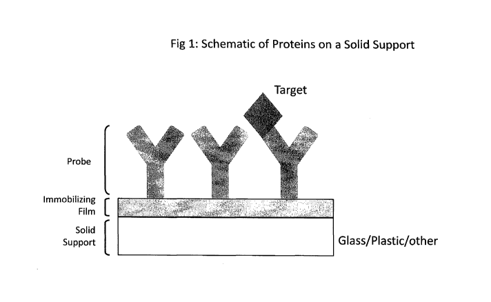
have a density of 10 to 1000 probe spots per cm2. Figure 1 is a schematic
showing a
capture probe (in this case, an antibody) affixed to a solid support via an
immobilizing
film. Note that Figure 1 is not to scale.
Besides photolithographic methods, robotic spotters are now the most common
instrument used for creating arrays. In some embodiments, protein microarrays
are
fabricated using non-contact piezoelectric robotic spotters manufactured by
companies
such as the Piezorray (Perkin-Elmer, Shelton, CT), GeSim (NanoPlotter),
Scienion and
Aushon. Very high-density microarrays containing over one-hundred antibodies
can be
prepared using robotic spotters.
In some embodiments, replicate spots of each analyte are included to increase
precision. In some cases, these replicates are scattered throughout the array
to reduce
spatial biases that may be present in a surface (See e.g., U.S. provisional
patent
application 60/972,928, herein incorporated by reference in its entirety).
B. Detection
When a sample is applied to an array, target analytes (e.g. proteins or
protein
fragments found in serum or some other biological sample) are captured by the
immobilized probes. Like other immunoassays, detection occurs using a detector
molecule such as a labeled antibody followed by a reporter molecule, often
containing a
fluorescent label. As in other methods, a number of wash steps are performed
in between
steps to remove non-specifically bound proteins.
In certain embodiments, the detection step is colorimetric. In certain
embodiments, the detection step involves a reaction that produces a
precipitate (See e.g.,
US 20030124522, herein incorporated by reference). In particular embodiments
the
detection step uses signal detection involving horseradish peroxidase, gold-
catalyzed
silver deposition, or alkaline phosphatase. These compositions and methods can
be used
to perform multiplexed assays for analytes in patient and other test samples.
In particular,
24
CA 02720747 2010-10-05
WO 2009/105670 PCT/US2009/034711
these methods have applications for multi-analyte immunoassays to measure
proteins in
human serum and plasma using inexpensive solid supports and colorimetric
detection
instrumentation.
In some embodiments, the presence of a precipitate is detected using an array
reader or other automated detection system. Array readers can measure a
variety of
optical outputs including, but not limited to, fluorescence, luminescence,
radioactivity,
colorimetric, optical waveguides, or surface plasmon resonance. In many cases,
the
bound molecule of interest is labeled in some way to make it detectable, such
as with a
fluorescent molecule, to generate an optical signal. Detection of optical
signals is
achieved using a variety of methods in these instruments, including, but not
limited to,
CCDs, CMOS chips, and/or PMTs. The concentrated light energy in an optical
waveguide can be used to excite fluorescently labeled molecules with higher
signal-to-
noise than conventional approaches. This excitation (and the concomitant
emission of
light) is used to detect the presence of fluorescently labeled molecules in
solution (like
proteins or DNA) at very low levels.
There are several types of colorimetric detection instruments available for
use
with colorimetric microarrays. In general, colorimetric detection instruments
are cheaper
than confocal laser scanners because they use an inexpensive light source and
detector
and in many cases, avoid expensive optics by using a fixed focus. The most
common
colorimetric scanners are sold by Epson and Hewlett Packard and are commonly
available at office supply stores. To use these scanners, slides are placed
face down on
the scanner bed. Samples are both illuminated and read by reflectance from
below
through a transparent glass surface. In this way, both transparent and opaque
solid
supports can be used with these types of colorimetric scanning devices.
In some embodiments, a colorimetric reader that scans through a transparent
microarray slide to allow the detection of light grey-to-black spots generated
from a
precipitating reaction. These types of scanners allow the detection of arrays
comprised of
light grey-to-black spots that can be visualized first by the naked eye and
subsequently
scanned. The grey or black level intensity is related to the quantity of
target molecule
that are hybridized or adsorbed onto an array spot. This type of technology
can be used to
detect any type of precipitating reagents using an optically clear slide.
These instruments
CA 02720747 2010-10-05
WO 2009/105670 PCT/US2009/034711
can now be purchased commercially, usually for less than a third of the price
of a typical
fluorescent microarray scanner. One example of an instrument that can read
colorimetric
arrays is Eppendorf's SILVERQUANT scanner, which can be used to scan standard
microscope slides (25x75 mm). In other embodiments, a colorimetric reader that
scans
through a transparent, SBS-compatible plate with arrays printed on the bottom
to allow
the detection of light grey-to-black spots generated from a precipitating
reaction is used.
One such instrument, the APiX VistaScan, is available from Gentel (Madison,
WI),
which allows scanning of transparent, SBS-compatible 96- or 384-well plates
with arrays
printed on them as well as scanning of standard microarray slides.
Additionally, flatbed
scanners that are capable of scanning in a transmission mode can also scan
through a
transparent microarray slides to allow the detection of light grey-to-black
spots generated
from a precipitating reaction. Examples of scanners capable of transmission
scanning
include the EPSON 4490, EPSON V700, and Cannon CanoScan 8800F. In many cases,
these transmission-based flatbed scanners can achieve equivalent or superior
S/N
compared to other colorimetric scanners. In testing performed in our
laboratory, we have
shown that reflection-based flatbed scanners yield inadequate S/N compared to
transmission-based colorimetric scanners.
In another embodiment, we have performed colorimetric assays using a 96-well
hybrid microarray and multiplexing device called SmartplexTM,
(ThermoScientific). The
device is fully compatible with microplate- and liquid-handling automation
uses a unique
approach to incorporate coated planar substrates such as aminosilane, epoxy
silane, and
poly-L-lysine coated glass. Nitrocellulose coated substrates such as the PATH
slide and
clear nitrocellulose coated glass or plastic substrates can also be used with
Smartplex.
The Smartplex device uses a three-piece design that incorporates (1) a frame
for holding
a planar substrate, (2) a rigid substrate, and (3) a bottomless, 96-well top
with adhesive
on the bottom that forms 96-chambers when joined to the substrate. For array
printing,
the bottom frame holds can be used to hold the substrate in place. For sample
processing
using standard liquid handling automation, the 3-piece device is assembled to
resemble
an SBS-compatible 96-well microplate. The fully assembled 3-piece device can
be read
scanned in a fluorescent scanner such as the Tecan LS Reloaded. The APiX
VistaScan
colorimetric reader can also scan the fully assembled 3-piece device provided
optically
26
CA 02720747 2010-10-05
WO 2009/105670 PCT/US2009/034711
clear substrates and surface chemistries are used. Transmission-based flatbed
scanners
can be used to read the Smartplex device provided the bottom frame is removed
for
scanning on the flatbed scanner. Without the capability to remove the bottom
frame, the
colorimetric array would be beyond the focal length of commercially available
flatbed
scanners. Therefore, use of the 3-piece design with colorimetric experiments
has unique
advantages and enables use of very low-cost transmission-based flatbed
scanners.
Reflection-based flatbed scanners do not have the focal length to image a
Smartplex or
standard 96-well plate.
Once read by a scanner or imager, the array readout is processed in order to
transform the image into quantitative data. Many software programs exist for
array
image processing, including ArrayVision (Imaging Research Inc/GE Healthcare
Life
Sciences), ScanArray Express (PerkinElmer Life Sciences Waltham,
Massachusetts),
MicroVigene (VigeneTech. Inc, Carlisle, MA). These programs include "spot
finding"
algorithms and turn microarray images into values. These programs often have
features
that subtract array background noise from spot values. Once values are
obtained for each
spot, values from standard calibration curves can be used to generate a curve-
fit, from
which the user can back-calculate the concentration of analytes in the sample
of interest.
C. Kits and Systems
In some embodiments, the present invention provides kits and systems for
performing and analyzing array data. In some embodiments, the kits and systems
comprise all of the components necessary, sufficient, or useful for
generating, performing
and analyzing arrays. For example, in some embodiments, kits and systems
include all of
the substrates (e.g., arrayed substrates), reagents, components, buffers,
normalization
standards, and controls needed for performing assays. In some embodiments,
kits and
systems further comprise software for collecting and analyzing data from
arrays. In some
embodiments, kits and systems comprise instructions for using the kits. In
some
embodiments, systems comprise automation equipment (e.g., robotics, etc.) for
automating assays.
II. Diagnostic and/or Clinical Methods
27
CA 02720747 2010-10-05
WO 2009/105670 PCT/US2009/034711
As described above, embodiments of the present invention provides devices and
systems for generation and detection of high density arrays. As described
above, in some
embodiments, multiplexed assays are performed. The present invention is not
limited to
detection of a particular analyte. The methods and compositions of the present
invention
find use in the detection of any number of diagnostic and research
applications.
The present invention is not limited to a particular detection assay.
Quantitative
multiplex immunoassays, single capture antibody arrays, multiplex serological
assays,
and biomarker profiling are all contemplated. Examples include, but are not
limited to,
immunoassays where antibodies or antigens are affixed to the array surface,
nucleic acid
based assays where a nucleic acid or probe is affixed to the array surface,
protein-protein
interaction assays where a protein is affixed to the surface, small molecule
detection
assays where a small molecule or capture reagent is affixed to the array
surface and drug
screening assays where a small molecule or target enzyme is affixed to the
array surface.
In particular, embodiments of the present invention (See e.g.., Example 1)
have
applications for multi-analyte immunoassays to measure proteins in human serum
and
plasma using inexpensive solid supports and colorimetric detection
instrumentation.
In some embodiments, protein arrays are used to measure protein abundance.
Protein abundance is most commonly measured using protein capture molecules
such as
antibodies, aptamers, antibody fragments, and others. Capture molecules can be
immobilized on surfaces and used to quantify protein abundance in a wide
variety of
samples, including, but not limited to, saliva, blood, plasma, serum, urine,
cell lysates,
tissue, or other biological fluids. Fluorescence-, luminescence-, and
colorimetric-based
detection using planar arrays have proven to be highly sensitive and rapid
methods for
multiplexed protein detection. The attractive cost, use of less sample,
improvement in
efficiency and chain-of-custody benefits of multiplexed protein measurement in
a single
sample has helped these assay become much more common, particularly
measurements
of cytokine proteins in human serum and plasma. However, prior to the present
invention, problems with assay sensitivity and reproducibility persist and
have limited the
broader utility and hence acceptance of these assays.
Protein analytes can be detected using a variety of detection steps that may
include detector antibodies (commonly a biotinlyated, fluorescent, or
otherwise-labeled
28
CA 02720747 2010-10-05
WO 2009/105670 PCT/US2009/034711
monoclonal or polyclonal antibody), secondary antibodies (such as a
biotinlyated,
fluorescent- or otherwise labeled anti-species antibody), and/or a detection
reagent (such
as fluorescent- or otherwise-labeled streptavidin, a substrate, or
precipitate).
The present invention provides several methods for improving the efficiency of
obtaining protein array results. Often, a single planar surface can contain
multiple arrays
to enable processing of standard calibration curves and/or multiple patient or
test samples
on a single slide. These multi-array surfaces are usually coupled to
multiplexing devices
(also called separators) that separate samples by forming multiple,
independent chambers
or wells. Examples of multiplexing devices include the ProPlateTM (Grace Bio-
Labs, Inc.
Bend, OR), FASTframeTM (Publication # W02005060678 or Application #
10/737,784),
or SIMplexTM products (Gentel Biosciences, Madison, WI). Commonly,
multiplexing
devices separate a single 3"xl" microarray slide into sixteen chambers (e.g. 2
x 8
format). The ProplateTM, FASTframeTM, and SIMp1ex64TM devices secure four
slides
(sixteen chambers each) to form a sixty-four well device. These devices have
been
designed to fit within the standard footprint of a multi-well plate as
established by the
Society of Biomolecular Sciences (SBS Standards). The footprint for most
multiwell
plates is approximately 85 mm x 125 mm with wells located in a standardized
format
depending upon the total number of wells. In this format, researchers can
incorporate an
eight-point standard curve- and process up to 56 samples using a single, 64-
well plate.
Alternatively, a researcher could incorporate two eight-point standard curves
and process
up to sixteen samples in triplicate using a single, 64-well plate to achieve
higher
precision.
More recently, researchers have coupled larger format slides to separators to
emulate the 96-well plate format standard (United States Patent: 7063979,
United States
Patent Application 20050277145; each of which is herein incorporated by
reference)
established by SBS. This also includes the SmartplexTM device
(ThermoScientific). This
format has the advantage of being more fully compatible with robotic liquid
handling
instruments and enables the processing of additional samples. For example, a
researcher
could incorporate a single eight-point standard curve and process up to 88
samples using
a single 96-well plate.
29
CA 02720747 2010-10-05
WO 2009/105670 PCT/US2009/034711
All of these multiplexing methods allow sample processing using automated
liquid handling robots to enable rapid and efficient collection of multi-
analyte data from
many samples. Additionally, automation helps reduce assay variation (thus
enabling more
precise quantitation).
In some embodiments, the present invention provides methods for differential
diagnosis of a disorder or identification of a patient subset, identification
of potential
responders to a specific drug, targeting of specific therapies, identifying
individuals at
risk for adverse events, and monitoring individual responses to drugs. These
applications
require very robust protein quantification technologies with high levels of
accuracy and
precision to meet this need.
Accordingly, in some embodiments, the present invention provides methods to
normalize microarray data across different wells and within a single well (See
e.g., above
description of replicate assays).
The applications of the present invention described herein are examples and
are
not intended to limit the present invention. The methods of the present
invention are
suitable for detection and quantitation of any number of targets and analytes.
EXPERIMENTAL
The following examples are provided to demonstrate and illustrate certain
preferred embodiments and aspects of the compositions and methods disclosed
herein,
but are not to be construed as limiting the scope of the claimed invention.
Example 1
Quantitative Multiplexed Immunoassay to Measure Human Cytokines
This example describes the use of precipitating reagents and optically clear
nitrocellulose-coated plastic slides to perform a quantitative multiplexed
immunoassay to
measure human cytokines in patient serum. For comparison, a similar assay was
performed on a commercially available PATH protein microarray slide (Gentel
Biosciences, Madison, WI) and fluorescence reagents.
Capture antibodies to six human cytokines were printed in sixteen sub-arrays
on
both optically clear nitrocellulose-coated plastic slides and PATH" slides
(see Fig. 3).
CA 02720747 2010-10-05
WO 2009/105670 PCT/US2009/034711
After printing, arrays were allowed to incubate for several days and
subsequently blocked
using Gentel Block Buffer (Gentel Biosciences). After blocking, the coated
plastic slides
and PATHTM slides were assembled in SIMplex64TM multiplexing devices to enable
processing of sixteen samples per slide. Antigens were diluted in a serum
matrix (PBS +
10% FBS) and applied to separate sample wells to create a standard dilution
curve.
Internal normalization standards are also included in all wells to improve
sensitivity and
reproducibility. To do this, solutions in all wells are spiked with (3-
galactosidase
normalization reagent such that the final concentration of (3-galactosidase
was equivalent
in all wells. Next, a cocktail of biotiny]ated detector antibodies was
incubated in each
well, followed by washing of the slides.
For optically clear nitrocellulose coated plastic slides, the SilverquantTM
detection
kit was used. The SilverquantTM detection kit includes all required reagents
to perform
gold particle catalyzed silver deposition. Briefly, the slides were placed
into the
SilverquantTM box and washed per kit instructions, blocked with the
SilverquantTM
blocker for 10 min, and then incubated with the anti-biotin-gold conjugate Ab
for 45 min.
Following more washes, the slides were incubated with the SilverquantTM silver
staining
reagent for 5 min and then washed with water and dried. Readout was performed
using
an Eppendorf SilverquantTM Scanner following manufacturer's instructions.
For PATHTM slides, streptavidin DY547 (Dyomics GmBH, Germany) was used at
a concentration of 10 ng/mL. The slides were incubated with the SA-Dy547
solution and
then washed, disassembled from the SIMplex64 device and dried. Readout was
performed using a Tecan LS Reloaded using 532 nm excitation. An example of
each slide
type after the human cytokine assay was completed is shown in Fig. 4.
Standard curves for both the coated plastic substrates and PATHTM slides are
shown in Fig 5 and Fig 6. It should be noted from these data that the standard
curves
generated from precipitating reagents and optically clear nitrocellulose-
coated plastic
substrates yield much more discrimination. The antigens for the most
concentrated
solution in the standard curve ranged from 0.6-2.0 ng/mL (fluorescence) or 0.2-
0.67
ng/mL (colorimetric). The subsequent six standards were 1:2 serial dilutions
of this
sample, with the eighth standard being a blank (i.e. no antigens present).
31
CA 02720747 2010-10-05
WO 2009/105670 PCT/US2009/034711
Further quantitative analysis indicated significantly improved reproducibility
and
sensitivity resulting from using precipitating reagents and optically clear
nitrocellulose-
coated plastic substrates. This is evident in the comparison of the limit-of-
detection
(LOD), and percent coefficient of variation (%CV), for the two approaches.
Table 1
shows the calculated LODs for six human cytokines using a fluorescence-based
assay and
PATHTM slides as well as a colorimetric assay and nitrocellulose-coated
plastic slides.
LOD was calculated using the blank signal plus two standard deviations in a 4-
parameter
fit of the standard curve. %CV was calculated spot-to-spot (n=5 spots), well-
to-well (n=4
wells across four slides), and day-to-day (n=3 days). For the reproducibility
data shown
in Tables 2-4, the %CV were calculated from data from standards #1-5 from the
standard
curves. The lowest three standards were not included because the average
signals were
small and thus the %CV were significantly impacted by small variations.
As is seen from Table 1, it is evident from these data that the use of
precipitating
reagents and optically clear nitrocellulose-coated plastic slides to perform a
quantitative
multiplexed immunoassay significantly improves sensitivity compared to a
similar
fluorescence-based assay. The present invention is not limited to a particular
mechanism.
Indeed, an understanding of the mechanism is not necessary to practice the
present
invention. Nonetheless, it is contemplated that one reason for the increased
sensitivity of
the assay compared to fluorescence-based assays and similar assays performed
on a
silanized glass slide is due to the unique properties of the colloidal
nitrocellulose film,
which creates a very low background signal when used in a precipitating assay.
Further benefits can be noted in assay reproducibility. Tables 2-4 show that
precipitating reagents and optically clear nitrocellulose-coated plastic
slides yield lower
spot-to-spot %CVs, lower well-to-well %CVs and roughly equivalent day-to-day
%CVs
compared to a similar fluorescence-based assay.
Table 1 Limit of Detection for each method (pg/mL).
Gentel
Gentel PATHTM
Antigen Plastic Colorimetric Fluorescence
IL-2 0.050 0.11
32
CA 02720747 2010-10-05
WO 2009/105670 PCT/US2009/034711
IL-6 0.022 0.13
EL-7 0.22 0.29
IL-8 0.009 0.042
IL-10 0.011 0.13
TNF-b 1.5 7.5
SUM 1.812 8.202
Table 2 Spot-to-Spot Reproducibility for each method (% CV).
Gentel
Gentel PATH
Antigen Plastic Colorimetric Fluorescence
IL-2 6% 7%
IL-6 5% 14%
IL-7 7% 8%
IL-8 4% 11%
IL-10 5% 13%
TNF-b 33% 14%
SUM 59% 67%
Table 3 Well-to-Well Reproducibility for each method (% CV).
Gentel
Gentel PATH
Antigen Plastic Colorimetric Fluorescence
IL-2 10% 10%
IL-6 9% 21%
IL-7 13% 28%
IL-8 8% 11%
IL-10 9% 19%
33
CA 02720747 2010-10-05
WO 2009/105670 PCT/US2009/034711
TNF-b 9% 17%
SUM 57% 105%
Table 4 Day-to-Day Reproducibility for each method (% CV).
Gentel
Gentel PATH
Antigen Plastic Colorimetric Fluorescence
IL-2 14% 14%
IL-6 11% 12%
IL-7 18% 12%
IL-8 8% 13%
IL-10 11% 14%
TNF-b 30% 19%
SUM 92% 84%
Example 2
Der p 2 mediated quantitative determination of allergen-specific IgE in human
serum
This example describes the use of precipitating reagents and optically clear
nitrocellulose-coated plastic slides to make quantitative determinations of
allergen-
specific IgE titers. A Chimeric anti- Der p 2 Immunoglobulin E (IgE) (Indoor
Biotechnologies) was used as a surrogate for quantitative determinations
encompassing a
large range of allergen-specific IgE titers in patient serum. Here,
quantitation capability
using both precipitating reagents and optically clear nitrocellulose-coated
plastic slides
and fluorescence-based measurements and a commercially available PATH slide
were
compared using the Der p 2 standard curve.
To do this, recombinant allergens including Cat (Fel d 1), Silver Birch (Bet v
la,
Bet v 2), Timothy Grass (Phl p 1, Phl p 2, Phl p 5a, Phl p 6), mold
(Alternaria alternata,
Alt a 1), dust mite (Der p 1, Der p 2, Der f 1), Dog (Can f 1) (Indoor
Biotechnologies)
were immobilized on a microarray using a robotic microarrayer. Recombinant f3-
galactosidase was also included in the array for use as an internal
normalization standard.
34
CA 02720747 2010-10-05
WO 2009/105670 PCT/US2009/034711
Arrays were printed in sixteen sub-arrays on both optically clear
nitrocellulose coated
plastic slides and PATHTM slides. After printing, arrays were allowed to
incubate for
four days and subsequently blocked using GenTe1TM Block Buffer. After
blocking, the
coated plastic slides and PATHTM slides were assembled in SIMplex64
multiplexing
devices to enable processing of sixteen sample wells for each slide and to
facilitate
automated washing.
Chimeric anti- Der p 2 IgE was diluted in a serum matrix (PBS + 10% FBS) and
applied to separate sample wells to create standard dilution curves. Internal
normalization standards were included in all wells to improve sensitivity and
reproducibility. To do this, solutions in all wells were spiked with 0-
galactosidase
normalization reagent such that the final concentration of 0-galactosidase was
equivalent
in all wells. Next, a biotinylated anti-human IgE- IgG was incubated on the
array,
followed by washing and detection.
For optically clear nitrocellulose coated plastic slides, the SilverquantTM
detection
kit was used. Briefly, the slides were placed into the SilverquantTM box and
washed per
kit instructions, blocked with the SilverquantTM blocker for 10 min, and then
incubated
with the anti-biotin-gold conjugate Ab for 45 min. Following more washes, the
slides
were incubated with the SilverquantTM silver staining reagent for 5 min and
then washed
with water and dried. Alternately, the steps performed in the SilverquantTM
box can be
performed in the SIMplex device. Readout was performed using an Eppendorf
SilverquantTM Scanner. Readout was repeated using the Gentel APiX VistaScan
Reader
and an EPSON V700 flatbed scanner.
For PATHTM slides, streptavidin DY649 (Dyomics GmBH, Germany) was used at
a concentration of 10 ng/mL. Readout was performed using a Tecan LS Reloaded
using
633 nm excitation.
Standard dilution curves for Chimeric anti- Der p 2 IgE on both the coated
plastic
substrates and PATHTM slides are shown in Fig 7(a) and Fig 7(b). It should be
noted
from these data that the standard curves generated from precipitating reagents
and
optically clear nitrocellulose-coated plastic substrates yield much more
discrimination.
Further quantitative analysis indicated significantly improved reproducibility
and
sensitivity resulting from using precipitating reagents and optically clear
nitrocellulose-
CA 02720747 2010-10-05
WO 2009/105670 PCT/US2009/034711
coated plastic substrates. This is evident in the comparison of the limit-of-
detection
(LOD), coefficient of variation (CV), and goodness-of-fit (R2) for the two
approaches.
LOD was calculated using the blank signal plus two standard deviations in a 4-
parameter
fit of the standard curve. LOQ was calculated using the blank signal plus
eight standard
deviations in a 4-parameter fit of the standard curve. Table 5 shows results
based on a
standard curve generated using Chimeric anti- Der p2 IgE.
Table 5
Experiment LOD LOD Dynamic Avg % CV
Range R2
Performed (pg/ml) (IU/ml) (range)
(Logs)
Initial 6.28, (10, 2,
Plastic 38.70 0.016 2.7 23, 4, 2, 1,
0.881
Colorimetric 2)
Optimized 18.4, (1, 7,
Plastic 6.048 0.003 5.4 39, 6, 37,
0.954
Colorimetric 18, 21)
Optimized 9.43 (5, 6,
Fluorescence 89.23 0.037 3.4 23, 7, 7, 5,
0.975
PATH 12,8)
All publications and patents mentioned in the above specification are herein
incorporated by reference. Various modifications and variations of the
described devices,
compositions, methods, systems, and kits of the invention will be apparent to
those
skilled in the art without departing from the scope and spirit of the
invention. Although
the invention has been described in connection with specific preferred
embodiments, it
should be understood that the invention as claimed should not be unduly
limited to such
specific embodiments. Indeed, various modifications of the described modes for
carrying
out the invention that are obvious to those skilled in art are intended to be
within the
scope of the following claims.
36