Note: Descriptions are shown in the official language in which they were submitted.
CA 02720776 2010-10-05
WO 2009/126868
PCT/US2009/040178
X-RAY GENERATOR WITH POLYCAPILLARY OPTIC
BACKGROUND OF THE INVENTION
1. Description of the Related Art
[0001] The
present invention relates systems for generating and focusing x-
ray radiation for analytical instruments including x-ray diffractometry, x-ray
spectrometry or other x-ray analysis applications.
2. Description of the Known Technology
[0002] There
are numerous analytical instruments and procedures for which
x-ray radiation is directed onto a target for analytical or metrology
applications.
Examples of such instruments include those based on the principles of x-ray
coherent scattering such as x-ray scattering and x-ray diffraction, and those
based
on the principle of x-ray fluorescence such as x-ray spectroscopy and x-ray
elemental mapping microscopy. In many such applications, there is a need to
direct
an intense beam of x-rays having controlled beam characteristics in its
interaction
with the target. These characteristics include spatial definition (divergence,
beam
size, focal spot size and intensity distribution at different locations),
spectrum purity
and intensity. However, these characteristic parameters can not be optimized
independently. Improving one often comes at price of others. X-rays are
inherently
difficult to direct. Different technologies have been employed to form x-ray
beams.
These include total reflection reflectors, optics based on total reflection
principle
such as capillary and polycapillary made of bundle of micro-sized waveguides,
natural crystals, and man-made layered structures called multilayer optics. In
some
cases, polychromatic radiation with energy spectrum over a relatively wide
range
may be desired. In other applications, highly monochromitized radiation is
desired.
Optics are made with selected technologies to match with the beam requirements
while maintain an acceptable cost.
[0003] X-ray
beam systems with excellent performance have been developed
with microfocusing sources and variety of beam conditioning optics. Typical
focal
spot projection of these microfocusing sources is less than 100 micrometers
and as
small as 10 micrometers. Future development of source technology and optics
1
CA 02720776 2010-10-05
WO 2009/126868
PCT/US2009/040178
technology may drive the brilliance even higher and spot size even smaller.
Both
stability of the spot size and spot position are critical for x-ray beams in
analytical
applications. In addition to superior performance, microfocusing sources use
much
less energy therefore has a lower operation cost and cause less environment
issues.
Sealed tube microfocusing sources, not only offers good performance, but also
offers good performance-cost ratio. Representative optics in a microfocusing
sources based beam system include multilayer optics, crystal optics, total
reflection
mirrors, mono-capillary optics and polycapillary optics. Optics can be
designed for
redirecting x-rays in one direction only, i.e. so-called one-dimensional
optics (1D
optics), or designed for redirecting x-rays in two perpendicular directions
either
through single interactions, two interactions or multiple interactions, i.e.
so called
two-dimensional optics (2D optics). For a highly intense beam, close coupling
to an
x-ray source is critical in order to acquire a large solid capture angle. To
obtain a
monochromatic beam, diffraction element should be a key part of the system.
[0004]
Multilayer optics naturally delivers monochromatic beams. The beam
characteristics, such as spatial definition, spectrum purity and intensity,
can be
optimized through various designs. Multilayer optics have been the major beam
conditioning optics for x-ray scattering and diffraction.
[0005] In many
analyses, such as in x-ray powder diffraction and thin film
analysis, the probe beam is conditioned typically by a one-dimensional optic,
meaning to redirect and form a beam in one direction only. These optics
include
planar multilayer optic, parabolic multilayer optic, and elliptical multilayer
optic.
These optics have a profile of cylinder curve, i.e. the curvature in the
direction
perpendicular the beam propagation direction is straight line, and the
curvature in
the direction of beam propagation direction is a profile of either straight
line (planar
optic), or part of a parabola (collimating optics), or part of an ellipse
(focusing optics).
These optics are typically very efficient and are capable in delivering high
flux
beams.
[0006] For many
other applications, such as single crystal crystallography
represented by small molecule crystallography and macro molecule
crystallography
(protein crystallography), the probe beam has to be a two-dimensional beam,
i.e. a
"pencil-like" beam formed in two perpendicular directions. Such a beam can be
2
CA 02720776 2010-10-05
WO 2009/126868
PCT/US2009/040178
formed by a two-dimensional optic. Multilayer two-dimensional optics are the
major
beam conditioning optics for the need of two-dimensional beam conditioning.
These
optics delivers beams with well defined spatial characteristics and good
spectrum
purity.
[0007] Optics
based on the waveguide principle, such as waveguide bundle
optics represented by polycapillary optics, have been used in x-ray micro-
spectrometry and selected x-ray diffraction applications. Comparing to
multilayer
optics, waveguide bundle optics offer much large capture angle and therefore
potentially much higher flux and brilliance. The issue with waveguide bundle
optics
is that the output, in nature, is x-rays with continuous spectrum and is not
suitable for
x-ray elastic scattering and x-ray diffraction.
[0008] Being
able to analyzing small sample is highly important, whether this
is because of a local interest on a large sample or acquiring adequate signal
strength from small available sample volume. High flux with well defined
spectrum
and spatial characteristics is often delivered by a focusing multilayer optic.
Such an
optic could consist of two cylinder elliptical mirrors; each of the mirrors
focuses x-
rays in one of the two perpendicular directions and the two mirrors are in a
so-called
Kirkpatrick-Baez geometry, either in sequential or "side-by-side" arrangement
as
depicted in US patent 6,041,099. Such an optic could also be part of an
ellipsoid
with multilayer coating inside, where a single reflection from the optic
directing the x-
rays in 2-dimensions.
[0009] Further
improving the intensity of a multilayer focusing optical system
depends on close coupling between source and optic. Unfortunately, the
coupling
distance is limited by the physically feasible dimension of the structure at
low d-
spacing end. The smallest layer thickness of the man-made layer structure is
limited
by the size of atoms. At extremely low d-spacing end, such as lower than 10
angstroms, the inter-layer roughness is high; the peak reflectivity is low;
and rocking
curve is narrow.
[0010] As it
can be seen, none of the solutions discussed above offers
efficient coupling with a source and meanwhile provide a beam with
controllable and
satisfactory spectrum. US patent 6,504,901 proposed an optical system coupled
with a x-ray focusing mirror. But the proposed solution failed to demonstrate
that the
3
CA 02720776 2012-10-17
system will deliver a monochromatic beam and failed to illustrate its
efficiency
improvement. In fact, the description of the patent leads to a solution which
is less
efficient and renders an optical scheme without practical significance. The
intention
seems that using a polycapillary optic to form a small, intense and low
divergence
"virtual source", the second optic, being a reflector limited with its capture
angle,
would be able to take the advantage of a source that is the small, more
intense and
with a lower divergence, and thus deliver a higher flux.
[0011] However, from physics law we know, that the first optic, the
polycapillary optic, as a kinematical system, i.e. without energy input, will
not be able
to convert a beam with large divergence into a beam with lower divergence
without
enlarging the focal spot size of the virtual source. This can also be
illustrated by
applying thermodynamics to the optical system: the entropy, or the ordering
represented by the spot size and divergence, of an isolated system without
external
energy input will at best be preserved and can not be reduced (or improved in
terms
of spot size and divergence). The description in US patent 6,504,901
"polycapillary
lens comprises a plurality of tapered capillaries arranged such that both the
diameter
of the focal spot of an x-ray source and the angular divergence of x-rays are
reduced" inevitably results in, in the best case, the same brilliance.
Therefore, the
performance of the system, in the best case, is equivalent to the performance
of the
direct coupling between the second optic and the source.
[0012] The low efficiency of the proposed system in US patent 6,504,901
could
also be illustrated in an geometrical manner, as well. The mechanism of x-ray
photons
propagation through a single capillary is multiple external total reflection.
It occurs in a
quite small range of incident angles, which is below 0.3 degrees for the wave
length
commonly used for diffraction experiments. A collimating waveguide bundle
optic has a
smaller cross section at a distance closer to the source. For an x-ray photon
propagating
inside a capillary, the incident angle at capillary wall gets smaller with
each consecutive total
reflection. On the other hand, if the optic is a focusing optic, the
reflection angle gets smaller
at first until the x-ray photons reach the point with largest diameter of the
polycapillary
optic, then gets larger with each consecutive reflection after passing the
point with
maximum diameter. When photons reach the exit of the proposed optic in the
patent
4
CA 02720776 2010-10-05
WO 2009/126868
PCT/US2009/040178
6,504,901 "bottle-shaped" optic where capillary diameter is smaller than at
optic
entrance, some portion of them will have incident angle larger than critical
angle of
external total reflection and will be lost, reducing optical system
efficiency.
BRIEF SUMMARY
[0013] In
satisfying the above need, as well as overcoming the enumerated
drawbacks and other limitations of the related art, the present invention
provides an
improved x-ray generating system. The x-ray generating system includes a
source
of x-ray radiation, a waveguide bundle based optic, such as polycapillary
optic, for
collecting the x-ray radiation produced by the source at large capture angle,
a
focusing diffractive optic for capturing beam from the first optic and
focusing the
monochromatic x-ray radiation to a focal point.
[0014] For
avoiding the issues identified previously for the focusing
polycapillary optic, the waveguide bundle optic should be designed in such a
fashion
that it provides a divergent beam, a collimated beam, or a slightly convergent
beam.
[0015]
Generally, the focusing optic can be a Kirkpatrick-Baez side-by-side
optic having multilayer Bragg x-ray reflecting surfaces that may be either
laterally or
laterally and depth graded. The focusing reflector can be parabolic, elliptic,
and
hyperbolic cylinder surfaces. The focusing optic can also be a doubly curved
optic,
such as paraboloidal, ellipsoidal, hyperboloidal, and toroidal optics, having
multilayer
Bragg x-ray reflecting surface that may be either laterally or laterally and
depth
graded.
[0016] The
coupling between the waveguide bundle optic and the diffractive
optic is in such a way that the geometric focus of the diffractive optic is at
the virtual
focus of the waveguide bundle optic or the other way around. If the waveguide
bundle optic is a collimating optic, the diffractive optic will be a parabolic
or
parabloidal optic having its focus at infinite; if the waveguide bundle optic
delivers a
divergent beam, the diffractive optic will be an elliptical or ellipsoidal
optic having its
geometric focus at the virtual focus of the divergent beam delivered by the
waveguide bundle optic; if the waveguide bundle optic delivers a slightly
focused
(convergent) beam, the diffractive optic will be hyperbolic and hyperboloidal
optic.
CA 02720776 2011-02-07
,
[0017] The x-
ray system in accordance with this invention seeks to overcome
the previously described design challenges of the prior art by providing a
waveguide
bundle based optical element closely coupled with an x-ray source which
captures x-
ray radiation from the source at a large capture angle and directs it to a
further
diffraction element in a controlled beam size and desired ray configuration.
For
example, collimated beam configurations can be readily provided. Through the
use
of suitable additional diffraction optics, such as the previously mentioned
Kirkpatrick-
Baez multilayer parabolic optic or an paraboloidal optic, a beam with
substantially
high intensity can be acquired.
[0017a] In
accordance with one aspect of the present invention, there is
provided an x-ray generating system comprising a microfocusing source of x-ray
radiation, a waveguide bundle optic for capturing the x-ray radiation produced
by
the microfocusing source, the waveguide bundle optic having an input for
receiving
x-ray radiation from the microfocusing source and an output for outputting a
collimated, or convergent x-ray beam, a diffractive optic with a diffractive
reflection
surface for focusing the x-ray beam from the wave guide bundle optic to a
focal
point, and wherein a sample receives radiation from the diffractive reflection
surface, the sample being located adjacent to the focal point.
[0018] Further
objects, features and advantages of this invention will become
readily apparent to persons skilled in the art after a review of the following
description, with reference to the drawings and claims that are appended to
and
form a part of this specification.
BRIEF DESCRIPTION OF THE DRAWINGS
[0019] Figure
1 illustrates an x-ray generating system having a waveguide
bundle optic, such as polycapillary optic, and a Kirkpatrick-Baez side-by-side
multilayer optic;
[0020] Figure
2 illustrates an x-ray generating system having a waveguide
bundle optic (such as polycapillary optic) and a doubly curved multilayer
optic; and
[0021] Figure
3 illustrates a more detailed view of the waveguide bundle optic
of Figures 1 and 2.
6
CA 02720776 2011-02-07
DETAILED DESCRIPTION
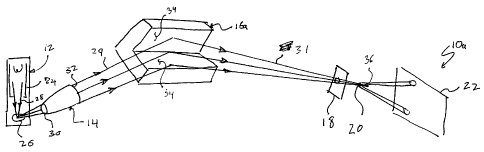
[0022] An x-ray
analysis system 10a includes an x-ray source 12, a
waveguide bundle optic (such as polycapillary optic) 14, a focusing optic 16a,
an
aperture 18, a sample 20, and an x-ray detector 22. The x-ray source 12 may be
a
laboratory source, such as a high brilliance rotating anode x-ray generator or
a
sealed tube microfocusing source. The x-ray source 12 generally includes an
electron beam focusing system 24 and a target 26. Electron beam 28 is guided
to
the target 26 by the e-beam focusing system 24.
6a
CA 02720776 2011-02-07
[0023] The waveguide bundle optic 14 includes an input 30 and an output 32.
The input 30 of the waveguide bundle optic is generally located about 3mm to
15mm, but not limited to, from the focus of the x-ray source 12. This distance
between the input 30 of the waveguide bundle optic 14 and x-ray source 12 is
better
known as the focal distance. Once x-rays are received by the input 30 of the
waveguide bundle optic 14, the waveguide bundle optic 14 guides the x-rays
from its
input to its output. The x-rays leaving the output 32 of the waveguide bundle
optic
are divergent, parallel, or slightly convergent.
[0024] The focusing optic 16a in this embodiment is a Kirkpatrick-Baez side-
by-side optic having multilayer Bragg x-ray reflecting surfaces 34 as
described in
U.S. Patent 6,041,099. The Bragg x-ray reflecting surfaces 34 generally have
graded-d spacing that is either lateral or lateral and depth graded.
[0025] The x-rays 29 received by the Bragg x-ray reflecting surfaces 34 of
the
focusing optic 16a are then reflected by the Bragg x-ray reflecting surfaces
34 to a
focal point 36. The surface shape of the mirrors of the diffractive optic
depends on
the design of the first waveguide bundle optic. If the first optic provides a
divergent
beam, the surfaces of the mirrors have an elliptical shape. If the first optic
forms a
collimating beam, the surfaces of the mirrors have a parabolic shape. lithe
first
optic provides slightly focusing beam, the surfaces of the mirrors have an
hyperbolic
shape. In any combination, the diffractive optic is positioned in such a way
that the
virtual focus of one of optics coincides with the real focus of other optic.
This
condition is critical and provides an effective acceptance by the diffractive
optic of all
the rays from the waveguide bundle optic. The reflected x-rays by the
diffractive
optic x-rays 31 are further defined by the aperture 18 in order to remove any
unnecessary x-rays. The sample 20 is located adjacent to the focal point 36
and
receives the reflected x-rays 31 shaped by the aperture 18. The sample 20 may
be
any sample, such as a biological sample, a polymer, or a crystallized protein,
whose
structure is the interest of study. The x-rays altered by the sample are
captured by
an x-ray detector 22.
[0026] Referring to Figure 2, another x-ray generating system 10b is shown.
The x-ray generating system 10b is similar to the x-ray generating system 10a
in
7
CA 02720776 2010-10-05
WO 2009/126868
PCT/US2009/040178
Figure 1, however, the focusing optic 16b of the x-ray generating system 10b
differs
from that of the focusing optic 16a of the x-ray generating system 10a. In
this
embodiment, the focusing optic 16b is a doubly curved optic, such as an
ellipsoidal,
paraboloidal or hyperboloidal optic, having multilayer Bragg x-ray reflecting
surface.
Additionally, the multilayer Bragg reflecting surface 35 of the reflecting
optic 16b has
graded-d spacing that may be laterally graded or laterally and depth graded.
[0027] Similar
to the embodiment shown in Figure 1, the pre-conditioned x-
rays 24 from the output 32 of the waveguide bundle optic, or polycapillary
optic, 14
are reflected by the diffractive optic 16b. The reflected x-rays 31 are then
focused
on a focal point 36. The sample 20 is located near the focal point 36 and is
configured to receive the reflected x-rays 31. Thereafter, a detector 22
receives x-
rays that have traveled through the sample 20 or are scattered or diffracted
by the
sample 20.
[0028]
Referring to Figure 3, a more detailed illustration of the waveguide
bundle optic 14, such as a polycapillary optic, is shown. The source 12
emitting the
x-rays 28 are separated from the input 30 of the waveguide bundle optic 14 by
a
distance f, known as the focal distance. As stated previously, the focal
distance f is
generally between about 3 millimeters to 15 millimeters but not limited to.
[0029] The
waveguide bundle optic 14 includes a plurality of hollow
waveguides 40 which are bundled together and plastically shaped into
configurations which allow efficient capture of divergent x-rays emerging from
the x-
ray source 12. In this example, the captured x-rays 28 are shaped by the
waveguide
bundle optic 14 into the collimated x-rays 29. Channel openings 42 located at
the
input 30 of the waveguide bundle optic 14 are pointing at the x-ray source 12.
The
optic could be shorter than shown on the Fig.3 providing divergent beam, or it
could
be longer providing a slightly convergent beam. In any of the described
embodiments, the diameters of the individual channel openings 42 at the input
30 of
the waveguide bundle optic 14 is smaller than the channel diameters at the
output
32.
[0030]
Generally, the hollow waveguides, or capillaries, 40 are made of glass
and have a diameter ranging from a few micrometers to sub-millimeters.
However,
8
CA 02720776 2012-10-17
the hollow capillaries may be made from carbon nanotubes with even smaller
diameter
of the channels.
[0031] As a person
skilled in the art will readily appreciate, the above
description is meant as an illustration of implementation of the principles of
this
invention. The scope of the claims should not be limited by the preferred
embodiments
set forth in the examples, but should be given the broadest interpretation
consistent
with the description as a whole.
9