Note: Descriptions are shown in the official language in which they were submitted.
CA 02721069 2015-09-14
SURGICAL INSTRUMENTS
FIELD
[0002] The present disclosure generally related to surgical instruments.
More
specifically, the disclosure relates to a suture anchor.
BACKGROUND
[0003] Suture anchors are well known in the art. Suture anchors are
typically used to
anchor soft tissues such as tendons, and the like, to bone using a suture. The
suture anchor is
typically secured in a bone and sutures, previously having been inserted into
tissue, are then
threaded through the suture anchor to tension the tissue and hold it in place.
The tissue is
tensioned to the bone via the suture attached to the anchor.
[0004] Typically, the suture is threaded through a small hole, or series
of holes in the
anchor and some suture anchors come pre-loaded with the sutures. The suture
can then be
knotted to prevent release of the suture from the suture anchor. However, the
need for
knotting can increase surgical time and provide a weak point for suture
breakage, hence a
need exists for a suture anchor that is fast to use, readily allows for re-
tensioning of a suture,
and does not introduce knotting weakness to the suture.
[0005] Surgical cannulas are used to enter areas within the body such as
the shoulder,
knee, or abdomen. The cannula provides a means for passing surgical
instruments into and
out of a subject. Cannulas are also used as a channel to introduce surgical
implements such
as surgical instruments, suture anchors, or sutures. Such surgical cannulas
typically have a
single chamber that is conducive to instruments or items touching and either
cross-
contaminating one another or disturbing the function of each other. Such
surgical cannulas
1
CA 02721069 2010-10-08
WO 2009/146155 PCT/US2009/039787
are also subject to the cannula readily pulling out of the subject or falling
into a subject and
requiring re-insertion or extraction. A need exists for cannulas that resist
the tendency to
push out of a subject, or fall into a subject. Also multi-chambered cannulas
are desired.
SUMMARY
[0006] In a first aspect, a surgical instrument is provided comprising
(a) an anchor
comprising (i) a wall comprising (1) an outer surface having threads; and (2)
an inner surface;
(iii) a first end; and a second end; and (b) a plug comprising a first end, a
second end, and an
outer wall; wherein the anchor and the plug form a suture anchor capable of
anchoring a
suture.
[0007] In one embodiment, the outer wall of the plug comprises threads
capable of
engaging the inner surface of the anchor via a friction fit. In another
embodiment, the inner
surface comprises threads. In another embodiment, the outer wall of the plug
and the inner
wall of the anchor each have threads capable of engaging each other in a screw-
like fashion
to secure the plug in the anchor.
[0008] In another embodiment, the plug is made of a compressible
material. For
example, the compressible material may be a polymer such as high density
polyethylene,
polyurethane, silicones, or a mixture thereof.
[0009] In some embodiments, the first end of the plug has the same
diameter as the
second end of the plug. In other embodiments, the first end of the plug has a
larger diameter
than the second end of the plug, such that the plug forms conical shape
similar to that of the
profile of a funnel.
[0010] In another aspect, methods of using a suture anchor is provided
comprising:
securing the anchor into a bone, draping a suture through an interior of the
anchor and
touching the inner surface; and inserting the plug in the anchor to secure and
tension the
suture. In some embodiments, the securing is via screwing or cementing of the
anchor in the
bone. Other embodiments, further comprise removing the plug; re-tensioning the
suture; and
replacing the plug in the anchor.
2
CA 02721069 2010-10-08
WO 2009/146155 PCT/US2009/039787
[0011] In another aspect, a suture anchor has an anchor body that has a
well, a top
surface, a bottom surface, and a transverse bore. The well has an outer
surface, an inner
surface, and an inner bottom surface. In such embodiments, the anchor and the
plug form a
suture anchor configured to secure a surgical suture. In some embodiments, the
transverse
bore is proximally located to the inner bottom surface. In some embodiments,
grooves are
formed in the outer surface extending from the transverse bore to the top
surface. In other
embodiments, the outer surface has rungs and the inner surface has threads. In
other
embodiments, the post is a threaded post.
[0012] In some embodiments, the outer surface has rungs, the inner
surface has, the
post is a threaded post, and the threads of the inner surface and the threads
of the post are
configured to engage in a screw-like fashion to secure the anchor plug in the
anchor body.
[0013] Methods of using such a suture anchor are also provided. Such
methods
include boring a nest in a bone; positioning the bottom surface of the anchor
body in the nest;
threading a suture through the transverse bore; driving the suture anchor into
the nest;
tensioning the suture; and securing the suture in the anchor body with the
anchor plug.
Alternatively, the threading of the suture precedes the positioning of the
anchor body in the
nest. In some embodiments, the methods also include loosening or removing the
anchor
plug, re-tensioning the suture, and tightening or replacing the anchor plug in
the anchor body.
[0014] In another aspect, surgical instrument is provided comprising (a)
a cannula
comprising a distal end, a proximal end and at least one chamber extending the
entire length
of the cannula from the proximal end to the distal end; (b) at least one
inflatable donut; and
(c) an air passageway having an air inlet; wherein the inflatable donut has a
circumference at
full inflation that is greater than a circumference of the cannula. In some
embodiments, the
cannula is capable of passing a surgical item into and out of a subject
undergoing surgery.
For example, surgical items include, but are not limited to, surgical
instruments, sutures, and
implants.
[0015] In some embodiments, the cannula comprises a divider that divides
the at least
one chamber extending the entire length of the cannula into at least two
chambers. In some
embodiments, the divider is a flexible diaphragm or divider. In other
embodiments, the
divider extends the full length of the cannula.
3
CA 02721069 2010-10-08
WO 2009/146155
PCT/US2009/039787
[0016] In other embodiments, the cannula further comprises a second
donut. In some
such embodiments, the second donut is rigid or inflatable. In other
embodiments, the second
donut is capable of preventing insertion of the cannula completely into a
subject. In yet other
embodiments, the second donut is formed integrally with the cannula. In yet
other
embodiments, the second donut is formed separately from the cannula and is
attached to the
cannula.
[0017] In other embodiments of the cannula, the inflatable donut is
capable of being
inflated via the air passageway. In some embodiments, a pump is used to
inflate the
inflatable donut via the air passageway. In other embodiments, an air-filled
syringe inserted
through a valve is used to inflate the inflatable donut via the air
passageway.
[0018] In another aspect, a method of using the cannula is provided
comprising:
making an incision in the subject at the location in which the cannula is to
be inserted;
inserting the cannula; and inflating the at least one inflatable donut. In
other embodiments,
the method further comprises: conducting a surgical procedure using the
cannula; deflating
the inflatable donut; and removing the cannula from the subject.
BRIEF DESCRIPTION OF THE DRAWINGS
[0019] FIG. 1 is a side view of a suture passer without a suture.
[0020] FIG. 2 is a side view of a suture passer having a suture threaded
through the
suture passer.
[0021] FIG. 3A is a side view of a suture anchor showing the plug and the
nest with
sutures passing through, prior to insertion of the plug into the nest,
according to one
embodiment. FIG. 3B is a side view of a suture anchor showing the plug and the
nest with
sutures passing through, prior to insertion of the plug into the nest,
according to another
embodiment.
[0022] FIG.
4 is a top view of a suture anchor showing the plug and the nest with a
sutures passing through, prior to insertion of the plug into the nest.
4
CA 02721069 2010-10-08
WO 2009/146155 PCT/US2009/039787
[0023] FIG. 5 is an illustration of a humerus with a hole bored through a
portion for
the passage of a suture to secure a rotator cuff bed.
[0024] FIG. 6 is an illustration of a humerus with a hole bored through a
portion for
the passage of a suture to secure a rotator cuff bed.
[0025] FIG. 7 is an illustration of a humerus with a hole bored through a
portion for
the passage of a suture to secure a rotator cuff bed and a suture anchor
secured in the
humerus.
[0026] FIG. 8 is an illustration of a humerus with a hole bored through a
portion for
the passage of a suture to secure a rotator cuff bed and a suture anchor
secured in the humerus
with the suture beginning to be tensioned.
[0027] FIG. 9 is a side view of a cannula.
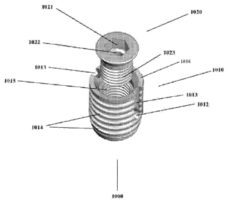
[0028] FIG. 10 is a perspective view of a suture anchor, according to one
embodiment.
[0029] FIG. 11 is a perspective view of an anchor body, according to one
embodiment.
[0030] FIG. 12 is a perspective view of an anchor plug, according to one
embodiment.
[0031] FIG. 13 is a side view of a suture anchor, according to one
embodiment.
DESCRIPTION
[0032] In one aspect, an instrument comprising a suture passer 100 is
described. As
shown in FIGS. 1 and 2, the suture passer 100 comprises a body 110 having a
tunnel 115, an
articulating arm 120, 190 connected to the body 110 proximally to a first end
of the body
110, and a fore end 130 distal to the first end of the body 110. In some
embodiments, the
suture passer 100, further comprises suture channels 184, 185 through which
sutures are
threaded to load the suture passer 100. In some embodiments, the suture passer
100 is an
arthroscopic instrument. The suture passer 100 may be used to grasp tissue and
pass a
CA 02721069 2010-10-08
WO 2009/146155
PCT/US2009/039787
sliding, locking suture in a single grasp of the tissue. The suture passer 100
grasps tissue 180
between the body 110 and the articulating arm 120. The suture passer 100 then
passes a
suture loop 140 through the tissue 180. This may be done by loading the suture
140 in a U
fashion. A needle passes through the tunnel 115 then penetrates the tissue 180
passing
through the suture loop 140. A second needle or a pass of the same needle
passes through a
channel within articulating arm 120 and is directed through the suture loop
140 and capturing
the other end of the suture. This pulls one end of the suture back in a
retrograde fashion
through the first loop 140 creating a locking stitch. The suture has one end
outside the body
110 and the other end loaded on the other side of the suture passer 100. This
allows for it to
be passed through the suture loop 140, ultimately forming a locking stitch.
One end of the
suture is passed below the tissue 180 and one end is passed above the tissue
180. When the
bottom suture is pulled longitudinally it pulls the suture loop 140 down
perpendicular to the
tissue 180 resulting in bringing it downward. When the top suture is pulled it
brings the
tissue 180 laterally or in line with the sutures. In one embodiment, the
articulating arm 120 is
connected to the body 110 by a joint or a hinge, such that the articulating
arm 120 may move
relative to the body 110 in a tweezer-like fashion.
[0033] In
other embodiments, the suture passer 100 will grasp tissue, thus allowing
for a loop of a single suture (or multiple sutures) to be placed from an
inferior aspect of the
tissue to a superior aspect of the tissue. A needle or grasping agent will
then reach through
this loop and pull the other end of the suture back through this suture loop.
This will create a
locking stitch with one end on the superior and one end on the inferior aspect
of the tissue.
This is accomplished by a needle driving the loop of suture through the
tissue. This needle
passes through a channel in the inferior arm of the suture passer. The second
needle or
grasping agent penetration runs parallel to the first but on the other side of
the tissue. This
needle may be have passage through the superior arm of the suture passer. This
allows it to
be on the other side of the tissue as the first arm or inferior arm and on the
same side as the
suture loop. Thus, going through the loop and pulling back the other end of
suture. The
result is a locking stitch with suture limbs on both sides of the tissue.
[0034] While
conventional suture anchors known to those of skill in the art may be
used to secure sutures required for tissue repair using the suture passer 100
described above,
in another aspect, a suture anchor 300 comprising an anchor 310, and a plug
320 is described
6
CA 02721069 2010-10-08
WO 2009/146155 PCT/US2009/039787
herein and is illustrated in FIGS. 3-8. Such suture anchors 300 may be used
with the suture
passers 100, described above, or the suture anchors 300 may be used in any
suturing
application known to those of skill in the art.
[0035] Suture anchors 300 embodied herein, allow for one or more points
of fixation
of a tissue to be anchored by a single anchor position. As described below,
the suture
anchors embodied herein are capable allowing the tensioning of a tissue with a
suture to be
adjustable and re-tensionable.
[0036] Referring to FIGS. 3-8, the anchor 310 comprises a wall having an
outer
surface 311, an inner surface 312, that may or may not have threads to secure
the plug 320, a
first end 313, and a second end 314. In the either case of the inner surface
312, having or not
having threads, it is a friction fit between the plug 320 and the inner
surface 312 that secures
the plug 320 into the suture anchor 300. In some cases, a diameter of the
first end 313 is
larger than a diameter of the second end 314 (FIG. 3B), while in other
embodiments, the first
end 313 and the second end 314 have the same diameter (FIG. 3A). The plug 320
comprises
an outer wall 321 having threads 325 such that when a suture(s) 330 is draped
into the anchor
310 and the plug 320 is inserted into the anchor 310, the plug 320 secures the
suture(s) 330
via a friction fit between the plug 320 and the inner surface 312 of the wall
311. FIGS. 5-8
further illustrate the suture anchor 300 secured in a humerus 510 and with
sutures 530
anchored in the suture anchor 300.
[0037] Anchor 310 may be secured in any bone via a screw mechanism on the
outer
surface 311 of the wall, or via a cementing of the anchor 310 to the bone, as
is known to
those of skill in the art. The anchor 310 may also have a means for driving
the screw
mechanism into bone. For example, the anchor 310 may have a hex-head, slot,
Phillips-type
head, or other shaped head that may be mated to a driver for screwing the
anchor 310 into
bone. Cementing of the anchor 310 to the bone may be accomplished using a
variety of bone
cements known to those of skill in the art. For example, curable polymers such
as
polymethylmethacrylate may be used.
[0038] Such suture anchors 300 allow for tightening, adjustment, or re-
tensioning of a
suture by loosening and/or removal of the plug 320 from anchor 310, adjusting
or re-
tensioning of the suture, and tightening and/or re-insertion of the plug 320
into the anchor
7
CA 02721069 2010-10-08
WO 2009/146155 PCT/US2009/039787
310. Such suture anchors 300 also allow for securing of the suture without the
tying of knots
(i.e. a knotless suture anchor) or replacement of sutures when re-tensioning
is required.
Suture anchors 300 may be used for the fixation of soft tissue to bone, or of
bone to bone.
[0039] Suture anchors 300 and plugs 320 may be made from a variety of
materials
known to those of skill in the art. For example, for the suture anchors 300
the material is
typically a rigid material such as a metal, a polymer, or a ceramic.
Biocompatible metals
include, but are not limited to stainless steel, titanium, tantalum, aluminum,
chromium,
molybdenum, cobalt, silver, and gold, or alloys of such metals that are known
to those of skill
in the art. Biocompatible polymers include, but are not limited to, high-
density
polyethylenes, polyurethanes, or blends of such polymers, as are known to
those of skill in
the art. Biocompatible polymers also include absorbable materials such as
polylactic acid,
polyglycolic acid, or mixtures thereof. Biocompatible ceramics include, but
are not limited to
alumina, silica, silicon carbide, silicon nitride, zirconia, and mixtures of
any two or more
thereof.
[0040] The plugs 320 may likewise be prepared from similar metals,
polymers, and
ceramics, however in some embodiments, the plugs 320 are prepared from
materials that may
be compressed. In such embodiments, the plug material is capable of being
compressed from
an uncompressed state to a compressed state, prior to or during insertion of
the plug 320 into
the suture anchor 300. Such compression allows for the material to recoil from
the
compressed state to the uncompressed state and thereby increasing the friction
fit between the
plug 320 and the suture anchor 300. Such materials that may be compressed
include, but are
not limited to, polyethylenes, silicones, polyesters, polyurethanes,
polylactic acid,
polyglycolic acid, or mixtures of any two or more thereof.
[0041] The anchor 300 may be used to secure sutures tensioning tissue
without tissue
to bone direct contact. Examples of such uses of suture tensioning without
tissue to bone
contact include, but are not limited to, pelvic surgery, bladder suspension
surgery, brow lift or
face lift surgery, hand surgery and the like.
[0042] Suture anchors 1000 are also embodied herein, and allow for one or
more
points of fixation of a tissue to be anchored by a single anchor position. As
described below,
8
CA 02721069 2010-10-08
WO 2009/146155 PCT/US2009/039787
the suture anchors embodied herein are capable allowing the tensioning of a
tissue with a
suture to be adjustable, and re-tensionable.
[0043] Referring to FIGS. 10-13, the anchor 1000 comprises an anchor body
1010
and a plug 1020. The anchor body 1010 has a central region, or well, that is
bored out to
accept the anchor plug 1020. The well is surrounded by a wall having an outer
surface 1017,
an inner surface 1018, and a top surface 1016. The well also has a bottom
inner surface (i.e.
the bottom of the well), and a bottom outer surface (i.e. the bottom of the
anchor body 1010).
The inner surface 1018 of the anchor body 1010 may have threads 1015 to accept
corresponding threads 1023 on the anchor plug 1020. The top edge of the inner
surface 1018
of the wall, proximal to the top surface 1016, may have a bevel 1015. The
outer surface 1017
of the wall may have rungs or ridges 1014 for securing the plug 1020 in bone
or other tissue.
The rungs or ridges 1014 provide anchoring ability to the anchor body 1010 and
the suture
anchor 1000 as a whole to prevent either from readily pulling out of the bone
or other tissue
when tensioning a suture, or over the time of implantation in a subject.
Alternatively, the
bored central region of the anchor body 1010 may not be threaded, but is a
smooth bore that
can accept an anchor plug via a friction fit. The anchor body 1010 may
accommodate sutures
that are draped into the anchor body 1010, and a friction fit anchor plug is
then inserted, or
the anchor body 1010 may accommodate sutures that are threaded through a
transverse bore
1012 in the anchor body 1010, to be secured in place by an anchor plug 1020.
[0044] The transverse bore 1012 in the anchor body 1010 is capable of
receiving one
or more sutures to be secured by the suture anchor 1000. The transverse bore
1012 is
configured proximally to the bottom of the well, such that a suture may be
secured between
the bottom of the well and a bottom face 1026 of the anchor plug 1020. Grooves
1013 are
provided that extend from the transverse bore 1012 to a top surface 1016 of
the anchor body
1010, to allow for movement of a suture through the anchor body 1010 when the
anchor body
1010 is in place in a bone. Therefore, once the anchor body 1010 is driven
into a bone or
other tissue, with a suture threaded through the transverse bore 1012, the
suture is movable in
the grooves 1013. The suture may be moved to the desired tension or secured in
the suture
anchor 1000 by engaging the anchor plug 1020 in the anchor body 1010 and
driving the
anchor plug 1020 until the plug engages the suture, thereby preventing
movement of the
9
CA 02721069 2010-10-08
WO 2009/146155 PCT/US2009/039787
suture. The suture is secured between a bottom face 1026 of the anchor plug
1020 and the
bottom of the well that is formed in the anchor body 1010.
[0045] The anchor plug 1020 may have a head 1024, a threaded post 1023
for
engaging the threaded inner surface 1018 of the anchor body 1010, and a bottom
face 1026
that is distal to the head 1024. The anchor plug 1020 may also have a bevel
1025 that is
complementary to the bevel 1015 of the inner surface 1018. When the anchor
plug 1020 is
fully engaged in the anchor body 1010, the bevel 1025 is configured to engage
the bevel 1015
of the inner surface 1018.
[0046] The anchor plug 1020 may also be configured to be engaged by a
complementary driving device such that the anchor plug 1020 may be tightened
or loosened
in the anchor body 1010. The head 1024 of the anchor plug 1020 is typically
shaped or has a
recessed area to accommodate engagement with a driving device. For example,
the anchor
plug 1020 may have a hexagonal drive 1021, as shown in FIGS. 10 and 11, or it
may have a
slotted drive, a Philips drive, a square drive, a star drive, a nut drive, or
other mechanism that
is known to those of skill in the art for engaging a complementary drive
device. The anchor
plug 1020 may be configured such that the top of the head 1024 of the anchor
plug 1020 is
flush with the top surface 1016 of the anchor body 1010, recessed in the
anchor body 1010,
or above the anchor body 1010, when the anchor plug 1020 is fully engaged in
the anchor
body 1010.
[0047] Such suture anchors 1000 allow for tightening, adjustment, or re-
tensioning of
a suture by tightening, loosening, re-tightening, and/or removing the anchor
plug 1020 from
anchor body 1010. Such suture anchors 1000 also allow for securing of the
suture without
the tying of knots or replacement of sutures when re-tensioning is required.
Suture anchors
1000 may be used for the fixation of soft tissue to bone, or of bone to bone.
[0048] Suture anchors 1000 and plugs 1020 may be made from a variety of
materials
known to those of skill in the art. For example, for the suture anchors 1000
the material is
typically a rigid material such as a metal, a polymer, or a ceramic.
Biocompatible metals
include, but are not limited to stainless steel, titanium, tantalum, aluminum,
chromium,
molybdenum, cobalt, silver, and gold, or alloys of such metals that are known
to those of skill
CA 02721069 2010-10-08
WO 2009/146155 PCT/US2009/039787
in the art. Biocompatible polymers include, but are not limited to, high-
density
polyethylenes, polyurethanes, or blends of such polymers, as are known to
those of skill in
the art. Biocompatible polymers also include absorbable materials such as
polylactic acid,
polyglycolic acid, or mixtures thereof. Biocompatible ceramics include, but
are not limited to
alumina, silica, silicon carbide, silicon nitride, zirconia, and mixtures of
any two or more
thereof.
[0049] The plugs 1020 may likewise be prepared from similar metals,
polymers, and
ceramics, however in some embodiments, the anchor plugs are prepared from
materials that
may be compressed. In such embodiments, the plug material is capable of being
compressed
from an uncompressed state to a compressed state, prior to or during insertion
of the plug into
the anchor body 1010. Such compression allows for the material to recoil from
the
compressed state to the uncompressed state and thereby increasing the friction
fit between the
plug and the anchor body 1010. Such materials that may be compressed include,
but are not
limited to, polyethylenes, silicones, polyesters, polyurethanes, polylactic
acid, polyglycolic
acid, or mixtures of any two or more thereof.
[0050] The anchor 1000 may be used to secure sutures tensioning tissue
without
direct contact of tissue to bone. Examples of such uses of suture tensioning
without tissue to
bone contact include, but are not limited to, pelvic surgery, bladder
suspension surgery, brow
lift or face lift surgery, hand surgery and the like.
[0051] Methods of using suture anchors 300, 1000 are also provided. For
example,
referring to FIGS. 5-8 and 10-13, suture anchor 1000 is capable of adjustably
retaining a
suture. In a typical procedure, a nest, or hole, is drilled into a bone. The
anchor body 1010,
is then placed at the top of the nest and inserted such that the transverse
bore 1012 is not
obscured in the bone. The suture is then threaded through a tissue to be
secured, and the ends
of the suture are threaded through the transverse bore 1012. The anchor body
1010 may then
be fully or partially driven into the nest, such that the suture is guided by
the grooves 1013
and is freely moving through the grooves 1013 and transverse bore 1012. The
anchor plug
1020 may then be engaged in the anchor body 1010 and driven into the anchor
body 1010
until sutures are nearly engaged. The tension of the suture may then be set by
the surgeon, or
other medical professional, and the anchor plug 1020 fully engaged to secure
the sutures
11
CA 02721069 2010-10-08
WO 2009/146155 PCT/US2009/039787
within the suture anchor 1000. To re-adjust the tension of the suture, the
anchor plug 1020
may be driven in a reverse direction to loosen the anchor plug 1020, thereby
allowing for free
movement of the suture and the process of tensioning the suture may be
repeated.
[0052] Referring now to FIG. 9, in another aspect, a cannula 900
comprising at least
one chamber 910, 920, an air passage 930 having a valve 940, and at least one
inflatable
donut 950 is described. The cannula 900 has a distal end 970 and a proximal
end 980. The
inflatable donut 950 is located at, or near the distal end 970 of the cannula
900. The cannula
may be used in both arthroscopic and endoscopic surgery. The cannula may be
used to
facilitate the passage of surgical items such as but not limited to
instruments, sutures, and
implants, into and out of a subject. The cannula 900 may be at least a single
chambered
passageway or the cannula 900 may be divided into multiple chambers, such as
two chambers
910, 920 as illustrated in FIG. 9, or more than two chambers, depending upon
the intended
use of the cannula for a given procedure or procedures. In some embodiments, a
flexible
diaphragm is used to divide cannula 900 into multiple chambers 910, 920. In
some such
embodiments, the flexible diaphragm extends the entire length of the cannula
900. The donut
950 is an inflatable donut that, when inflated, has a larger diameter than a
diameter of the
cannula 900. Cannula 900 may also comprise a second donut 960, that may be
rigid or
inflatable. The second donut 960 may be located at or near the proximal end
980. In one
aspect, the cannula 900 is inserted through the skin of a subject and the
donut 950 is inflated
via the air passage 930. In one embodiment, inflation of donut 950 is via a
pump connected
to the valve 940, and subsequent filling of the air chamber 930 and donut 950
with air from
the pump. In another embodiment, inflation of donut 950 is via insertion of an
air-filled
syringe through the valve 940, and subsequent filling of the air chamber 930
and donut 950
with air from the syringe. The valve 940 may comprise rubber(s), silicone(s),
or other
materials known to those of skill in the art to be useful for the insertion
and removal of
syringes or other devices that may be used for inflation of donut 950. Other
methods of
inflating the donut 950 will be readily apparent to those of skill in the art.
Inflation of donut
950 prevents inadvertent removal of the cannula from the subject during
surgical procedures.
The presence of inflatable donut 950 allows for less trauma to an insertion
point in the skin of
a subject by allowing for a cannula 900 of small diameter to be inserted, but
then the larger
diameter donut 950 prevents removal. In embodiments where the second donut 960
is
present, the second donut 960 prevents the inadvertent full insertion of the
cannula into a
12
CA 02721069 2010-10-08
WO 2009/146155 PCT/US2009/039787
subject beyond the surface of the skin of the subject. As noted above, because
the second
donut 960 does not pass through the skin of a subject, the second donut may be
made of a
rigid material or the second donut 960 may be inflatable. The second donut 960
may be
integrally formed with cannula 900 or it may be formed separately and attached
to cannula
900. In embodiments, where both the first donut 950 and the second donut 960
are inflatable,
the donuts 950, 960 may be simultaneously inflatable in a "dumbbell" formation
allowing for
the inflation both within, via the first donut 950, and external, 960, to the
body together.
[0053] Generally, cannulas are used to enter areas within the body such
as the
shoulder, knee or abdomen. Cannulas are also used as a channel to introduce
surgical
implements such as surgical instruments, suture anchors, or sutures. The
cannulas embodied
herein allow for separate chambers which allow multiple instruments or items
to be entered
into the joint but partitioned from one another. Another feature is an
expandable, inflatable
device on the end of the cannula which prevents expulsion of the cannula from
the cavity as
intracavitary pressure increases. The inflatable device, i.e. inflatable
donut, locks the cannula
in place.
[0054] In another aspect, methods for using instruments described herein,
are
provided. For example in some embodiments, methods are disclosed for using the
suture
passer 100, suture anchor 300, and cannula 900 are described. The embodied
methods allow
for tissue repair. In some embodiments, the methods provided allow for
arthroscopic rotator
cuff repair, by attempting to recreate the true native footprint of the
rotator cuff of a subject.
In some embodiments, such methods comprise preparing the rotator cuff bed,
boring a tunnel
510 (FIGS. 5-8), or hole, through a portion of bone such as a humerus 520,
passing a suture
530 through the tunnel 510, suturing the tissue using a suture passer 100, and
anchoring the
sutures 530 in the suture anchor 300, thereby securing the rotator cuff
muscles to the bone
[0055] In some embodiments of the methods, the suture passer 100 descends
through
one chamber 950, 960 of the cannula 900, gasping tissue. The suture passer 100
passes a
locking stitch as described above, followed by removal of the suture passer
100, with the
sutures remaining in the chamber 950, 960 of the cannula 900. The other
chamber 950, 960
of the cannula 900 has a humerus drill inserted. A small hole is bored in a
greater tuberosity.
One limb of a suture is then passed through the bone. A suture anchor 300 is
then placed into
13
CA 02721069 2010-10-08
WO 2009/146155 PCT/US2009/039787
the greater tuberosity. Sutures may be placed through the suture anchor 300
either before or
after insertion. If not previously completed, the suture anchor 300 is then
fixated in the bone.
The sutures are then tensioned thus tensioning the tissue. The plug 320 of the
suture anchor
300 is then engaged in the anchor 310 and locked into position, thus securing
the sutures.
This step can be repeated to alter the tension of the sutures and therefore re-
tensioning the
sutures and tissue.
[0056] For the purposes of this disclosure and unless otherwise
specified, "a" or "an"
means "one or more."
[0057] One skilled in the art will readily realize that all ranges
discussed can and do
necessarily also describe all subranges therein for all purposes, and that all
such subranges
also form part and parcel of this invention. Any listed range can be easily
recognized as
sufficiently describing and enabling the same range being broken down into at
least equal
halves, thirds, quarters, fifths, tenths, etc. As a non-limiting example, each
range discussed
herein can be readily broken down into a lower third, middle third and upper
third, etc.
[0058] While some embodiments have been illustrated and described, it
should be
understood that changes and modifications can be made therein in accordance
with ordinary
skill in the art without departing from the invention in its broader aspects
as defined in the
following claims.
14