Note: Descriptions are shown in the official language in which they were submitted.
CA 02721687 2010-10-15
WO 2009/140508 PCT/US2009/043973
METHODS FOR PREDICTING A PATIENT'S RESPONSE TO EGFR
INHIBITORS
PRIORITY
[001] This application claims priority to US Provisional Application
61/142,809
filed January 6, 2009 and US Provisional Application No. 61/053,094 filed May
14,
2008, each of which is hereby incorporated by reference in its entirety.
FIELD OF THE INVENTION
[002] The present invention relates to individualizing cancer treatment, and
particularly to individualizing cancer treatment by evaluating a patient for
responsiveness to an EGFR inhibitor prior to therapy with such agent.
BACKGROUND
[003] Epidermal growth factor receptor (EGFR) inhibitors have been approved
or tested for treatment of a variety of cancers, including non-small cell lung
cancer
(NSCLC), head and neck cancer, colorectal carcinoma, and Her2-positive breast
cancer, and are increasingly being added to standard therapy. EGFR inhibitors,
which
may target either the intracellular tyrosine kinase domain or the
extracellular domain
of the EGFR target, are generally plagued by low population response rates,
leading
to ineffective or non-optimal chemotherapy in many instances, as well as
unnecessary
drug toxicity and expense. For example, a reported clinical response rate for
treatment of breast carcinoma with lapatinib (a small molecule EGFR tyrosine
kinase
inhibitor) is about 10% [New England J. Med. 2006; 355:2733-43], a reported
clinical
response rate for treatment of colorectal carcinoma with cetuximab (a chimeric
monoclonal antibody targeting the extracellular domain of EGFR) is about 11%
[New
England J. Med. 2004; 351:337-45], and a reported clinical response rate for
treatment of NSCLC with erlotinib is about 8.9% [13].
1
CA 02721687 2010-10-15
WO 2009/140508 PCT/US2009/043973
[004] Thus, there is a need for predicting patient responsiveness to EGFR
inhibitors prior to treatment with such agents, so as to better individualize
patent
therapy.
[005] For example, small molecules including gefitinib and erlotinib have been
developed that inhibit the intracellular tyrosine kinase domain of EGFR, thus
blocking
EGFR signaling. The addition of gefitinib or erlotinib to first-line platinum-
based
chemotherapy in patients with NSCLC did not show a clear survival benefit
[6,7,8-
11]. However, patients that reported never smoking did benefit with the
addition of
erlotinib [10]. Erlotinib showed a survival advantage when administered as
monotherapy in the second- or third-line setting [12,13] and was approved for
this
indication by the FDA in 2004. For gefitinib, subgroup analyses showed a
statistically significant survival benefit in Asians and those reporting
having never
smoked. Of course, not all patients that fit these demographic criteria
respond to the
inhibitor, and thus more predictive and/or definitive tests are necessary to
guide the
use of these agents selectively in responsive subpopulations.
[006] The identification of surrogate biomarkers is one potential strategy for
identifying responsive patients prior to treatment. Investigations are ongoing
to
identify biomarkers of response to EGFR inhibitors, including for NSCLC
(reviewed
in [14]). Several somatic mutations in the tyrosine kinase domain of EGFR may
be
associated with likelihood of response to EGFR tyrosine kinase inhibition [15-
17].
However, validating and performing multiple molecular analyses to guide
treatment
can be complicated and costly. Furthermore, such biomarkers may not correlate
to
clinical response for all patients, and thus may be better suited to provide
prognostic,
not predictive, information.
[007] A chemotherapy sensitivity and resistance assay (CSRA) that is able to
predict tumor response to EGFR inhibitors is needed to assist clinical
decision
making.
SUMMARY OF THE INVENTION
[008] The present invention provides methods for individualizing chemotherapy
for cancer treatment, and particularly for evaluating a patient's
responsiveness to one
or more epidermal growth factor receptor (EGFR) inhibitors prior to treatment
with
2
CA 02721687 2010-10-15
WO 2009/140508 PCT/US2009/043973
such agents. Particularly, the invention provides an in vitro chemoresponse
assay for
predicting a patient's response to an EGFR inhibitor, such as an EGFR tyrosine
kinase
inhibitor or a molecule targeting the extracellular domain of EGFR. The method
generally comprises culturing malignant cells from a patient's specimen (e.g.,
biopsy
specimen), contacting the cultured cells with an EGFR inhibitor that is a
candidate
treatment for the patient, and evaluating the cultured cells for a response to
the drug.
In certain embodiments, monolayer(s) of malignant cells are cultured from
explants
prepared by mincing tumor tissue, and the cells of the monolayer are suspended
and
plated for chemosenstivity testing. The in vitro response to the drug as
determined by
the method of the invention is correlative with the patient's in vivo response
upon
receiving the EGFR inhibitor during chemotherapeutic treatment (e.g., in the
course
of standardized or individualized chemotherapeutic regimen).
[009] In certain embodiments, the EGFR inhibitor is a tyrosine kinase
inhibitor
such as erlotinib, gefitinib, or lapatinib, or a molecule that targets the
EGFR
extracellular domain (e.g., cetuximab). While such agents may have relatively
low
population response rates, the invention provides a convenient in vitro assay
to predict
whether a particular patient will be responsive to an EGFR inhibitor, thus
avoiding
ineffective treatment as well as unnecessary toxicity and expense. As
described
herein, the method of the invention predicts responsiveness to EGFR inhibitors
at a
rate that matches reported clinical response rates for the EGFR inhibitors.
DESCRIPTION OF THE FIGURES
[010] Figure 1 shows in vitro response of three human non-small cell lung
cancer (NSCLC) cell lines (H292, Calu-3, H358) after a 72-hour treatment with
erlotinib. H292 is Responsive to erlotinib, Calu-3 is Intermediate Responsive,
and
H358 is Non-Responsive.
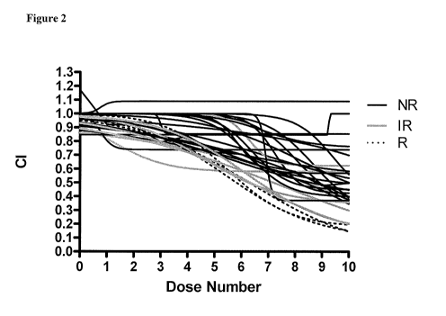
[011] Figure 2 shows in vitro chemoresponse to erlotinib in primary cultures
of
lung cancer. In vitro response of 34 lung cancer tumors after a 72-hour
treatment with
erlotinib is shown. 3 (8.8%) specimens are Responsive to erlotinib, 7 (20.6%)
are
Intermediate Responsive, and 24 (70.6%) are Non-Responsive. These results are
consistent with a reported clinical response rate of 8.9% in NSCLC patients
[13].
3
CA 02721687 2010-10-15
WO 2009/140508 PCT/US2009/043973
[012] Figure 3 shows in vitro chemoresponse to: (A) four immortalized cell
lines (SK-OV3, BT474, MDA-MB-231, MCF7), and (B) to 55 primary cultures of
breast carcinomas. All four cell lines (BT474, MDA-MB-23 1, MCF7, SK-OV3) were
responsive to lapatinib treatment, with EC50 values of approximately 10 uM.
Dose-
response curves of the 55 primary breast cultures revealed that 9% of the
specimens
tested were responsive to lapatinib, 15% had an intermediate response and 76%
were
non-responsive. These results are consistent with a reported clinical response
rate of
10% for lapatinib in breast carcinoma [New England J. Med. 2006; 355:2733-43].
[013] Figure 4 shows in vitro chemoresponse to four different immortalized
cell
lines (NCI-H292, NCI-H522, NCI-H1666, Calu3), and to 54 primary cultures of
human colorectal tumor specimens. Two of the examined cell lines showed
response
to cetuximab treatment; EC50 values for NCI-H292 and NCI-H1666 were 825 nM
and 13 nM, respectively. NCI-H522 and Calu3 were non-responsive to cetuximab.
Dose-response curves of the 54 primary colorectal cultures revealed that 8% of
the
cultures tested were responsive to cetuximab, 22% had an intermediate
response, and
70% were non-responsive. These results are consistent with a reported clinical
response rate of 11 % for cetuximab in colorectal carcinoma patients [New
England J.
Med. 2004; 351:337-45].
DETAILED DESCRIPTION OF THE INVENTION
[014] The present invention provides methods for individualizing chemotherapy
for cancer treatment, and particularly, provides an in vitro chemoresponse
assay for
evaluating a patient's responsiveness to one or more EGFR inhibitors prior to
treatment with such agents. The method generally comprises culturing malignant
cells from a patient's specimen (e.g., biopsy), contacting the cultured cells
with an
EGFR inhibitor, and evaluating the cultured cells for a response to the drug.
The in
vitro response to the drug as determined by the method of the invention is
correlative
with an in vivo response upon receiving the EGFR inhibitor during
chemotherapeutic
treatment.
Chemoresponse Assay
[015] The present invention supports individualized chemotherapy decisions for
cancer patients, and particularly with candidate EGFR inhibitors. The patient
4
CA 02721687 2010-10-15
WO 2009/140508 PCT/US2009/043973
generally has a cancer for which an EGFR is a candidate treatment, for
example,
alone or in combination with other therapy. For example, the cancer may be
selected
from breast, ovarian, colorectal, endometrial, thyroid, nasopharynx, prostate,
head and
neck, liver, kidney, pancreas, bladder, brain, and lung. In certain
embodiments, the
tumor is a solid tissue tumor and/or is epithelial in nature. For example, the
patient
may be a Her2-positive breast cancer patient, a colorectal carcinoma patient,
NSCLC
patient, head and neck cancer patient, or endometrial cancer patient.
[016] The present invention involves conducting chemoresponse testing with one
or a panel of chemotherapeutic agents on cultured cells from a cancer patient,
including one or more EGFR inhibitors. In certain embodiments, the
chemoresponse
method is as described in U.S. Patent Nos. 5,728,541, 6,900,027, 6,887,680,
6,933,129, 6,416,967, 7,112,415, and 7,314,731 (all of which are hereby
incorporated
by reference in their entireties). The chemoresponse method may further employ
the
variations described in US Published Patent Application Nos. 2007/0059821 and
2008/0085519, both of which are hereby incorporated by reference in their
entireties.
Such chemoresponse methods are commercially available as the ChemoFx Assay
(Precision Therapeutics, Inc, Pittsburgh, PA).
[017] Briefly, in certain embodiments, cohesive multicellular particulates
(explants) are prepared from a patient's tissue sample (e.g., a biopsy sample)
using
mechanical fragmentation. This mechanical fragmentation of the explant may
take
place in a medium substantially free of enzymes that are capable of digesting
the
explant. However, in some embodiments, some enzymatic treatment may be
conducted. Generally, the tissue sample is systematically minced using two
sterile
scalpels in a scissor-like motion, or mechanically equivalent manual or
automated
opposing incisor blades. This cross-cutting motion creates smooth cut edges on
the
resulting tissue multicellular particulates. The tumor particulates each
measure from
about 0.25 to about 1.5 mm3, for example, about 1 mm3.
[018] After the tissue sample has been minced, the particles are plated in
culture
flasks (e.g., about 5 to 25 explants per flask). For example, about 9 explants
may be
plated per T-25 flask, or about 20 particulates may be plated per T-75 flask.
For
purposes of illustration, the explants may be evenly distributed across the
bottom
surface of the flask, followed by initial inversion for about 10-15 minutes.
The flask
CA 02721687 2010-10-15
WO 2009/140508 PCT/US2009/043973
may then be placed in a non-inverted position in a 37 C CO2 incubator for
about 5-10
minutes. Flasks are checked regularly for growth and contamination. Over a
period
of a few weeks a cell monolayer will form. Further, it is believed (without
any
intention of being bound by the theory) that tumor cells grow out from the
multicellular explant prior to stromal cells. Thus, by initially maintaining
the tissue
cells within the explant and removing the explant at a predetermined time
(e.g., at
about 10 to about 50 percent confluency, or at about 15 to about 25 percent
confluency), growth of the tumor cells (as opposed to stromal cells) into a
monolayer
is facilitated. Further, in certain embodiments, the tumor explant may be
agitated to
substantially release tumor cells from the tumor explant, and the released
cells
cultured to produce a cell culture monolayer. The use of this procedure to
form a cell
culture monolayer helps maximize the growth of representative tumor cells from
the
tissue sample.
[019] Prior to the chemotherapy assay, the growth of the cells may be
monitored,
and data from periodic counting may be used to determine growth rates which
may or
may not be considered parallel to growth rates of the same cells in vivo in
the patient.
If growth rate cycles can be documented, for example, then dosing of certain
active
agents may be customized for the patient. Monolayer growth rate and/or
cellular
morphology and/or epithelial character may be monitored using, for example, a
phase-contrast inverted microscope. Generally, the monolayers are monitored to
ensure that the cells are actively growing at the time the cells are suspended
for drug
exposure. Thus, the monolayers will be non-confluent when the cells are
suspended
for chemoresponse testing.
[020] A panel of active agents may then be screened using the cultured cells,
including one or more EGFR inhibitors. Generally, the agents are tested
against the
cultured cells using plates such as microtiter plates. For the
chemosensitivity assay, a
reproducible number of cells is delivered to a plurality of wells on one or
more plates,
preferably with an even distribution of cells throughout the wells. For
example, cell
suspensions are generally formed from the monolayer cells before substantial
phenotypic drift of the tumor cell population occurs. The cell suspensions may
be,
without limitation, about 4,000 to 12,000 cells/ml, or may be about 4,000 to
9,000
cells/ml, or about 7,000 to 9,000 cells/ml. The individual wells for
chemoresponse
testing are inoculated with the cell suspension, with each well or "segregated
site"
6
CA 02721687 2010-10-15
WO 2009/140508 PCT/US2009/043973
containing about 102 to 104 cells. The cells are generally cultured in the
segregated
sites for about 4 to about 30 hours prior to contact with an agent.
[021] Each test well is then contacted with at least one pharmaceutical agent,
or
a sequence of agents. In addition to at least one EGFR inhibitor (as discussed
in more
detail below), the panel of chemotherapeutic agents may comprise at least one
agent
selected from a platinum-based drug, a taxane, a nitrogen mustard, a kinase
inhibitor,
a pyrimidine analog, a podophyllotoxin, an anthracycline, a monoclonal
antibody, and
a topoisomerase I inhibitor. For example, the panel may comprise 1, 2, 3, 4,
or 5
agents selected from bevacizumab, capecitabine, carboplatin, cecetuximab,
cisplatin,
cyclophosphamide, docetaxel, doxorubicin, epirubicin, etoposide, 5-
fluorouracil,
gemcitabine, irinotecan, oxaliplatin, paclitaxel, panitumumab, tamoxifen,
topotecan,
and trastuzumab, in addition to other potential agents for treatment. In
certain
embodiments, the chemoresponse testing includes one or more combination
treatments, such combination treatments including one or more agents described
above. Generally, each agent in the panel is tested in the chemoresponse assay
at a
plurality of concentrations representing a range of expected extracellular
fluid
concentrations upon therapy.
[022] The efficacy of each agent in the panel is determined against the
patient's
cultured cells, by determining the viability of the cells (e.g., number of
viable cells).
For example, at predetermined intervals before, simultaneously with, or
beginning
immediately after, contact with each agent or combination, an automated cell
imaging
system may take images of the cells using one or more of visible light, UV
light and
fluorescent light. Alternatively, the cells may be imaged after about 25 to
about 200
hours of contact with each treatment. The cells may be imaged once or multiple
times, prior to or during contact with each treatment. Of course, any method
for
determining the viability of the cells may be used to assess the efficacy of
each
treatment in vitro.
[023] While any grading system may be employed, in certain embodiments the
grading system may employ from 2 to 10 response levels, e.g., about 3, 4, or 5
response levels. For example, when using three response grades, the three
grades may
correspond to a responsive grade, an intermediate responsive grade, and a non-
responsive grade. In certain embodiments, the patient's cells show a
heterogeneous
7
CA 02721687 2010-10-15
WO 2009/140508 PCT/US2009/043973
response across the panel of agents, making the selection of an agent
particularly
crucial for the patient's treatment.
[024] The output of the assay is a series of dose-response curves for tumor
cell
survivals under the pressure of a single or combination of drugs, with
multiple dose
settings each (e.g., ten dose settings). To better quantify the assay results,
the
invention employs in some embodiments a scoring algorithm accommodating a dose-
response curve. Specifically, the chemoresponse data are applied to an
algorithm to
quantify the chemoresponse assay results by determining an adjusted area under
curve
(aAUC) (see US Application No. 12/252,073, which is hereby incorporated by
reference in its entirety).
[025] In some embodiments, the agents are designated as, for example,
sensitive,
or resistant, or intermediate, by comparing the aAUC test value to one or more
cut-off
values for the particular drug (e.g., representing sensitive, resistant,
and/or
intermediate aAUC scores for that drug). The cut-off values for any particular
drug
may be set or determined in a variety of ways, for example, by determining the
distribution of a clinical outcome within a range of corresponding aAUC
reference
scores. That is, a number of patient tumor specimens are tested for
chemosenstivity/resistance to a particular drug prior to treatment, and aAUC
quantified for each specimen. Then after clinical treatment with that drug,
aAUC
values that correspond to a clinical response (e.g., sensitive) and the
absence of
significant clinical response (e.g., resistant) are determined. Cut-off values
may
alternatively be determined from population response rates. For example, where
a
patient population is known to have a response rate of 30% for the tested
drug, the
cut-off values may be determined by assigning the top 30% of aAUC scores for
that
drug as sensitive. Further still, cut-off values may be determined by
statistical
measures.
EGFR Inhibitors
[026] In accordance with the present invention, cultured cells may be tested
for
their responsiveness to any candidate EGFR inhibitor (e.g., an EGFR inhibitor
that is
a candidate treatment for the patient). The EGRF inhibitor may be an EGFR
tyrosine
kinase inhibitor, or may alternatively target the extracellular domain of the
EGFR
target.
8
CA 02721687 2010-10-15
WO 2009/140508 PCT/US2009/043973
[027] In certain embodiments, the EGFR inhibitor is a tyrosine kinase
inhibitor
such as Erlotinib, Gefitinib, or Lapatinib, or a molecule that targets the
EGFR
extracellular domain (e.g., Cetuximab or Panitumumab).
[028] Erlotinib hydrochloride (e.g., as marketed as TarcevaTM) is used to
treat
non-small cell lung cancer, pancreatic cancer and several other types of
cancer.
Erlotinib specifically targets the epidermal growth factor receptor (EGFR)
tyrosine
kinase, which is highly expressed and occasionally mutated in various forms of
cancer. It binds in a reversible fashion to the adenosine triphosphate (ATP)
binding
site of the receptor to inhibit receptor signaling. Erlotinib has shown a
survival
benefit in the treatment of lung cancer in phase III trials. It has been
approved for the
treatment of locally advanced or metastatic non-small cell lung cancer that
has failed
at least one prior chemotherapy regimen. The FDA has further approved the use
of
erlotinib in combination with gemcitabine for treatment of locally advanced,
unresectable, or metastatic pancreatic cancer. It has been reported that
responses
among patients with lung cancer are seen most often in females who were never
smokers, particularly Asian women and those with adenocarcinoma cell type.
[029] Gefitinib acts in a similar manner to erlotinib (marketed as Tarceva),
and
is marketed under the trade name IressaTM. Research on gefitinib-sensitive non-
small
cell lung cancers has shown that a mutation in the EGFR tyrosine kinase domain
may
be responsible for activating anti-apoptotic pathways. These mutations may
confer
increased sensitivity to tyrosine kinase inhibitors such as gefitinib and
erlotinib. Of
the types of non-small cell lung cancer histologies, adenocarcinoma most often
harbors these mutations. These mutations are more commonly seen in Asians,
women, and non-smokers (who also tend to more often have adenocarcinoma).
[030] Gefitinib is indicated for the treatment of locally advanced or
metastatic
non-small cell lung cancer (NSCLC) in patients who have previously received
chemotherapy. There is also potential for use of gefitinib in the treatment of
other
cancers where EGFR overexpression is involved.
[031] Lapatinib inhibits the tyrosine kinase activity associated with two
oncogenes, EGFR (epidermal growth factor receptor) and HER2/neu (Human EGFR
type 2). Over expression of HER2/neu can be responsible for certain types of
high-
risk breast cancers in women. Lapatinib is a protein kinase inhibitor shown to
9
CA 02721687 2010-10-15
WO 2009/140508 PCT/US2009/043973
decrease tumor-causing breast cancer stem cells. Lapatanib inhibits receptor
signal
processes by binding to the ATP-binding pocket of the EGFR/HER2 protein kinase
domain, preventing self-phosphorylation and subsequent activation of the
signal
mechanism.
[032] Lapatinib is used as a treatment for women's breast cancer in patients
who
have HER2-positive advanced breast cancer that has progressed after previous
treatment with other chemotherapeutic agents, such as anthracycline, taxane-
derived
drugs, or trastuzumab (Herceptin,Genentech).
[033] Cetuximab is a chimeric monoclonal antibody targeting EGFR, and is
given by intravenous injection for treatment of metastatic colorectal cancer
and head
and neck cancer. Cetuximab may act by binding to the extracellular domain of
the
EGFR, preventing ligand binding and activation of the receptor. This blocks
the
downstream signaling of EGFR resulting in impaired cell growth and
proliferation.
Cetuximab has also been shown to mediate antibody dependent cellular
cytotoxicity
(ADCC).
[034] Cetuximab is used in metastatic colon cancer and is given concurrently
with the chemotherapy drug irinotecan (Camptosar), a form of chemotherapy that
blocks the effect of DNA topoisomerase I, resulting in fatal damage to the DNA
of
affected cells. Cetuximab was approved by the FDA for use in combination with
radiation therapy for treating squamous cell carcinoma of the head and neck
(SCCHN) or as a single agent in patients who have had prior platinum-based
therapy.
[035] Panitumumab is a recombinant, human IgG2 kappa monoclonal antibody
that binds specifically to the human epidermal growth factor receptor (EGFR).
Panitumumab is indicated as a single agent for the treatment of EGFR-
expressing,
metastatic colorectal carcinoma with disease progression on or following
fluoropyrimidine-, oxaliplatin-, and irinotecan-containing chemotherapy
regimens.
[036] EGFR inhibitors are generally plagued by low population response rates,
leading to ineffective or non-optimal chemotherapy in many instances, as well
as
unnecessary drug toxicity and expense. For example, a reported clinical
response
rate for treatment of breast carcinoma with lapatinib is 10% [New England J.
Med.
2006; 355:2733-43], a reported clinical response rate for treatment of
colorectal
CA 02721687 2010-10-15
WO 2009/140508 PCT/US2009/043973
carcinoma with cetuximab is 11% [New England J. Med. 2004; 351:337-45], and a
reported clinical response rate for treatment of NSCLC with erlotinib is 8.9%
[13].
[037] The method of the invention predicts patient responsiveness to EGFR
inhibitors at rates that match reported clinical response rates for the EGFR
inhibitors.
EXAMPLES
EXAMPLE 1: ERLOTINIB
[038] Three human lung tumor-derived immortalized cell lines were tested in
this study: H292, H358, and Calu3 (American Type Culture Collection, Manassas,
VA). These cell lines were seeded at 40,000 cells in T25 flasks (PGC
Scientifics,
Frederick, MD) and allowed to grow for one week to approximately 90%
confluence.
[039] Patient tumor specimens: Primary cell cultures were established using
tumor specimens procured for research purposes from the following sources:
National
Disease Research Interchange (Philadelphia, PA), Cooperative Human Tissue
Network (Philadelphia, PA), Forbes Regional Hospital (Monroeville, PA),
Jameson
Hospital (New Castle, PA), Saint Barnabas Medical Center (Livingston, NJ),
Hamot
Medical Center (Erie, PA), and Windber Research Institute (Windber, PA). The
tumors were removed from the patient at the time of surgery, placed in the
supplied
125-mL bottle containing sterile McCoy's shipping medium (Mediatech, Herndon,
VA), and shipped overnight to Precision Therapeutics, Inc. laboratories
(Pittsburgh,
PA).
[040] Erlotinib hydrochloride was kindly provided by OSI Pharmaceuticals
(Melville, NY) as a lyophilized powder. The drug was reconstituted to 5 mM in
100% DMSO and frozen at -80 C.
[041] Cell lines and tissue specimens were processed and tested with the
ChemoFx assay as described elsewhere [21]. Also see, US Patent Nos.
5,728,541,
6,900,027, 6,887,680, 6,933,129, 6,416,967, 7,112,415, and 7,314,731; and US
Application Nos. 10/399,563, 11/504,098, 11/595,967, 11/713,662, and
11/785,984,
all of which are hereby incorporated by reference in their entireties, and
especially
with regard to tissue processing and cell culturing techniques, and assays.
Ten doses
of erlotinib were prepared by serial dilution. The same 10-dose concentration
range
11
CA 02721687 2010-10-15
WO 2009/140508 PCT/US2009/043973
was used for the cell lines and the tissue specimens. For each dose, a
cytotoxic index
(CI) was calculated according to the following formula: CI = Mean cell count
dose x/
mean cell count control, which represents the ratio of cells killed as a
result of the
treatment. Cell counts were the average of 3 replicates at each dose for
primary
cultures and 9 replicates at each dose for immortalized cell lines. Dose
response
curves were generated using the CI at each dose. Adjusted areas under the
curve
(aAUC) were calculated for each dose-response curve as previously described
[20].
Assay results were classified as responsive (R; assay score ?7.48),
intermediate
responsive (IR; assay score 6.89-7.47), or non-responsive (NR; assay score <
6.88).
Results
[042] The chemoresponse assay was performed on 3 NSCLC lung cancer cell
lines (H292, H358, and Calu-3) to determine whether the assay was able to
detect
sensitivity of the cells to erlotinib. The 3 cell lines exhibited a
heterogeneous
response to erlotinib (Figure 1). The assay prediction of response for H358
was non-
responsive (NR), for Calu-3 was intermediate responsive (IR), and for H292
responsive (R).
[043] Table I. Comparison of response to erlotinib treatment: in vitro
chemoresponse assay and ex vivo human tumor xenograft outcomes on NSCLC cell
lines.
Cell Line ChemoFx Assay Xenograft TGI (%)
Designation [22]
H292 R 85
Calu3 IR 67
H358 NR 25
R=responsive, IR = intermediate responsive, NR=non-responsive; TGI=tumor
growth
inhibition
[044] Each dose-response curve includes 9 replicates at each dose for each
cell
line tested; each assay included 3 replicates, and 3 assays were run per cell
line. The
coefficient of variance (CoV) was calculated for each cell line using the Log
EC50
12
CA 02721687 2010-10-15
WO 2009/140508 PCT/US2009/043973
values (by dose number) for each assay. H292 had a CoV of 7%, H358 was 9%, and
Calu-3 was 3%.
[045] Table II. Coefficient of Variance for the ChemoFx Assay in evaluating
response to Erlotinib in 3 NSCLC cell lines.
Mean
Cell Line CoV
(Log EC50*)
H292 4.731 7%
Calu3 6.715 3%
H358 5.925 9%
*By dose number
CoV=coefficient of variance
[046] The in vitro responsiveness to erlotinib of the 3 NSCLC cell lines was
compared with published reports of the responsiveness of human tumor
xenografts
[22]. The sensitivity of tumor growth inhibition in xenografts derived from
these cell
lines is consistent with the in vitro prediction of response in the same cell
lines (Table
I).
[047] Of the 34 lung cancer patient specimens evaluated in this study, 22
(64.7%) were confirmed to be NSCLC, 11 (32.4%) were of unconfirmed lung cancer
subtype, and 1 (2.9%) was confirmed as not NSCLC (mesothelioma). The 34 tumor
specimens exhibited heterogeneity of in vitro response to erlotinib (Figure
2). Of the
34 patient specimens, 3 (8.8%) were assay responsive to erlotinib, 7 (20.6%)
were
intermediate responsive, and 24 (70.6%) were non-responsive.
[048] These results indicate that the assay described herein is able to
distinguish
tumor response to erlotinib in patients with lung carcinoma. The invention is
thus
useful as a decision support tool to assist oncologists in making treatment
decisions
involving erlotinib in lung cancer patients. The approach described above was
to first
conduct the assay on NSCLC cell lines to determine its ability to distinguish
in vitro
response to erlotinib. The responses of the three NSCLC cell lines tested
(H292,
H358, Calu-3) to erlotinib in the current study were similar to the responses
observed
in previously published studies using other types of chemoresponse assays
[23,24]. In
13
CA 02721687 2010-10-15
WO 2009/140508 PCT/US2009/043973
addition, the assay is shown to be highly reproducible (i.e. low process
variability) in
assessing chemoresponse to erlotinib in 3 separate NSCLC cell lines.
[049] To confirm the range of responses observed, we next compared the in
vitro
sensitivity of these cell lines to the observed outcomes of ex vivo human
tumor
xenografts derived from those same cell lines as an estimation of correlation
with
clinical response. Corresponding sensitivities support the hypothesis that in
vitro
response may predict clinical response.
[050] Having evidence that the assay prediction of response to erlotinib of
the
NSCLC cell lines corresponded to responsiveness of human tumor xenografts
produced from the same cell lines, we next examined human lung tumor
specimens.
The assay was able to distinguish sensitivity to erlotinib among 34 human
tumor
specimens. Our finding that 8.8 % of the tumors were responsive to erlotinib
is
similar to the 8.9 % reported response rate in a phase 3, randomized, double
blind,
placebo-controlled study of previously treated NSCLC patients [13].
[051] Currently, erlotinib is FDA approved only as second- or third-line
treatment for advanced NSCLC. Reports from clinical trials to date have not
shown a
benefit of erlotinib in first line treatment [8-11]. However, subgroup
analyses have
shown that groups of patients differed in their sensitivity and clinical
response to
erlotinib [10,13,25]. Investigators have speculated about the potential
findings had
the large, first-line studies been conducted on selected populations showing
increased
sensitivity [6]. Thus, an accurate and reliable test to identify erlotinib-
sensitive
subpopulations of NSCLC patients would be of crucial benefit. Much interest
has
been focused on identifying patients sensitive to EGFR inhibition using
molecular
profiles, such as EGFR mutations and amplifications as well as increased gene
number [26,27]. A chemoresponse assay that can reproducibly and reliably
identify
sensitive patients by in vitro tumor response would be a superior alternative
to support
clinical decision making, in some embodiments, may be performed alongside
molecular profiling.
EXAMPLE 2: LAPATINIB
[052] Lapatinib (Tykerb ) is a small molecule tyrosine kinase inhibitor which
targets the intracellular domain of both the epidermal growth factor receptor
and
Her2, thereby inhibiting cell growth and proliferation. Lapatinib is currently
FDA-
14
CA 02721687 2010-10-15
WO 2009/140508 PCT/US2009/043973
approved to treat Her2 positive breast cancer which has previously been
treated with
anthracycline and taxane therapies and trastuzumab. Due to the low population
response rate of lapatinib, a biomarker which can identify patients with an
increased
likelihood for response would be of great clinical utility. This example
demonstrates
an in vitro chemoresponse assay developed to predict sensitivity and
resistance of
primary cultures of human breast tumor specimens to lapatinib.
[053] The chemoresponse assay for lapatinib was developed using four different
immortalized cell lines (SK-OV3, BT474, MDA-MB-23 1, MCF7). In addition to
cell
lines, the chemoresponse assay was also performed on 55 primary cultures of
breast
carcinomas. All cultures were confirmed to contain keratin-positive epithelial
cells
using fluorescence immunocytochemistry. Cell lines and specimens were treated
with
a 10 dose concentration range of lapatinib for 72 hours and stained with DAPI;
remaining live cells were counted on an inverted fluorescent microscope.
Resulting
dose-response curves were analyzed and categorized as responsive, intermediate
responsive, or non-responsive.
[054] All four cell lines (BT474, MDA-MB-231, MCF7, SK-OV3) were
responsive to lapatinib treatment, with EC50 values of approximately 10 uM
(Figure
3A). Dose-response curves of the 55 primary breast cultures revealed that 9%
of the
specimens tested were responsive to lapatinib, 15% had an intermediate
response and
76% were non-responsive. These results are consistent with a reported clinical
response rate of 10% (Figure 3B).
[055] In conclusion, these results demonstrate that in vitro chemoresponse
testing is useful in predicting patient response to lapatinib. This test may
increase the
efficacy of the current chemotherapy decision-making process for patients.
EXAMPLE 3: CETUXIMAB
[056] Cetuximab (Erbitux ) is a chimeric monoclonal antibody that binds to the
extracellular domain of the epidermal growth factor receptor. This interaction
interferes with binding of the ligand and causes internalization of the
receptor which
blocks the downstream signaling of EGFR, resulting in impaired cell growth and
proliferation. Cetuximab has also been shown to mediate antibody dependent
cellular
cytotoxicity. Cetuximab is FDA-approved to treat head and neck cancer and
colorectal carcinomas; it is also being evaluated in clinical trials for use
in other
CA 02721687 2010-10-15
WO 2009/140508 PCT/US2009/043973
cancers, including non-small cell lung and endometrial cancer. Due to the low
population response rate of cetuximab, a test that can identify patients with
an
increased likelihood for response would be of great clinical utility. The
current
example demonstrates an in vitro chemoresponse assay to predict response of
primary
cultures of human colorectal tumor specimens to cetuximab.
[057] The chemoresponse assay was developed using four different
immortalized cell lines (NCI-H292, NCI-H522, NCI-H1666, Calu3). The
chemoresponse assay was also performed on 54 primary cultures of human
colorectal
tumor specimens. Cell lines and specimens were treated with a 10 dose
concentration
range of cetuximab for 72 hours, stained with a nuclear dye, and remaining
post-
treatment live cells were counted. Resulting dose-response curves were
analyzed.
[058] Two of the examined cell lines showed response to cetuximab treatment;
EC50 values for NCI-H292 and NCI-H1666 were 825nM and l3nM, respectively
(Figure 4A). NCI-H522 and Calu3 were deemed non-responsive to cetuximab.
Dose-response curves of the 54 primary colorectal cultures revealed that 8% of
the
cultures tested were responsive to cetuximab, 22% had an intermediate
response, and
70% were deemed non-responsive (Figure 4B). These results are consistent with
a
reported clinical response rate of 11 % for cetuximab in colorectal carcinoma
patients.
[059] These results demonstrate that in vitro chemoresponse testing is useful
for
predicting patient responsiveness to cetuximab. Use of these embodiments in
practice
could increase the efficacy of the current chemotherapy decision-making
process for
patients.
16
CA 02721687 2010-10-15
WO 2009/140508 PCT/US2009/043973
References
The following references are hereby incorporated by reference in their
entireties:
1. American Cancer Society Cancer Facts & Figures 2008. 2008;
2. Jemal A, Siegel R, Ward E et al. Cancer statistics, 2008. CA Cancer J Clin
2008;58:71--96.
3. Schiller JH, Harrington D, Belani CP et al. Comparison of four chemotherapy
regimens for advanced non-small-cell lung cancer. N Engl J Med 2002;346:92--
98.
4. Sandler A, Gray R, Perry MC et al. Paclitaxel-carboplatin alone or with
bevacizumab for non-small-cell lung cancer. N Engl J Med 2006;355:2542--2550.
5. Giaccone G HER1/EGFR-targeted agents: predicting the future for patients
with
unpredictable outcomes to therapy. Ann Oncol 2005;16:538--548.
6. Sequist LV, Lynch TJ EGFR Tyrosine Kinase Inhibitors in Lung Cancer: An
Evolving Story. Annu Rev Med 2008;59:429--442.
7. Ciardiello F, Tortora G EGFR antagonists in cancer treatment. N Engl J Med
2008;358:1160--1174.
8. Giaccone G, Herbst RS, Manegold C et al. Gefitinib in combination with
gemcitabine and cisplatin in advanced non-small-cell lung cancer: a phase III
trial--
INTACT 1. J Clin Oncol 2004;22:777--784.
9. Herbst RS, Giaccone G, Schiller JH et al. Gefitinib in combination with
paclitaxel
and carboplatin in advanced non-small-cell lung cancer: a phase III trial--
INTACT 2.
J Clin Oncol 2004;22:785--794.
10. Herbst RS, Prager D, Hermann R et al. TRIBUTE: a phase III trial of
erlotinib
hydrochloride (OSI-774) combined with carboplatin and paclitaxel chemotherapy
in
advanced non-small-cell lung cancer. J Clin Oncol 2005;23:5892--5899.
11. Gatzemeier U, Pluzanska A, Szczesna A et al. Phase III study of erlotinib
in
combination with cisplatin and gemcitabine in advanced non-small-cell lung
cancer:
the Tarceva Lung Cancer Investigation Trial. J Clin Oncol 2007;25:1545--1552.
17
CA 02721687 2010-10-15
WO 2009/140508 PCT/US2009/043973
12. Thatcher N, Chang A, Parikh P et al. Gefitinib plus best supportive care
in
previously treated patients with refractory advanced non-small-cell lung
cancer:
results from a randomised, placebo-controlled, multicentre study (Iressa
Survival
Evaluation in Lung Cancer). Lancet 2005;366:1527--1537.
13. Shepherd FA, Rodrigues PJ, Ciuleanu T et al. Erlotinib in previously
treated non-
small-cell lung cancer. N Engl J Med 2005;353:123--132.
14. Eberhard DA, Giaccone G, Johnson BE Biomarkers of response to epidermal
growth factor receptor inhibitors in Non-Small-Cell Lung Cancer Working Group:
standardization for use in the clinical trial setting. J Clin Oncol
2008;26:983--994.
15. Paez JG, Janne PA, Lee JC et al. EGFR mutations in lung cancer:
correlation with
clinical response to gefitinib therapy. Science 2004;304:1497--1500.
16. Pao W, Miller V, Zakowski M et al. EGF receptor gene mutations are common
in
lung cancers from "never smokers" and are associated with sensitivity of
tumors to
gefitinib and erlotinib. Proc Natl Acad Sci U S A 2004;101:13306--13311.
17. Sequist LV, Joshi VA, Janne PA et al. Response to treatment and survival
of
patients with non-small cell lung cancer undergoing somatic EGFR mutation
testing.
Oncologist 2007; 12:90--98.
18. Gallion H, Christopherson WA, Coleman RL et al. Progression-free interval
in
ovarian cancer and predictive value of an ex vivo chemoresponse assay. Int J
Gynecol
Cancer 2006;16:194--201.
19. Herzog TJ, Fader AN, Fensterer JE et al. A chemoresponse assay and
survival in
primary ovarian cancer. J Clin Oncol 2008;ASCO Annual Meetings Proceedings:
20. Mi Z, Holmes FA, Hellerstedt B et al. Feasibility assessment of a
chemoresponse
assay to predict pathologic response in neoadjuvant breast cancer. Anticancer
Res
2008;
21. Brower SL, Fensterer JE, Bush JE The ChemoFx assay: an ex vivo
chemosensitivity and resistance assay for predicting patient response to
cancer
chemotherapy. 2008;57--78.
18
CA 02721687 2010-10-15
WO 2009/140508 PCT/US2009/043973
22. Thomson S, Buck E, Petti F et al. Epithelial to mesenchymal transition is
a
determinant of sensitivity of non-small-cell lung carcinoma cell lines and
xenografts
to epidermal growth factor receptor inhibition. Cancer Res 2005;65:9455--9462.
23. Li T, Ling YH, Goldman ID et al. Schedule-dependent cytotoxic synergism of
pemetrexed and erlotinib in human non-small cell lung cancer cells. Clin
Cancer Res
2007;13:3413--3422.
24. Yauch RL, Januario T, Eberhard DA et al. Epithelial versus mesenchymal
phenotype determines in vitro sensitivity and predicts clinical activity of
erlotinib in
lung cancer patients. Clin Cancer Res 2005;11:8686--8698.
25. Perez-Soler R, Chachoua A, Hammond LA et al. Determinants of tumor
response
and survival with erlotinib in patients with non--small-cell lung cancer. J
Clin Oncol
2004;22:3238--3247.
26. Bonomi PD, Buckingham L, Coon J Selecting patients for treatment with
epidermal growth factor tyrosine kinase inhibitors. Clin Cancer Res
2007;13:s4606--
s4612.
27. Sequist LV, Bell DW, Lynch TJ et al. Molecular predictors of response to
epidermal growth factor receptor antagonists in non-small-cell lung cancer. J
Clin
Oncol 2007;25:587--595.
19