Note: Descriptions are shown in the official language in which they were submitted.
CA 02722078 2010-11-16
WO 2008/085185 PCT/US2007/009103
TITLE OF THE INVENTION
Functional Porous Substrates for Attaching Biomolecules
FIELD OF THE INVENTION
This invention relates to functional porous substrates and, more
particularly, to such substrates used in a microarray application for
detection of biomolecules.
BACKGROUND OF THE INVENTION
Owing to their high throughput screening capability, microarrays
have become an essential tool for the healthcare and pharmaceutical'
industries where researchers are working to diagnose disease or
discover new drugs. Moreover, agriculture and homeland defense firms
are utilizing microarrays to uncover information regarding the presence
of harmful pathogenic bacteria. Such simultaneous screening is possible
by printing many microscopic spots, typically 10-250 m in size, of
biological molecules (i.e., biomolecules such as nucleic acid fragments,
antibodies, peptides, proteins, pathogens, cells and the like) as probes
onto the same substrate to forma microarray. A high density microarray
developed for research purposes typically comprises between 1000 to
50000 probe spots arranged in a predetermined regular pattern on a
substrate, thus leading to a spot density of about 50 spots/cm2 to 2500
spots/cm2. The dimension of the substrate can vary, but generally the
.substrate is the size of a 1 inch by 3 inch microscope slide. It is critical
that the substrate surface be reactive and capable of binding probe bio-
molecules of known sequence. In use, the microarray is hybridized with
target bio-molecules of unknown sequence in order to simultaneously
detect the response of the target with the different probes spotted on the
array surface. Typically, targets are labeled with fluorescent dyes and
fluorescence based detection techniques are most commonly used to
quantify the response of the target biomolecule to the probes following
hybridization. The composite quantitative response of the target to all
the probes spotted on the microarray substrate is the data resulting from
the microarray experiment.
Microarray experiments can be employed to detect the
expression levels of various genes or proteins for a given organism (i.e.
human, mouse, plant, bacteria, etc). Highly expressed genes or
1
CA 02722078 2010-11-16
WO 2008/085185 PCTNS2007/009103
proteins are much easier to detect because their concentration in a given
sample is often the greatest. However, when expression levels are low
or samples are scarce, sensitive and reliable detection technology
becomes critical. This type of detection technology is increasingly
important for studying protein-protein interactions or protein biochemical
activity since the concentration of proteins can not be amplified via
enzymatic reactions such as the polymerase chain reaction.
As a result, within the microarray industry, there is an overriding
need for confident detection of low abundant protein and/or nucleic
acids. When attempting to accurately measure or detect such low levels
in a microarray experiment, it is imperative that researchers employ
system components that maximize sensitivity and overall signal to noise
ratio. A number of approaches can be employed to impact sensitivity
and signal to noise ratio and three of the common ones are as follows:
(1) improvements in the sensitivity or detection limits of scanning
devices, (2) increased amplification of the fluorescent signal via labeling
methods, and (3) the employment of a highly sensitive substrate. The
present invention focuses on enhancing signal to noise ratio through the
employment of a highly sensitive microarray substrate.
An increase in signal strength can be achieved by increasing the
number of binding sites per unit area (functional site density), which
ultimately impacts the retention of immobilized bio-molecular probes and
the emission of an increased signal when hybridized with fluorescently
labeled target molecules. Signal clarity can also be enhanced through a
reduction in the inherent auto-fluorescence of the materials and/or
system used for detection. These approaches will ultimately influence
the signal to noise ratio, either by increasing the signal strength, and/or
reducing the noise. Several prior art approaches have been attempted.
Many common methods used to manufacture high density
microarrays use non-porous, two-dimensional glass substrates
containing functional sites for binding samples of interest. Glass is
preferred because of its inertness and low inherent auto-fluorescence
which contributes less noise to the signal being detected, usually
measured by fluorescence-based techniques. Examples of such
commercially available substrates are UltraGAPS II slides (Corning Inc.,
Life Sciences, Oneonta, NY), Nexterion Slides (Schott North America,
Inc., Louisville, KY), and Array-It slides (Telechem International Inc.,
Sunnyvale, CA). One drawback to using non-porous glass is that the
2
CA 02722078 2010-11-16
WO 2008/085185 PCT/US2007/009103
functional site density is quite low resulting in relatively weak signals,
which makes it very difficult to detect the sample of interest, especially
when trying to detect low expressing genes or proteins. This effect can
be minimized by increasing the volume or concentration of the sample of
interest, however, the approach can only be employed if a large enough
sample is available. Often, researchers are highly limited by the
quantity, concentration or volume of a given sample. A common
approach to increasing functional site density has been through the use
of porous substrates to increase the accessible surface area containing
the functional sites. Tanner et al. (US patent 6750023) teach a method
of forming a functional material for attaching an array of biological or
chemical analytes by applying an inorganic porous layer to an inorganic
non-porous understructure.
Alternate approaches using organic polymers as functional
materials have been attempted. Haddad et al. (WO 01/66244) teach
making arrays utilizing textured non-porous functional materials created
from oriented polymer films. Porous organic polymers have also been
used in microarray substrates and examples of such commercially
available materials are Vivid Microarray Slides (Pall Corporation, East
Hills, NY) and CAST' slides (Schleicher & Schuell Biosciences, Inc.,
Keene, NH), both using porous nylon membranes.
Phase inversion is a common technique used to make
microporous membranes from organic polymers. Use of such
membranes as microarray substrates is described in detail in U.S. Patent
Applications 2003/0219816 of Solomon et al and 2004/0157320 of
Andreoli et at. A variety of microporous materials are discussed in the
literature, with nylon and nitrocellulose being the most common. Nylon
affords the benefits that it can be readily rendered microporous and has
a natural affinity for DNA. Similarly, nitrocellulose is known to be
effective in binding proteins. In the case of nylon and nitrocellulose,
binding with DNA and/or proteins is reliant on the inherent functional
groups present in the nylon or nitrocellulose polymer backbone.
Consequently, the functional site density afforded by these materials is
limited. Moreover, the pore size of phase inversion membranes may not
be small enough to prevent lateral spot spreading which leads to
crosstalk thereby limiting the array density. Another common problem
with using organic polymers such as nylon or nitrocellulose resides with
the fact that these materials possess inherently high auto-fluorescence.
3
CA 02722078 2010-11-16
WO 2008/085185 PCT/US2007/009103
Since fluorescence-based detection is the most commonly used
technique to quantify the hybridized target biomolecules, high auto-
fluorescence contributes to increased background noise thereby
adversely affecting the clarity of the fluorescent signal. Use of pigments
such as carbon black has been shown to reduce the auto-fluorescence.
Alternatively, as taught by Montagu (WO 2004/018623), the background
noise can also be reduced by the use of a thin (less than about 5 p)
functional material.
The need exists for an array substrate that can be easily
fabricated, provides high functional site density and exhibits low auto-
fluorescence to maximize signal to noise ratio. The present invention
addresses all of these needs along with providing very high level of
precision.
SUMMARY OF THE INVENTION
This invention provides a substrate comprising a microporous
microstructure, an interlayer over at least a portion of the microstructure
and a functional layer attached to the interlayer, the functional layer
having functional sites with a density of at least 50 nanomoles/cm2.
In another aspect, this invention provides a method of creating a
functionalized article comprising the steps of (1) providing a microporous
substrate having a microstructure, (2) depositing an interlayer over the
microstructure, (3) and attaching a functional layer to the interlayer such
that the article has a functional site density of at least 50 nanomoles/cm2.
In yet another aspect, this invention provides an article comprising
a support layer adjacent to a polytetrafluoroethylene substrate
comprising a porous microstructure, an interlayer over at least a portion
of the microstructure and a functional layer attached to the interlayer, the
functional layer having functional sites with a density of at least 50
nanomoles/cm2.
In still another aspect, this invention provides an article comprising
a support layer adjacent to a polytetrafluoroethylene substrate
comprising a porous microstructure, an interlayer over at least a portion
of the microstructure and a functional layer attached to the interlayer, the
functional layer having functional sites with a density of at least 50
nanomoles/cm2 , and a biomolecule attached to the functional material.
In a further aspect, this invention provides a method of measuring
biomolecules comprising the steps of-
4
CA 02722078 2010-11-16
WO 2008/085185 PCT/US2007/009103
(a) providing a support layer,
(b) functionalizing the support layer,
(c) disposing an adhesive on at least part of the support layer,
(d) attaching a microporous polytetrafluoroethylene substrate
S having a node and fibril microstructure to the support layer via
the adhesive,
(e) functionalizing the microporous polytetrafluoroethylene
substrate to form functional sites,
(f) binding biomolecules to the functional sites, and
(g) detecting the amount of biomolecules bound to the
functionalized layer.
In a further aspect, this invention provides a method of preparing
a microarray substrate comprising
(a) providing a support layer,
(b) optionally functionalizing said support layer,
(c) disposing an adhesive on at least a part of said support layer,
(d) attaching a microporous polytetrafluoroethylene substrate
having a node and fibril microstructure to said support layer via
said adhesive, and
e) functionalizing said microporous polytetrafluoroethylene
substrate.
In another aspect, this invention provides a microarray substrate
comprising an auto-fluorescence level less than 100 RFU at a
wavelength of 635 m and a functional site density greater than 50
nanomoles/cm2.
In another aspect, this invention provides a microarray substrate
comprising an auto-fluorescence level less than 1000 RFU at a
wavelength of 532 m and a functional site density greater than 50
nanomoles/cm2.
In another aspect, this invention provides a microarray substrate
comprising a signal to noise ratio for the Cy5 dye greater than 130,
preferably greater than 150.
In another aspect, this invention provides a microarray substrate
comprising a signal to noise ratio for the Cy3 dye is greater than 90,
preferably greater than 110.
In another aspect, this invention provides a microarray substrate
comprising a 1.5 fold precision level of at least 99%.
5
CA 02722078 2010-11-16
WO 2008/085185 PCT/US2007/009103
In another aspect, this invention provides a microarray substrate
comprising a 1.2 fold precision level of at least 76%.
BRIEF DESCRIPTION OF THE DRAWINGS
Fig. 1 is a transverse cross-sectional view of one end of an
exemplary embodiment of the present invention.
Fig. 2(A) is a scanning electron micrograph of the microporous
surface of a microarray of an exemplary embodiment of the present
invention prior to being functionalized.
Fig. 2(B) is a scanning electron micrograph of the microporous
surface of a microarray of an exemplary embodiment of the present
invention subsequent to being functionalized.
Fig. 2(C) is a scanning electron micrograph of the microporous
surface of a microarray of an exemplary embodiment of the present
invention subsequent to being functionalized.
Fig. 3(A) is a scatter plot of normalized Cy3 and Cy5 signal
intensity of microarrays using the substrate of an exemplary embodiment
of the present invention.
Fig. 3(B) is a scatter plot of normalized Cy3 and Cy5 signal
intensity of microarrays using a substrate of the prior art.
DETAILED DESCRIPTION OF THE INVENTION
The present invention is directed toward improved functional
porous and microporous substrates with high functional group density
and low auto-fluorescence which when used as a microarray substrate
for bio-analytical detection provide heretofore unobtainable high signal-
to-noise ratios with high precision level. These attributes derive from the
unique combination of the selection of the porous and microporous
materials and a method for functionalizing these materials. A microarray
can be defined as a tool used to sift through and analyze the information
contained within a genome. This tool contains different bio-molecular
(nucleic acid, protein, cell, etc) probes that are chemically attached to a
substrate, which can be a microchip, a glass slide or a microsphere-size
bead. In the following discussion, "porous material" refers to a material
with pores that extend through the entire cross-section thereby making
the material permeable to fluids. Porous materials are typically
6
CA 02722078 2010-11-16
WO 2008/085185 PCT/US2007/009103
characterized by the mean flow pore size. Alternatively, porous materials
can be characterized by bubble point which is a measure of the
maximum pore size. Both the mean flow pore size as well the bubble
point can be measured by pressure-flow tests. Microporous materials
are a subset of porous materials where the mean flow pore size is less
than about 1 m or the bubble point is greater than about 10psi.
Porous materials of this invention are planar in nature and can be
in the form of membranes or sheets. The porous substrates of this
invention are permeable to fluids due to the presence of interconnecting
pores that traverse the entire cross-section. The surface area of this
microstructure is considerably higher than that of a non-porous material
of equal volume. The present invention utilizes this microstructure and
its attendant high surface area to volume ratio in creating the high
functional density substrate. This internal surface area, better referred to
as specific surface area, is related to the pore size of the porous
material; the surface area increases as the average pore size of the
material decreases. Typically, the specific surface area of the porous
material is at least 0.1 m2/gm and preferably greater than 1 m2/gm and
most preferably greater than 10 m2/gm as measured by standard gas
adsorption techniques.
A "functional site" as used herein is a site located at either an
external or internal surface of the porous substrate. Functional sites may
be generated using the surface modification techniques described
herein, and are useful for providing binding sites to which biomolecules
may be attached. In certain preferred embodiments, the biomolecules
that are attached to the functional sites serve as probe molecules to
which a target biomolecule (typically an analyte in solution) can be
bound, either covalently or non-covalently. Non-limiting examples of
biomolecules contemplated by the invention include nucleic acids,
oligonucleotides, and antibodies. "Functional group" as used herein is a
group of atoms that reacts as a single unit and determines the properties
of the functional site. A functional substrate is a porous substrate which
has functional sites residing on the surface of its microstructure. The
term "functionalize" refers to the process in which a functional group or
groups is attached to the microstructure of a porous substrate.
Porous materials with their inherent high specific surface area to
volume ratios offer more area for functionalization than non-porous
substrates, such as non-porous glass. As mentioned earlier, use of such
7
CA 02722078 2010-11-16
WO 2008/085185 PCT/US2007/009103
functional porous substrates for microarray applications have been
taught in the prior art literature such as in US Patent Application
2004/0157320 to Andreoli and to US Patent 6750023 to Tanner.
Although these teachings take advantage of the increased internal
surface area afforded by the porous microstructure, they rely on the
inherent functional group density of the porous material for binding sites.
For example, Andreoli teaches the use of porous nylon as the substrate
with the functionality provided by the amide groups within the chemical
structure of the nylon molecule. In comparison, Tanner teaches the use
of porous glass but relies on the presence of surface hydroxyl.group on
the glass surface for subsequent functionalization through silane
treatment.
The present invention takes a novel approach in creating the high
functional density substrate for microarray application. The inventive
approach starts with a porous material that does not rely on the inherent
chemical nature of the material for creating the functional groups.
Instead, the microstructure of the porous material is substantially coated
with an intermediate layer containing a reactive functionality, such as
hydroxyl functionality. The functional substrate is then created by
reacting appropriate functional chemistries with the hydroxyl functionality
of the intermediate layer. Functionalizing the substrate may thus include
the step of depositing an interlayer over the porous micro-structure. In
this approach, choice of the intermediate layer and not of the porous
material, now controls the density of the functional groups. All prior art
materials that were subsequently functionalized in accordance with these
teachings of the present invention exhibited much higher functional
group density.
The high functional density substrate of the present invention is
obtained by starting with a porous material, preferably in a planar form
such as membranes or sheets. The porous materials can be organic or
inorganic in nature. Non-limiting examples of such organic porous
materials could be porous sheets of ultahighmolecular weight
polyethylene (UHMWPE) sold by Porex Corporation, polypropylene (PP)
or polytetrafluoroethylene (PTFE) available from Small Parts,
Incorporated (Miami Lakes, FL). Membranes made from organic
polymers are typically microporous in nature and are available
commercially. Examples of such materials are expanded PTFE (ePTFE)
membranes available from W. L. Gore and Associates, nylon and
8
CA 02722078 2010-11-16
dF
WO 2008/085185 PCT/US2007/009103
polyvinylidenefluoride (PVDF) membranes available from Pall Corp
under BiodyneTM and BiotraceTM brand names respectively, PP
membrane available from Osmonics Inc. under PolySepTM brandname
and PTFE membrane from Porex Corporation under MuporTM brand
name. Inorganic porous materials are typically available as rigid sheets.
Such materials typically are obtained by sintering inorganic materials
such as metals, ceramics and metal oxides. Porous glass is a common
example of such sintered material and is available from companies such
as R&H Filter company (Georgetown, DE) or Advanced Glass &
Ceramics (Holden, MA). Through proper choice of the porous material
and subsequent functionalizing chemicals, the high functional density
substrate of the present invention can also be made to have low auto
fluorescence. Generally, materials devoid of conjugated bonds in their
chemical structure exhibit low fluorescing properties. Examples of such
porous materials are those that are made from materials such as PTFE,
UHMWPE, PP, and glass.
Expanded PTFE (ePTFE) is particularly preferred as the porous
material because of its low auto-fluorescence as well as for its chemical
inertness and high temperature stability. Methods for making ePTFE are
described in U.S. Patents 3,953,566 to Gore. Expanded PTFE is a
microporous form of PTFE consisting of irregular shaped pores.
Whereas the exceptionally high surface area to volume ratio of
microporous expanded PTFE (ePTFE) suggests that it might serve well
in this application, the irregular pore shape makes it an unlikely
candidate. Surprisingly, however, the irregularity of the ePTFE structure,
with its non-circular pores, does not compromise the performance;
indeed, ePTFE is the most preferred porous material. The pores of
expanded PTFE are created by an expansion-by-stretching process
performed at elevated temperatures. Expansion creates a microporous
structure in which nodes are interconnected by fine fibrils. Preferred
ePTFE materials are made in accordance with the teachings of U.S.
Patent 4,187,390 to Gore.
The choice of pore size is a key factor in selecting the porous
material. In order to be an effective microarray substrate, the pores must
be small enough to inhibit lateral spreading of the solution during the
spotting process. It is believed that if the pores are too large, the
spotting liquid will spread in all directions and the spots will run into each
other thereby leading to crosstalk and contamination. The spot to spot
9
CA 02722078 2010-11-16
WO 2008/085185 PCT/US2007/009103
distance can be increased to avoid this problem, however, this
compromises the number of spots that can be placed within a given
area. On the other hand, the pores must be large enough to enable the
bio-molecules to enter the pores and to allow reagents to enter and exit
the pores freely during washing processes. Bubble point measurement
is a standard technique to characterize the maximum pore size of porous
materials.
Whereas most porous substrates can be used to create the high
functional density substrate of the present invention, for microarray
applications, the bubble point should be at least 0.007 MPa (1 psi),
preferably at least 0.070 MPa (10 psi). Preferred ePTFE materials
possess bubble point values of at least 0.070 MPa (10 psi). Most
preferably, the ePTFE material has bubble point values of at least 0.207
MPa (30 psi).
The microstructure of microporous ePTFE material consists of
nodes interconnected by fibrils and can be characterized by its average
fibril length. The fibril length can be measured by taking a scanning
electron micrograph of the surface of the ePTFE membrane at
reasonably high magnification (such as at 20,000x) and then measuring
the length of the fibrils between the nodes. Thirty such measurements
are taken of fibrils and the average fibril length is reported as the
average of these measurements. Larger average fibril lengths are
typically associated with lower bubble point and higher mean flow pore
size. For the preferred ePTFE materials, it is believed that the average
fibril length should be between 0.5 to 5 m, preferably between 0.5 to 3
m and most preferably between 0.5 to 2 m.
The high functional site density substrate of this invention can be
formed by using a porous material ranging in thickness from 5 m and
above. Increased thickness provides higher internal surface for
attachment of functional groups thereby leading to increased functional
density. For microarray applications, however, there is a limit to the
thickness of the porous material. An excessively thick material is not
desirable since such a material may absorb excessive amounts of probe
solution during contact printing thereby causing the printing pins to
rapidly become dry, thus affecting the spot clarity. In addition,
excessively thick materials are difficult to process particularly during the
washing step after hybridization. Inadequate washing of the
hybridization liquids from the material can lead to residual reagents
CA 02722078 2010-11-16
WO 2008/085185 PCT/US2007/009103
causing increased auto-fluorescence of the substrate. For a microarray
substrate, the preferred thickness of the porous material is about 250 gm
or less, most preferably about 125 m or less.
The present invention relies on the internal surface of the porous
material to attach the functional group. Internal surface area is a function
of the thickness and the specific surface area of the porous material.
Expectedly, specific surface area is an important consideration for
selection of the porous material. However, specific surface area is
related to the pore size. Typically, the smaller the pore size, the greater
the specific surface area. Consistent with pore size requirements,
porous material with any specific surface area can be converted into a
high functional site density of this invention. However, for a viable
microarray substrate, the porous material should possess a specific
surface area of at least 1 m2/gm. The preferred ePTFE material has at
least a 1 m2/gm of specific surface area and most preferably at least 10
m2/gm of specific surface area.
The porous material that can be used for the present invention is
preferred to be free of any additives, particularly additives that can
contribute to increased auto-fluorescence. However, if needed, the
porous material can contain additives such as pigments, fillers, colorants,
UV absorbers and the like.
The porous materials are converted into high functional density
porous substrates of this invention by first depositing an intermediate
layer of hydroxyl containing functional coating on the entire
microstructure of the porous material and subsequently using
organosilane chemistry to react with the hydroxyl group of the
intermediate layer. Details of this conversion process are described
below primarily for the preferred ePTFE material. However, the
conversion method can similarly be applied to a large variety of porous
materials described in the preceding sections.
Because of the inherent hydrophobicity of ePTFE, polar solutions
such as microarray printing and hybridization buffers do not wet the
substrate material. Also, due to the chemical structure of ePTFE, bio-
molecules such as nucleic acids or proteins do not efficiently bind to the
material. Consequently, for effective binding of biomolecules, the ePTFE
surface must be modified and functional groups need to be subsequently
attached. Surface modification of ePTFE by coating its microstructure
using organic polymers in order to render its surface hydrophilic has
11
CA 02722078 2010-11-16
WO 2008/085185 PCT/US2007/009103
been described in U.S. Patents 5130024 and 5897955 to Fujimoto and to
Drumheller, respectively. Similar hydrophilic treatment of ePTFE using
inorganic sol-gel formulations has been described in Japanese patent
publication number 08-250101 and in US Patent application
2004/0081886A1 to Zuckerbrod.
For the present invention, it is preferred that the surface
modification be performed using low auto-fluorescing hydrophilic
coatings that provide hydroxyl groups capable of subsequent reaction
with silanes. Non-limiting examples of organic polymers that are suitable
for such hydrophilic coatings are polyvinyl alcohol, polyethyleneglycol,
polypropylene glycol, polyglycidol, poly(vinyl alcohol-co-ethylene),
poly(ethyleneglycol-co-propyleneglycol), poly(ethyleneglycol-co-
propyleneglycol), polyvinyl acetate -co-vinyl alcohol, either alone or in
combination. Optionally, these polymeric coatings can be covalently
cross-linked to themselves in situ by using suitable cross-linking agents
such as aldehydes, epoxides, anhydrides etc. Polyvinyl alcohol (PVOH)
is the preferred organic polymer for the hydrophilic treatment of ePTFE.
The optional cross-linking can be achieved by the use of aldehydes such
as glutaraldehyde.
Sol-gel solution, as described below, is a more preferred solution
inasmuch as it renders the ePTFE surface more amenable for
subsequent functionalization by providing a larger number of hydroxyl
groups. Sol-gel is a technique for preparing specialty metal oxide
glasses and ceramics by hydrolyzing chemical intermediates or mixtures
of chemical intermediates that pass sequentially through a solution state
and a gel state before being dehydrated to a glass or ceramic. The
details of the sol-gel treatment used to make ePTFE hydrophilic are
described in the Japanese patent publication number 08-250101.
The preferred sol-gel coating solution is derived from
tetraethylorthosilicate (TEOS), tetramethylorthosilicate, or a sol-gel
coating derived from sodium silicate solution or a colloidal silica
suspension. Sol-gel coating solution derived from TEOS is the most
preferred. Hydrophilic coatings, described above, can also be used to
surface modify other microporous materials including but not limited to
those made from nylon, ultrahigh molecular weight polyethylene
(UHMWPE), polypropylene, porous PTFE, PVDF, porous glass, and the
like. The hydrophilic treatment of the ePTFE and other microporous
materials can be achieved by a number of ways. Usually, this treatment
12
CA 02722078 2010-11-16
WO 2008/085185 PCT/1JS2007/009103
is achieved by applying a solution of the organic polymer or the inorganic
sol-gel to membranes by commonly known methods such as dip coating,
spraying, spin coating, brushing, roller coating, or Meyer bar coating.
Care must be taken to add only enough of the coating to render the
surface hydrophilic while maintaining the porosity of the material. Adding
excessive amounts of the hydrophilic coating also will increase the auto-
fluorescence of the substrate.
The hydrophilic treatment step, described above, renders the
ePTFE and other microporous materials hydrophilic by depositing a
hydroxyl containing coating over the entire microstructure. In a
subsequent step, the hydroxyl groups are reacted with low auto-
fluorescing organosilanes to obtain the desired functional groups
depending on the specific biomolecules to be attached. For example, if
complementary DNA (cDNA) molecules are to be attached to the
substrate, amine functionality is most suitable and such functionality can
be introduced by reacting the hydrophilized ePTFE material with suitable
straight or branched aminosilane, aminoalkoxysilane, aminoalkylsilane,
aminoarylsilane. Examples of silanes that can be used are y-
aminopropyltrimethoxysilane, y-aminopropyltriethoxysilane, N-(beta-
aminoethyl)-y-aminopropyltrimethoxysilane, N-(beta-aminoethyl)-y-
aminopropyltriethoxysilane. Such amine functionality can also be
introduced through organosilane coupled dendrimers available from
companies such as Dendritech, Inc. (Midland, MI). Using this approach,
through selection of appropriate organosilanes, different reactive
functional groups can be attached to the ePTFE substrates. For the
same functional group, the placement of the functional group from the
attachment site on the surface can also be controlled by the size of the
linker molecule used in the organosilane selected. Non-limiting
examples of reactive functional groups that can be attached are amines,
epoxides, adehydes, carboxyls, anhydrides, hydroxyl, acrylates,
methacrylates, esters, thiols, azides, sulfonates and phosphonates to
name a few. If desired, more than one functional group can be
deposited on the substrate by reacting a mixture of silanes with different
organofunctional groups with the hydroxyl groups from the intermediate
layer. The functional groups can be further reacted with other chemical
reagents to create the desired functionality for the targeted end use. For
example, epoxide groups can be further reacted with diols to recreate
hydroxyl functionality or amine functional groups can be further
13
CA 02722078 2010-11-16
WO 2008/085185 PCT/US2007/009103
crosslinked through use of maleimide - NHS based cross-linkers to
create a functionality that can react with biomolecules possessing amine
functionality such as antibodies.
The silane treatment can be achieved by treating the hydrophilic
ePTFE material with a dilute solution of the silane in an organic solvent
at low pH. Details of such silane treatment and the variety of silanes with
different organofunctional groups and different linker size can be
obtained from the brochure "Silane Coupling Agents: Connecting Across
Boundaries" available from Gelest, Inc. of Morrisville, PA. Similar
information and chemicals are also available from other companies such
as United Chemical Technologies (Bristol, PA), Dow-Corning (Midland,
MI), and GE Advanced Materials (Wilton, CT). For the present invention,
the silane treatment can be achieved through conventional liquid coating
processes such as dip coating, spraying, spin coating, brushing, roller
coating, or Meyer bar coating. Alternatively, the silane can be deposited
on the membrane microstructure through vapor phase coating. Care
must be taken to add only enough of the silane to functionalize the
ePTFE while maintaining the porosity of the material.
The process of the present invention dramatically improves the
functionality of a variety of membranes as evidenced by significantly
improved amine density values which can be measured using assays
familiar to those skilled in the art. Most remarkably, treated ePTFE and
other microporous membranes of the present invention exhibit well over
an order of magnitude improvement in amine group density compared to
prior art materials. Improvements to functionalized membranes of this
magnitude were surprising and unexpected.
The functional group density can be measured in terms of moles
of functional groups per unit superficial area of the substrate or per unit
volume of the substrate. Using the inventive method, porous materials
with amino group density ranging between 0 and 5 nanomoles/cm2 were
converted to substrates with functional group densities in the range of 50
to 1300 nanomoles/cm2.depending on the specific chemical nature of the
porous material. Given the thicknesses of these substrates, the
functional density per unit volume translates to 2500 to 150,000
nanomoles/cm3. For example, ePTFE and microporous PP materials
which were devoid of any amino functionality were converted into amino
functional substrates with amino functional density of 416 and 487
nanomoles/cm2 using the inventive method. Since the inventive method
14
CA 02722078 2010-11-16
WO 2008/085185 PCT/US2007/009103
generates functional sites by reacting different organosilanes with the
hydroxyl groups deposited during the intermediate layer coating, the
above functional group density values are not limited only to amine
functional groups. Rather, the functional site density is expected to
depend primarily on the density of hydroxyl groups resulting from the
intermediate layer coating. It is therefore anticipated that through
suitable choice of organofunctional silane compounds, functional site
density greater than 50 nanomoles/cm2 or greater than 2500
nanomoles/cm3 can be achieved irrespective of the specific nature of the
functional groups chosen.
Apart from use in microarray applications, the highly functional
substrates of this invention can be used for effective binding and capture
of a large variety of biomolecules in other applications such as in
diagnostic devices, active filtration applications, blotting applications, and
the like.
This remarkable increase in functional density of the substrate can
be achieved while keeping auto-fluorescence quite low. This
combination of high functional density and low auto-fluorescence is
highly desirable in a microarray substrate since it maximizes the
fluorescence signal from the hybridized target biomolecules over the
background noise. The auto-fluorescence of the substrate can be
determined by scanning the substrate prior to printing biomolecules
using microarray scanners available commercially from several vendors
such as Axon Instruments (Union City, CA) and Perkin-Elmer (Wellesley,
MA). Scanning can be done at multiple wavelengths of interest,
depending on the type of scanner used. Average fluorescence values
can be calculated at the wavelengths of interest from the scanned
substrate data. It should be noted that scanning can be performed at
different instrument settings such as laser power, focus depth and PMT
gain. Signal intensities are function of these settings. Therefore auto-
fluorescence values should be accompanied by the scanner settings for
a given instrument.
A GenePix 4000A scanner (Axon) was used to measure the auto-
fluorescence of the substrates for this invention. The laser power and
the focus depth of this scanner were fixed at 100% and 0 m
respectively, and all measurements were done at a PMT setting of 350.
Using the method of this invention, for low fluorescence porous materials
such as PTFE, UHMWPE, PP, glass, etc.; high functional density
CA 02722078 2010-11-16
WO 2008/085185 PCT/US2007/009103
substrates can be made with auto-fluorescence level less than 1000
relative fluorescence units (RFU) and less than 100 RFU for the 532 nm
(green) and 635 nm (red) wavelengths, respectively. In most cases the
auto-fluorescence level of such high functional site density substrates
can be much lower, typically less than 100 RFU and 30 RFU at 532 nm
and 635 nm wavelengths, respectively.
The high functional density and low auto-fluorescence porous
substrate is most suitable for microarray applications as it is expected to
provide increased signal intensity over background noise. There are
various ways such substrates can be used in microarray application.
The substrate can be used as is or alternatively the substrate can be
converted into a cassette or into the shape of a microscope slide through
combining it with other plastic, ceramic or metallic parts through insert
molding or other assembly techniques such ultrasonic bonding, RF
welding, heat welding, or the like. By choosing the appropriate
functionality, it is possible to attach large variety of biomolecules to the
high functional site density substrate of this invention. Non-limiting
examples of biomolecules that can be attached are nucleic acids,
proteins, peptides, oligonucleotides, antibodies, cells, enzymes, and
pathogens, to name a few.
For many applications, in order to ease both handling and printing,
it is desirable to support the high density functional site substrate of this
invention with a support layer. This support layer can be both flexible as
well as rigid. The flexible support layer can be plastic films and metal
foils. However, for traditional microarray application the support layer is
typically rigid. Such rigid support layer can be made from a stiff material
as long as the material maintains dimension stability at hybridization
temperatures and does not get affected by the reagents used during
printing, hybridization, washing and drying steps involved in typical
microarray experiment. Non-limiting examples of materials that are
suitable as the support layer are glass, metals, ceramics, and plastics. A
glass microscope slide is most commonly used as the support layer.
In an embodiment of the present invention, the functional
substrate may be adhesively bonded, at least partially, to the rigid
support to create a composite microarray substrate. The resulting
adhesive bond should be strong enough to survive processing steps of
printing, hybridization, washing and drying steps involved with a typical
microarray experiment. The adhesive therefore needs to possess the
16
CA 02722078 2010-11-16
WO 2008/085185 PCTIUS2007/009103
appropriate thermal and chemical resistance. It is also desired that the
adhesive exhibits as low an auto-fluorescence level as possible.
Typically, adhesives containing no conjugated bonds in their chemical
structure are likely to demonstrate low auto-fluorescence. The adhesive
chosen must bond well to the rigid support as well as to the functional
porous layer. If needed, the support surface can be treated to enhance
the bond with the adhesive. Treating the support surface with
organosilane is an example of a surface treatment that can be used to
enhance adhesion. Alternatively, adhesion-promoting additives such as
silane coupling agents can be added to the adhesive to promote better
bond between the adhesive and the rigid support. For acceptable bond
to the functional porous substrate, the adhesive needs to penetrate into
the porous structure. If the adhesive viscosity or surface tension is not
low enough to allow this penetration, the adhesive can be solvated with
low viscosity and/or low surface tension solvents to promote this
penetration. However, caution must be exercised to ensure that the
adhesive does not penetrate excessively into the cross-section of the
functional substrate since this would reduce the availability of the
functional groups as well as increase the auto-fluorescence level of the
composite microarray substrate
Various kinds of adhesive can be used. The adhesive can be
thermosetting in nature. Examples of such adhesives include but are not
limited to epoxies, acrylics, silicones. These types of adhesives can be
applied either to the support layer or to the functional layer, contacted to
the other surface to be bonded, and then cured through application of
energy in the form heat, UV radiation or the like. TRA-BOND FDA2
available from Tra-Con, Incorporated (Bedford, MA) is an example of a
two-part thermosetting epoxy that can be used. If the adhesive is in
liquid form, it can be applied by a variety of commonly used methods
such as spraying, brushing, roller coating, etc. If the adhesive is in the
form of a partially cured film, it can be laminated through application of
pressure and/or heat. The adhesive can also be pressure sensitive in
nature and belong to different chemical families. Acrylics (e.g., 3M
9461 P Adhesive transfer tape from 3M Corporation, St. Paul, MN) and
silicones (e.g. Dow-Corning MD7-4602 from Dow-Corning Corporation,
Midland, MI) are commonly used pressure sensitive adhesives (PSAs).
In this case, the adhesive is applied either to the support or the functional
substrate and bonded to the other material through application of
17
CA 02722078 2010-11-16
WO 2008/085185 PCT/US20071009103
pressure and/or heat. If the adhesive is in liquid form, it is applied as
indicated above, dried if necessary to remove any volatiles and then
bonded to its counterpart. Finally, the adhesive can be thermoplastic.
Examples of such adhesives with low fluorescing properties are
fluoroplastics (DyneonTM THV Fluorothermoplastic from Dyneon LLC,
Oakdale, MN) ; eFEPTm from Daikin America, Inc. , Orangeburg, NY;
Teflon FEP (Dupont Fluoroproducts, Wilmington, DE), Topas cyclic
olefin copolymers from Ticona, Chatham, NJ, to name a few. The film
form of these materials can be used to bond the functional layer to the
support layer through application of heat and pressure in a lamination
step. If available in resin form, the thermoplastic material can be
dissolved in a suitable solvent and applied as a thin layer on either the
functional membrane or the support layer, dried to remove the volatiles
and bonded to its counterpart. The porous material layer can be
adhesively bonded to the support prior to functionalization and then the
composite can be functionalized through the steps of hydrophilic
treatment and silane treatment as discussed previously.
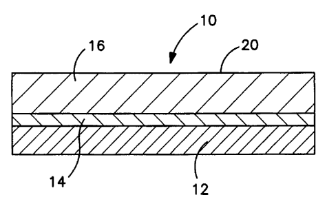
Fig. 1 shows an article 10 according to an exemplary embodiment
of the present invention. A support layer 12 has an adhesive 14
disposed thereon. A microporous fluoropolymer, ePTFE, substrate layer
16 is attached to support layer 12 by adhesive 14.
Support layer 12 is any rigid surface capable of bonding to
microporous fluoropolymer layer 16, with or without the use of an
adhesive. Glass is preferred for support layer 12. The surface of
support layer 12 is optionally treated before adhesive 14 (if used) and
microporous fluoropolymer layer 16 are applied.
A scanning electron micrograph of the surface 20 of the ePTFE
material is shown in Fig. 2(A). This figure indicates the presence of
nodes 22 interconnected by fibrils 24. The photomicrograph also depicts
the irregular pores of this material.
A scanning electron micrograph of the surface of the porous
substrate of this invention using ePTFE as the starting material is shown
in Figs. 2(B) and 2(C) at two different magnifications. The microstructure
of the porous ePTFE materials was first treated with silica sol-gel to
create an intermediate layer which was then reacted with an
aminosilane. The coated nodes 26 and coated fibrils 28 of the
microstructure are shown in Fig. 2(C).
18
CA 02722078 2010-11-16
wo 2008/085185 PCT/US2007/009103
Using the inventive method, composite microarray substrates
employing the functional ePTFE layer were made that exhibit the desired
features of unusually high functional group density and low auto-
fluorescence. In particular, depending on the characteristics of the
ePTFE, composite substrates can be made with functional group density
of at least 50 nanomoles/cm2, preferably of at least 100 nanomoles/cm2
and most preferably of at least 250 nanomoles/cm2. This is at least an
order of magnitude higher than functional group densities obtained for
prior art substrates. For example, depending on the specific
characteristics of the ePTFE material used, the amine densities
measured for the composite microarray substrates range from about 100
to 400 nanomoles/cm2. In comparison, the amine densities measured for
aminosilane treated non-porous glass slide (Corning UltragapsTM) and
porous nylon membrane based VividTM microarray slide were about 4.8
and 6.5 nanomoles/cm2 respectively.
While providing high functional site density, the inventive
composite microarray substrate maintains its auto-fluorescence at a low
level. The auto-fluorescence of the composite substrate can be
determined by scanning the substrate prior to printing biomolecules
using GenePix 4000A microarray scanner at a PMT setting of 350.
Using the method of this invention, a composite microarray substrate
comprising a functional ePTFE layer can be made with auto-
fluorescence level less than 1000 relative fluorescence units (RFU) and
less than100 RFU for the 532nm (green) and 635nm wavelengths,
respectively. In most cases the auto-fluorescence level of the composite
microarray substrates can be much lower, typically less than 200 RFU
and 30 RFU at 532nm and 635nm wavelengths, respectively.
The composite microarray substrate of this invention provides a
versatile surface for immobilization of biomolecules. Other than its use in
typical microarray analysis, the inventive substrate can also be used as a
substrate with a variety of biomolecules attached to it in any arbitrary
pattern. Examples of biomolecules that can be attached are nucleic
acids, proteins, peptides, oligonucleotides, antibodies, cells, pathogens,
to name a few.
The performance of the composite microarray substrate was
determined by conducting an evaluation in which a DNA microarray was
created using the composite substrate. The microarray was then
hybridized with cDNA labeled with two fluorescent dyes, namely Cy3 and
19
CA 02722078 2010-11-16
WO 2008/085185 PCT/US2007/009103
Cy5, that emit fluorescent signals at two different wavelengths. The
fluorescent signals, at two different wavelengths, from each spot (and its
vicinity) within the hybridized slides were then detected using a
microarray scanner using a laser light source and a photo multiplier tube
(PMT) as the detector. The scanner detects the fluorescent light
intensities from the hybridized microarray substrate and the data is
stored in the form of a scanned image of the substrate representing
intensities on a color scale. The raw signal intensity data, thus obtained,
were statistically analyzed using standard microarray data analysis
software (such as GenePix Pro from Axon Instruments, Genetraffic
from Lobion informatics.or Scanarray Express from Perkin-Elmer) to
determine some key performance metrics such as signal to noise ratio
and precision level. These performance criteria were determined for the
composite microarray substrate of this invention as well as for substrates
representing the prior art. Details of microarray data analysis are readily
available in books such as "DNA Microarrays", edited by Ulrike A Nuber,
Taylor & Francis, NY, 2005 or "Microarray analysis" by Mark Schena,
John Wiley & Sons, Hoboken, NY, 2003.
Signal to noise ratio (SNR) is a key performance measure for a
microarray substrate. The quality of the signal from a spot within a
microarray depends on its intensity relative to its immediate
surroundings, also known as local background noise. As the signal
intensity from a spot approaches the intensity of the local background
noise, the error in each measurement becomes potentially higher. At a
given wavelength, the SNR for a spot can be easily computed by first
determining the net signal intensity, which is the difference between the
median signal intensity (S) for all pixels representing a spot and its
median local background (B) for all pixels representing the immediate
area just outside the spot. The background noise (NB) is estimated by
calculating the standard deviation of the local background. SNR for the
spot is then defined as:
SNR = (S-B)/NB
in which S, B, and NB are expressed in relative fluorescence units
(RFU).
Typically, the SNR is determined for individual spots in an array.
The average SNR (ASNR) is the average of all the SNR for individual
spots in an array. Using microarray data analysis software, SNR
CA 02722078 2010-11-16
WO 2008/085185 PCT/US2007/009103
calculations can be performed automatically for the large number of
spots within a typical microarray.
High ASNR is always desired since it provides higher confidence
in the accuracy of the data obtained from a microarray experiment. The
composite microarray substrate of this invention provides remarkably
high ASNR as compared to substrates of the prior art. In general, on
average, the inventive composite substrate exhibits ASNR which is at
least twice that obtained from aminosilane treated glass slides at both of
the wavelengths. For example, the performance of a functionalized
ePTFE membrane adhered to a glass slide far exceeds that of all prior
art materials. It exhibits average signal to noise ratios for Cy5 and Cy3
of at least about 191 and 94, respectively. The most commonly used
prior art slide exhibits signal to noise ratios for Cy5 and Cy3 of about 110
and 62, respectively
It was surprising to notice that the composite microarray substrate
of the present invention not only provides high ASNR, but was also very
effective in stabilizing the fluorescent signal obtained. It is well known in
the art that the signal from Cy5 dye is extremely unstable particularly
under the influence of ozone. In fact, due to seasonal variation in ozone
level in the ambient, it is not unusual to see the stability of the Cy5 signal
deteriorate when the ambient ozone levels increase. It has now been
found that the composite microarray substrate of this invention
employing functional ePTFE layer is remarkably more effective in
stabilizing the Cy5 signal as compared to that seen on substrates of the
prior art. For example, in summer months when ambient ozone levels
were high, the Cy5 SNR for the inventive substrate was about 7.7 times
that of the Cy5 SNR on aminosilane treated glass slide and this ratio.
Within 24 hours, this ratio increased to 38.9 as the Cy5 signal on the
glass slide reduced drastically whereas the Cy5 signal was relatively
more stable on the composite substrate of this invention.
In addition to high ASNR, precision is another performance
measure that is highly desirable. In a microarray experiment, when the
same target is labeled with two different fluorescent dyes (Cy3 & Cy5); it
is expected that signals from both the wavelengths should provide the
same information. In other words, if the Cy3 signal intensity is plotted
against the Cy5 signal intensity on a graph with identical x and y axes,
ideal data should lie on the 1:1 (or 45 degree) line. In reality, the data
generally deviates from this line and the further this deviation is from the
21
CA 02722078 2010-11-16
WO 2008/085185 PCT/US2007/009103
1:1 line, the less reliable the data becomes. A measure of the precision
of the data can be obtained by devising a measure of how close the data
are to the 1:1 line. If there are M number of data points and out of that
set if N data points lie outside the Z-fold up and Z-fold down boundaries,
the Z-fold precision level can be defined as
Pz = Z-fold % Precision level = 100 x (1-(N/M))
in which Z-fold up and Z-fold down boundaries represent relationships
where the Cy3 signal intensity is Z times or 1/Z times that of the Cy5
signal intensity respectively. For example, 2-fold up implies that the Cy3
signal intensity is twice that of the Cy5 signal intensity and 2-fold down
implies that the Cy3 signal intensity is half that of the Cy5 signal
intensity. Higher Pz values at lower Z levels indicate more precise and
reliable data. The composite microarray substrate of the present
invention exhibits remarkably high precision level. Typically, P1.5 and P1.2
were at least 99% and at least 90%, respectively, for the substrates of
this invention. In comparison, respective values for aminosilane treated
glass slide were 96 and 73%, respectively. Clearly, the composite
substrate described here yields extremely precise and reliable data when
used in a microarray experiment.
EXAMPLES
Test Methods
Thickness Measurement
Membrane thickness was measured by placing the membrane
between the two plates of a Kafer FZ1000/30 thickness snap gauge
(Kafer Messuhrenfabrik GmbH, Villingen-Schwenningen, Germany). The
average of the three measurements was used.
Bubble Point Measurement
The bubble point and mean flow pore size were measured
according to the general teachings of ASTM F31 6-03 using a Capillary
Flow Porometer (Model CFP 1500 AEXL, Porous Materials Inc., Ithaca,
NY). The sample membrane was placed into the sample chamber and
wet with SilWick Silicone Fluid (Porous Materials Inc., Ithaca, NY) having
a surface tension of 19.1 dynes/cm. The bottom clamp of the sample
chamber had a 2.54 cm diameter, 3.175 mm thick porous metal disc
insert (40 micron porous metal disk, Mott Metallurgical, Farmington, CT,)
and the top clamp of the sample chamber had a 3.175 mm diameter
22
CA 02722078 2010-11-16
WO 2008/085185 PCT/US2007/009103
hole. Using the Capwin software (version 6.62.1) the following
parameters were set as specified in the table immediately below. The
values presented for bubble point and mean flow pore size were the
average of two measurements.
Parameter Set Point Parameter Set Point
maxflow cGm 200000 min time (sec) 30
bublfow cc/m 100. resslew cts 10
F/PT (old bubltime 40 19, flowslew (cts) 50
minbppres (PSI) 0 egiter 3
zerotime (sec) 1 aveiter 20
v2incr (cts) 10 max if (PSI) 0.1
re inc (cts) 1 max if cc/m
5500
1
pulse delay (sec) 2 sartp PSI
0
max re (PSI) 500 sartf cc/m
pulse width (sec) 0.2
Functional Group Density Measurement
A ninhydrin based assay was used to determine the density of the
functional amino groups. The assay was based on the teachings of
Sarin et.al. (Sarin, V.K., Kent, S.B.H., Tam, J.P. & Merrifield, R.B. (1981)
Anal. Biochem. 117, 147-157). In this assay, ninhydrin was reacted with
the substrates of this invention. The reaction product within the resulting
liquid was sprectroscopically determined to arrive at the concentration of
amine functionality. The assay used about 1cm2 size specimens
obtained from the sample substrates and the following procedure was
employed:
Reagent A - In a beaker, 40 g of phenol and 10 ml of absolute
ethanol were mixed and warmed until a clear liquid was obtained. In a
separate beaker, 0.042g of potassium cyanide (KCN) was dissolved in
65 ml of water. Approximately 2 ml of this KCN solution was then diluted
with 100 ml of absolute pyridine in a separate bottle. In a separate
container labeled "Reagent A," 6 ml of the phenol/ethanol solution was
mixed with 12.5 ml of the KCN/pyridine solution.
Reagent B - 2.5 g of ninhydrin was dissolved in 10 ml of absolute
ethanol.
23
CA 02722078 2010-11-16
WO 2008/085185 PCT/US2007/009103
Sample Analysis
In a test tube, 800 pl of Reagent A and 200 pl of Reagent B were
added. The test tube was placed in a heating block set at 100 C and the
block was placed over a shaker. The shaker was run at 110rpm for 10
min. The test tube was then removed and placed in a water bath.
Ethanol was added to the tube until the total volume was 2 ml and the
solution was well-mixed. 200 pl aliquots of this mix were pipetted into a
glass 96 well plate and the absorbance at 570 nm was measured using a
spectrophotometer.
Data Analysis -
The amine density for each sample was calculated from the
following relationship using the absorbance value after the blank
absorbance was subtracted out.
Amine Density (nanomoles/cm2) _
[Absorbance Sample * Volume (L) * 109 (nmol/mol)]/ [Ext. Co.570 (M-' cm') *
Pathlength (cm) * Area Sample (cm2)],
in which Volume = 2ml=0.002 L, Ext. Co. = Extinction Coefficient =
15,000 M" cm'-, and the pathlength used was 0.4146 cm.
For each sample, three measurements were made and the amine
density value was reported as the average of the three replicates.
In the case of unsupported functional substrates, the thickness of
the substrate was directly measured. In this case, the functional group
density was expressed as:
Functional group density (nanomoles/cm3) = functional group density
(nanomoles/cm2)/ substrate thickness (cm)
Auto-fluorescence Measurement
The auto-fluorescence of the unsupported substrates and the
microarray slides prepared with these functionalized substrates was
measured using an Axon Genepix 4000A (Axon Instruments Inc., Union
City, CA) scanner with a PMT setting of 350 and a resolution of 10 m.
Auto fluorescence was measured at wavelengths of 635 nm and 532 nm.
Slides samples (including rigid Vycor substrates) were placed in the slide
holder with the substrate facing down and scanned for auto
24
CA 02722078 2010-11-16
WO 2008/085185 PCT/US2007/009103
fluorescence. In the case of unsupported substrates such as
membranes, the samples were draped around a plain glass microscope
slide and placed in the slide holder with the substrate facing down and
scanned for auto fluorescence. The scanned image was analyzed using
GenePix Pro 5.0 software. Auto fluorescence values were recorded at
4800 discrete locations within the slide over a rectangular area. The top
left corner of this area was 2.27 mm from the left edge and 12.21 mm
from the top edge of the 25.4 mm x 76.2 mm sample. The bottom right
hand corner of this area was 18.04 mm from the left edge and 59.82 mm
from the bottom edge of the sample. The average auto fluorescence
values at the two wavelengths were reported for each sample slide and
each substrate.
Signal to Noise Measurement
Signal to noise measurements of the microarray slides for the
present invention were conducted by the Microarray Centre at University
Health Network (UHN), Toronto, Canada. A 1718 clone set from the
human genome was printed on the slide using a printing solution of the
DNA in 3x SSC at a concentration of 0.2 pg/ml. The printed array was
organized in 32 blocks arranged in 8 rows and 4 columns with a grid-to-
grid distance of 4500 p. Within each grid, there were 120 features
arranged in 10 rows and 12 columns. The feature size was 100 m and
the feature-to-feature distance was 200 p. Humidity level during printing
was controlled between 55-60%. Following printing, the printed probes
on the slide were dried at 95 C for 1 minute and then cross-linked at
2500 micro Joules of power using a UV StratalinkerTM 1800
(Stratagene).
The following labeling protocol was used to generate labeled
cDNA from 10pg total RNA.
Reverse Transcription
= In a 0.5pl tube, combine 8.0 pl of 5X First Strand buffer (Superscript II,
Invitrogen), 1.5 pI of AncT primer (5'-T20VN, 100pmol/pl), 3.0 WI of
dNTP-dTTP (6.67 mM each of dATP, dCTP, dGTP), 3.0 pl of 2 mM
dTTP, 3.0 pl of 2mM AA-dUTP (Sigma, catalog no. A-0410), 4.0 pl of
0.1 M DTT, 1.0 pl of control RNA ( artificial Arabadopsis transcripts (2-
CA 02722078 2010-11-16
WO 2008/085185 PCT/US2007/009103
ng/pl), optional), 0.1-10 pg of total RNA (0.1-0.5 pg mRNA or 5-10
pg total RNA), and 40 pl of nuclease-free water.
= Incubate the labeling reaction at 65 C for 5 minutes, then at 42 C for 2
minutes (to partially cool solution). It is not necessary for the
5 incubation to occur in the dark.
= Add 2 pl reverse transcriptase (Superscript It, Invitrogen) and incubate
at 42 c for 2 hours.
= Add 8 pl of 1 M sodium hydroxide and heat to 65 C for 15 minutes to
hydrolyze RNA.
10 = Add 8 pl of 1 M hydrochloric acid and 4 pi of 1 M tris-HCL, pH 7.5 to
neutralize the solution.
Amino allyl-cDNA purification
Purification was performed using CyScribe TM GFX TM Purification kit
(GE Amersham, catalog no. 27-9606-02). Each sample was purified in
one GFX column, using the following protocol.
= Add 500pl of capture buffer to each column.
= Transfer cDNA product (approx 62 pl) to the column, pipette up and
down several times to mix, spin at 13800xg for 30 seconds and discard
flow-through.
= Add 600 pl of 80% ethanol and spin at 1300 rpm for 30 seconds and
discard flow-through; repeat this step for a total of 3 washes.
= Spin the column for an additional 30 seconds to ensure all ethanol is
removed.
= Transfer the GFX column to a fresh tube and add 60 pl of 0.017 M
sodium bicarbonate, pH 9.
= Incubate the GFX column at room temperature for 1 minute.
= Spin at 13800xg for 1 minute to elute purified labelled cDNA.
= Use Speed Vac to completely dry sample. Resuspend in 7p1 nuclease-
free water.)
Preparing Monofunctional Reactive Cyanine Dye & Labeling
= Alexa 647 / Alexa 555 fluors (Invitrogen) and Cy5/Cy3 (Amersham)
were used in this study and both will be referred to as Cy5 & Cy3
respectively. The Alexa fluors are sold individually packaged. Add 3 PI
of DMSO per tube to resuspend the dye. Add entire contents of the
tube to each labeling reaction. The Cy dyes come in packages of 5 l.
Add 45 l of DMSO to each tube. Again, 3 l of the resuspended dye
was added to each labeling reaction.
26
CA 02722078 2010-11-16
WO 2008/085185 PCT/US2007/009103
= Add 3 pi dye to 7 I aminoallyl-labelled cDNA, mix by pipetting up and
down, and incubate in the dark at room temperature for 1 hour.
= Add 4.5 pl of 4M hydroxylamine to quench non-conjugated dye.
Incubate in the dark at room temperature for 15 minutes.
Purification of Fluorescent Labeled Probe
= Add 35 pl water to each reaction to bring each reaction volume to
about 50 pl.
= Combine the Cy5 and Cy3-labelled samples that will be co-hybridized.
= Add 500 pL of capture buffer to each column.
= Transfer labelled-cDNA product (approx. 100 pL) to the column, pipette
up and down several times to mix, spin at 13,800xg for 30 seconds and
discard flow-through.
= Add 600 pL 80% ethanol and spin at 13,800xg for 30 seconds and
discard flow-through; repeat this step for a total of 3 washes.
= Spin the column for an additional 30 seconds to ensure all ethanol is
removed.
= Transfer the GFX column to a fresh tube and add 60 pL elution buffer
(provided with kit)
= Incubate the GFX column at room temperature for 1 minute.
= Spin at 13,800xg for 1 minute to elute purified fluor-labelled cDNA.
= SpeedVac sample to dryness (on high heat; be careful not to over-dry)
and resuspend in 5pL nuclease-free water
Hybridization
= A prehybridization step is not required.
= Make enough solution for all your hybridizations - make 100 pl per
slide and an additional 100 pi for pipetting error.
= To each 100 pL of DIG Easy Hyb solution (Roche), add 5 pL of yeast
tRNA (Invitrogen; 10 mg/ml) and 5 pL of calf thymus DNA (Sigma;
10mg/ml). Incubate the mixture at 65 C for 2 minutes and cool to room
temperature.
= Add 100 pi of the prepared hybridization solution to each pooled pair of
Cy5 and Cy3-labelled cDNA (about 5 pL).
= Mix the hybridization solution with the labelled-cDNA, incubate at 65 C
for 2 minutes, and cool to room temperature
= Place cover slip (24x60 mm, non-lifter slip) onto a reliable surface (the
corner of a tip box works well) and pipette the hybridization mixture
onto the cover slip. Lay the slide "array-side" down on top of the cover
slip (do not actually put the slide down on the cover slip simply hold it
27
CA 02722078 2010-11-16
WO 2008/085185 PCT/US2007/009103
on top of the cover slip until the slide is wetted enough to pick up the
cover slip). Quickly flip the slide, with cover slip stuck to it, over so the
cover slip is on top of the slide.
= Carefully place the slide(s) into hybridization chamber(s). The
S hybridization chambers that we use are plastic microscope slide boxes
containing a small amount of DIG Easy Hyb solution in the bottom (to
keep a humid environment). Clean plain microscope slides are placed
at every second or third slide position in the slide box to create rails or
a platform onto which the hybridization arrays can be placed. Each
hybridization chamber can hold two or three hybridization slides
(depending on which direction. the slides are placed). The lid is
carefully placed onto the box and the box is then wrapped with plastic
wrap.
= Incubate on a level surface in a 37 C incubator overnight (about 16-18
hours)
Washing
= Remove the cover slip by quickly but gently dipping the array in 1X
SSC (let the cover slip slide off gently; hold the slide at the bar-code
end with forceps). Place the slide into a staining rack and place into a
staining dish (Evergreen Scientific through Diamed cat# E/S258-4100-
000) with fresh 1X SSC.
= When all of the arrays have been removed from the hybridization
chambers, wash for 3 sets of 15 minutes each at 50 C in clean slide
staining boxes containing pre-warmed (at 50 C) 1X SSC/0.1% SDS
with gentle occasional agitation
= After the washes are complete, rinse the slides twice in room
temperature 1X SSC (plunging 4-6 times) and then in 0.1X SSC
= Spin slides dry at 89xg for 5 minutes in a slide box lined with WHATMAN
paper. Alternatively, slides can be dried in a 50 mL Falcon tube (and
spun at 89xg for 5 min)
= Arrays should be stored in the dark. It is recommended that arrays be
scanned as soon as possible after they are washed (at least within two
days). The hybridized slides of this invention were scanned using
Scanarray TM 4000 scanner (Perkin Elmer, Wellesly, MA) at laser
power setting varying between 65-75 and a PMT setting ranging from
50-55.
The TIFF images were quantified using ArrayVision v.8.0 (Imaging
Research Inc.). The data and images were then loaded into
28
CA 02722078 2010-11-16
WO 2008/085185 PCT/US2007/009103
GeneTrafficTM (Lobion Informatics) for normalization. The "Lowess, sub-
grid" method was chosen for normalization in GeneTrafficTM. Normalized
intensity values were downloaded from the GeneTrafficTM database. The
average S/N was calculated in Excel for each slide type. The standard
deviation for each replicate was calculated in Excel for each slide type.
Each spot appears twice on every array so even where only one array
was tested there were 2 replicates to calculate the standard deviation of
the S/N between them. Where more arrays were used the standard
deviation of S/N was calculated across all the replicate arrays and the
replicate spots (2 per array).
Signal to noise ratio was also measured for Ultragaps slides from
Corning Life Sciences. The procedure was identical to that mentioned
above except that less (80 NI) hybridization buffer was used. Also,
scanning was performed at a different setting which was determined to
be optimum for these slides. Specifically, the laser power setting used
varied between 95 to 100 and the PMT setting ranged from 70-80.
Precision Level
Precision level measurements were performed using arrays that
were labeled with the same sample of. RNA in both the Cy5 and Cy3
channels. Ideally, all data points would fall exactly on a 45 line drawn
through the origin on a scatter plot of normalized Cy3 signal intensity
against normalized Cy5 signal intensity on a log-log plot for all the data
points (M) on the slide. From this plot, the number of data points (N) that
lie outside the Z-fold up and Z-fold down limits were determined. The
Consistency or Specificity or Precision level is defined as
Z-fold Precision Level, % = 100 x (1-(N/M))
Functionalized Substrate Examples
Sol-gel Solution
A precursor solution was prepared by allowing 40.7 parts
tetraethoxysilane (Dynasil A made by Degussa Corporation, Parsippany,
NJ), 14.1 parts deionized water, 44.8 parts ethanol and 0.4 parts
hydrochloric acid (37%) to react for 24 hours at 65 C. The solution was
then cooled and stored in a freezer until further use.
29
CA 02722078 2010-11-16
WO 2008/085185 PCT/US2007/009103
Silane Solution
A silane solution was prepared by mixing 2 parts of
aminopropyltriethoxysilane (A0750, United Chemical Technologies,
Bristol, PA) to 98 parts of a 95/5 (w/w) mixture of ethanol/water. This
solution was prepared just prior to use and was allowed to stand for at
least 5 minutes prior to its use.
Example 1
This example describes a highly functional microporous substrate
of the present invention obtained by starting with ePTFE as the porous
material. The surprising advantages of the process and articles of the
present invention are apparent when examining the substantial
improvement of the treated versus the untreated membranes.
Material type: ePTFE
Membrane
An expanded polytetrafluoroethylene (ePTFE) membrane made in
accordance with the teachings of U.S. Patent 4,187,390 was obtained.
The ePTFE membrane was about 74 p thick and had a bubble point of
about 0.434MPa (63 psi). Water beading on the surface of the
membrane attested to its hydrophobicity.
Sol-gel Treatment
The ePTFE membrane was treated by mounting the membrane
on embroidery hoops for ease of handling and then immersing the
membrane in a solution obtained by diluting the sol-gel solution with
equal amounts of ethanol by weight. After 5 minutes, the membrane was
removed and immersed in deionized water for 5 minutes. Following the
rinse step, the membrane was air dried and then heated at 150 C for 5
minutes. At this stage the membrane was hydrophilic and water readily
wet the membrane.
Aminosilane Treatment
The hydrophilic membrane was further treated with aminosilane to
provide functional amino groups on its microstructure. This was
accomplished by immersing the membrane in the silane solution for 5
minutes, then rinsing it in isopropyl alcohol (IPA) for 2 minutes and
CA 02722078 2010-11-16
WO 2008/085185 PCT/US2007/009103
heating the membrane at 110 C for 10 minutes. The membrane was
silane treated again by repeating the identical steps of 5 minute
immersion in silane solution, 2 minute rinse in IPA and heating at 110 C
for 10 minutes.
Final membrane
The resulting functionalized ePTFE membrane was 34 m thick
and a ninhydrin assay indicated the amine density to be 416.4
nanomoles/cm2 (or 121435 nanomoles/cm3). The auto fluorescence of
the functionalized ePTFE membrane was measured as 21.2 and 30.4
respectively at 635nm and 532 nm. For comparison purposes, an
identical but untreated ePTFE membrane was tested for the presence of
any functional amino group using the ninhydrin assay; no amino groups
were detected.
Example 2
This example also describes a highly functional microporous
substrate of the present invention obtained by starting with ePTFE as the
porous material. Example 1 was repeated except that PVOH was
substituted for Sol-gel. Again, the article of the present invention
performed much better than the untreated membrane of the same type
as described in Example 1.
Material type: ePTFE
PVOH Treatment
The ePTFE membrane used in Example I was treated by
mounting the membrane in an embroidery hoop for ease of handling and
then immersing the membrane in IPA for 5 minutes, then in deionized
water for 2 minutes, then in 5 wt.% aqueous solution of polyvinyl alcohol
(P1180, Spectrum Chemicals, Gardena, CA) for 10 minutes. The
membrane was removed and immersed in deionized water for 10
minutes. Following the rinse step, the membrane was air dried overnight
under ambient conditions and then heated at 110 C for 10 minutes. At
this stage the membrane was hydrophilic and water readily wet the
membrane.
31
CA 02722078 2010-11-16
WO 2008/085185 PCT/US2007/009103
Aminosilane Treatment
The hydrophilic membrane was further treated with aminosilane to
provide functional amino groups on its microstructure. This was
accomplished by immersing the membrane in the silane solution for 5
minutes, then rinsing in isopropyl alcohol (IPA) for 2 minutes and heating
the membrane at 110 C for 10 minutes. The membrane was again
silane treated by repeating the identical steps of 5 minute immersion in
silane solution, 2 minute rinse in IPA and heating at 110 C for 10
minutes.
Final Membrane
The resulting membrane was tested using the ninhydrin assay
and the average density of the functional amino groups was detected to
be 118.5 nanomoles/cm2.
Example 3
This example describes a highly functional microporous substrate
of the present invention obtained by starting with microporous nylon as
the porous material. The surprising advantages of the process and
articles of the present invention are apparent when examining the
substantial improvement of the treated versus the untreated membranes.
Material type: microporous nylon
Membrane
A commercial microporous nylon membrane with surface
treatment (HYBOND N+) was obtained from Amersham Biosciences Corp.,
Piscataway, NJ.
The membrane was about 150 m thick and had a bubble point of
about 12.5 psi. Ninhydrin assay indicated that the functional amino
group density of the membrane as obtained from the vendor was 9.7
nanomoles/cm2 (or 638 nanomoles/cm3).
Sol-gel Treatment and Aminosilane Treatment
The membrane was functionalized using the steps outlined in
Example 1.
32
CA 02722078 2010-11-16
WO 2008/085185 PCT/US2007/009103
Final Membrane
The resulting membrane was about 147 m thick and possessed a
functional group density of 1093 nanomoles/cm2 (74199
nanomoles/cm3). This marked increase in functional amino group density
was a consequence of the method of the present invention.
Example 4
This example describes a highly functional microporous substrate
of the present invention obtained by starting with a porous ultrahigh
molecular weight polyethylene (UHMWPE) sheet as the porous material.
The surprising advantages of the process and articles of the present
invention are apparent when examining the substantial improvement of
the treated versus the untreated membranes.
Material type: porous UHMWPE
Membrane
A commercial porous UHMWPE sheet (Porex 9619) was obtained
from Porex Corporation, Fairburn, GA. The sheet was about 1524 pm
thick and its bubble point was determined to be about 0.009 MPa (1.3
psi).
Sol-gel Treatment and Aminosilane Treatment
The porous sheet was functionalized using the steps outlined in
Example 1.
Final Membrane
The resulting sheet was about 1524 pm thick and the functional
group density was 426.9 nanomoles/cmZ (2801 nanomoles/cm3). The
auto-fluorescence of the functionalized UHMWPE sheet was measured
as 31.1 and 107.2 RFU respectively at 635nm and 532 nm. For
comparison purposes, the commercially obtained porous UHMWPE
sheet was tested for the presence of any functional amino group using
the ninhydrin assay. The assay indicated that the functional amino group
density of the membrane was 1.1 nanomoles/cmZ ( or 7.0
nanomoles/cm3). The auto fluorescence of the commercially available
UHMWPE sheet was measured as 23.9 and 48.7 RFU respectively at
635 nm and 532 nm. This marked increase in functional amino group
33
CA 02722078 2010-11-16
WO 2008/085185 PCTIUS2007/009103
density without significantly increasing the auto fluorescence level was
due to the method of the present invention.
Example 5
This example describes a highly functional microporous substrate
of the present invention obtained by starting with microporous
polypropylene as the porous material. The surprising advantages of the
process and articles of the present invention are apparent when
examining the substantial improvement of the treated versus the prior art
microporous polypropylene membranes.
Material type: microporous polypropylene
Membrane
A commercial microporous polypropylene membrane (Polysep,
0.1 , catalog no. M01WP320F5) was obtained from GE Osmonics Inc.,
Watertown, MA. The membrane was about 86 p thick and its bubble
point was determined to be about 0.135 MPa (19.6 psi).
Sol-gel Treatment and Aminosilane Treatment
The membrane was functionalized using the steps outlined in
Example 1.
Final Membrane
The resulting membrane was about 74 pm thick and the functional
group density was now 486.8 nanomoles/cm2 (66087 nanomoles/cm3).
For comparison purposes, the commercially obtained microporous
polypropylene membrane was tested for the presence of any functional
amino group using the ninhydrin assay. The assay could not detect any
functional amino groups on this membrane.
Example 6
This example describes a highly functional microporous substrate
of the present invention obtained by starting with porous PTFE as the
porous material. The surprising advantages of the process and articles
of the present invention are apparent when examining the substantial
improvement of the treated versus the untreated membranes.
34
CA 02722078 2010-11-16
WO 2008/085185 PCT/US2007/009103
Material type: porous PTFE
Membrane
A commercial porous polytetrafluoroethylene (PTFE) membrane
(Mupor PM17Y) was obtained from Porex Corporation, Fairburn, GA.
The membrane was about 152 m thick and its bubble point was
determined to be about 0.044 MPa (6.4 psi).
Sol-gel Treatment and Aminosilane Treatment
The porous membrane was functionalized using the steps outlined
in Example 1.
Final Membrane
The resulting membrane was about 152 gm thick and the
functional group density was now 78.6 nanomoles/cm2 (5158
nanomoles/cm3). No ninhydnn assay was performed on the untreated
membrane since no functional amino groups were expected to be
present.
Example 7
This example describes a highly functional microporous substrate
of the present invention obtained by starting with microporous
polyvinylidenefluoride as the porous material. The surprising advantages
of the process and articles of the present invention are apparent when
examining the substantial improvement of the treated versus the
untreated membranes.
Material type: microporous polyvinylidenefluoride membrane
Membrane
A commercial microporous polyvinylidenefluoride membrane
(PVDF-Plus Transfer membrane, 0.22 p, catalog no. PV2HY320F5) was
obtained from GE Osmonics Inc., Watertown, MA. The membrane was
about 152 p thick and its bubble point was determined to be about 0.135
MPa (19.6 psi).
Sol-gel Treatment and Aminosilane Treatment
CA 02722078 2010-11-16
WO 2008/085185 PCT/US2007/009103
The membrane was functionalized using the steps outlined in
Example 1.
Final Membrane
The resulting membrane was about 154 m thick and the
functional group density was now 420.6 nanomoles/cm2 (27146
nanomoles/cm3). The auto-fluorescence of the functionalized PVDF
membrane was measured as 173.4 and 526.5 RFU respectively at
635nm and 532 nm. No ninhydrin assay was performed on the
untreated membrane since no functional amino groups were expected to
be present. The auto fluorescence of the untreated PVDF membrane
was measured as 36.6 and 58.2 RFU respectively at 635 nm and 532
nm.
Example 8
This example describes a highly functional microporous substrate
of the present invention obtained by starting with porous glass as the
porous material. This example describes the use of porous glass as the
substrate. The surprising advantages of the process and articles of the
present invention are apparent when examining the substantial
improvement of the treated versus the untreated substrates.
Material type: porous glass
Substrate
Fabricated 25.4mmx76.2mmxlmm thick rectangular slides made
from Vycor 7930 porous glass were obtained from Advanced Glass &
Ceramics, Holden, MA.
Aminosilane Treatment
Following the teachings of prior art, the porous glass slide was
functionalized using just the silane treatment steps specified in Example
1.
Sol-gel Treatment and Aminosilane Treatment
Another sample of the porous glass slide was also functionalized
using both the sot-gel and aminosilane steps outlined in Example 1.
36
CA 02722078 2010-11-16
WO 2008/085185 PCT/US2007/009103
Final Substrates
The slide that was only silane treated showed an amino group
density of 1169.6 nanomoles/cm2 (or 12118 nanomoles/cm) and the
auto fluorescence levels to be 23.9 and 93.7 RFU respectively at 635nm
and 532nm. In comparison, the porous glass slide that was
functionalized using the method of the present invention as outlined in
Example 1 (i.e., treated with both sol-gel and aminosilane) indicated
functional amino group density of 1311.7 nanomoles/cm2 (or 13590
nanomoles/cm3). The auto fluorescence of this slide was measured as
36.3 and 332 RFU respectively at 635 nm and 532 nm. The ninhydrin
assay was not performed on the untreated substrate since no functional
amino groups were expected to be present. The auto-fluorescence of
the untreated porous glass slide was measured to be 23.1 and 39.4 RFU
respectively at 635 nm and 532 nm.
Composite Microarray Substrate Examples
Comparative Examples
Commercially available microarray slides were obtained and
analyzed for functional amino group density using the ninhydrin assay
and for auto fluorescence levels. The slides from Corning, Telechem
and Erie Scientific are all non-porous glass slides with aminofunctional
surfaces. In comparison, the Vivid Microarray slide from Pall Corp. is a
microporous nylon polymer membrane adhesively bonded to a glass
slide. Results for these commercial microarray slides are summarized in
Table 1.
37
CA 02722078 2010-11-16
WO 2008/085185 PCT/US2007/009103
TABLE 1
Name Source Amino Group 635nm - 532nm -
Density, Avg. RFU Avg. RFU
nanomoles/cm2
Ultragaps slide Corning Life 4.8 21 23.4
Sciences
Array-It Telechem 0.9 21 26.5
Superamine 2 International
slide
Aminofunctional Erie 1.0 -- --
slide Scientific
Company
Vivid Pall 6.5 33.5 176
Microarray slide Corporation
Example 9
An ePTFE membrane was bonded to a glass slide, then
functionalized.
Plain pre-cleaned glass microscope slides (VWR, catalog no.
48300) were treated with the silane solution described above by dipping
the slides in the solution for 5 minutes, then rinsing them in IPA for 2
minutes and then heating them at 110 C for 10 minutes. The silane
treated slides were then bonded to an ePTFE membrane that was 74 m
thick having a bubble point of about 0.434 MPa (63psi) and having an
.average fibril length of 1.2 m. The bonding was performed by spraying
a 40 wt.% solution of TRABOND FDA2 epoxy adhesive (Tar-Con Inc.,
Bedford, MA) in methyl ethyl ketene onto the silane-treated glass slides
using an air-brush kit (McMaster-Carr, catalog no. 9546T13).
The adhesive treated slides were placed on top of the ePTFE
membrane that was secured in an embroidery hoop. The adhesive was
then cured for 60 minutes in a forced air oven set at 110 C. Following
curing, the excess ePTFE membrane was trimmed off the glass slide
using a razor blade. The resulting composite slide had a layer of ePTFE
membrane attached to one its surfaces. The membrane surface was
hydrophobic. A ninhydrin assay indicated that no functional amino
groups were present. Auto fluorescence of the membrane surface was
38
CA 02722078 2010-11-16
WO 2008/085185 PCT/US2007/009103
expected to be about 21 and 22 RFU respectively at 635 nm and 532
nm.
The composite slides were then place in a slide rack (Wheaton,
catalog no. 900403) and the slides were treated with sol-gel by
immersing them in the sol-gel solution diluted with equal parts in weight
of ethanol. After 5 minutes of immersion, the slides were removed and
rinsed in de-ionized water for 5 minutes. The rinsed slides were air dried
and then heated at 150 C for 5 minutes. At this stage, the ePTFE
membrane was extremely hydrophilic as evidenced by the fact that it
readily wet with water. These sol-gel treated slides were further treated
by immersing them into the silane solution for 5 minutes, then rinsing in
IPA for 2 minutes and then heating them in an oven set at 110 C for
10minutes. At this stage, the ePTFE membrane surface of the slide
possessed amino functionality. A ninhydrin assay indicated the functional
amino group density to be 338.6 nanomoles/cm2. The auto-fluorescence
level of the membrane surface of the slide was measured to be 22.7 and
191.2 RFU at 635 nm and 532 nm, respectively.
Comparing these results with those of the prior art presented in
Table 1 demonstrates that the present invention provides a microarray
slide with significantly higher functional amino group density while
maintaining the auto fluorescence level comparable to porous polymer
membrane based commercial products.
Example 10
Plain pre-cleaned glass microscope slides (VWR, catalog no.
48300) were wiped clean with acetone and then bonded to an ePTFE
membrane that was 74 pm thick having a bubble point of about 0.434
MPa (63psi) and having an average fibril length of 1.2 m. The bonding
was done by manually spraying a 40 wt.% solution of TRABOND FDA2
epoxy adhesive containing 1.8% (on epoxy solids) of 3-
glycidoxypropoyltrimethoxysilane (G6720, United Chemical
Technologies, Bristol, PA) in methyl ethyl ketone on the glass slides by
using an air-brush kit (McMaster-Carr, catalog no. 9546T13) and then
placing the adhesive treated slides on top of the ePTFE membrane
mounted on an embroidery hoop and curing the adhesive in an air
circulating oven at 80 C for 18 hours. Following curing, the excess
39
CA 02722078 2010-11-16
WO 2008/085185 PCT/US2007/009103
ePTFE membrane was manually trimmed from the glass slide using a
razor blade. The resulting composite slide had a layer of ePTFE
membrane attached to one surface. The membrane surface was
hydrophobic and ninhydrin assay indicated that no functional amino
groups are present. Auto fluorescence of the membrane surface is
expected to be about 21 and 22 RFU at 635 nm and 532 nm
respectively.
The composite slides were then placed in a slide rack (Wheaton,
catalog no. 900403) and the slides were treated with sol-gel solution by
immersing it in the sol-gel solution diluted with equal parts in weight of
ethanol. After 5 minutes of immersion, the slides are removed and
rinsed in de-ionized water for 5 minutes. The rinsed slides were air dried
and then heated at 150 C for 5 minutes. At this stage, the ePTFE
membrane is extremely hydrophilic and readily wets out with water.
These slides were further treated by immersing the sol-gel treated slides
into the silane solution for 5 minutes, then rinsing in IPA for 2 minutes
and then heating them at 110 C for 1 Ominutes. At this stage, the auto-
fluorescence level of the ePTFE membrane surface on the slide was
measured to be 27.4 and 350.8 RFU at 635 nm and 532 nm,
respectively.
Example 11
The composite microarray slide described in Example 10 was
processed at UHN in December 2005 to determine the average signal to
noise ratio. For comparison, Ultragaps microarray slides (Corning) were
also processed at that time. A similar comparison was attempted using
Vivid microarray slide (Pall Corp.) using the same protocol. However, it
was not possible to print the complete array on these slides using the
contact printing method used here.
Ambient ozone level is well known to have a significant effect on
the stability of the signal in the Cy5 channel. Samples of the prior art
that are measured in the summer months exhibit much lower signal to
noise ratio values than identical samples measured in colder months.
The previously described inventive (Example 9) and prior art samples of
this example had also been tested in March of 2005.
CA 02722078 2010-11-16
WO 2008/085185 PCT/US2007/009103
The signal to noise data appear in Table 2.
TABLE 2
Slide Description Number of Average Signal Average Signal
Slides Used to Noise Ratio, to Noise ratio,
C5 C3
inventive sample 2 205.8 116.1
tested
December.
UltraGaps 5 110.1 81.35
sample
tested
December
inventive sample 2 191.5 93.7
tested March
UltraGaps 2 87.8 40.2
sample
tested March
This data demonstrates the significantly higher signal to noise
ratio values for the inventive articles when compared against prior art
articles tested in the same time frame. The data also indicate the
seasonal influence on the performance of the slides. The Cy5 signals
from the inventive microarray substrate show much less seasonal
influence than those substrates representing the prior art. The invention
sample shows about a 7% variance, while the conventional sample
shows about a 25% variance. The inventive sample thus has enhanced
stability (defined as less than 20%, and preferably less than 10%,
variability under the above conditions).
The Figures 3(A) and 3(B) show the scatter plots for inventive and
prior art (Ultragaps) samples of this example, respectively, that were
tested in December 2005. Also shown in these figures are the 2-fold, 1.5
fold and 1.2 fold limits from which different precision levels were
calculated. The precision level values are summarized in Table 3.
41
CA 02722078 2010-11-16
WO 2008/085185 PCT/US2007/009103
TABLE 3
Slide Number of P2, 2-fold P1.5, 1.5-fold P1.2, 1.2-fold
Description slides precision level, precision precision
Tested % level, % level, %
Inventive 2 100 99.99 99.92
sample
prior art 5 99.88 98.88 92.73
sample
This data indicates that the microarray slides of the present
invention yield significantly higher precision levels as fold limits are made
smaller. That is, as the fold limits are made tighter, the precision level of
the inventive article is seen to maintain its extremely high precision level,
whereas the precision of the prior art article is seen to significantly
decrease. Consequently, substrates of the present invention generate
more useful data for a given microarray experiment compared prior art
substrates. Also, the higher precision level afforded by the inventive
substrate leads to higher degree of confidence in the microarray data,
thereby requiring less test replication.
Example 12
Another composite microarray slide prepared as per the
procedure described in Example 9 was processed at UHN in March 2005
to determine the average signal to noise ratio. In this case, a 7407 clone
set for the mouse genome was used as the probe. For comparison,
Ultragaps microarray slides (Corning) were also processed
simultaneously. Signal to noise ratios were determined and the results
are summarized in Table 4.
TABLE 4
Slide Description Number of Average Signal Average Signal
Slides Used to Noise Ratio, to Noise ratio,
C 5 C y3
Inventive sample 2 75.8 54.5
UltraGaps 2 34.5 34.1
sample
42
CA 02722078 2010-11-16
WO 2008/085185 PCT/US2007/009103
The inventive microarray exhibited far higher signal to noise ratio
for both wavelengths compared to microarrays of the prior art.
Scatter plots of normalized Cy5 and Cy3 signal intensity were also
created for inventive and prior art (Ultragaps) samples of this example.
The 2-fold, 1.5 fold and 1.2 fold limits were calculated from the scatter
plots. The precision level values are summarized in Table 5.
TABLE 5
Slide Number of P2, 2-fold P1.5, 1.5-fold P1.2, 1.2-fold
Description slides precision level, precision precision
Tested % level, % level, %
Inventive 2 99.98 99.91 97.87
sample
UltraGaps 2 99.47 96.09 73.12
sample
These data indicate that the microarray slides of the present
invention yield significantly higher precision level as fold limits are made
smaller. That is, as the fold limits are made tighter, the precision level of
the inventive article is seen to maintain its extremely high precision level,
whereas the precision of the prior art article is seen to significantly
decrease. Consequently, substrates of the present invention generate
more useful data for a given microarray experiment compared prior art
substrates. Also, the higher precision level afforded by the inventive
substrate leads to higher degree of confidence in the microarray data,
thereby requiring less test replication.
Example 13
The composite microarray slide prepared as per the procedure
described in Example 9 was processed at UHN in June 2005 to
determine the average signal to noise ratio. In this case, a 19008 clone
set from human genome was used as the probe. For comparison,
Ultragaps microarray slides (Corning) were processed simultaneously.
In order to study the signal stability, the same slides were scanned on
sequential days. The signal to noise ratios were determined and the
results are summarized in Table 6.
43
CA 02722078 2010-11-16
WO 2008/085185 PCT/US2007/009103
TABLE 6
inventive Inventive Ultragaps Ultragaps
sample sam le sample sample
Dye Type C5 C3 C5 C3
Number of 3 3 3 3
Slides
Tested
Average 88.9 54.8 11.5 25.6
S/N Ratio
on Dayl
Average 58.3 38.1 1.5 20.6
S/N Ratio
on Da y2
Average 53.3 30.7 1.05 20.9
S/N Ratio
on Da y3
The data demonstrate that the microarray substrate of the present
invention is significantly more effective in retaining the Cy5 signal over a
longer time period. . .
44