Note: Descriptions are shown in the official language in which they were submitted.
CA 02722242 2015-10-06
- 1-
METHODS AND DEVICES FOR DETERMINING VALUES
INDICATIVE OF THE PRESENCE AND/OR AMOUNT
OF NUCLEIC ACIDS
PRIORITY CLAIM
This application claims priority from U.S. Application No. 60/951,358, filed
July 23,
2007.
FIELD OF THE INVENTION
The present invention relates to assays, for instance assays for
polynucleotides.
BACKGROUND
The presence of a pathogen in a biological sample can be determined by
assaying the
sample for a polynucleotide associated with the presence of the pathogen.
Bacteria,
mold, and viruses are examples of pathogens that can be determined based on an
assay for associated polynucleotides.
EP 0 637 999 discloses devices for amplifying a preselected polynucleotide in
a
sample by conducting a polynucleotide polymerization reaction. The devices
comprise a substrate microfabricated to define a sample inlet port and a
mesoscale
flow system, which extends from the inlet port. The mesoscale flow system
includes a
polynucleotide polymerization reaction chamber in fluid communication with the
inlet port which is provided with reagents required for polymerization and
amplification of a preselected polynucleotide. The devices may be utilized to
implement a polymerase chain reaction (PCR) in the reaction chamber (PCR
chamber). The PCR chamber is provided with the sample polynucleotide,
polymerase, nucleoside triphosphates, primers and other reagents required for
the
polymerase chain reaction, and the device is provided with means for theimally
controlling the temperature of the contents of the reaction chamber at a
temperature
CA 02722242 2010-01-05
WO 2009/013321 PC T/EP2008/059670
- 2 -
controlled to dehybridize double-stranded polynucleotide, to anneal the
primers, and
to polymerize and amplify the polynucleotide.
However, it may be difficult to properly coordinate various tasks of
conventional
microfluidic devices.
SUMMARY
There may be a need for a device and a method enabling sample analysis in a
simple
manner. According to an exemplary embodiment, a device is provided, the device
comprising a rigid substrate, a flexible cover element at least partially
covering the
substrate, a first structure formed in the substrate, adapted for
accommodating liquids
and adapted for releasing contents of one or more cells, spores, or viruses,
the
contents including the target molecules (for instance a dried buffer in the
structure or
chamber or well), a second structure (which may differ from the first
structure)
formed in the substrate, adapted for accommodating liquids and comprising at
least
one binding member adapted for capturing the target molecules and for
determining
a value indicative for the presence and/or amount of the target molecules, a
microfluidic network interconnecting at least the first structure and the
second
structure, and an actuator member adapted for effecting a fluid flow between
the first
structure and the second structure by pressing the flexible cover element
against the
substrate to selectively close a portion of the microfluidic network.
According to another exemplary embodiment, a device is provided, the device
comprising a structure adapted for accommodating liquids, wherein the
structure
comprises at least one binding member and is in fluid communication with a
microfluidic network, and a control unit adapted for controlling a fluid flow
through
the microfluidic network in such a manner that target molecules are captured
at the at
CA 02722242 2010-01-05
WO 2009/013321 PC T/EP2008/059670
- 3 -
least one binding member, adapted for controlling an amplification of the
target
molecules in the structure, and adapted for controlling detection of compounds
indicative for the presence and/or amount of the target molecules and captured
at the
at least one binding member.
According to still another exemplary embodiment, a method is provided, the
method
comprising accommodating liquids in a structure comprising at least one
binding
member and being in fluid communication with a microfluidic network,
controlling a
fluid flow through the microfluidic network in such a manner that target
molecules
are captured at the at least one binding member, amplifying the target
molecules in
the structure, and detecting compounds indicative for the presence and/or
amount of
the target molecules and captured at the at least one binding member.
According to still another exemplary embodiment, a device is provided, the
device
comprising a structure adapted for accommodating liquids, wherein the
structure
comprises a first binding member adapted for capturing a first compound and
comprises a second binding member (which may differ from the first binding
member) adapted for capturing a second compound (which may differ from the
first
compound) indicative for the presence and/or amount of the first compound.
According to an exemplary embodiment, a device may be provided in which a
sample is guided, under the control of a control unit, through a micro fluidic
device in
such a manner as to perform a predefined analysis task. In the device, a
central
well/central structure (which may also be denoted as second well or second
structure)
may be provided which may perform several or all solid phase coupling
procedures
needed during the analysis. In the structure (which may be denoted as a
central well),
it may be possible to capture target molecules of a sample (for purification
or
separation purposes), to amplify target molecules (for instance by polymerase
chain
CA 02722242 2010-01-05
WO 2009/013321 PC T/EP2008/059670
- 4 -
reaction, PCR), and to perform a (for instance optical) detection procedure
which
allows to derive information regarding the presence/absence or even the
quantity of
target molecules.
Therefore, a powerful and fully automatic biochemical analysis system may be
provided, which may allow deriving, in a fast and accurate manner and without
the
requirement of much manpower, a biochemical or medical result. For instance,
with
such a device, it may be possible to detect nucleic acids associated with an
HIV
infection in a whole blood sample of a patient, in a qualitative or in a
quantitative
manner.
Next, further exemplary embodiments of the devices will be explained. However,
these embodiments also apply to the method.
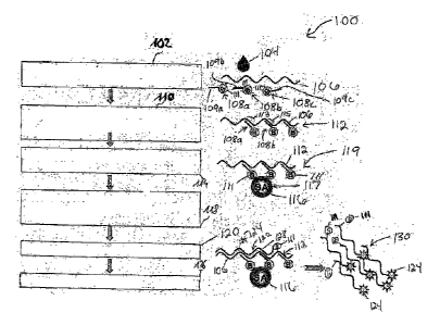
According to an exemplary embodiment, the compounds being detected in the
central well are the target molecules. For this purpose, the central well may
be
provided with specific binding members (for instance binding members which
differ
from other binding members needed for capturing the target molecules). In
other
words, in such an embodiment the target molecules (e.g. nucleic acids
originating
from free and from cell-associated viruses such as HIV comprising RNA
originating
from free viruses, RNA originating from cell-associated viruses, pro-viral
DNA,
reverse transcribed viral DNA, i.e. the "intermediates" of viral replication,
and
transcripts derived from pro-viral DNA, i.e. RNA molecules obtained by
transcription of the host DNA genome) may be bound to the binding members.
Alternatively, it is also possible to provide specific compounds such as
reporter
compounds which may have the capability to bind, for instance, to a PCR
product, to
RNA or to DNA. In such a scenario, the reporter compounds may be the compounds
CA 02722242 2010-01-05
WO 2009/013321 PC T/EP2008/059670
- 5 -
which are detected, thereby allowing to indirectly derive information
regarding the
presence and/or amount of target molecules in a sample.
The at least one binding member may be adapted for capturing the target
molecules.
For example, the at least one binding member may comprise labelled beads
capable
of capturing complexes including target molecules such as total viral nucleic
acids.
The at least one binding member may be adapted for capturing compounds
indicative
for the presence and/or amount of the target molecules. Thus, not only the
separate
individual target molecules can be detected directly, but it is also possible
to detect
target molecules indirectly, for instance by detecting reporter compounds
captured on
a binding member.
The at least one binding member may comprise a first binding member adapted
for
capturing the target molecules and may comprise a second binding member (which
may differ from the first binding member) adapted for capturing reporter
compounds
indicative for the presence and/or amount of the target molecules. Therefore,
two
different kinds of compounds may be provided, one specifically for capturing
the
target molecules after lysing, e.g. capture molecules comprising a binding
portion
specific to a region of a target polynucleotide and an anchor group; the other
one for
detection purposes, e.g. reporter compounds capable of forming complexes with
the
target polynucleotide, the forming of complexes with the target polynucleotide
inhibiting capturing of the reporter compound by the second binding member. In
other words, capturing may be functionally decoupled from detection. For
example,
the first binding member may be beads being configured to bind complexes
comprising a capture molecule and a target molecule, e.g. by binding an anchor
group of the capture molecule, whereas the second binding member may be a
surface
of the central well capable of capturing reporter compounds. The surface of
the
CA 02722242 2010-01-05
WO 2009/013321 PC T/EP2008/059670
- 6 -
central well being the second binding member may comprise one or more
different
reporter specific capture molecules being capable of capturing a reporter
compound
on the surface.
The structure, that is to say the central member at which the various solid
phase
coupling procedures occur, may be a well. A "well" may be an indentation or a
recess formed in a substrate and providing a sample chamber in which various
analysis procedures may be performed. Such a well may be a cylindrical
structure or
pot having a volume in the order of magnitude between microliters and
millilitres.
The central well or second structure may be irreversibly sealable, e.g. by
sealing an
inlet and, optionally, an outlet of the central well.
The microfluidic network may comprise a channel or a plurality of
interconnected
channels. A "channel" may denote a fluidic structure (for instance an
essentially one-
dimensional structure) having a length which is significantly larger than a
width and
a height, thereby providing a path along which liquids may be transported. A
single
channel may be provided, or several channels may be interconnected to form a
channel system. Such a channel system may allow a liquid flow from one channel
to
another channel at bifurcations of such a system. One or more wells may be
integrated in such a channel system.
In addition to a structure as described above, e.g. the "central" structure,
the
microfluidic network may comprise at least one further structure. In other
words,
apart from the channels and the central well, further microfluidic members may
be
provided, such as further channels and/or further wells. Therefore, a complex
system
of wells and channels may be provided.
CA 02722242 2010-01-05
WO 2009/013321 PC T/EP2008/059670
- 7 -
At least one further structure (such as a lysis structure or a lysis well) may
be adapted
for releasing contents of one or more cells, spores, or viruses, the contents
including
the target molecules. Thus, such a further structure may be denoted as a lysis
chamber in which biological compounds such as cells are forced to release
their
contents, for subsequent analysis. In other words, the further structure may
comprise
a structure comprising biochemical agents performing such tasks for releasing
the
contents, thereby providing a modified sample to be transported to the central
well.
To this end, the further structure such as a lysis structure may comprise a
lysing
reagent, for example chaotropic salts or a reagent comprising one or more
detergents
which disintegrate the cellular membranes and/or viral cap sids. Alternatively
or in
addition, the further structure, e.g. the lysis well, may be adapted to heat
the sample
in order to destroy cellular membranes and/or viral capsids (e.g., by
employing or
comprising a temperature control unit and/or temperature regulating unit as
described
below).
The at least one further structure may also comprise capture probes capable of
forming complexes with the target molecules. Therefore, it may be possible to
lyse a
sample in the presence of capture molecules with anchor groups.
At least one further structure (such as a well comprising PCR reagents ) may
comprise at least one substance promoting amplification of the target
molecules. In
other words, a further well may be provided which comprises biochemical agents
needed for, i.e. promoting the amplification. However, although PCR agents may
be
included in the further structure, the actual PCR amplification procedure may
be
carried out at another position, namely in the central well. However,
according to
exemplary embodiments, as will be explained below in more detail, it may be
advantageous to transport the sample from the central well through the well
including the amplification substances well back to the central well again to
avoid
CA 02722242 2010-01-05
WO 2009/013321 PC T/EP2008/059670
- 8 -
loss of sample material. Substances promoting amplification may be substances
needed for PCR (such as enzyme, primer, buffer, etc.) and are described in
detail
below.
The at least one further structure may also be a well. Therefore, a plurality
of wells
connected by the microfluidic network may be provided. However, it may also be
possible to perform lysing and/or to provide amplification material in other
structures
than wells, for instance in channels.
The device may comprise a substrate, on and/or in which the structure(s) may
be
formed. Therefore, fluid accommodating components of the device may be
monolithically integrated in the substrate. Alternatively, structure(s) may be
formed
on a substrate, for instance printed or spotted. Examples for materials of a
rigid
substrate which may properly cooperate with a flexible cover element are
polycarbonate, polypropylene, polystyrene, PET, PMMA, polyethylene, acrylic
glass, PU, PEEK, PVC, glass, and the like.
Particularly, the substrate may be rigid allowing to cooperate with one or
more
flexible cover elements at least partially covering the substrate in a very
efficient
manner. Particularly, the flexible cover element may cover the rigid
substrate, and an
actuator may press the cover element against the substrate to selectively
close
channels (for performing valve functions or the like).
According to an exemplary embodiment, the substrate may have a first surface
and a
second surface opposing the first surface. The structure may be provided on
and/or in
the first surface (particularly a first main surface) of the substrate. The
main surface
is the principal surface of the substrate upon which the structure is
configured. A
further structure may be provided on and/or in the second surface
(particularly a
CA 02722242 2010-01-05
WO 2009/013321 PC T/EP2008/059670
- 9 -
second main surface) of the substrate. A fluidic connection structure may be
provided, particularly a through hole penetrating the substrate and/or a
groove in a
surface portion of the substrate connecting the first surface with the second
surface.
Such a fluidic connection structure may be arranged between the first and the
second
surface and may be configured to provide a fluid communication of the
structure
with the further structure. In such an embodiment, the substrate may be
processed at
two opposing main surfaces to thereby form microfluidic structures. These
structures
may be connected by the connection structure which may comprise channels
formed
along a surface of the substrate, or directly going through the substrate.
Therefore, a
device may be provided in which both main surface portions of the substrate
may be
used in a very efficient manner, since both main surfaces of such a substrate
may be
processed for providing liquid transport tasks. Optionally, such a substrate
may be
covered on one or both sides with a (particularly flexible) cover element,
thereby
allowing to control fluid flow through fluidic structures on both surfaces
efficiently,
for instance by actuators acting on flexible portions on one or both main
surfaces.
Thus, a central substrate may be provided having fluidic structures on both
sides.
Particularly, this may allow manufacturing a cartridge formed by three layers,
namely the substrate and two at least partially flexible cover elements. Such
a three
layer structure may have a (for instance flexible) base element and a (for
instance
flexible) cover element sandwiching an intermediate layer (for instance being
rigid)
accommodating the micro fluidic structures. Base element and/or cover element
may
cover the central substrate entirely or only partially, for instance at
positions at which
a cover function is desired as a basis for an actuator based control (see, for
instance,
FIG. 21).
In addition to the substrate, the device may comprise at least one further
substrate,
wherein a further structure may be provided on and/or in the further
substrate. The
substrate and the further substrate may be adapted to be connectable or
mountable or
CA 02722242 2010-01-05
WO 2009/013321 PC T/EP2008/059670
- 10 -
assemblable or installable reversibly or detachably to one another in such a
manner
that the structure and the further structure may be brought in fluid
communication in
an operation state in which the substrate is connected or mounted or assembled
or
installed with the further substrate. According to such an embodiment, a
modular
construction may be provided in which a device may be formed by combining
several modules which can be flexibly connected to one another. A
corresponding
cartridge may be formed by a modular construction set, wherein each of the
modules
may have the following properties and may be used in combination with other
cooperatively formed modules:
- it comprises a chamber having at least two fluid connections;
- the chamber comprises a rigid component and an elastic component;
- at least one fluid connection may be closable by the motion of the
elastic
component, and a mixing of the content of the chamber may be effected.
The at least one binding member may be adapted such that a plurality of solid
phase
coupling procedures during an analysis of the target molecules occur at the at
least
one binding member. The term "solid phase coupling procedure" may particularly
include any kind of anchoring and hybridization, etc., at a
functionalization/binding
member. In this context, the "binding member or support member" may include
any
substance, surface or functionalization being configured to bind an anchor
group of
capture molecules and/or a surface being configured to capture
polynucleotides.
Solid phase coupling procedures may include any procedure in which molecules
to
be analyzed or detected are specifically bound to a solid surface, that is to
say are
bound not in a solution but on a solid surface.
The at least one binding member may be adapted such that all solid phase
coupling
procedures during an analysis of the target molecules occur at the at least
one
binding member. In other words, in such an embodiment, no solid phase coupling
CA 02722242 2010-01-05
WO 2009/013321 PC T/EP2008/059670
- 11 -
procedures occur at another well than at the central well/structure. This may
allow
performing all solid phase coupling procedures in a single well, allowing for
a
miniature and high performance device. The at least one binding member may be
adapted such that exactly two solid phase coupling procedures during an
analysis of
the target molecules occur at the at least one binding member. These two solid
phase
coupling procedures may relate to capturing target molecules from a multi-
component sample, and to detecting compounds indicative of the presence or
absence or the quantity of the target molecules. In the described embodiment,
these
two procedures are performed in a single well allowing to synergistically use
provisions of the well for both such tasks. Combining such two tasks in one
well may
keep liquid flow paths short, keep the device small, and keep the analysis
time short.
In some embodiments, the at least one binding member may be adapted such that
exactly three solid phase coupling procedures during an analysis of the target
molecules occur at the at least one binding member. These three solid phase
coupling
procedures may relate to capturing target molecules from a multi-component
sample,
capturing nucleic acids resulting from reverse transcription of target nucleic
acids,
and to detecting compounds indicative of the presence or absence or the
quantity of
the target molecules. In the described embodiment, these three procedures are
performed in a single well allowing to synergistically use provisions of the
well for
all such tasks. Combining such three tasks in one well may keep liquid flow
paths
short, keep the device small, and keep the analysis time short.
Alternatively, the at least one binding member may be adapted such that
exactly one
solid phase coupling procedure during an analysis of the target molecules in
the
sample occurs at the at least one binding member. Such an embodiment may be
particularly advantageous, when the entire biochemical analysis or experiment
only
CA 02722242 2010-01-05
WO 2009/013321 PC T/EP2008/059670
- 12 -
comprises a single solid phase coupling procedure, for instance is only
foreseen for
sample purification, not for detection.
At least a portion of the device located adjacent to the at least one binding
member
may be transparent for electromagnetic radiation in a range of wavelengths
between
essentially 1 nm and essentially 10 [tm to thereby allow for an
electromagnetic
radiation based detection of the compounds indicative for the presence and/or
amount of the target molecules and captured at the at least one binding
member. In
such embodiments, particularly a portion of the substrate close to the central
well
may be transparent for electromagnetic radiation used for detection purposes,
particularly for electromagnetic radiation in the near-infrared, optical and
ultraviolet
domain. By taking this measure, it may be possible to perform also the
detection on
the basis of electromagnetic radiation (for instance a fluorescence-based
detection) in
the central well. When the portion of the device located adjacent to the at
least one
binding member is transparent for electromagnetic radiation in a range of
wavelengths between essentially 400 [im and essentially 800 p.m, an optical
detection
of the compounds is enabled.
The device may comprise or may be connectable with a temperature manipulation
unit adapted for manipulating a temperature of liquids located in the
structure. Such a
temperature manipulation unit may comprise a heating and/or cooling element
which
allows to bring a sample to a specific temperature, or to conduct a specific
temperature pattern or sequence.
The temperature manipulation unit may be adapted for manipulating a
temperature of
liquids located in the structure in accordance with a temperature sequence for
performing a polymerase chain reaction (PCR). Such a polymerase chain reaction
may require temperature cycles to, for instance about 95 C, about 55 C and
about
CA 02722242 2010-01-05
WO 2009/013321 PC T/EP2008/059670
- 13 -
72 C. Such a sequence of temperatures usually has to be performed for specific
predefined time intervals, and has to be repeated a predefined plurality of
times.
The at least one binding member may be configured to bind an anchor group of a
capture molecule. Particularly, the at one least binding member may be
configured to
capture polynucleotides.
The at least one binding member may comprise at least one of the group
consisting
of capture molecules, e.g. reporter specific capture molecules, arranged on a
surface
of the structure (for instance immobilized in the well), capture molecules
arranged on
particles (for instance on beads), capture molecules arranged on a porous
surface of
the structure (for instance a porous glass structure), and one or more
different capture
molecules, e.g. reporter specific capture molecules, arranged on different
locations
with respect to a surface of the structure (for instance different kinds of
capture
molecules being immobilized in an array-like manner in the well, for instance
in the
context of a competitive assay). In some embodiments, the at least one binding
member also may comprise capture molecules for capturing an anchor group such
as
biotin.
The structure may have a volume in a range between essentially 1 [d and
essentially
1 ml, particularly in a range between essentially 20 [d and essentially 300
pl. For
example, a well having a volume of essentially 100 111 may be provided.
The substrate may have a groove configured to receive a cannula for supplying
liquids to the device. In such an embodiment, it may be very easy for a user
to handle
the device, since the cannula for sample supply simply has to be placed in the
groove
to be brought in proper accordance and cooperation with the micro fluidic
channel
CA 02722242 2010-01-05
WO 2009/013321 PC T/EP2008/059670
- 14 -
system, thereby allowing for an easy analysis which may be performed even by
users
which are not specifically skilled or trained.
The substrate may have a window portion adjacent the structure and being
transparent for electromagnetic radiation in a range of wavelengths between
essentially 1 nm and essentially 10 [Lm (that is to say for near infrared,
optical or
ultraviolet radiation), particularly in a range of wavelengths between
essentially 400
nm and essentially 800 nm (that is to say particularly for optical radiation),
to thereby
allow for an electromagnetic radiation based detection of a meniscus of a
liquid
flowing through (more precisely reaching) the structure or the micro fluidic
network.
In such an embodiment, an optically transparent window portion of the
substrate may
be detected by a radiation detector. When a meniscus of a fluid pumped through
the
microfluidic network or the structure passes the window portion, this may
abruptly
change the transmission properties through the window portion in a
characteristic
manner, thereby generating a signal at a radiation detector indicative for the
fact that
the meniscus has reached a specific region in the device. This signal may be
useful
for triggering purposes, or as a control signal for actuators, because the
cooperative
motion of actuators and/or the control of temperature manipulation units can
be
brought in proper accordance with the present position of a sample being
pumped
through the device. For instance, by taking such a measure, it may be detected
that a
predefined volume of water or buffer has been pumped into the device, when an
overflow occurs.
At least one of the group consisting of the structure and the further
structure may
comprise two fluid openings. Such fluid openings may be a fluid inlet and a
fluid
outlet.
CA 02722242 2010-01-05
WO 2009/013321 PC T/EP2008/059670
- 15 -
The cover element may be a flexible cover element. Particularly in cooperation
with
a rigid substrate, the cover element and the substrate may form three-
dimensionally
sealed channels which can be properly controlled by actuators acting on the
cover
element. When the cover element is at least partially deformable at a specific
position under the influence of an external force, it may be possible to
selectively
enable or disable a flow of liquids by opening or closing the structure or the
microfluidic network. Beyond this, a transport of liquids along the structure
is
possible with such a cover element.
Particularly when an actuator member is provided and adapted for being
actuated to
deform the cover element, a high performance lab-on-chip may be provided which
has integrated mixing, pumping and/or valve functions.
Any one of the structures may comprise one or more substances being
biologically,
biochemically and/or chemically active. Therefore, when such substances, which
may include capture molecules, reporter-specific capture molecules, detectable
markers, lysing reagents and PCR reagents, are present in the wells in dried
form,
particularly in lyophilized form, it is possible to provide a device which a
user simply
has to fill with liquids (such as water, buffers and sample) to perform a
fully
automatic analysis. When the necessary biochemical components are provided in
the
different wells, a user can simply start an experiment on the basis of a
sequence
stored in the control unit and may provide water or buffers to different inlet
chambers. The remainder will be performed by the fully automatic device.
The channel may have a width (that is a dimension in a surface plane of the
substrate
and perpendicular to a fluid flow direction) in a range between essentially 50
.t.m and
essentially 1 mm, particularly in a range between essentially 100 1..im and
essentially
300 ttm. For example, a width of the channel may be essentially 200 ium. A
height
CA 02722242 2010-01-05
WO 2009/013321 PC T/EP2008/059670
- 16 -
(that is a dimension in a direction perpendicular to a surface plane of the
substrate
and perpendicular to a fluid flow direction) of the channel may be in a range
between
essentially 20 um and essentially 300 [(m, particularly in a range between
essentially
50 um and essentially 200 um. For example, a height of the channel may be
essentially 100 p.m. In contrast to this, a length of the channel may be much
larger
than the width and the height, for instance may be larger than 1 mm,
particularly may
be larger than 1 cm or may even be several centimetres.
The structure may comprise a material adapted as a transport medium for
liquids. For
example, the material may comprise at least one of the group consisting of a
solid
material, a gel material, a liquid material, and a combination thereof.
Therefore, the
structure may be a recess or may be formed by material serving as a carrier
for the
liquids.
The cover element may comprise a flexible membrane or a flexible sealing. Such
a
flexible membrane or flexible sealing may be made of materials such as latex,
thereby enabling the cover element to be flexibly deformed under the influence
of a
mechanical force (for instance generated by an actuator member).
The device may comprise an actuator member adapted for being actuated for
deforming the cover element to thereby control a fluid flow property of
liquids in the
structure and/or in the microfluidic network. Such an actuator member may be
under
the control of the control unit and may have a plurality of cooperating pins
or stencils
acting on the flexible cover element to thereby selectively open or close
channels,
temporarily reduce the volume of a channel or well for pumping or mixing
purposes,
etc.
CA 02722242 2010-01-05
WO 2009/013321 PC T/EP2008/059670
- 17 -
The actuator member may particularly be adapted for controlling a fluid flow
property of liquids along a straight portion of a channel. When a fluid flows
along a
straight channel, a perpendicularly arranged actuator member may efficiently
disable
a fluid flow when this channel is closed at a specific portion.
The actuator member may be adapted for functioning as a valve, as a fluid
mixer,
and/or as a fluid pump.
More particularly, the actuator member may comprise a plurality of actuator
elements adapted for being cooperatively actuated for deforming the cover
element
to thereby control the fluid flow property of liquids in accordance with a
fluid flow
scheme defined by the control unit. Therefore, when a user has selected a
specific
experiment or assay, which involves the transport of fluids and samples
through
various channels, the control unit simply controls the individual stencils of
the
actuator member to provide such a reversible compression of the flexible cover
element, to thereby fully automatically perform the assay.
The control unit may be adapted to control the actuator member to deform the
cover
element in such a manner that target molecules are captured at the at least
one
binding member, that the target molecules are amplified in the structure, and
that
compounds indicative for the presence and/or amount of the target molecules
and
captured at the at least one binding member are detected. Thus, the control
unit may
be the central regulator of the device harmonizing the function of the various
components.
The actuator member may comprise one or more pins configured to be
reciprocated.
By moving a pin in a forward direction, a channel may be closed by pressing
the
flexible cover element towards the substrate in this channel. When the pin is
moved
CA 02722242 2010-01-05
WO 2009/013321 PC T/EP2008/059670
- 18 -
backwardly, the channel may be opened again to allow for a fluid flow. In some
embodiments, the one or more pins may have an at least partially elastic tip.
The actuator member may further be provided to be movable in a direction
perpendicular to a main surface of the substrate. By reciprocating in a
direction
which is perpendicular to the planar substrate, an efficient opening and
closing may
be made possible. Particularly, the actuator member may be provided movably to
selectively close at least a part of the structure to disable a transport of
liquids
through the structure. In another operation mode, the actuator member may be
moved to selectively open at least a part of the structure to enable a
transport of
liquids through the structure.
The actuator member may be adapted for reciprocating perpendicular to a main
surface of the substrate for selectively enabling or disabling a fluid flow of
liquids
through the structure. The use of reciprocating actuators may allow for
reversibly and
selectively enabling or disabling fluid flows, allowing for a very flexible
operation of
the device and allowing for using the device multiple times (in contrast to
approaches
in which channels are closed irreversibly for performing a one-way valve
function).
The actuator member may be adapted for reciprocating in a perpendicular
direction
to a main surface of the substrate for pumping liquids through the structure.
Therefore, it is possible that the actuator member controls a volume or height
of the
structure. The actuator member may also selectively close the structure.
Closing a
structure may be performed in the context of a valve function, of a mixing
function
or of a pumping function. However, it is also possible to use such an actuator
during
a detection phase, since it is possible to compress the structure and/or
binding
members for detection purposes to increase the local concentration of target
CA 02722242 2010-01-05
WO 2009/013321 PC T/EP2008/059670
- 19 -
molecules to be detected and/or to remove background signals. This may allow
increasing the accuracy.
A drive unit may be provided for mechanically driving the actuator member,
wherein
the drive unit may be controllable by the control unit. Such a drive unit may
comprise a pneumatic drive mechanism, a hydraulic drive mechanism, or an
electromagnetic drive mechanism.
The at least one binding member may comprise a three-dimensional medium, for
instance a gel, particles, beads or a porous matrix. The three-dimensional
medium
may be arranged and configured to be reversibly compressible by moving the
actuator member. By taking this measure, a very accurate detection may be made
possible, because the local concentration of the molecules to be detected may
be
selectively increased by compressing the three-dimensional medium (such as
beads)
having attached thereto compounds or complexes indicative for the presence or
the
quantity of the target molecules.
The device may be adapted as a biosensor assay device, a microfluidic
cartridge, or a
lab-on-chip. Therefore, on a small scale, various biochemical functions may be
combined to perform an entire biochemical experiment.
A temperature sensor may be provided and adapted for sensing a temperature of
liquids transported through the device. The temperature sensor may be
integrated in a
substrate to thereby sense the temperature of the liquids flowing through the
microfluidic network. Alternatively, the temperature sensor may be arranged at
the
actuator member, for instance at a tip of a stencil-like actuator, so that the
actuator,
when pressing the cover element against the substrate, may simultaneously
measure
the local temperature of the fluid.
CA 02722242 2010-01-05
WO 2009/013321 PC T/EP2008/059670
- 20 -
The device may comprise a temperature manipulation unit adapted for
manipulating
a temperature of liquids, and preferably arranged at the actuator member. Such
a
temperature manipulation unit may also be integrated within the substrate, for
example in the form of heating wires integrated in the substrate and heating
sample
in the well. Alternatively, such a temperature manipulation unit may be an
external
device such as an external electromagnetic radiation source wherein
electromagnetic
radiation (for instance from a laser) may be directed onto a well resulting in
a heating
of the fluid in the well using the electromagnetic radiation as an energy
source.
Further alternatively, the temperature manipulation unit may include not or
not only
a heating element, but also a cooling element. For such an embodiment, a
Peltier
cooler may be implemented with low effort.
A temperature manipulation unit may be provided and adapted for manipulating a
temperature of liquids, wherein the temperature manipulation unit may comprise
a
first heating element and a second heating element, the structure being
arranged
between the first heating element and the second heating element. By providing
two
such heating plates, one being a continuous plate and the other one being an
annular
plate, heating may be performed without disabling the device to be operated
with an
electromagnetic radiation based detector, since a recess in the annular plate
may
allow electromagnetic radiation to be directed onto the central well and may
allow
fluorescence radiation to be detected through the recess and the second
heating
element.
According to an exemplary embodiment at least one of the heating/cooling
elements
is flexibly mounted. Flexibly mounting the heating/cooling elements may allow
for
an easy insertion of a structure, e.g. the second structure or central well,
between the
first and second heating/cooling elements. Further, flexibly mounting at least
one of
CA 02722242 2010-01-05
WO 2009/013321 PC T/EP2008/059670
- 21 -
the heating/cooling elements may allow for flexibly adapting the flexible
heating/cooling element to the surface of the structure, e.g. the second
structure or
central well, so that the flexible heating/cooling element is forced to
contact the
surface of the structure and thus also allows for an efficient thermal
conductance.
According to an exemplary embodiment the flexibly mounting is a flexible
mounting
of the whole heating/cooling element.
According to a further exemplary embodiment the flexibly mounting is a
flexibility
of the heating/cooling element as such.
Further, also two heating/cooling elements may be flexibly mounted. The both
heating/cooling elements may be arranged in a butterfly fashion to sandwich
the
probe device. In the same fashion a single heating/cooling element may be
arranged
with a pressing counter plate. This may avoid any scratches when inserting the
probe
device, in particular when the heating/cooling elements will be moved towards
the
surfaces of the probe device after the probe device has reached its final
position.
In some embodiments, each of the heating element or cooling element, or both,
is a
Peltier element.
A temperature regulation unit may be provided and adapted for regulating a
temperature of liquids in the structure. Such a regulation entity may include
the
measurement of the actual temperature and, on the basis of this measurement,
the
performance of a heating and/or cooling performance to thereby adjust the
temperature to a desired value.
CA 02722242 2010-01-05
WO 2009/013321 PC T/EP2008/059670
- 22 -
A detection unit may be provided and adapted for detecting, in the structure,
compounds indicative for the presence and/or amount of the target molecules
and
captured at the at least one binding member. Such a detection unit may
comprise an
optical detection unit, particularly a fluorescence detection unit.
The substrate and the cover element may be separate components which are
connected to one another. Alternatively, the substrate and the cover element
may be
made of different materials.
A transport unit may be provided and adapted for transporting liquids through
the
structure and/or the microfluidic network. Such a transport unit may comprise
a
pump, particularly one of the group consisting of a compressed-air pump, a
hydraulic
pump, a peristaltic pump, and a vacuum pump. Furthermore, the device may be
adapted in such a manner, during normal use, the gravitational force promotes
the
flow of liquids through the device in a desired manner. Therefore, in the
absence of
the activity of a transport unit, liquids may directly flow in a desired
direction.
However, when the transport unit is switched on, the influence of the
transport unit
may be larger than the influence of the gravitation, thereby allowing to
selectively
initiate a fluid flow in a direction against the gravitational force.
Therefore, the
combination of gravity and a special transport unit may be highly advantageous
and
may allow for an energy-saving operation.
The transport unit may be adapted for transporting liquids by actuating a gas
bubble
in the structure and/or in the micro fluidic network. By moving a gas bubble
through
the device, the transport of the liquids through the device may be supported
or
promoted.
CA 02722242 2010-01-05
WO 2009/013321 PC T/EP2008/059670
- 23 -
At least one filter, particularly at least one fit, may be arranged at the
structure (that
is to say at an inlet and/or at an outlet of the central well) and may be
adapted for
preventing the at least one binding member (for instance beads) arranged in
the
structure, from being washed out of the structure. Under the influence of a
fluid flow,
a mechanical force may act on the beads or other binding members in the
structure.
However, when a fit, that is to say a porous filter element which may be made
of a
sinter material, is provided at an inlet and/or an outlet of the structure it
may be
securely prevented that the beads are washed out of the central chamber. The
fit may
be provided with an annular shape to allow for being inserted into a
correspondingly
shaped annular groove in the device.
The at least one binding member may comprise a surface functionalization. The
term
"surface functionalization" may denote the fact that the surface is processed
in such a
manner as to perform a specific binding function. In such an embodiment, the
binding member may be part of or coupled to or attached to the surface of the
well.
The substrate and the cover element may be in direct contact to one another.
Alternatively, the substrate may be free of a direct contact with the cover
element.
Various geometrical realizations are possible.
A portion of the substrate located adjacent to the structure may be
transparent for
electromagnetic radiation in a range of wavelengths between essentially 400 nm
and
essentially 800 nm to thereby allow for an optical detection in the structure.
Therefore, visible light may be used for detection purposes. Such a detection
may be
performed on the basis of light absorption, light reflection, or fluorescence
generation, for instance using fluorescence labels attached to molecules or
complexes
to be detected.
CA 02722242 2010-01-05
WO 2009/013321 PC T/EP2008/059670
- 24 -
The at least one binding member may be adapted such that at least two solid
phase
coupling procedures during an analysis of the target molecules occur at
exactly one
of the at least one binding member. In other words, one and the same binding
member may be used for multiple solid phase coupling procedures. For example,
beads with attached groups may be used for capturing target molecules out of
the
sample, and may be used later for capturing compounds such as amplified and
labelled target molecules as a basis for a subsequent detection.
Alternatively, the at least one binding member may be adapted such that at
least two
solid phase coupling procedures during an analysis of the target molecules
occur at
different ones of the at least one binding member. E.g., the device includes
multiple
binding members and at least one binding member is adapted such that at least
two
solid phase coupling procedures during an analysis of the target molecules
occur at
two or more of the at least one binding member. In such a configuration, for
example, capturing molecules from a sample on the one hand, and detecting
components indicative of the target molecules on the other hand are captured
using
two different kinds of binding members. For example, beads may be provided for
capturing the target molecules out of a sample. On the other hand, capture
molecules,
e.g. reporter specific capture molecules immobilized in the well may be used
in the
context of a competitive assay for capturing the components indicative of the
presence or amount of target molecules in the sample, e.g. reporter compounds.
According to another exemplary embodiment of the invention, a method is
provided
comprising forming complexes, each comprising a target nucleic acid and a
capture
molecule, wherein each capture molecule comprises a binding portion specific
to a
region of the target nucleic acid and an anchor group; contacting the
complexes with
a binding member, the binding member being configured to bind the anchor group
of
the capture molecule to bind the complexes to the binding member; subjecting
one or
CA 02722242 2010-01-05
WO 2009/013321 PC T/EP2008/059670
- 25 -
more target nucleic acids to an amplification; capturing the amplified target
nucleic
acids with respect to the binding member; and determining a value indicative
for the
presence and/or amount of the captured target nucleic acids.
The one or more target nucleic acids may be single-stranded or double-stranded
nucleic acids.
The method may further comprise subjecting the target nucleic acids to reverse
transcription prior to subjecting one or more target nucleic acids to
amplification.
The method may further comprise releasing the captured amplified target
nucleic
acids from the binding member and repeating the steps of subjecting one or
more
target nucleic acids to amplification and capturing the amplified target
nucleic acid
with respect to the binding member. In such an embodiment, the cycle of
releasing
the captured amplified target nucleic acids from the binding member and
repeating
the steps of subjecting one or more target nucleic acids to amplification and
capturing the amplified target nucleic acids with respect to the binding
member may
be performed at least 10 times or at least 20 times.
A value indicative for the presence and/or amount of the captured target
nucleic acids
may be determined after at least one cycle, e.g. after each cycle, of
releasing the
captured amplified target nucleic acids from the binding member and repeating
the
steps of subjecting one or more target nucleic acids to amplification and
capturing
the amplified target nucleic acids with respect to the binding member.
The binding member may comprise one or more capture molecules capable of
capturing the target nucleic acids. In such an embodiment, the target nucleic
acids
CA 02722242 2010-01-05
WO 2009/013321 PC T/EP2008/059670
- 26 -
are captured with respect to the binding member by the one or more capture
molecules.
The binding member may further comprise particles.
The step of forming complexes each comprising a target nucleic acid and a
capture
molecule may be performed spatially separated from the step of contacting the
complexes with a binding member.
The method may further comprise labeling the target nucleic acids. The target
nucleic acids may be labeled by adding or more detectable markers, e.g. prior
to or
during subjecting one or more target nucleic acids to amplification and/or
prior to
capturing the amplified target nucleic acids with respect to the binding
member. The
one or more detectable markers may be fluorescent markers.
Determining a value indicative for the presence and/or amount of the captured
target
nucleic acids may comprise time-dependent monitoring of the one or more
indicative
values obtained.
The method may further comprise providing the one or more target nucleic acids
prior to forming complexes each comprising a target nucleic acid and a capture
molecule. The step of providing one or more target nucleic acids may comprise
releasing the target nucleic acids from biological material. In such an
embodiment,
the biological material may be selected from the group consisting of one or
more
prokaryotic cells, one or more eukaryotic cells, one ore more erythrocytes,
and one
or more viral particles as well as mixtures thereof. Further, releasing the
target
nucleic acids from biological material may comprise contacting the biological
material with a lysing reagent.
CA 02722242 2010-01-05
WO 2009/013321 PC T/EP2008/059670
- 27 -
Providing the one or more target nucleic acids may comprise providing a sample
comprising the one or more target nucleic acids wherein the sample may be
selected
from the group consisting of whole blood, plasma, serum, urine, sputum, saliva
and
cerebrospinal fluid.
Providing the one or more target nucleic acids may be performed spatially
separated
from the contacting complexes each comprising a target nucleic acid and a
capture
molecule, subjecting the one or more target nucleic acids to amplification,
capturing
the amplified target nucleic acids with respect to the binding member and
determining a value indicative for the presence and/or amount of the captured
target
nucleic acids.
The method may further comprise separating the one or more target nucleic
acids
from concomitant material.
In a further embodiment, the method according this exemplary embodiment is
performed in a device as described above. E.g., the method may be performed in
a
device, comprising a rigid substrate; a flexible cover element at least
partially
covering the substrate; a first structure formed in the substrate, adapted for
accommodating liquids and adapted for releasing contents of one or more cells,
spores, or viruses, the contents including target molecules such as target
nucleic
acids; a second structure formed in the substrate, adapted for accommodating
liquids
and comprising at least one binding member adapted for capturing the target
molecules and for determining a value indicative for the presence and/or
amount of
the target molecules; a microfluidic network interconnecting at least the
first
structure and the second structure; and an actuator unit adapted for effecting
a fluid
flow between the first structure and the second structure by pressing the
flexible
CA 02722242 2010-01-05
WO 2009/013321 PC T/EP2008/059670
- 28 -
cover element against the substrate to selectively close a portion of the
micro fluidic
network. Further, the method may be performed in a device, comprising a
structure
adapted for accommodating liquids, wherein the structure comprises at least
one
binding member and is in fluid communication with a microfluidic network; and
a
control unit adapted for controlling a fluid flow through the microfluidic
network in
such a manner that target molecules such as target nucleic acids are captured
at the at
least one binding member, adapted for controlling an amplification of the
target
molecules in the structure, and adapted for controlling detection of compounds
captured at the at least one binding member.
The device may comprise a first structure adapted for accommodating liquids.
In
such an embodiment, the complexes each comprising a target nucleic acid and a
capture molecule are formed in the first structure.
Further, the device may comprise a second structure configured for detecting
one or
more target nucleic acids and comprising a cover element covering the second
well
and an actuator unit adapted for being actuated to deform the cover element.
In such
an embodiment, determining a value indicative for the presence and/or amount
of the
captured target nucleic acids may be performed in the second structure.
Further, subjecting one or more target nucleic acids to amplification and/or
capturing
the amplified target nucleic acids with respect to a binding member may also
be
performed in the second structure.
Determining a value indicative for the presence and/or amount of the captured
target
nucleic acids may be performed with the actuator actuated to deform the cover
element. The cover element may be deformed in such a way that the volume of
the
second structure or central well or detection well is reduced. In such an
embodiment,
CA 02722242 2010-01-05
WO 2009/013321 PC T/EP2008/059670
- 29 -
the volume of the second well may be re-increased after determining a value
indicative for the presence and/or amount of the captured target nucleic
acids.
According to another exemplary embodiment of the invention, a method is
provided,
comprising:
providing an amount of a reporter compound; a first binding member being
configured to bind an anchor group of a capture molecule; a second binding
member
capable of capturing the reporter compound; an amount of a target nucleic acid
capable of forming complexes with the reporter compound; the forming of
complexes with a reporter compound inhibiting capturing of the reporter
compound
by the second binding member; and an amount of capture molecules wherein each
capture molecule comprises a binding portion specific to a region of the
target
nucleic acids and an anchor group;
forming complexes each comprising a target nucleic acid and a capture
molecule;
contacting the complexes with the first binding member to bind the complexes
to the
first binding member;
releasing at least a subset of the amount of target nucleic acid from the
first binding
member;
forming complexes of a subset of the amount of a reporter compound with at
least a
subset of the amount of target nucleic acid;
capturing a remaining subset of the amount of reporter compound not in complex
with a target nucleic acid on the second binding member; and
determining a value indicative for the presence and/or amount of reporter
compound
captured on the second binding member.
The reporter compound may comprise one or more detectable labels, e.g. two
detectable labels. The one or more detectable labels may be fluorescent
labels.
CA 02722242 2010-01-05
WO 2009/013321 PC T/EP2008/059670
- 30 -
Further, the reporter compounds may be oligonucleotides.
The method may further comprise determining a value indicative for the
presence
and/or amount of target nucleic acid based on the value indicative for the
presence
and/or amount of reporter compound captured on the second binding member.
The method may further comprise releasing the remaining subset of the amount
of
reporter compound from the second binding member after the step of determining
a
value indicative for the presence and/or amount of reporter compound captured
on
the second binding member; forming complexes of a subset of the amount of
reporter
compound with at least a subset of the amount of target nucleic acid;
capturing a
remaining subset of the amount of reporter compound not in complex with a
target
nucleic acid on the second binding member; and determining the value
indicative for
the presence and/or amount of reporter compound captured on the second binding
member. In such an embodiment, the steps of releasing, forming complexes,
capturing and determining may be performed N additional times, wherein N is an
integer greater than or equal to 1, e.g. N > 5, N > 10 or N > 20.
Further, the step of forming complexes of a subset of the amount of reporter
compound with at least a subset of the amount of target nucleic acid and the
step of
capturing a remaining subset of the amount of reporter compound not in complex
with a target nucleic acid on the second binding member may be performed
concomitantly.
The method may further comprise subjecting the target nucleic acid to
amplification.
In such an embodiment, amplification of the target nucleic acid may be
initiated prior
to the step of forming complexes of a subset of the amount of reporter
compound
with at least a subset of the amount of target nucleic acid.
CA 02722242 2010-01-05
WO 2009/013321 PC T/EP2008/059670
-31 -
The value indicative for the presence and/or amount of reporter compound
captured
on the second binding member may be determined before the steps of forming of
complexes of a subset of the amount of reporter compound with at least a
subset of
the amount of target nucleic acid and of capturing a remaining subset of the
amount
of reporter compound not in complex with a target nucleic acid on the second
binding member are in chemical equilibrium. Particularly, the value indicative
for
the presence and/or amount of reporter compound captured on the second binding
member may be determined 1 s to 120 s after initiating the steps of forming
complexes of a subset of the amount of reporter compound with at least a
subset of
the amount of target nucleic acid, and of capturing a remaining subset of the
amount
of reporter compound not in complex with a target nucleic acid on the second
binding member.
The method may further comprise subjecting the target nucleic acids to reverse
transcription prior to subjecting them to amplification.
The second binding member may comprise one or more different reporter specific
capture molecules being capable of capturing a reporter compound on the second
binding member. In such an embodiment, the capture molecules may be
oligonucleotides. The different reporter specific capture molecules may be
arranged
on different locations with respect to the second binding member. Further, the
reporter compounds may be captured on the second binding member by forming
complexes with the reporter-specific capture molecules. At least a part of an
interaction site of the reporter compound being capable of forming a complex
with a
target nucleic acid may also be capable of forming a complex with a reporter
specific
capture molecule. The reporter specific capture molecules and the target
nucleic acid
may compete for forming a complex with the reporter compound.
CA 02722242 2010-01-05
WO 2009/013321 PC T/EP2008/059670
- 32 -
The amplification may comprise a step of denaturing double-stranded nucleic
acids.
Double-stranded nucleic acids may comprise complexes of reporter compounds
with
target nucleic acids, complexes of reporter compounds with reporter specific
capture
molecules, double strands of reporter compounds and double strands of target
nucleic
acids.
The amplification may further comprise a step of annealing primer molecules to
target nucleic acids. In this embodiment, the annealing step may be performed
concomitantly with the step of forming complexes of a subset of the amount of
reporter compound with at least a subset of the amount of target nucleic acid
and/or
the step of capturing a remaining subset of the amount of reporter compound
not in
complex with a target nucleic acid on the second binding member.
The amplification may be a cyclic amplification, e.g. a PCR. Performing the
PCR
may comprise using a polymerase having exonuclease activity. The cyclic
amplification may comprise at least 10 cycles or at least 20 cycles.
The value indicative for the presence and/or amount of reporter compound
captured
on the second binding member may be determined after at least one cycle, e.g.
after
each cycle, of the cyclic amplification. Further, the value indicative for the
presence
and/or amount of target nucleic acid may be determined each time after
determining
the value indicative for the presence and/or amount of reporter compound
captured
on the second binding member.
Determining the value indicative for the presence and/or amount of reporter
compound captured on the second binding member may comprise time-dependent
monitoring of the indicative value.
CA 02722242 2010-01-05
WO 2009/013321 PC T/EP2008/059670
- 33 -
Further, the value indicative for the presence and/or amount of target nucleic
acid
may be determined based on a calibration curve correlating the value
indicative for
the presence and/or amount of reporter compound with a value indicative for
the
presence and/or amount of target nucleic acid.
The method of this exemplary embodiment may also be performed in a device as
described above. E.g., the method may be performed in a device, comprising a
rigid
substrate; a flexible cover element at least partially covering the substrate;
a first
structure formed in the substrate, adapted for accommodating liquids and
adapted for
releasing contents of one or more cells, spores, or viruses, the contents
including
target nucleic acids; a second structure formed in the substrate, adapted for
accommodating liquids and comprising at least one binding member adapted for
capturing the target nucleic acids and for determining a value indicative for
the
presence and/or amount of the target nucleic acids; a microfluidic network
interconnecting at least the first structure and the second structure; and an
actuator
unit adapted for effecting a fluid flow between the first structure and the
second
structure by pressing the flexible cover element against the substrate to
selectively
close a portion of the microfluidic network. The method may also be performed
in a
device, comprising a structure adapted for accommodating liquids, wherein the
structure comprises at least one binding member and is in fluid communication
with
a microfluidic network; and a control unit adapted for controlling a fluid
flow
through the microfluidic network in such a manner that target nucleic acids
are
captured at the at least one binding member, adapted for controlling an
amplification
of the target molecules in the structure, and adapted for controlling
detection of
compounds captured at the at least one binding member.
CA 02722242 2010-01-05
WO 2009/013321 PC T/EP2008/059670
- 34 -
The device may further comprise a first structure adapted for accommodating
liquids.
In such an embodiment, the step of forming complexes each comprising a target
nucleic acid and a capture molecule is performed in the first structure.
The device may further comprise a second structure adapted for accommodating
liquids and the first and, optionally, the second binding member may be
provided in
the second structure. In such an embodiment, forming complexes each comprising
a
target nucleic acid and a capture molecule; contacting the complexes with the
first
binding member to bind the complexes to the first binding member; releasing at
least
a subset of the amount of target nucleic acid from the first binding member;
forming
complexes of a subset of the amount of a reporter compound with at least a
subset of
the amount of target nucleic acid;
capturing a remaining subset of the amount of reporter compound not in complex
with a target nucleic acid on the second binding member; and determining a
value
indicative for the presence and/or amount of reporter compound captured on the
second binding member is performed in the second structure, e.g. the central
well.
Determining a value indicative for the presence and/or amount of the captured
reporter compounds may be performed with the actuator actuated to deform the
cover element. The cover element may be deformed in such a way that the volume
of the central well or second structure or detection well is reduced. In such
an
embodiment, the volume of the central well may be increased again after
determining
a value indicative for the presence and/or amount of the captured reporter
compounds.
Providing the one or more target nucleic acids may comprise providing a sample
comprising the one or more target nucleic acids. The sample may be a liquid
sample
CA 02722242 2010-01-05
WO 2009/013321 PC T/EP2008/059670
- 35 -
having a volume of I pi to 50 pl. Further, the sample may be a liquid whole
blood
sample.
The method may further comprise adding an amount of a quencher compound
capable of forming complexes with the reporter compound not in complex with
target molecules or reporter specific capture molecules. The quencher compound
may comprise one or more moieties interfering with the generation of a
detectable
signal by a label (e.g., a quencher group "hijacking" the emissions that
resulted from
excitation of a fluorophor). E.g. the quencher groups may be capable of
suppressing
or inhibiting signals emitted by a detectable label of the reporter compound,
e.g. a
fluorescence signal. In such an embodiment, the quencher compound may be
capable of forming complexes with the reporter compound not in complex with
target molecules or reporter specific capture molecules such that the one or
more
quencher groups are in close proximity to the detectable label of the reporter
compound within the complex.
The quencher compound may be an oligonucleotide. In this embodiment, the
quencher oligonucleotide may comprise at least one specific sequence region
which
is complementary to a sequence region of a reporter oligonucleotide, thus
allowing
base-pairing between the quencher compound and the reporter compound.
The quencher group may include usual quenchers such as for instance Black Hole
Quenchers (Biosearch Technologies), Qxl quenchers (AnaSpec) and Iowa black
quenchers.
The quencher compounds may be provided in the second structure of a device as
described above. In such an embodiment, the quencher compound may form a
complex with a reporter compound not captured on the second binding member.
CA 02722242 2010-01-05
WO 2009/013321 PC T/EP2008/059670
- 36 -
The second structure of a device as described above may be irreversibly sealed
before initiating amplification of the target nucleic acids. Irreversibly
sealing the
second structure may be achieved by sealing (e.g. welding) an inlet and,
optionally
an outlet of the second structure, e.g. by heat-sealing channels and/or valves
connected with the second structure.
According to another exemplary embodiment, a method is provided, comprising
amplifying at least one target polynucleotide to form double-stranded
amplicons,
contacting the amplicons with a surface configured to selectively bind the
amplicons
(e.g., with an anchor group), and with the amplicons bound to the surface by
an
anchor group, optically determining the presence of the amplicons. The method
may
further comprise releasing the amplicons from the surface after the step of
optically
detecting, subjecting the released amplicons to at least one more
amplification cycle,
contacting the resulting amplicons with the surface, and with the amplicons
bound to
the surface by the anchor group, optically determining the presence of the
amplicons.
The method may further comprise performing the steps of releasing, subjecting,
contacting, and optically determining a number N additional times, where N is
an
integer greater than or equal to 1. Particularly, N > 5, more particularly N?
10, and
still more particularly N > 20.
The method may further comprise, prior to the step of amplifying, providing
the
target polynucleotides, forming complexes each comprising a target
polynucleotide
released from a pathogen and at least one capture molecule, each capture
molecule
comprising a binding portion specific to a region of the target polynucleotide
and an
anchor group, and contacting the complexes with the surface, the surface being
configured to non-selectively bind the anchor group of the capture molecule to
non-
selectively bind the complexes and the surface. In such a method, providing
the
CA 02722242 2010-01-05
WO 2009/013321 PC T/EP2008/059670
- 37 -
polynucleotides may comprise releasing contents of one or more cells, spores,
or
viruses, the contents including the target polynucleotides. The step of
releasing may
comprise contacting a sample comprising the one or more cells, spores, or
viruses
with a lysing reagent and the capture molecules. The step of contacting the
sample
with the lysing reagent and capture molecules may comprise contacting the
sample
with the lysing reagent and capture molecules in lyophilized form.
In such a method, the step of providing the target polynucleotides may include
providing concomitant materials, and the method may further include separating
the
surface-bound complexes and the concomitant materials. In such a method, the
concomitant materials may include contents of at least one cell, spore, or
virus from
which the polynucleotides have been released. The surface may be a surface of
a
particle.
According to another exemplary embodiment, a method is provided, comprising
providing one or more target polynucleotides, forming complexes each
comprising a
target polynucleotide and at least one capture molecule, each capture molecule
comprising a binding portion specific to a region of the target polynucleotide
and an
anchor group, and contacting the complexes with a surface, the surface being
configured to non-selectively bind the anchor group of the capture molecule to
non-
selectively bind the complexes and the surface. In such a method, the step of
providing may comprise releasing the contents of one or more cells, spores, or
viruses and the contents comprises the polynucleotides. The method may further
comprise separating the surface-bound complexes and other contents released
from
the one or more cells, spores, or viruses.
According to another exemplary embodiment, a method is provided, the method
comprising forming a composition of matter comprising an amount of a reporter
CA 02722242 2010-01-05
WO 2009/013321 PC T/EP2008/059670
- 38 -
compound, a binding member capable of capturing the reporter compound, and an
amount of a target nucleic acid capable of forming complexes with the reporter
compound, the forming of complexes with the reporter compound inhibiting
capturing of the reporter compound by the binding member; forming complexes of
a
subset of the amount of reporter compound with at least a subset of the amount
of
target nucleic acid; capturing a remaining subset of the amount of reporter
compound
not in complex with a target nucleic acid on the binding member; and
determining a
value indicative for the presence and/or amount of reporter compound captured
on
the binding member.
In other words, the method may comprise allowing a subset of the amount of
reporter
compound to form a complex with at least a subset of the amount of target
nucleic
acid, and allowing a remaining subset of the amount of reporter compound not
in
complex with a target nucleic acid to be captured on the binding member.
The method may be performed in a device selected from the group consisting of
a
biosensor assay device, a micro-fluidic cartridge, and a lab-on-chip.
In some embodiments, the method further comprises determining a value
indicative
for the presence and/or amount of target nucleic acid based on the value
indicative
for the presence and/or amount of reporter compound captured on the binding
member. The determination of the value indicative for the presence and/or
amount of
reporter compound captured on the binding member may comprise time-dependent
monitoring of the indicative value. In specific embodiments, the value
indicative for
the presence and/or amount of target nucleic acid is determined based on a
calibration curve correlating the value indicative for the presence and/or
amount of
reporter compound with the value indicative for the presence and/or amount of
target
nucleic acid.
CA 02722242 2010-01-05
WO 2009/013321 PC T/EP2008/059670
- 39 -
In other embodiments, the method further comprises releasing the remaining
subset
of the amount of reporter compound from the binding member after the steps of
forming complexes of a subset of the amount of reporter compound with at least
a
subset of the amount of target nucleic acid, capturing a remaining subset of
the
amount of reporter compound not in complex with a target nucleic acid on the
binding member, and determining a value indicative for the presence and/or
amount
of reporter compound captured on the binding member.
The steps of releasing, forming complexes, capturing, and determining a value
indicative for the presence and/or amount of reporter compound and/or of
target
nucleic acid may be performed a number N additional times, where N is an
integer
greater than or equal to 1. In specific embodiments, N is 5, 10 or 20.
The method may further comprise, prior to the step of forming complexes:
capturing
at least a subset of the amount of reporter compound on the binding member;
determining a value indicative for the presence and/or amount of reporter
compound
captured on the binding member; and releasing captured reporter compounds from
the binding member.
In some embodiments, the step of forming complexes of a subset of the amount
of
reporter compound with at least a subset of the amount of target nucleic acid
and the
step of capturing a remaining subset of the amount of reporter compound not in
complex with a target nucleic acid on the binding member are performed
concomitantly.
In further embodiments, the method comprises subjecting the target nucleic
acid to
amplification. Amplification of the target nucleic acid may be initiated prior
to the
CA 02722242 2010-01-05
WO 2009/013321 PC T/EP2008/059670
- 40 -
step of forming complexes of a subset of the amount of reporter compound with
at
least a subset of the amount of target nucleic acid.
The value indicative for the presence and/or amount of reporter compound
captured
on the binding member may be determined before the forming of complexes of a
subset of the amount of reporter compound with at least a subset of the amount
of
target nucleic acid and the capturing of a remaining subset of the amount of
reporter
compound not in complex with a target nucleic acid on the binding member are
in
chemical equilibrium. In some embodiments, the value indicative is determined
1 s
to 120 s after initiating the steps of forming complexes of a subset of the
amount of
reporter compound with at least a subset of the amount of target nucleic acid
and of
capturing a remaining subset of the amount of reporter compound not in complex
with a target nucleic acid on the binding member.
The reporter compounds may comprise one or more detectable labels, e.g. two
detectable labels. In specific embodiments, the one or more detectable labels
are
fluorescent labels. In other specific embodiments, the reporter compounds are
oligonucleotides.
In other embodiments, the method further comprises subjecting the target
nucleic
acids to reverse transcription prior to subjecting them to amplification.
In other embodiments, the step of forming a composition of matter comprises
forming a composition of matter comprising an amount of a first reporter
compound,
an amount of a first target nucleic acid capable of forming complexes with the
first
reporter compound, the forming of complexes with the first reporter compound
inhibiting capturing of the first reporter compound by the binding member, an
amount of a second reporter compound, and an amount of a second target nucleic
CA 02722242 2010-01-05
WO 2009/013321 PC T/EP2008/059670
- 41 -
acid capable of forming complexes with the second reporter compound, the
forming
of complexes with the second reporter compound inhibiting capturing of the
second
reporter compound by the binding member.
The binding member used in the method may comprise one or more different
capture
molecules being capable of capturing a reporter compound on the binding
member.
The capture molecules may also be denoted as reporter specific capture
molecules. In
specific embodiments, the capture molecules are oligonucleotides. The
different
capture molecules may also be arranged on different locations with respect to
the
binding member.
The reporter compounds may be captured on the binding member by forming
complexes with the capture molecules. In specific embodiments, at least a part
of an
interaction site of the reporter compound being capable of forming a complex
with a
target nucleic acid is also capable of forming a complex with a capture
molecule. In
other specific embodiments, the capture molecules and the target nucleic acid
compete for forming a complex with the reporter compound.
In other embodiments, the amplification comprises a step of denaturing double
stranded nucleic acids. The double stranded nucleic acids may comprise
complexes
of reporter compounds with target nucleic acids, complexes of reporter
compounds
with capture molecules, double strands of reporter compounds, and double
strands of
target nucleic acids.
The amplification may also comprise a step of annealing primer molecules to
target
nucleic acids. The annealing step may be performed concomitantly with the step
of
forming complexes of a subset of the amount of reporter compound with at least
a
subset of the amount of target nucleic acid and/or with the step of capturing
a
CA 02722242 2010-01-05
WO 2009/013321 PC T/EP2008/059670
- 42 -
remaining subset of the amount of reporter compound not in complex with a
target
nucleic acid on the binding member
The amplification may be a cyclic amplification. In specific embodiments, the
cyclic
amplification is a PCR. The cyclic amplification may comprise at least 10 or
at least
20 cycles. In other embodiments, performing the PCR comprises using a
polymerase
having exonuclease activity.
The value indicative for the presence and/or amount of reporter compound
captured
on the binding member may be determined after at least one cycle of the cyclic
amplification. In specific embodiments, this value is determined after each
cycle of
the cyclic amplification. In other embodiments, the value indicative for the
presence
and/or amount of target nucleic acid is determined each time after determining
the
value indicative for the presence and/or amount of reporter compound captured
on
the binding member.
The method may further comprise adding an amount of a quencher compound
capable of forming complexes with the reporter compound not in complex with
target molecules or reporter specific capture molecules. The quencher compound
may comprise one or more moieties interfering with the generation of a
detectable
signal by a label (e.g., a quencher group "hijacking" the emissions that
resulted from
excitation of a fluorophor). E.g. the quencher groups may be capable of
suppressing
or inhibiting signals emitted by a detectable label of the reporter compound,
e.g. a
fluorescence signal. In such an embodiment, the quencher compound may be
capable of forming complexes with the reporter compound not in complex with
target molecules or reporter specific capture molecules such that the one or
more
quencher groups are in close proximity to the detectable label of the reporter
compound within the complex.
CA 02722242 2010-01-05
WO 2009/013321 PC T/EP2008/059670
- 43 -
The quencher compound may be an oligonucleotide. In this embodiment, the
quencher oligonucleotide may comprise at least one specific sequence region
which
is complementary to a sequence region of a reporter oligonucleotide, thus
allowing
base-pairing between the quencher compound and the reporter compound.
The quencher group may include usual quenchers such as for instance Black Hole
Quenchers (Biosearch Technologies), Qxl quenchers (AnaSpec) and Iowa black
quenchers.
According to another exemplary embodiment, a method is provided, the method
comprising introducing a liquid whole blood sample into a device adapted for
accommodating a sample in a fluid state; and determining a value indicative of
the
presence and/or amount of nucleic acids associated with a viral infection in
the whole
blood sample based on an analysis performed in the device. Particularly, the
value
determined may be indicative of the presence and/or amount of total nucleic
acids
associated with a viral infection. The volume of the whole blood sample
introduced
into the device may be 1 p1 to 50 1.
In some embodiments, the method further comprises determining a value
indicative
of the viral load in an infected patient based on the value indicative of the
presence
and/or amount of total nucleic acids associated with a viral infection.
In other embodiments, the fluid whole blood sample is introduced into the
device
directly from a patient. Particularly, the fluid whole blood sample may be
obtained
from a puncture at a fingertip of the patient. The method may further comprise
contacting the blood obtained from the puncture at the fingertip with a
capillary
while the capillary remains in contact with the fingertip. In one embodiment,
the
CA 02722242 2010-01-05
WO 2009/013321 PC T/EP2008/059670
- 44 -
method further comprises connecting the capillary to the device after
contacting the
capillary and the blood.
According to another exemplary embodiment, a method is provided, the method
comprising providing a fluid sample having a volume of 1 I to 50 I; and
determining a value indicative of the presence and/or amount of nucleic acids
associated with a viral infection in the fluid sample. In some embodiments,
the
method further comprises introducing the fluid sample into a device adapted
for
accommodating a sample in a fluid state; and determining a value indicative of
the
presence and/or amount of nucleic acids associated with a viral infection in
the fluid
sample based on an analysis performed in the device. The value determined may
be
indicative of the presence and/or amount of total nucleic acids associated
with a viral
infection.
In some embodiments, the method further comprises determining a value
indicative
of the viral load in an infected patient based on the value indicative of the
presence
and/or amount of total nucleic acids associated with a viral infection.
In further embodiments, the fluid sample is a whole blood sample which may be
an
untreated whole blood sample. Furthermore, the volume of the fluid sample may
be 1
IA to 10 1.
In particular embodiments, the viral infection is an infection with HIV.
The device employed in embodiments of the methods may be adapted for detecting
nucleic acids associated with a viral infection in a fluid sample. In further
embodiments, the device is selected from the group consisting of a biosensor
assay
device, a micro-fluidic cartridge, and a lab-on-chip.
CA 02722242 2010-01-05
WO 2009/013321 PC T/EP2008/059670
- 45 -
In some embodiments, the analysis performed in the device further comprises
releasing nucleic acids from the sample, which may involve contacting the
fluid
sample with a lysing reagent.
The analysis may also comprise forming complexes, wherein each complex
comprises a nucleic acid associated with a viral infection and a capture
molecule, and
wherein each capture molecule comprises an anchor group and a binding portion
specific to a region of the nucleic acid associated with a viral infection.
In other embodiments, the analysis performed in the device further comprises
contacting the complexes with a first binding member of the device, the first
binding
member being configured to bind the anchor group of the capture molecule and
thus
to bind the complexes to the first binding member. The step of forming
complexes
may be performed spatially separated from the step of contacting the complexes
with
the first binding member.
In some embodiments, the analysis performed in the device further comprises
the
amplification of the nucleic acids to be detected, typically by PCR. The
amplified
nucleic acids may be captured with respect to the first binding member.
The analysis may further comprise the provision of an amount of a reporter
compound capable of forming complexes with the nucleic acid associated with a
viral infection, and a second binding member capable of capturing the reporter
compound, the forming of complexes with the nucleic acid inhibiting capturing
of
the reporter compound by the second binding member.
CA 02722242 2010-01-05
WO 2009/013321 PC T/EP2008/059670
- 46 -
In some embodiments, the method also comprises forming complexes of a subset
of
the amount of reporter compound with at least a subset of the amount of
nucleic acid
associated with a viral infection; capturing a remaining subset of the amount
of
reporter compound not in complex with a nucleic acid associated with a viral
infection on the second binding member; and determining a value indicative of
the
presence and/or amount of reporter compound captured on the second binding
member; and, optionally, determining one or more values indicative for the
amount
of nucleic acids associated with a viral infection based on the value
indicative of the
amount of reporter compound.
Furthermore, the method may comprise subjecting the nucleic acids associated
with a
viral infection to amplification while allowing the reporter molecules to be
released
from the second binding member.
In another exemplary embodiment, the present invention is directed to the use
of a
method, as defined herein, for detecting HIV and/or for determining the HIV
load in
a patient.
In another exemplary embodiment, the present invention relates to the use of
the
amount of total viral nucleic acids as a diagnostic marker. In particular
embodiments,
the amount of total viral nucleic acids is determined by a method as described
herein.
In other particular embodiments, the total viral nucleic acids used as a
diagnostic
marker are HIV nucleic acids. The amount of total HIV nucleic acids used as a
marker may be indicative for detecting HIV, determining the HIV load in a
patient,
monitoring disease progression in a patient infected with HIV and/or
monitoring the
efficiency of antiviral treatment of a patient infected with HIV. The amount
of total
HIV nucleic acids may comprise nucleic acids originating from free and from
cell-
CA 02722242 2015-10-06
- 47 -
associated viruses, which, in turn, may comprise RNA originating from free
viruses, RNA originating from cell-associated viruses, pro-viral DNA, reverse
transcribed viral DNA, and transcribed pro-viral RNA.
A device may be provided which is configured to perform any one of the above
described methods.
In accordance with an aspect of the present invention there is provided a
method
comprising:
introducing an untreated fluid whole blood sample into a first structure of a
device, the device including a microarray and being adapted for accommodating
a
sample in a fluid state;
in the first structure of the device, releasing contents from one or more
cells,
spores or viruses, wherein the sample is modified by release of the content;
separating one or more target nucleic acids from concomitant material;
amplifying the one or more target nucleic acids in presence of the
microarray; and
determining a value indicative of the presence and/or amount of nucleic
acids associated with a viral infection in the whole blood sample by
performing a
microarray analysis in the device.
In accordance with a further aspect of the present invention there is provided
a
method, comprising:
(a) providing an amount of a reporter oligonucleotide comprising one or
more
detectable labels;
a first binding member being configured to bind an anchor group of a
capture nucleic acid;
a second binding member capable of capturing the reporter oligonucleotide
wherein the second binding member comprises one or more different reporter
specific capture on gonucleotides being capable of capturing a reporter
oligonucleotide on the second binding member by forming complexes with the
reporter oligonucleotide, wherein the different reporter specific capture
oligonucleotides are arranged on different locations with respect to the
second
CA 02722242 2015-10-06
- 47a -
binding member;
an amount of a target nucleic acid capable of forming complexes with the
reporter oligonucleotide, wherein at least a part of an interaction site of
the reporter
oligonucleotide being capable of forming a complex with the target nucleic
acid is
also capable of forming a complex with a reporter specific capture
oligonucleotide,
the forming of complexes of the target nucleic acid with the reporter
oligonucleotide inhibiting capturing of the reporter oligonucleotide by the
second
binding member; and
an amount of the capture nucleic acid wherein each capture nucleic acid
comprises a binding portion specific to a region of the target nucleic acid
and the
anchor group;
wherein the amount of the target nucleic and the amount of the capture
nucleic acid are provided in a first structure, the first binding member and
the
second binding member are provided in a second structure, and the amount of
the
reporter oligonucleotide is provided in a different structure;
(b) forming complexes each comprising a target nucleic acid and a capture
nucleic acid in the first structure;
(c) contacting the complexes with the first binding member in the second
structure to bind the complexes to the first binding member and removing
unbound
material;
(d) releasing at least a subset of the amount of target nucleic acid from
the first
binding member in the second structure;
(e) subjecting the target nucleic acid to amplification in the second
structure;
(f) folining complexes of a subset of the amount of reporter
oligonucleotide
with at least a subset of the amount of amplified target nucleic acid in the
second
structure;
(g) capturing a remaining subset of the amount of reporter oligonucleotide
not
in complex with an amplified target nucleic acid on the second binding member
in
the second structure; and,
(h) determining a value indicative for the presence and/or amount of
reporter
oligonucleotide captured on the second binding member.
CA 02722242 2015-10-06
- 47b -
In accordance with a further aspect of the present invention there is provided
a
biochemical analysis system, comprising:
an actuator member adapted for being actuated for deforming a flexible
cover element of a microfluidic cartridge to thereby control a flow property
of
liquids in a structure and/or in a microfluidic network of the microfluidic
cartridge;
a fluorescence detection unit adapted for detecting, in the structure,
compounds indicative for the presence and/or amount of target molecules and
captured by at least one binding member;
a drive unit for mechanically driving the actuator member;
a transport unit adapted for transporting liquids through the structure and
/or
the microfluidic network;
a control unit adapted to control the actuator member to deform the flexible
cover element; and
a temperature manipulation unit for manipulating a temperature of liquids,
wherein the temperature manipulating unit comprises a first and a second
heating
element, the structure being arranged between the first heating element and
the
second heating element, wherein at least one of the heating elements is
flexibly
mounted, thus allowing for flexibly adapting the heating element to a surface
of the
structure, and wherein the first heating element is continuous and the second
heating element has a recess to allow electromagnetic radiation to be directed
onto
the structure and to allow fluorescence radiation to be detected through the
recess
and the second heating.
In accordance with a further aspect of the present invention there is provided
a
device, comprising:
(a) a rigid substrate;
(b) a flexible cover element at least partially covering the substrate;
(c) a first structure formed in the substrate, adapted for accommodating
liquids and adapted for releasing contents of one or more cells, spores, or
viruses,
the contents including target molecules;
(d) a second structure formed in the substrate, adapted for accommodating
CA 02722242 2015-10-06
- 47e -
liquids and comprising at least one binding member adapted for
capturing the target molecules and for determining a value indicative for the
presence and/or amount of the target molecules;
(e) a microfluidic network interconnecting at least the first structure and
the
second structure;
(0 an actuator unit adapted for effecting a fluid flow between the first
structure
and the second structure by pressing the flexible cover element against the
substrate
to selectively close a portion of the microfluidic network.
In accordance with a further aspect of the present invention there is provided
a
device, comprising:
(a) a structure adapted for accommodating liquids, wherein the structure
comprises at least one binding member and is in fluid communication
with a microfluidic network;
(b) a control unit adapted for controlling a fluid flow through the
microfluidic network in such a manner that target molecules are
captured at the at least one binding member, adapted for controlling an
amplification of the target molecules in the structure, and adapted for
controlling detection of compounds captured at the at least one binding
member.
In accordance with a further aspect of the present invention there is provided
a
device, comprising: a structure adapted for accommodating liquids, wherein the
structure comprises a first binding member adapted for capturing a first
compound
and a second binding member adapted for capturing a second compound indicative
for the presence and/or amount of the first compound.
The aspects defined above and further aspects are apparent from the examples
of
embodiment to be described hereinafier and are explained with reference to
these
examples of embodime.
- 47d -
In accordance with a further aspect of the present invention there is provided
a
method comprising:
introducing an untreated whole blood sample having a volume of I I to
50 I into a device, the device being adapted for accommodating a sample in a
fluid state and comprising a microarray, wherein the microarray comprises a
defined spatial arrangement of capture nucleic acid molecules on a support
member, wherein each predetermined region of the microarray comprises only one
species of capture nucleic acid molecules;
in a first structure of the device, releasing nucleic acids from the sample;
amplifying nucleic acids associated with a viral infection in a second
structure of the device in presence of the microarray; and
determining a value indicative of the presence and/or amount of nucleic
acids
associated with a viral infection in the whole blood sample by performing an
analysis on the microarray in the second structure of the device, wherein the
microarray comprises capture nucleic acid molecules which are capable of
forming
a complex with a nucleic acid to be detected.
BRIEF DESCRIPTION OF THE DRAWINGS
Exemplary embodiments will be described in more detail hereinafter but to
which the
invention is not limited. The illustration in the drawings is schematically.
In different
drawings, similar or identical elements arc provided with the same reference
signs.
Figure la is a flow chart of a polynucleotide assay method according to an
exemplary
embodiment.
Figure lb is a view of a detection system useful in performing the method of
Figure
la according to an exemplary embodiment.
CA 2722242 2017-09-14
- 47e -
Figure lc is a view of the detection system of Figure lb, with the detection
system
being shown in an actuated state for performing a detection step of the method
of
Figure la according to an exemplary embodiment.
Figure Id shows an amplicon bound to a particle.
CA 2722242 2017-09-14
CA 02722242 2010-01-05
WO 2009/013321 PC T/EP2008/059670
- 48 -
Figure le shows an amplicon bound to a particle.
Figure 2 is an assay device according to an exemplary embodiment suitable for
use
in the detection system of Figs. lb and lc.
Figure 3 is the assay device of Figure 2 shown with a stencil actuator for
operating
the device.
Figure 4 shows the results (RT-PCR product curves and gel electrophoresis) of
assays performed with either fresh or lyophilized lysis buffer, wherein the
lysis
buffer can be stored as lyophilized pellet without loss of function.
Figure 5 shows the effect of the amount of streptavidin sepharose slurry used
to
capture an oligonucleotide (i.e. HIV RNA) from a blood-lysis mixture, wherein
the
results of assays performed with 200 1, 100 1 or 50 1 of streptavidin
sepharose
slurry reveal that binding capacity of 50 1 of slurry is sufficient to
capture
substantially all RNA molecules.
Figure 6 shows the effect of the amount of streptavidin sepharose slurry used
to
capture an oligonucleotide (i.e. HIV RNA) from a blood-lysis mixture, wherein
the
results of assays performed with 10 il and 7 AI of streptavidin sepharose
slurry
reveal that binding capacity of 10 tl of slurry is sufficient to capture
substantially all
RNA molecules.
Figure 7 shows the effect of the incubation time for the complex formation
(i.e.
hybridization) between the polynucleotide to be analysed and the capture
probes,
wherein a substantial amount of polynucleotide is not recovered after 2 min of
CA 02722242 2010-01-05
WO 2009/013321 PC T/EP2008/059670
- 49 -
incubation time, while after 10 min of incubation no RNA can be detected in
the
supernatant.
Figure 8 shows the effect of the incubation time for the complex formation
(i.e.
hybridization) between the polynucleotide to be analysed and the capture
probes.
Figure 9 shows the effect of the incubation time for the capture step (i.e.
binding of
the biotin anchor groups of the complexes to the streptavidin sepharose
particles),
wherein it is shown that 5 minutes of incubation time are sufficient to
capture all
polynucleotide molecules (i.e. no RNA molecules are detected in the
supernatant).
Figure 10 show the results (RT-PCR product curves) of assays performed with
either
fresh or lyophilized strepavidin sepharose particles after storage of several
hours or
seven days, wherein the strepavidin sepharose particles can be lyophilized and
reconstituted without loss of function.
Figure 11 show the results (RT-PCR product curves) of assays performed with
either
fresh or lyophilized wash buffers, wherein the wash buffers can be lyophilized
and
reconstituted without loss of function.
Figure 12 show the results (RT-PCR product curves and agarose gel
electrophoresis)
of tests performed to show the compatibility of the strepavidin sepharose
particles
with RT-PCR, wherein 10 1 of strepavidin sepharose particle slurry can be
applied
to a RT-PCR amplification without loss of amplification efficiency.
Figure 13 shows the specificity of the assay according to an exemplary
embodiment,
wherein the results (RT-PCR product curves and agarose gel electrophoresis)
show
that neither the HIV-RNA binds non-specifically (i.e. in the absence of
capture
CA 02722242 2010-01-05
WO 2009/013321 PC T/EP2008/059670
- 50 -
probes) to the strepavidin sepharose particles nor does any RNA of human blood
cells which is also released during the lysis step is captured/amplified.
Figure 14 shows fluorescent images of the detection of amplicons on
strepavidin
sepharose particles, wherein biotin-labelled amplicons were captured on
strepavidin
sepharose particles and visualized after hybridization of a fluorescently
labelled
probe to the captured amplicon.
Figure 15 illustrates that the agarose gel electrophoresis shows that
polynucleotides
(i.e. HIV-RNA) captured on strepavidin sepharose particles can be used
directly as a
template for the amplification without further processing steps (i.e. elution,
dilution
or concentration).
Figure 16 shows the respective fluorescent images of strepavidin sepharose
particles,
wherein more fluorescent strepavidin sepharose particles are detected in the
positive
probe as compared to the negative probe.
Figure 17 schematically illustrates a device according to an exemplary
embodiment.
Figure 18 illustrates a front side of a device according to another exemplary
embodiment.
Figure 19 illustrates a back side of the device of Figure 18.
Figure 20 illustrates a plan view of a device according to an exemplary
embodiment.
Figure 21 illustrates a cross-sectional view of a device according to an
exemplary
embodiment.
CA 02722242 2010-01-05
WO 2009/013321 PC T/EP2008/059670
-51 -
Figure 22 schematically illustrates an exemplary embodiment of the competitive
method for the detection of polynucleotides according to the present
invention.
Figure 23 shows the results of an exemplary embodiment of the competitive
assay
according to the present invention for determining the amount of human
poliovirus 1
DNA in a sample.
Figure 24 shows the principle as well as the results of an exemplary
embodiment of
an array-based competitive assay according to the present invention for
determining
the amount of a HIV gag/env PCR product in a sample.
Figure 25 illustrates different steps during the assay shown in Figure 24.
Figure 26 schematically illustrates an exemplary embodiment of the competitive
method for the detection of polynucleotides according to the present
invention.
Figure 27 shows the results of an exemplary embodiment of the competitive
assay
according to the present invention for determining the amount of HIV subtype B
and
HIV subtype 02 in a sample.
Figure 28 shows the results of an exemplary embodiment of the competitive
assay
according to the present invention for determining different amounts of HIV
subtype
B in a sample.
Figure 29 shows the results of a PCR-based assay determining the respective
copy
numbers of HIV-1 RNA in blood plasma and whole blood samples from HIV-
CA 02722242 2010-01-05
WO 2009/013321 PC T/EP2008/059670
- 52 -
positive patients. Included in the analysis are only those samples in which at
least 40
copies of HIV-1 RNA have been detected.
Figure 30 shows the results of the PCR-based assay shown in Figure 29 for
those
blood plasma samples in which no or less than 40 copies of H1 V-1 RNA have
been
detected.
Figure 31 shows the results of another PCR-based assay according to Figure 29.
Figures 32 to 34 depict the respective plasma and whole blood viral loads of
different
HIV-positive patients receiving an antiviral therapy.
Figure 35 depicts typical time courses of viral copy numbers in whole blood
and
blood plasma samples.
Figure 36 schematically illustrates a device according to another exemplary
embodiment.
DETAILED DESCRIPTION
Analysis of biological samples may include determining whether one or more
polynucleotides (for instance, a DNA, RNA, mRNA, or rRNA) are present in the
sample. For example, one may analyze a sample to determine whether a
polynucleotide indicative of the presence of a particular pathogen is present.
According to an exemplary embodiment of the invention, a method for the
analysis
comprises forming complexes, each comprising a target nucleic acid and a
capture
molecule, wherein each capture molecule comprises a binding portion specific
to a
CA 02722242 2010-01-05
WO 2009/013321 PC T/EP2008/059670
- 53 -
region of the target nucleic acid and an anchor group; contacting the
complexes with
a binding member, the binding member being configured to bind the anchor group
of
the capture molecule to bind the complexes to the binding member; subjecting
one or
more target nucleic acids to a amplification; capturing the amplified target
nucleic
acids with respect to the binding member; and determining a value indicative
for the
presence and/or amount of the captured target nucleic acids.
The term "target nucleic acid", as used herein, denotes any nucleic acid
molecule that
can be detected by using the method (i.e. target nucleic acids that are
capable of
forming complexes with a capture molecule; see below). Examples of such
nucleic
acid molecules include naturally occurring nucleic acids such as
deoxyribonucleic
acid (DNA) or ribonucleic acid (RNA) as well as artificially designed nucleic
acids,
e.g., nucleic acid analogs such as inter alia peptide nucleic acids (PNA) or
locked
nucleic acids (LNA), that are chemically synthesized or generated by means of
recombinant gene technology (see, for example, Sambrook, J. et al. (1989)
Molecular, Cloning: A Laboratory Manual, 2nd ed., Cold Spring Harbor
Laboratory
Press, Cold Spring Harbor, NY). Specific examples of naturally occurring
nucleic
acids include DNA sequences such as genomic DNA or cDNA molecules as well as
RNA sequences such as hnRNA, mRNA or rRNA molecules or the reverse
complement nucleic acid sequences thereof. Such nucleic acids can be of any
length
and can be either single-stranded or double-stranded molecules. Typically,
target
nucleic acids are 10 to 10000 nucleotides in length, e.g., 20 to 2000
nucleotides, 30
to 1000 nucleotides or 50 to 500 nucleotides. As used herein, the term
"nucleotide" is
to be understood as referring to both ribonucleotides and deoxyribonucleotides
(i.e.
RNA and DNA molecules).
The target nucleic acid may be a nucleic acid associated with viral
infections. A
nucleic acid associated with viral infections denotes any nucleic acid
molecule of
CA 02722242 2010-01-05
WO 2009/013321 PC T/EP2008/059670
- 54 -
viral origin (i.e. whose nucleotide sequence is identical or complementary to
a
corresponding sequence within the virus genome) that is present in a liquid
sample to
be analyzed that has been infected by one or more virus species. The viruses
infecting the host, from which the liquid sample is obtained, may be any DNA
virus
(i.e. a virus having a DNA genome) or RNA virus (i.e. a virus having a RNA
genome) (reviewed, e.g., in: Biichen-Osmond, C. (2003). Taxonomy and
Classification of Viruses. In: Manual of Clinical Microbiology, 8th ed., vol.
2, p.
1217-1226, ASM Press, Washington DC). Examples of DNA viruses include inter
alia the families of Papovaviridae (e.g. papillomavirus), Adenoviridae (e.g.
adenovirus), and Herpesviridae (e.g. Epstein-Barr virus, cytomegalovirus).
Examples
of RNA viruses include inter alia the families of Picornaviridae (e.g.
poliovirus,
rhinovirus) Flaviviridae (e.g. hepatitis C virus), Filoviridae (e.g. Marburg
virus,
ebolavirus), and Retroviridae (e.g. human immunodeficiency virus (HIV)). In
some
embodiments of the invention, the nucleic acids to be detected are associated
with
infections caused by members of the Retroviridae, particularly they are
associated
with HIV infections. The term "HIV", as used herein, refers to both the HIV-1
and
HIV-2 species and to any subtypes derived thereof
Since many DNA viruses as well as the Retroviridae (notably, the replication
of the
Retroviridae generally requires reverse transcription of the RNA virus genome
into
DNA), can integrate their genetic information into the host cell's genome in
form of a
latent pro-virus, the term "nucleic acids associated with viral infections"
does not
only refer to nucleic acids originating from free and from cell-associated
viruses but
also to pro-viral DNA molecules being integrated into the host's genome,
reverse
transcribed viral DNA molecules (i.e. the "intermediates" of viral
replication), and
transcripts derived from pro-viral DNA (i.e. RNA molecules obtained by
transcription of the host DNA genome).
CA 02722242 2010-01-05
WO 2009/013321 PC T/EP2008/059670
- 55 -
Typically, the target nucleic acids are not subjected in isolated form to the
method
according to the invention but in form of a sample that is supposed to
comprise one
or more species of target nucleic acids. The term "one or more species", as
used
herein, refers to one or more different types of nucleic acids such as
molecules
having different nucleotide sequences and/or molecules descending from
different
origins (e.g., nucleic acids derived from different pathogens infecting a host
cell).
The term "sample", as used herein, refers to any liquid, which is to be
analyzed by
using the invention, and which is supposed to comprise one or more species of
target
nucleic acids to be detected. Thus, a sample may comprise purified nucleic
acid
preparations dissolved in water or a suitable buffer (e.g. Tris/EDTA) as well
as
various biological samples. Examples of liquid samples that can be analyzed
using
the invention include inter alia organic and inorganic chemical solutions,
drinking
water, sewage, human and non-human body fluids such as whole blood, plasma,
serum, urine, sputum, salvia or cerebrospinal fluid, cellular extracts from
animals,
plants or tissue cultures, prokaryotic and eukaryotic cell suspensions, phage
preparations and the like.
The term "whole blood", as used herein, refers to blood with all its
constituents. In
other words, whole blood comprises both blood cells such as erythrocytes,
leukocytes, and thrombocytes, and blood plasma in which the blood cells are
suspended.
The sample may further comprise one or more additional agents such as
diluents,
solvents or buffers that may result from an optional purification and/or
processing of
the sample prior to subjecting it to the inventive method. However, in some
embodiments of the invention, the sample analyzed is an untreated sample such
as an
untreated whole blood sample. The term "untreated", as used herein, is to be
CA 02722242 2010-01-05
WO 2009/013321 PC T/EP2008/059670
- 56 -
understood that after collecting the sample (e.g., by blood withdrawal from a
patient)
and before subjecting it to the inventive method no further sample processing
(e.g.,
fractionation methods, drying/reconstitution, and the like) occurs.
A typical nucleic acid detection method involving such untreated samples is
described below.
The volume of the fluid sample to be analyzed may be in the range of 1 jtl to
50
typically in the range of 1 pi to 45 ill or 1 j.il to 40 )11 or 1 111 to 3010
or 1 pi to 25 pI
or 1 1.1.1 to 20 111 or 1 !,i1 to 15 IA In particular embodiments, the volume
of the fluid
sample is in the range of 1 gl to 10 pI. However, in case whole blood samples
are
analyzed sample volumes exceeding 50 1 are within the scope of the invention
as
well.
The term "capture molecule", as used herein, denotes any molecule that shows a
specific binding behavior and/or a characteristic reactivity, which makes it
suitable
for the formation of complexes with a target nucleic acid. Nucleic acids are
typically
used as capture molecules. Examples of nucleic acids that can be used as
capture
molecules include naturally occurring nucleic acids such as deoxyribonucleic
acid
(DNA) or ribonucleic acid (RNA) as well as nucleic acid analogs such as inter
alia
peptide nucleic acids (PNA) or locked nucleic acids (LNA). Specific examples
of
naturally occurring nucleic acids include DNA sequences such as genomic DNA or
cDNA molecules as well as RNA sequences such as hnRNA, mRNA or rRNA
molecules or the reverse complement nucleic acid sequences thereof. Such
nucleic
acids can be of any length and can be either single-stranded or double-
stranded
molecules. Typically, nucleic acid capture molecules are single-stranded
oligonucleotides having a length of 10 to 150 nucleotides, e.g. of 20 to 100
nucleotides or 30 to 70 nucleotides. In specific embodiments, the capture
molecules
CA 02722242 2010-01-05
WO 2009/013321 PC T/EP2008/059670
- 57 -
are used as primers in a PCR in order to amplify any target nucleic acid of
interest
being present in a given fluid sample.
In some embodiments, the capture molecules used in the invention may comprise
at
least one specific sequence region (i.e. the binding portion referred to
above), which
is complementary to a sequence region of a target nucleic acid (e.g., a
nucleic acid
associated with a viral infection), thus allowing base-pairing between the
capture
molecules and the nucleic acid to be detected. Typically, the specific binding
region
is at least 20 nucleotides in length, e.g. at least 30 nucleotides, or at
least 40
nucleotides. Particularly, the nucleotide sequence of the binding region of
the capture
molecules is complementary to the corresponding nucleotide sequence of the
target
nucleic acid. As used herein, the term "nucleotide" is to be understood as
referring to
both ribonucleotides and deoxy-ribonucleotides (i.e. RNA and DNA molecules).
The capture molecules may be provided (e.g. in lyophilized or dried form) in
one or
more of the at least one structure adapted for accommodating liquids of the
device as
described above prior to the introduction of the fluid sample to be analyzed.
Alternatively, the capture molecules may be introduced into the device along
with
the sample (i.e. concomitantly) or after the sample has already been
introduced.
Within the scope of the invention one or more species of capture molecules may
be
employed. The term "one or more species" denotes one or more different types
of
capture molecules such as one or more nucleic acid molecules having different
nucleotide sequences. More than one species of capture molecule concomitantly
used
are also referred to as "library". Such libraries comprise at least two but
may also
comprise many more different molecules, e.g. at least 5 different species, at
least 10
different species, at least 30 different species and so forth. The libraries
may also be
present in form of array elements or any other spatial arrangement.
CA 02722242 2010-01-05
WO 2009/013321 PC T/EP2008/059670
- 58 -
In some embodiments of the invention, the analysis performed in the device
further
comprises contacting the complexes comprising a target nucleic acid to be
detected
and a capture molecule with a binding member of the device, the binding member
being configured to bind the anchor group of the capture molecule in order to
bind
the complexes to the binding member.
The terms "binding member" or "support member", as used herein, refers to any
matrix, to which capture molecules, and thus also any complexes comprising
such
capture molecule, can be coupled via the anchor group of the capture molecules
by
covalent or non-covalent interactions. Examples of such matrices comprise
inter alia
the substrates of array elements or synthetic particles such as magnetic beads
(e.g.,
paramagnetic polystyrol beads, also known as Dynabeads ) and latex beads as
well
as porous surfaces such as CPG and the like. Depending on the type of capture
molecule, the type of anchor group, and the intended application, in each case
a large
variety of linkages are possible. For example, in case the anchor group of the
capture
molecules may be a biotin moiety, which may be coupled to an avidin or a
streptavidin group being attached to the binding member. Alternatively, the
capture
molecules may comprise a stretch of adenosine residues (e.g. 10 adenosine
residues)
that will interact with a corresponding stretch of thymidine residues bound to
the
binding member. Specific coupling reagents including anchor groups are
commercially available from different providers and well established in the
art (see,
for example, Sambrook, J. et al., supra; Ausubel, F.M. et al., supra, and
Lottspeich,
F., and Zorbas H., supra).
The binding member may be provided in one or more of the at least one
structures of
the device described above prior to the introduction of the fluid sample to be
analyzed. Thereby, the binding member may be provided in the same one or more
CA 02722242 2010-01-05
WO 2009/013321 PC T/EP2008/059670
- 59 -
structures as the capture molecules or in at least one different structure.
Typically,
the step of forming complexes of capture molecules with target nucleic acids
is
performed spatially separated from the step of contacting the complexes with
the
binding member, i.e. in different structures or wells or reaction chambers of
the
device. E.g., the step of forming complexes of capture molecules with target
nucleic
acids is performed in the "lysis well" and the step of contacting the
complexes with
the binding member is performed in the "central well" referred to in FIG. 17.
In such
embodiments, capture molecules and the binding member are usually provided in
different structures adapted for accommodating liquids. Instead of providing
the
binding member in the device prior to adding the sample, the binding member
may
be introduced into the device along with the sample (i.e. concomitantly) or
after the
sample has already been introduced.
In particular embodiments, the method further comprises subjecting the target
nucleic acid to amplification, that is, to increase their amount present in
the sample
before subjecting the same to the further analysis in order to facilitate
further
detection. Typically, target nucleic acid amplification is achieved by means
of a
cyclic amplification. The cyclic amplification may comprise any number of
amplification cycles that is equal or greater than two. Usually, cyclic
amplification
reaction comprises at least 10 or at least 20 cycles.
An exemplary cyclic amplification is a polymerase chain reaction (PCR). PCR is
an
established standard method in molecular biology that is described in detail,
e.g., in
Sambrook et al., supra; and in Ausubel, F.M. et al., supra. Typically, PCR is
used for
the amplification of double-stranded DNA molecules by employing a thermostable
DNA polymerase. In some embodiments, the DNA polymerase used in the cyclic
amplification has exonuclease activity, particularly 5'¨>3' exonuclease
activity.
CA 02722242 2010-01-05
WO 2009/013321 PC T/EP2008/059670
- 60 -
Examples of such DNA polymerases include inter alia Taq DNA polymerase or Tth
DNA polymerase (which are commercially available from multiple providers).
In case the target nucleic acid is a RNA molecule, the method of the invention
may
further comprise subjecting the target nucleic acid to reverse transcription
(that is, to
produce a DNA molecule from a corresponding RNA molecule) prior to subjecting
them to amplification. Reverse transcription is another standard method in
molecular
biology and also described, e.g., in Sambrook et al., supra; and in Ausubel,
F.M. et
al., supra.
For this purpose, i.e. nucleic acid amplification, the device as described
above may
further comprise one or more temperature control units and/or temperature
regulating
units for controlling and/or regulating the temperature within the reaction
chamber.
Such a temperature control unit and/or temperature regulating unit may
comprise one
or more separate heating and/or cooling elements, which may directly contact
one or
more reaction chambers of the device. Typically, the one or more heating
and/or
cooling elements are made of a heat conductive material. Examples of such heat
conductive materials include inter alia silicon, ceramic materials like
aluminium
oxide ceramics, and/or metals like high-grade steel, aluminium, copper, or
brass. An
exemplary detailed description of a temperature control unit and/or
temperature
regulating suitable for performing the present invention can also be found in
the
International Patent Application WO 01/02094, whose relevant contents are
herewith
explicitly referred to.
For example, controlling/regulating the temperature within a structure adapted
for
accommodating liquids may also be achieved by using a chamber body made of an
electrically conductive material. The term "chamber body", as used herein, is
understood to denote a solid body surrounding at least partially the at least
one
CA 02722242 2010-01-05
WO 2009/013321 PC T/EP2008/059670
- 61 -
structure or reaction chamber of the device. The at least one structure may be
at least
in part an integral component of the chamber body (i.e. is made of the same
material
as the chamber body). Examples of electrically conductive materials include
electrically conductive synthetic materials, such as polyamide with 5 to 30%
carbon
fibres, polycarbonatc with 5 to 30% carbon fibres, polyamidc with 2 to 20%
stainless
steel fibres, and polyphenylenc sulfide with 5 to 40% carbon fibres.
Furthermore, the
chamber body may be designed to comprise swellings and diminutions, which
allow
specific heating of the reaction chamber or the corresponding surfaces.
The structure for accommodating liquids may be filled with a solution
comprising
the target nucleic acids to be amplified in such a manner that the pressure in
the
structure is increased. The pressure increase in the structure may force the
flexible
cover elements of the structure against the heating element and/or cooling
element.
Measuring the temperature in the structure can be performed by various methods
well established in the art, for example by using integrated resistance
sensors, semi-
conductor sensors, light waveguide sensors, polychromatic dyes or liquid
crystals.
Furthermore, the temperature in the reaction chamber can be determined by
using an
integrated temperature sensor in the chamber body, a pyrometer or an infrared
sensor, or by measuring the temperature-dependent alteration of parameters
such as
the refraction index at the surface on which detection takes place or the pH
value of
the sample, for example by measuring the colour alteration of a pH-sensitive
indicator.
Usually, amplification such as a PCR comprises three basic steps ¨
denaturation,
annealing of the primers, and extension of the primers ¨ that are iteratively
performed in a cyclic manner. However, the amplification may further comprise
an
initial denaturation step prior to the first "true" amplification cycle and/or
a final
CA 02722242 2010-01-05
WO 2009/013321 PC T/EP2008/059670
- 62 -
extension step after completion of the final amplification cycle,
respectively. In some
embodiments, target nucleic acid amplification comprises (at least) a step of
denaturing double-stranded nucleic acids and/or a combined step of annealing
and
extending the primer molecules at the target nucleic acids (i.e. a "two-step
PCR").
Typically, the denaturation step involves the heating of the sample to be
analyzed to
a temperature of 94-95 C, typically for 0.5 s to 5 min, thus resulting in the
strand-
dissociation of double-stranded nucleic acid templates. Subjecting a sample to
be
analyzed to such denaturation step results in (i.e. allows) the simultaneous
denaturation of the double stranded nucleic acids in the sample including
double-
stranded target nucleic acids and complexes of capture molecules with target
nucleic
acids (attached to the binding member), the latter resulting in the release of
the target
nucleic acids from the binding member.
Typically, the annealing step involves the cooling down of the sample to be
analyzed
to a temperature of 40-65 C, typically for 1 s to 5 min, to allow the
association (i.e.
the hybridization/base-pairing) of the primer molecules to the denaturated
nucleic
acid template strands. The reaction temperature employed depends on the
chemical
and/or physical properties of the primer molecules to be annealed such as
their
nucleotide sequence composition, melting temperature, their tendency for intra-
molecular folding (e.g., the formation of double-stranded hairpin or turn
structures),
and the like. Within some embodiments of the present invention, subjecting a
sample
to be analyzed to such annealing step results in (i.e. allows) the re-
association of
double-stranded target molecules, and the forming of complexes of target
nucleic
acids with capture molecules, the latter resulting in the capturing or re-
capturing of
the target nucleic acids on the binding member. Thus, in some embodiments of
the
invention, the annealing step is performed concomitantly with the step of
capturing
CA 02722242 2010-01-05
WO 2009/013321 PC T/EP2008/059670
- 63 -
target nucleic acids on the binding member by forming complexes with the
capture
molecules.
Finally, a typical extension step involves the extension of the hybridized
primer
molecules to produce full-length copies of the DNA template strands by a DNA
polymerase. The length of the amplified DNA fragment is determined by the 5'
ends
of the pair of primers employed. Typically, the elongation step is performed
at a
temperature of 70-72 C for 1 s to 10 min. Within some embodiments of the
present
invention, subjecting a sample to be analyzed to such extension step may
result in the
replication of the target nucleic acids to be analyzed by allowing the
complexes of a
primer with a target nucleic that have been formed during the annealing step
to be
extended to generate double-stranded amplified nucleic acid fragments
optionally
having incorporated a detectable marker that subsequently may be detected.
In some embodiments, e.g. for safety reasons, the central well or second
structure
may be irreversibly sealed prior to initiating amplification of the target
nucleic acids.
Irreversibly sealing the central well may be achieved by sealing an inlet and,
optionally, an outlet of the central well. For instance, a channel and/or a
value
connected with the central well may be heat-sealed or welded. Plastics
channels or
valves e.g. may be heat-sealed by contacting a hot pin with the channel or
valve so
that the plastics are melted and the channel or valve is locked.
In specific embodiments, the method further comprises capturing the target
nucleic
acids that have been amplified, typically by subjecting the sample to be
analyzed to
PCR, with respect to the binding member (i.e. immobilizing the target nucleic
acids
thereon). As already described above, the target nucleic acids may be captured
with
respect to the binding member by forming complexes with the capture molecules
which are still coupled to the binding member via the anchor group.
CA 02722242 2010-01-05
WO 2009/013321 PC T/EP2008/059670
- 64 -
The method may further comprise releasing the captured amplified target
nucleic
acids from the binding member and repeating the steps of subjecting one or
more
target nucleic acids to amplification and capturing the amplified target
nucleic acid
with respect to the binding member. The term "releasing", as used herein,
denotes
the detachment or unbinding of the target nucleic acids from the binding
member.
This may be accomplished, for example, enzymatically via the cleavage of any
covalent bonds or in cases, where the target nucleic acids are bound to the
binding
member by nucleic acid capture molecules via complementary base-pairing, by
increasing the temperature in the structure, in which the assay is performed,
thus
resulting in nucleic acid strand separation (i.e. denaturation).
In such an embodiment, the cycle of releasing the captured amplified target
nucleic
acids from the binding member and repeating the steps of subjecting one or
more
target nucleic acids to amplification and capturing the amplified target
nucleic acids
with respect to the binding member may be performed at least 5 times, at least
10
times, at least 20 times, at least 30 times, at least 50 times or at least 100
times. The
step of determining a value indicative for the presence and/or amount of the
captured
target nucleic acids may be performed after at least one cycle, e.g. after
each cycle of
releasing the captured amplified target nucleic acids from the binding member
and
repeating the steps of subjecting one or more target nucleic acids to
amplification and
capturing the amplified target nucleic acids with respect to the binding
member.
The step of forming complexes, each comprising a target nucleic acid and a
capture
molecule, wherein each capture molecule comprises a binding portion specific
to a
region of the target nucleic acid and an anchor group may be performed
spatially
separated from the step of contacting the complexes with a binding member, the
binding member being configured to bind the anchor group of the capture
molecule
CA 02722242 2010-01-05
WO 2009/013321 PC T/EP2008/059670
- 65 -
to bind the complexes to the binding member. In such an embodiment, the method
is
performed in a device which comprises at least two structures adapted for
accommodating lipids. The at least two structures may be in fluid
communication,
e.g. with a microfluidic network. E.g., the method may be performed in device
500
as illustrated in Figures 18 and 19. The complexes each comprising a target
nucleic
acid and a capture molecule may be formed in the first structure 502. The
complex
may then be transferred to the second structure 512, in which the complexes
are
contacted with a binding member as described above which is configured to bind
an
anchor group of the capture molecule.
The term "determining a value indicative for the presence and/or amount of the
captured target nucleic acids", as used herein, refers to the
detection/determination of
parameters such as electrical conductivity, redox potential, optical
absorption,
fluorescence intensity or bioluminescence that allow for qualitative and/or
quantitative measurements of the target nucleic acids captured (or re-
captured) on the
binding member. Only one of these parameters may be determined but it is also
possible to determine more than one parameter (e.g., electrical conductivity
and the
intensity of a fluorescence signal caused by a suitable label), either
concomitantly or
consecutively.
For performing the detection reaction, the target nucleic acids may be
labelled with
one or more detectable labels. The term "one or more detectable label", as
used
herein, refers to any compound or moiety that comprises one or more
appropriate
chemical substances or enzymes, which directly or indirectly generate a
detectable
compound or signal in a chemical, physical or enzymatic reaction. Such a label
may
thus be necessary for or will facilitate detection of the reporter compound of
interest
by being capable of forming interactions with said reporter compound. As used
herein, the term is to be understood to include both detectable labels as such
(also
CA 02722242 2010-01-05
WO 2009/013321 PC T/EP2008/059670
- 66 -
referred to as "markers") as well as any compounds coupled to one or more such
detectable markers. Furthermore, moieties interfering with the generation of a
detectable signal by a label (e.g., a quencher "hijacking" the emissions that
resulted
from excitation of the fluorophor, as long the quencher and the fluorophor are
in
close proximity to each other) may also belong to the detectable labels. The
detectable labels may be incorporated or attached to the target nucleic acids,
e.g., in
form of modified and/or labelled ribonucleotides, deoxynucleotides or
dideoxynucleotides.
Detectable markers or labels that may be used include any compound, which
directly
or indirectly generates a detectable compound or signal in a chemical,
physical or
enzymatic reaction. Labeling can be achieved by methods well known in the art
(see,
for example, Sambrook, J. et al., supra; and Lottspeich, F., and Zorbas H.,
supra).
The labels can be selected inter alia from fluorescent labels, enzyme labels,
colored
labels, chromogenic labels, luminescent labels, radioactive labels, haptens,
biotin,
metal complexes, metals, and colloidal gold. All these types of labels are
well
established in the art. An example of a physical reaction that is mediated by
such
labels is the emission of fluorescence or phosphorescence upon irradiation or
excitation or the emission of X-rays when using a radioactive label. Alkaline
phosphatase, horseradish peroxidase, [3-galactosidase, and 13-lactamase are
examples
of enzyme labels, which catalyze the formation of chromogenic reaction
products. In
specific embodiments, the detectable labels are fluorescent labels. Numerous
fluorescent labels are well established in the art and commercially available
from
different suppliers (see, for example, The Handbook - A Guide to Fluorescent
Probes
and Labeling Technologies, 10th ed. (2006), Molecular Probes, Invitrogen
Corporation, Carlsbad, CA, USA).
CA 02722242 2010-01-05
WO 2009/013321 PC T/EP2008/059670
- 67 -
For detecting such labels, a detection system may be used which is suitable
for
determining values indicative for the presence and/or amount of reporter
compound
captured on a support member. The detection system may be connected to the
device
500. Typically, the detection system is positioned opposite to one of the
second
structure 512, optionally opposite to a particular surface region where
detection takes
place. The selection of a suitable detection system depends on several
parameters
such as the type of labels used for detection or the kind of analysis
performed.
Various optical and non-optical detection systems are well established in the
art. A
general description of detection systems that can be used with the method can
be
found, e.g., in Lottspeich, F., and Zorbas H., supra.
Typically, the detection system is an optical detection system. In some
embodiments,
performing the method involves simple detection systems, which may be based on
the measurement of parameters such as fluorescence, optical absorption,
resonance
transfer, and the like.
In further embodiments, detection systems are based on the comparison of the
fluorescence intensities of spectrally excited nucleic acids labelled with
fluorophores.
Fluorescence is the capacity of particular molecules to emit their own light
when
excited by light of a particular wavelength resulting in a characteristic
absorption and
emission behavior. In particular, quantitative detection of fluorescence
signals is
performed by means of modified methods of fluorescence microscopy (for review
see, e.g., Lichtman, J.W., and Conchello, J.A. (2005) Nature Methods 2, 910-
919;
Zimmermann, T. (2005) Adv. Biochem. Eng. Biotechnol. 95, 245-265). Thereby,
the
signals resulting from light absorption and light emission, respectively, are
separated
by one or more filters and/or dichroites and imaged on suitable detectors.
Data
analysis is performed by means of digital image processing. Image processing
may
be achieved with several software packages well known in the art (such as
CA 02722242 2010-01-05
WO 2009/013321 PC T/EP2008/059670
- 68 -
Mathematica Digital Image Processing, EIKONA, or Image-PRO). Another suitable
software for such purposes is the Iconoclust software (Clondiag Chip
Technologies
GmbH, Jena, Germany).
Suitable detection systems may be based on "classical" methods for measuring a
fluorescent signal such as epifluorescence or darkfield fluorescence
microscopy
(reviewed, e.g., in: Lakowicz, J.R. (1999) Principles of Fluorescence
Spectroscopy,
2nd e
a Plenum Publishing Corp., NY).
Another optical detection system that may be used is confocal fluorescence
microscopy, wherein the object is illuminated in the focal plane of the lens
via a
point light source. Importantly, the point light source, object and point
light detector
are located on optically conjugated planes. Examples of such confocal systems
are
described in detail, for example, in Diaspro, A. (2002) Confocal and 2-photon-
microscopy: Foundations, Applications and Advances, Wiley-Liss, Hobroken, NJ.
The fluorescence-optical system is usually a fluorescence microscope without
an
autofocus, for example a fluorescence microscope having a fixed focus.
Further fluorescence detection methods that may also be used include inter
alia total
internal fluorescence microscopy (see, e.g., Axelrod, D. (1999) Surface
fluorescence
microscopy with evanescent illumination, in: Lacey, A. (ed.) Light Microscopy
in
Biology, Oxford University Press, New York, 399-423), fluorescence lifetime
imaging microscopy (see, for example, Dowling, K. et al. (1999) J. Mod. Optics
46,
199-209), fluorescence resonance energy transfer (FRET; see, for example,
Periasamy, A. (2001)1 Biomed. Optics 6, 287-291), bioluminescence resonance
energy transfer (BRET; see, e.g., Wilson, T., and Hastings, J.W. (1998) Annu.
Rev.
Cell Dev. Biol. 14, 197-230), and fluorescence correlation spectroscopy (see,
e.g.,
Hess, S.T. et al. (2002) Biochemistg 41, 697-705).
CA 02722242 2010-01-05
WO 2009/013321 PC T/EP2008/059670
- 69 -
In specific embodiments, detection is performed using FRET or BRET, which are
based on the respective formation of fluorescence or bioluminescence quencher
pairs. The use of FRET is also described, e.g., in Liu, B. et al. (2005) Proc.
Natl.
Acad. Sci. USA 102, 589-593; and Szollosi, J. et al. (2002) J. Biotechnol. 82,
251-
266. The use of BRET is detailed, for example, in Prinz, A. et al. (2006)
Chembiochem. 7, 1007-1012; and Xu, Y. et al. (1999) Proc. Natl. Acad. Sci. USA
96,
151-156.
Determining one or more values indicative for the presence and/or amount of
the
captured target nucleic acids may comprise time-dependent monitoring of the
one or
more indicative values obtained (i.e. the repeated performing of the
determination/detection step and monitoring the course of the indicative value
over
time).
The step of providing the target nucleic acids may comprise releasing the
target
nucleic acids from biological material comprised in the sample. To this end,
the
sample may be heated in order to destroy cellular membranes and/or viral
capsids
(e.g., by employing a temperature control unit and/or temperature regulating
unit as
described below). In some embodiments, this releasing step comprises
contacting the
fluid sample with a lysing reagent, for example a reagent comprising one or
more
detergents which disintegrate the cellular membranes and/or viral capsids.
Such
lysing reagents are well known in the art (see, for example, Sambrook, J. et
al.
(1989) Molecular, Cloning: A Laboratory Manual, 2nd ed., Cold Spring Harbor
Laboratory Press, Cold Spring Harbor, NY) and commercially available by many
suppliers.
CA 02722242 2010-01-05
WO 2009/013321 PC T/EP2008/059670
- 70 -
The method may further comprise separating the one or more target nucleic
acids
from concomitant material.
Providing the target nucleic acids may be performed spatially separated from
the
steps of contacting the complexes each comprising a target nucleic acid and a
capture
molecule with the binding member, subjecting the target nucleic acids to
amplification, capturing the amplified target nucleic acids with respect to
the binding
member and determining a value indicative for the presence and/or amount of
the
captured target nucleic acids. E.g., the target nucleic acids may be provided
in the
same structure 502 in which the complexes each comprising a target nucleic
acid and
a capture molecule are formed.
In a further embodiment, the method is performed in a device as described
above.
For example, the device may comprise a first well 502 and the complexes each
comprising a target nucleic acid and a capture molecule are formed in the
first well
502. Further, the device may comprise a second well 512 and determining a
value
indicative for the presence and/or amount of the captured target nucleic acids
may be
performed in the second well 512 configured for detecting one or more target
nucleic
acids. The second well 512 may comprise a cover element covering the second
well
and an actuator unit adapted for being actuated to deform the cover element.
Further,
subjecting one or more target nucleic acids to amplification and/or (re-
)capturing the
amplified target nucleic acids with respect to the binding member may also be
performed in the second well 512.
Determining a value indicative for the presence and/or amount of the captured
target
nucleic acids may be performed with the actuator actuated to deform the cover
element. In such an embodiment, the cover element may be deformed in such a
way
that the volume of the detection well 512 is reduced. Further, the volume of
the
CA 02722242 2010-01-05
WO 2009/013321 PC T/EP2008/059670
- 71 -
second well may be re-increased after determining the value indicative for the
presence and/or amount of the captured target nucleic acids.
According to another exemplary embodiment of the invention, a method is
provided,
comprising
a) providing an amount of a reporter compound; a first binding member being
configured to bind an anchor group of a capture molecule; a second binding
member capable of capturing the reporter compound; an amount of a target
nucleic acid capable of forming complexes with the reporter compound; the
forming of complexes with a reporter compound inhibiting capturing of the
reporter compound by the second binding member; an amount of capture
molecules wherein each capture molecule comprises a binding portion
specific to a region of the target nucleic acids and an anchor group;
b) forming complexes each comprising a target nucleic acid and a capture
molecule;
c) contacting the complexes with the first binding member to bind the
complexes to the first binding member;
d) releasing at least a subset of the amount of target nucleic acid from
the first
binding member;
e) forming complexes of a subset of the amount of a reporter compound with
at
least a subset of the amount of target nucleic acid;
0 capturing a remaining subset of the amount of reporter compound not
in
complex with a target nucleic acid on the second binding member; and
g) determining a value indicative for the presence and/or amount of
reporter
compound captured on the second binding member.
The term "reporter molecule" or "reporter compound", as used herein, denotes
any
molecule that is capable of forming complexes with one or more target nucleic
acids
CA 02722242 2010-01-05
WO 2009/013321 PC T/EP2008/059670
- 72 -
and that can be captured on a support member, e.g. the second binding member,
wherein the forming of complexes with the target nucleic acids inhibits the
capturing
of the reporter compound on the support member, e.g. the second binding
member.
Thereby, the term "capable of forming complexes", as used herein, refers to
any
interaction between a reporter molecules and a target nucleic acid. In other
words,
the term denotes the binding of the molecules to each other that may be
accomplished via a common or different binding regions comprised in the
reporter
molecule that mediate the interaction with the target (such as via Watson-
Crick base
pairing between complementary nucleotide sequences). Typically, the
interaction is
reversible. Analogously, the term "being captured on a support member" or
"being
captured on the second binding member" also denotes any direct or indirect
(for
example, via capture molecules; see below) interaction of a reporter molecule
with a
given support member. This interaction is generally reversible as well.
In general, the reporter molecules may be nucleic acid molecules (i.e. RNA or
DNA
molecules as described above) having a length of 10 to 100 nucleotides, for
example
15 to 50 nucleotides, 15 to 40 nucleotides or 20 to 30 nucleotides. Usually,
the
reporter molecules are single-stranded nucleic acid molecules (i.e.
oligonucleotides).
The reporter compound is configured such that the binding of such a reporter
molecule to a target nucleic acid to be detected inhibits the capturing of the
reporter
molecule on the second binding member. The nucleic acid reporter molecules may
comprise at least one specific binding region (herein also referred to as
"interaction
site") that is not only capable of interacting with the target nucleic acid
(e.g., by
binding to an at least partially complementary sequence region of the target
nucleic
acid, thus allowing, e.g., Watson-Crick base-pairing between the reporter
molecule
and the target nucleic acid to be detected), but also of being captured on the
second
binding member. Typically, the specific binding region comprised in the
reporter
molecule is at least 12 nucleotides in length, e.g. at least 15 nucleotides,
at least 18
CA 02722242 2010-01-05
WO 2009/013321 PC T/EP2008/059670
- 73 -
nucleotides or at least 22 nucleotides. In particular embodiments, the
nucleotide
sequence of the binding portion of the reporter molecules is complementary to
the
corresponding nucleotide sequence of the target nucleic acid.
One or more species of reporter molecules may be employed. The term "one or
more
species" denotes one or more different types of reporter molecules such as one
or
more nucleic acid molecules having different nucleotide sequences.
A "first binding member" as used herein may be a binding member as described
above. E.g., a first binding member may refer to any solid matrix to which
capture
molecules, and thus also any complexes comprising such capture molecules, can
be
coupled via the anchor group of the capture molecules by covalent or non-
covalent
interactions. Examples of such matrices comprise inter alia synthetic
particles such
as magnetic beads (e.g., paramagnetic polystyrol beads, also known as
Dynabeads )
and latex beads.
A "second binding member", as used herein, may be a binding member as
described
above. E.g., a second binding member refers to any solid matrix, on which the
reporter molecules can be captured either directly (e.g., via an anchor group
comprised in the reporter molecule) or in an indirect manner via one or more
species
of reporter specific capture molecules capable of capturing a reporter
molecule to the
second binding member by covalent or non-covalent interactions. Examples of
second binding members that can be used comprise inter alia the substrates of
array
elements (e.g., microscope slides, wafers or ceramic materials).
The term "reporter specific capture molecule", as used herein, denotes any
molecule
being e.g. attached to or immobilized on the second binding member that shows
a
specific binding behavior and/or a characteristic reactivity, which makes it
suitable
CA 02722242 2010-01-05
WO 2009/013321 PC T/EP2008/059670
- 74 -
for the formation of complexes with a reporter molecule (i.e. the binding to
the
reporter molecule). Nucleic acids are typically used as capture molecules.
Examples
of nucleic acids that can be used as reporter specific capture molecules
include
naturally occurring nucleic acids such as deoxyribonucleic acid (DNA) or
ribonucleic acid (RNA) as well as nucleic acid analogs such as inter alia
peptide
nucleic acids (PNA) or locked nucleic acids (LNA). Specific examples of
naturally
occurring nucleic acids include DNA sequences such as genomic DNA or cDNA
molecules as well as RNA sequences such as hnRNA, mRNA or rRNA molecules or
the reverse complement nucleic acid sequences thereof Such nucleic acids can
be of
any length and can be either single-stranded or double-stranded molecules.
Typically, reporter specific capture molecules are single-stranded
oligonucleotides
having a length of 10 to 100 nucleotides, e.g. of 15 to 50 nucleotides or 20
to 30
nucleotides.
The reporter specific capture molecules may comprise at least one specific
sequence
region (i.e. the binding region), which is configured to bind a reporter
molecule, for
example, to interact with a complementary sequence region of a reporter
molecule
via base-pairing between the reporter specific capture molecules and the
nucleic acid
to be detected. Typically, the specific binding region is at least 12
nucleotides in
length, e.g. at least 15 nucleotides, at least 18 nucleotides or at least 22
nucleotides.
In particular embodiments, the nucleotide sequence of the binding region of
the
reporter specific capture molecules is complementary to the corresponding
nucleotide sequence of the reporter molecule.
In some embodiments, at least a part of an interaction site of the reporter
compound
being capable of forming a complex with a target nucleic acid is also capable
of
forming a complex with a reporter specific capture molecule. In other words,
the
reporter specific capture molecules and the target nucleic acids compete for
forming
CA 02722242 2010-01-05
WO 2009/013321 PC T/EP2008/059670
- 75 -
a complex with the reporter compound, that is, the respective binding regions
comprised in the reporter specific capture molecules and the target nucleic
acids
recognize the same or at least similar corresponding sequence(s) of a reporter
molecule. The term "similar sequences", as used herein, denotes sequences that
differ
only in one or more single nucleotide mismatches (i.e. non-complementary pairs
of
nucleotides) or by one or more single nucleotide additions, insertions or
deletions
(i.e. additional or lacking nucleotide residues). Thus, the respective binding
regions
comprised in the reporter specific capture molecules and the target nucleic
acids are
at least partially identical. The term "partially identical", as used herein,
denotes
sequences differing only in one or more single nucleotides, as described
above, or
sequences having overlapping binding sites, i.e. sequences sharing a common
nucleotide sequence but differ in at least one other part of the sequence
region.
However, it is also possible that the respective binding regions comprised in
the
reporter specific capture molecules and the target nucleic acids recognize
different,
non-overlapping (e.g., adjacent) sequences of a reporter molecule but binding
of
either the reporter specific capture molecule or the target nucleic acid to
the reporter
molecule sterically interferes with the binding of the other one.
In some embodiments, the chemical equilibrium between the steps of forming of
complexes of reporter compound and target nucleic acid on the one hand and
capturing of reporter compound on the second binding member (e.g. by forming
complexes with a reporter specific capture molecule) on the other hand may be
influenced by varying the degree of similarity and/or partial identity of the
sequences
of the reporter specific capture molecule (with respect to the reporter
compound
sequences) and the reporter compound (with respect to the target nucleic acid,
respectively, as described above.
For instance, the reporter specific capture molecule sequences may be selected
such
CA 02722242 2010-01-05
WO 2009/013321 PC T/EP2008/059670
- 76 -
that the binding region with respect to the reporter compound sequence is
shorter or
longer than that of the binding region of the reporter compound sequence with
respect to the target nucleic acid sequence. In this way, the binding affinity
of the
reporter compound with respect to the target nucleic acid compared to that of
the
reporter compound with respect to the reporter specific capture molecule may
be
increased or decreased.
One or more species of reporter specific capture molecules may be employed.
The
term "one or more species" denotes one or more different types of reporter
specific
capture molecules such as one or more nucleic acid molecules having different
nucleotide sequences. More than one species of reporter specific capture
molecule
concomitantly used are also referred to as "library". Such libraries comprise
at least
two but may also comprise many more different molecules, e.g. at least 10
different
species, at least 20 different species, at least 50 different species and so
forth. The
libraries may also be arranged on different locations with respect to the
second
binding member. For example, they may be present in form of arrays or any
other
spatial arrangement.
The term "array" (also referred to as "microarray"), as used herein, refers to
a defined
spatial arrangement (layout) of capture molecules such as reporter specific
capture
molecules on a binding member, e.g. the second binding member (also referred
to as
"substrate"), wherein the position of each molecule within the array is
determined
separately. Typically, the microarray comprises defined sites or predetermined
regions, i.e. so-called "array elements" or "spots", which may be arranged in
a
particular pattern, wherein each array element typically comprises only one
species
of capture molecules. The arrangement of the capture molecules such as
reporter
specific capture molecules on the support, e.g. the second binding member can
be
generated by means of covalent or non-covalent interactions. However, the
capture
CA 02722242 2010-01-05
WO 2009/013321 PC T/EP2008/059670
- 77 -
molecules may also be directly immobilized within the reaction chamber of a
device
used for performing the method (see below).
A "target nucleic acid" may be a target nucleic acid as described above. E.g.,
the
target nucleic acid may be a nucleic acid associated with viral infections
such as
HIV.
Typically, the target nucleic acids are not subjected in isolated form to the
method
according to the invention but in form of a sample as described above that is
supposed to comprise one or more species of target nucleic acids. The term
"one or
more species", as used herein, refers to one or more different types of
nucleic acids
such as molecules having different nucleotide sequences and/or molecules
descending from different origins (e.g., nucleic acids derived from different
pathogens infecting a host cell).
The term "sample", as used herein, refers to any liquid sample as described
above.
Examples of liquid samples that can be analyzed include inter alia human and
non-
human body fluids such as whole blood. In some embodiments of the invention,
the
sample analyzed is an untreated sample such as an untreated whole blood sample
as
described above. The volume of the fluid sample to be analyzed may be in the
range
of 1 1.1.1 to 50 111, typically in the range of 1 I to 45 1 or 1 .4.1 to 40
I or 1 h' to 30 1
or 1 ill to 25 1 or 1 1 to 20 I or 1 h' to 15 1. In particular
embodiments, the
volume of the fluid sample is in the range of 1 h1 to 10 1. However, in case
whole
blood samples are analyzed sample volumes exceeding 50 IA are within the scope
of
the invention as well.
The term "determining a value indicative for the presence and/or amount of
reporter
compound captured on the second binding member", as used herein, refers to the
CA 02722242 2010-01-05
WO 2009/013321 PC T/EP2008/059670
- 78 -
detection/determination of parameters such as electrical conductivity, redox
potential, optical absorption, fluorescence intensity or bioluminescence that
allow for
qualitative and/or quantitative measurements of the reporter molecules
captured (or
re-captured) on the second binding member. Only one of these parameters may be
determined but it is also possible to determine more than one parameter (e.g.,
electrical conductivity and the intensity of a fluorescence signal caused by a
suitable
label), either concomitantly or consecutively.
In some embodiments, the method further comprises determining a value
indicative
for the presence and/or amount of target nucleic acid based on the value
indicative
for the presence and/or amount of reporter compound captured on the second
binding
member. That is, the presence and/or amount of the one or more target nucleic
acids
present in a particular sample may be calculated based on the difference
between the
presence and/or amount of reporter compound being present prior to the forming
of
target nucleic acid/reporter molecule complexes and the amount of reporter
compound being captured on the second binding member after said complex
formation.
For performing the detection reaction, the reporter compound may comprise one
or
more detectable labels as described above. For instance, the reporter compound
may
comprise two detectable labels. In specific embodiments, the detectable labels
are
fluorescent labels. Numerous fluorescent labels are well established in the
art and
commercially available from different suppliers (see, for example, The
Handbook - A
Guide to Fluorescent Probes and Labeling Technologies, 10th ed. (2006),
Molecular
Probes, Invitrogen Corporation, Carlsbad, CA, USA).
For detecting such labels, the device used for performing the method may
further
comprise a detection system suitable for determining values indicative for the
CA 02722242 2010-01-05
WO 2009/013321 PC T/EP2008/059670
- 79 -
presence and/or amount of reporter compound captured on the second binding
member. E.g., a detection system suitable for determining values indicative
for the
presence and/or amount of target nucleic acids captured on a binding member as
described above may be used.
In some embodiments, the method further comprises releasing the remaining
subset
of the amount of reporter compound from the second binding member after the
steps
of forming complexes of a subset of the amount of reporter compound with at
least a
subset of the amount of target nucleic acid, capturing a remaining subset of
the
amount of reporter compound not in complex with a target nucleic acid on the
second binding member, and determining the value indicative for the presence
and/or
amount of reporter compound captured on the second binding member. The term
"releasing", as used herein, denotes the detachment or unbinding of the
reporter
molecules from the second binding member. This may be accomplished, for
example, enzymatically via the cleavage of any covalent bonds or in cases,
where the
nucleic acid reporter molecules are bound to the second binding member by
reporter
specific nucleic acid capture molecules via complementary base-pairing, by
increasing the temperature in the structure, in which the assay is performed,
thus
resulting in nucleic acid strand separation (i.e. denaturation).
In further embodiments, the steps of releasing, forming complexes, capturing,
and
determining are repeated N additional times, where N is an integer greater
than or
equal to 1. In other words, the method is performed in a cyclic manner. In
specific
embodiments, the integer N is 5, 10 or 20.
Further, the step of forming complexes of a subset of the amount of reporter
compound with at least a subset of the amount of target nucleic acid and the
step of
capturing a remaining subset of the amount of reporter compound not in complex
CA 02722242 2010-01-05
WO 2009/013321 PC T/EP2008/059670
- 80 -
with a target nucleic acid on the second binding member may be performed
concomitantly.
In particular embodiments, the method further comprises subjecting the target
nucleic acids to amplification, that is, to increase their amount present in
the sample
before subjecting the same to the further analysis in order to facilitate
further
detection. Typically, target nucleic acid amplification is achieved by means
of a
cyclic amplification. The cyclic amplification may comprise any number of
amplification cycles that is equal or greater than two. Usually, cyclic
amplification
reaction comprises at least 10 or at least 20 cycles.
An exemplary cyclic amplification is a polymerase chain reaction (PCR) as
described
above. Typically, PCR is used for the amplification of double-stranded DNA
molecules by employing a thermostable DNA polymerase. In some embodiments,
the DNA polymerase used in the cyclic amplification has exonuclease activity,
particularly 5'3' exonuclease activity. Examples of such DNA polymerases
include
inter alia Taq DNA polymerase or Tth DNA polymerase (which are commercially
available from multiple providers). By means of this 5'3' exonuclease activity
the
DNA polymerase may nucleolytically attack the labelled 5'-termini of reporter
molecules that are bound to the target nucleic acids resulting in a
progressive
degradation of such reporter molecules. As a result, the amount of reporter
compound that is captured on the second binding member additionally decreases
during each cycle of the amplification reaction. Optionally, the DNA
polymerase
employed may also exhibit 3'¨>5' exonuclease activity ("proofreading
activity") for
removing an incorrect nucleotide that has been added to the nascent DNA strand
at a
particular sequence position. Examples of such DNA polymerases having both
exonuclease activities include inter alia Pwo DNA polymerase, and Pfu DNA
polymerase (both enzymes are also commercially available from various
suppliers).
CA 02722242 2010-01-05
WO 2009/013321 PC T/EP2008/059670
- 81 -
If the target nucleic acid is a RNA molecule, the method may further comprise
subjecting the target nucleic acid to reverse transcription as described above
prior to
subjecting them to amplification.
Amplification of the target nucleic acid may be initiated prior to the step of
forming
complexes of a subset of the amount of reporter compound with at least a
subset of
the amount of target nucleic acid. That is, the target nucleic acid is
subjected to
amplification while allowing reporter compounds to form a complex with a
target
nucleic acid, and reporter compounds not in complex with a target nucleic acid
to be
re-captured on the second binding member.
For this purpose, i.e. nucleic acid amplification, a device 500 as illustrated
in Figures
18 and 19 may be used for performing the method which may further comprise one
or more temperature control units and/or temperature regulating units as
described
above for controlling and/or regulating the temperature within the structure
or
reaction chamber, e.g. the central well 502.
The structure for accommodating liquids may be filled with a solution
comprising
the target nucleic acids to be amplified in such a manner that the pressure in
the
structure is increased, whereby the pressure increase in the structure forces
the one or
more flexible cover elements of the structure against the heating element
and/or
cooling element. For instance, for performing amplification of the nucleic
acid
targets the structure may be filled such that the one or more flexible cover
elements
carry out a convex bending thus pressing the one or more cover elements
against the
heating element and/or cooling element and allowing for an efficient thermal
conductance.
CA 02722242 2010-01-05
WO 2009/013321 PC T/EP2008/059670
- 82 -
Measuring the temperature in the reaction chamber can be performed as
described
above.
Usually, amplification such as a PCR comprises three basic steps ¨
denaturation,
annealing of the primers, and extension of thc primers ¨ that arc iteratively
performed in a cyclic manner. However, thc amplification may further comprise
an
initial denaturation step prior to the first "true" amplification cycle and/or
a final
extension step after completion of the final amplification cycle,
respectively. In some
embodiments of the method, target nucleic acid amplification comprises (at
least) a
step of denaturing double-stranded nucleic acids and/or a combined step of
annealing
and extending the primer molecules at the target nucleic acids (i.e. a "two-
step
PCR").
The denaturation step involves the heating of the sample to be analyzed to a
temperature of 94-95 C, typically for 0,5 s to 5 min, thus resulting in the
strand-
dissociation of double-stranded nucleic acid templates. Subjecting a sample to
be
analyzed to such denaturation step may further result in (i.e. allow) the
simultaneous
denaturation of the double stranded nucleic acids in the sample including
double-
stranded target molecules, double-stranded reporter molecules, complexes of
reporter
compounds with target nucleic acids, and complexes of reporter compounds with
reporter specific capture molecules (attached to the second binding member),
the
latter resulting in the release of the reporter compounds from the second
binding
member.
The annealing step involves the cooling down of the sample to be analyzed to a
temperature of 40-65 C, typically for 1 s to 5 min, to allow the association
(i.e. the
hybridization/base-pairing) of the primer molecules to the denaturated nucleic
acid
template strands. The reaction temperature employed depends on the chemical
and/or
CA 02722242 2010-01-05
WO 2009/013321 PC T/EP2008/059670
- 83 -
physical properties of the primer molecules to be annealed such as their
nucleotide
sequence composition, melting temperature, their tendency for intra-molecular
folding (e.g., the formation of double-stranded hairpin or turn structures),
and the
like. Subjecting a sample to be analyzed to such annealing step may further
result in
(i.e. allow) the re-association of double-stranded target molecules, the re-
association
of double-stranded reporter molecules, the forming of complexes of reporter
compounds with nucleic acid targets, and the forming of complexes of reporter
compounds not in complex with a target nucleic acid with reporter specific
capture
molecules, the latter resulting in the capturing or re-capturing of the
reporter
compounds on the second binding member. Thus, in some embodiments, the
annealing step is performed concomitantly with the step of forming complexes
of a
subset of the amount of reporter compound with at least a subset of the amount
of
target nucleic acid and/or the step of capturing a remaining subset of the
amount of
reporter compound not in complex with a target nucleic acid on the second
binding
member.
Finally, the extension step involves the extension of the hybridized primer
molecules
to produce full-length copies of the DNA template strands by a DNA polymerase.
The length of the amplified DNA fragment is determined by the 5' ends of the
pair of
primers employed. Typically, the elongation step is performed at a temperature
of
70-72 C for 1 s to 10 min. Subjecting a sample to be analyzed to such
extension step
may further result in the replication of the target nucleic acids to be
analyzed by
allowing the complexes of a subset of the amount of reporter compound with at
least
a subset of the amount of target nucleic that have been formed during the
annealing
step to be extended to generate double-stranded amplified nucleic acid
fragments
having incorporated an optionally labelled reporter compound that subsequently
may
be detected.
CA 02722242 2010-01-05
WO 2009/013321 PC T/EP2008/059670
- 84 -
In some embodiments, e.g. for safety reasons, the central well or second
structure
may be irreversibly sealed prior to initiating amplification of the target
nucleic acids.
Irreversibly sealing the central well may be achieved by sealing an inlet and,
optionally, an outlet of the central well. For instance, a channel and/or a
value
connected with the central well may be heat-sealed or welded. Plastics
channels or
valves may be heat-sealed by contacting a hot pin with the channel or valve so
that
the plastics are melted and the channel or valve is locked.
For performing the detection reaction, the reporter compounds may be labelled
with
one or more detectable labels as described above, e.g. fluorescent labels. The
detectable labels may be incorporated or attached to the reporter molecules,
e.g., in
form of modified and/or labelled ribonucleotides, deoxynucleotides or
dideoxynucleotides. For detecting such labels, detections systems as described
above, e.g. optical detection systems, may be used.
The detection/determination of a value indicative for the presence and/or
amount of
the target nucleic acids may be performed only once or more than once during
the
assay performed. In case, more than one detection step during a single assay
is
performed, in some embodiments the mean value of the results obtained may be
calculated. The data obtained in one or more cycles of detection may be
analyzed and
mathematically processed using appropriate computer software known by persons
skilled in the art in order to determine inter alia the presence, the length
or the
sequence of one or more target nucleic acids and/or to calculate its/their
amount.
In some embodiments, particularly if the reporter compound is in excess of the
target
nucleic acid, the value indicative for the presence and/or amount of reporter
compound captured on the second binding member is determined before the
forming
of complexes of a subset of the amount of reporter compound with at least a
subset
CA 02722242 2010-01-05
WO 2009/013321 PC T/EP2008/059670
- 85 -
of the amount of target nucleic acid and the capturing of a remaining subset
of the
amount of reporter compound not in complex with a target nucleic acid on the
second binding member are in chemical equilibrium. For example, the
determination/detection step is performed during the annealing step of an
amplification reaction. However, it is also possible to perform the
determination/detection reaction after completion of the annealing step (i.e.
during or
after completion of the elongation step).
In a further embodiment, the value indicative for the presence and/or amount
of
reporter compound captured on the second binding member is determined 1 s to
120
s (e.g., 1, 5, 10, 15, 20, 30, 60 or 120 s) after initiating the steps of
forming
complexes of a subset of the amount of reporter compound with at least a
subset of
the amount of target nucleic acid and of capturing a remaining subset of the
amount
of reporter compound not in complex with a target nucleic acid on the second
binding member.
In other embodiments, the value indicative for the presence and/or amount of
reporter compound captured on the second binding member is determined after at
least one cycle of the cyclic amplification comprising denaturation,
annealing, and
elongation steps, e.g., during or after completion of the annealing step. In
specific
embodiments, said value is determined after each cycle of the cyclic
amplification. In
other specific embodiments, the value indicative for the presence and/or
amount of
target nucleic acid is determined each time after determining the value
indicative for
the presence and/or amount of reporter compound captured on the second binding
member.
In some embodiments, determining the value indicative for the presence and/or
amount of reporter compound captured on the second binding member comprises
CA 02722242 2010-01-05
WO 2009/013321 PC T/EP2008/059670
- 86 -
time-dependent monitoring of the indicative value (i.e. the repeated
performing of
the determination/detection step and monitoring the course of the indicative
value
over time).
In further embodiments, the value indicative for the presence and/or amount of
target
nucleic acid is determined based on a calibration curve correlating the value
indicative for the presence and/or amount of reporter compound with the value
indicative for the presence and/or amount of target nucleic acid.
The method may be performed in a device as described above comprising a
structure
adapted for accommodating liquids, wherein the structure comprises at least
one
binding member and is in fluid communication with a microfluidic network; and
a
control unit adapted for controlling a fluid flow through the microfluidic
network in
such a manner that target molecules are captured at the at least one binding
member,
adapted for controlling an amplification of the target molecules in the
structure, and
adapted for controlling detection of compounds captured at the at least one
binding
member. E.g., the method may be performed in a device comprising a rigid
substrate; a flexible cover element at least partially covering the substrate;
a first
structure formed in the substrate, adapted for accommodating liquids and
adapted for
releasing contents of one or more cells, spores or viruses, the contents
including
target molecules; a second structure formed in the substrate, adapted for
accommodating liquids and comprising at least one binding member adapted for
capturing the target molecules and for determining a value indicative for the
presence
and/or amount of the target molecules; a micro fluidic network interconnecting
at
least the first structure and the second structure; and an actuator unit
adapted for
effecting a fluid flow between the first structure and the second structure by
pressing
the flexible cover element against the substrate to selectively close a
portion of the
microfluidic network.
CA 02722242 2010-01-05
WO 2009/013321 PC T/EP2008/059670
- 87 -
E.g., a device 500 may be used which comprises a first well 502. In such an
embodiment, the step of forming complexes each comprising a target nucleic
acid
and a capture molecule is performed in the first well.
The device 500 may comprise a second well 512. In such an embodiment, the
first
binding member and the second binding member are provided in the second well
and
the steps of contacting the complexes with the first binding member to bind
the
complexes to the first binding member; releasing at least a subset of the
amount of
target nucleic acid from the first binding member; forming complexes of a
subset of
the amount of a reporter compound with at least a subset of the amount of
target
nucleic acid; capturing a remaining subset of the amount of reporter compound
not in
complex with a target nucleic acid on the second binding member; and
determining a
value indicative for the presence and/or amount of reporter compound captured
on
the second binding member are performed in the second well.
Determining a value indicative for the presence and/or amount of the captured
reporter compounds may be performed with the actuator actuated to deform the
cover element. The cover element may be deformed in such a way that the volume
of the central well or second structure or detection well is reduced. In such
an
embodiment, the volume of the central well may be increased again after
determining
a value indicative for the presence and/or amount of the captured reporter
compounds.
The method may further comprise adding an amount of a quencher compound
capable of forming complexes with the reporter compound not in complex with
target molecules or reporter specific capture molecules. The quencher compound
may comprise one or more moieties interfering with the generation of a
detectable
CA 02722242 2010-01-05
WO 2009/013321 PC T/EP2008/059670
- 88 -
signal by a label (e.g., a quencher group "hijacking" the emissions that
resulted from
excitation of a fluorophor). For example, the quencher groups may be capable
of
suppressing or inhibiting signals emitted by a detectable label of the
reporter
compound, e.g. a fluorescence signal. In such an embodiment, the quencher
compound may be capable of forming complexes with the reporter compound not in
complex with target molecules or reporter specific capture molecules such that
the
one or more quencher groups are in close proximity to the detectable label of
the
reporter compound within the complex.
The quencher compound may be an oligonucleotide. In this embodiment, the
quencher oligonucleotide may comprise at least one specific sequence region
which
is complementary to a sequence region of a reporter oligonucleotide, thus
allowing
base-pairing between the quencher compound and the reporter compound.
The quencher group may include usual quenchers such as for instance Black Hole
Quenchers (Biosearch Technologies), Qxl quenchers (AnaSpec) and Iowa black
quenchers.
The quencher compounds may be provided in the second structure of a device as
described above. In such an embodiment, the quencher compound may form a
complex with a reporter compound not captured on the second binding member.
The second structure of a device as described above may be irreversibly sealed
before initiating amplification of the target nucleic acids. Irreversibly
sealing the
second structure may be achieved by sealing (e.g. welding) an inlet and,
optionally
an outlet of the second structure, e.g. by heat-sealing channels and/or valves
connected with the second structure.
CA 02722242 2010-01-05
WO 2009/013321 PC T/EP2008/059670
- 89 -
According to another exemplary embodiment of the invention the method
comprises:
- forming a composition of matter comprising:
an amount of a reporter compound,
a binding member capable of binding the reporter compound, and
an amount of a target nucleic acid capable of binding the reporter compound,
the binding of the target nucleic acid to the reporter compound inhibiting
binding of the reporter compound to the binding member;
- binding a subset of the amount of reporter compound with at least a
subset of
the amount of target nucleic acid;
- binding a remaining subset of the amount of reporter compound not in
complex with a target nucleic acid on the binding member; and
- determining a value indicative for the presence and/or amount of reporter
compound bound to the binding member.
The term "target nucleic acid", as used herein, denotes any nucleic acid
molecule that
can be detected by using the method (i.e. target nucleic acids that are
capable of
forming complexes with a reporter compound; see below). Examples of such
nucleic
acid molecules include naturally occurring nucleic acids such as
deoxyribonucleic
acid (DNA) or ribonucleic acid (RNA) as well as artificially designed nucleic
acids,
e.g., nucleic acid analogs such as inter alia peptide nucleic acids (PNA) or
locked
nucleic acids (LNA), that are chemically synthesized or generated by means of
recombinant gene technology (see, for example, Sambrook, J. et al. (1989)
Molecular, Cloning: A Laboratory Manual, 2nd ed., Cold Spring Harbor
Laboratory
Press, Cold Spring Harbor, NY). Specific examples of naturally occurring
nucleic
acids include DNA sequences such as genomic DNA or cDNA molecules as well as
RNA sequences such as hnRNA, mRNA or rRNA molecules or the reverse
complement nucleic acid sequences thereof. Such nucleic acids can be of any
length
and can be either single-stranded or double-stranded molecules. Typically,
target
CA 02722242 2010-01-05
WO 2009/013321 PC T/EP2008/059670
- 90 -
nucleic acids are 10 to 10000 nucleotides in length, e.g., 20 to 2000
nucleotides, 30
to 1000 nucleotides or 50 to 500 nucleotides. As used herein, the term
"nucleotide" is
to be understood as referring to both ribonucleotides and deoxyribonucleotides
(i.e.
RNA and DNA molecules).
Typically, the target nucleic acids are not provided in isolated form to the
method but
in form of a sample that is supposed to comprise one or more species of target
nucleic acids. The term "one or more species", as used herein, refers to one
or more
different types of nucleic acids such as molecules having different nucleotide
sequences and/or molecules descending from different origins (e.g., nucleic
acids
derived from different pathogens infecting a host cell).
The term "sample", as used herein, refers to any liquid, which is to be
analyzed by
using the method, and which is supposed to comprise one or more species of
target
nucleic acids to be detected. Thus, the term sample comprises purified nucleic
acid
preparations dissolved in water or a suitable buffer (e.g. Tris/EDTA) as well
as
various biological samples. Examples of liquid samples that can be analyzed
using
the method include inter alia organic and inorganic chemical solutions,
drinking
water, sewage, human and non-human body fluids such as whole blood, plasma,
serum, urine, sputum, salvia or cerebrospinal fluid, cellular extracts from
animals,
plants or tissue cultures, prokaryotic and eukaryotic cell suspensions, phage
preparations and the like.
The sample may further comprise one or more additional agents such as
diluents,
solvents or buffers that may result from an optional purification and/or
processing of
the sample prior to subjecting it to the inventive method. However, in some
embodiments, the sample analyzed is an untreated sample such as an untreated
whole
blood sample. The term "untreated", as used herein, is to be understood that
after
CA 02722242 2010-01-05
WO 2009/013321 PC T/EP2008/059670
- 91 -
collecting the sample (e.g., by blood withdrawal from a patient) and before
subjecting it to the inventive method no further sample processing (e.g.,
fractionation
methods, drying/reconstitution, and the like) occurs.
A typical nucleic acid detection method involving such untreated samples is
described below.
The term "reporter molecule" or "reporter compound", as used herein, denotes
any
molecule that is capable of forming complexes with one or more target nucleic
acids
and that can be captured on a binding member, wherein the forming of complexes
with the target nucleic acids inhibits the capturing of the reporter compound
on the
binding member. Thereby, the term "capable of forming complexes", as used
herein,
refers to any interaction between a reporter molecules and a target nucleic
acid. In
other words, the term denotes the binding of the molecules to each other that
may be
accomplished via a common or different binding regions comprised in the
reporter
molecule that mediate the interaction with the target (such as via Watson-
Crick base
pairing between complementary nucleotide sequences). Typically, the
interaction is
reversible. Analogously, the term "being captured on a binding member" also
denotes any direct or indirect (for example, via capture molecules; see below)
interaction of a reporter molecule with a given binding member. This
interaction is
generally reversible as well.
In general, the reporter molecules may be nucleic acid molecules (i.e. RNA or
DNA
molecules as described above) having a length of 10 to 100 nucleotides, for
example
15 to 50 nucleotides, 15 to 40 nucleotides or 20 to 30 nucleotides. Usually,
the
reporter molecules are single-stranded nucleic acid molecules (i.e.
oligonucleotides).
The reporter compound is configured such that the binding of such a reporter
molecule to a target nucleic acid to be detected inhibits the capturing of the
reporter
CA 02722242 2010-01-05
WO 2009/013321 PC T/EP2008/059670
- 92 -
molecule on the binding member. The nucleic acid reporter molecules may
comprise
at least one specific binding region (herein also referred to as "interaction
site") that
is not only capable of interacting with the target nucleic acid (e.g., by
binding to an at
least partially complementary sequence region of the target nucleic acid, thus
allowing, e.g., Watson-Crick base-pairing between the reporter molecule and
the
target nucleic acid to be detected), but also of being captured on a binding
member.
Typically, the specific binding region comprised in the reporter molecule is
at least
12 nucleotides in length, e.g. at least 15 nucleotides, at least 18
nucleotides or at least
22 nucleotides. In particular embodiments, the nucleotide sequence of the
binding
portion of the reporter molecules is complementary to the corresponding
nucleotide
sequence of the target nucleic acid.
One or more species of reporter molecules may be employed. The term "one or
more
species" denotes one or more different types of reporter molecules such as one
or
more nucleic acid molecules having different nucleotide sequences.
The term "binding member" or "support member", as used herein, refers to any
solid
matrix, on which the reporter molecules can be captured either directly (e.g.,
via an
anchor group comprised in the reporter molecule) or in an indirect manner via
one or
more species of capture molecules capable of capturing a reporter molecule to
the
binding member by covalent or non-covalent interactions. Examples of binding
members that can be used comprise inter alia the substrates of array elements
(e.g.,
microscope slides, wafers or ceramic materials) or synthetic particles such as
magnetic beads (e.g. paramagnetic polystyrol beads, also known as Dynabeads0)
and
latex beads.
The term "capture molecule", as used in this embodiment, denotes any molecule
being comprised on (e.g., that attached to or immobilized on) the binding
member
CA 02722242 2010-01-05
WO 2009/013321 PC T/EP2008/059670
- 93 -
that shows a specific binding behavior and/or a characteristic reactivity,
which makes
it suitable for the formation of complexes with a reporter molecule (i.e. the
binding
to the reporter molecule). Capture molecules as used in this embodiment may
also be
denoted as reporter specific capture molecules. Nucleic acids are typically
used as
capture molecules. Examples of nucleic acids that can be used as capture
molecules
have been described above in connection with target and reporter molecules,
respectively. Such nucleic acids can be of any length and can be either single-
stranded or double-stranded molecules. Typically, nucleic acid capture
molecules are
single-stranded oligonucleotides having a length of 10 to 200 nucleotides,
e.g. of 15
to 100 nucleotides or 20 to 70 nucleotides.
The capture molecules may comprise at least one specific sequence region (i.e.
the
binding region), which is configured to bind a reporter molecule, for example,
to
interact with a complementary sequence region of a reporter molecule via base-
pairing between the capture molecules and the nucleic acid to be detected.
Typically,
the specific binding region is at least 15 nucleotides in length, e.g. at
least 20
nucleotides, at least 40 nucleotides or at least 50 nucleotides. In particular
embodiments, the nucleotide sequence of the binding region of the capture
molecules
is complementary to the corresponding nucleotide sequence of the reporter
molecule.
In some embodiments, at least a part of an interaction site of the reporter
compound
being capable of forming a complex with a target nucleic acid is also capable
of
forming a complex with a capture molecule. In other words, the capture
molecules
and the target nucleic acids compete for forming a complex with the reporter
compound, that is, the respective binding regions comprised in the capture
molecules
and the target nucleic acids recognize the same or at least similar
corresponding
sequence(s) of a reporter molecule. The term "similar sequences", as used
herein,
denotes sequences that differ only in one or more single nucleotide mismatches
(i.e.
CA 02722242 2010-01-05
WO 2009/013321 PC T/EP2008/059670
- 94 -
non-complementary pairs of nucleotides) or by one or more single nucleotide
additions, insertions or deletions (i.e. additional or lacking nucleotide
residues).
Thus, the respective binding regions comprised in the capture molecules and
the
target nucleic acids are at least partially identical. The term "partially
identical", as
used herein, denotes sequences differing only in one or more single
nucleotides, as
described above, or sequences having overlapping binding sites, i.e. sequences
sharing a common nucleotide sequence but differ in at least one other part of
the
sequence region. However, it is also possible that the respective binding
regions
comprised in the capture molecules and the target nucleic acids recognize
different,
non-overlapping (e.g., adjacent) sequences of a reporter molecule but binding
of
either the capture molecule or the target nucleic acid to the reporter
molecule
sterically interferes with the binding of the other one.
In some embodiments, the chemical equilibrium between the steps of forming of
complexes of reporter compound and target nucleic acid on the one hand and
capturing of reporter compound on the second binding member (e.g. by forming
complexes with a reporter specific capture molecule) on the other hand may be
influenced by varying the degree of similarity and/or partial identity of the
sequences
of the reporter specific capture molecule (with respect to the reporter
compound
sequences) and the reporter compound (with respect to the target nucleic acid,
respectively, as described above.
For instance, the reporter specific capture molecule sequences may be selected
such
that the binding region with respect to the reporter compound sequence is
shorter or
longer than that of the binding region of the reporter compound sequence with
respect to the target nucleic acid sequence. In this way, the binding affinity
of the
reporter compound with respect to the target nucleic acid compared to that of
the
reporter compound with respect to the reporter specific capture molecule may
be
CA 02722242 2010-01-05
WO 2009/013321 PC T/EP2008/059670
- 95 -
increased or decreased.
One or more species of capture molecules may be employed. The term "one or
more
species" denotes one or more different types of capture molecules such as one
or
more nucleic acid molecules having different nucleotide sequences. More than
one
species of capture molecule concomitantly used are also referred to as
"library". Such
libraries comprise at least two but may also comprise many more different
molecules, e.g. at least 5 different species, at least 10 different species,
at least 30
different species and so forth. The libraries may also be arranged on
different
locations with respect to the binding member. For example, they may be present
in
form of arrays or any other spatial arrangement.
The term "array" (also referred to as "microarray"), as used herein, refers to
a defined
spatial arrangement (layout) of capture molecules on a binding member (also
referred
to as "substrate"), wherein the position of each molecule within the array is
determined separately. Typically, the microarray comprises defined sites or
predetermined regions, i.e. so-called "array elements" or "spots", which may
be
arranged in a particular pattern, wherein each array element typically
comprises only
one species of capture molecules. The arrangement of the capture molecules on
the
support can be generated by means of covalent or non-covalent interactions.
However, the capture molecules may also be directly immobilized within the
reaction chamber of a device used for performing the method (see below).
In a first step, the method may comprise forming a composition of matter
including
an amount of a reporter compound, a binding member, and an amount of a target
nucleotide. The term "forming a composition", as used herein, denotes any
combining or mixing of the components described above. This may be achieved by
introducing the components either simultaneously, consecutively or separately
into
CA 02722242 2010-01-05
WO 2009/013321 PC T/EP2008/059670
- 96 -
one or more reaction chambers of an analytical device suitable for performing
the
method. Alternatively, it is also possible to mix the individual components
before
introducing the mixture into the device.
As already described above, the method may also be performed with more than
one
reporter compound and more than one target nucleotide. Thus, in some
embodiments, the step of forming a composition of matter comprises forming a
composition of matter comprising:
- an amount of a first reporter compound,
- an amount of a first target nucleic acid capable of forming complexes
with the
first reporter compound, the forming of complexes with the first reporter
compound inhibiting capturing of the first reporter compound by the
binding
member,
- an amount of a second reporter compound, and
- an amount of a second target nucleic acid capable of forming complexes
with
the second reporter compound, the forming of complexes with the second
reporter compound inhibiting capturing of the second reporter compound by
the binding member.
The term "device", as used herein, denotes any instrumentation suitable for
assaying
samples by means of the method. Typical devices for use in the method are
described
herein. Exemplary embodiments of such a device are illustrated in Figures 17
to 19.
Further devices suitable for performing the method are described in the
European
patent application EP 06 122 695 and the international patent application WO
2007/051861, the relevant contents both of which are hereby explicitly
referred to as
well.
CA 02722242 2010-01-05
WO 2009/013321 PC T/EP2008/059670
- 97 -
Typically, the devices may comprise at least one structure for accommodating
liquid
samples (herein also referred to as "reaction chamber" or "reaction space").
The term
"reaction chamber", as used herein, denotes the space formed within the device
between a base surface and a top surface (also referred to as first and second
surfaces), in which at least one step of the actual analysis, e.g., the
detection of the
target nucleic acids, is performed. The base and top surfaces may be located
opposite
or substantially opposite to each other. For example, they may be arranged in
parallel
or substantially parallel to each other.
In some embodiments, the reaction chamber may comprise two or more sub-
chambers. This can be achieved by providing the first surface and/or the
second
surface with one or more partitions or cavities, which serve as lateral
sidewalls
between the two or more sub-chambers.
In a further embodiment, a device used in the method comprises more than one
reaction chamber in order to perform multiple assays of one sample in parallel
or to
perform different steps of an assay in a serial manner in different reaction
chambers.
To this end, the reaction chambers may be in fluid communication with each
other.
The term "in fluid communication with each other", as used herein, denotes any
interconnection between the individual reaction chambers, either directly or
indirectly via an additional means such as a common sample introduction
passage,
filling unit, processing unit or the like. However, as used herein, the term
does not
necessarily mean that, after introducing a sample, the reaction chambers are
in
permanent fluid communication with each other. It is also possible that the
reaction
chambers are in transient fluid communication, for example achieved by
unidirectional or bidirectional valves at the connections between the reaction
chambers.
CA 02722242 2010-01-05
WO 2009/013321 PC T/EP2008/059670
- 98 -
The reporter molecules and/or the capture molecules may be provided (e.g. in
lyophilized or dried form) in one or more of the at least one reaction chamber
(or in
one or more sub-chambers) of the device, in which the detection assay is
performed,
prior to the introduction of the sample (comprising the target nucleic acids)
to be
analyzed. The reporter molecules and/or the capture molecules may be provided
in
the same reaction chambers (or sub-chambers) or in different ones.
Alternatively, the
reporter molecules and/or the capture molecules may be introduced into the
device
along with the sample (i.e. concomitantly) or after the sample has already
been
introduced.
Analogously, the binding member may be provided in one or more of the at least
one
reaction chamber (or in one or more sub-chambers) of the device, in which the
detection assay is performed, prior to the introduction of the sample
(comprising the
target nucleic acids) to be analyzed. The binding member may be provided in
the
same reaction chambers (or sub-chambers) as the reporter molecules and/or the
capture molecules or in different ones. For example, it may be possible to
perform
the step of forming complexes of reporter molecules with the target nucleic
acids
spatially separated from the step of capturing the reporter molecules to the
binding
member, i.e. in different reaction chambers (or sub-chamber) of the device. In
such
embodiments, the individual components are usually not provided in the same
reaction chambers. Instead of providing the binding member in the device prior
to
adding the sample, it may be introduced into the device along with the sample
(i.e.
concomitantly) or after the sample has already been introduced.
In specific embodiments, the device used in the method is a device selected
from the
group consisting of a biosensor assay device, a micro-fluidic cartridge, and a
lab-on-
chip.
CA 02722242 2010-01-05
WO 2009/013321 PC T/EP2008/059670
- 99 -
After forming the composition of matter, the method may comprise the step of
forming complexes of a subset of the amount of reporter compound with at least
a
subset of the amount of target nucleic acid. In other words, the reporter
molecules
may be allowed to bind to the target nucleic acids, for example by forming
double-
stranded nucleic acid molecules via base pairing of complementary nucleotide
sequences of the reporter compound and the target nucleic acid, respectively.
The
fact that a subset of the amount of reporter compound forms complexes with at
least
a subset of the amount of target nucleic acid present denotes that the total
concentration of reporter molecules present at the beginning of the assay may
exceed
the total concentration of target nucleic acids present.
Subsequently, the remaining amount of reporter compound that is not in complex
with a target nucleic acid may be captured (i.e. bound) on the binding member
via
the one or more binding regions comprised in the reporter molecule described
above
(either directly or by binding to capture molecules being attached to the
binding
member). Since the forming of complexes of the target nucleic acids with the
reporter molecules inhibits the capturing of the reporter molecule on the
binding
member, the forming of target nucleic acid/reporter molecule complexes
decreases
the amount of reporter molecules that can be captured on the binding member as
compared to the amount being present prior to performing the step of forming
target
nucleic acid/reporter molecule complexes.
In specific embodiments of the inventive method, the step of forming complexes
of a
subset of the amount of reporter compound with at least a subset of the amount
of
target nucleic acid and the step of capturing a remaining subset of the amount
of
reporter compound not in complex with a target nucleic acid on the binding
member
are performed concomitantly.
CA 02722242 2010-01-05
WO 2009/013321 PC T/EP2008/059670
- 100 -
Finally, the method may comprise determining a value indicative for the
presence
and/or amount of reporter compound captured on the binding member. The term
"determining a value indicative for the presence and/or amount of reporter
compound
captured on the binding member", as used herein, refers to the
detection/determination of parameters such as electrical conductivity, rcdox
potential, optical absorption, fluorescence intensity or bioluminescence that
allow for
qualitative and/or quantitative measurements of the reporter molecules
captured (or
re-captured) on the binding member. Only one of these parameters may be
determined but it is also possible to determine more than one parameter (e.g.,
electrical conductivity and the intensity of a fluorescence signal caused by a
suitable
label), either concomitantly or consecutively.
In some embodiments, the method further comprises determining a value
indicative
for the presence and/or amount of target nucleic acid based on the value
indicative
for the presence and/or amount of reporter compound captured on the binding
member. That is, the presence and/or amount of the one or more target nucleic
acids
present in a particular sample may be calculated based on the difference
between the
presence and/or amount of reporter compound being present prior to the forming
of
target nucleic acid/reporter molecule complexes and the amount of reporter
compound being captured on the binding member after said complex formation.
For performing the detection reaction, the reporter compound may comprise one
or
more detectable labels as described above, e.g. fluorescent labels. For
instance, the
reporter compound may comprise two detectable labels. The detectable labels
may
be incorporated or attached to the reporter molecules, e.g., in form of
modified
and/or labelled ribonucleotides, deoxynucleotides or dideoxynucleotides.
CA 02722242 2010-01-05
WO 2009/013321 PC T/EP2008/059670
- 101 -
For detecting such labels, the device used for performing the method may
further
comprise a detection system as described above suitable for determining values
indicative for the presence and/or amount of reporter compound captured on a
binding member, e.g. an optical detection system. The detection system may be
connected to the reaction chamber. Typically, the detection system is
positioned
opposite to one of the at least one reaction chamber, optionally opposite to a
particular surface region where detection takes place.
In some embodiments, the method further comprises releasing the remaining
subset
of the amount of reporter compound from the binding member after the steps of
forming complexes of a subset of the amount of reporter compound with at least
a
subset of the amount of target nucleic acid, capturing a remaining subset of
the
amount of reporter compound not in complex with a target nucleic acid on the
binding member, and determining the value indicative for the presence and/or
amount of reporter compound captured on the binding member. The term
"releasing", as used herein, denotes the detachment or unbinding of the
reporter
molecules from the binding member. This may be accomplished, for example,
enzymatically via the cleavage any covalent bonds or in cases, where the
nucleic acid
reporter molecules are bound to the binding member by nucleic acid capture
molecules via complementary base-pairing, by increasing the temperature in the
reaction chamber, in which the assay is performed, thus resulting in nucleic
acid
strand separation (i.e. denaturation).
In further embodiments, the steps of releasing, forming complexes, capturing,
and
determining are repeated N additional times, where N is an integer greater
than or
equal to 1. In other words, the method is performed in a cyclic manner. In
specific
embodiments, the integer N is 5, 10 or 20.
CA 02722242 2010-01-05
WO 2009/013321 PC T/EP2008/059670
- 102 -
In some embodiments, prior to the step of forming complexes, the method
further
comprises capturing at least a subset of the amount of reporter compound on
the
binding member; determining a value indicative for the presence and/or amount
of
reporter compound captured on the binding member; and releasing captured
reporter
compounds from the binding member. Thus, performing these additional steps
enables the determination of the amount of reporter compound initially present
before allowing the formation of complexes between receptor compound and
target
nucleic acid. Comparing the value obtained with that determined after
capturing the
subset of reporter compound not in complex with a target nucleic acid on the
binding
member provides a measure for the presence and/or amount of target nucleic
acid
present in a sample.
In particular embodiments, the method further comprises subjecting the target
nucleic acid to amplification, that is, to increase their amount present in
the sample
before subjecting the same to the further analysis in order to facilitate
further
detection. Typically, target nucleic acid amplification is achieved by means
of a
cyclic amplification. The cyclic amplification may comprise any number of
amplification cycles that is equal or greater than two. Usually, cyclic
amplification
reaction comprises at least 10 or at least 20 cycles.
An exemplary cyclic amplification is a polymerase chain reaction (PCR) as
described
above. Typically, PCR is used for the amplification of double-stranded DNA
molecules by employing a thermostable DNA polymerase. In some embodiments,
the DNA polymerase used in the cyclic amplification has exonuclease activity,
particularly 5'3' exonuclease activity. Examples of such DNA polymerases
include
inter alia Taq DNA polymerase or Tth DNA polymerase (which are commercially
available from multiple providers). By means of this 5'3 exonuclease activity
the
DNA polymerase may nucleolytically attack the labelled 5'-termini of reporter
CA 02722242 2010-01-05
WO 2009/013321 PC T/EP2008/059670
- 103 -
molecules that are bound to the target nucleic acids resulting in a
progressive
degradation of such reporter molecules. As a result, the amount of reporter
compound that is captured on the binding member additionally decreases during
each
cycle of the amplification reaction. Optionally, the DNA polymerase employed
may
also exhibit 3'5' exonuclease activity ("proofreading activity") for removing
an
incorrect nucleotide that has been added to the nascent DNA strand at a
particular
sequence position. Examples of such DNA polymerases having both exonuclease
activities include inter alia Pwo DNA polymerase, and Pfu DNA polymerase (both
enzymes are also commercially available from various suppliers).
If the target nucleic acid is a RNA molecule, the method may further comprise
subjecting the target nucleic acid to reverse transcription as described above
prior to
subjecting them to amplification.
Amplification of the target nucleic acid may be initiated prior to the step of
forming
complexes of a subset of the amount of reporter compound with at least a
subset of
the amount of target nucleic acid. That is, the target nucleic acid is
subjected to
amplification while allowing reporter compounds to form a complex with a
target
nucleic acid, and reporter compounds not in complex with a target nucleic acid
to be
re-captured on the binding member
For this purpose, i.e. nucleic acid amplification, the device used in the
method may
further comprise one or more temperature control units and/or temperature
regulating
units as described above for controlling and/or regulating the temperature
within the
reaction chamber.
Measuring the temperature in the reaction chamber can be performed as
described
above. Usually, amplification such as a PCR comprises three basic steps ¨
CA 02722242 2010-01-05
WO 2009/013321 PC T/EP2008/059670
- 104 -
denaturation, annealing of the primers, and extension of the primers ¨ that
are
iteratively performed in a cyclic manner. However, the amplification may
further
comprise an initial denaturation step prior to the first "true" amplification
cycle
and/or a final extension step after completion of the final amplification
cycle,
respectively. In some embodiments of the inventive method, target nucleic acid
amplification comprises (at least) a step of denaturing double-stranded
nucleic acids
and/or a combined step of annealing and extending the primer molecules at the
target
nucleic acids (i.e. a "two-step PCR").
The denaturation step involves the heating of the sample to be analyzed to a
temperature of 94-95 C, typically for 0,5 s to 5 min, thus resulting in the
strand-
dissociation of double-stranded nucleic acid templates. Subjecting a sample to
be
analyzed to such denaturation step may further result in (i.e. allow) the
simultaneous
denaturation of the double stranded nucleic acids in the sample including
double-
stranded target molecules, double-stranded reporter molecules, complexes of
reporter
compounds with target nucleic acids, and complexes of reporter compounds with
capture molecules (attached to the binding member), the latter resulting in
the release
of the reporter compounds from the binding member.
The annealing step involves the cooling down of the sample to be analyzed to a
temperature of 40-65 C, typically for 1 s to 5 min, to allow the association
(i.e. the
hybridization/base-pairing) of the primer molecules to the denaturated nucleic
acid
template strands. The reaction temperature employed depends on the chemical
and/or
physical properties of the primer molecules to be annealed such as their
nucleotide
sequence composition, melting temperature, their tendency for intra-molecular
folding (e.g., the formation of double-stranded hairpin or turn structures),
and the
like. Subjecting a sample to be analyzed to such annealing step may further
result in
(i.e. allow) the re-association of double-stranded target molecules, the re-
association
CA 02722242 2010-01-05
WO 2009/013321 PC T/EP2008/059670
- 105 -
of double-stranded reporter molecules, the forming of complexes of reporter
compounds with nucleic acid targets, and the forming of complexes of reporter
compounds not in complex with a target nucleic acid with capture molecules,
the
latter resulting in the capturing or re-capturing of the reporter compounds on
the
binding member. Thus, in some embodiments, the annealing step is performed
concomitantly with the step of forming complexes of a subset of the amount of
reporter compound with at least a subset of the amount of target nucleic acid
and/or
the step of capturing a remaining subset of the amount of reporter compound
not in
complex with a target nucleic acid on the binding member.
Finally, the extension step involves the extension of the hybridized primer
molecules
to produce full-length copies of the DNA template strands by a DNA polymerase.
The length of the amplified DNA fragment is determined by the 5' ends of the
pair of
primers employed. Typically, the elongation step is performed at a temperature
of
70-72 C for 1 s to 10 min. Subjecting a sample to be analyzed to such
extension step
may further result in the replication of the target nucleic acids to be
analyzed by
allowing the complexes of a subset of the amount of reporter compound with at
least
a subset of the amount of target nucleic that have been formed during the
annealing
step to be extended to generate double-stranded amplified nucleic acid
fragments
having incorporated an optionally labelled reporter compound that subsequently
may
be detected.
The detection/determination of a value indicative for the presence and/or
amount of
the target nucleic acids may be performed only once or more than once during
the
assay performed. In case, more than one detection step during a single assay
is
performed, the mean value of the results obtained is calculated. The data
obtained in
one or more cycles of detection may be analyzed and mathematically processed
using appropriate computer software known by persons skilled in the art in
order to
CA 02722242 2010-01-05
WO 2009/013321 PC T/EP2008/059670
- 106 -
determine inter alia the presence, the length or the sequence of one or more
target
nucleic acids and/or to calculate its/their amount.
In some embodiments, particularly if the reporter compound is in excess of the
target
nucleic acid, the value indicative for the presence and/or amount of reporter
compound captured on the binding member is determined before the forming of
complexes of a subset of the amount of reporter compound with at least a
subset of
the amount of target nucleic acid and the capturing of a remaining subset of
the
amount of reporter compound not in complex with a target nucleic acid on the
binding member are in chemical equilibrium. For example, the
determination/detection step is performed during the annealing step of an
amplification reaction. However, it is also possible to perform the
determination/detection reaction after completion of the annealing step (i.e.
during or
after completion of the elongation step).
In a further embodiment, the value indicative for the presence and/or amount
of
reporter compound captured on the binding member is determined 1 s to 120 s
(e.g.,
1, 5, 10, 15, 20, 30, 60 or 120 s) after initiating the steps of forming
complexes of a
subset of the amount of reporter compound with at least a subset of the amount
of
target nucleic acid and of capturing a remaining subset of the amount of
reporter
compound not in complex with a target nucleic acid on the binding member.
In other embodiments, the value indicative for the presence and/or amount of
reporter compound captured on the binding member is determined after at least
one
cycle of the cyclic amplification comprising denaturation, annealing, and
elongation
steps, e.g., during or after completion of the annealing step. In specific
embodiments,
said value is determined after each cycle of the cyclic amplification. In
other specific
embodiments, the value indicative for the presence and/or amount of target
nucleic
CA 02722242 2010-01-05
WO 2009/013321 PC T/EP2008/059670
- 107 -
acid is determined each time after determining the value indicative for the
presence
and/or amount of reporter compound captured on the binding member.
In some embodiments, determining the value indicative for the presence and/or
amount of reporter compound captured on the binding member comprises time-
dependent monitoring of the indicative value (i.e. the repeated performing of
the
determination/detection step and monitoring the course of the indicative value
over
time).
In further embodiments, the value indicative for the presence and/or amount of
target
nucleic acid is determined based on a calibration curve correlating the value
indicative for the presence and/or amount of reporter compound with the value
indicative for the presence and/or amount of target nucleic acid.
The method may further comprise adding an amount of a quencher compound
capable of forming complexes with the reporter compound not in complex with
target molecules or reporter specific capture molecules. The quencher compound
may comprise one or more moieties interfering with the generation of a
detectable
signal by a label (e.g., a quencher group "hijacking" the emissions that
resulted from
excitation of a fluorophor). E.g. the quencher groups may be capable of
suppressing
or inhibiting signals emitted by a detectable label of the reporter compound,
e.g. a
fluorescence signal. In such an embodiment, the quencher compound may be
capable of forming complexes with the reporter compound not in complex with
target molecules or reporter specific capture molecules such that the one or
more
quencher groups are in close proximity to the detectable label of the reporter
compound within the complex.
CA 02722242 2010-01-05
WO 2009/013321 PC T/EP2008/059670
- 108 -
The quencher compound may be an oligonucleotide. In this embodiment, the
quencher oligonucleotide may comprise at least one specific sequence region
which
is complementary to a sequence region of a reporter oligonucleotide, thus
allowing
base-pairing between the quencher compound and the reporter compound.
The quencher group may include usual quencher groups such as for instance
Black
Hole Quenchers (Biosearch Technologies), Qxl quenchers (AnaSpec) and Iowa
black
quenchers.
The quencher compounds may be provided in the second structure of a device as
described above. In such an embodiment, the quencher compound may form a
complex with a reporter compound not captured on the second binding member.
The second structure of a device as described above may be irreversibly sealed
before initiating amplification of the target nucleic acids. Irreversibly
sealing the
second structure may be achieved by sealing (e.g. welding) an inlet and,
optionally
an outlet of the second structure, e.g. by heat-sealing channels and/or valves
connected with the second structure.
According to another exemplary embodiment, a method is provided, the method
comprising:
- introducing a fluid whole blood sample into a device adapted for
accommodating a sample in a fluid state; and
- determining a value indicative of the presence and/or amount of nucleic
acids
associated with viral infections in the whole blood sample based on an
analysis
performed in the device.
CA 02722242 2010-01-05
WO 2009/013321 PC T/EP2008/059670
- 109 -
Particularly, the value determined is indicative of the presence and/or amount
of total
nucleic acids associated with a viral infection.
According to another exemplary embodiment, a method is provided, the method
comprising:
- providing a fluid sample having a volume of 1 ul to 50 iul; and
- determining a value indicative of the presence and/or amount of nucleic
acids
associated with viral infections in the sample based on an analysis performed
in the device.
Optionally, the method may further comprise introducing the fluid sample into
a
device adapted for accommodating a sample in a fluid state; and determining a
value
indicative of the presence and/or amount of nucleic acids associated with
viral
infections in the whole blood sample based on an analysis performed in the
device.
Particularly, the value determined is indicative of the presence and/or amount
of total
nucleic acids associated with a viral infection.
The term "fluid sample" or "liquid sample", as used herein, denotes a liquid
which is
to be analyzed by the method, and which is supposed to comprise one or more
nucleic acids to be detected (i.e. nucleic acids associated with a viral
infection).
Typically, the fluid sample to be analyzed is a biological sample. Examples of
fluid
samples that can be analyzed include inter alia human and non-human body
fluids
such as whole blood, blood plasma, blood serum, urine, sputum, salvia or
cerebrospinal fluid, cellular extracts, tissue cultures, and the like. In some
embodiments, the fluid samples to be analyzed are blood samples (i.e., for
example,
whole blood, blood plasma, and blood serum), particularly whole blood samples.
CA 02722242 2010-01-05
WO 2009/013321 PC T/EP2008/059670
- 1 1 0 -
The volume of the fluid sample to be analyzed may be in the range of 1 I to
50 I,
typically in the range of 1 pi to 45 1 or 1 I to 40 1 or 1 I to 30 il or 1
IA to 25 Al
or 1 I to 20 I or 1 I to 15 I. In particular embodiments, the volume of
the fluid
sample is in the range of 1 I to 10 I. However, in case whole blood samples
are
analyzed sample volumes exceeding 50 I are within the scope of the invention
as
well.
The term "whole blood", as used herein, refers to blood with all its
constituents. In
other words, whole blood comprises both blood cells such as erythrocytes,
leukocytes, and thrombocytes, and blood plasma in which the blood cells are
suspended.
The term "blood plasma" (or "plasma"), as used herein, denotes the blood's
liquid
medium and is an substantially aqueous solution containing water, blood plasma
proteins, and trace amounts of other materials such as serum albumin, blood
clotting
factors, immunoglobulins (antibodies), hormones, carbon dioxide, various other
proteins and various electrolytes (mainly sodium and chloride).
The term "blood serum" (or "serum"), as used herein, refers to plasma from
which
the clotting proteins have been removed.
In further embodiments, the fluid sample introduced into the device is an
untreated
whole blood sample. The term "untreated", as used herein, is to be understood
that
after collecting the sample (e.g., by blood withdrawal from a patient) and
before
subjecting it to the inventive method no further sample processing (e.g.,
fractionation
methods, drying the whole blood, e.g. on filter paper, for sample storage, and
reconstitution of dried blood samples by re-dissolving in water, and the like)
occurs.
CA 02722242 2010-01-05
WO 2009/013321 PC T/EP2008/059670
- 1 1 1 -
However, the storage of the samples per se, for example in a refrigerator or
freezer,
is not to be considered a processing step as defined above. Thus, the sample
may be
introduced into the device immediately after collection or it may be
introduced into
the device after storage of the sample for one or more hours to one or more
days or
weeks.
In addition, since whole blood samples comprise blood-clotting factors, which
will
cause the formation of blood clots upon prolonged storage of the samples and
whose
presence may thus interfere with the subsequent analysis, the addition of anti-
coagulants (i.e. inhibitors of blood clotting) is also not a treatment of the
sample
within the meaning of the present invention. Multiple compounds acting as anti-
coagulants are well known in the art. Examples of anti-coagulants include
inter alia
natural or synthetic (i.e. obtained by chemical synthesis and/or recombinant
DNA
technology) vitamin K antagonists, natural or synthetic direct thrombin
inhibitors,
citrate, oxalate, heparin and ethylene-diamine-tetraacetic acid (EDTA).
In other embodiments, the fluid whole blood sample is introduced into the
device
directly (i.e. in untreated form, as defined above) from a patient.
Particularly, the
fluid whole blood sample may be obtained from a puncture at a fingertip of the
patient. For example, after puncturing the fingertip the leaking blood may be
collected by contacting the blood with a capillary such that the blood is
introduced
by capillary force without external manipulation. The capillary may then be
positioned relative to the assay device employed such that the blood can pass
or can
be actively transferred into the device. Alternatively, the punctured
fingertip may be
positioned immediately adjacent to one of the openings of the device, which
are
detailed below (e.g. by pressing the finger tip directly on such an opening)
such that
the blood leaking from the puncture may be introduced into the device.
CA 02722242 2010-01-05
WO 2009/013321 PC T/EP2008/059670
- 112 -
The term "nucleic acids associated with viral infections", as used herein,
denotes any
nucleic acid molecule of viral origin (i.e. whose nucleotide sequence is
identical or
complementary to a corresponding sequence within the virus genome) that is
present
in a fluid sample to be analyzed that has been infected by one or more virus
species.
The viruses infecting the host, from which the fluid sample is obtained, may
be any
DNA virus (i.e. a virus having a DNA genomc) or RNA virus (i.e. a virus having
a
RNA genome) (reviewed, e.g., in: Biichen-Osmond, C. (2003). Taxonomy and
Classification of Viruses. In: Manual of Clinical Microbiology, 8th ed., vol.
2, p.
1217-1226, ASM Press, Washington DC). Examples of DNA viruses include inter
alia the families of Papovaviridae (e.g. papillomavirus), Adenoviridae (e.g.
adenovirus), and Herpesviridae (e.g. Epstein-Barr virus, cytomegalovirus).
Examples
of RNA viruses include inter alia the families of Picornaviridae (e.g.
poliovirus,
rhinovirus) Flaviviridae (e.g. hepatitis C virus), Filoviridae (e.g. Marburg
virus,
ebolavirus), and Retroviridae (e.g. human immunodeficiency virus (HIV)). In
some
embodiments, the nucleic acids to be detected are associated with infections
caused
by members of the Retroviridae, particularly they are associated with HIV
infections.
The term "HIV", as used herein, refers to both the HIV-1 and HIV-2 species and
to
any subtypes derived thereof
Since many DNA viruses as well as the Retroviridae (notably, the replication
of the
Retroviridae generally requires reverse transcription of the RNA virus genome
into
DNA), can integrate their genetic information into the host cell's genome in
form of a
latent pro-virus, the term "nucleic acids associated with viral infections"
does not
only refer to nucleic acids originating from free and from cell-associated
viruses but
also to pro-viral DNA molecules being integrated into the host's genome,
reverse
transcribed viral DNA molecules (i.e. the "intermediates" of viral
replication), and
transcripts derived from pro-viral DNA (i.e. RNA molecules obtained by
transcription of the host DNA genome).
CA 02722242 2010-01-05
WO 2009/013321 PC T/EP2008/059670
- 113 -
In particular embodiments, the methods are intended to determine the amount of
total
viral nucleic acids in a fluid sample to be analyzed. In other words, the
method may
aim at the detection of all different species (i.e. both RNA and DNA
molecules) and
cellular subsets (i.e. spatially separated nucleic acid pools) of nucleic
acids
associated with a viral infection of a patient. Typically, the nucleic acids
to be
detected are associated with a single type of viral infection such as a HIV
infection.
However, it may also be possible that a patient suffers from a co-infection
with
different types of viruses. The method may further comprise determining the
presence and/or the (total) amount of the nucleic acids associated with the
respective
different types of viruses either concomitantly in a single analysis or in a
plurality of
separate analysis (which may, however, be performed using the same fluid
sample).
Thus, in case a patient has been infected with HIV, then the nucleic acids
associated
with the HIV infection that may be present in a whole blood sample obtained
from
that patient comprise RNA molecules originating from free HIV (i.e. virus
particles
freely circulating in the plasma), RNA molecules originating from cell-
associated
HIV (i.e. virus particles attached to any type of blood cells), pro-viral HIV
DNA
molecules being integrated into the host's genome, reverse transcribed HIV DNA
molecules, and HIV transcripts derived from pro-viral DNA. However, a blood
plasma sample obtained from the same patient only comprises RNA molecules
originating from free HIV, since all other HIV nucleic acid species are
associated
with the patient's blood cells that have been removed.
The term "device", as used herein, denotes any instrumentation suitable for
assaying
samples by means of the methods described above provided that the device is
"adapted for accommodating the samples in a fluid state", which means that the
device is configured such that the fluid (i.e. liquid) state of the sample is
maintained
CA 02722242 2010-01-05
WO 2009/013321 PC T/EP2008/059670
- 114 -
while the sample is accommodated in the device. That is, the sample is not in
any
way dried in the device before nucleic acid analysis takes place, such as by
applying
the sample on filter paper and allowing excess liquid to evaporate. Herein,
such a
device is also referred to as a "microfluidic device".
Typical devices for use in the method are described herein. Exemplary
embodiments
of such a device are illustrated in Figures 17 to 19. Further devices suitable
for
performing the method are described in the European patent application EP 06
122
695 and the international patent application WO 2007/051861, the relevant
contents
both of which are hereby explicitly referred to as well.
The devices used for performing the methods comprise at least one structure
for
accommodating liquid samples (herein also referred to as "reaction chamber" or
"reaction space"). The term "reaction chamber", as used herein, denotes the
space
formed within the device between a base surface and a top surface (also
referred to as
first and second surfaces), in which at least one step of the actual analysis,
e.g., the
detection of the target nucleic acids, is performed. The base and top surfaces
may be
located opposite or substantially opposite to each other. For example, they
may be
arranged in parallel or substantially parallel to each other.
In some embodiments, at least a part of the at least one reaction chamber is
made of a
transparent material, that is, a light-permeable material, to facilitate
nucleic acid
detection. Examples of suitable transparent materials include inter alia
glasses or
glass-like materials (e.g., acrylic glass) as well as synthetic polymers
(e.g.,
polymethylmethacrylate, acryl or polyethylene).
In other embodiments, at least a part of the at least one reaction chamber is
flexible
or elastically deformable. That is, at least one or more parts of the reaction
chamber
CA 02722242 2010-01-05
WO 2009/013321 PC T/EP2008/059670
- 115 -
are made of an elastically deformable material, for example an elastic
membrane
(e.g., silicone rubber).
In some devices used, the at least one reaction chamber may comprise two or
more
sub-chambers. This can be achieved by providing the first surface and/or the
second
surface with one or more partitions or cavities, which serve as lateral
sidcwalls
between the two or more sub-chambers.
In some embodiments, a device comprises more than one reaction chamber in
order
to perform multiple assays of one sample in parallel or to perform different
steps of
an assay in a serial manner in different reaction chambers (see also FIG. 17).
To this
end, the reaction chambers may be in fluid communication with each other. The
term
"in fluid communication with each other", as used herein, denotes any
interconnection between the individual reaction chambers, either directly or
indirectly via an additional means such as a common sample introduction
passage,
filling unit, processing unit or the like (also referred to as a "microfluidic
network").
However, as used herein, the term does not necessarily mean that, after
introducing a
sample, the reaction chambers are in permanent fluid communication with each
other. It is also possible that the reaction chambers are in transient fluid
communication, for example achieved by unidirectional or bidirectional valves
at the
connections between the reaction chambers.
In the assay devices used in the method the distance between the base surface
and the
top surface of at least one of the at least one reaction chamber may be
variable via
one or more actuators (also referred to as displacers). An actuator denotes a
means
for allowing the vertical movement of the base surface and/or the top surface,
or at
least one or more parts thereof, relative to each other. Thus, the variation
of the
distance between said surfaces may not necessarily occur over the entire
surface area
CA 02722242 2010-01-05
WO 2009/013321 PC T/EP2008/059670
- 116 -
but may also be locally restricted to at least one part of the surface area of
either one
or both of said surfaces. Typically, the distance between the base surface
and/or the
top surface is reduced, for example by applying pressure via the actuator(s)
to at least
a part of either one or both of said surfaces. An actuator may constitute an
integral
part of the base surface or the top surface (e.g., configured as a bulge or
buckle) or
may represent an independent, i.e. self-contained, entity (such as a tappet or
a stencil)
located outside the reaction chamber.
The variation of the distance between the top surface and the base surface via
the one
or more actuators may result in the displacement of at least a part of the
sample
within a particular reaction chamber and/or in the movement (transport) of at
least a
part of the sample between different reaction chambers (or sub-chambers) in
which
different method steps may take place. That is, by operating the actuator(s)
the
sample is moved within or between the at least one reaction chamber of the
device.
The repetitive and alternating reduction and re-increasing of the distance
between
said surfaces will thus also result in a corresponding forward and backward
movement of the sample within the reaction chamber (i.e. the mixing of a
sample).
Instead of varying the distance between the base surface and the top surface
of a
reaction chamber via one or more actuators transport or movement of a fluid
sample
in the device may inter alia be accomplished by means of a pump, in particular
by
employing a vacuum pump or a peristaltic pump.
A reaction chamber of a device used herein may further comprise one or more
microarrays (herein also referred to as "arrays" or "array elements") being
disposed
on the base surface and/or the top surface of the at least one reaction
chamber. As
used herein, a "microarray" denotes a defined spatial arrangement (layout) of
capture
molecules (e.g., one or more species of probe molecules or a substance
library; cf.
CA 02722242 2010-01-05
WO 2009/013321 PC T/EP2008/059670
- 117 -
also below) on a support member (also referred to as "substrate" or "binding
member"), wherein the position of each molecule within the microarray is
determined separately. Typically, the microarray comprises defined sites or
predetermined regions, i.e. so-called array elements or spots, which may be
arranged
in a particular pattern, wherein each array element typically comprises only
one
species of capture molecules. The arrangement of the capture molecules on the
support can be generated by means of covalent or non-covalent interactions.
Suitable
substrates for microarrays include inter alia microscope slides, wafers or
ceramic
materials. However, the capture molecules may also be directly immobilized on
the
base surface and/or the top surface.
A reaction chamber of a device used in the method may further comprise one or
more openings, which may be lockable and/or sealable, and which may be used
for
the direct introduction of a sample to be analyzed as well as any additional
reagents,
detection agents or the like that may optionally also be required for
performing the
method. Alternatively, such openings may also be used for the attachment of
any
additional (supplementary) modules of the device that have not been designed
as
integral parts of the device, such as inter alia filling units, processing
units,
temperature control units, specific detection units, and waste containers.
In specific embodiments, the device is a device selected from the group
consisting of
a biosensor assay device, a micro-fluidic cartridge, and a lab-on-chip.
In some embodiments, the device is adapted for detecting nucleic acids in a
fluid (i.e.
liquid) sample. In other words, the device further comprises a detection
system as
described above, e.g. an optical detection system, that may be connected to
the
reaction chamber. Typically, the detection system is positioned opposite to
one of the
at least one reaction chamber, optionally opposite to a particular surface
region
CA 02722242 2010-01-05
WO 2009/013321 PC T/EP2008/059670
- 118 -
where detection takes place. The selection of a suitable detection system
depends on
several parameters such as the type of labels used for detection or the kind
of
analysis performed. In some embodiments, performing the method involves simple
detection systems, which may be based on the measurement of parameters such as
fluorescence, optical absorption, resonance transfer, and the like.
Typically, the devices and systems are self-contained. That is, they do not
necessarily
require removal and/or replacement of the sample and/or any other reagents in
the
reaction chamber while performing an assay. Thus, such devices may only
comprise
a sample inlet port but no outlet port.
The fluid sample to be analyzed may be introduced directly into the device via
one or
more openings of the at least one reaction chamber, which may be lockable
and/or
sealable. The sample may be transferred, optionally along with additional
reagents
(such as buffers or other diluents, dyes, labels, assay reagents or enzymes
for
performing the detection analysis), into the reaction chamber by using a
suitable
pressure-generating means, for example, a pipette, a syringe or an automated
unit,
which may be, for example, a functional unit of a processing apparatus.
Alternatively, the sample may also be introduced into the reaction chamber by
capillary force without any external manipulation, for example by placing the
sample
immediately adjacent to one of the openings being present in any of the
surfaces
defining the reaction chamber.
The method may be performed without the requirement to remove and/or replace
the
sample and/or any other reagents in the reaction chamber while performing the
method. However, some applications may require the introduction of additional
reagents into the reaction chamber such as one or more agents comprising any
labels
in order to allow further detection of the nucleic acids of interest. Such
additional
CA 02722242 2010-01-05
WO 2009/013321 PC T/EP2008/059670
- 119 -
solutions may also be directly introduced into the reaction chamber, as
described
above, either before introducing the sample or concomitantly with the sample
or after
the sample has been introduced into the reaction chamber. In some embodiments,
the
additional reagents are provided within the device before adding the sample,
particularly in lyophilized or dry form such as powders, granules or pellets.
Alternatively, introducing the sample to be analyzed, and optionally of
further
reagents, may also be possible in an indirect manner by means of one or more
filling
units which may be an integral part of the device or it may be designed as a
separate
part that can be attached to the reaction chamber for filling the same and
detached
after use. Any container that is capable of holding a liquid sample to be
analyzed and
that can be (reversibly) connected to the reaction chamber may be used as
filling
unit. For example, the filling unit may a capillary suitable for taking of a
blood
sample.
The device may comprise an integrated or a detachable separate waste
container,
which serves for taking up surplus media from the reaction chamber.
Optionally, the
waste container comprises a further gaseous, liquid, or solid filler medium
such as
inter alia cellulose, filter materials, and silica gels, which binds the
surplus
substances reversibly or irreversibly. Furthermore, the waste container may
comprise
one or more air vents or may be provided with a vacuum in its interior for
improving
the transfer of surplus material to the waste container.
After the sample, and optionally any additional reagents, have been introduced
into
the reaction chamber or have been transferred from the one or more filling
units into
the reaction chamber, the sample may optionally be incubated in the reaction
chamber for a given period of time to allow proper diffusion throughout the
reaction
CA 02722242 2010-01-05
WO 2009/013321 PC T/EP2008/059670
- 120 -
space. Typically, the incubation period is in the range of 1 s to 30 min, e.g.
in the
range of 10 s to 15 min, or in the range of 30 s to 10 min.
In some embodiments, the analysis performed in the device further comprises
releasing the nucleic acids from the fluid sample to be analyzed. To this end,
the
sample may be heated in order to destroy cellular membranes and/or viral
capsids
(e.g., by employing a temperature control unit and/or temperature regulating
unit as
described below). In some embodiments, this releasing step comprises
contacting the
fluid sample with a lysing reagent as described above.
In further embodiments, the analysis performed in the device further comprises
amplifying the nucleic acids associated with viral infections, that is, to
increase their
amount present in the sample before subjecting the same to the further
analysis in
order to facilitate further detection. Typically, nucleic acid amplification
is achieved
by means of a polymerase chain reaction (PCR) as described above.
Inter alia for this purpose, i.e. nucleic acid amplification, the device used
in the
method may further comprises a temperature control unit and/or temperature
regulating unit as described above for controlling and/or regulating the
temperature
within the reaction chamber.
Measuring the temperature in a reaction chamber can be performed as described
above.
In some embodiments, the analysis performed in the device further comprises
forming complexes, each complex comprising a nucleic acid associated with a
viral
infection and a capture molecule, wherein the capture molecule comprises a
binding
CA 02722242 2010-01-05
WO 2009/013321 PC T/EP2008/059670
- 121 -
portion specific to a region of the nucleic acid associated with a viral
infection and an
anchor group.
The term "capture molecule", as used in this embodiment, denotes any molecule
that
shows a specific binding behavior and/or a characteristic reactivity, which
makes it
suitable for the formation of complexes with a nucleic acid to be detected.
Nucleic
acids are typically used as capture molecules. Examples of nucleic acids that
can be
used as capture molecules include naturally occurring nucleic acids such as
deoxyribonucleic acid (DNA) or ribonucleic acid (RNA) as well as nucleic acid
analogs such as inter alia peptide nucleic acids (PNA) or locked nucleic acids
(LNA). Specific examples of naturally occurring nucleic acids include DNA
sequences such as genomic DNA or cDNA molecules as well as RNA sequences
such as hnRNA, mRNA or rRNA molecules or the reverse complement nucleic acid
sequences thereof. Such nucleic acids can be of any length and can be either
single-
stranded or double-stranded molecules. Typically, nucleic acid capture
molecules are
single-stranded oligonucleotides having a length of 10 to 150 nucleotides,
e.g. of 20
to 100 nucleotides, 25 to 80 nucleotides or 30 to 70 nucleotides. In specific
embodiments, the capture molecules are used as primers in a PCR in order to
amplify
any target nucleic acid of interest being present in a given fluid sample.
In some embodiments, the capture molecules comprise at least one specific
sequence
region (i.e. the binding portion referred to above), which is complementary to
a
sequence region of a nucleic acid associated with a viral infection (i.e. the
target
nucleic acid), thus allowing base-pairing between the capture molecules and
the
nucleic acid to be detected. Typically, the specific binding region is at
least 12
nucleotides in length, e.g. at least 15 nucleotides, at least 18 nucleotides
or at least 22
nucleotides. Particularly, the nucleotide sequence of the binding region of
the capture
molecules is complementary to the corresponding nucleotide sequence of the
target
CA 02722242 2010-01-05
WO 2009/013321 PC T/EP2008/059670
- 122 -
nucleic acid. As used herein, the term "nucleotide" is to be understood as
referring to
both ribonucleotides and deoxy-ribonucleotides (i.e. RNA and DNA molecules).
The capture molecules may be provided (e.g. in lyophilized or dry form) in one
or
more of the at least one reaction chamber of the device prior to the
introduction of
the fluid sample to be analyzed. Alternatively, the capture molecules may be
introduced into the device along with the sample (i.e. concomitantly) or after
the
sample has already been introduced.
One or more species of capture molecules may be employed. The term "one or
more
species" denotes one or more different types of capture molecules such as one
or
more nucleic acid molecules having different nucleotide sequences. More than
one
species of capture molecule concomitantly used are also referred to as
"library". Such
libraries comprise at least two but may also comprise many more different
molecules, e.g. at least 5 different species, at least 10 different species,
at least 30
different species, and so forth. The libraries may also be present in form of
array
elements or any other spatial arrangement.
In other embodiments, the analysis performed in the device further comprises
contacting the complexes comprising a nucleic acid to be detected and a
capture
molecule with a first binding member of the device, the first binding member
being
configured to bind the anchor group of the capture molecule in order to bind
the
complexes to the first binding member.
The term "first binding member", as used in herein, refers to any solid
matrix, to
which the capture molecules, and thus also any complexes comprising such
capture
molecule, can be coupled via the anchor group of the capture molecules by
covalent
or non-covalent interactions. Examples of such matrices comprise inter alia
the
CA 02722242 2010-01-05
WO 2009/013321 PC T/EP2008/059670
- 123 -
substrates of array elements (cf. above) or synthetic particles such as
magnetic beads
(e.g., paramagnetic polystyrol beads, also known as Dynabeads ) and latex
beads.
Depending on the type of capture molecule, the type of anchor group, and the
intended application, in each case a large variety of linkages are possible.
For
example, the anchor group of the capture molecules may be a biotin moiety,
which
may be coupled to an avidin or a streptavidin group being attached to the
binding
member. Alternatively, the capture molecules may comprise a stretch of
adenosine
residues (e.g. 10 adenosine residue) that will interact with a corresponding
stretch of
thymidine residues bound to the binding member. Specific coupling reagents are
commercially available from different providers and well established in the
art (see,
for example, Sambrook, J. et al., supra; Ausubel, F.M. et al., supra, and
Lottspeich,
F., and Zorbas H., supra).
The first binding member may be provided in one or more of the at least one
reaction
chamber of the device prior to the introduction of the fluid sample to be
analyzed.
Thereby, the binding member may be provided in the same one or more reaction
chambers as the capture molecules or in at least one different reaction
chamber.
Typically, the step of forming complexes of capture molecules with nucleic
acids
associated with a viral infection is performed spatially separated from the
step of
contacting the complexes with the first binding member, i.e. in different
reaction
chambers of the device (e.g. the "lysis well" and the "central well" referred
to in FIG.
17). In such embodiments, the capture molecules and the first binding member
are
usually provided in different reaction chambers. Instead of providing the
first binding
member in the device prior to adding the sample, the first binding member may
be
introduced into the device along with the sample (i.e. concomitantly) or after
the
sample has already been introduced.
In specific embodiments, the method further comprises capturing the target
nucleic
CA 02722242 2010-01-05
WO 2009/013321 PC T/EP2008/059670
- 124 -
acids that have been amplified, typically by subjecting the sample to be
analyzed to
PCR, with respect to the first binding member (i.e. immobilizing the target
nucleic
acids thereon).
In other specific embodiments, the analysis performed in the device further
comprises providing reporter molecules comprising an interaction site capable
of
forming a complex with a nucleic acid associated with a viral infection and
capable
of being captured on a second binding member of the device.
The term "reporter molecule" or "reporter compound", as used herein, denotes
any
molecule that is capable of interacting with both a target nucleic acid to be
detected
and a second binding member. Thereby, the interaction occurs via a common or
different binding regions comprised in the reporter molecule. In general, the
reporter
molecules are nucleic acid molecules (i.e. RNA or DNA molecules as described
above) having a length of 10 to 100 nucleotides, for example 15 to 50
nucleotides, or
to 30 nucleotides. Usually, the reporter molecules are single-stranded nucleic
acid
molecules (i.e. oligonucleotides). In some embodiments, the nucleic acid
reporter
molecules comprise a single binding region that is not only capable of
interacting
with the target nucleic acid but also of being captured on a second binding
member.
20 Typically, the interactions are reversible. The step of capturing on the
second binding
member may be achieved by means of an anchor group comprised in the reporter
molecules, as has been described above for the capture molecules. In general,
the
specific binding region comprised in the reporter molecule is at least 12
nucleotides
in length, e.g. at least 15 nucleotides, at least 18 nucleotides or at least
22
nucleotides. Particularly, the nucleotide sequence of the binding portion of
the
reporter molecules is complementary to the corresponding nucleotide sequence
of the
target nucleic acid.
CA 02722242 2010-01-05
WO 2009/013321 PC T/EP2008/059670
- 125 -
The reporter molecules may be provided (e.g. in lyophilized/dry form) in one
or
more of the at least one reaction chamber of the device used in the invention
prior to
the introduction of the fluid sample to be analyzed. Thereby, the reporter
molecules
may be provided in the same one or more reaction chambers as the capture
molecules
and/or the first binding member or in at least one different reaction chamber.
Alternatively, however, the reporter molecules may also be introduced into the
device along with the sample (i.e. concomitantly) or after the sample has
already
been introduced.
In some embodiments, the reporter molecules and reporter specific capture
molecules
as described above compete for binding to a nucleic acid associated with a
viral
infection, that is, the respective binding regions comprised in the reporter
specific
capture molecules and the reporter molecules recognize the same or at least
similar
corresponding sequence(s) of the target nucleic acid. The term "similar
sequences",
as used herein, denotes sequences that differ only in one or more single
nucleotide
mismatches (i.e. non-complementary pairs of nucleotides) or by one or more
single
nucleotide additions, insertions or deletions (i.e. additional or lacking
nucleotide
residues). In other words, the respective binding regions comprised in the
reporter
specific capture molecules and the reporter molecules are at least partial
identical.
The term "partial identical", as used herein, denotes sequences differing only
in one
or more single nucleotides, as described above, or sequences having
overlapping
binding sites, that is sequences sharing a common nucleotide sequence but
differ in
at least one other part of the sequence region. However, it is also possible
that the
respective binding regions comprised in the competing capture molecules and
the
target nucleic acids recognize different, non-overlapping (e.g., adjacent)
sequences of
a reporter molecule but binding of either the capture molecule or the target
nucleic
acid to the reporter molecule sterically interferes with the binding of the
other one.
CA 02722242 2010-01-05
WO 2009/013321 PC T/EP2008/059670
- 126 -
Typical reporter molecules for use in the present invention as well as a
typical
competitive assay for the detection of nucleic acids associated with viral
infections
are described herein.
The term "second binding member", as used herein, may refer to the same type
of
solid matrix as the first binding member (i.e. the first and second binding
member
may be identical) or to a different type of solid matrix. The second binding
member
may be provided in one or more of the at least one reaction chamber of the
device
used in the invention prior to the introduction of the fluid sample to be
analyzed.
Thereby, the second binding member may be provided in the same one or more
reaction chambers as the capture molecules and/or the first binding member
and/or
the reporter molecules or in at least one different reaction chamber. Instead
of
providing the second binding member in the device prior to adding the sample,
the
second binding member may be introduced into the device along with the sample
(i.e. concomitantly) or after the sample has already been introduced.
In some embodiments, the assay performed in the device further comprises:
- allowing reporter molecules to be released from the second binding
member,
released reporter molecules to form a complex with a nucleic acid associated
with a viral infection, and reporter molecules not in complex with a nucleic
acid associated with a viral infection to be re-captured on the second binding
member;
- determining one or more values indicative for the amount of reporter
molecules which are captured on the second binding member; and/or
- determining one or more values indicative for the amount of nucleic acids
associated with a viral infection based on the values indicative for the
amount
of reporter molecules.
CA 02722242 2010-01-05
WO 2009/013321 PC T/EP2008/059670
- 127 -
The step of releasing the reporter molecules from the second binding member
may be
accomplished by increasing the temperature in the one or more reaction
chambers of
the device, in which the second binding member is provided. The variation of
the
temperature may be achieved by employing one or more temperature control
and/or
temperature regulating unit as described above. Such a temperature increase
may, for
example, occur during the denaturation step of a PCR performed in the device.
The
forming of complexes between reporter molecules and a target nucleic acid
and/or
the re-capturing of reporter molecules not in complex with a target nucleic
acid on
the second binding member can be accomplished by decreasing the temperature in
the respective one or more reaction chambers, for example during the annealing
and/or elongation step(s) of a PCR. Experimental setups and temperature
profiles for
performing PCR amplifications are well established in the art. Thus, in some
embodiments, the nucleic acids associated with a viral infection are further
subjected
to amplification, while allowing reporter molecules to be released from the
second
binding member, released reporter molecules to form a complex with a nucleic
acid
associated with a viral infection, and reporter molecules not in complex with
a
nucleic acid associated with a viral infection to be re-captured on the second
binding
member.
In some embodiments, the method further comprises introducing one or more
agents
each comprising one or more detectable moieties into the reaction chamber of
the
device before performing the actual detection reaction. That is, the agents
comprising
one or more detectable moieties may be introduced into the reaction chamber
before
introducing the sample (i.e. they may be provided in one or more reaction
chambers),
concomitantly with the sample, or after the sample has been introduced either
directly or via a filling unit, as described above.
CA 02722242 2010-01-05
WO 2009/013321 PC T/EP2008/059670
- 128 -
The term "agent comprising one or more detectable moieties", as used herein,
refers
to any compound that comprises one or more appropriate chemical substances or
enzymes (i.e. one or more "moieties"), which directly or indirectly generate a
detectable compound or signal in a chemical, physical or enzymatic reaction.
Such
an agent may thus be necessary for or will facilitate detection of the target
nucleic
acids and/or reporter compounds of interest by being capable of forming
interactions
with said target nucleic acids and/or reporter compounds. As used herein, the
term is
to be understood to include both detectable markers as such (also referred to
as
"labels") as well as any compounds coupled to one or more such detectable
markers.
Furthermore, moieties interfering with the generation of a detectable signal
by a label
(e.g., a quencher "hijacking" the emissions that resulted from excitation of
the
fluorophor, as long the quencher and the fluorophor are in close proximity to
each
other) also belong to the detectable labels. The detectable markers may also
be part
of or being coupled to the capture molecules and/or the target nucleic acids
and/or
the reporter molecules, for example in form of modified and/or labelled
ribonucleotides, deoxynucleotides or dideoxynucleotides.
Detectable markers or labels that may be used are described above and include
fluorescent labels.
The term "determining a value indicative for the presence and/or amount of
nucleic
acids associated with viral infections", as used herein, refers to the
detectionldetermination of parameters such as electrical conductivity, redox
potential, optical absorption, fluorescence intensity or bioluminescence that
allow for
qualitative and/or quantitative measurements of the target nucleic acids
present in a
given fluid sample. Only a single of these parameters may be determined but it
is
also possible to determine more than one parameter (e.g., electrical
conductivity and
CA 02722242 2010-01-05
WO 2009/013321 PC T/EP2008/059670
- 129 -
the intensity of a fluorescence signal caused by a suitable label), either
concomitantly
or consecutively.
In some embodiments, the method further comprises the determination of one or
more values indicative for the viral load in a patient based on the value
indicative of
the presence and/or amount of total nucleic acids associated with a viral
infection.
The term "viral load", as used herein, refers to the amount of viruses present
in a
given volume of blood (usually calculated as the copy number of viruses
present per
ml of blood). The virus copy number may be determined inter alia based on the
total
concentration of viral nucleic acids present in a given sample by employing
appropriate computer software packages well known in the art (see above).
The detection reaction may be performed in a particular reaction chamber of
the
device used (also referred to as "detection chamber") or in a particular
segment of a
reaction chamber referred to as "detection zone" (e.g. an area located between
those
one or more parts of the base surface and/or the top surface of the reaction
chamber
that are made of a transparent material). For quantitative measurements, a
device
comprising a detection chamber and/or a detection zone having known volumes,
respectively, may be employed.
The detection/determination of a value indicative for the presence and/or
amount of
the target nucleic acids may be performed only once or more than once during
the
assay performed. In case of more than one detection steps during a single
assay the
mean value of the results obtained is calculated. The data obtained in one or
more
cycles of detection may be analyzed and mathematically processed using
appropriate
computer software known by persons skilled in the art in order to determine
inter
alia the presence, the length or the sequence of one or more target nucleic
acids
and/or to calculate its/their amount.
CA 02722242 2010-01-05
WO 2009/013321 PC T/EP2008/059670
- 130 -
According to another exemplary embodiment, the present invention relates to
the use
of a method, as defined herein, for detecting HIV in a given fluid sample,
particularly
in a whole blood sample (i.e. for determining the mere presence of the virus)
as well
as to the use for determining the HIV load in a patient (i.e. for determining
the
amount of virus present).
According to another exemplary embodiment, the present invention relates to
the use
of the amount of total viral nucleic acids, e.g. as determined by a method as
defined
herein, as a diagnostic marker. In particular embodiments, the total viral
nucleic
acids used as a diagnostic marker are HIV nucleic acids.
It has been found that sample fractionation or other processing steps may
cause false-
negative assay results because all those polynucleotides being present in the
"discarded" portions of the sample will thus be distracted from further
analysis. This
is of particular importance not only in applications where the reliable
detection of
rare polynucleotides (i.e. nucleic acids present only in a very low copy
number) is
required, for example in order to prove the onset of a pathogenic condition at
an
early stage, but also in any uses aiming at the accurate quantitative
determination of
one or more polynucleotides present in a sample, e.g., for using this data as
a marker
for assessing disease state and/or progression.
For example, upon infecting their host viruses may immediately undergo
replication
resulting in an ongoing virus release and thus virus propagation and
spreading.
Additionally or alternatively, at least some types of viruses may run through
a
latency (i.e. quiescent) state before reproduction, e.g. in form of a provirus
integrated
into the host cell's genome. Thus, for determining whether a patient is
infected by a
particular virus the method may comprise not only detection of the nucleic
acids
CA 02722242 2010-01-05
WO 2009/013321 PC T/EP2008/059670
- 131 -
originating from actively replicated virus particles but also of the pro-viral
nucleic
acids as intimate parts of the target cells themselves. Furthermore, viruses
may occur
as free particles or bound to the surface of host cells used as "transport
vehicles".
One clinically important example of a virus that may concomitantly occur in an
infected patient as a free particle and/or as a particle attached to host
cells and/or as a
provirus is human immunodeficiency virus (HIV).
As already indicated above, the life cycles of viruses can be highly diverse.
Typically, upon infection viruses replicate in the host cells from which the
progeny is
released after assembly of new viral particles. However, instead of
immediately
replicating upon infecting a host some viruses integrate their genetic
information into
the host cell's genome in form of a latent pro-virus. Furthermore, viruses may
spread
within the host solely in form of free viral particles circulating, e.g., in
the
bloodstream. Other viruses do not only occur as free viral particles but also
in form
of cell-associated viruses that remain attached, for example to blood cells,
using
them as transport vehicles. Notably, such diverse virus pools may also exhibit
different life-spans (in vivo half-lives) in the host, that is, they may be
detectable for
different periods of time (cf. also FIG. 35). For example, in case of HIV
infections it
has been shown that free viruses circulating in the blood plasma have a life-
span of
0.3 days (i.e. an in vivo half-life of 0.24 days) in average, whereas infected
cells (e.g.,
leukocytes harboring a HIV provirus) have a mean life-span of 2.2 days (i.e.
an in
vivo half-life of 1.6 days) (see, for example, Perelson, A.S. et al. (1996)
Science 271,
1582-1586).
Therefore, using a measure that includes all the different states of a viral
life cycle
and detects all different (spatially restricted) viral pools that may occur in
a given
host with high sensitivity is suitable to reliably detect a particular virus
in an affected
patient and/or to accurately determine the viral load. This may be of
particular
CA 02722242 2010-01-05
WO 2009/013321 PC T/EP2008/059670
- 132 -
importance in patients having only a rather low viral load (e.g., less than
5000 viral
copies/ml blood plasma or less than 2000 viral copies/ml blood plasma), where
even
minor changes in virus copy numbers may be indicative, e.g., for the onset of
a re-
infection or the incident efficacy of an antiviral therapy.
It has been found that the amount of total viral nucleic acids, particularly
the amount
of total viral nucleic acids in an untreated sample obtained from a patient,
represents
such a measure which may thus represent a superior clinical marker for
diagnosing a
viral infection.
For example, in case a patient has been infected with HIV, then the nucleic
acids
associated with the HIV infection that may be present in a whole blood sample
obtained from that patient comprise RNA molecules originating from free HIV
(i.e.
virus particles freely circulating in the plasma), RNA molecules originating
from
cell-associated HIV (i.e. virus particles attached to any type of blood
cells), pro-viral
HIV DNA molecules being integrated into the host's genome, reverse transcribed
HIV DNA molecules, and HIV transcripts derived from pro-viral DNA. However, a
blood plasma sample obtained from the same patient only comprises RNA
molecules
originating from free HIV, since all other HIV nucleic acid species are
associated
with the patient's blood cells that have been removed. Therefore, it is
evident that the
amount of total HIV nucleic acids originating from a whole blood sample
represents
a more authentic diagnostic marker for HIV than the amount of total HIV
nucleic
acids originating from a blood plasma sample (cf. also FIG. 35)
The amount of total HIV nucleic acids used as a marker may be indicative for
detecting HIV, determining the HIV load in a patient, monitoring disease
progression
in a patient infected with HIV and/or monitoring the efficiency of antiviral
treatment
of a patient infected with HIV. The amount of total HIV nucleic acids may
comprise
CA 02722242 2010-01-05
WO 2009/013321 PC T/EP2008/059670
- 133 -
nucleic acids originating from free and from cell-associated viruses, which,
in turn,
may comprise RNA originating from free viruses, RNA originating from cell-
associated viruses, pro-viral DNA, reverse transcribed viral DNA, and
transcribed
pro-viral RNA.
Referring to FIG. la, a method 100 for determination of molecular targets
includes a
lysing step 102 (for lysing a sample, for instance whole blood, in the
presence of
capture molecules with anchor groups), a complex formation step 110 (for
forming a
complex of HIV nucleic acids and capture probes with anchor groups, for
instance
hybridization), a capture step 114 (for capturing complexes onto a solid
matrix, via
anchor groups), a wash step 118 (for removing all unbound material, for
instance
nucleic acids, proteins, low molecular weight contaminants etc.), an
amplification
step 120 (for amplifying and labelling captured nucleic acids) and a detection
step
126 (for detecting amplicons). According to method 100, polynucleotides are
released from one or more target pathogens of a sample. Released
polynucleotides
that are associated with the target pathogens are captured at a surface. The
captured
polynucleotides are separated from concomitant materials (for instance,
amplification
inhibitors) of the sample. The separated captured polynucleotides are
amplified to
form amplicons. The presence of the polynucleotides is determined by detecting
the
amplicons. Because the amplified polynucleotides are associated with the
target
pathogens, the presence and/or identity of the one or more target pathogens
can be
determined (for instance, qualitatively and/or quantitatively). In an
exemplary
embodiment, method 100 includes determination of viral load based on a
determination of one or more viruses present in a blood sample. Next, various
steps
of method 100 will be discussed.
In lysing step 102, polynucleotides 106 are released from pathogens present in
a
blood sample 104. Polynucleotides can be released from target pathogens as
desired
CA 02722242 2010-01-05
WO 2009/013321 PC T/EP2008/059670
- 134 -
(for instance, thermally, chemically, mechanically, or by combination
thereof). In an
exemplary embodiment, polynucleotides are released by combining sample 104
with
a lysing liquid that includes materials that lyse pathogens in the sample.
Examples of
liquids capable of lysing pathogens are found in Boom R., Sol CJ., Salimans
M.M.,
Jansen C.L., Wertheim-van Dilien P.M., van der Noordaa J., Rapid And Simple
Method For Purification Of Nucleic Acids, J. Clin. Microbiol. 1990
Mar;28(3):495-
503, which is incorporated herein by reference.
An exemplary lysing liquid includes one or more of a denaturant (for instance,
guanidine thiocyanate (GuSCN) (for instance, about 4.57 M)), a pH buffer (for
instance, Tris-HC1, (for instance, pH 6.4, 45 mM), a chelator (for instance,
EDTA
20mM), and a detergent (for instance, Triton X-100 1.2% (w/v) and/or saponin
(for
instance, 0.2 % )), a salt (for instance, MgC12 (for instance, 75 mM) and/or
ZnC12 (for
instance, 1 mM)).
Lysing step 102 typically includes forming a mixture comprising released
polynucleotides 106, concomitants of sample 104 (for instance, cellular
components,
amplification inhibitors, proteins, and other materials), and capture
molecules 108i.
Each capture molecule 108i includes a polynucleotide binding portion 109i and
a
biotin anchor group 111. Each polynucleotide binding portion 109i is a
polynucleotide sequence complementary to (for instance, specific for) a
different
region of polynucleotide 106. For example, capture molecule 108a includes a
binding
portion 109a complementary to a target region 113 of polynucleotide 106 and
capture molecule 108b includes a binding portion 109b complementary to a
different
target region 115 of polynucleotide 106. Typically, at least one (for
instance, two or
more, three or more, four or more) different capture molecules are used for
each
polynucleotide to be determined.
CA 02722242 2010-01-05
WO 2009/013321 PC T/EP2008/059670
- 135 -
In some embodiments, polynucleotides 106 are released from target pathogens in
the
presence of capture molecules 108i. This can be accomplished by, for example,
essentially simultaneously combining sample 104 with the capture molecules
108i
and lysing liquid components. Sample 104 may be combined with the capture
molecules 108i and components of the lysing liquid may be combined with the
capture molecules 108i and lysing liquid components a liquid state or in a
dried (for
instance, lyophilized) state.
In alternative embodiments, polynucleotides are released from pathogens of
sample
104 and the resulting mixture is combined with capture molecules 108i. For
example,
sample 104 and the lysing liquid components excluding capture molecules 108i
may
be combined and allowed to incubate for a period of time prior to combining
the
incubated mixture with capture molecules 108i.
In an exemplary embodiment, polynucleotides 106 are HIV-RNA and binding
portions 109i of capture molecules 108i are complementary to regions thereof.
Turning to complex formation step 110, one or more capture molecules 108i
combine with (for instance, hybridize with) polynucleotide 106 to form a
complex
112. Complex formation step 110 can be performed by, for example, allowing
released polynucleotides 106 to incubate for a period of time in the presence
of
capture molecules 108i sufficient to form complexes 112. In some embodiments,
the
incubation period is at least about 60 seconds (for instance, at least about
120
seconds, at least about 360 seconds). In some embodiments, the incubation
period is
about 600 seconds or less (for instance, about 480 seconds or less, about 420
seconds
or less). In an exemplary embodiment, the incubation period is about 5
minutes.
CA 02722242 2010-01-05
WO 2009/013321 PC
T/EP2008/059670
- 136 -
For each polynucleotide to be determined, the total concentration of capture
molecules 108i is typically sufficient to capture most (for instance, at least
60%, at
least 75%, at least 90%, essentially all) of the polynucleotide in complexes
112. In
some embodiments, the concentration of each of one or more (for instance, most
or
all) of capture molecules 108i is at least about 0.1 iuM (for instance, at
least about 0.
25 gM, at least about 0.5 04). The concentration in of each of one or more
(for
instance, most or all) of capture molecules is typically about 2 gM or less
(for
instance, about 1.5 gM or less, about 1 ,tA4 or less). In an exemplary
embodiment,
the concentration of each of one or more (for instance, most or all) of
capture
molecules is about 0.625 M.
Turning to capture step 114, complexes 112 and capture particles 117 are
combined
to form capture complexes 119. Each capture complex 119 includes one or more
complexes 112 and a capture particle 117. Complexes 112 are typically bound
non-
selectively to particle 117. Each capture particle 117 includes a streptavidin
capture
surface 116. Capture particles 117 capture each complex 112 by interaction
between
one or more biotin anchor groups 111 of capture molecules 108i and
streptavidin
capture surface 116. Exemplary capture particles 117 include streptavidin
sepharose
beads (Amersham) having a diameter of about 34 gm pre-washed with diH20 to
remove ethanol. Approximately 10000 to 20000 beads are used per assays,
corresponding to a binding capacity of about 3 nmol of biotin per 10 gl of
whole
blood.
Typically, capture step 114 is initiated after incubating sample 104 with
polynucleotides 106 and capture molecules 108i for a time sufficient to form
complexes 112. For example, sample 104 can be incubated in the presence of the
lysing liquid and capture molecules 108i prior to combining the resulting
mixture
with capture particles 117.
CA 02722242 2010-01-05
WO 2009/013321 PC T/EP2008/059670
- 137 -
Typically, the total concentration of capture molecules 108i and particles 117
is
sufficient to quantitatively capture each of one or more selected
polynucleotides 106
associated with each of one or more target pathogens in sample 104. Thus, for
each
polynucleotide 106 to be determined, substantially all (for instance, at least
75%, at
least 90%, at least 95%, at least 97.5%, or essentially all) of the
polynucleotide is
captured by capture molecules 108i and particles 117.
Turning to wash step 118, capture complexes 119 are separated from concomitant
material (for instance, nucleic acids, proteins, cellular components, lysing
reagents,
and the like) not captured by particles 117. In some embodiments, capture
complexes
119 are filtered using a filter with pores small enough to prevent passage of
complexes 119 but large enough to permit passage of material not captured by
particles 117.
Capture complexes 119 can be washed with a wash liquid to enhance separation
of
concomitant material. In some embodiments, at two or more different wash
liquids
are used. In some embodiments, a first wash liquid contains a detergent to
remove
low molecular weight substances, proteins and other cellular components
adhering to
the particles via hydrophobic interaction and a second wash liquid removes the
detergent which might otherwise interfere with the subsequent amplification
process.
An exemplary first wash liquid includes 0.15 M LiC1, 0.1% SDS (since SDS is a
PCR inhibitor, it may be removed prior to a PCR procedure), 10 mM Tris-HC1 pH
8.0, and 1 mM EDTA). An exemplary second wash liquid includes 0.15 M LiC1, 10
mM Tris-HC1 pH 8.0, 1 mM EDTA. Suitable wash liquids are described in, for
example, U.S. patent publication number 20040215011A1.
CA 02722242 2010-01-05
WO 2009/013321 PC T/EP2008/059670
- 138 -
Turning to amplification step 120, polynucleotides 106 are amplified using
probes
122. Typically, the amplification is a PCR amplification. In an exemplary
embodiment, polynucleotides 106 are RNA and the amplification is RT-PCR. In
some embodiments, the pathogen is HIV.
Referring to FIG. id, in some embodiments, amplification step 120 produces
amplicons 130. Under hybridizing conditions (for instance, temperatures),
amplicons
130 are captured by the immobilized capture probe molecules 108a, 108b and
108c
at streptavidin surface 116 of particles 117. Amplicons are labelled with a
fluorescent
a labelling agent 125 comprising an optical label 124 (for instance, a
fluorescent
label) and a polynucleotide portion 129 complementary to a region of the
amplicon
130.
Referring to FIG. le, in an alternative embodiment, each probe 122 includes an
optical label 124 (for instance, a fluorescent label). Other probes 108j
include a
polynucleotides portion 109j complementary to a region of amplicon 130 and
also
carries a biotin anchor group 111. Probe molecules 108j are captured to the
streptavidin surface 116 of particles 117. Amplification step 124 produces
directly
labelled amplicons 130, each including a label 124. Amplicons 130 are captured
by
the immobilized probe molecules 108j onto the streptavidin surface 116 of
particles
117. Binding portions 109j of probes 108j may be the same as, or different
from,
probes 108i used in capture step 114. Probes 108j and/or beads 117 can be
combined
with polynucleotides 106 along with other components used to perform
amplification
step 120.
In detection step 126, amplicons 130 are detected (for instance, by
fluorescent
detection of labels 111). Detection step 126 can be performed with amplicons
130
captured at streptavidin surface 116 of particles 117. Detection step 126 can
be
CA 02722242 2010-01-05
WO 2009/013321 PC T/EP2008/059670
- 139 -
performed without first combining amplicons with a liquid free of probes 122.
For
example, detection step 126 can be performed with captured amplicons 130
present
between first and second surfaces after reducing a distance the surfaces. An
embodiment of this method for performing detection step 126 is discussed next
with
respect to FIG. lb and FIG. lc.
Referring to FIG. lb and FIG. lc, a system 200 for performing at least
detection
step 126 of method 100 includes a microfluidic cartridge 202, a detection
system
210, a stencil actuator 212, and a processor 218, in communication with
detection
system 210 and actuator 212.
Cartridge 202 includes a first substrate 206 and a second substrate 208, which
together define a detection chamber 204. First substrate 206 is typically
optically
transmissive (for instance, clear) with respect to a wavelength of light
useful for
exciting and detecting fluorescence from labels 124 of amplicons 130. First
substrate
206 can be formed of, for example, a polymer, glass, or silica. Second
substrate 208
is formed of a pliable or flexible material (for instance, an elastomeric
polymer).
First substrate 206 is generally less flexible than second substrate 208.
Actuator 212 includes a stencil 214 and a stencil driver 236 configured to
drive
stencil toward and away from second substrate 208. Stencil driver 236 can be
actuated by, for example, compressed air, electromagnets, piezo electric or
another
suitable actuation. As seen in FIG. lc, when actuated toward a wall 238 of
second
substrate 208, stencil 214 reduces a distance "d" between inner wall 232 of
first
substrate 206 and inner wall 234 of second substrate 208. In the reduced
distance
state of FIG. lc, at least some capture particles 117 with captured amplicons
130
remain between surfaces 232, 234. In contrast, much of the liquid surrounding
particles 117 is displaced from between surfaces 232, 234.
CA 02722242 2010-01-05
WO 2009/013321 PC T/EP2008/059670
- 140 -
Detection system 210 is configured to detect the presence of amplicons 130
with
cartridge 202 in the reduced distance state of FIG. lc. Detection system 210
includes
a light source 246 (for instance, a laser), an imaging detector 240, and an
optical
system 242. In use, light source 246 illuminates material present between
inner
surfaces 232, 234 of substrates 206, 208. Fluorescence 250 emitted from labels
124
from amplicons 130 is detected. The detected fluorescence 250 is indicative of
the
presence of amplicons 130. Processor 218 receives a signal from detection
system
210 indicative of the detected fluorescence. Processor 218 can determine the
presence of amplicons 130 and, therefore, the presence of the corresponding
pathogens in sample 104.
In general, liquid remaining between inner surfaces 232,234 emits background
fluorescence 252 not associated with the presence of amplicons 130. The
intensity of
background fluorescence 252 is generally proportional to the amount of liquid
remaining between inner surfaces 232,234. The intensity of label fluorescence
250
from labels 124 of amplicons 130, however, is spatially localized in the
vicinity of
particles 117. Imaging detector 240 receives and detects both label
fluorescence 250
and background fluorescence 252. However, because of the displacement of
liquid
from between inner surfaces 232,234 in the reduced distance state of FIG. lc,
the
signal-to-noise of label fluorescence 252 relative to background fluorescence
250 is
higher than in the un-reduced state of FIG. lb.
An exemplary embodiment of method 100 can be performed as follows. Between
about 5 and 10 iLt1 of capillary blood (for instance finger tip, earlap) is
obtained from
an individual. The blood sample is combined with about 90 Id of a lysis buffer
including lysing components and capture molecules 108i. The resulting mixture
is
incubated with agitation for about 5 min at 21 C. The incubated mixture is
combined
CA 02722242 2010-01-05
WO 2009/013321 PC T/EP2008/059670
- 141 -
with an amount of particles 117 equivalent to about 10 p.1 of slurry,
corresponding to
a binding capacity of 3 nmol biotin, i.e. particles are purchased as a slurry
of
particles in 20% ethanol). The mixture with particles is incubated with
agitation for
about 5 min at 21 C. After incubation, supernatant is removed by the stencil
actuator
system 350 for operating cartridge 300. The particles are washed with a first
wash
buffer (for instance, 3 times with 50 jtl volume each time) and then with a
second
wash buffer (for instance, 3 times with 50 Al each time). After washing, the
supernatant is removed. The washed particles are combined with an
amplification
medium and subjected to qRT-PCR amplification for detection (for instance,
quantization) of captured polynucleotides 106.
Referring to FIG. 14, fluorescence from amplicons 130 is detected (for
instance,
using the reduced distance mode of an instrument such as that shown in FIG. lb
and
FIG. 1c). Amplicons 130 can be detected after each of multiple different
heating and
cooling cycles of the amplification. In this way, the build up of amplicon
concentration can be followed in time. Amplicons are typically detected while
bound
to particles 117.
While method 100 has been described as including a step of releasing
polynucleotides from pathogens, method 100 can include other steps for
providing
polynucleotides. In some embodiments, polynucleotides are released from non-
pathogenic cells (for instance, plant, human, animal, or the like). In some
embodiments, the polynucleotides are products of a gene expression analysis.
In
some embodiments, polynucleotides are provided without requiring a releasing
step
and/or as polynucleotides already released from a cell or other biological
sample.
CA 02722242 2010-01-05
WO 2009/013321 PC T/EP2008/059670
- 142 -
Next, an embodiment of an assay system and a microfluidic cartridge will be
discussed typically capable of performing most (for instance all) steps of
method
100.
Referring to FIG. 2, a microfluidic cartridge 300 includes a first substrate
301, a
second substrate 303 and a micro fluidic network 305. First and second
substrates
301,303 may have properties similar to those described for substrates 206,208
of
cartridge 202.
Microfluidic network 305 is configured to receive a sample and various reagent
materials, permit operations to be performed on these materials (for instance,
mixing,
transport, and incubation), and to facilitate detection of amplicons
indicative of the
presence of one or more target pathogens.
Microfluidic network 305 includes a sample inlet 302 connected by a channel
304 to
a lysis chamber 306, which is connected by a channel 308 and a junction 307 to
a
detection chamber 332; a first liquid inlet 310 is connected by a channel 312
to a first
reagent chamber 314, which is connected by a channel 316 to junction 307; a
second
liquid inlet 318 is connected by a channel 319 to a second reagent chamber
320,
which is connected by a channel 322 to junction 307; and a third liquid inlet
324 is
connected by a channel 326 to an amplification-labelling reagent chamber 328,
which is connected by a channel 330 to junction 307. Junction 307 is connected
to a
waste chamber 334 via a waste channel 336. Detection chamber 332 is connected
to
waste chamber 334 via a waste channel 340, which includes a filter sized to
prevent
passage of particles 317 but to permit passage of un-captured material as
described in
wash step 118 of method 100.
CA 02722242 2010-01-05
WO 2009/013321 PC T/EP2008/059670
- 143 -
Typically, reagent chambers 306, 314, 320, 328 include lyophilized reagents
(for
instance, as pellets) used to perform steps as described for method 100. In
use, a
liquid (for instance, water, buffer, aqueous solvent, or other liquid) is
introduced to
the inlet corresponding to a chamber. The liquid solubilises the lyophilized
reagents
to form a liquid. In an exemplary embodiment, lysis chamber 306 includes
lyophilized reagents to facilitate lysing of target pathogens and capture
molecules
308i corresponding to polynucleotides of the pathogens. Typically, lyophilized
reagents of chamber 306 are solubilised by the sample (for instance, a whole
blood
sample) alone or in combination with added liquid. In an exemplary embodiment,
chamber 314 includes lyophilized reagents to form a wash liquid (for instance,
a first
wash liquid (buffer)) when combined with a liquid introduced to inlet 310. In
an
exemplary embodiment, chamber 320 includes lyophilized reagents to form a wash
liquid (for instance, a second wash liquid (buffer)) when combined with a
liquid
introduced to inlet 318. In an exemplary embodiment, chamber 328 includes
lyophilized reagents to form an amplification mixture (for instance, a second
wash
liquid (buffer)) when combined with a liquid introduced to inlet 324.
Prior to use of device 300, particles 116 are typically disposed within
network 305
downstream of chamber 306. For example, particles 116 may be disposed within
detection chamber 332 prior to use. Particles 116 can be washed with liquids
from
chambers 306,314,320,328 by appropriate actuation of stencils as discussed
next.
Referring to FIG. 3, microfluidic cartridge 300 is shown in combination with a
stencil actuator system 350 for operating cartridge 300. Actuator system 300
includes
an actuator base 352 and multiple stencils 354i. Each stencil 354i is actuated
by a
corresponding stencil driver similar to stencil driver 236. In use, cartridge
300 is
positioned with flexible substrate 303 facing actuator base 352 and stencils
354i.
Each stencil 354i corresponds spatially to a different location of
microfluidic
CA 02722242 2010-01-05
WO 2009/013321 PC T/EP2008/059670
- 144 -
network 305. For example, stencil 354d corresponds to waste channel 336. When
actuated, stencil 354d compresses substrate 303 overlying channel 336 thereby
obstructing channel 336 and preventing the passage of fluid there along.
Thus, the flexible property of the second substrate 303 or cover element
ensures that
it can be deformed in a reversible manner when a stencil 354i exerts a
mechanical
force onto a dedicated portion of the flexible second substrate 303. In other
words, if
a reversible valve action is desired, the deformation of the second substrate
303 is
reversible to that extent that when the force applied by the stencil 354i is
removed,
the second substrate 303 returns towards its original position such that fluid
can
again pass along a corresponding channel 336.
In contrast to this, the rigid property of the first substrate 301 refers to
the fact that
the material of the first substrate 301 is configured in such a manner that,
upon
exertion of a force by a stencil 354i onto the first substrate 301, no
deformation of
the first substrate 301 occurs which could have an influence on the valve
function.
Consequently, the second substrate 303 provides for flexibility, whereas the
first
substrate 301 provides for stability.
Other stencils correspond similarly to other channels of network 305. Stencils
354a,
354c respectively correspond to waste channel 340 and junction 307. Actuation
of
stencils 354a, 354c seals detection chamber 332 allowing multiple heating and
cooling cycles to be performed without significant loss of liquid therein.
Filter 341
permits particles 116 within chamber 332 to be washed with liquids from
chambers
306,314,320,328 without loss of the particles. Still other stencils
respectively
correspond to chambers 306, 314, 320, 328, and 332. Repetitive actuation of
these
stencils can be used to agitate material (for instance, liquid) within the
chambers to
facilitate mixing (for instance, of samples and reagents). Sequential
actuation of
CA 02722242 2010-01-05
WO 2009/013321 PC T/EP2008/059670
- 145 -
stencils along a channel can be used to move liquids along the channel.
Contents of a
chamber can be emptied by, for example, actuation of respective stencils
operating
upstream, downstream, and upon the chamber.
In one embodiment, the substrate is sufficiently reversible in that upon
repeated
stencil actuations and removals (e.g. at least ten, or at least fifty), the
substrate
returns toward its original position so that the portion of a micro fluidic
network
underlying a particular stencil can be repeatedly obstructed and reopened.
Cartridge 300 can be operated as follows. An amount (for instance, between
about 5-
10iul) of sample (for instance, whole blood) and an optional amount (for
instance,
between about 5 and 50 pi) of liquid (for instance, water) is introduced to
chamber
306 network 305 via inlet 302. An amount of liquid (for instance, between
about 20
and 200 iul) is introduced to chambers 314,320,328 via corresponding inlets.
The
respectively introduced sample and optional liquid resolublises lyophilized
reagents
present in chambers 306,314,320,328. Stencils corresponding to each chamber
are
actuated to agitate the liquid reagent mixture therein to facilitate mixing.
Within lysis
chamber 306, the lysis buffer releases polynucleotides 106 from pathogens (for
instance, as in lysing step 102). The released polynucleotides combine with
capture
molecules 108i to form complexes 112 (for instance, as in complex formation
step
110).
The lysing mixture of chamber 306 is moved to the detection chamber 332 and
combined with particles 116 and incubated to form capture complexes 119 (for
instance, as in capture step 114). The mixture within chamber 332 can be
agitated for
instance using a stencil. At the end of the capture step 114 incubation,
liquid/supernatant is removed from detection chamber 332 to waste chamber 334
with the stencil actuator system 350 for operating cartridge 300.
CA 02722242 2010-01-05
WO 2009/013321 PC T/EP2008/059670
- 146 -
After removal of liquid/supernatant from waste chamber 332, wash liquid from
chambers 314,320 is moved through chamber 332 to separate concomitants from
complexes 119 (for instance, as in wash step 118). Chamber 332 can be agitated
via
stencil 354b during washing.
After separating concomitants from complexes 119 within chamber 332,
amplification reagents from chamber 328 are moved to detection chamber 332 and
the resulting contents are subjected to multiple PCR cycles (for instance, as
in
amplification step 120).
After each of one or more amplification cycles, stencil 354b is actuated to
reduce a
distance between opposed inner surfaces of detection chamber 332. Complexes
119,
if present, remain trapped between the inner surfaces whereas other contents
are
relatively displaced as discussed with respect to device 200 in FIG. lc.
Detection is
typically performed using a fluorescence detection system (for instance, as
described
for device 200). Detection is typically performed with amplicons 130 of
complexes
112 in the hybridized state and bound to particles 117 as complexes 119 (for
instance, as in detection step 126). After each cycle, the population of
amplicons 130
increases. The fluorescence intensity resulting from capture complexes 119
increases
accordingly. The fluorescence intensity increase with cycle number can be
monitored
to determine the threshold cycle at which the amplicons 130 can by
quantitized.
Because polynucleotides 106 are captured quantitatively (for instance, as in
capture
step 114), the quantitative detection of amplicons 130 permits the amount of
polynucleotides 106 present in the sample to be determined quantitatively.
Thus, for
example, where the pathogen is a virus (for instance, HIV), the viral load
within the
sample (for instance, whole blood) can be determined.
CA 02722242 2010-01-05
WO 2009/013321 PC T/EP2008/059670
- 147 -
Cartridge 300 may further include an array including multiple immobilized
polynucleotides each corresponding to a polynucleotide sequence of a different
pathogen subtype. After detection step 126, hybridization of amplicons 130 is
performed to determine the pathogen subtype. In an exemplary embodiment, the
array includes polynucleotides configured to determine a subtype of HIV.
While operation of cartridge 300 has been described as including the addition
of
liquid reagents, liquid reagents may be stored on the cartridge as in blister
packs and
released during use.
Other examples of systems suitable for optically determining the presence of
label
124 are described in each of the following applications: the U.S. continuation
of
International Patent Application PCT/EP2005/004923, filed on May 6, 2005,
which
designates the United States and claims priority to German Patent Application
DE 10
2004 022 263, filed May 6, 2004, the U.S. continuation having serial no. US
11/593,
021 and being filed November 6, 2006.
Next, referring to FIG. 4 to FIG. 16, various steps during an analysis
procedure
according to an exemplary embodiment will be explained.
FIG. 4 illustrates a lysis.
FIG. 5 to FIG. 10 illustrate capturing of RNA complexes onto a solid matrix.
FIG. 11 illustrates washing.
FIG. 12 and FIG. 13 illustrate amplification.
CA 02722242 2010-01-05
WO 2009/013321 PC T/EP2008/059670
- 148 -
FIG. 14 to FIG. 16 illustrate detection.
Fig 17a. illustrates an exemplary system 400 for performing at least the steps
of
capturing targets from a sample, amplification of the target and detection of
one or
more values indicative of the presence of the target in the sample.
Fig. 17b. illustrates an exemplary system 400 for performing at least the
steps of
capturing targets from a sample, amplification of the target and detection of
one or
more values indicative of the presence of the target in the sample in operated
state.
Fig. 17c illustrates an exemplary embodiment for valve unit 435 depicted in
figs. 17a
and 17b.
Fig. 17d illustrates an exemplary embodiment for valve 2 depicted in fig. 17c.
Referring to fig. 17a and b, the exemplary system 400 comprises a micro
fluidic
cartridge 401, a detection system 455, a system for heating and/or cooling at
least a
part of the cartridge 451, actuator members 441-444 and actuators 437-440, a
valve
unit 435, a compressor 431, a liquid reservoir 461 and a processor 471.
Cartridge 401 comprises a substrate 402 and a first cover element 403 which
together
define a first and a second well 408 and 407. The first cover element 403 is
at least
partially flexible to allow the cover element to be reversibly pressed towards
substrate 402. The cartridge further comprises a second cover element which
defines
together with substrate 402 channels 410, 411, 412. In some embodiments, the
second cover element is also at least partially flexible. Channels and wells
are
interconnected by holes 413, 414, 415, 416 to form a microfluidic network.
CA 02722242 2010-01-05
WO 2009/013321 PC T/EP2008/059670
- 149 -
In various embodiments, the substrate 402 may be any physical body made of any
suitable material, such as plastics, glass, metal or a semiconductor. It may
be any
essentially planar (i.e. two-dimensional) or non-planar (i.e. three-
dimensional)
surface. An example for such a three-dimensional object is a physical body
having a
cavity or well comprising a reaction chamber (in which a biological, chemical
or
biochemical reaction may occur) comprising fluidic paths (like channels).
The first well 408 which may also be denoted as a lysis well is adapted for
accommodating fluids and for releasing contents of cells, spores, or viruses,
the
contents including target molecules to be analyzed by the system 400. E.g.,
the first
well 408 is adapted for releasing contents of cells, spores, or viruses by
comprising
lysing reagents 409 as described above. The lysing reagents 409 may be
provided in
dried form.
A second well 407 which also may be denoted as a central well is adapted for
accommodating fluids and comprises particles 406 as first binding members, the
particles adapted for capturing target in complex with capture molecules and,
optionally, a second binding member 417 adapted for capturing reporter
molecules.
The second well 407 further comprises filter elements 405 to prevent passage
of
particles 406 but to permit passage of gases, liquids and substances solved in
the
liquids.
Wells 407 and 408 are interconnected by channel 411 via through holes 415 and
414.
More generally, the first and second wells 408, 407 can be any structure, i.e.
any
physical entity which can serve as a carrier for receiving samples or
substances.
Particularly, such structures may include recesses such as grooves, wells or
channels,
CA 02722242 2010-01-05
WO 2009/013321 PC T/EP2008/059670
- 150 -
or may also cover a material in which substances may be accommodated and
through
which the substances may be moved, such as gels.
In various embodiments, binding members comprise a component which is
configured to bind molecules having a specific configuration. Such binding
members
may or may not be molecules immobilized on a surface. A binding capability may
also result directly from a surface configuration (for instance a porous
surface
structure). It is also possible that binding members are provided as or on
three-
dimensional elements such as beads or a porous support. The surface of such a
three-
dimensional element or further molecules attached to the surface of the three-
dimensional element, e.g. particles, may then serve as binding members.
Different
binding members being sensitive to different molecules may also be arranged
(for
instance in a matrix-like manner) on a surface of a structure. Examples for
binding
members are described above with respect to the various methods disclosed
herein.
Volumes of the lysis well 408 and of the central well 407 may be 100 111.
In an exemplary embodiment, the width of the channels 410-412 is 200 gm, and a
height of the channels 410-412 is 100 gm.
In various embodiments, such a microfluidic network may comprise one or more
channels and/or wells, which may be interconnected to one another. For
example, the
various channels of such a microfluidic network may be bifurcated or branched
to
thereby allow for a transport of liquids through the microfluidic network
along
predefined paths (not shown).
CA 02722242 2010-01-05
WO 2009/013321 PC T/EP2008/059670
- 151 -
The system 400 also comprises an actuator system comprising actuator members
441, 442, 443, 444 driven by pneumatic actuators 437, 438, 439, 440, a valve
unit
435, a compressor 431 and a reservoir for compressed air 433.
Compressor 431 may constantly adjust a defined pressure in the reservoir for
compressed air 433.
Each of the actuator members 441, 442, 443, 444 is actuated by a corresponding
actuator. In use, cartridge 401 is positioned with the at least partially
flexible cover
element 403 facing the actuators and actuator members. Each actuator member
corresponds spatially to a different location of microfluidic network of
cartridge 401.
For example, actuator member 442 corresponds to hole 414 leading to well 407
via
channel 411 and hole 415. When actuated, actuator member 442 compresses the at
least partially flexible cover 403 overlying hole 414 thereby obstructing hole
414 and
preventing the passage of fluid there along. Other actuator members correspond
similarly to other structures. E.g., actuator members 443 and 444 respectively
correspond to holes 415 and 416. Actuation of actuator members 415, 416 seals
second well 407 allowing e.g. multiple heating and cooling cycles to be
performed
without significant loss of liquid therein.
For exemplary actuation of actuator member 442 the control unit sends a signal
to
the valve unit. The valve unit opens the pneumatic connection 436 to actuator
438
thereby applying a pressure to the actuator 438. Thus, actuator member 442
moves
out and compresses the at least partially flexible cover 403 overlying hole
414. To
release the actuator member, the control unit sends a respective signal to the
valve
unit. The valve unit closes the pneumatic connection leading to actuator 438
thereby
moving back the actuator member 442 and releasing the at least partially
flexible
cover 403 overlying hole 414.
CA 02722242 2010-01-05
WO 2009/013321 PC T/EP2008/059670
- 152 -
The actuator member may be adapted to elastically deform the first flexible
cover
403 to perform various tasks. E.g., as described above actuator member 442 is
adapted to compress the at least partially flexible cover 403 overlying hole
414
thereby obstructing hole 414 and preventing the passage of fluid there along
while
actuator member 441 is adapted to move a liquid within well 408 by repeatedly
pressing and releasing the first flexible cover overlying well 408.
In one embodiment, an actuator member may be an element which is able to be
moved to selectively open or close individual ones of the structures of the
microfluidic network by mechanical forces. For example, such an actuator
member
may be a pin or a stencil which may be pressed against a flexible cover
element to
press the latter onto a surface of the substrate, thereby selectively opening
or closing
the channels.
In some embodiments, the tip of the actuator member 441, 442, 443, 444 is made
of
an elastic material such as silicone, gum or the like. The diameter of the
actuator
members 442, 443 and 444 maybe 1.5 times the diameter of holes 414, 415 and
416.
A typical diameter for holes 414, 415 and 416 is 0,5mm.
As described above, a pneumatic valve unit 435 is provided which is coupled to
the
actuators 437-440. The valve unit 435 receives drives signals from a control
unit 471.
Thus, the control unit 471 controls the operation of the actuator members 441-
444.
The control unit 471 such as a microprocessor is provided and adapted for
controlling an analysis of a fluidic sample in such a manner that target
molecules of
the fluidic samples are captured at the binding members 406. The control unit
471
further controls an amplification of the target molecules in the central well
407.
CA 02722242 2010-01-05
WO 2009/013321 PC T/EP2008/059670
- 153 -
Moreover, the control unit 471 controls a detection of compounds indicative
for the
presence and/or amount of the target molecules and captured at the binding
members
417. All solid phase coupling procedures during an analysis of the target
molecules
occur at the binding members 406 in the central well 407. Particularly, no
solid phase
coupling procedures occur in the lysis well 408.
In an embodiment, a control unit may be an electronic component which is
capable
of controlling the function of one or more other components of the device, and
which
may particularly coordinate the function of the individual components. In the
control
unit, a code or an algorithm may be stored or may be user-defined in software,
in
hardware, or in hybrid form (i.e. comprising software and hardware
components), in
a manner to be capable of performing a specific analysis, experiment or assay.
Particularly, such a control unit may include a processor having processing
capability
(optionally having also storage capability) and being configured to perform a
specific
experimental protocol. Particularly, such a control unit may be a
microprocessor or a
CPU (central processing unit).
The temperature of fluids in the central well 407 can be manipulated by a
temperature manipulation unit comprising an pneumatic cooler 453, a
temperature
sensor (not shown) and a heating and/or cooling plate 451 arranged in vicinity
of an
upper surface of the substrate 402 and a second annular heating and/or cooling
plate
451 having a central recess 459 to allow for an optical detection of molecules
in the
central well 407. In some embodiments, the heating and/or cooling plates
comprise a
temperature sensor for adjusting the temperature of the heating plates and/or
of the
second well. The control unit 471 may control the temperature distribution of
the
plates 451 to thereby manipulate the temperature of liquids in the central
structure
407 (for instance in accordance with a temperature sequence for performing a
polymerase chain reaction, to amplify target molecules during the analysis).
CA 02722242 2010-01-05
WO 2009/013321 PC T/EP2008/059670
- 154 -
Particularly, the temperature manipulation unit 451 has the capability to
raise the
temperature of the liquids located in the central well 407 up to 95 C.
The heating/cooling elements or plates 451 may be flexibly mounted. The
flexible
mounting may be a flexible mounting of the whole heating/cooling element. A
frame
supporting the heating/cooling element 451 may be for example hinged to a
carrier
structure, so that the hinge allows the flexible position of the whole
heating/cooling
element 451.
Alternatively or in addition, the flexibly mounting may also be a flexibility
of the
heating/cooling element 451 as such. This can for example be achieved by a
flexible
layer, which layer comprises the heating/cooling source/drain. Any kind of
actuator
may be provided behind the flexible layer to actuate the heating/cooling
element
layer. Such an actuator may for example be an inflatable air pillow. However,
the
flexible layer may also be provided with a resilient member on the back side
allowing a flexible matching when being pressed against the structure.
Thus, by flexibly mounting at least one of the heating/cooling elements, an
efficient
thermo transition can be carried out, since the flexible heating/cooling
element 451
can be flexibly adapted to the structure or the probe device.
The heating/cooling element 451 may be a combined flexible whole
heating/cooling
element 451 and flexible as such heating/cooling element 451.
As a matter of fact, also two heating/cooling elements 451 may be flexibly
mounted.
The both heating/cooling elements may be arranged in a butterfly fashion to
sandwich the probe device. In the same fashion a single heating/cooling
element 451
may be arranged with a pressing counter plate. This may avoid any scratches
when
CA 02722242 2010-01-05
WO 2009/013321 PC T/EP2008/059670
- 155 -
inserting the probe device, in particular when the heating/cooling elements
451 will
be moved towards the surfaces of the probe device after the probe device has
reached
its final position.
Between the substrate 402 and the cover element 404, a fluid interface 418 is
provided allowing inserting liquids such as water or buffers or gases such as
air into
the microfluidic system via channel 410 and hole 413. Another interface 482
may be
provided which allows inserting a sample 481 into the microfluidic system.
In some embodiments, the substrate 402 is, at least partially, optically
transparent to
thereby allow for an optical radiation based detection of the components in
the
central well 407, as will be explained in the following.
A detector system 455 comprising an optical light source (not shown) such as a
laser
diode is adapted for generating an electromagnetic radiation beam impinging
through the recess 459 in the second heating element 451 into the central
chamber
407. In the presence of fluorescence markers in this chamber 407, a secondary
electromagnetic light beam is generated which may propagate through the recess
459
in the second heating element 451 and may be detected by a detector (not
shown) in
the detector system 455 such as a photodiode. A detection signal of the
detector
system 455 indicative for the concentration of the target molecules may be
provided
to the control unit 471 for further processing via control unit interface 456.
Thus, as
can be taken from FIG. 17, the control unit 471 also coordinates the function
of the
detector system 455.
In some embodiments, during detection a detection actuator 457 compresses the
central well to reduce the distance between the flexible cover elements 403
and 404
or between the flexible cover elements 403 and 404 and the substrate 402
thereby
CA 02722242 2010-01-05
WO 2009/013321 PC T/EP2008/059670
- 156 -
removing liquid comprising material which has not bound to one of the binding
members 406 or 417 from the detection zone.
A liquid supply 461 is provided for pumping liquids such as water or buffers
through
the microfluidic network formed by the wells 408, 407, by the through holes
413,
414, 415, 416 and by the channels 410, 411, 412.
The transport of liquids through the device 400 may also be performed by
sucking
the the liquid by a negative pressure (not shown).
An optical sensor 464 may provided to control the fluid level in chamber 408
as
exemplary explained in the following. If well 408 is to be filled with liquid
from
liquid supply 461 the control unit 471 sends an according signal to valve unit
435 via
interface 446. The valve unit opens a valve to apply pressure on liquid supply
461
via pneumatic connection 463 thereby pressing liquid from the liquid supply
461 into
well 408 via liquid connection 462, channel 410 and hole 413.
When optical sensor 464 detects a signal indicative for the presence of the
liquid in
well 408, the sensor sends a signal to the control unit 471 via interface 465.
The
control unit 471 then sends a signal to valve unit 435. The valve unit closes
the valve
thereby stopping the pressure on liquid supply 461 thereby stopping the
movement of
the liquid out of well 408.
Other optical sensors may be provided to control the liquid levels in other
structures
such as channels (410, 411, 412, sensors not shown) or wells (407, sensors not
shown).
CA 02722242 2010-01-05
WO 2009/013321 PC T/EP2008/059670
- 157 -
In various embodiments, the sample 481 may comprise any solid, liquid or
gaseous
substance, or a combination thereof. For instance, the substance may be a
liquid or
suspension, furthermore particularly a biological substance (such as blood,
particularly whole blood). Such a substance may comprise proteins,
polypeptides,
nucleic acids, lipids, carbohydrates, viruses, bacteria, etc. In embodiments,
a sample
is a composition of matter possibly comprising a target.
As can be taken from FIG. 17, the control unit 471 also controls the pump 431
via
interface 447. A reservoir 433 for compressed air may be provided so as to
harmonize the pumping procedure with the performance of the actuators 437-440,
of
the pneumatic cooler 453 and with the detection actuator 457.
The system 400 further comprises a user interface unit 472 which may also be
denoted as an input/output device. Via the user interface unit 472, a user may
define
an experiment run by the system 400. In other words, the user interface 472
may
enable a user to program the system 400 so as to perform a specific assay.
Such a
user interface 472 may comprise a graphical user interface (GUI) having a
display
unit such as an LCD, a plasma device, or a cathode ray tube. Furthermore,
input
elements can be provided at the user interface 472 such as a keypad, joystick,
buttons, a trackball or even a microphone of a voice recognition system. The
user
interface 472 is connected to the control unit via a data connection.
Referring to figs. 17c and d, in some embodiments the valve unit 435 consists
of a
number (n) of single valves (2). Each valve is made of a rotor (2.1)
comprising
channels (2.3) and a stator (2.2) both consecutive mounted and fixed with 4
springs
to apply a constant pressure. Each valve has 4 holes (a, b, c, d), a is
connected with
CA 02722242 2010-01-05
WO 2009/013321 PC T/EP2008/059670
- 158 -
the ventilation, b) is connected to the compressor, c) is connected with the
pneumatic
actuator and d) is connected to the ventilation site of the actuator
The carrier (3) connected to a ball screw (4) that is placed inside the tube.
A slot
within the tube (6) enables the carrier to move. Rotation movement of the
driving
shaft (5) will result in a movement of the ball screw and the connected
carrier in x-
direction. That enables a movement of the carrier to the position of each
valve (2).
The carrier will lock into the rotor (2.1).
A 90 movement of the tube (6) will result in a 90 movement of the carrier
(3) and
rotor (2.1). The rotor and the pockets in the rotor disc will open or close
the valve
connections. (a,b and c,d; d,a and b,c).
In the following, referring to FIG. 18 and FIG. 19, a device 500 according to
another
exemplary embodiment will be explained.
FIG. 18 shows a front view and FIG. 19 shows a back view of the device 500.
The device 500 comprises a groove 501, formed in a substrate 402, for
inserting a
cannula (not shown) via which a sample may be supplied to the device 500. A
lysis
chamber 502 is provided in which materials needed for lysing may be stored in
a
dried form. A central well 512 serves for performing all solid phase coupling
procedures required for operating the device 500. Additional wells 504, 506,
508,
and 510 are provided in which various further substances are provided in dried
form
and which may serve for washing procedures, a PCR procedure, etc. A waste
chamber 514 is provided as a well in which liquids can be transported which
are no
longer needed for the analysis.
CA 02722242 2010-01-05
WO 2009/013321 PC T/EP2008/059670
- 159 -
Although not shown in FIG. 18 and FIG. 19, a liquid absorbent material may be
provided in the waste chamber 514 which can absorb fluids entering the waste
chamber 514. By taking this measure, undesired back flow of liquids from the
waste
chamber 514 into other portions of the device 500 may be securely prevented to
thereby avoid any contamination. For instance, svvellable polymers (which may
also
be used in diapers) may be employed for such a purpose.
As can be taken particularly from FIG. 18, a plurality of fluid connection
ports 520,
524, 521, 525, 540, 542, 544, 545, 548, 578, 580, 558, 562, 564, 560, 561,
552, 550,
516, 554, 530, 528, 532 and 526 are provided connecting various ones of
channels,
which will be explained in the following.
As can be taken from FIG. 19, additional fluid connection ports 541, 560, 566,
519,
512 are shown. Furthermore, a plurality of channels 538, 522, 518, 527, 529,
536,
572, 574, 576, 539, 562, 570, 546, 556, 568 and 534 are foreseen to connect
the
various fluid connection ports 520, 524, 521, 525, 540, 542, 544, 545, 548,
578, 580,
558, 562, 564, 560, 561, 552, 550, 516, 554, 530, 528, 532, 526, 541, 560,
566, 519
and wells 502, 504, 506, 508, 510 and 512. Beyond this, a fluid inlet port 593
is
shown via which fluids such as water may be injected into the device 500. Via
a fluid
outlet port 594, fluid (such as air removed for reducing a pressure) may be
removed
from the device 500. A further fluid inlet/outlet port 597 is shown as well.
A first window portion 598 accessible by a light barrier and a second window
portion
599 accessible by a light barrier are shown which may serve to detect
optically when
a meniscus of a fluid column within the device 500 passes transparent window
portions 598, 599 related to the light barriers. When one of the light
barriers detects
that one of the chambers corresponding to the window portions 598, 599 is full
with
a liquid or overflows, this may be detected optically and may serve to
generate a
CA 02722242 2010-01-05
WO 2009/013321 PC T/EP2008/059670
- 160 -
control signal for controlling a control unit (not shown in FIG. 18 and FIG.
19) to
control the operation of the device 500 correspondingly.
When a first portion of a cannula is inserted into the groove 501, a second
portion of
the cannula may be inserted into a patient to take a blood sample from the
patient and
to directly inject the whole blood sample into the device 500.
Although not shown in FIG. 18 and FIG. 19, any one of the fluid connection
ports
520, 524, 521, 525, 540, 542, 544, 545, 548, 578, 580, 558, 562, 564, 560,
561, 552,
550, 516, 554, 530, 528, 532, 526, 541, 560, 566, 519 may be covered by a
flexible
member which may be compressed by an actuator pin (not shown in FIG. 18 and
FIG. 19) so that the pins may serve for selectively opening or closing any
individual
one of the fluid connection ports 520, 524, 521, 525, 540, 542, 544, 545, 548,
578,
580, 558, 562, 564, 560, 561, 552, 550, 516, 554, 530, 528, 532, 526, 541,
560, 566,
519, thus fulfilling a valve function.
Although not shown in FIG. 18 and FIG. 19, any one of the wells 502, 504, 506,
508,
510 and 512 may be covered by a flexible member which may be compressed by an
actuator pin (not shown in FIG. 18 and FIG. 19) so that the pins may serve for
selectively pressing on the wells 502, 504, 506, 508, 510 and 512, thus
serving as
mixers or pumps.
As can be taken from FIG. 18, a component 587 forming the central well 512 is
a
moulded plastic member which can be inserted into grooves 585, 583 of the
substrate
402. This plastic member 587 may be patterned or structured from both sides so
that
components 590, 591, 578, 548, 580, 558, etc. are formed.
CA 02722242 2010-01-05
WO 2009/013321 PC T/EP2008/059670
- 161 -
In the following, an assay performed in the device 500, particularly based on
the
central well 512, will be explained which may allow to perform a determination
of
HIV load in a fast manner, for instance in less than one hour.
Within the central chamber 512, beads may be provided. These beads may be
configured to capture target molecules (for instance HIV RNA) from a
previously
lysed sample. E.g., the beads may be configured to bind an anchor group of a
capture
molecule to bind complexes comprising a target polynucleotide and the capture
molecule, wherein the capture molecule comprises a binding portion specific to
a
region of the target polynucleotide and the anchor group.
Reference numeral 541 denotes a connection to pressurized air (see arrow in
FIG.
19) so that pressurized air may pass through elements 538, 518, 516 and will
enter
the well 502. Thus, it is possible to pump the well 502 empty using
pressurized air.
In case that a blood sample supplied via the groove 501 should be diluted with
water,
such water may be supplied via fluid inlet port 593.
In one embodiment, a whole blood sample (or any other sample) may be
transported
in the well 502, for instance for lysing. Blood may be soaked into the device
500 by
first compressing the chamber, applying the blood to a capillary, the
capillary in
contact with the lysing chamber 502, then releasing the lysing chamber 502
thereby
soaking the blood into the device 500.
For this purpose, the corresponding lysing agents as described above are
provided in
dried form in the lysis well 502. The lysis well may further comprise the
capture
molecules each comprising an anchor group and a binding portion specific to a
region of the target polynucleotide. The sample which now may comprise
complexes
each comprising a target polynucleotide and a capture molecule may then be
CA 02722242 2010-01-05
WO 2009/013321 PC T/EP2008/059670
- 162 -
transported via components 554, 556 (via pressurized air) to the component
558. In
this scenario, component 552 is closed by a corresponding actuator. Via
components
558, 580, the sample may be transported into the central well 512. For this
purpose,
grooves 591, 590 of the central well 512 may be equipped with filters such as
frits
(not shown in FIG. 18 and FIG. 19) preventing beads in the central well 502
from
being removed from this well 502 under the influence of the streaming force of
the
fluids. Thus, via the filter or frit in the grooves 591, 590, the lysed sample
may be
transported via component 576 into the central well 512.
In the central well 512, a first binding member such as beads or a surface
functionalization may be provided so that targets or complexes comprising a
target
polynucleotide and a capture molecule may bind on solid capture structures in
the
central chamber 512. An incubation may be performed so that the beads properly
mix with the sample material.
An air stream presses the liquid (i.e. non-captured components of the lysed
sample)
from the central well 512 via components 558, 560, 561 into the waste 514.
Thus,
many of the sample components which have not been captured by the beads in the
central well 512 are transported into the waste chamber 514. Thus, only
targets
remain in the central well 512, and the remainder of the whole blood sample is
now
in the waste 514. Thus, the central well 512 now houses the beads together
with
complexes comprising capture probes and targets.
Subsequently, the central well 512 may be washed, wherein components for a
wash
buffer provided in a solid manner in a wash well 504 are used to produce a
wash
buffer. Such a washing procedure may be advantageous since, after the
capturing
procedure, some impurities may still be present in the chamber 502,
particularly
CA 02722242 2010-01-05
WO 2009/013321 PC T/EP2008/059670
- 163 -
when a whole blood sample is used or the sample is supplied via a cannula
inserted
into the groove 501.
The wash liquid may be pumped, under the influence of air pressure, via
components
541, 540, 542, 546, 548, 578, 591, 574, 512.
As already indicated above, a wash buffer is prepared in the wash well 504. In
the
wash well 504, salts for such a wash buffer may be present in dried form. For
preparing the wash buffer, water may be transported from component 566 via
components 564, 562, 570, 552 (while component 554 is closed), 527 (components
532, 525, 530 are closed), so that water is supplied to component 521 (open).
Water
may be pumped in the wash well 504 until a transparent window coupled to
component 520 is filled with water, which may be detected by detecting a
meniscus
passing the light barrier adjacent the transparent window next to component
520.
Upon receipt of a corresponding detection signal, the supply of water may be
terminated.
An actuator (not shown) may then reciprocate upwardly and downwardly to
compress a flexible cover element covering the wash well 504 to perform mixing
to
dissolve the salts provided therein.
Water filled channels may then be emptied by a corresponding control of the
various
valves and by supplying pressurized air, so that the water may be pumped into
the
waste chamber 514.
The prepared wash buffer in the wash well 504 may then be pressed into the
central
well 512 so that a washing procedure may be performed in the central well 512.
After this washing, the wash solution may be pumped in the waste chamber 514.
CA 02722242 2010-01-05
WO 2009/013321 PC T/EP2008/059670
- 164 -
Next, a reverse transcription may be performed to convert target RNA into a
corresponding DNA. Such a procedure is specifically necessary in case of
detecting
Retroviridae such as HIV, and may be dispensable in other cases, for instance
when
DNA viruses are detected. To perform such a reverse transcription, components
required for reverse transcription such as a primer, an enzyme and a buffer
may be
pumped from a reverse transcription well 508 into the central well 512.
Optionally, the components in the reverse transcription well 508 may also
comprise
another set of further capture molecules which may have the specific
capability of
capturing DNA molecules in the central well 512 produced during reverse
transcription.
Since, after the reverse transcription, target DNA does not remain at the
beads of the
chamber 512, transporting the solution into the waste container 514 would
reduce the
amount of sample. For this purpose, the sample is now pumped from the central
well
512 into the PCR well 510, and may dissolve the PCR salts within this sample,
wherein the PCR buffer in the PCR well 510 may comprise polymerase, reporter
molecules capable of forming complexes with the target polynucleotide, primer,
and/or buffer. Alternatively, the reverse transcription buffer contains
capture
molecules directed to the synthesised DNA strands and capturing these strands
takes
place the same way like the initial capturing of HIV nucleic acids. After
this, the
sample may be pumped back into the central well 512.
However, the actual PCR amplification is then performed in the central well
512. For
this purpose, a PCR is performed in the central well 512 by performing a
temperature
cycle, that is to say by repeating e.g. 40 times a procedure with 5 s at 95 C
and 10 s
at 60 C. In another embodiment a temperature cycle comprising 3 or more
different
CA 02722242 2010-01-05
WO 2009/013321 PC T/EP2008/059670
- 165 -
temperatures, e.g. comprising 30 cycles of 20 s at 95 C, 30 s at 55 C and 30 s
at
72 C, can be performed. However, other PCR cycling protocols can be performed
in
the central chamber, too.
In some embodiments, for adjusting the temperature in the central well 512 two
heating and/or cooling plates may be provided above and below the central well
512.
In another embodiment, one of the two heating and/or cooling wells or plates
may be
continuous and the other one may have a recess to allow for a subsequent
optical
detection.
In some embodiments, the volume of the sample pumped from the PCR well 510
into
the central well 512 may be such that the pressure in the central well is
increased.
This pressure increase forces the flexible cover elements of the central well
against
the two heating and/or cooling plates allowing, amongst others, for an
efficient
thermal transfer between the heating/cooling plates and the central well.
In some embodiments, during the amplification the detection may take place as
described above.
E.g., in a first embodiment, a competitive assay of capture molecules may be
performed in the central well 512. Thus, in this embodiment, a first binding
member
such as beads are used for capturing the complexes each comprising a target
molecule and a capture molecule, and a second binding member comprising an
array
of reporter specific capture molecules immobilized in the central well 512 is
used for
detection. The competitive assay comprises forming complexes of a subset of
the
amount of reporter compound with at least a subset of the amount of target
nucleic
acid, the forming of these complexes inhibiting the capturing of the reporter
compound by the array of reporter specific capture molecules immobilized in
the
CA 02722242 2010-01-05
WO 2009/013321 PC T/EP2008/059670
- 166 -
central well 512. The reporter specific capture molecules immobilized in the
central
well 512 are capable of capturing at least a remaining subset of the amount of
reporter compound not in complex with a target polynucleotide. By providing an
array of different kinds of reporter specific capture molecules in the well
512 for
detection, it is possible to distinguish between different types of the HI
virus, for
instance type 1 HIV and type 2 HIV, and it may be even possible to distinguish
between different subtypes of the HI virus.
In a second embodiment, it is possible to use the same binding member, e.g.
beads,
which have already been used for the capturing procedure also for the
detection. In
this embodiment, a capture oligonucleotide being attached to the beads via an
anchor
group may hybridize with a complex of amplified target DNA, which itself may
comprise a fluorescence label.
The captured reporter compounds or the captured target molecules may be
detected
by an optical detection for instance using the fluorescence label as described
above.
Particularly, an optical system having a light source (not shown) and a light
detector
(not shown) may be operated in a manner so as to measure the time dependence
of
the signal during the PCR, which allows deriving the viral load of HIV. In
other
words, the time dependence of the fluorescence signal may be acquired and
evaluated.
In the following, referring to FIG. 20, a device 600 according to an exemplary
embodiment will be explained.
The embodiment of FIG. 20 is similar to the embodiments of FIG. 18, 19, so
that
corresponding components are denoted with the same reference numerals. For the
sake of simplicity and clarity, the channels and fluid ports are not denoted
with
CA 02722242 2010-01-05
WO 2009/013321 PC T/EP2008/059670
- 167 -
reference numerals in FIG. 20. For corresponding explanation, reference is
made to
FIG. 18 and FIG. 19.
FIG. 20 shows a window portion 602 related to the well 504 and a window
portion
604 related to the well 506 to enable for a meniscus detection and therefore
an
overflow detection as a basis for determining control signals for controlling
actuators
acting on the wells 504, 506 and acting on the various fluid communication
ports.
The direction of the gravity vector k is indicated to show in which position
the
device 600 can be operated in some embodiments. In these embodiments, the
operation of the device 600 is based on a combination of the gravitational
force and
liquid transportation forces provided via a pressure air connection 606, and a
water
supply connection 608. Furthermore, a vent connection 610 and a vent
connection
612 are provided for venting the corresponding fluidic structures.
FIG. 20 schematically shows a portion 613 which can be, as an alternative to
the
integral solution of FIG. 20, be provided as a separate module which can be
combined with other modules to form a user-defined device in which the various
modules are assembled together.
In the following, referring to FIG. 21, a device 700 according to another
exemplary
embodiment will be explained.
The device 700 comprises a rigid substrate 704 in which a first through hole
709 and
a second through hole 707 are formed. On a first main surface of the substrate
704, a
first well 720 and a second well 708 are formed. On an opposing main surface
of the
substrate 704, a channel 706 is formed. The channel 706 is in fluid
communication
with the wells 720, 708 via the through holes 709, 707, respectively.
CA 02722242 2010-01-05
WO 2009/013321 PC T/EP2008/059670
- 168 -
On an upper surface of the rigid substrate 704, a first flexible cover element
708 is
formed and adhered to the rigid substrate 704. On a lower surface of the
substrate
704, a second cover element 705 is formed and laminated to the rigid substrate
704.
As can further be taken from FIG. 21, a first actuator member 701 and a second
actuator member 702 are provided, the first actuator member 701 being adapted
for
pressing on a first portion of the cover element 720 to selectively close the
through
hole channel 709 or the entire well 720. In a corresponding manner, the second
actuator element 702 may selectively open or close the well 708 and/or the
through
hole 707. Thus, the flow of a fluid through channel 706 into one or both of
the wells
720 or 708 can be controlled.
Figure 22 represents a schematic illustration of an exemplary competitive
assay
according to the present invention. A labelled nucleic acid reporter molecule
(shown
as a grey sinuous line) is attached via a nucleic acid capture molecule (shown
as a
black sinuous line) on a binding member (here exemplified as a bead). The
target
nucleic acid to be detected is present in the sample in double-stranded form
(the two
strands are shown as light grey/black sinuous lines). Subjecting the sample to
a
denaturation step (of a cyclic amplification reaction) allows the strands of
the target
nucleic acid to dissociate and the reporter molecule to be released from the
binding
member. During the subsequent annealing step, a subset of the amount of
reporter
molecule is allowed to form complexes with at least a subset of the amount of
the
target nucleic acid, wherein the forming of target nucleic acid/reporter
molecule
complexes inhibits the capability of the reporter molecule of being captured
on the
binding member due to a competition of the capture molecule and the nucleic
acid
target for binding the reporter molecule. The remaining subset of the amount
of
reporter compound not in complex with a target nucleic acid is allowed to be
re-
CA 02722242 2010-01-05
WO 2009/013321 PC T/EP2008/059670
- 169 -
captured on the binding member. At this stage, a value indicative for the
presence
and/or amount of reporter compound captured on the binding member, and based
thereon a value indicative for the presence and/or amount of the target
nucleic acid,
is determined by detecting a signal generated by the label comprised in the
receptor
molecule. Consecutively or concomitantly to the annealing step, the extension
step of
the amplification reaction is performed. Then, the sample may be subjected to
another amplification cycle.
Figure 23 shows the results of an exemplary competitive assay according to the
present invention for determining the amount of human poliovirus 1 DNA
(designated "EV" for "enterovirus DNA") in a sample in comparison to a
standard
Taq-man assays performed with the same target. Two samples, each containing
104
DNA copies were analyzed in parallel: the first sample (label "probe" in the
diagram)
was subjected to PCR amplification using a Rotor-Gene 6000 real-time rotary
PCR
analyzer (Corbett Life Sciences, Sydney, Australia) according to the
manufactures
instructions. The PCR primer employed resulted in the amplification of a 150
bp
DNA fragment. Detection of the fragment was accomplished by means of a so-
called
Taqmae) probe comprising a 6-carboxy-fluorescein (FAM) label at its 5'
terminus
and a 6-carboxytetramethylrhodamine-succinimidylester (TAMRA) label at its 3'
terminus, respectively (Invitrogen Corporation, Carlsbad, CA, USA). In total,
50
PCR cycles were performed. The second sample ("competitive assay")
additionally
included a reporter molecule having the same nucleotide sequence as the Taqman
probe but comprises a CY3 carbocyanine label (Invitrogen Corporation,
Carlsbad,
CA, USA) at its 3' terminus instead of FAM/TAMRA labels and was amplified
using
a device according to one embodiment of the present invention. The
fluorescence
signals obtained detected during amplification are shown in the diagram.
CA 02722242 2010-01-05
WO 2009/013321 PC T/EP2008/059670
- 170 -
Figure 24 illustrates the principle and shows the results of an exemplary
array-based
competitive assay according to the present invention for determining the
amount of a
HIV gag/env PCR product in a sample. FIG. 24A schematically illustrates the
principle of the assay (cf. also FIG. 22). Initially, no amplified PCR
product, i.e.
target nucleic acid is present. Labelled fluorescent nucleic acid reporter
molecules
arc bound to reporter-specific probes captured on the substrate of an array.
If no PCR
product is produced, the amount of reporter molecule hybridizing to the
reporter-
specific probes remains constant after each cycle of the amplification
reaction and
thus the fluorescence signal determined remains constant as well. If a PCR
product is
synthesized, the amount of reporter molecule hybridizing to the reporter-
specific
probes decreases after each PCR cycle and, as a result, the fluorescence
signal
determined decreases accordingly. FIG. 24B shows the results of an array-based
competitive assay for determining the amount of a 151 bp HIV1 gag/ env PCR
product. Different amounts of fragment (corresponding to 104-106 copies) along
with
a reporter molecule ('anti_cdso29_5'CY3") comprising at its 5' terminus a CY3
carbocyanine label (Invitrogen Corporation, Carlsbad, CA, USA) were subjected
to
36 cycles of PCR amplification. Two different types of probe molecules ¨ a non-
specific one ("ara_54986_NH2") and a reporter-specific one ("cdso29_NH2") ¨
were
captured on an array substrate in an arrangement as shown in FIG. 25A and
disposed
within the reaction chamber of the assay device employed. The CT values
("threshold"; i.e. a measure for the onset of the exponential amplification
phase,
where the increase in fluorescence and thus DNA amount occurs in a linear
manner)
were determined using the Iconoclust software (Clondiag Chip Technologies
GmbH,
Jena, Germany) and plotted versus the respective DNA concentrations employed
to
generate a calibration curve (FIG. 24C). In all samples employing the receptor-
specific probe a progressive decrease in fluorescence intensity was observed
as the
number of PCR cycles increased. In contrast, in the sample using the non-
specific
probe no fluorescence was observed (FIG. 24B).
CA 02722242 2010-01-05
WO 2009/013321 PC T/EP2008/059670
- 171 -
Figure 25 depicts the array employed in the assay shown in FIG. 24 at
different
stages of the PCR amplification. The arrangement of the different spots on the
array
substrate is schematically illustrated in FIG. 25A. The black circles denote
spots
(four parallel samples), where the specific probe (cf. FIG. 24) was used for
capturing
the reporter molecules, whereas the white circles refer to spots (four
parallel
samples), where the non-specific probe was used for capturing the reporter
molecules. The grey circles represent positive controls, where the fluorescent
label
was spotted on the array substrate. FIG. 25B shows photographs of the array
(corresponding to the 105 DNA copies-samples in FIG. 24B) that were taken
after
amplification cycles 1, 12, 18, and 21, respectively. In the samples captured
on the
array via the specific probe molecules a decrease in fluorescence signal
intensity can
be observed during the course of the PCR amplification.
Figures 26A-D represent a schematic illustration of an exemplary embodiment of
the
competitive method for the detection of polynucleotides according to the
present
invention. As shown in FIG. 26A, initially, no amplified PCR product, i.e.
target
nucleic, acid is present. Labelled nucleic acid reporter molecules (shown as a
black
sinuous line and denoted as target/probe specific reporter) are bound to
reporter-
specific probes captured on the substrate of an array. The signal corresponds
to that
of labelled internal control molecules (shown as light grey sinuous line)
which are
bound to internal control-specific probes captured on the substrate of an
array. As
shown in FIG. 26B, if the PCR enters into the early exponential phase the
reporter
molecules not only bind to reporter-specific probes captured on the substrate
but also
bind to the reporter-specific region of the PCR product. Thus, if a PCR
product is
synthesized, the amount of reporter molecule hybridizing to the reporter-
specific
probes captured on the substrate decreases and, as a result, the signal
determined
decreases accordingly. The signal decreases significantly when the PCR is in
the
CA 02722242 2010-01-05
WO 2009/013321 PC T/EP2008/059670
- 172 -
exponential phase (see FIG. 26C). The signal on the reporter-specific probes
captured on the substrate remains low when the PCR reaches the plateau phase
(see
FIG. 26D).
Figure 27 shows the results of an exemplary embodiment of the competitive
assay
according to the present invention for determining the amount of HIV subtype B
and
HIV subtype 02 in a sample. In the experiment underlying FIG. 27A, only HIV
Subtype B was present in the sample. It can be seen that the signal
corresponding to a
labelled nucleic acid reporter molecule specific for HIV subtype 02 (HIV sub
02)
remains constant whereas the signal corresponding to a labelled nucleic acid
reporter
molecule specific for HIV subtype B (HIV sub B) decreases significantly after
about
13 cycles of the PCR (cf. FIG. 26). In the experiment underlying FIG. 27B,
only
HIV Subtype 02 was present in the sample. It can be seen that the signal
corresponding to a labelled nucleic acid reporter molecule specific for HIV
subtype
B (HIV sub B) remains constant whereas the signal corresponding to a labelled
nucleic acid reporter molecule specific for HIV subtype 02 (HIV sub 02)
decreases
significantly after about 25 cycles of the PCR (cf. FIG. 26).
Figure 28 shows the results of an exemplary embodiment of the competitive
assay
according to the present invention for determining different amounts of HIV
subtype
B in a sample. If 106 copies of HIV are present in the sample the signal
corresponding to a labelled nucleic acid reporter molecule specific for HIV
subtype
B (HIV sub B) decreases significantly after about 13 cycles of the PCR (see
FIG.
28A). If only 104 copies of HIV are present in the sample the signal
corresponding to
a labelled nucleic acid reporter molecule specific for HIV subtype B (HIV sub
B)
decreases significantly after about 19 cycles of the PCR (see FIG. 28B). It is
apparent from Figure 28 that the amount of PCR cycles required before a
decrease in
the signal is detectable allows conclusions as to the amount of target nucleic
acid
CA 02722242 2010-01-05
WO 2009/013321 PC T/EP2008/059670
- 173 -
present in the sample to be analyzed.
Figure 29 depicts the results of a PCR-based assay (COBAS AmpliPrep/COBAS
TaqtVlan HIV assay; Roche Diagnostics, Mannheim, Germany) determining the
respective copy numbers of HIV-1 RNA in blood plasma and whole blood samples
from 52 patients infected with HIV. In brief, whole blood samples of the
patients
were obtained by venous puncture. EDTA was added to the samples in order to
prevent coagulation. Blood plasma was purified by centrifugation of the whole
blood
samples for 5 min at 4000 x g and removal of the cell debris. 1 ml of the
plasma
samples and 10 ul of the whole blood samples (mixed with 990 111 phosphate
buffered saline) were processed automatically in the COBAS AmpliPrep/COBAS
TaqMan 48 devices (Roche Diagnostics, Mannheim, Germany) according to the
manufacturer's instructions. The virus copy numbers (per ml sample volume)
were
automatically calculated by the COBAS AmpliLink software package and are shown
as a scattered plot of the whole blood samples versus the plasma samples. The
values
obtained for the whole blood samples were multiplied by a factor 100 to
correct for
the different blood sample volumes (10 I whole blood versus 1 ml plasma). For
those blood plasma samples in which less than 40 virus copies were detected
the
copy number were calculated as described for FIG. 30. Due to the log scale,
samples
resulting in negative results (i.e. 0 virus copies per ml) in either blood or
plasma
samples are not shown.
Figure 30 depicts the results of the assay shown in FIG. 29 for those blood
plasma
samples in which no or less than 40 copies of HIV-1 RNA were detected. The
virus
copy numbers (per ml sample) were calculated manually by creating a
calibration
curve based on all calculated values for the respective copy numbers/ml sample
(i.e.
the threshold value given in the AmpliLink result file obtained).
CA 02722242 2010-01-05
WO 2009/013321 PC T/EP2008/059670
- 174 -
Figure 31 depicts the results of another assay as shown in FIG. I determining
the
respective copy numbers of HIV-I RNA in blood plasma and whole blood samples
from 245 patients infected with HIV (including the 52 patients investigated in
FIG.
29).
Figure 32 shows the determination of the respective plasma and whole blood
viral
loads of a HIV-positive patient (patient #028) receiving a HIV antiviral
therapy. The
viral loads were determined according to the assay of FIG. 29. Whole blood and
plasma samples were collected at different days during the regimen. Due to
compliance problems (i.e. the patient had not taken the medicament as
prescribed) a
sudden increase of the HIV load was observed in the whole blood samples (but
not in
the plasma samples) about 60 days after onset of monitoring the patient's
response to
drug treatment.
Figure 33 shows the determination of the respective plasma and whole blood
viral
loads of two HIV-positive patients receiving a HIV antiviral therapy: patient
#003
(FIG. 33A), and patient #004 (FIG. 33B). The viral loads were determined
according to the assay of FIG. 29. Whole blood and plasma samples were
collected
at different days during the regimen. In both patients, low viral loads in
plasma and
relatively high viral loads in whole blood were observed. From these data, it
can be
seen that the virus is still actively replicating during the regimen but that
the
replicating HIV pool is mainly cell-associated and thus the viral load in the
plasma
samples remains very low.
Figure 34 shows the determination of the respective plasma and whole blood
viral
loads of two HIV-positive patients receiving a HIV antiviral therapy: patient
#009
(FIG. 34A), and patient #066 (FIG. 34B). The viral loads were determined
according to the assay of FIG. 29. Whole blood and plasma samples were
collected
CA 02722242 2010-01-05
WO 2009/013321 PC T/EP2008/059670
- 175 -
at different days during the regimen. In patient #009, the viral load in
plasma was
below the limit of detection but in whole blood (apparently mainly cell-
associated)
HIV could be detected. In patient #066, due to compliance problems (i.e. the
patient
had not taken the medicament as prescribed) an increase of the HIV load was
observed both in the whole blood samples until 35 days after onset of
monitoring the
patient's response to drug treatment. Afterwards, another therapy was started
resulting in a decrease of the viral loads. Even though the respective time
courses
observed for whole blood and plasma viral loads were similar, in absolute
numbers,
the viral load in the whole blood samples was at any time higher than that of
the
plasma samples.
Figure 35 depicts typical time courses of viral copy numbers observed in whole
blood and blood plasma samples. The designation "Idl" denotes the lower
detection
limit of the analysis performed. At any time, the viral copy numbers observed
in the
whole blood samples are higher than the respective copy numbers of the blood
plasma samples (cf. "dcl" and "dc2" designating "differences in copy number").
In
many cases, however, in the plasma samples the virus copy numbers decrease
earlier
and increase later than in the whole blood samples, respectively (cf. "dtl"
and "dt2"
designating "differences in time").
In the following, referring to Figure 36 a device 800 according to another
exemplary
embodiment will be explained.
A lysis chamber 802 is provided in which materials needed for lysing may be
stored
in a dried form. A central well 812 serves for performing all solid phase
coupling
procedures required for operating the device 800 as well as the amplification
of the
target. Additional wells 804, 806, and 808 are provided in which various
further
substances are provided in dried form and which may serve for washing
procedures,
CA 02722242 2010-01-05
WO 2009/013321 PC T/EP2008/059670
- 176 -
a PCR procedure, etc. For instance, in the original state before starting the
assay,
lysis well 802, PCR/RT buffer well 808, wash buffer well 806 and wash buffer
well
804 contain the appropriate agents for the respective step of the assay. A
waste
chamber 814 is provided as a well in which liquids can be transported which
are no
longer needed for the analysis.
Although not shown in FIG. 36, a liquid absorbent material may be provided in
the
waste chamber 814 which can absorb fluids entering the waste chamber 814. By
taking this measure, undesired back flow of liquids from the waste chamber 814
into
other portions of the device 800 may be securely prevented to thereby avoid
any
contamination. For instance, swellable polymers (which may also be used in
diapers)
may be employed for such a purpose.
Waste chamber 814 may include an opening 815 for venting the device 800. The
opening may be capped by a filter which only allows gas to pass and which
prevents
liquids, aerosols and macromolecules such as DNA or RNA from leaving the
device.
Beyond this, a fluid reservoir 816 is shown via which fluids may be stored
within the
device and/or injected into the device 800. In some embodiments, the fluid
reservoir
816 is a reservoir containing water or another solvent which may be needed for
analysis, wherein the reservoir has a variable volume. By lowering the volume
e.g.
by applying an external force via an actuator 817 the content of reservoir 816
may be
injected into device 800. In some embodiments, fluid reservoir 816 is a
chamber
which is connected to the micro fluidic network via a septum (not shown).
Before
starting the analysis, the chamber is fluidically separated from the fluidic
network.
When starting the analysis, the septum will be opened thereby connecting the
fluid
reservoir with the fluidic network allowing the content of the reservoir,
under an
external force, to be released into the channels and wells of device 800.
CA 02722242 2010-01-05
WO 2009/013321 PC T/EP2008/059670
- 177 -
As can be taken particularly from FIG. 36, a plurality of fluid connection
ports 820-
826 are provided. Although not shown in FIG. 36, any one of the fluid
connection
ports 820-826 may be covered by a flexible member which may be compressed by
an
actuator pin (not shown in FIG. 36) so that the pins may serve for selectively
opening
or closing any individual one of the fluid connection ports 820-826 thus
fulfilling a
valve function, e.g. as explained in FIG. 17a and b or FIG. 21.
Furthermore, a microfluidic network of a plurality of channels 830-837 are
foreseen
to connect the various fluid connection ports 820-826 and wells 802, 804, 806,
808
and 812.
Window portions 840 accessible by light barriers are shown which may serve to
detect optically when a meniscus of a fluid column within the device 800
passes
transparent window portions 840 related to the light barriers. When one of the
light
barriers detects that one of the chambers corresponding to the window portions
840
is full with a liquid or overflows, this may be detected optically and may
serve to
generate a control signal for controlling a control unit (not shown in FIG.
36) to
control the operation of the device 800 correspondingly.
Although not shown in FIG. 36, any one of the wells 802, 804, 806, 808 and 812
may be covered by a flexible member which may be compressed by an actuator pin
(not shown in FIG. 36) so that the pins may serve for selectively pressing on
the
wells 802, 804, 806, 808 and 812, thus serving as mixers or pumps.
Reference numeral 818 denotes a connection to pressurized air so that
pressurized air
may be introduced into device 800. For example, when fluid connection ports
821-
824 and 826 are closed and fluid connection ports 820 and 825 are open,
pressurized
CA 02722242 2010-01-05
WO 2009/013321 PC T/EP2008/059670
- 178 -
air may flow via connection 818 into well 802, via connection port 820,
channel 830,
fluid connection port 825 into well 812. Thus, in case well 802 is filled with
a liquid
it is possible to pump the content of well 802 into well 812 using pressurized
air. For
instance, the device may comprise a septum which may be punched in, in order
to
pump air or gas into the device. By pumping air or gas into the device,
liquids may
be forced or pressed from wells 802, 804, 806 and 808 into the central well
812 by
opening the respective valves or fluid connecting ports.
In the following, an exemplary assay performed in the device 800, particularly
based
on the central well 812, will be explained which may allow to perform a
determination of HIV load in a fast manner, for instance in less than one
hour.
Within the central chamber 812, a binding member such as beads 812a may be
provided. The binding member may be configured to capture target molecules
(for
instance HIV RNA and DNA) from a previously lysed sample. E.g., the binding
member may be configured to bind an anchor group, such as biotin, of a capture
molecule to bind complexes comprising a target polynucleotide and the capture
molecule, wherein the capture molecule comprises a binding portion specific to
a
region of the target polynucleotide and the anchor group. The central well 812
may
be equipped with filters such as fits (not shown in FIG. 36) preventing beads
in the
central well 812 from being removed from this well under the influence of the
streaming force of the fluids.
In one embodiment, a whole blood sample (or any other sample) may be
transported
in the well 802, for instance for lysing. Blood may be introduced or pressed
into the
device 800 by first applying the blood to a capillary 801, the capillary 801
being in
contact with the lysing chamber 802, then closing the capillary with a plug
(not
CA 02722242 2010-01-05
WO 2009/013321 PC T/EP2008/059670
- 179 -
shown) thereby increasing the pressure within capillary 801 and forcing or
pressing
the blood sample into the lysing chamber 802.
For this purpose, the corresponding lysing agents as described above are
provided in
dried form in the lysis well 802. The lysis well may further comprise the
capture
molecules each comprising an anchor group and a binding portion specific to a
region of the target polynucleotide. The sample which may now comprise
complexes
each comprising a target polynucleotide and a capture molecule may then be
transported into the central well 812.
In the central well 812, a first binding member such as beads 812a or a
surface
functionalization may be provided so that targets or complexes comprising a
target
polynucleotide and a capture molecule may bind on solid capture structures in
the
central chamber 812. An incubation may be performed so that the beads properly
mix with the sample material.
The central well 812 may be in fluid communication via e.g. a channel 836 with
a
further well 850 or chamber serving as a pneumatic spring. The further well
850, also
denoted as spring well, may be adapted for receiving a content of the central
well
812. The central well 812, out of which the content is displaced when the
central well
812 is accommodated with liquids is connected via the microfluidic network.
The
filling substance, in normal cases air, being included in the central well 812
before
accommodating the liquid can then be stored in the spring well 850. It should
be
noted that the spring well may store any substances irrespective of the
consistency,
i.e. liquids and gases.
The spring well 850 may also be adapted to build up a pressure when receiving
a
content from the central well 812. The well 850 may also be adapted to release
a
CA 02722242 2010-01-05
WO 2009/013321 PC T/EP2008/059670
- 180 -
build up pressure into the central well 812 by releasing the content, e.g. gas
or air,
into the central well. Thus, the well 850 may serve as a pneumatic spring,
allowing
displacement of the liquid from the central well 812 through opened valve 825.
In
this embodiment, an active displacement by e.g. an external pressured air
supply may
be rendered superfluous, since the build up pressure instead of e.g. external
pressured
air serves for displacing an accommodated liquid from the central well 812.
For instance, when filling the central well 812, air or gas included in the
pneumatic
spring will be compressed and the pressure within the pneumatic spring will
increase.
Thus, when filling the central well the liquid may be introduced under
pressure, e.g.
with a pressure of 500 to 2000 mbar, for instance 800 mbar. If the filling
pressure is
reduced (e.g. by opening valve 825), the pressure in the pneumatic spring 850
will be
greater than the filling pressure, so that the inserted solution will be
pressed out or
forced out of the central well 812.
The pneumatic spring 850 may be dimensioned such that the built-up accumulated
pressure therein is sufficient to entirely empty the contents of the central
well into the
waste container 814. For instance, the volume of the pneumatic spring well 850
may
be essentially as large as of the central well 812. In some embodiments, the
volume
of the pneumatic spring is twice or triple the volume of central well 812 or
half the
volume of central well 812. For instance, the volume of the pneumatic spring
well
850 may be between 50 and 300 1, such as 100 Ill, 150 IA, 200 IA or 250 pl.
The well 850 may be provided with a channel 836 as a fluid communication, i.e.
a
liquid or gas communication, between the well 850 and central well 812. The
orifice
of the channel 836 on the central well side may be is positioned in a
direction
opposite a direction of gravity in a normal operating position. This means
that the
orifice is at the upper part of the central well 812 in normal gravity
conditions. Thus,
CA 02722242 2010-01-05
WO 2009/013321 PC T/EP2008/059670
- 181 -
it can be avoided that the accommodating liquid of the central well 812 is
unintentionally released into the well 850. The well 850 may be in fluid
communication with the central well 812 via a microfluidic network. Thus, well
850
may serve as a pressure reservoir or a pressured gas spring and may be
remotely
located from the second structure.
According to an exemplary embodiment, a detecting device 840, e.g. a light
barrier,
is provided between the central well 812 and well 850, which detecting device
840 is
adapted for detecting presence of an accommodating liquid accommodated in the
central well 812. The device 840 may be anywhere in the fluid connection
between
the central well 412 and the well 850.
By releasing the built-up pressure in the spring well 850 (e.g. by opening
fluid
connection ports 825 and 826) the liquid (i.e. non-captured components of the
lysed
sample) is forced or pressed from the central well 812 into the waste 814.
Thus,
sample components which have not been captured by the beads in the central
well
812 are transported into the waste chamber 814. Thus, only targets remain in
the
central well 812, and the remainder of the whole blood sample is now in the
waste
814. Thus, the central well 812 now houses the beads together with complexes
comprising capture probes and targets.
Subsequently, the central well 812 may be washed, wherein components for a
wash
buffer provided in a solid manner in a wash buffer well 804 are used to
produce a
wash buffer. Such a washing procedure may be advantageous since, after the
capturing procedure, some impurities may still be present in the chamber 812.
As already indicated above, a wash buffer is prepared in the wash buffer well
804. In
the wash well 804, salts for such a wash buffer may be present in dried form.
For
CA 02722242 2010-01-05
WO 2009/013321 PC T/EP2008/059670
- 182 -
preparing the wash buffer, water from liquid reservoir 816 may be transported
via
channels 830, 833 and fluid connection port 821 (while fluid connection ports
820,
822, 823, 825 and 826 are closed) into the wash well 804 until a transparent
window
840 coupled to component 821 is filled with water, which may be detected by
detecting a meniscus passing the light barrier adjacent the transparent window
next
to well 804. Upon receipt of a corresponding detection signal, the supply of
water
may be terminated.
In some embodiments, an actuator (not shown) may then reciprocate upwardly and
downwardly to compress a flexible cover element covering the wash well 804 to
perform mixing to dissolve the salts provided therein.
The prepared wash buffer in the wash buffer well 804 may then be pumped into
the
central well 812 by applying a pressure via components 818, 831, 804, 821,
833,
830, 825 so that a washing procedure may be performed in the central well 812.
By
pumping the content of well 804 into well 812 the pressure in well 850
increases.
After this washing, the wash solution may be pumped in the waste chamber 814
by
releasing the pressure of well 850, e.g. as described above.
Then, the buffers in wells 806 and 808 may be prepared and transferred into
and out
from chamber 812 equally.
Next, a reverse transcription followed by PCR may be performed to convert
target
RNA into a corresponding DNA and subsequently amplify the DNA. Such a
procedure is specifically necessary in case of detecting Retroviridae such as
HIV,
and the reverse transcription step may be dispensable in other cases, for
instance
when DNA viruses are detected. To perform such a reverse transcription PCR,
CA 02722242 2010-01-05
WO 2009/013321 PC T/EP2008/059670
- 183 -
components required for reverse transcription such as a primer, an enzyme and
a
buffer may be pumped from a RT/PCR well 808 into the central well 812.
PCR amplification is then performed in the central well 812. For this purpose,
a PCR
is performed in the central well 812 by performing a temperature cycle, that
is to say
by repeating e.g. 40 times a procedure with 5s at 95 C and 10 s at 60 C. In
another
embodiment a temperature cycle comprising 3 or more different temperatures,
e.g.
comprising 30 or 40 cycles of 20s at 95 C, 30s at 55 C and 30 s at 72 C, can
be
performed. However, other PCR cycling protocols can be performed in the
central
chamber, too.
In some embodiments, for adjusting the temperature in the central well 812 two
heating plates may be provided above and below the central well 812. In
another
embodiment, one of the two heating wells may be continuous and the other one
may
have a recess to allow for a subsequent optical detection. In some
embodiments, the
heating plates are heating and/or cooling plates, such as Peltier elements.
In some embodiments, during the amplification the detection may take place as
described above.
For instance, in a first embodiment, a competitive assay of capture molecules
may be
performed in the central well 812. Thus, in this embodiment, a first binding
member
such as beads are used for capturing the complexes each comprising a target
molecule and a capture molecule, and a second binding member comprising an
array
of reporter specific capture molecules immobilized in the central well 812 is
used for
detection. The competitive assay comprises forming complexes of a subset of
the
amount of reporter compound with at least a subset of the amount of target
nucleic
acid, the forming of these complexes inhibiting the capturing of the reporter
CA 02722242 2010-01-05
WO 2009/013321 PC T/EP2008/059670
- 184 -
compound by the array of reporter specific capture molecules immobilized in
the
central well 812. The reporter specific capture molecules immobilized in the
central
well 812 are capable of capturing at least a remaining subset of the amount of
reporter compound not in complex with a target polynucleotide. By providing an
array 812b of different kinds of reporter specific capture molecules in the
well 812
for detection, it is possible to distinguish between different types of the HI
virus, for
instance type 1 HIV and type 2 HIV, and it may be even possible to distinguish
between different subtypes of the HI virus.
In another embodiment, it is possible to use the same binding member, e.g.
beads,
which have already been used for the capturing procedure also for the
detection. In
this embodiment, a capture oligonucleotide being attached to the beads via an
anchor
group may hybridize with a complex of amplified target DNA, which itself may
comprise a fluorescence label.
The captured reporter compounds or the captured target molecules may be
detected
by an optical detection for instance using the fluorescence label as described
above.
Particularly, an optical system having a light source (not shown) and a light
detector
(not shown) may be operated in a manner so as to measure the time dependence
of
the signal during the PCR, which allows deriving the viral load of HIV. In
other
words, the time dependence of the fluorescence signal may be acquired and
evaluated.
According to an exemplary embodiment, the central well 812 will be
irreversibly
sealed before starting PCR. This sealing can be carried out by welding an
inlet and, if
necessary, an outlet. In case of the presence of a third structure, e.g. a
pneumatic
spring or spring well 850 , it is possible to seal only the inlet. The sealing
can be
carried out for example by using a hot pin which is pressed into the valve 825
or onto
CA 02722242 2010-01-05
WO 2009/013321 PC T/EP2008/059670
- 185 -
the channel, causing the plastic to melt and thus sealing the valve or the
channel. The
PCR chamber can thus be safely sealed.
According to a further exemplary embodiment, the central well for PCR is
filled such
that a flexible first and second face or cover layer carries out a convex
bending. The
layers can thus be forced or pressed against the heating/cooling elements,
thus
allowing for an efficient thermal transition between the heating/cooling
elements and
the central well.
In some embodiments, for the test the capillary 801 of the device will be
filled with
blood. When covering the capillary with a cover (not shown in Fig. 36), the
blood
will be supplied to the lysis chamber or lysis well. The device will now be
inserted
into the detector and the assay will start. Firstly, all chambers or wells
(except the
central well) will be filled with water. The respective valves or fluid
connecting ports
will be opened and the water out of the reservoir 816 will be pumped into the
wells
until a light barrier or detecting device 840 at the upper section of the well
802
signals that the well 802 is filled. The water flow for the respective well
will be
stopped and the next well will be filled. By filling with water, the dried
agents or
reagents in the respective well will be dissolved. When the respective
solutions are
ready to use, firstly, the content of the lysis well 802 will be pumped into
the central
well 812. For this purpose, the valve or fluid connecting port below the lysis
well as
well as the valve or fluid connecting ports connecting the central well will
be opened
so that a fluid connection will be established between both wells. When the
lysis mix
containing the target nucleic acids flows into the central well 812, the
target nucleic
acids will be captured via the capture molecules onto the binding matrix in
the
central well. In order to increase the efficiency of capturing the of target
nucleic
acids, the lysis mix will be pumped into the central well a plurality number
of times,
by moving between the central well and the lysis well a plurality number of
times.
CA 02722242 2010-01-05
WO 2009/013321 PC T/EP2008/059670
- 186 -
Then, the lysis solution will be pumped into the waste container by using the
pneumatic spring 850 as described above.
The invention is further described by the following examples, which are solely
for
the purpose of illustrating specific embodiments of this invention, and are
not to be
construed as limiting the scope of the invention in any way.
EXAMPLES
Example 1: Competitive assay for determining human poliovirus 1 DNA
The principle of the competitive assay performed is schematically shown in
FIG. 22.
DNA of human poliovirus 1 isolate TCDC01-861 (GenBank accession number
AF538843) cloned into a suitable expression vector (pCR4-3)2.1-TOPO ,
Clontech,
Inc. Palo Alto, CA, USA) was used as a DNA template (herein also designated
"EV"
(enterovirus) DNA).
Two samples, each containing 104 DNA copies were analyzed in parallel: the
first
sample was subjected to PCR amplification using a Rotor-Gene 6000 real-time
rotary
PCR analyzer (Corbett Life Sciences, Sydney, Australia) according to the
manufactures instructions.
The second sample additionally included a reporter molecule having the same
nucleotide sequence as the Taqman probe but comprises a CY3 carbocyanine
label
(lnvitrogen Corporation, Carlsbad, CA, USA) at its 3' terminus instead of
FAM/TAMRA labels and was amplified using directly in a reaction chamber of an
assay device, in which the array was disposed on the heatable base surface.
CA 02722242 2010-01-05
WO 2009/013321 PC T/EP2008/059670
- 187 -
The following PCR primers were used:
forward PCR primer:
pr_for_EV_02: 5'-CAAACCAGTGATTGGCCTGTCGTAACG-3'
(corresponding to the nucleotide positions 492-518 of AF538843)
reverse PCR primer:
pr_rev_EV_01: 5'-TTCACCGGATGGCCAATCCAATTCG-3'
(corresponding to the nucleotide positions 617-641 of AF538843)
Thus, PCR resulted in the amplification of a 150 bp DNA fragment.
PCR samples contained 200 nM (final concentration) each of the PCR primers as
well as the EnzymMix and the reaction buffer of the Ultrasense RT-PCR Kit
(Invitrogen Corporation, Carlsbad, CA, USA) according to manufactures
instructions.
Furthermore, for detecting the amplified PCR fragment using the Rotor-Gene
6000
real-time rotary PCR analyzer the according PCR sample contained 100 nM (final
concentration) of a dual-labelled so-called Taqman0 probe comprising a 6-
carboxy-
fluorescein (FAM) label at its 5' terminus (i.e. the fluorophor) and a 6-
carboxy-
tetramethyl-rhodamine-succinimidylester (TAMRA) label at its 3' terminus (i.e.
the
quencher), respectively (both labels were purchased from Invitrogen
Corporation,
Carlsbad, CA, USA). The probe has the following sequence:
HP EV2 001: FAM-5'-ACCGACTACTTTGGGTGTCCGTGTTT-3'-
TAMRA
(corresponding to the nucleotide positions 536-561 of AF538843)
For performing the competitive analysis, the PCR sample further contained 20
nM
(final concentration) of a reporter molecule having the same sequence but a
different
CA 02722242 2010-01-05
WO 2009/013321 PC T/EP2008/059670
- 188 -
label as the Taqmang probe, namely a CY3 carbocyanine label at its 3' terminus
(Invitrogen Corporation, Carlsbad, CA, USA):
EV2 02CY3: 5'-ACCGACTACTTTGGGTGTCCGTGTTT-3'-CY3
(corresponding to the nucleotide positions 536-561 of AF538843)
Real-time PCR was performed according to the following temperature profile: 2
min
at 94 C, and subsequently 50 cycles of 5 s at 94 C, 30 s at 62 C, and 30 s at
72 C.
During PCR fluorescence signals for both reactions are shown in fig. 23.
Example 2: Array-based competitive assay for determining 111171 zazi env DNA
The principle of the competitive assay performed is schematically shown in
FIG.
24A. DNA of a synthetic H1V1 gag/env fusion construct (EMBL accession number
A06258) cloned into the EcoRI endonuclease restriction site of the expression
vector
pCR 2.1-TOPO (Clontech, Inc. Palo Alto, CA, USA) was used as a DNA
template.
Furthermore, the following PCR primers were used:
forward PCR primer:
cdia: 5'-TGAAGGGTACTAGTAGTTCCTGCTATGTC-3'
(corresponding to the nucleotide positions 214-232 of A06258)
reverse PCR primer:
cdis: 5'-ATCAAGCAGCCATGCAAATGTT-3'
(corresponding to the nucleotide positions 384-405 of A06258)
Thus, PCR resulted in the amplification of a 151 bp DNA fragment having the
following sequence: 5'-ATC AAG CAG CCA TGC AAA TGT TAA AAG AGA
CA 02722242 2010-01-05
WO 2009/013321 PC T/EP2008/059670
- 189 -
CCA TCA ATG AGG AAG CTG CAG AAT GGG ATA GAT TUC ATC CAG
TCC ATG GAG GGC CTA TTG CAC CAG GCC AGA TGA GAG AAC CAA
GGG GAA GTG ACA TAG CAG GAA CTA CTA GTA CCC TTC A-3'.
PCR was performed directly in the reaction chamber of the assay device, in
which
the array was disposed on the heatable base surface. PCR samples contained 200
nM
(final concentration) each of the PCR primers as well as the EnzymMix and the
reaction buffer of the Ultrasense RT-PCR Kit (Tnvitrogen Corporation,
Carlsbad, CA,
USA). For generating a calibration curve, different amounts of DNA template
(in 1
iul) were used corresponding to 0, 104, 105, and 106 DNA copies (each
performed in
quadruplicate).
For performing the competitive analysis, the PCR sample further contained 10
nM
(final concentration) of a reporter molecule having a CY3 carbocyanine label
at its 5'
terminus (Invitrogen Corporation, Carlsbad, CA, USA):
anti_cdso29_5'CY3: CY3-5'-TCCCATTCTGCAGCTTCCTCATTGATGGT-3'
(complementary to the cdso29_NH2 probe molecule described below)
PCR was performed according to the following temperature profile: 30 s at 95
C,
and subsequently 36 cycles of 5 s at 95 C, 30 s at 50 C, and 30 s at 72 C.
The interaction of the reporter molecule with the two types of probes was
determined
in each cycle at the end of the annealing step using an optical detection
system
positioned opposite to the top surface of the assay device and the Iconoclust
software
package (Clondiag Chip Technologies GmbH, Jena, Germany). The exposure time
during data acquisition was 2.5 s.
CA 02722242 2010-01-05
WO 2009/013321 PC T/EP2008/059670
- 190 -
Two different types of probe molecules were captured on the array substrate in
an
arrangement as shown in FIG. 25A. Fluorescent labels alone were used as
positive
controls. The following probes were employed:
non-specific probe:
ara 54986 NH2: 5'-ACCAGCTTTGAACCCAACAC-3'
receptor-specific probe:
cdso29_NH2: 5'-ACCATCAATGAGGAAGCTGCAGAATGGGA-3'
The CT values ("thresholds"), as a measure for the onset of the exponential
amplification phase, where the increase in fluorescence and thus DNA amount
occurs in a linear manner, were determined using the Iconoclust software
(Clondiag
Chip Technologies GmbH, Jena, Germany) and plotted versus the respective DNA
concentrations employed to generate a calibration curve (FIG. 24C). The mean
CT
values determined were as follows: 22.0 in the 104 DNA copies-samples; 18.5 in
the
105 DNA copies-samples; and 15.0 in the 106 DNA copies-samples.
In all samples employing the receptor-specific probe a progressive decrease in
fluorescence intensity was observed as the number of PCR cycles increased. In
contrast, in the sample using the non-specific probe no fluorescence was
observed
(FIG. 24B).
The arrangement of the different spots on the array substrate is schematically
illustrated in FIG. 25A. The black circles denote spots (four parallel
samples), where
the specific probe (cf. FIG. 24) was used for capturing the reporter
molecules,
whereas the white circles refer to spots (four parallel samples), where the
non-
specific probe was used for capturing the reporter molecules. The grey circles
represent positive controls, where the fluorescent label was spotted on the
array
substrate.
CA 02722242 2010-01-05
WO 2009/013321 PC T/EP2008/059670
- 191 -
FIG. 25B shows photographs of the array (corresponding to the 105 DNA copies-
samples in FIG. 24B) that taken after amplification cycles 1, 12, 18, and 21,
respectively. In the samples captured on the array via the specific probe
molecules a
progressive decrease in fluorescence signal intensity can be observed during
the
course of the PCR amplification that, in turn, corresponds to a concomitant
increase
of the amount of PCR product amplified that can be quantified by comparison
with a
corresponding calibration curve.
Example 3: Determination of the HIV load in blood samples of HIV-positive
patients
Blood samples were initially obtained from 52 patients infected with HIV, who
were
medicated at the HIV ambulance, Friedrich-Schiller-University Jena, Germany.
The
patients have not been grouped according to their gender, age, etiology of the
HIV
infection, clinical symptoms, HIV species/subtypes present, accompanying
diseases,
and the like.
Whole blood samples of the patients were obtained from the patients by venous
puncture. EDTA in a final concentration of 5 mM was added to the samples in
order
to prevent coagulation (i.e. the formation of blood clots) of the samples. The
samples
were stored at 4 C and analyzed within 24 hours after sample collection.
Blood plasma was purified from the whole blood samples by centrifugation for 5
min
at 4000 x g and removal of the cell debris. 1 ml of the plasma samples and 10
)11 of
the whole blood samples (mixed with 990 jtl phosphate buffered saline) were
subjected to further analysis. A sample volume of 1 ml is required for
performing the
COBAS AmpliPrep/ COBAS TaqMan HIV assay used for virus detection (Roche
CA 02722242 2010-01-05
WO 2009/013321 PC T/EP2008/059670
- 192 -
Diagnostics, Mannheim, Germany).
The whole blood and plasma samples were processed automatically in the COBAS
AmpliPrep device according to the manufacturer's instructions. In brief, 850
jul of the
samples were lysed in a chaotropic buffer in the presence of proteinase K in
order to
release any nucleic acids. Furthermore, a negative control (without any
nucleic acid)
and two positive controls, which contain an RNA standard corresponding to a
viral
load of about 500 copies/ml and 500.000 copies/ml, respectively, were
prepared.
The nucleic acids present in the samples were purified by non-specific capture
onto
magnetic silica particles. After washing, the nucleic acids were eluted from
the silica
particles by adding 75 j.il elution buffer. 50 j.tl of the eluate were mixed
with 50 jil of
COBAS TaqMan master mix (also comprising HIV specific PCR primers) and
transferred to the COBAS TaqMan 48 device for performing a quantitative RT-PCR
according to the manufacturer's instructions.
The viral load of the samples (i.e. the HIV copy number/ml sample) ¨
normalized
with respect to a standard RNA added to each sample before start of processing
¨
were automatically calculated by the COBAS AmpliLink software package. The
values obtained for the whole blood samples were multiplied by a factor 100 to
correct for the different blood sample volumes (10 jil whole blood versus 1 ml
plasma).
Notably, included in this automatic data analysis are only those samples, in
which at
least 40 copies of HIV-I RNA were detected, which represents the detection
limit of
the AmpliLink software. Any samples having less than 40 copies of HIV-1 RNA
(i.e.
in fact corresponding to 40 copies of HIV-1 RNA/ml plasma and 4.000 copies of
copies of HIV-1 RNAlml whole blood when corrected for the different sample
CA 02722242 2010-01-05
WO 2009/013321 PC
T/EP2008/059670
- 193 -
volumes used) were analyzed manually. The virus copy numbers/ml sample were
calculated by creating a calibration curve based on all calculated values for
the
respective copy numbers/m1 sample (i.e. the threshold value given in the
AmpliLink
result file obtained).
The results obtained are summarized in the following Table 1, which
illustrates the
respective numbers of whole blood and plasma samples, in which no
("negative"),
less than 40 copies/ml ("< 40"), and more than 40 copies/ml ("positive") of
HIV
RNA were detected.
The results ¨ expressed as scattered plots of the values calculated from the
whole
blood samples versus those calculated from the corresponding plasma samples ¨
are
also shown in FIGS. 30 and 31. In particular, FIG. 29 illustrates the results
obtained
by the automatic data analysis performed by the AmpliLink software, whereas
FIG.
30 depicts the results obtained by manual calculation for those plasma and
whole
blood samples, in which no or less than 40 copies of HIV RNA were detected.
CA 02722242 2010-01-05
WO 2009/013321 PC T/EP2008/059670
- 194 -
TABLE 1
j.d whole blood
Number of samples
negative < 40 positive
negative 13 8 6
ct <40 3 8 8
TE, positive 0 4 18
Form the results obtained it becomes apparent that the use of plasma samples
for
5 detecting HIV may lead to false-negative results. The virus load in the
plasma
samples in several of the HIV-infected patients analyzed were negative, which
suggests the absence of any infection HIV particles circulating in the blood
stream,
even though in the corresponding whole blood samples HIV RNA in considerable
copy numbers could be detected.
These results were corroborated in a subsequent analysis comprising a
collective of
245 HIV-infected patients (including the 52 patients investigated in the first
analysis). The assay was performed as described above. The results obtained ¨
expressed as a scattered plot ¨ are shown in FIG. 31 and also summarized in
the
following Table 2.
TABLE 2
Samples analyzed total number negative positive <40
1 ml plasma 245 109 (44%) 42 (17%)
10 111 whole blood 245 50(20%) 36(15%)
61% of the 245 plasma samples analyzed were HIV-negative or contained less
than
40 copies of HIV-1 RNA (i.e. in fact corresponding to 40 copies of HIV-1
RNA/m1
CA 02722242 2010-01-05
WO 2009/013321 PC T/EP2008/059670
- 195 -
plasma and 4.000 copies of copies of HIV-1 RNA/ml whole blood when corrected
for the different sample volumes used), whereas this portion represents only
35% of
the 245 whole blood samples analyzed. In other words, in 65 (43%) patients
whose
plasma samples comprised no or less than 40 copies of H1 V-1 RNA the
corresponding whole blood samples were in fact HIV-positive (i.e. more than 40
copies of HIV-1 RNA could be detected).
It is tempting to speculate that this "additional" pool of HIV is mainly
attributable to
those HIV particles being attached to blood cells such as neutrophils, B
lymphocytes,
platelets, and erythrocytes (cf. the discussion in the background section
above) that
are considered to represent an important marker for the continuous viral
replication
in infected cells. Thus, assay methods using plasma samples will fail to
detect
nucleic acids originating from cell-associated HIV and thus give rise to false-
negative results that may potentially be detrimental for the patients
affected.
Accordingly, the amount of total HIV nucleic acids appear to represent a more
accurate and reliable diagnostic marker than the viral load in the plasma.
Example 4: Use of the HIV load in whole blood samples as a diagnostic marker
The respective plasma and whole blood viral loads of five HIV-positive
patients
from the above collective of subjects receiving a HIV antiviral therapy
(namely,
patients #028, #003, #004, #009, and #066) were determined according to the
assay
described in Example 1 at different time points during the regimen. The
respective
results obtained are summarized in the following Tables 3 to 7 as well as
FIGS. 33 to
35.
With regard to patient #028, whole blood and plasma samples were collected at
different days (i.e. at day 5, 25, 61, and 68) after onset of monitoring the
patient's
CA 02722242 2010-01-05
WO 2009/013321 PC T/EP2008/059670
- 196 -
response to HIV therapy. The assay results obtained are shown in Table 3 as
well as
in FIG. 32.
TABLE 3: HIV #028
Day of sample Viral load plasma Viral load whole
collection (copies/ml) blood (copies/m1)
5 0 280
25 7 100
61 0 410
68 0 16384
Surprisingly, a dramatic and sudden increase of the HIV load in the whole
blood was
observed in the sample collected on day 68 after onset of monitoring the
patient's
response to drug treatment, whereas the HIV load in the plasma remains
undetectable. After reporting this observation to the HIV ambulance at the
Jena
University Hospital it turned out that the patient has stopped taking the
medicament
as prescribed during the regimen. Apparently, this compliance problem has led
to an
increase in HIV replication which can only be detected in whole blood.
Even though the cause of this phenomenon remains unclear, it appears that the
overall increase in HIV copy numbers is mainly attributable to an increasing
number
HIV particles remaining attached to blood cells, i.e. a HIV pool that cannot
be
detected in plasma samples.
With regard to patients #003 and #004, whole blood and plasma samples were
collected at different days (i.e. at day 0, 60, 98, and 172 for #003; and at
day 0, 42,
98, and 158 for #004) after onset of monitoring the patient's response to HIV
therapy.
The assay results obtained are shown in Tables 4 and 5 as well as in FIGS. 34A
and
CA 02722242 2010-01-05
WO 2009/013321 PC T/EP2008/059670
- 197 -
34B, respectively.
TABLE 4: HIV #003
Day of sample Viral load plasma Viral load whole
collection (copies/10 1) blood (copies/10 til)
0 0.26 260.5
60 5.65 2565
98 2.04 2460
172 2.31 2470
TABLE 5: HIV #004
Day of sample Viral load plasma Viral load whole
collection (copies/10 1) blood (copies/10 til)
0 0.24 21.1
42 0.20 112
98 2.19 259
158 0.97 249
In both patients, low viral loads in plasma and relatively high viral loads in
whole
blood were consistently observed. From these data, it can be seen that the
virus is
still actively replicating during the regimen but that the replicating HIV
pool is again
mainly cell-associated and thus not detectable in plasma samples.
With regard to patients #009 and #066, whole blood and plasma samples were
collected at different days (i.e. at day 0, 66, and 136 for #009; and at day
0, 11,35,
and 42 for #066) after onset of monitoring the patient's response to HIV
therapy. The
assay results obtained are shown in Tables 6 and 7 as well as in FIGS. 35A and
35B,
CA 02722242 2010-01-05
WO 2009/013321 PC T/EP2008/059670
- 198 -
respectively.
TABLE 6 HIV #009
Day of sample Viral load plasma Viral load whole
collection (copies/10 1) blood (copies/10 til)
0 0 43.5
66 0 15.7
136 0 3.6
TABLE 7: HIV #066
Day of sample Viral load plasma Viral load whole
collection (copies/10 1) blood (copies/10 til)
0 2.3 61.4
11 1400 5410
35 4650 6675
42 75.8 292
In patient #009, the viral load in plasma was below the limit of detection but
in
whole blood HIV could be detected. Again, this pool of actively replicating
HIV thus
appears to be mainly cell-associated.
In patient #066, due to compliance problems (i.e. the patient had not taken
the
medicament as prescribed) an increase of the HIV load was observed both in the
whole blood samples until 35 days after onset of monitoring the patient's
response to
drug treatment. Afterwards, another therapy was started resulting in a
decrease of the
viral loads. Even though the respective time courses observed for whole blood
and
CA 02722242 2010-01-05
WO 2009/013321 PC T/EP2008/059670
- 199 -
plasma viral loads were similar, in absolute numbers, the viral load in the
whole
blood samples was at any time higher than that of the plasma samples.
Thus, based on these results the HIV load in whole blood appears to represent
a more
meaningful and significant diagnostic marker than the HIV load in plasma, not
only
for monitoring disease progression in a patient infected with HIV but also for
monitoring the efficiency of antiviral treatment.
It should be noted that the term "comprising" does not exclude other elements
or
features and the "a" or "an" does not exclude a plurality. Also elements
described in
association with different embodiments may be combined.
It should also be noted that reference signs in the claims shall not be
construed as
limiting the scope of the claims.