Note: Descriptions are shown in the official language in which they were submitted.
CA 02723580 2010-11-04
WO 2009/143610 PCT/CA2009/000720
MODULATING INTERSTITIAL PRESSURE AND
ONCOLYTIC VIRAL DELIVERY AND DISTRIBUTION
BACKGROUND
Oncolytic virus therapy is unique in the sense that, although it is a large
molecule
and is dependent upon solvent drag to assist effective delivery, these agents
are able to
replicate themselves and propagate in tumor targets, lyse target cells,
release progeny and
retarget adjacent cells. Thus, oncolytic viruses mitigate the total dependency
on
convection for delivery throughout the tumor mass.
SUMMARY
Provided herein are methods of treating a proliferative disorder in a subject
comprising decreasing interstitial pressure and/or increasing vascular
permeability in the
subject and administering to the subject an oncolytic virus. Such methods
improve
oncolytic viral delivery and distribution.
The details of one or more aspects are set forth in the accompanying drawings
and
the description below. Other features, objects, and advantages will be
apparent from the
description and drawings, and from the claims.
BRIEF DESCRIPTION OF THE DRAWINGS
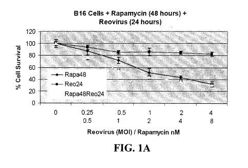
Figures 1 A, 1 B, and 1 C are graphs showing the effect of reovirus and
rapamycin
on B 16.F 10 cells in vitro. Cells (5 x 10³ per well) were seeded in 96 well
plates and
allowed to adhere overnight. Culture medium was replaced with doubling
dilutions of
rapamycin and/or reovirus, corresponding to 2, 1, 0.5 and 0.25 times the
previously
determined ED50, diluted in fresh culture medium and incubation continued for
48h.
Medium was then removed and percentage cell survival compared to untreated
cells was
determined using the MTS assay.
Figures 2A and 2B are graphs showing reovirus and rapamycin are synergistic in
vivo. B16.F10 tumors were seeded subcutaneously in C57B1/6 mice and treated
with
intratumoral reovirus T3D 5 x 10⁸ TCID50 on day 1 and 4, and intraperitoneal
rapamycin
5mg/kg on day 1, 4, 8 and 12 either alone or in combination, or with control
treatment
(intratumoral PBS, intraperitoneal PBS). Figure 2A is a graph showing the
average tumor
diameter of B16.F10 tumors in C57B1/g mice treated with reovirus and
rapamycin.
Figure 2B is a graph showing the survival data for C57B1/g mice with B16.F10
tumors
treated with reovirus and rapamycin.
-1-
CA 02723580 2010-11-04
WO 2009/143610 PCT/CA2009/000720
Figure 3 is a graph showing Treg depletion + IL-2 enhances systemic delivery
of
reovirus to subcutaneous tumors. C57B1/6 mice were seeded with subcutaneous
B16
tumors. Nine days later, mice received an intraperitoneal injection of anti-
CD25 antibody
PC-61or a control IgG. Twenty-four hours later, mice were injected
intraperitoneally
with PBS or with recombinant human IL-2 at a dose of 75,000 units/injection
three times
a day for 3 d. On the fourth day, a single further injection of IL-2 was
given. Two hours
after this last injection of IL-2/PBS, mice received an intravenous injection
of reovirus
(3.75 x10⁹ TCID50) followed 24 h later by a second similar injection of virus.
72 h later,
tumors were explanted and dissociated and viral titers recovered from
freeze/thaw lysates
of tumors from mice treated as shown were determined (3 mice per group).
Figures 4A and 4B are graphs showing CPA-mediated Treg modification, with IL-
2 and lower-dose reovirus, is therapeutic against established tumors. For
Figure 4A,
C57B1/6 mice were seeded with subcutaneous B16 tumors. Nine days later, mice
received an intraperitoneal injection of either CPA (100 mg/kg) or anti-CD25
antibody
PC-61 or PBS. Twenty-four hours later, mice were injected intraperitoneally
with PBS or
with recombinant human IL-2 at a dose of 75,000 units/injection three times a
day for 3 d.
On the fourth day, a single further injection of IL-2 was given. Two hours
after this last
injection of IL-2/PBS, mice received an intravenous injection of reovirus at a
lower than
maximal achievable dose of 1 x 10⁸ TCID50 followed 24h later by a second
similar
injection of virus. Survival of mice (tumor <1.0 cm in any diameter) with time
after
tumor seeding is shown (n = 7 per group). The median survival times of groups
treated
with reovirus alone (median survival, 21d), CPA/IL-2 (23 d), PC-61/reovirus
(22 d), or
CPA/reovirus (21 d) were not significantly different from each other and none
of these
treatments generated any long-term survivors. Median survival times of groups
treated
with IL-2/reovirus (25 d), PC-61/IL-2/reovirus (24d), or CPA/IL-2/reovirus (25
d) were
significantly longer (P = 0.04) than these other groups. Treatment with PC-
61/IL-
2/reovirus or CPA/IL-2/reovirus led to long-term survivors and both of these
were
significantly more therapeutic. **, P < 0.01. Figure 4B is a graph showing
neutralizing
antibodies against reovirus in serum recovered from mice 7 to 10 days after
the final viral
injection of the mice as described in Figure 4A.
DETAILED DESCRIPTION
Large, biological agents for the treatment of neoplasia may be limited by
intratumoral interstitial pressure and/or reduced vascular permeability.
Further, diffusion
seems to be the most important mode of passive transport of small molecules
(i.e., MW
-2-
CA 02723580 2010-11-04
WO 2009/143610 PCT/CA2009/000720
4000 Da) in tissues, whereas convection or solvent drag typically is the major
mechanism
of movement of large proteins (MW >40,000 Da).
Interstitial pressure within a tumor mass may be the result of increased
microvascular pressure (MVP), which is dependent upon the arteriovenous
pressure
difference and geometric and viscous resistance to blood flow (i.e., the
result of the
decrease in vessel diameter which is a function of the physical stress induced
on the
vessel by the growth of solid tumors decreasing vessel diameter). As such, the
intratumoral environment is one which results in increased interstitial
pressure and/or
decreased vascular permeability and may inhibit delivery of large molecules.
Agents that decrease hydrostatic pressure in a tumor create a situation where
the
hydrostatic pressure outside of the tumor mass would be greater than that of
the tumor
itself. This situation aids in the delivery of large molecules, such as
oncolytic viruses.
Thus, provided herein are methods of treating a proliferative disorder in a
subject
comprising decreasing interstitial pressure and/or increasing vascular
permeability in a
subject in need of treatment and administering to the subject in need of
treatment an
oncolytic virus. Optionally, the oncolytic virus is administered at the same
time, before
or after decreasing interstitial pressure and/or increasing vascular
permeability in the
subject.
Optionally, the interstitial pressure in the subject is decreased by an agent
that
decreases interstitial pressure and/or increases vascular permeability. Thus,
agents that
decrease interstitial pressure, optionally, increase vascular permeability, as
well.
Alternatively, an agent that decreases interstitial pressure can be used in
combination with
an agent that increases vascular permeability.
Agents suitable for use in the provided methods include a taxane. Suitable
taxanes for use in the provided methods include, but are not limited to, taxol
(paclitaxel),
larotaxel, and taxotere (docetaxel). Other agents include, but are not limited
to,
vasopressin; TNF; interleukin-1 (IL-1); interferon-K (IFN-K); substance P;
proteinase
inhibitors such as N-alpha-tosyl-L-lysyl-chloromethyl-ketone (TLCK), tosyl
phenylalanyl
chloromethyl ketone (TPCK) and leupeptin; vascular endothelial growth factor
(VEGF);
nitroglycerine; serotonin; plasma kinins such as bradykinin; platelet-
activating factor
(PAF); prostaglandin E1 (PGE1); histamine; imatinib; zona occludens toxin
(ZOT);
interleukin-2; nitric oxide inhibitors such as L-N-monomethyl arginine (L-
NMMA) and
L-N-nitro-arginine methyl ester (L-NAME); and human growth factor receptor
tyrosine
kinase inhibitors such as gefitinib. See Martin et al., Immunology 64(2):301-5
(1988);
Zhou et al., Radiat. Res. 168(3):299-307 (2007); Watanabe et al., Inflammation
Research
-3-
CA 02723580 2010-11-04
WO 2009/143610 PCT/CA2009/000720
17(5-6):472-7 9 (1986); U.S. Publication No. 2005/0101559; Moasser et al., J.
Magn.
Reson. Imaging 26(6):1618-25 (2007); and Vlahovic et al., Br. J. Cancer
97(6):735-40
(2007), which are incorporated herein by reference in their entireties at
least for the
agents described therein and methods of making and using the agents.
Optionally, the interstitial pressure in the subject is decreased by lowering
extracellular calcium ion concentrations. Low extracellular calcium ion
concentration
conditions also can be used to enhance vascular permeability. For example, a
low
calcium ion concentration fluid can be perfused through the vasculature of the
tissue to
which the oncolytic virus is administered. Suitable perfusate calcium ion
concentrations
may range from about 40 or 50 Tmol/L to about 500 Tmol/L, more preferably from
about
50 Tmol/L to about 200 Tmol/L. A perfusate calcium concentration of about 50
Tmol/L
is provided. Calcium ion (e.g., Ca2) concentration can also be lowered, for
example,
through use of a suitable buffer such as a chelating agent, for example,
ethylenebis(oxyethylenenitrilo)tet- racetic acid (EGTA),
ethylenediaminetetracetic acid
(EDTA), or 1,2-bis-(2-aminophenoxy)ethane-N,N,N',N'-tetraacetic acid (BAPTA).
See
U.S. Publication No. 2005/0101559, which is incorporated by reference herein
in its
entirety. Thus, provided herein are methods of treating a proliferative
disorder in a
subject comprising administering to the subject a low calcium ion
concentration fluid that
decreases interstitial pressure and an oncolytic virus. Optionally, the method
further
comprises administering an agent that increases vascular permeability.
Optionally, the interstitial pressure of a tumor can be reduced by removal of
excess interstitial fluid. Removal of excess interstitial fluid is
accomplished by any
known method, including, for example, by an artificial lymphatic system (ALS).
Such
methods are described in, for example, U.S. Publication No. 2001/0047152; U.S.
Patent
No. 5,484,399; U.S. Publication No. 2005/0165342; and U.S. Publication No.
2003/0149407, which are incorporated by reference herein, in their entireties.
Thus,
provided herein are methods of treating a tumor in a subject comprising
reducing in the
subject the excess interstitial fluid of a tumor and administering to the
subject an
oncolytic virus. Optionally, the excess interstitial fluid is removed prior to
administration
of the oncolytic virus. Optionally, the method further comprises administering
an agent
that increases vascular permeability.
If the oncolytic virus is administered systemically, permeabilizing
photodynamic
therapy (P-PDT) can be used to enhance delivery of the oncolytic virus by
enhancing
vascular permeability. P-PDT induced vascular leakiness allows the therapeutic
agents to
leave the vasculature and distribute into hyperproliferative tissue (e.g. the
tumor bed) in
-4-
CA 02723580 2010-11-04
WO 2009/143610 PCT/CA2009/000720
higher concentrations than achievable without prior permeabilizing PDT. See
U.S.
Publication No. 2004/0010218, which is incorporated by reference herein in its
entirety.
Thus, provided herein are methods of treating a proliferative disorder in a
subject
comprising administering to the subject a permeabilizing photodynamic
therapeutic agent
and an oncolytic virus. Optionally, the permeabilizing photodynamic
therapeutic agent is
administered prior to administration of the oncolytic virus. Optionally, the
method
further comprises administering an agent that decreases interstitial pressure.
Optionally, the provided methods further comprise administering to the subject
an
immunosuppressive agent. Optionally, the immunosuppressive agent is an agent
that
inhibits a pro-inflammatory cytokine. As used herein, a pro-inflammatory
cytokine refers
to a cytokine that directly or indirectly stimulates the immune system. Pro-
inflammatory
cytokines include, but are not limited to, IL-11, IL-3, IL-6, IL-12 p70, IL-
17, MIP-1I, and
RANTES. Thus, provided herein are methods of treating a proliferative disorder
in a
subject comprising administering to the subject in need of treatment, an agent
that
decreases interstitial pressure, an agent that inhibits a pro-inflammatory
cytokine and an
oncolytic virus. Optionally, the agent that decreases interstitial pressure is
administered
to the subject first, followed by administration of the agent that inhibits a
pro-
inflammatory cytokine and the oncolytic virus. Optionally, the oncolytic virus
is then
administered after the agent that inhibits the pro-inflammatory cytokine. The
agent that
inhibits the pro-inflammatory cytokine, optionally, inhibits the expression or
activity of
the pro-inflammatory cytokine. Optionally, the agent blocks T-cell responses
while
having little to no effect on B-cell activity. Thus, the agent inhibits pro-
inflammatory
cytokines but does not inhibit or minimally inhibits production of NARA.
Optionally, the
agent is a platinum compound. Suitable platinum compounds also include, but
are not
limited to, cisplatin, carboplatin, metaplatin, and oxaliplatin. Optionally,
the agent that
decreases interstitial pressure is paclitaxel, the agent that inhibits a pro-
inflammatory
cytokine is carboplatin and the oncolytic virus is a reovirus.
Other agents that inhibit pro-inflammatory cytokines include, but are not
limited
to, TNF-I antibodies such as infliximab, CDP571, CDP870, and adalimumab;
recombinant, human soluble p55 TNF receptors such as onercept; soluble TNF
receptor
and Fc fragment fusion proteins such as etanercept; pegylated Fab fragments of
humanized antibody to TNF such as certolizumab pegol; chimeric antibodies to
anti-I
chain of IL-2 receptor such as basiliximab or daclizumab; IL-12p40 antibodies
such as
ABT-874; IL-6 receptor antibodies such as MRA or tocilizumab; IFN-K antibodies
such
as fontolizumab; antibodies that inhibit IL-1 binding to the IL-1 receptor
such as
-5-
CA 02723580 2010-11-04
WO 2009/143610 PCT/CA2009/000720
AMG108; caspase-l inhibitors that inhibit cytokine-release such as
diarylsulphonylurene;
IL-15 antibodies such as mepolizumab; IL-8 antibodies such as ABX-IL-8; IL-9
antibodies including IL-9 monoclonal antibodies; recombinant human IL-21 also
referred
to as 494C 10; inhibitors of TNF-I, IL-19, IL-6 and granulocyte monocyte-
colony
stimulating factor expression such as biophylum sensitivum; NF-PB signaling
blockers
that inhibit pro-inflammatory cytokine expression such as simvastatin; and
inhibitors of
IL-6 expression and NF-PB activation such as (-)-epigallocatechin-3-gallate
(EGCG).
Other agents that inhibit pro-inflammatory cytokines include human recombinant
lactoferrin, which inhibits cellular release of proinflammatory cytokines and
prometastatic cytokines (including IL-6, IL-8, granulocyte macrophage colony-
stimulating factor and TNF-a). Inhibitors of dendritic cell derived IL-12 and
IL-18, such
as rapamycin and sanglifehrin, are also suitable for use in the provided
methods.
Rapamycin is an immunosuppressant that inhibits T cell mTOR kinase activation,
and
Sanglifehrin A is a cyclophilin-binding immunosuppressant that also inhibits
IL-2
dependent T cell proliferation. Also suitable for use in the provided methods
is dietary
rutin, which suppresses the induction of pro-inflammatory cytokines such as IL-
1 R, IL-6,
and GM-CS.
Optionally, the provided methods further include the step of selecting a
subject
with a proliferative disorder. Thus, provided is a method of treating a
proliferative
disorder in a subject comprising selecting a subject with a proliferative
disorder,
administering to the subject in need of treatment an agent that decreases
interstitial
pressure and an oncolytic virus. Optionally, the proliferative disorder is a
ras-mediated
proliferative disorder. Thus, the provided methods, optionally, further
comprise the step
of selecting a subject with a ras-mediated proliferative disorder. Optionally,
the
proliferative disorder is a proliferative disorder characterized by interferon-
resistance,
p53-deficiency or Rb-deficiency.
Optionally, the subject is in need of enhanced delivery of an oncolytic virus.
Thus, provided herein are methods of enhancing delivery of an oncolytic virus
to a
subject with a proliferative disorder comprising administering to the subject
an agent that
decreases interstitial pressure and administering to the subject the oncolytic
virus. Such
methods can also comprise the step of selecting a subject with a proliferative
disorder.
Optionally, the provided methods comprise the step of diagnosing the phenotype
of the proliferative disorder, for example, by determining whether the
proliferative
disorder is a ras-mediated proliferative disorder. By way of another example,
the
provided methods comprise the step of determining whether the proliferative
disorder is
-6-
CA 02723580 2010-11-04
WO 2009/143610 PCT/CA2009/000720
an interferon-resistant tumor, p53 deficient tumor or an Rb-deficient tumor.
Such
methods for determining whether a proliferative disorder has a certain
phenotype are
known. See, for example, U.S. Patent No. 7,306,902, which is incorporated
herein by
reference in its entirety.
Oncolytic viruses that are used in the provided methods include, but are not
limited to, oncolytic viruses that are members in the family of myoviridae,
siphoviridae,
podpviridae, teciviridae, corticoviridae, plasmaviridae, lipothrixviridae,
fuselloviridae,
poxyiridae, iridoviridae, phycodnaviridae, baculoviridae, herpesviridae,
adnoviridae,
papovaviridae, polydnaviridae, inoviridae, microviridae, geminiviridae,
circoviridae,
parvoviridae, hepadnaviridae, retroviridae, cyctoviridae, reoviridae,
birnaviridae,
paramyxoviridae, rhabdoviridae, filoviridae, orthomyxoviridae, bunyaviridae,
arenaviridae, leviviridae, picornaviridae, sequiviridae, comoviridae,
potyviridae,
caliciviridae, astroviridae, nodaviridae, tetraviridae, tombusviridae,
coronaviridae,
glaviviridae, togaviridae, and barnaviridae. Immunoprotected viruses and
reassortant or
recombinant viruses of these and other oncolytic viruses are also encompassed
by the
provided methods. Furthermore, a combination of at least two oncolytic viruses
can also
be employed to practice the provided methods. A few oncolytic viruses are
discussed
below, and a person of ordinary skill in the art can practice the present
methods using
additional oncolytic viruses as well according to the disclosure herein and
knowledge
available in the art.
Normally, when a virus enters a cell, double-stranded RNA Kinase (PKR) is
activated, blocking protein synthesis, and the virus cannot replicate in this
cell. Some
viruses have developed a system to inhibit PKR and facilitate viral protein
synthesis as
well as viral replication. For example, adenovirus makes a large amount of a
small RNA,
VAl RNA. VAl RNA has extensive secondary structures and binds to PKR in
competition with the double-stranded RNA (dsRNA) which normally activates PKR.
Since it requires a minimum length of dsRNA to activate PKR, VA1 RNA does not
activate PKR. Instead, it sequesters PKR by virtue of its large amount.
Consequently,
protein synthesis is not blocked, and adenovirus can replicate in the cell.
Ras-activated neoplastic cells are not subject to protein synthesis inhibition
by
PKR because ras inactivates PKR. These cells are therefore susceptible to
viral infection
even if the virus does not have a PKR-inhibitory system. Accordingly, if the
PKR
inhibitors in adenovirus, vaccinia virus, herpes simplex virus, or
parapoxvirus orf virus
are mutated so as not to block PKR function anymore, the resulting viruses do
not infect
normal cells due to protein synthesis inhibition by PKR, but they replicate in
ras-activated
-7-
CA 02723580 2010-11-04
WO 2009/143610 PCT/CA2009/000720
neoplastic cells which lack PKR activities. By way of example, reoviruses
selectively
replicate and lyse ras-activated neoplastic cells.
Accordingly, a virus, modified or mutated such that it does not inhibit PKR
function, selectively replicates in ras-activated neoplastic cells while
normal cells are
resistant. Optionally, the oncolytic virus is an adenovirus mutated in the VA1
region, a
vaccinia virus mutated in the K3L and/or E3L region, a vaccinia virus mutated
in the
thymidine kinase (TK) gene, a vaccinia virus mutated in the vaccinia growth
factor
(VGF) gene, a herpes virus mutated in the y134.5 gene, a parapoxvirus orf
virus mutated
in the OV20.OL gene, or an influenza virus mutated in the NS-1 gene.
Vaccinia viruses mutated in the viral thymidine kinase (TK) gene are unable to
make nucleotides needed for DNA replication. In normal cells, the cellular TK
levels are
usually very low and the virus is unable to replicate. In tumors, loss of the
tumor
suppressor Rb or an increase in cyclin activity, leads to E2F pathway
activation and high
levels of TK expression. Thus, cancer cells have high TK levels and the
mutated vaccinia
virus can replicate and spread.
The vaccinia growth factor (VGF) gene is a homolog of mammalian epidermal
growth factor (EGF) and can bind and activate the EGF Receptor (EGFR).
Vaccinia
viruses mutated in the VGF gene are growth restricted to cells with activated
EGF
pathways, which is commonly mutated in cancers.
The viruses can be modified or mutated according to the known structure-
function
relationship of the viral PKR inhibitors. For example, since the amino
terminal region of
E3 protein interacts with the carboxy-terminal region domain of PKR, deletion
or point
mutation of this domain prevents anti-PKR function (Chang et al., PNAS 89:4825-
4829
(1992); Chang, H. W. et al., Virology 194:537-547 (1993); Chang et al., J.
Virol.
69:6605-6608 (1995); Sharp et al., Virol. 250:301-315 (1998); and Romano et
al., Mol.
and Cell. Bio. 18:7304-7316 (1998)). The K3L gene of vaccinia virus encodes
pK3, a
pseudosubstrate of PKR. Truncations or point mutations within the C-terminal
portion of
K3L protein that is homologous to residues 79 to 83 in eIF-2 abolish PKR
inhibitory
activity (Kawagishi-Kobayashi, M., et al., Mol. Cell. Biology 17:4146-4158
(1997)).
Another example is the Delta24 virus, which is a mutant adenovirus carrying a
24
base pair deletion in the E I A region (Fueyo, J., et al., Oncogene 19(1):2-12
(2000)). This
region is responsible for binding to the cellular tumor suppressor Rb and
inhibiting Rb
function, thereby allowing the cellular proliferative machinery, and hence
virus
replication, to proceed in an uncontrolled fashion. Delta24 has a deletion in
the Rb
binding region and does not bind to Rb. Therefore, replication of the mutant
virus is
-8-
CA 02723580 2010-11-04
WO 2009/143610 PCT/CA2009/000720
inhibited by Rb in a normal cell. However, if Rb is inactivated and the cell
becomes
neoplastic, Delta24 is no longer inhibited. Instead, the mutant virus
replicates efficiently
and lyses the Rb-deficient cell.
In addition, vesicular stomatitis virus (VSV) selectively kills neoplastic
cells (and
interferon can be added). A herpes simplex virus 1 (HSV-1) mutant defective in
ribonucleotide reductase expression, hrR3, replicates in colon carcinoma cells
but not
normal liver cells (Yoon, S. S., et al., FASEB J. 14:301-311(2000)). Newcastle
disease
virus (NDV) replicates preferentially in malignant cells, and the most
commonly used
strain is 73-T (Reichard, K. W., et al., J. of Surgical Research 52:448-453
(1992); Zorn,
U. et al., Cancer Biotherapy 9(3):22-235 (1994); Bar-Eli, N., et al., J.
Cancer Res. Clin.
Oncol. 122: 409-415 (1996)). Vaccinia virus propagates in several malignant
tumor cell
lines. Encephalitis virus has an oncolytic effect in a mouse sarcoma tumor,
but
attenuation may be required to reduce its infectivity in normal cells. Tumor
regression
has been described in tumor patients infected with herpes zoster, hepatitis
virus,
influenza, varicella, and measles virus (for a review, see Nemunaitis, J.,
Invest. New
Drugs 17:375-386 (1999)).
Optionally, the oncolytic virus is a reovirus. Reovirus refers to any virus
classified in the reovirus genus, whether naturally occurring, modified, or
recombinant.
Reoviruses are viruses with a double-stranded, segmented RNA genome. The
virions
measure 60-80 nm in diameter and possess two concentric capsid shells, each of
which is
icosahedral. The genome consists of double-stranded RNA in 10-12 discrete
segments
with a total genome size of 16-27 kbp. The individual RNA segments vary in
size. Three
distinct but related types of reoviruses have been recovered from many
species. All three
types share a common complement-fixing antigen.
The human reovirus includes three serotypes: type 1 (strain Lang or T1L), type
2
(strain Jones, T2J), and type 3 (strain Dearing or strain Abney, T3D). The
three serotypes
are easily identifiable on the basis of neutralization and hemagglutinin-
inhibition assays.
A reovirus according to this disclosure can be a type 3 mammalian
orthoreovirus. Type 3
mammalian orthoreoviruses include, without limitation, Dearing and Abney
strains (T3D
or T3A, respectively). See, for example, ATCC Accession Nos. VR-232 and VR-
824. As
described previously, reoviruses use a host cell's ras pathway machinery to
downregulate
double-stranded RNA-activated protein kinase (PKR) and thus replication in the
cell.
See, for example, U.S. Patent Nos. 6,110,461; 6,136,307; 6,261,555; 6,344,195;
6,576,234; and 6,811,775, which are incorporated by reference herein in their
entireties.
-9-
CA 02723580 2010-11-04
WO 2009/143610 PCT/CA2009/000720
The reovirus may be naturally occurring or modified. The reovirus is naturally-
occurring when it can be isolated from a source in nature and has not been
intentionally
modified by humans in the laboratory. For example, the reovirus can be from a
field
source, that is, from a human who has been infected with the reovirus. The
reovirus may
also be selected or mutagenized for enhanced oncolytic activity.
The reovirus may be modified but still capable of lytically infecting a
mammalian
cell having an active ras pathway. The reovirus may be chemically or
biochemically
pretreated (e.g., by treatment with a protease, such as chymotrypsin or
trypsin) prior to
administration to the proliferating cells. Pretreatment with a protease
removes the outer
coat or capsid of the virus and may increase the infectivity of the virus. The
reovirus may
be coated in a liposome or micelle (Chandran and Nibert, J. of Virology
72(1):467-75
1998). For example, the virion may be treated with chymotrypsin in the
presence of
micelle-forming concentrations of alkyl sulfate detergents to generate a new
infectious
subviral particle (ISVP).
The reovirus may be a recombinant reovirus. For example, the recombinant
reovirus can be a reassortant reovirus, which includes genomic segments from
two or
more genetically distinct reoviruses. Recombination/reassortment of reovirus
genomic
segments may occur following infection of a host organism with at least two
genetically
distinct reoviruses. Recombinant/reassortant viruses can also be generated in
cell culture,
for example, by co-infection of permissive host cells with genetically
distinct reoviruses.
Accordingly, the provided methods include the use of a recombinant reovirus
resulting
from reassortment of genome segments from two or more genetically distinct
reoviruses,
including but not limited to, human reovirus, such as type 1 (e.g., strain
Lang), type 2
(e.g., strain Jones), and type 3 (e.g., strain Dearing or strain Abney); non-
human
mammalian reoviruses; or avian reovirus. Optionally, the provided methods
include the
use of recombinant reoviruses resulting from reassortment of genome segments
from two
or more genetically distinct reoviruses wherein at least one parental virus is
genetically
engineered, comprises one or more chemically synthesized genomic segment, has
been
treated with chemical or physical mutagens, or is itself the result of a
recombination
event. Optionally, the provided methods include the use of the recombinant
reovirus that
has undergone recombination in the presence of chemical mutagens, including
but not
limited to, dimethyl sulfate and ethidium bromide, or physical mutagens,
including but
not limited to, ultraviolet light and other forms of radiation.
Optionally, the provided methods include the use of reoviruses with mutations
(including insertions, substitutions, deletions or duplications) in one or
more genome
-10-
CA 02723580 2010-11-04
WO 2009/143610 PCT/CA2009/000720
segments. Such mutations can comprise additional genetic information as a
result of
recombination with a host cell genome or can comprise synthetic genes. For
example,
mutant reoviruses as described herein can contain a mutation that reduces or
essentially
eliminates expression of a sigma3 polypeptide or that results in the absence
of a
functional sigma3 polypeptide as described in U.S. Serial No. 12/124,522,
which is
incorporated by reference herein in its entirety. A mutation that eliminates
expression of
a sigma3 polypeptide or that results in the absence of a functional sigma3
polypeptide can
be in the nucleic acid encoding the sigma3 polypeptide (i.e., the S4 gene) or
in a nucleic
acid that encodes a polypeptide that regulates the expression or function of
the sigma3
polypeptide.
As used herein, a mutation that reduces the expression of a sigma3 polypeptide
refers to a mutation that results in a decrease in the amount of sigma3
polypeptides,
compared to a reovirus expressing wild type levels of sigma3 polypeptide, of
at least 30%
(e.g., at least 40%, 50%, 60%, 70%, 80%, 90%, or 95%). As used herein, a
mutation that
essentially eliminates expression of a sigma3 polypeptide refers to a mutation
that results
in a decrease in the amount of sigma3 polypeptides, relative to the amount of
sigma3
polypeptides produced by a wild type reovirus, of at least 95% (e.g., 96%,
97%, 98%,
99%, or 100%). As used herein, a mutation that results in a decrease in or
absence of a
functional sigma3 polypeptide refers to a mutation that allows expression of
the sigma3
polypeptide but that results in a sigma3 polypeptide that is not able to
assemble or
incorporate into the viral capsid. It would be understood that it may be
desirable or
necessary for sigma3 polypeptides to retain other functionalities (e.g., the
ability to bind
RNA) in order that the mutant reovirus retain the ability to propagate.
A mutation in a sigma3 polypeptide as described herein can result in a sigma3
polypeptide that is incorporated into the capsid at levels that are reduced
relative to a
sigma3 polypeptide that does not contain the mutation (e.g., a wild type
sigma3
polypeptide). A mutation in a sigma3 polypeptide as described herein also can
result in a
sigma3 polypeptide that cannot be incorporated into a viral capsid. Without
being bound
by any particular mechanism, a sigma3 polypeptide may have reduced function or
lack
function due, for example, to an inability of the sigma3 polypeptide and the
mul
polypeptide to bind appropriately, or due to a conformational change that
reduces or
prohibits incorporation of the sigma3 polypeptide into the capsid.
In addition to a mutation that abolishes or reduces expression of the sigma3
polypeptide or that results in a non-functional or reduced-function sigma3
polypeptide, a
mutant reovirus as described herein also can contain one or more further
mutations (e.g.,
-11-
CA 02723580 2010-11-04
WO 2009/143610 PCT/CA2009/000720
a second, third, or fourth mutation) in one of the other reovirus capsid
polypeptides (e.g.,
mul, lambda2, and/or sigmal). Reoviruses containing a mutation affecting the
sigma3
polypeptide and, optionally, a further mutation in any or all of the other
outer capsid
proteins can be screened for the ability of such mutant reoviruses to infect
and cause lysis
of cells. For example, neoplastic cells that are resistant to lysis by wild
type reovirus can
be used to screen for effective mutant reoviruses described herein.
For example, a further mutation can reduce or essentially eliminate expression
of
a mul polypeptide or result in the absence of a functional mul polypeptide.
The mul
polypeptide, which is encoded by the M2 gene, is likely involved in cell
penetration and
may play a role in transcriptase activation. Each virion contains about 600
copies of mu l
polypeptides, which are present in the form of 1:1 complexes with sigma3
polypeptides.
The mul polypeptide is myristolated on its N-terminus, and then the
myristolated N-
terminal 42 residues are cleaved off, resulting in a C-terminal fragment
(muIC).
Additionally or alternatively, a further mutation can reduce or essentially
eliminate
expression of a lambda2 polypeptide or result in the absence of a functional
lambda2
polypeptide, and/or a further mutation can reduce or essentially eliminate
expression of a
sigmal polypeptide or result in the absence of a functional sigmal
polypeptide. The
lambda2 polypeptide is encoded by the L2 gene, is involved in particle
assembly, and
exhibits guanylyltransferase and methyltransferase activity. The sigmal
polypeptide is
encoded by the Si gene, is involved in cell-attachment and serves as the viral
hemagglutinin.
For example, the reovirus has a lambda-3 polypeptide having one or more amino
acid modifications; a sigma-3 polypeptide having one or more amino acid
modifications;
a mu-1 polypeptide having one or more amino acid modifications; and/or a mu-2
polypeptide having one or more amino acid modifications, as described in U.S.
Serial No.
12/046,095, which is incorporated by reference herein in its entirety. By way
of example,
the one or more amino acid modifications in the lambda-3 polypeptide are a Val
at residue
214, an Ala at residue 267, a Thr at residue 557, a Lys at residue 755, a Met
at residue
756, a Pro at residue 926, a Pro at residue 963, a Leu at residue 979, an Arg
at residue
1045, a Val at residue 1071, or any combination thereof, numbered relative to
GenBank
Accession No. M24734. 1. It is noted that, when the amino acid sequence is a
Val at
residue 214 or a Val at residue 1071, the amino acid sequence further includes
at least one
additional change in the amino acid sequence. Optionally, the lambda-3
polypeptide
includes the sequence shown in SEQ ID NO: 18. Further by way of example, the
one or
more amino acid modifications in the sigma-3 polypeptide are a Leu at residue
14, a Lys
-12-
CA 02723580 2010-11-04
WO 2009/143610 PCT/CA2009/000720
at residue 198, or any combination thereof, numbered relative to GenBank
Accession No.
K02739. It is noted that, when the amino acid sequence is a Leu at residue 14,
the amino
acid sequence further includes at least one additional change in the amino
acid sequence.
Optionally, the sigma-3 polypeptide includes the sequence shown in SEQ ID
NO:14.
Further by way of example, the one or more amino acid modifications in the mu-
1
polypeptide is an Asp at residue 73 numbered relative to GenBank Accession No.
M20161.1. Optionally, the mu-1 polypeptide includes the sequence shown in SEQ
ID
NO: 16. Also by way of example, the amino acid modification mu-2 polypeptide
is a Ser
at residue 528 numbered relative to GenBank Accession No. AF461684.1.
Optionally, the
mu-1 polypeptide includes the sequence shown in SEQ ID NO: 15. A reovirus as
described herein having one or more modifications can further include a
reovirus sigma-2
polypeptide. Such a sigma-2 polypeptide has a Cys at one or more of position
70, 127,
195, 241, 255, 294, 296, or 340, numbered relative to GenBank Accession No.
NP_694684.1. Optionally, the sigma-2 polypeptide includes the sequence shown
in SEQ
ID NO: 12.
Optionally, the reovirus has a L1 genome segment having one or more nucleic
acid modifications; a S4 genome segment having one or more nucleic acid
modifications;
a Ml genome segment having one or more nucleic acid modifications; and/or a M2
genome segment having one or more nucleic acid modifications, as described in
U.S.
Serial No. 12/046,095, which is incorporated by reference herein in its
entirety. By way
of example, the one or more nucleic acid modifications in the L1 genome
segment are a T
at position 660, a G at position 817, an A at position 1687, a G at position
2283, an ATG
at positions 2284-2286, a C at position 2794, a C at position 2905, a C at
position 2953,
an A at position 3153, or a G at position 3231, numbered relative to GenBank
Accession
No. M24734.1. Optionally, the L1 genome segment includes the sequence shown in
SEQ
ID NO:8. Further by way of example, the one or more nucleic acid modifications
in the
S4 genome segment is an A at position 74 and an A at position 624, numbered
relative to
GenBank Accession No. K02739. Optionally, the S4 genome segment includes the
sequence shown in SEQ ID NO:4. Further by way of example, the nucleic acid
modification in the M2 genome segment can be a C at position 248, numbered
relative to
GenBank Accession No. M20161.1. The M2 genome segment, for example, includes
the
sequence shown in SEQ ID NO:6. Also by way of example, the nucleic acid
modification
in the M1 genome segment is a T at position 1595, numbered relative to GenBank
Accession No. AF461684.1. Optionally, the M1 genome segment includes the
sequence
shown in SEQ ID NO:5. A reovirus as described herein can include any
modification or
-13-
CA 02723580 2010-11-04
WO 2009/143610 PCT/CA2009/000720
combination of modifications disclosed herein. Optionally, a reovirus as
described herein
includes genomic segments having the sequences shown in SEQ ID NOs:I-10 or the
polypeptides shown in SEQ ID NOs:11, 12, and 16-21, and either or both SEQ ID
NO:13
or 14. Optionally, a reovirus as disclosed herein is identified as IDAC
Accession No.
190907-01.
Sindbis virus (SIN) can be used in the methods described herein. Sindbis virus
is
a member of the alphavirus genus of the togaviridae family. The Sindbis virus
genome is
a single-stranded RNA of 11703 nucleotides, capped at the 5' terminus and poly-
adenylated at the 3' terminus. The genome consists of a 49S untranslated
region (UT),
nonstructural proteins nsP 1, nsP2, nsP3, and nsP4 followed by a promoter. The
promoter
is followed by a 26S UT, structural proteins C, E3, E2, 6K, and E1 and finally
a 3' UT
and a poly-adenylated terminus. The genomic 49S RNA is of plus sense, is
infectious,
and serves as mRNA in the infected cell.
Sindbis vectors systemically and specifically infect/detect and kill
metastasized
tumors in vivo, leading to significant suppression of tumor growth and
enhanced survival
(Hurtado et al., Rejuvenation Res. 9(1):36-44 (2006)). Sindbis virus infects
mammalian
cells using the Mr 67,000 laminin receptor, which is elevated in tumor versus
normal
cells. Tumor overexpression of the laminin receptor may explain the
specificity and
efficacy that Sindbis vectors demonstrate for tumor cells in vivo. Sindbis
does not have to
undergo genetic manipulation to target cancer cells or to be injected directly
into tumors.
Sindbis injected anywhere into a subject travels through the bloodstream to
the target area
(Tseng et al., Cancer Res. 64(18):6684-92 (2004). Sindbis can also be
genetically
engineered to carry one or more genes that suppress the immune response to the
virus
and/or genes that stimulate an immune response against the tumor such as, for
example,
antitumor cytokine genes such as interleukin- 12 and interleukin- 15 genes.
The oncolytic virus may be naturally occurring or modified. The virus may be
chemically or biochemically pretreated (e.g., by treatment with a protease,
such as
chymotrypsin or trypsin) prior to administration to the neoplastic cells.
Pretreatment with
a protease removes the outer coat or capsid of the virus and may increase the
infectivity
of the virus. The virus may be coated in a liposome or micelle (Chandran and
Nibert, J.
of Virology 72(l):467-75 (1998)) to reduce or prevent an immune response from
a
mammal which has developed immunity to the virus. For example, the virion may
be
treated with chymotrypsin in the presence of micelle forming concentrations of
alkyl
sulfate detergents to generate a new infectious subvirion particle. The
oncolytic virus
may also be a reassortant virus or an ISVP.
-14-
CA 02723580 2010-11-04
WO 2009/143610 PCT/CA2009/000720
The present methods include using any oncolytic virus according to the
disclosure
herein and knowledge available in the art. The oncolytic virus may be
naturally occurring
or modified. The oncolytic virus is naturally-occurring when it can be
isolated from a
source in nature and has not been intentionally modified by humans in the
laboratory. For
example, the oncolytic virus can be from a field source, that is, from a human
who has
been infected with the oncolytic virus.
The oncolytic virus may be a recombinant oncolytic virus. For example, the
recombinant oncolytic virus results from the reassortment of genomic segments
from two
or more genetically distinct oncolytic viruses, also referred to herein as a
reassortant.
Reassortment of oncolytic virus genomic segments may occur following infection
of a
host organism with at least two genetically distinct oncolytic viruses.
Recombinant
viruses can also be generated in cell culture, for example, by co-infection of
permissive
host cells with genetically distinct oncolytic viruses. Optionally, the
methods include the
use of recombinant oncolytic virus resulting from reassortment of genome
segments from
two or more genetically distinct oncolytic viruses wherein at least one
parental virus is
genetically engineered, comprises one or more chemically synthesized genomic
segment,
has been treated with chemical or physical mutagens, or is itself the result
of a
recombination event. Optionally, the methods include the use of the
recombinant
oncolytic virus that has undergone recombination in the presence of chemical
mutagens,
including but not limited to dimethyl sulfate and ethidium bromide, or
physical mutagens,
including but not limited to ultraviolet light and other forms of radiation.
Optionally, the methods include the use of oncolytic viruses with mutations
including (insertions, substitutions, deletions or duplications) in one or
more genome
segments. Such mutations can comprise additional genetic information as a
result of
recombination with a host cell genome, or that comprise synthetic genes such
as, for
example, genes encoding agents that suppress anti-viral immune responses.
Optionally, the oncolytic virus is a mutant oncolytic virus. For example, the
oncolytic virus may be modified by incorporation of mutated coat proteins,
such as for
example, into the virion outer capsid. The mutant oncolytic virus is,
optionally, a mutant
reovirus. Mutant reoviruses as described herein can contain a mutation that
reduces or
essentially eliminates expression of a sigma3 polypeptide or that results in
the absence of
a functional sigma3 polypeptide as described in U.S. Serial No. 12/124,522,
which is
incorporated by reference herein in its entirety. Optionally, the mutant
reoviruses used in
the provided methods are mutated as described in U.S. Serial No. 12/046,095,
which is
incorporated by reference herein in its entirety.
-15-
CA 02723580 2010-11-04
WO 2009/143610 PCT/CA2009/000720
A mutation as referred to herein can be a substitution, insertion or deletion
of one
or more nucleotides. Point mutations include, for example, single nucleotide
transitions
(purine to purine or pyrimidine to pyrimidine) or transversions (purine to
pyrimidine or
vice versa) and single- or multiple-nucleotide deletions or insertions. A
mutation in a
nucleic acid can result in one or more conservative or non-conservative amino
acid
substitutions in the encoded polypeptide, which may result in conformational
changes or
loss or partial loss of function, a shift in the reading frame of translation
(frame-shift)
resulting in an entirely different polypeptide encoded from that point on, a
premature stop
codon resulting in a truncated polypeptide (truncation), or a mutation in a
virus nucleic
acid may not change the encoded polypeptide at all (silent or nonsense). See,
for
example, Johnson and Overington, 1993, J. Mol. Biol. 233:716-38; Henikoff and
Henikoff, 1992, Proc. Natl. Acad. Sci. USA 89:10915-19; and U.S. Patent No.
4,554,101,
for disclosure on conservative and non-conservative amino acid substitutions.
Mutations can be generated in the nucleic acid of an oncolytic virus using any
number of methods known in the art. For example, site directed mutagenesis can
be used
to modify a reovirus nucleic acid sequence. One of the most common methods of
site-
directed mutagenesis is oligonucleotide-directed mutagenesis. In
oligonucleotide-
directed mutagenesis, an oligonucleotide encoding the desired change(s) in
sequence is
annealed to one strand of the DNA of interest and serves as a primer for
initiation of
DNA synthesis. In this manner, the oligonucleotide containing the sequence
change is
incorporated into the newly synthesized strand. See, for example, Kunkel,
1985, Proc.
Natl. Acad. Sci. USA 82:488; Kunkel et al., 1987, Meth. Enzymol. 154:367;
Lewis and
Thompson, 1990, Nucl. Acids Res. 18:3439; Bohnsack, 1996, Meth. Mol. Biol.
57:1;
Deng and Nickoloff, 1992, Anal. Biochem. 200:81; and Shimada, 1996, Meth. Mol.
Biol.
57:157. Other methods are used routinely in the art to modify the sequence of
a protein
or polypeptide. For example, nucleic acids containing a mutation can be
generated using
PCR or chemical synthesis, or polypeptides having the desired change in amino
acid
sequence can be chemically synthesized. See, for example, Bang and Kent, 2005,
Proc.
Natl. Acad. Sci. USA 102:5014-9 and references therein.
Viruses can be purified using standard methodology. See, for example, Schiff
et
al., "Orthoreoviruses and Their Replication," Ch 52, in Fields Virology, Knipe
and
Howley, eds., 2006, Lippincott Williams and Wilkins; Smith et al., 1969,
Virology
39(4):791-810; and U.S. Patent Nos. 7,186,542; 7,049,127; 6,808,916; and
6,528,305,
which are incorporated by reference herein in their entireties. As used
herein, purified
viruses refer to viruses that have been separated from cellular components
that naturally
-16-
CA 02723580 2010-11-04
WO 2009/143610 PCT/CA2009/000720
accompany them. Typically, viruses are considered purified when they are at
least 70%
(e.g., at least 75%, 80%, 85%, 90%, 95%, or 99%) by dry weight, free from the
proteins
and other cellular components with which they are naturally associated.
Provided herein are pharmaceutical compositions comprising the oncolytic
viruses. Also provided herein are pharmaceutical compositions comprising
therapeutic
agents, for example, the agents that decrease interstitial pressure and/or
increase vascular
permeability. Optionally, the pharmaceutical composition comprises the
oncolytic virus
and the agent that decreases interstitial pressure and/or increases vascular
permeability.
Optionally, the pharmaceutical composition comprises the oncolytic virus, the
agent that
decreases interstitial pressure and/or vascular permeability and the agent
that inhibits pro-
inflammatory cytokines. Thus, the provided pharmaceutical compositions can
comprise
one agent or more than one agent. For example, each of the oncolytic virus,
the agent that
decreases interstitial pressure and/or vascular permeability and the agent
that inhibits pro-
inflammatory cytokines can be contained within separate pharmaceutical
compositions or
the same composition. If the oncolytic virus and agents are contained within
separate
pharmaceutical compositions, the compositions can be administered
concomitantly or
sequentially.
The herein provided compositions are administered in vitro or in vivo in a
pharmaceutically acceptable carrier. A pharmaceutically acceptable carrier can
be a solid,
semi-solid, or liquid material that can act as a vehicle, carrier or medium
for the reovirus.
Thus, compositions containing a reovirus and/or one or more of the provided
agents can
be in the form of tablets, pills, powders, lozenges, sachets, elixirs,
suspensions,
emulsions, solutions, syrups, aerosols (as a solid or in a liquid medium),
ointments
containing, for example, up to 10% by weight of the active compound, soft and
hard
gelatin capsules, suppositories, sterile injectable solutions, and sterile
packaged powders.
Optionally, the compositions containing an oncolytic virus are suitable for
infusion. For intravenous infusions, there are two types of fluids that are
commonly used,
crystalloids and colloids. Crystalloids are aqueous solutions of mineral salts
or other
water-soluble molecules. Colloids contain larger insoluble molecules, such as
gelatin;
blood itself is a colloid. The most commonly used crystalloid fluid is normal
saline, a
solution of sodium chloride at 0.9% concentration, which is close to the
concentration in
the blood (isotonic). Ringer's lactate or Ringer's acetate is another isotonic
solution often
used for large-volume fluid replacement. A solution of 5% dextrose in water,
sometimes
called D5 W, is often used instead if the patient is at risk for having low
blood sugar or
high sodium.
-17-
CA 02723580 2010-11-04
WO 2009/143610 PCT/CA2009/000720
Some examples of suitable carriers include phosphate-buffered saline or
another
physiologically acceptable buffer, lactose, dextrose, sucrose, sorbitol,
mannitol, starches,
gum acacia, calcium phosphate, alginates, tragacanth, gelatin, calcium
silicate,
microcrystalline cellulose, polyvinylpyrrolidone, cellulose, sterile water,
syrup, and
methyl cellulose. A pharmaceutical composition additionally can include,
without
limitation, lubricating agents such as talc, magnesium stearate, and mineral
oil; wetting
agents; emulsifying and suspending agents; preserving agents such as methyl-
and
propylhydroxy-benzoates; sweetening agents; and flavoring agents.
Pharmaceutical
compositions can be formulated to provide quick, sustained or delayed release
of a mutant
reovirus after administration by employing procedures known in the art. In
addition to
the representative formulations described below, other suitable formulations
for use in a
pharmaceutical composition can be found in Remington: The Science and Practice
of
Pharmacy (21th ed.) ed. David B. Troy, Lippincott Williams & Wilkins, 2005.
For
preparing solid compositions such as tablets, a mutant reovirus can be mixed
with a
pharmaceutical carrier to form a solid composition. Optionally, tablets or
pills can be
coated or otherwise compounded to provide a dosage form affording the
advantage of
prolonged action. For example, a tablet or pill can comprise an inner dosage
and an outer
dosage component, the latter being in the form of an envelope over the former.
The two
components can be separated by an enteric layer which serves to resist
disintegration in
the stomach and permit the inner component to pass intact into the duodenum or
to be
delayed in release. A variety of materials can be used for such enteric layers
or coatings,
such materials including a number of polymeric acids and mixtures of polymeric
acids
with such materials as shellac, cetyl alcohol, and cellulose acetate.
Liquid formulations that include a reovirus and/or agent for oral
administration or
for injection generally include aqueous solutions, suitably flavored syrups,
aqueous or oil
suspensions, and flavored emulsions with edible oils such as corn oil,
cottonseed oil,
sesame oil, coconut oil, or peanut oil, as well as elixirs and similar
pharmaceutical
vehicles.
Compositions for inhalation or insufflation include solutions and suspensions
in
pharmaceutically acceptable, aqueous or organic solvents, or mixtures thereof,
and
powders. These liquid or solid compositions may contain suitable
pharmaceutically
acceptable excipients as described herein. Such compositions can be
administered by the
oral or nasal respiratory route for local or systemic effect. Compositions in
pharmaceutically acceptable solvents may be nebulized by use of inert gases.
Nebulized
solutions may be inhaled directly from the nebulizing device or the nebulizing
device
-18-
CA 02723580 2010-11-04
WO 2009/143610 PCT/CA2009/000720
may be attached to a face mask tent or intermittent positive pressure
breathing machine.
Solution, suspension, or powder compositions may be administered, orally or
nasally,
from devices which deliver the formulation in an appropriate manner.
Another formulation that is optionally employed in the methods of the present
disclosure includes transdermal delivery devices (e.g., patches). Such
transdermal
patches may be used to provide continuous or discontinuous infusion of the
viruses and
agents as described herein. The construction and use of transdermal patches
for the
delivery of pharmaceutical agents is well known in the art. See, for example,
U.S. Patent
No. 5,023,252. Such patches can be constructed for continuous, pulsatile, or
on-demand
delivery of mutant reoviruses.
As described above, viruses and/or other agents can, if necessary, be coated
in a
liposome or micelle to reduce or prevent an immune response in a mammal that
has
developed immunity toward a virus or agent. Such compositions are referred to
as
immunoprotected viruses or agents. See, for example, U.S. Patent Nos.
6,565,831 and
7,014,847.
In the provided methods, the oncolytic virus is administered, for example,
systemically, in a manner so that it can ultimately contact the target tumor
or tumor cells.
The route by which the virus is administered, as well as the formulation,
carrier or
vehicle, depends on the location as well as the type of the target cells. A
wide variety of
administration routes can be employed. For example, for a solid tumor that is
accessible,
the virus can be administered by injection directly to the tumor. For a
hematopoietic
tumor, for example, the virus can be administered intravenously or
intravascularly. For
tumors that are not easily accessible within the body, such as metastases, the
virus is
administered in a manner such that it can be transported systemically through
the body of
the mammal and thereby reach the tumor (e.g., intravenously or
intramuscularly).
Alternatively, the virus can be administered directly to a single solid tumor,
where it then
is carried systemically through the body to metastases. The virus can also be
administered subcutaneously, intraperitoneally, intrathecally or
intraventricularly (e.g., for
brain tumor), topically (e.g., for melanoma), orally (e.g., for oral or
esophageal cancer),
rectally (e.g., for colorectal cancer), vaginally (e.g., for cervical or
vaginal cancer),
nasally, by inhalation spray or by aerosol formulation (e.g., for lung
cancer).
Optionally, the virus is administered continuously to a subject at least once
per
day or up to intermittently or continuously throughout the day on consecutive
days, for a
period of time. Thus, the virus is administered, for example, to subjects by
means of
intravenous administration in any pharmacologically acceptable solution, or as
an
-19-
CA 02723580 2010-11-04
WO 2009/143610 PCT/CA2009/000720
infusion over a period of time. For example, the substance may be administered
systemically by injection (e.g., IM or subcutaneously) or taken orally daily
at least once
per day, or administered by infusion in a manner that results in the daily
delivery into the
tissue or blood stream of the subject. When the virus is administered by
infusion over a
period of time, the period of time is, for example, 1, 2, 3, 4, 5, 6, 7, 8, 9,
10, 12, or 24
hours, or any time between 1 and 24 hours, inclusive, or more. Optionally, the
period of
time is 5, 15, 30, 60, 90, 120, 150 or 180 minutes, or any time between 5 and
180
minutes, inclusive, or more. Thus, for example, the virus is administered by
infusion for
60 minutes. Administrations can be repeated daily for 2, 3, 4, 5, 6, 7, 8, 9,
10, 14, 21, 28
days or any number of days between 2 and 28 days, inclusive, or longer.
The agents that decrease interstitial pressure and/or vascular permeability or
other
therapeutic agents (i.e., the agents that inhibit pro-inflammatory cytokines)
of the
provided methods are also administered via a wide variety of administration
routes. Thus,
the agents are administered via any of several routes of administration,
including,
topically, orally, parenterally, intravenously, intraperitoneally,
intramuscularly,
subcutaneously, intracavity, transdermally, intrahepatically, intracranially,
nebulization/inhalation, or by instillation via bronchoscopy. Optionally, the
therapeutic
agents are administered continuously in the manner set forth in the
description above with
respect to oncolytic viruses. Thus, for example, the agent is administered to
subjects by
means of intravenous administration in any pharmacologically acceptable
solution, or as
an infusion over a period of time. Optionally, the agents are administered
locally at or
near the site of the tumor. Alternatively, the agents are administered
systemically. The
agents that decrease interstitial pressure and/or vascular permeability are
administered in
an amount that is sufficient (i.e., an effective amount) to decrease
interstitial pressure
and/or increase vascular permeability. Agents that inhibit pro-inflammatory
cytokines are
administered in an amount sufficient (i.e., an effective amount) to inhibit
one or more pro-
inflammatory cytokines. By way of example, effective amounts of taxanes
include from
about 40-300 mg/m2 of tumor volume; or any amount in between 40 and 300 mg/m2,
inclusive. Thus, effective amounts of taxanes include 130-225 mg/m2. By way of
another example, effective amounts of platinum compounds include from about 5-
1000
mg/m2, or any amount in between 5 and 1000 mg/m2, inclusive. Thus, for example
effective amounts of cisplatin include from about 175-200 mg/m2 and effective
mounts
for carboplatin include from about 200-600 mg/m2. Effective amounts of other
agents
range from 0.001-10,000 mg/kg body weight or any amount in between 0.001 and
10,000
mg/kg body weight, inclusive. Optionally, effective amounts of platinum
compounds
-20-
CA 02723580 2010-11-04
WO 2009/143610 PCT/CA2009/000720
include approximately 2 to 7 mg/mL minute (AUC) as calculated by the Calvert
formula.
Optionally, effective amounts of platinum compounds include approximately 5 or
6
mg/mL minute (AUC) as calculated by the Calvert formula. Optionally, the
platinum
compounds are administered as an intravenous infusion over a period of 30
minutes.
The viruses as disclosed herein are administered in an amount that is
sufficient
(i.e., an effective amount) to treat the proliferative disorder. A
proliferative disorder is
treated when administration of a virus to proliferating cells affects lysis
(e.g., oncolysis)
of the affected cells, resulting in a reduction in the number of abnormally,
proliferating
cells, a reduction in the size of a neoplasm, and/or a reduction in or
elimination of
symptoms (e.g., pain) associated with the proliferating disorder. As used
herein, the term
oncolysis means at least 10% of the proliferating cells are lysed (e.g., at
least about 20%,
30%, 40%, 50%, or 75% of the cells are lysed). The percentage of lysis can be
determined, for example, by measuring the reduction in the size of a neoplasm
or in the
number of proliferating cells in a mammal, or by measuring the amount of lysis
of cells in
vitro (e.g., from a biopsy of the proliferating cells). An effective amount of
a virus will
be determined on an individual basis and may be based, at least in part, on
the particular
virus used; the individual's size, age, gender; and the size and other
characteristics of the
abnormally, proliferating cells. For example, for treatment of a human,
approximately
103 to 1012 plaque forming units (PFU) of a virus are used, depending on the
type, size
and number of proliferating cells or neoplasms present. The effective amount
can be, for
example, from about 1.0 PFU/kg body weight to about 1015 PFU/kg body weight
(e.g.,
from about 102 PFU/kg body weight to about 1013 PFU/kg body weight).
Optionally, the
effective amount is about 1x10⁸ to about 1x10¹² TCID50. Optionally, the
effective
amount is about 1x10¹⁰ TCID50.
By way of example, 175 mg/m2 of the agent that decreases interstitial pressure
and/or increases vascular permeability, such as paclitaxel, is administered to
the subject
and 3x10¹⁰ TCID50 or lxl010 TCID50 of a reovirus is administered to the
subject.
Optionally, 200 mg/m2 of the agent that decreases interstitial pressure and/or
increases
vascular permeability, such as paclitaxel, is administered to the subject and
3x10¹⁰
TCID50 or 1x10¹⁰ TCID50 of a reovirus is administered to the subject.
Optionally, the
agent that decreases interstitial pressure and/or increases vascular
permeability is
administered as a three hour intravenous infusion. Optionally, the reovirus is
administered as a one hour intravenous infusion.
By way of another example, 175 mg/m2 of the agent that decreases interstitial
pressure and/or increases vascular permeability, such as paclitaxel, is
administered to the
-21-
CA 02723580 2010-11-04
WO 2009/143610 PCT/CA2009/000720
subject; 5mg/ml minute (AUC as calculated by the Calvert formula) of an agent
that
inhibits pro-inflammatory cytokines, such as carboplatin, is administered to
the subject;
and 3x10¹⁰ TCID50 or lxl010 TCID50 of a reovirus is administered to the
subject.
Optionally, 200 mg/m2 of the agent that decreases interstitial pressure and/or
increases
vascular permeability, such as paclitaxel, is administered to the subject;
6mg/ml minute of
an agent that inhibits pro-inflammatory cytokines is administered to the
subject; and
3x10¹⁰ TCID50 or lxl010 TCID50 of a reovirus is administered to the subject.
Optionally,
the agent that decreases interstitial pressure and/or increases vascular
permeability is
administered as a three hour intravenous infusion. Optionally, the agent that
inhibits pro-
inflammatory cytokines is administered as a thirty minute intravenous
infusion.
Optionally, the reovirus is administered as a one hour intravenous infusion.
Optimal dosages of viruses and therapeutic agents and compositions comprising
viruses and agents depend on a variety of factors. The exact amount required
will vary
from subject to subject, depending on the species, age, weight and general
condition of
the subject, the severity of the disease being treated, the particular virus
or vector used
and its mode of administration. Thus, it is not possible to specify an exact
amount for
every composition. However, an appropriate amount can be determined by one of
ordinary skill in the art using only routine experimentation given the
guidance provided
herein.
Effective dosages and schedules for administering the compositions may be
determined empirically. For example, animal models for a variety of
proliferative
disorders can be obtained from the Jackson Laboratory, 600 Main Street, Bar
Harbor,
Maine 04609 USA. Both direct (e.g., histology of tumors) and functional
measurements
(e.g., survival of a subject or size of a tumor) can be used to monitor
response to
therapies. These methods involve the sacrifice of representative animals to
evaluate the
population, increasing the animal numbers necessary for the experiments.
Measurement
of luciferase activity in the tumor provides an alternative method to evaluate
tumor
volume without animal sacrifice and allowing longitudinal population-based
analysis of
therapy.
The dosage ranges for the administration of compositions are those large
enough
to produce the desired effect in which the symptoms of the disease are
affected. The
dosage should not be so large as to cause adverse side effects, such as
unwanted cross-
reactions and anaphylactic reactions. The dosage can be adjusted by the
individual
physician in the event of any counterindications.
-22-
CA 02723580 2010-11-04
WO 2009/143610 PCT/CA2009/000720
Dosages vary and are administered in one or more dose administrations daily,
for
one or several days. The provided viruses and therapeutic agents are
administered in a
single dose or in multiple doses (e.g., two, three, four, six, or more doses).
For example,
where the administration is by infusion, the infusion can be a single
sustained dose or can
be delivered by multiple infusions. Treatment may last from several days to
several
months or until diminution of the disease is achieved.
Combinations of the provided viruses and therapeutic agents are administered
either concomitantly (e.g., as an admixture), separately but simultaneously
(e.g., via
separate intravenous lines into the same subject), or sequentially (e.g., one
of the
compounds or agents is given first followed by the second). Thus, the term
combination
is used to refer to either concomitant, simultaneous, or sequential
administration of two or
more agents. By way of example, the agent that decreases interstitial pressure
is
administered prior to or at the same time as the oncolytic virus. By way of
another
example, the agent that decreases interstitial pressure is administered first
or second, the
agent that inhibits a pro-inflammatory cytokine is administered first or
second and the
oncolytic virus is administered third. Optionally, the agent that decreases
interstitial
pressure is administered first, and the agent that inhibits a pro-inflammatory
cytokine is
administered at the same time as the oncolytic virus. When one compound is
administered prior to another compound, the first compound is administered
minutes,
hours, days, or weeks prior to administration of the second compound. For
example, the
first compound can be administered at 1, 2, 3, 4, 5, 6, 7, 8, 9, 10, 12, 24,
36, 48, 60, or 72
hours, or any time between 1 and 72 hours, inclusive, prior to administration
of a second
compound. Optionally, the first compound is administered more than 72 hours
prior to
the second compound. By way of another example, the first compound can be
administered at 1, 5, 15, 30, 60, 90, 120, 150 or 180 minutes, or any time
between 1 and
180 minutes, inclusive, prior to administration of a second compound.
Optionally, the
first compound is administered at 1, 2, 3, 4, 5, 6, 7, 14, 21, or 28 days, or
any amount in
between 1 and 28, inclusive, days prior to administration of the second
compound.
Optionally, the first compound is administered more than 28 days prior to the
second
compound. For example, the agent(s) that decreases interstitial pressure
and/or increases
vascular permeability is administered from about 1 to 8 hours prior to
administration of
the oncolytic virus. By way of another example, the agent(s) that decreases
interstitial
pressure and/or increases vascular permeability is administered first at a
time of four, six,
eight or ten hours prior to administration of the oncolytic virus, the agent
that inhibits pro-
inflammatory cytokines is administered second at a time of one hour prior to
-23-
CA 02723580 2010-11-04
WO 2009/143610 PCT/CA2009/000720
administration of the oncolytic virus and the oncolytic virus is administered
third (i.e.,
one hour after administration of the agent that inhibits pro-inflammatory
cytokines).
Oncolytic viruses or a pharmaceutical composition comprising such viruses are
optionally packaged into a kit. The kit also includes one or more agents or
pharmaceutical compositions comprising such agents that decrease interstitial
pressure
and/or increase vascular permeability. The kit, optionally, also includes one
or more
agents that inhibit a pro-inflammatory cytokine, one or more chemotherapeutic
agents,
one or more immunosuppressive agents, and/or one or more anti-anti-virus
antibodies. A
pharmaceutical composition can be formulated in a unit dosage form. The term
"unit
dosage forms" refers to physically discrete units suitable as unitary dosages
for human
subjects and other mammals, each unit containing a predetermined quantity of a
mutant
reovirus calculated to produce the desired therapeutic effect in association
with a suitable
pharmaceutically acceptable carrier.
The provided methods may be combined with other tumor therapies such as
chemotherapy, radiotherapy, surgery, hormone therapy and/or immunotherapy.
Thus, the
oncolytic virus may be administered in conjunction with surgery or removal of
the
neoplasm. Therefore, provided herewith are methods for the treatment of a
solid
neoplasm comprising surgical removal of the neoplasm and administration of an
oncolytic
virus at or near to the site of the neoplasm.
The compositions in the provided methods are, optionally, administered in
conjunction with or in addition to known anticancer compounds or
chemotherapeutic
agents. Chemotherapeutic agents are compounds which may inhibit the growth of
tumors. Such agents, include, but are not limited to 5-fluorouracil; mitomycin
C;
methotrexate; hydroxyurea; cyclophosphamide; dacarbazine; mitoxantrone;
anthracyclins
(epirubicin and doxurubicin); antibodies to receptors, such as herceptin;
etoposide;
pregnasome; hormone therapies such as tamoxifen and anti-estrogens;
interferons;
aromatase inhibitors; progestational agents; and LHRH analogs.
As used herein, the term proliferative disorder refers to any cellular
disorder in
which the cells proliferate more rapidly than normal tissue growth. A
proliferative
disorder includes, but is not limited to, neoplasms, which are also referred
to as tumors.
A neoplasm can include, but is not limited to, pancreatic cancer, breast
cancer, brain
cancer (e.g., glioblastoma), lung cancer, prostate cancer, colorectal cancer,
thyroid cancer,
renal cancer, adrenal cancer, liver cancer, neurofibromatosis 1, and leukemia.
A
neoplasm can be a solid neoplasm (e.g., sarcoma or carcinoma) or a cancerous
growth
-24-
CA 02723580 2010-11-04
WO 2009/143610 PCT/CA2009/000720
affecting the hematopoietic system (e.g., lymphoma or leukemia). Other
proliferative
disorders include, but are not limited to neurofibromatosis.
Generally, in proliferating disorders for which oncolytic virus is used as a
treatment, one or more of the proliferating cells associated with the disorder
may have a
mutation in which the ras gene (or an element of the ras signaling pathway) is
activated,
either directly (e.g., by an activating mutation in ras) or indirectly (e.g.,
by activation of
an upstream or downstream element in the ras pathway). Activation of an
upstream
element in the ras pathway includes, for example, transformation with
epidermal growth
factor receptor (EGFR) or Sos. See, for example, Wiessmuller and Wittinghofer,
1994,
Cellular Signaling 6(3):247-267; and Barbacid, 1987, Ann. Rev. Biochem. 56,
779-827.
Activation of a downstream element in the ras pathway includes, for example,
mutation
within B-Raf. See, for example, Brose et al., 2002, Cancer Res. 62:6997-7000.
A
proliferative disorder that results, at least in part, by the activation of
ras, an upstream
element of ras, or an element in the ras signaling pathway is referred to
herein as a ras-
mediated proliferative disorder. In addition, the oncolytic virus is useful
for treating
proliferative disorders caused by mutations or dysregulation of PKR. See, for
example,
Strong et al., 1998, EMBO J. 17:3351-62.
As used herein the terms treatment, treat, treating or ameliorating refers to
a
method of reducing the effects of a disease or condition or symptom of the
disease or
condition. Thus in the disclosed method, treatment can refer to a 10%, 20%,
30%, 40%,
50%, 60%, 70%, 80%, 90% or 100% reduction or amelioration in the severity of
an
established disease or condition or symptom of the disease or condition. For
example, the
method for treating cancer is considered to be a treatment if there is a 10%
reduction in
one or more symptoms of the disease in a subject as compared to control. Thus
the
reduction can be a 10, 20, 30, 40, 50, 60, 70, 80, 90, 100% or any percent
reduction in
between 10 and 100 as compared to native or control levels. It is understood
that
treatment does not necessarily refer to a cure or complete ablation of the
disease,
condition or symptoms of the disease or condition.
As used herein, the term subject can be a vertebrate, more specifically a
mammal
(e.g., a human, horse, pig, rabbit, dog, sheep, goat, non-human primate, cow,
cat, guinea
pig or rodent), a fish, a bird or a reptile or an amphibian. The term does not
denote a
particular age or sex. Thus, adult and newborn subjects, whether male or
female, are
intended to be covered. As used herein, patient or subject may be used
interchangeably
and can refer to a subject with a disease or disorder. The term patient or
subject includes
human and veterinary subjects.
-25-
CA 02723580 2010-11-04
WO 2009/143610 PCT/CA2009/000720
Disclosed are materials, compositions, and components that can be used for,
can
be used in conjunction with, can be used in preparation for, or are products
of the
disclosed methods and compositions. These and other materials are disclosed
herein, and
it is understood that when combinations, subsets, interactions, groups, etc.
of these
materials are disclosed that while specific reference of each various
individual and
collective combinations and permutation of these compounds may not be
explicitly
disclosed, each is specifically contemplated and described herein. For
example, if an
inhibitor is disclosed and discussed and a number of modifications that can be
made to a
number of molecules including the inhibitor are discussed, each and every
combination
and permutation of the inhibitor, and the modifications that are possible are
specifically
contemplated unless specifically indicated to the contrary. Likewise, any
subset or
combination of these is also specifically contemplated and disclosed. This
concept
applies to all aspects of this disclosure including, but not limited to, steps
in methods of
using the disclosed compositions. Thus, if there are a variety of additional
steps that can
be performed it is understood that each of these additional steps can be
performed with
any specific method steps or combination of method steps of the disclosed
methods, and
that each such combination or subset of combinations is specifically
contemplated and
should be considered disclosed.
Throughout this application, various publications are referenced. The
disclosures
of these publications in their entireties are hereby incorporated by reference
into this
application.
A number of aspects have been described. Nevertheless, it will be understood
that
various modifications may be made. Furthermore, when one characteristic or
step is
described it can be combined with any other characteristic or step herein even
if the
combination is not explicitly stated. Accordingly, other aspects are within
the scope of
the claims.
Example
Example 1. Reovirus, Paclitaxel and Carboplatin Protocols for Humans.
This is a study design of reovirus given intravenously with paclitaxel and
carboplatin every 3 weeks.
Paclitaxel is administered as a 3 hour intravenous infusion at a dose of 175
mg/m2 or 200 mg/m2. Carboplatin is then administered as a 30 minute
intravenous
infusion at a dose calculated by the Calvert formula (AUC 5 mg/mL minute or 6
mg/mL minute with GFR measured by 51 Cr EDTA). After paclitaxel and
carboplatin
-26-
CA 02723580 2010-11-04
WO 2009/143610 PCT/CA2009/000720
administration, reovirus is then administered as a 1 hour intravenous infusion
at a dose
of 1x10¹⁰ or 3x10¹⁰ TCID50.
On days 2 through 5, only reovirus will be administered, using the same dose
and method as used on Day 1.
Table 2 - Dosing Methods
Paclitaxel Carboplatin
Dose (mg/m) Dose Reovirus dose
Day 1 only
AUC mg/mL (TCID50)
min Days 1 - 5
Day 1 only
Method 1 175 5 1XI010
Method 2 175 5 3x10
Method 3 200 6 1XI010
Method 4 200 6 3x10
Example 2. Reovirus and mTOR Inhibitors.
Using a constant ratio combination design and combination index method based
on the Chou and Talalay median-effect principle (Chou and Talalay, Trends
Pharmacol.
Sci. 4:450-454 (1983)), the effect of reovirus combined with rapamycin on
B16.F10 cells
was assessed.
Cells (5 x 10³ /well) were seeded in 96 well plates and allowed to adhere
overnight. Culture medium was replaced with doubling dilutions of rapamycin
and/or
reovirus, corresponding to 2, 1, 0.5 and 0.25 times the previously determined
ED50,
diluted in fresh culture medium and incubation continued for 48h. At this
time, medium
was removed and percentage cell survival compared to untreated cells was
determined
using the MTS assay. Data were analyzed using the CalcuSyn program.
The effect of sequencing was assessed by adding the rapamycin 24 hours before
or after the reovirus. Of note, at 24h little if any cell death was seen with
reovirus. The
interaction was antagonistic (combination index value (CIV) of more than one)
if the
rapamycin preceded or was given concomitantly with reovirus (figures 1 a and 1
b,
respectively). A synergistic interaction (CIV of less than one) was observed
between
reovirus and rapamycin only when the rapamycin was given after the reovirus
(Figure
1 c).
-27-
CA 02723580 2010-11-04
WO 2009/143610 PCT/CA2009/000720
In the in vivo setting, combined reovirus and rapamycin therapy reduced the
growth of subcutaneously implanted tumors and prolonged the median survival
time of
mice. B16.F10 tumors were seeded subcutaneously in C57B1/6 mice and treated
with
intratumoral reovirus T3D 5x10⁸ TCID50 on day 1 and 4, and intraperitoneal
rapamycin
5mg/kg on day 1, 4, 8 and 12 either alone or in combination, or with control
treatment
(intratumoural PBS, intraperitoneal PBS).
The diameter of each tumor was measured and an average calculated for each
group. Combined reovirus T3D/rapamycin treatment resulted in markedly reduced
tumor
growth compared to single agent treatments or control treatment (Figure 2A).
Survival was plotted as a Kaplan-Meier curve. Median survival time for control
treated mice was 7 days. There was no improvement in median survival with
rapamycin
alone. Reovirus alone prolonged median survival time to 9 days. Combined
therapy
increased survival time to > 15 days (Logrank test p=0.0216) (Figure 2B).
Example 3. Reovirus, Cyclophosphamide (CPA) and IL-2.
Preconditioning of C57B1/6 mice with Treg depletion (PC-61) and/or IL-2
enhanced the localization of intravenously delivered reovirus to subcutaneous,
established
B16 tumors (Fig. 3). However, the high dose of reovirus (3.75 x 10⁹ TCID50)
used in this
experiment resulted in toxicities. Therefore, the therapeutic efficacy of PC-
61 or CPA
(which mimics the effects of PC-6 1) + IL-2 + reovirus was tested wherein the
viral dose
of reovirus was reduced to 1 x 10⁸ TCID50 per injection. Under these
conditions,
equivalent therapy of subcutaneous B 16 tumors was observed using either PC-61
+ IL-2
or CPA + IL-2 at levels that were significantly better than any of the control
treatments (P
< 0.01; Fig. 4A). None of the mice treated with the preconditioning regimens
and
intravenous reovirus developed toxicities. Despite the lack of observable
toxicity,
reovirus was, however, recovered from both the lungs and the hearts of mice
treated with
CPA + IL-2 + reovirus. This is in contrast to mice treated with PC-61 + IL-2 +
reovirus
where virus was recovered only from the lungs and not from the hearts.
Therefore,
preconditioning with CPA + IL-2 enhanced the therapy produced by systemic
delivery of
intravenously delivered reovirus to a level indistinguishable from that
induced by PC-61
+ IL-2.
Previously, it was shown that a higher dose of CPA (150 mg/kg) can modulate
levels of NAb against reovirus to allow for repeat administration of the virus
(Qiao et al.,
Clin. Cancer Research 14:259-69 (2008)). Therefore, although NAb to reovirus
has not
been shown to have any inhibitory role in the therapeutic effects seen in the
virus-naive
-28-
CA 02723580 2010-11-04
WO 2009/143610 PCT/CA2009/000720
C57B1/6 mice in Fig. 4A, their serum was tested for levels of NAb. As
expected, serum
from mice treated with reovirus alone contained high levels of neutralizing
activity
against reovirus (Fig. 4B). Pretreatment with either IL-2 or PC-61 showed a
trend toward
increasing the level of neutralizing activity in the serum, although these
values were very
variable. Pretreatment with CPA before reovirus administration reduced this
neutralizing
activity significantly (P < 0.01), which was maintained with the combination
of CPA +
IL-2 (Fig. 4B). Combination of Treg depletion by PC-61 + IL-2 maintained
levels of
neutralization at those observed in mice treated with reovirus alone (Fig.
4B). Therefore,
use of CPA in combination with IL-2 + reovirus not only enhances antitumor
therapy
(Fig. 4A) but also modulates levels of anti-reovirus antibody.
In summary, these data show that PC-61 + IL-2 enhanced intratumoral
localization of systemically delivered reovirus by 2 to 3 logs compared with
mice treated
with PBS/reovirus alone. This is due to IL-2-induced vascular leakage at the
tumor site,
which increased the ability of systemically delivered virus to localize into
established
tumors. Further, the data show that CPA-mediated Treg modification, with IL-2
and
reovirus, is therapeutic against established tumors.
-29-