Note: Descriptions are shown in the official language in which they were submitted.
CA 02723595 2010-11-04
WO 2009/140161 PCT/US2009/043285
ELECTROPORATION OF ADHERENT CELLS
WITH AN ARRAY OF CLOSELY SPACED
ELECTRODES
CROSS-REFERENCE TO RELATED APPLICATION
[0001] This application claims the benefit of United States Provisional Patent
Application
No. 61/052,728, filed May 13, 2008, the contents of which are incorporated
herein by
reference.
BACKGROUND OF THE INVENTION
1. Field of the Invention
[0002] This invention lies in the field of transfection, the process by which
exogenous
molecular species are inserted into membranous structures by rendering the
membrane
permeable on a transient basis while the structures are in contact with a
liquid solution of the
species, thereby allowing the species to pass through the membrane.
2. Description of the Prior Art
[0003] Certain biologic and biochemical techniques involve the introduction of
exogenous
species into biological cells. The process of introduction is termed
transfection, and
transfections of high efficiency are those in which the exogenous species has
successfully
entered a high proportion of the cells of the population being treated and in
which the
viability of the cells has either been maintained throughout or restored after
the procedure.
Of the various transfection techniques, electroporation, which is the use of
an electric field to
cause a transient permeabilization of the cell membrane, has received the most
attention.
Transfection has been performed both on cells that are suspended in a buffer
solution and on
adherent cells, i.e, cells that are immobilized on a solid surface which is
often the surface on
which the cells have been grown. Achieving high efficiency is a continuing
challenge in all
1
CA 02723595 2010-11-04
WO 2009/140161 PCT/US2009/043285
forms of electroporation, but even more so in the electroporation of adherent
cells.
Disclosures of the electroporation of adherent cells are found in the
following published
documents:
[0004] Jarvis et al., United States Patent No. US 6,897,069 B1, issued May 24,
2005
[0005] Lee et al., United States Patent Application Publication No. US
2007/0155016 Al,
published July 5, 2007
[0006] Vassanelli et al., United States Patent Application Publication No. US
2007/0115015 Al, published July 5, 2007
[0007] Huang et al., United States Patent Application Publication No. US
2005/070510 Al,
published August 4, 2005
[0008] Acker, United States Patent Application Publication No. US 2004/0029240
Al,
published February 12, 2004
[0009] Zimmerman et al., United States Patent Application Publication No. US
2003/0148524 Al, published August 7, 2003
[0010] Meyer, United States Patent No. US 6,261,815 Bl, issued July 17, 2001,
issued July
17, 2001
[0011] Korenstein et al., United States Patent No. 5,964,726, issued October
12, 1999
[0012] Casnig, United States Patent No. 5,134,070, issued July 28, 1992
[0013] Raptis, United States Patent No. 6,001,617, issued December 124, 1999
[0014] While the documents in the above list present a variety of approaches
to improving
the efficiency and uniformity of transfection, these qualities remain elusive
and are a
continuing goal. In addition to the difficulties presented by the adherent
nature of the cells,
transfection efficiency also suffers from the variation to which different
membranous
structures are exposed to the same electric field in any electroporation
procedure. When the
structures are biological cells, for example, a typical cell population
contains cells of different
degrees of maturity or cells at different stages of their life cycles. A cell
population of a
single cell line can thus include cells of different sizes. The voltage across
a single cell will
be proportional to the cell diameter, and thus for a given field intensity,
the voltage difference
across a small cell will be lower than that across a large cell. A voltage
difference that is too
2
CA 02723595 2010-11-04
WO 2009/140161 PCT/US2009/043285
low will fail to render the cell wall sufficiently porous to allow the
molecules to penetrate the
wall, while a voltage that is too high will cause lysis of the cell.
SUMMARY OF THE INVENTION
[0015] The present invention resides in a method and apparatus for
electroporation of
adherent cells or other immobilized membranous structures, in which individual
cells are
exposed to a highly focused electric field that does not vary with the cell
size. In accordance
with this invention, an array of electric fields, produced by an array of
pairs of closely spaced
electrodes distributed within a plane that is positioned substantially
parallel to the solid
surface on which the adherent cells reside. The electrode array is close
enough to the solid
surface that the electric fields intersect the surface without the electrodes
contacting the cells.
The electrodes within each adjacent pair are close enough to each other that
no more than one
biological cell resides within length of the shortest distance between the
electrodes. The
electrodes are either dot-form electrodes (i.e., electrodes in the form of
dots) or elongated
strip electrodes such as exposed lengths of wire. In the case of dot-form
electrodes, the
shortest distance referred to above is the distance between the two adjacent
and oppositely
polarized dots. In the case of elongated wire or strip electrodes, the
shortest distance is the
distance along a line perpendicular to the electrodes themselves. By stating
that no more than
one cell resides within the shortest distance between the two electrodes, it
is meant herein that
the electrodes are close enough that the distance between them is equal to or
less than the
width of a single cell, or that if the distance is greater than the width of a
single cell, the cells
themselves are spaced far enough apart on the solid surface that no more than
one cell will
reside within the distance separating the electrodes. This is true even though
the electrodes
and the cells are in different, although substantially parallel, planes, i.e.,
the distance between
the electrodes is compared to the cell width and/or the spacing of the cells
by projection of
the electrodes into the plane of the cells, or vice versa. In most cases, the
cells will be either
equal to or larger in diameter than the shortest distance between the adjacent
electrodes.
Cells of relatively large diameters will therefore be exposed to electric
fields from more than
one pair of adjacent electrodes, including pairs that share a common
electrode.
[0016] In embodiments where the electrodes are a series of parallel lines or
traces, the
widths of the lines or traces are in the micron range, substantially less than
the diameters of
the cells, and lines of positive polarity will preferably alternate with lines
of negative polarity.
3
CA 02723595 2010-11-04
WO 2009/140161 PCT/US2009/043285
Electroporation of the cells across a two-dimensional area is readily achieved
either by
energizing all of the line electrodes simultaneously (with positively charged
electrodes
alternating with negatively charged electrodes) or by energizing adjacent
pairs of line
electrodes in sequence. In embodiments utilizing dot electrodes, the dots have
diameters in
the micron range, substantially less than the diameters of the cells, and are
preferably
arranged in a straight line or in two or more parallel straight lines, with
polarities alternating
among the dots in a single line or between the dots of adjacent parallel
lines, i.e., the dots of
one line having a polarity opposite that of the dots in an adjacent line. In
all cases,
electroporation of the cells across a two-dimensional area is readily achieved
either by
energizing all electrodes simultaneously or by energizing adjacent pairs in
sequence. With a
single line of dot-form electrodes or a narrow strip of parallel lines of dot-
form electrodes,
electroporation of the cells across a two-dimensional area is achieved by
mounting the
electrodes on a movable support and traversing the area with the support.
[0017] These and other objects, features, and advantages of the invention will
be more
apparent from the description that follows.
BRIEF DESCRIPTION OF THE DRAWINGS
[0018] FIG. 1 is an end view of an electrode support for use in certain
embodiments of the
present invention.
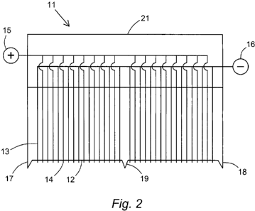
[0019] FIG. 2 is a side view of the electrode support of FIG. 1.
[0020] FIG. 3 is a top view of a surface for adherent cells and a mobile
electrode support in
accordance with certain embodiments of the invention.
[0021] FIG. 4 is a top view of an alternative surface for adherent cells and a
mobile
electrode support in accordance with certain other embodiments of the
invention.
[0022] FIG. 5 is a perspective view of an electroporation apparatus that
utilizes a two-
dimensional array of parallel-line electrodes in accordance with certain
embodiments of the
invention.
[0023] FIG. 6 is a second perspective view of the apparatus of FIG. 5 with
parts separated
to show their internal surfaces.
4
CA 02723595 2010-11-04
WO 2009/140161 PCT/US2009/043285
[0024] FIG. 7 is a view of the underside of the electrode support serving as
one of the parts
of the apparatus of FIGS. 5 and 6.
[0025] FIG. 8 is a cross section of a vessel containing the electroporation
apparatus of
FIGS. 5, 6, and 7.
DETAILED DESCRIPTION OF THE INVENTION
AND PREFERRED EMBODIMENTS
[0026] The most typical adherent cells are biological cells that are adherent
to the surface
on which they are grown. The concerns that apply to such cells can also arise
in the
electroporation of cells that are immobilized for other purposes or that have
become
immobilized by other means. The present invention is thus directed to the
electroporation of
adherent membranous structures in general, including such structures as
vesicles and
liposomes in addition to cells. The terms "cell" and "biological cell" will be
used herein for
convenience to collectively denote all such membranous structures. Examples of
the species,
referred to herein as "exogenous species" or "transfecting species," that will
pass through the
membranes of these cells during the electroporation, are nucleic acids
including DNA, RNA,
plasmids, and genes and gene fragments, and proteins, pharmaceuticals, and
enzyme
cofactors. Further examples of exogenous species will be apparent to those
skilled in the art.
[0027] The solid surface to which the cells adhere can be the surface of any
material that is
capable of serving as an immobilizing support for the cells. Such surfaces can
be the surfaces
of glass, polycarbonate, polystyrene, polyvinyl, polyethylene, polypropylene,
or a variety of
other materials known to cell biologists. Microporous membranes used in
membrane-based
cell culture can also be used. Examples are membranes of hydrophilic
poly(tetrafluoroethylene), cellulose esters, polycarbonate, and polyethylene
terephthalate. A
membrane that is otherwise flexible can made flat and rigid by placing the
membrane over a
support such as a flat screen or a block of solid glass or polymeric material.
Adherence of the
cells to the surface can be achieved by conventional means, including the
inherent adherence
when the cells are grown on the surface, as well as adherence through
immunological or
affinity-type binding, electrostatic attraction, and covalent coupling.
[0028] For embodiments in which the electrodes are dots, referred to herein as
"dot-form
electrodes," each electrode can be formed in a variety of ways. One example is
by passing an
electric wire through the bore of a glass pipet or microcapillary such that
the wire is either
5
CA 02723595 2010-11-04
WO 2009/140161 PCT/US2009/043285
exposed at the open end of the pipet or microcapillary or protrudes a short
distance from the
open end. Another example is by plating an electrical trace on an electrically
insulating block
or chip, preferably a block or chip with a sharply angled edge over which the
trace passes,
using conventional methods such as those employed in semiconductor
manufacture. Once
formed, the trace can be insulated by conventional masking material at all
points except at the
edge where the exposed trace serves as the dot-form electrode. Parallel line
electrodes are
also readily formed by conventional semiconductor manufacturing methods.
[0029] When a series of dot-form electrodes is used, the electrodes are
preferably arranged
in a straight line or in two or more parallel straight lines. Since the
adherent cells are
typically grown on a flat surface, and preferably a surface that is optically
flat, the straight
line of electrodes allows the electrodes to be positioned at a uniform height
above the surface.
The flat surface affords the cells their best opportunity for growth, for
interaction among
neighboring cells, and for uniform exposure to the electric fields. The
straight line of dot-
form electrodes is convenient for sweeping a two-dimensional area of cells
with the
electrodes.
[0030] The ability of the electrodes to form consistent electric fields for
substantially all
cells within the influence of the electrodes regardless of cell size is
achieved by the
narrowness or small diameter of the exposed surface of an individual
electrode, the spacing
between the adjacent electrodes, and the height of the electrodes above the
surface on which
the cells reside. These and other dimensions can vary with the nature of the
cells, i.e.,
whether they be biological cells of various sources and cell lines, or
liposomes, vesicles, or
other membranous structures. Nevertheless, and particularly in the case of
biological cells
whose diameters are within the range of about 10 microns to about 20 microns,
best results
will be achieved in most cases with electrodes whose exposed surfaces are
about 3 microns to
about 20 microns in width or diameter, preferably about 5 microns to about 10
microns, and
most preferably about 8 microns. For best results as well, the spacing between
adjacent
electrodes is about 20 microns to about 75 microns, preferably about 30
microns to about 50
microns, and most preferably about 40 microns. Best results are also achieved
in most cases
when the height of the electrodes above the surface on which the cells reside
is from about 25
microns to about 100 microns, preferably from about 25 microns to about 50
microns, and
most preferably about 40 microns. This height can be set by incorporating
spacing legs,
ridges, piers, or the like into the structure of the support on which the
electrodes are mounted.
6
CA 02723595 2010-11-04
WO 2009/140161 PCT/US2009/043285
[0031] An example of a linear array of dot-form electrodes is shown in FIGS.
1, 2, 3, and 4.
The electrodes in this example are positioned along the sharp edge of a wedge-
shaped block.
FIG. 1 is an end view of the block 11, with the sharp edge 12 shown at the
bottom. FIG. 2 is
a side view of the block, again with the sharp edge 12 of the wedge at the
bottom. The
electrodes are formed by electrical traces 13 that are plated on the block
surface in parallel
lines extending down one face of the wedge, across the sharp edge 12, and up
the other face.
The traces are electrically insulated with masking material at all points
along their lengths
except at the edge 12 where the gaps in the masking form a line of dots 14.
The traces are
electrically connected in alternating fashion, so that odd-numbered traces can
be connected to
one pole 15 of a power source and even-numbered traces to the other pole 16.
At the ends of
the sharp edge 12 of the block are legs 17, 18, with an additional leg 19 at
the center of the
edge. These legs, which are of equal length and are extensions of the block 11
or of the mask
layer, contact the surface on which the cells reside to fix the height of the
dot electrodes 12
above the surface with the cells in between.
[0032] The scanning of a two-dimensional surface (on which the cells reside)
with the
block-supported electrodes is demonstrated in FIGS. 3 and 4 which offer top
views of two
surfaces, respectively. FIGS. 3 and 4 also show the top edge 21 of the block
and the
movement of the block. The surface 31 in FIG. 3 is a circular surface, and the
block 11 scans
the surface by rotating about its center 32, as indicated by the arrows 33.
The surface 41 in
FIG. 4 is a square or rectangular surface which the block 11 scans by moving
laterally, in the
direction of the arrow 42. Movement of the block in both cases is achieved by
conventional
means, such as a stepper motor or a dc motor.
[0033] An example of a two-dimensional array of parallel-line electrodes is
shown in FIGS.
5, 6, 7, and 8. FIG. 5 shows the combination of the electrode block 51 and a
cell plate 52 in
an assembled structure. Both the cells and the electrodes are internal to the
assembled
structure and therefore not visible in this view. FIG. 6 shows the block 51
and cell plate 52
separated (with exaggerated dimensions to more clearly illustrate the
component parts), so
that both the cells 53 and the electrodes 54 are visible. The cells 53 are
grown on the
optically flat surface 55 of the cell plate, and the electrodes 54 are plated
onto the flat
undersurface 56 of the electrode block. Optical flatness is not a requirement
for the
undersurface 56, despite the optical flatness of the cell plate surface 55,
but the closer the
surface is to optical flatness the more effectively the system will function
in achieving
uniform electroporation of the cells. Surrounding the flat undersurface 56 on
which the line
7
CA 02723595 2010-11-04
WO 2009/140161 PCT/US2009/043285
electrodes are plated is a ridge or raised edge 57 which, when the block 51 is
pressed against
the cell plate 52, will contact the cell plate surface 55 outside of the area
occupied by the cells
53 and set the electrodes 54 at the desired distance above the plate surface
55 and hence the
cells 53. To allow the space between the electrodes 54 and the cells 53 to be
filled with the
solution of the transfecting species that will enter the cells and to allow
free movement of the
solution through the space, the electrode block 51 contains a series of holes
58 (also visible in
FIG. 5). The line electrodes 54 are most clearly seen in FIG. 7, which is a
plan view of the
undersurface 56 of the electrode block 51. The line electrodes are connected
to a positive
pole 71 and a negative pole 72 of a power source in alternating manner. Line
electrodes of
positive polarity will thus alternate with line electrodes of negative
polarity. Finally, FIG. 8
shows an electroporation cell 81 consisting of a reservoir 82 in which the
assembled structure
of the electrode block 51 and a cell plate 52 as depicted in FIG. 5 are
placed, together with
the solution 83 of the transfecting species, the block and cell plate assembly
fully immersed
in the solution.
[0034] In each of the configurations shown in the drawings, whether of dot-
form electrodes
or line electrodes, all of the electrodes can be energized or pulsed
simultaneously, or adjacent
pairs can be energized in succession along the length of the array. When
adjacent pairs are
individually energized, each pair will produce an electric field that will
encompass cells on
the portion of the cell plate surface that is between the electrodes in the
pair, and the
electrodes are spaced such that no more than a single cell will reside within
the field of a
single pair of electrodes.
[0035] Power sources and energization protocols that are known in the
electroporation art
can be used. The use of a pulsed electric field is preferred, and the pulse
duration will
typically be within the range of about 1 microsecond to about 1 second,
preferably from
about 50 microseconds to about 10 milliseconds.
[0036] In the claim or claims appended hereto, the term "a" or "an" is
intended to mean
"one or more." The term "comprise" and variations thereof such as "comprises"
and
"comprising," when preceding the recitation of a step or an element, are
intended to mean
that the addition of further steps or elements is optional and not excluded.
All patents, patent
applications, and other published reference materials cited in this
specification are hereby
incorporated herein by reference in their entirety. Any discrepancy between
any reference
material cited herein and an explicit teaching of this specification is
intended to be resolved
8
CA 02723595 2010-11-04
WO 2009/140161 PCT/US2009/043285
in favor of the teaching in this specification. This includes any discrepancy
between an art-
understood definition of a word or phrase and a definition explicitly provided
in this
specification of the same word or phrase.
9