Note: Descriptions are shown in the official language in which they were submitted.
CA 02724604 2015-10-26
1
MINIMALLY INVASIVE LEVATOR AVULSION REPAIR
Priority Claim
This application claims the benefit of U.S. Provisional Application Serial No.
61/057,027, filed
May 29, 2008.
FIELD OF THE INVENTION
The invention relates to apparatus and methods for treating pelvic conditions
by use of a
pelvic implant to support pelvic tissue. The pelvic conditions include
conditions of the female or
male anatomy, and specifically include treatments that involve supporting
pelvic muscles and
organs, such as to levator defects, female or male urinary and fecal
incontinence, among other
conditions.
BACKGROUND OF THE INVENTION
Pelvic health for men and women is a medical area of increasing importance, at
least in
part due to an aging population. Examples of common pelvic ailments include
incontinence
(fecal and urinary) and pelvic tissue prolapse (e.g., female levator bulging
and vaginal
prolapses). Urinary incontinence can further be classified as including
different types, such as
stress urinary incontinence (SUI), urge urinary incontinence, mixed urinary
incontinence, among
others. Other pelvic floor disorders include cystocele, rectocele, enterocele,
and prolapse such as
anal, uterine and vaginal vault prolapse. A cystocele is a hernia of the
bladder, usually into the
vagina and introitus. Pelvic disorders such as these can result from weakness
or damage to
normal pelvic support systems, including the levator muscles.
Pelvic implants, sometimes referred to as slings, hammocks, have been
introduced for
implantation in the body to treat pelvic conditions such as prolapse and
incontinence conditions.
See, for example, commonly assigned U.S. Patent Nos. 6,382,214, 6,641,524,
6,652,450, and
6,911,003, and publications and patents cited therein. The implantation of
these implants
involves the use of implantation tools that create
CA 02724604 2010-11-16
WO 2009/145911 PCT/US2009/003300
- 2 -
transvaginal, transobturator, supra-pubic, or retro-pubic exposures or
pathways. A
delivery system for coupling the sling ends to ends of elongate insertion
tools, to
draw sling extension portions through tissue pathways, is also included.
One specific area of pelvic health is trauma of the pelvic floor, e.g., of the
levator ("levator ani") or coccygeus muscle (collectively the pelvic floor).
The
pelvic floor is made up of the levator and coccygeus muscles, and the levator
is
made up of components that include the puborectalis muscle, the pubococcygeus
muscle, and the iliococcygeous muscle. For various reasons, the levator may
suffer
weakness or injury that can result in various symptoms such as prolapse,
incontinence, and other conditions of the pelvis.
SUMMARY OF THE INVENTION
The invention relates to methods of treating pelvic conditions, especially by
supporting levator tissue. Levator defects (weakness or injury) can affect any
portion of the levator, and can be especially common in the pubic portion of
the
levator ani, including the pubococcygeus and puborectalis muscles. Such
defects are
relatively common, for instance, in women with vaginal prolapse. Defects can
also
be present at the iliococcygeus muscle. Still other defects are in the form of
a
paravaginal defect, such as avulsion of the inferiomedial aspects of the
levator ani
from the pelvic sidewall; avulsion can refer to tissue being detached from the
pubic
bone, and may precede prolapse conditions. Another levator defect is levator
ballooning, which refers to distension of levator muscles.
A different levator defect is a defect of the levator hiatus, which can reduce
the stability of the pelvic floor and may result in sexual dysfunction,
defecatory
dysfunction, rectal prolapse, and fecal incontinence and possibly urinary
incontinence. Levator hiatus is also believed to play a significant role in
the
progression of prolapse. Embodiments of methods of the invention can address
any
of the conditions, as well as related conditions and symptoms.
The present patent application describes pelvic implants and methods for
treating pelvic conditions by treating defects of the pelvic floor (coccygeus
or
levator), such as weakness or injury, or by otherwise supporting levator
muscle.
Useful methods can involve methods and implants that can restore natural
pelvic
CA 02724604 2010-11-16
WO 2009/145911 PCT/US2009/003300
- 3 -
floor anatomy using an implant (e.g., graft) in the form of a hammock, sling,
and the
like, to augment injured, weakened, or attenuated levator musculature. The
levator
musculature or "levator ani" can include the puborectalis, pubococcygeus,
iliococcygeus, among others.
Embodiments of implants useful according to the invention can be of a size
and shape to address a desired pelvic floor condition, generally of a size and
shape
to conform to levator tissue, optionally to additionally contact or support
other tissue
of the pelvic region such as the anal sphincter, rectum, perineal body, etc.
The
implant can be of a single or multiple pieces that is or are shaped overall to
match a
portion of the levator, e.g., that is circular, oblong trapezoidal,
rectangular, that
contains a combination of straight, angled, and arcuate edges, etc. The
implant may
include attached or separate segments that fit together to extend beside or
around
pelvic features such as the rectum, anus, vagina, and the like, optionally to
attach to
the feature.
In one embodiment, the implant can include a tissue support portion, which
may contact levator tissue upon implantation. The implant can additionally
include
one or more guide extension portions that extends beyond the tissue support
portion
and extend out of an incision to guide a support backer member that may be
used to
tension the tissue support portion of the implant.
Optionally, the tissue support portion can include one or more tissue
fasteners (e.g., self-fixating tip, soft tissue anchor, etc.), connected to a
portion of the
tissue support portion in order to fix the tissue support portion to tissue. A
sheath
may also be provided that extends over at least a portion of the tissue
support portion
to aid in easing implantation.
In an example embodiment, the tissue support portion and optionally the
guide extension portion, tissue fastener, etc., may optionally be coated with
antimicrobial coatings to prevent infection or coatings to encourage ingrowth
or
inhibit rejection. For tissue support portions and extension portions,
biocompatible
materials are contemplated such as porcine dermis or meshes with growth
factors.
A method as described herein may improve or treat a condition of the pelvic
region, such as any of the pelvic conditions described. The method may support
levator tissue, for treatment of prolapse; fecal incontinence; a torn,
weakened, or
CA 02724604 2010-11-16
WO 2009/145911 PCT/US2009/003300
- 4 -
damaged levator muscle (meaning any portion of the levator muscle); levator
avulsion, levator ballooning, treatment to support a perineal body; a method
of
perineal body repair; a method of treating the levator hiatus by tightening or
reducing the size of the levator hiatus; and combinations of one or more of
these.
The method may also be more general, as a treatment of more general conditions
such as urinary continence that is believed to be caused by or contributed to
by a
weakened levator.
The method may be prophylactic or medically necessary. A prophylactic
treatment may be a preventative treatment for potential disease or condition
that
does not yet exist but that may be likely to exist. For example preventative
treatment may be useful upon a grade one or two prolapse, for reinforcement of
current prolapse repair, or post-partum. A medically necessary procedure may
take
place when a disease is present and in need of immediate treatment, such as in
the
case of perineal descent, fecal incontinence, urinary incontinence,
reinforcement of
current prolapse repair, and rectal prolapse.
An implant can be placed to contact pelvic tissue as desired, to support the
tissue, such as levator tissue, and can optionally be secured to the tissue to
be
supported, e.g., by suturing or anchoring. The implant can additionally be
secured
to tissue of the pelvic region for additional support, such as to tissue such
as:
sacrotuberous ligament; sacrospinous ligament; anococcygeal ligament
("anococcygeal body ligament"); periostium of the pubic bone (e.g., in a
region of
the ischial tuberosity); pubourethral ligament; ischial spine (e.g., at a
region of the
ischial spine); ischial tuberosity; arcus tendineus (used synonymously herein
with
the term "white line"), e.g., through a tissue path between levator ani muscle
and
obturator internus muscle and attached at the arcus tendineus; obturator
internus
muscle. Alternately, an extension guide portion of an implant can be extended
through a tissue path that leads to an external incision such as: by passing
through
tissue of the obturator foramen.
According to exemplary methods, an implant can be introduced through an
incision that allows access to levator tissue, optionally with or without some
amount
of dissection. The incision can be any of a variety of incisions that provide
such
access, such as a small external perirectal incision that can allow a tissue
path to
CA 02724604 2010-11-16
WO 2009/145911 PCT/US2009/003300
- 5 -
extend from the external perirectal incision to levator tissue; an external
suprapubic
incision; an external incision that can be used to pass a portion of an
implant through
an obturator foramen, a Kraske incision under the rectum; an incision at the
perineum; and a vaginal incision. An implant or a portion of the implant can
be
accessed or placed into position using the incision, to support tissue of the
levator.
Preferably the implant can be placed by dissecting a plane or region of
dissection
that includes the ischorectal fossa. Anatomical landmarks included with this
region
of dissection can include the ischial spine, the obturator internus, the arcus
tendineus.
One embodiment of implant can be a synthetic or biologic implant having a
tissue support portion. The tissue support portion can be sized and shaped to
support
levator tissue. The precise form can depend on the type of condition being
treated.
Certain embodiments of a tissue support portion may optionally include a
segment
or support for addressing levator hiatus opening, perineal descent, rectal
prolapse,
fecal incontinence, etc.
The invention contemplates various methods of supporting levator tissue.
Exemplary methods include steps that involve creating a single medial incision
(a
transvaginal incision or a perineal incision) or an incision near the rectum,
anus, or
perineum; and dissecting within a plane or region of dissection including the
ischorectal fossa. An implant can be inserted to contact tissue of the
levator, over a
desired area. Optionally, the implant can be a single piece or multiple pieces
or
portions, and may include one or more tissue fasteners that can be secured to
tissue
in the pelvic region. An implant may include materials or components such as
those
used in the Elevate, MiniArc, SPARC and Monarc systems (from American Medical
Systems), include connectors for engagement between a needle of an insertion
tool
and an distal end of an extension portion, as well as helical, straight, and
curved
needles.
In one aspect, the invention relates to pelvic implant for supporting tissue
of
the pelvic floor (e.g., levator tissue, coccygeus tissue, or combinations of
these), and
related surgical systems and kits. In another aspect, the invention provides a
system
and method of repairing levator avulsion. Namely, a small incision can be made
to
deploy a relatively small implant, with anchoring device, unilaterally or
bilaterally.
CA 02724604 2010-11-16
WO 2009/145911 PCT/US2009/003300
- 6 -
The implant is delivered and deployed via a needle or introducer device that
can be
anatomically (curved, helical, etc.) shaped and configured for traversal along
the
vaginal sidewall to advance through the obturator membrane such that a first
anchor
of the implant is secureable to or proximate the tissue defect or avulsion.
The needle
and implant are then retrieved or withdrawn, with the anchored tissue being
pulled
back to or proximate the tissue tear site or damaged area. At this point,
adjustment
can be made to the anchor devices, implant length, or by employing other known
adjustment systems or methods to remove slack in the implant or facilitate
tautness
in the repaired tissue.
Various implant and anchoring devices can be implemented for the present
invention. For instance, various mesh supports and fixating anchoring end
portions
can be employed.
BRIEF DESCRIPTION OF DRAWINGS
Other features and advantages of the present invention will be seen as the
following description of particular embodiments progresses in conjunction with
the
drawings. Drawings are schematic and not to scale.
Figure 1A illustrates example of health anatomy of a pelvic region.
Figure 1B illustrates example of a pelvic region having a defect.
Figure 2 illustrates exemplary features or components of the invention for
repairing defects in pelvic anatomy.
Figure 3A illustrates an exemplary implant of the invention.
Figure 3B illustrates an exemplary implant of the invention.
Figure 3C illustrates a carrier for carrying support backer members of the
invention.
Figure 3D illustrates a carrier for carrying support backer members of the
invention coupled to a portion of the implant.
Figure 4A illustrates an implant disposed upon a delivery tool.
Figures 4B-4G illustrates example embodiments of delivery tools.
Figure 5A illustrates an example of process for repairing a pelvic defect.
Figure 5B illustrates another step in a process for repairing a pelvic defect.
Figure 5C illustrates another step in a process for repairing a pelvic defect.
CA 02724604 2015-10-26
7
Figure 5D illustrates another step in a process for repairing a pelvic defect.
Figure 6 illustrates an example of a support backer member.
Figure 7 illustrates a cross section of Figure 6 above.
Figures 8A-9B illustrates example embodiments of delivery tools used to
deliver the
support backer member.
Figure 10A illustrates an example of delivering the support backer member.
Figure 11A illustrates an example of the implant prior to adjusting to repair
a defect.
Figure 11B illustrates an example of the implant after adjustment with a
defect repaired.
The preceding description of the drawings is provided for example purposes
only and
should not be considered limiting. The following detailed description is
provided for more
detailed examples of the invention. Other embodiments not disclosed or
directly discussed are
also considered to be within the scope of the invention. It is not the
intention of the inventor to
limit the scope of the invention by describing one or more example
embodiments.
DETAILED DESCRIPTION
The following description is meant to be illustrative only and not limiting.
Other
embodiments of this invention will be apparent to those of ordinary skill in
the art in view of this
description.
The invention relates to surgical implants, insertion tools, kits, and
assemblies, and
related methods for treating pelvic defect and disorders related to levator
avulsion, ballooning,
etc. As a secondary effect the methods of treating levator avulsion or
ballooning may treat,
improve, or prevent a condition such as prolapse, incontinence (urinary or
fecal incontinence),
conditions of the perineal body, conditions of the levator hiatus, levator
ballooning, and
combinations of two or more of these.
According to various embodiments, a surgical implant can be used to support
levator
tissue in a region of a levator avulsion. The implant may be placed to contact
or support levator
tissue at a location in a region of a levator avulsion. A "region of a levator
avulsion" refers to a
location of levator tissue that can be contacted by a
CA 02724604 2010-11-16
WO 2009/145911 PCT/US2009/003300
- 8 -
support portion of an implant to allow the implant to be manipulated or
tensioned in
a manner that will cause approximation of tissue of the levator muscle, to at
least in
part remedy the avulsion; the region may be at a surface of the levator tissue
or at a
location at an interior of the tissue, and may be at a location that is
considered to be
either "inferior" to ("caudal" to) the avulsed tissue or "superior" to the
avulsed
tissue; optionally the region can be proximal to the avulsion, such as within
3
centimeters, within 2 centimeters, or within 1 centimeter from tissue of an
avulsion.
As an example, a portion of implant (e.g., a portion of support portion, such
as a short length of the support portion) can be placed within the tissue of
the levator
muscle ("embedded" in the muscle or "tunneled" through a length of the
muscle).
According to such an example, a portion of implant may enter (or exit) levator
tissue
inferior to the avulsed tissue, be embedded in the belly of the levator muscle
and
extend through an obturator foramen. Alternately, the implant may extend
through
the levator muscle tissue inferior to the avulsed tissue, traverse the
avulsion, and
continue on a superior side of the avulsion, then exit at a location superior
to the
avulsed tissue, such as at a location near the obturator internus muscle.
Generally,
the implant can exit the levator muscle tissue on the obturator internus side
of the
levator muscle, extend through tissue of the obturator internus muscle,
through the
obturator foramen, toward the anterior external incision, and exit the patient
at the
anterior incision.
The implant can be used to support the levator tissue to remedy a levator
avulsion such as by approximating levator tissue, supporting levator tissue,
or both,
to cause levator tissue to move to close the avulsion. In the event that an
avulsion
involves a detachment of levator tissue from tissue or bone at a superior
region of
levator tissue ¨ e.g., a detachment at or near tissue of an arcus tendineus,
tissue of
an obturator internus muscle, a pubic bone, an ischial spine, or any
combination of
these ¨ the levator tissue can be approximated in a direction to allow the
detached
tissue to be moved closer to the location of the detachment. After this tissue
approximation, the implant can be maintained in the implanted position to
continue
to support the levator tissue to prevent subsequent return of the levator
muscle to the
avulsed condition, and to potentially prevent future conditions of avulsion or
related
prolapse, incontinence, levator ballooning, or other pelvic condition.
CA 02724604 2010-11-16
WO 2009/145911 PCT/US2009/003300
- 9 -
Turning now to the figures, figure 1 shows anatomy relevant to methods and
devices of embodiments of the invention. In particular, figure 1, illustrates
a superior
view of tissue at different levels of the pelvic region, including
ischiococcygeous
muscle 20, iliococcygeus muscle 22, puborectalis and pubococcygeous muscles
24,
ischial spine 26, pubic symphasis 228, coccyx 30, arcus tendineus 32, and
obturator
foramen 34. Vagina 36, urethra 37, rectum 38, and pelvic bone 39 are also
shown.
Continuing with figure 1, a levator avulsion (not illustrated) may typically
be located
along the superior portion of the levator muscle extending between the ischial
spine,
along the arcus tendineus, and to the obturator intemus muscle, for example at
a
superior portion of any one or more of the puborectalis muscle, pubococcygeus
muscle, or iliococcygeous muscle. According to embodiments of the invention,
an
implant can be placed to approximate, support, or approximate and support, one
or
more of these muscles to treat the avulsion.
Figure 1B illustrates relevant anatomy (on a patient's right side), as
described, and includes an exemplary depiction of avulsion 40. Although the
avulsion is demonstrated as being located in the iliococcygeus muscle it
should be
understood that the avulsion could be located in any of the pelvic muscles and
the
implant of the present invention may be used to repair such defect.
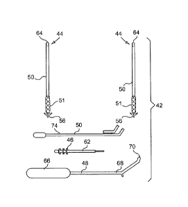
Generally, figure 2 illustrates various parts or components of the pelvic
defect repair system 42. Figure 2 shows an implant 44 with one-way frictional
adjusting engagement members or support backer members 46. The system 42 may
also include an implant delivery device 48 to deliver the implant 44 to a
tissue
approximate a defect and a backer delivery device 50 to deliver one or more
support
backer members 46 to a tissue used to support the tissue via the implant 44.
As particularly illustrated in figures 3A and 3B, the implant 44 may include a
tissue support portion 51 made from any material capable of permitting tissue
ingrowth such as synthetic or biological mesh materials. The tissue support
portion
51 may have first 52 and second 54 opposed ends. Attached or connected to the
first
end 52 of the tissue support portion 51 may be a tissue anchor or self
fixating tip 56
that is designed to engage tissue proximate a defect such that the tissue
support
portion 51 may support and/or contact the tissue proximate the defect. By
contacting or connecting with the tissue proximate the defect the tissue
support
CA 02724604 2010-11-16
WO 2009/145911 PCT/US2009/003300
- 10 -
portion 51 may permit tissue ingrowth, thereby providing additional support to
the
musculature.
As particularly illustrated in figure 3B, the implant 44 may also have an
extension guide portion 58 disposed on or connected to the second end 54 of
the
tissue support portion 51 to guide the support backer member(s) 46 toward a
portion
of the tissue support portion 51. In one embodiment, the extension guide 58
may
comprise a generally rigid, generally flexible or bendable rod or shaft. In
other
embodiments, as illustrated in figure 3A the extension guide 58 may comprise
one or
more sutures 60 that are connected or sewn to a portion of the tissue support
portion
51. In yet other embodiments, the extension guide 58 may comprise mesh, wire
or
any other material or structure capable of guiding the support backer
member(s) 46
toward the tissue support portion 51.
As illustrated in figures 3C and 3D, the system 42 may also include a carrier
62 to carry and transfer one or more of the support backer members 46 to the
extension guide portion 58. In one embodiment, the carrier 62 may comprise a
rod
or shaft having a mating feature such as a shaft having a reduced outer
diameter or
axially extending bore that can fit into or onto a free end 64 of the
extension guide
portion 58. The carrier 62 ensures that the support backer members 46 are
properly
transferred to the extension guide portion 58 of the implant 44.
Turning to figure 4A, the implant delivery device 48 of the system 42 may
include a handle 66 having a needle 68 extending away from a portion of the
handle
66. As particularly illustrated in figure 4A, the self fixating tip 56 is
designed to
engaged, connect or couple to a tip or free end 70 of the needle 68. In one
embodiment, the self fixating tip 56 may have a channel or bore that may be
keyed
to ensure proper connection between it and the free end 70 of the needle 68.
The
self fixating tip 56 may also be connected to the free end 70 of the needle 60
by a
mechanism that permits controlled release into a tissue mass such as a levator
muscle.
Referring to figures 4B-4G, the needle 68 may have any of a variety of
shapes to facilitate placement of the implant 44 in a therapeutic location
designed to
repair pelvic defects. As particularly illustrated in figures 4B -4E, the
needle 68 may
have a portion between the handle 66 and its free end 70 that is generally
helical.
CA 02724604 2010-11-16
WO 2009/145911 PCT/US2009/003300
- 11 -
The system 42 may include multiple delivery tools 48 designed for insertion of
the
implant 44 in different anatomical locations such as the left and right
obturator
foramen. As particularly illustrated in figures 4D and 4E, the needle 68 may
be bent
or have a slight bend located between the handle 66 and the free end 70 of the
needle
68. The delivery tools 48 may be sterilized or disposable. The needle 68 may
also
have a number of notches or measurements 72 disposed along its length to
permit a
physician to determine a length of the needle 68 disposed in a patient.
As illustrated in figure 5A, an incision 72 can be made in a patient's dermis
proximate their obturator foramen. A physician can attach the self fixating
tip 56 to
the free end 70 of the needle 68. The free end 70 of the needle 68 and the
self
fixating tip 56 can then be inserted into the incision 72. As illustrated in
figure 5B,
the needle 68 and self fixating tip 56 may be passed through the obturator
foramen
34 (not shown) and into the levator tissue as shown in figure 10A. As
particularly
illustrated in figure 11A, the self fixating tip 56 may be inserted into
levator muscle
inferior the defect or any other tissue such as tissue surrounding the urethra
or
rectum.
As illustrated in figure 5B, the extension guide portion 58 of the implant 44
may extend out of the incision 72. At this particular point a physician may
remove
the delivery tool 48 or maintain it in contact with the implant 44. The
carrier 62 can
be coupled to the extension guide 58 and one or more support backer members 46
moved down onto the extension guide portion 58. The backer delivery device 50,
which may comprise a shaft 74 having an engagement end 76 and an engagement
configuration attached to or formed on the shaft 74 for engaging a support
backer
member 46, may be moved along the extension guide 58 to engage the support
backer member 46 and move it through the incision 72.
As the support backer member 46 is moved toward the obturator foramen by
the backer delivery device 50 it engages the tissue support portion 51 of the
implant
44. The support backer member 46, which may comprise a ring 78 having an
aperture and a plurality of inwardly radiating engagement portions or flanges,
flaps
or teeth 79 (as illustrated in figures 6 and 7) extending into the aperture
engage the
tissue support portion 51 of the implant 44. The support backer member 46 may
be
designed to permit movement along the extension guide portion 58 and onto the
CA 02724604 2015-10-26
12
tissue support portion 51 but resist movement in a reverse direction. As the
support backer
member 46 is disposed proximate the obturator foramen the physician may pull
on the extension
guide portion 58 to place the tissue support portion 51 in tension. In another
embodiment, the
physician may permit a predetermined amount of slack in the tissue support
portion 51 between
the obturator foramen and the self fixating tip 56 in the tissue to create a
backstop for organs
and/or tissue.
In one embodiment, the engagement end 76 of the backer member delivery device
50
may include one or more solid rings and aperture 80 or arms 81 and 82 defining
an opening for
receiving the extension guide portion 58. In one embodiment of the invention,
as illustrated in
figures 9A and 9B, the engagement end 76 may include multiple apertures 80,
80' and/or arms
81, 81' and 82, 82'. In this particular embodiment, a backer member 46 can be
disposed between
the pairs of arms 81, 81' and 82, 82'. The aperture 80 or apertures 80 and 80'
may have a central
axis that is generally parallel to or generally angled to a longitudinal axis
of the shaft 74 of the
backer member delivery tool 50. The shaft 74 of the backer delivery tool 50
may have spaced
apart notches, markings and the like to permit a physician to determine a
length or depth of the
backer member delivery tool 50 disposed in a patient.
The following patent documents permit one skilled in the art to better
understand the
invention and its various embodiments: US Patent Publication No. US
2004/0039453 Al; US
Patent Publication No. US 2005/0250977 Al; US Patent Publication No. US
2005/0245787 Al;
US Patent No. 6,652,450; US Patent No. 6,612,977; US Patent No. 6,802,807; US
Patent No.
7,048,682; US Patent No. 6,641,525; US Patent No. 6,911,003; US Patent No.
7,070,556; US
Patent No. 6,354,991; US Patent No. 6,896,651; US Patent No. 6,652,449; US
Patent No.
6,862,480; US Patent No. 6,712,772; and PCT Application Serial No. Unknown,
filed June 15,
2007, titled "Surgical Implants, Tools and Methods for Treating Pelvic
Conditions" (Attorney
Docket No. AMS- 3419-PCT). (See International Patent Application No.
PCT/US2007/014120,
entitled "Surgical Implants, Tools, and Methods for Treating Pelvic
Conditions, filed June 15,
2007) PCTUS2007/004015, filed February 16, 2007, titled Surgical Articles and
Methods
CA 02724604 2015-10-26
13
for Treating Pelvic Conditions. WO 2007/016083, published February 8, 2007,
and entitled
"Methods and Symptoms for Treatment of Prolapse,"); including tissue at or
near an ischial
spine, e.g., at a region of an ischial spine.
Embodiments of exemplary implants that may be useful as discussed herein can
include a
tissue support portion 51 and no extension portions 58. Other embodiments can
include one, two,
three, or more extension portions 58 attached to a tissue support portion 51.
An exemplary
urethral sling can be an integral mesh strip or hammock with supportive
portions consisting of or
consisting essentially of a tissue support portion 51 and zero, one, or two
extension portions 58.
An implant may include portions or sections that are synthetic or of
biological material
(e.g., porcine, cadaveric, etc.), and that may be resorbable or non-
resorbable. Extension portions
may be, e.g., a synthetic mesh such as a polypropylene mesh. The tissue
support portion may be
synthetic (e.g., a polypropylene mesh) or biologic.
The implant 44, either or both of the tissue support portion 51 or a guide
extension
portion 58, may comprise variable weave meshes with varying elasticities such
as a mesh that is
highly elastic around the anus to allow stool to pass.
Some example of commercially available materials may include MarleXTM
(polypropylene) available from Bard of Covington, RI, ProleneTM
(polypropylene) and
Mersilene (polyethylene terephthalate) Hernia Mesh available from Ethicon, of
New Jersey,
Gore-TeXTm (expanded polytetrafluoroethylene) available from W. L. Gore and
associates,
Phoenix, Arizona, and the polypropylene sling material available in the
SPARCTM sling system,
available from American Medical Systems, Inc. of Minnetonka, Minnesota.
Commercial
examples of absorbable materials include DexonTM (polyglycolic acid) available
from Davis and
Geck of Danbury, Connecticut, and VicrylTM available from Ethicon.
Dimensions of an implant can be as desired and useful for any particular
installation
procedure, treatment, patient anatomy, to support a specific tissue or type of
tissue, and to extend
to a desired location of internal supportive tissue or an
CA 02724604 2010-11-16
WO 2009/145911 PCT/US2009/003300
- 14 -
external incision. Exemplary dimensions can be sufficient to allow the tissue
support portion to contact tissue of the levator, coccygeus, rectum, external
anal
sphincter, etc., or any desired portion of one or more of these. Optionally,
one or
more guide extension portions 58 can extend from the tissue support portion 51
to a
desired internal or external anatomical location to allow the guide extension
portion
58 to be secured to anatomy of the pelvic region, to support the tissue
support
portion 51.
Dimensions of guide extension portions 58 according to the invention can
allow the guide extension portion 58 to reach between a tissue support portion
51
placed to support tissue of the pelvic floor (at an end of the extension
portion
connected to the tissue support portion) and a location at which the distal
end of the
guide extension portion 58 pass through an external incision.
An implant 44 can be of a single or multiple pieces that is or are shaped
overall to match a portion of the levator, e.g., that is completely or
partially circular,
trapezoidal (non-symmetric or symmetric), rectangular, rhomboidal, etc. The
implant may be multiple pieces to fit beside or around pelvic features such as
the
rectum or anus. Alternately, the implant 44 may be irregular (while optionally
symmetrical) to reach different areas of the levator.
To contact tissue of the pelvic floor, the implant 44 or any portion thereof
can be a continuous or a non-continuous sling, and of one or multiple pieces
or
segments. A continuous implant may be substantially continuous between edges,
to
be placed over a level surface area of levator tissue. A non-continuous
implant may
include breaks or cuts that allow much of the implant to be placed on a level
surface
of levator tissue, with portions being formed to extend around tissue
structure
extending from or to the levator tissues, such as the anus, rectum, etc.
An embodiment of a non-continuous sling may be designed to cover or
contact area of the levator, coccygeus, or both, and also reach around to
contact a
posterior side of the rectum or external anal sphincter. For example, a
portion of an
implant could attach to the lateral sides of the external anal sphincter and
extend
toward or in the direction of the obturator foramen, or any other suspensory
structure
(e.g., supportive tissue), but need not engage tissue of the obturator foramen
directly.
In this embodiment, the tissue support portion of the implant need not
necessarily be
CA 02724604 2015-10-26
directly under the anus to provide the corrective action for fecal
incontinence. An advantage to of
this approach is to allow the anus to expand unrestricted to facilitate normal
rectal function and
may give the levator plate (or plates) the support necessary to be leveraged.
Embodiments of implants can include a segment that is located anterior to the
anus, such
5
as in contact with levator tissue or tissue of the perineal body, anterior to
the anus. Alternate
implants may be designed to replace the perineal muscle or attach to the
superior portion of the
external sphincter. The various embodiments disclosed herein are also
applicable to men and can
be implanted via an incision in the perineal floor (see attached figures).
An implant, e.g., at a tissue support portion 51 can optionally include a
tissue fastener
10
such as a soft tissue anchor, a self-fixating tip, a biologic adhesive, a
tissue clamp, opposing male
and female connector elements that securely engage when pushed together, or
any other device
to secure a distal end of an extension portion to tissue of the pelvic region.
Exemplary tissue
fasteners are discussed, e.g., in PCT/SU2007/014120 "Surgical Implants, Tools,
and Methods for
Treating Pelvic Conditions, filed June 15, 2007. The implant may also have
extension portions
15
that do not include a tissue fastener at a distal end thereof, for example if
the distal end is
designed to be secured to tissue by other methods (e.g., suturing), or is
intended to pass through a
tissue path ending in an external incision. Exemplary self-fixating tips are
described, for
example, in PCT/US2007/004015 "Surgical Articles and Methods for Treating
Pelvic
Conditions," filed February 16, 2007.
A self-fixating tip 56 can be made out of any useful material, generally
including
materials that can be molded or formed to a desired structure and connected to
or attached to an
end of an extension portion of an implant. Useful materials can include
plastics such as
polyethylene, polypropylene, and other thermoplastic or thermoformable
materials, as well as
metals, ceramics, and other types of biocompatible and optionally
bioabsorbable or bioresorbable
materials. Exemplary bioabsorbable materials include, e.g., polyglycolic acid
(PGA), polylactide
(PLA), copolymers of PGA and PLA.
CA 02724604 2015-10-26
16
Alternate embodiments of self-fixating tips 56 do not require and can exclude
an internal
channel for engaging a delivery tool 48. These alternate embodiments may be
solid, with no
internal channel, and may engage a delivery tool 48, if desired, by any
alternate form of
engagement, such as, for example, by use of a delivery tool 48 that contacts
the self-fixating tip
56 at an external location such as by grasping the base (on a side or at the
face of the proximal
base end) or by contacting a lateral extension.
Examples of commercial implants include those sold by American Medical
Systems, Inc.,
of Minnetonka MN, under the trade names Apogee , Perigee , and ElevateTM for
use in treating
pelvic prolapse (including vaginal vault prolapse, cystocele, enterocele,
etc.), and Sparc ,
BioarcO, MonarcO, and MiniArcTM for treating urinary incontinence. Implants
useful according
to the present description can include one or more features of these
commercial implants.
Generally, transobturator tissue approaches are described at pending
application
11/347,047 "Transobturator Methods for Installing Sling to Treat Incontinence,
and Related
Devices," filed February 3, 2006, and at US publication 2005/0143618
(11/064,875) filed
February 24, 2005.
Also straight, helical and curved needles, as described in U.S. Publication
no.
2005/0250977; 2005/0245787 and 2004/0039453, can also be used with their
associated
tunneling paths and techniques.
In a related embodiment, a depth limiting feature such as a sheath design or a
mechanical
stop or a bend in the needle to facilitate correct depth placement. Also
inside out as opposed to
the outside in implantation approach is a possible variation to the described
embodiments
(similar to the ISCP methods and techniques).
Examples of various tissue paths, relevant anatomy, implant materials,
features of
implants (e.g., connectors, tensioning devices), insertion tools, are
described, for example, in
U.S. Publication Nos. 2002/0161382 (10/106,086) filed March 25, 2002;
2005/0250977
(10/840,646) filed May 7, 2004; and 2005/0245787 (10/834,943) filed April 30,
2004;
2005/0143618 (11/064,875) filed February 24, 2005; and U.S. Patent Nos.
6,971,986
(10/280,341) filed October 25, 2002;
CA 02724604 2015-10-26
17
6,802,807 (09/917,445) filed July 27, 2001; 6,612,977 (09/917,443) filed July
27, 2001;
6,911,003 (10/377,101) filed March 3, 2003; 7,070,556 (10/306,179) filed
November 27, 2002,
PCT/US2007/004015 "Surgical Articles and Methods for Treating Pelvic
Conditions," filed
February 16, 2007; PCT/US2007/014120 "Surgical Implants, Tools, and Methods
for Treating
Pelvic Conditions, filed June 15, 2007.