Note: Descriptions are shown in the official language in which they were submitted.
CA 02724826 2015-11-30
1
MEDICAL INSTRUMENT INCORPORATING X-RAY MARKERS
AND MR MARKERS
The present invention relates to a medical instrument. In particular, the
present invention
concerns a medical instrument which can be detected by means of magnetic
resonance
tomography.
WO 2007/000148 A2 discloses a rod-type body serving for forming medical
instruments
such as catheters or guiding wires for catheters. This rod-type body consists
of one or
more filaments and a non-ferromagnetic matrix material enclosing the
filaments. A
doping agent made of particles which create MRT artifacts is introduced into
the matrix
material.
A detailed explanation of magnetic resonance tomography (MRT) or magnetic
resonance
imaging can be found in the Internet at http:/en.wikipedia.org/wiki/MRT.
US 2003/0055449 Al shows a balloon catheter in which the balloon is formed
from a
polymeric material comprising a ferromagnetic or paramagnetic material so that
it is
visible during the magnetic resonance examination.
US 5,154,179 discloses a catheter which is formed e.g. from an extruded
plastic hose,
ferromagnetic particles being contained in the plastic material of the plastic
hose. This
catheter is visible in magnetic resonance tomography. Further, it is suggested
to provide
such a catheter with a material which is opaque for X-rays. It is preferred to
use non-
ferrous materials for these X-ray markers.
DE 101 07 750 Al describes a guiding wire which is supposed to be suitable for
magnetic
resonance tomography. This guiding wire comprises a core made of a metallic
front part.
Ropes made of an electrically non-conductive plastic material are arranged
between an
outer jacket and the core. This plastic material is supposed to be reinforced
with glass
fibers or carbon fibers. Carbon fibers are, however, electrical conductors so
that they
cannot be used for magnetic resonance tomography.
Further, medical equipment is known from EP 1 206 945 Al, which is provided
with
paramagnetic metallic compounds and/or a paramagnetic metal so that they are
visible in
a magnetic resonance imaging process.
CA 02724826 2010-11-18
2
WO 87/02893 discloses poly-chelating substances for the imaging enhancement
and
spectral enhancement for magnetic resonance imaging. These substances comprise
different complexes in which metal ions, in particular gadolinium ions are
immobilized.
The relaxivity of gadolinium(III) complexes is explained in chapter 1.6.1 of
the inaugural
dissertation by Daniel Storch, entitled "Neue, radioaktiv markierte Magnet-
Resonanz-
aktive Somatostatinanaloga zur besseren Diagnose und zielgerichteten
Radionuklid-
therapie von neuroendokrinen Tumoren", Basel, 2005. The paramagnetic
relaxation of
the water molecules which are in the vicinity of the gadolinium(III) ion is
the result of the
dipole-dipole-interaction between the nuclear spin and the fluctuating local
magnetic field
of the magnetic resonance imaging apparatus, caused by the unpaired electrons.
The
magnetic field around the paramagnetic center, i.e. the gadolinium(III) ion,
disappears
with increasing distance. This is why it is decisive to bring the protons in
close proximity
to the metal ion. Concerning gadolinium(III) complexes, this means that the
water
molecules are to be transported into the first coordination sphere of the
metal ion. These
"inner-sphere" H20 molecules are exchanged with the surrounding water
molecules and
transmit the paramagnetic effect in this way.
DE 100 40 381 Cl discloses fluoroalkyl-containing complexes with residual
sugars.
These complexes can be provided with paramagnetic metal ions so that they can
serve as
contrast agents in magnetic resonance imaging. These metal ions are in
particular the
bivalent and trivalent ions of the elements of the atomic numbers 21 to 29,
42, 44 and 58
to 70. Suitable ions are, for instance, the chromium(III), iron(II),
cobalt(II), nickel(II),
copper(II), praseodymium(III), neodymium(III), samarium(III) and
ytterbium(III) ions.
Gadolinium(III), erbium(III), dysprosium(III), holmium(III), erbium(III),
iron(III) and
manganese(II) ions are particularly preferred because of their strong magnetic
moment.
EP 1 818 054 Al discloses the use of gadolinium chelates for the purpose of
marking
cells.
US 6,458,088 B1 describes a guiding wire provided for magnetic resonance
imaging, this
guiding wire comprising a glass body. The glass body is provided with a
protective layer
which is made of polymeric material and can be additionally provided with
fibers. The
distal end of the guiding wire can be formed from a metal section such as
nitinol. This
metal section should have a length which is clearly shorter than the
wavelength of the
magnetic resonance field.
CA 02724826 2015-08-10
,
3
WO 2005/120598 Al discloses a catheter guiding wire comprising a PEEK core.
This core
is provided with a coating. The coating is provided with a contrast agent. The
contrast
agent is iron powder having a grain size of less than 10 pm.
WO 97/17622 discloses a medical instrument comprising an electrically non-
conductive
body which is provided with an ultra-thin coating made of an electrically
conductive
material so that the medical instrument is visible in a magnetic resonance
tomography
process without unduly affecting the image.
WO 99/060920 A and WO 2002/022186 A each show a coating for a medical
instrument
comprising a paramagnetic ion which is complexed in the coating. The
paramagnetic ion
is in particular gadolinium. This coating is visible during the MRT
examination.
The invention is based on the object to provide a medical instrument which can
be
inserted in a human or animal body and is very versatile as regards its use in
an MRT
examination.
According to a first aspect of the present invention, a medical instrument is
provided
which can be inserted in a human or animal body, the medical instrument
including an
instrument body. The instrument body comprises at least one rod-type body
having poor
electrical conductivity and being formed from a matrix material and non-
metallic
filaments. This medical instrument is distinguished in that the rod-type body
is doped
with an X-ray marker and the medical instrument comprises an MR marker.
By providing an X-ray marker as well as an MR marker, the medical instrument
can
be seen in both X-ray examinations and MRT. The introduction of the X-ray
marker into
the medical instrument can be easily realized by the use of a rod-type body
having an
appropriate doping. Such rod-type bodies can be produced as a mass product
with
different doping agents at a favorable price and with an exact dosage of the
marker
particles. During the production of a medical instrument, the visualization of
the medical
instrument in X-ray examinations can be ensured by using the respective rod-
type body
with an X-ray marker.
According to a second aspect, the medical instrument according to the
invention is
designed for being inserted into a human or animal body, said instrument
comprising an
instrument body having a surface which may come into contact with the human or
animal body. The surface area of the instrument body is provided with
immobilized active
MR markers.
CA 02724826 2010-11-18
4
Active MR markers are markers which interact with the protons in the water or
fat
molecule and result in a quicker relaxation of the protons adjoining the
marker when
these have undergone an induced orientation due to the applied magnetic field.
The
reduction of the relaxation time caused by the marking process results in
strong MRT
signals, bringing about a correspondingly high contrast in the images created
hereby.
By the use of an immobilized active MR marker on the surface of the instrument
body
in connection with at least one rod-type body doped with a marker, the high
contrast of
an active MR marker in MRT and the versatile field of application of passive
markers is
combined in a simple way. The passive markers may be designed both for X-ray
and MRT
examinations. It is preferred that the medical instrument comprises several
rod-type
bodies which are doped differently.
Medical instruments provided with active MR markers on their surface have a
very
flexible field of application with respect to the sequences used in an MRT
examination
and also are uniformly visible in MRT examinations with different sequences.
The active MR markers comprise an element or a combination of elements or a
compound of an element from the group consisting of gadolinium, cerium, praseo-
dymium, neodymium, promethium, samarium, europium, terbium, dysprosium,
holmium,
erbium, thulium, ytterbium and lutetium. These elements can be bound in a
complex in
the form of ions. They can also be present, however, in the form of salts or
alloys.
It is particularly preferred that gadolinium is used as an active MR marker.
This
element is preferably immobilized by means of a complex, in particular a
chelate
complex.
The complexes can either be covalently bound to the surface of the instrument
body
or embedded in a coating which is capable of swelling and formed on the
surface of the
instrument body.
Spacers can be arranged between the complexes and the surface of the
instrument
body so that the active MR marker is arranged so as to be spaced from the
surface of the
instrument body. This measure makes sure that the body fluid flows over and
around the
markers and the majority of the MR markers is in close proximity to protons of
water
and/or fat molecules.
When a coating is provided which is capable of swelling and contains the MR
markers, body fluid is absorbed by the coating capable of swelling while the
medical
instrument is inserted in the human or animal body so that protons of water
molecules
CA 02724826 2015-08-10
will bind closely to the MR markers, resulting in the interaction which
shortens the
relaxation time.
The invention will now be exemplified in more detail on the basis of the
embodiments
illustrated in the drawings in which:
5 Figure 1 shows a guiding wire according to a first
embodiment of the invention
in cross-section,
Figure 2 shows a guiding wire according to a further embodiment of
the
present invention in cross-section,
Figure 3 shows a test equipment with several rods which are provided
with
different markers,
Figures 4a to 4f show images which have been created by the test equipment by
means of MRT or computer tomography,
Figure 5 shows a guiding wire according to a further embodiment of
the
invention in cross-section,
Figure 6 shows a guiding wire according to a further embodiment of the
invention in a longitudinal section, and
Figures 7a to 7e show images which have been created by further test equipment
by
means of MRT or computer tomography.
The invention will be exemplified in the following on the basis of a guiding
wire 1 for
a catheter. The guiding wire 1 is made from a material which does not create
any MRT
artifacts. A material of this kind is, for example, a ceramic or plastic
material such as
PEEK, PEBAX, PE, PP, PU, silicone, polylactic acid polymers, aromatic
polyamides or
memory plastic materials. The plastic material is in particular reinforced
with fibers. Apart
from the above-mentioned plastic materials, epoxy resin can also be used as a
matrix
material. The fibers are glass fibers or ceramic fibers or Kevlar fibers,
DacronTM, plant-
based fibers (e.g. silk, sisal, hemp etc.). Materials which do not create any
MRT artifacts
must be free from electrically conductive sections. The electrically
conductive sections
should have a length of not more than 15 cm, in particular not more than 10 cm
or 5 cm.
This is why it is possible to use electrically conductive fibers such as coal-
based or carbon
fibers, or electrically conductive wires provided that the sections are
electrically insulated
from one another to a sufficient extent. They must not be formed from a
ferromagnetic,
paramagnetic, ferrimagnetic or anti-ferromagnetic material.
CA 02724826 2010-11-18
6
The guiding wire is an elongated body with a circular cross-section and a
diameter of
usually not more than 2 mm (e.g. 0.7 mm). On its surface 2, active MR markers
3 are
immobilized on the guiding wire.
Active MR markers are markers which interact with a proton-containing medium
such
as water or fat molecules in such a way that they bring about a quicker
relaxation of the
protons adjoining the MR marker after their induced orientation by an applied
magnetic
field. Such MR markers comprise, for instance, an element or a combination of
elements
or a compound of an element from the group consisting of gadolinium, cerium,
praseo-
dymium, neodymium, promethium, samarium, europium, terbium, dysprosium,
holmium,
erbium, thulium, ytterbium, lutetium. These elements are preferably
immobilized by
means of a complex, in particular by means of a chelate complex. They can also
be
present as salts or in alloys.
Typical chelating agents are EDTA (ethylenediaminetetraacetic acid), DTPA
(diethylenetriaminepentaacetic acid) and DOTA (1,4,7,10-tetrazacyclododecane-
N,N1',N",N" tetraacetic acid).
Basically, chemical macromolecules (inter alia polylysines, dendrimers) or
biological
macromolecules (proteins, sugars, inter alia dextran) are suitable as
complexes.
In the present exemplary embodiment, the MR markers are gadolinium(III)
chelate
complexes, the chelate complexes being bound to the surface 2 of the guiding
wire 1 by
means of a covalent bond. It is preferred that spacer molecules are provided
between
the chelate complexes and the surface 2 so that the MR markers are arranged so
as to
be spaced from the surface 2. Polyethylene glycol is suited for being used as
a spacer
molecule, for instance.
The covalent bond between the chelates, spacers and the instrument body formed
from a polymer can be realized through amino, quaternary ammonium, hydroxyl,
carboxyl, sulfhydryl, sulfate, sulfonium, thiol groups, reactive nitrogen
groups, etc. (in
each case for chelating agents and polymers).
The guiding wire 1 is used for inserting catheters into blood vessels. During
inserting
the guiding wire 1 in the blood vessel, the surface 2 of the guiding wire 1
comes into
contact with blood. Blood flows over and around the MR markers 3 which are
arranged
so as to be spaced from the surface 2 so that water molecules are attached to
the
majority of the MR markers 3. The MR markers interact with the water molecules
such
that their relaxation time is reduced. In an MRT examination, these water
molecules
produce a high-contrast signal. This is why the guiding wire becomes clearly
visible in the
CA 02724826 2015-08-10
7
image created by MRT. The active MR markers 3 immobilized on the surface 2 of
the
guiding wire 1 ensure a uniform contrast in all known sequences (for instance
T1-
weighted, 12-weighted, gradient echo sequence etc.). With conventional medical
instruments provided with passive MR markers (e.g. WO 2007/000148 A2) it is
also
possible to readily detect these markers by means of MRT, but the passive MR
markers
bring about a disturbance of the field lines which are pronounced to differing
extents at
different sequences; this often has the effect that with certain sequences the
image is
disturbed to such a large or small extent that it can not be used for the
medical
examination. This is why medical instruments provided with passive MR markers
cannot
be used with all sequences, or the concentration of the passive MR markers is
so low or
so high that they are not visible any more with certain sequences and result
in
excessively strong signals overlaying the surrounding structures,
respectively.
Medical instruments, provided with active MR markers on their surface like the
guiding wire described above, have a considerably more flexible field of
application with
respect to the sequences compared to instruments with passive MR markers due
to the
other underlying physical effect and are also uniformly visible in MRT
examinations with
different sequences.
Figure 2 shows a second exemplary embodiment of an instrument according to the
invention, which again is a guiding wire 1 comprising a surface 2. The body of
the
guiding wire is designed like the body of the guiding wire according to the
first exemplary
embodiment. The surface 2 is provided with a coating 4 capable of swelling.
Such
coatings which are able to swell are formed from polyvinylpyrrolidone (PVP),
for instance.
Such coatings with swelling ability are available from BASF AG inter alia
under the trade
name of ColidoneTM or CollidoneTM.
Active MR markers are embedded in the coating with swelling ability. To give
an
example, a gadolinium(III) chelate complex is used as an MR marker.
When immersed in an aqueous or fatty environment, the coating 4 with swelling
ability absorbs water molecules or fat molecules so that the water or fat
molecules attach
to the active MR markers. The MR markers interact with the protons contained
in water
and fat molecules so that their relaxation time is reduced and they are
visible in an MRT
examination.
This embodiment of the guiding wire can also be detected in MRT by means of
any
sequences. This is why this guiding wire has a very flexible range of use with
respect to
MRT.
CA 02724826 2010-11-18
8
As a rule, the active MR markers are toxic in elementary or free form. When
the
active markers are bound in complexes, however, they are usually well
tolerated by the
human and animal bodies. The higher the binding constant in the chelate
complex, the
lower the dissociation of the MR marker from the complexing agent and hence
the risk of
elementary MR markers migrating freely into the body fluid. With the
invention, the
active MR markers are immobilized on the respective medical instrument so that
after the
examination they are removed from the human or animal body together with the
instrument. Therefore, there is a minimum danger in terms of a toxic effect.
The invention has been explained above on the basis of two guiding wires.
However,
the invention is not limited to guiding wires. Within the scope of the
invention, any
instruments which can be inserted in human or animal bodies can be realized
according
to the invention by immobilizing active MR markers in the surface area of the
instrument
body in such a manner that they are able to interact with the protons in the
body
medium. Such instruments are, for instance, catheters, stents or implants. The
instrument body is preferably formed from a material which does not create any
MRT
artifacts or only small ones so that the contrast is primarily caused by the
active MR
markers arranged in the surface area. Materials of this kind are preferably
plastic
materials, in particular glass-fiber reinforced plastics. They can also be
ceramic materials
and composite materials from ceramics and plastics.
According to a further aspect of the present invention, the medical
instruments are
provided with both MR and X-ray markers. It is preferred that active MR
markers are
used as MR markers in the way explained above. It is also possible, however,
to use
passive MR markers. Passive MR markers are paramagnetic, ferromagnetic,
ferrimagnetic
and anti-ferromagnetic metals, metal alloys and metallic compounds. They are
preferably
embedded in a plastic matrix in the form of particles. The passive MR markers
are
preferably the following metals or metallic compounds: Cobalt (Co), nickel
(Ni), molyb-
denum (Mo), zirconium (Zr), titanium (Ti), manganese (Mn), rubidium (Rb),
aluminum
(Al), palladium (Pd), platinum (Pt), chromium (Cr) or chromium dioxide (Cr02),
and in
particular iron (Fe) and iron oxide (FeO, Fe203, Fe304). The concentration of
the passive
MR markers is to be selected such that they are visible with the desired
sequences, give
a good reproduction of the medical instrument in at least one MR sequence, but
do not
superpose or impair the imaging of the surrounding body tissue in this
process. The
active MR markers arranged on the surface are preferred, however, as they can
be used
in a much more flexible way.
1
CA 02724826 2015-08-10
-
9
For the X-ray markers, however, the following metals or other elements are
used:
Barium (Ba), tungsten (W), tantalum (Ta), osmium (Os), praseodymium (Pr),
platinum
(Pt), gold (Au) and lead (Pb). These elements can be used as X-ray markers in
elementary form or also in compounds such as barium sulfate.
Usually, the X-ray markers hardly have an influence on the imaging in an MRT
process. In X-ray examinations, for instance in computer tomography or
screenings,
however, they can be easily detected by means of X-rays.
Some markers can be generally used as both X-ray and passive MR markers, where
the imaging function depends on the concentration in each case. As will be
explained in
more detail below, iron produces image signals in both MRT and X-ray
examination.
However, the iron concentrations required for the X-ray examination are so
high that the
image will be disturbed in MRT. Markers which can be used as both X-ray and MR
markers are used in such a concentration that they do not disturb either the
MRT or the
X-ray examination. As a rule, the concentrations of these markers are adjusted
such that
they only produce an image signal in magnetic resonance imaging and are hardly
visible
during the X-ray examination. The situation is a similar one if platinum is
used but here
the difference in the effect is not so marked between the two imaging methods.
The X-ray markers are formed from particles which are embedded in a rod-type
body.
The rod-type body in turn is part of the medical instrument which may comprise
several
of these rod-type bodies which can be provided with the same or also with
different
markers, including passive MR markers. Such a rod-type body is preferably
designed as
described in WO 2007/000148 A2. Concerning this matter, reference is made to
this
document.
The rod-type body is formed from a matrix material enclosing non-metallic
filaments
and the particles of the respective marker. The matrix material is preferably
a plastic
material such as epoxy resin, PEEK, PEBAX, PE, PP, PU, silicone, polylactic
acid polymers.
The filaments are glass fibers, ceramic fibers, DacronTm, Kevlar or plant-
based fibers
(e.g. silk, sisal, hemp etc.), for instance.
The rod-type body is designed so as to have a poor electrical conductivity.
Basically,
the particles of the markers can have a good electrical conductivity (e.g.
iron or platinum
particles). However, they are to be provided in such a concentration that they
are
insulated from one another by the matrix material and at least do not form an
electrical
conductor which has a length of more than 15 cm and preferably of not more
than 10 cm
or 5 cm.
CA 02724826 2015-08-10
The use of such rod-type bodies which normally have a diameter of 0.1 to 0.7
mm
and preferably of 0.1 to 0.3 mm, allows the simple manufacture of medical
instruments;
such medical instrument can be realized in a simple way with different markers
by
forming it from rod-type bodies provided with different doping agents. The rod-
type
5 bodies can be embedded in a further, primary matrix material for forming
the medical
instrument. They can also be braided to form a medical instrument.
A medical instrument comprising at least one X-ray marker and at least one MR
marker can thus be used for both X-ray and MRT examinations and is clearly
visible in
each case without any disturbance of the imaging process caused by one of the
two
10 markers.
Fig. 3 shows test equipment for testing different markers in different imaging
methods. The test equipment comprises five test rods 5 arranged on a plastic
plate 6.
The test rods are each formed from a two-component epoxy resin. One of the
test rods
5/1 consists exclusively of the epoxy resin. Two of the test rods, 5/2 and
5/3, are doped
with tungsten powder, and two further test rods 5/4, 5/5 are doped with an
iron powder.
The iron powder is sold by the Roth company under the trade name of
EisenrothipuranTM
under number 3718.1. It has a purity of at least 99.5 /0. The grain size is
in the range of
4 to 6 pm. The tungsten powder is tungsten fine powder 99+ from the Merck KGaA
company, marketed under number 1.12406.0100. It has a purity of at least 99.0
0/0. The
grain size is smaller than 20 pm. The tungsten powder is paramagnetic. The
test rod 5/2
comprises tungsten powder in an amount of 10 % by weight. The test rod 5/3
comprises
tungsten powder in an amount of 1 % by weight. The test rod 5/4 comprises iron
powder
in an amount of 10 % by weight. The test rod 5/5 comprises iron powder in an
amount
of 1 % by weight.
This test equipment was arranged in a tub (filled with water at 37 C) such
that a
water layer having a thickness of at least 5 mm was underneath the test
equipment and
a water layer having a thickness of at least 25 mm was above the test
equipment.
This test equipment was subjected to an MRT process with a Ti-weighted
sequence
(Figs. 4a, 4b), a gradient echo EPI sequence (Fig. 4d), a T2-weighted sequence
(Fig. 4e)
and a gradient echo sequence (Fig. 4f). Further, the test equipment was
subjected to an
X-ray examination (CT) (Fig. 4c).
The Figures clearly show that the iron particles, even with comparably low
concentrations, are the reason for substantial artifacts in an MRT process,
which artifacts
have such a disturbing impact on the image in the vicinity of the iron-
containing area that
CA 02724826 2010-11-18
11
it is useless for analysis. This is true in particular for the MRT examination
by means of
the gradient echo sequence (Fig. 4f).
Tungsten, however, having an atomic number which is much higher than that of
iron,
can hardly be seen in the MRT examinations as the test rods 5/2 and 5/3 do not
produce
a higher contrast than the test rod 5/1 which is not doped at all. The test
rod 5/2 with an
amount of 10 % by weight of tungsten powder can be seen very well in the X-ray
examination (Fig. 4c). Even the test rod 5/3 which is provided with a very low-
rate
tungsten doping can still be seen in the X-ray examination.
Basically, it can be said that the elements of the X-ray markers generally
have a
higher atomic number than the elements of the MR markers, with an overlapping
area
existing, too. With the exception of platinum (atomic number 78), the
preferred passive
MR markers have an atomic number of not higher than 46 (palladium). The
preferred X-
ray markers, however, have an atomic number of at least 56 (barium).
This results in the realization of a medical instrument which can be seen in
both MRT
and CT and does not induce any disturbances in the image.
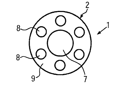
Fig. 5 shows a further example of the medical instrument according to the
invention
which is a guiding wire 1. This guiding wire 1 comprises seven rod-type bodies
7, 8. A
central rod-type body 7 is arranged in the center of the guiding wire 1. Six
radial rod-type
bodies 8 are arranged around the central rod-type body 7 so as to be equally
spaced
from each other. All rod-type bodies 7, 8 are embedded in a sheathing matrix
9. The
surface of the sheathing matrix 9 defines the surface of the guiding wire 1.
As explained above, the rod-type bodies 7, 8 are formed from a matrix material
containing non-metallic filaments. The above explanation of the rod-type
bodies also
applies to the rod-type bodies 7, 8 unless otherwise stated below.
The central rod-type body 7 has a larger diameter than the radial rod-type
bodies 8.
This results in the central rod-type body 7 having a higher stiffness than the
radial rod-
type bodies 8. As the central rod-type body 7 is arranged in the center of the
guiding
wire 1, its higher stiffness has a smaller effect on the flexural rigidity of
the whole
medical instrument than the radial rod-type bodies 8 as it is arranged on the
bending line
of the medical instrument. The radial rod-type bodies 8 have a higher
flexibility and this
is why they do not affect the flexural rigidity of the medical instrument too
much.
Therefore, a medical instrument is obtained which has a suitable flexibility.
CA 02724826 2010-11-18
12
The embodiment illustrated in Fig. 5 is very advantageous as it results in a
very thin
guiding wire with high strength and flexibility, and due to the radial
arrangement of the
radial rod-type bodies 8 the guiding wire 1 has a high torsional stiffness.
Further, the strength and flexibility of the medical instrument can be changed
by a
different number of the rod-type bodies and also by a modified arrangement,
for instance
without the central rod-type body. The flexibility of the guiding wire is an
essential
feature and to be individually adapted to different applications. The
flexibility of the
guiding wire can be varied by varying the diameter of the central rod-type
body and/or of
the radial rod-type bodies as well as by changing the composition of the
sheathing
matrix. In order that the medical instrument has the desired strength and
flexibility, it is
useful that all rod-type bodies are fully enclosed by the sheathing matrix.
The radial rod-type bodies 8 may extend parallel to the central rod-type body
7.
However, they can also be arranged in a spiral arrangement around the central
rod-type
body 7.
The central rod-type body has a diameter of 0.1 to 0.4 mm, preferably from
approximately 0.2 to 0.3 mm. The central rod-type body is doped with tungsten
nano
particles (particle size approximately 40 to 50 nm), for example.
The amount of the tungsten particles in relation to the matrix material of the
rod-type
body is 50 % by weight. In the present embodiment, an epoxy resin adds the
remaining
50 % by weight. The rod-type body additionally comprises glass fibers.
It has turned out that the tungsten nano particles during manufacturing the
rod-type
body have had an advantageous influence on the flowability of the epoxy resin.
The
undoped rod-type bodies are extruded with the addition of aerosils in order to
improve
the flowability. In case tungsten particles are used, adding such aerosils to
the epoxy
resin is not necessary. It has turned out that the smaller the particles, the
better the
viscosity of the epoxy resin.
With a high amount of tungsten particles, these act as both X-ray and MR
markers.
The weight proportion of the tungsten particles in relation to the matrix
material should
be at least 1:2 to 2:1. The higher the amount of the tungsten particles, the
better their
effect as MR markers. This effect as MR markers also depends on the size of
the rod-type
body and hence on the absolute amount of the tungsten particles and the
particle size of
the tungsten particles. Tungsten particles with a size from a few pm to
approximately 20
pm are hardly suited as MR markers as explained above on the basis of Figs.
4a, 4b and
4d to 4f. The smaller the tungsten particles, the higher their effect as MR
markers. It has
CA 02724826 2010-11-18
13
tuned out that the weight proportion of the tungsten particles in relation to
the matrix
material can be adjusted up to a range of 2:1 to 3:1.
The radial rod-type bodies 8 have a diameter from 0.10 to 0.25 mm, preferably
from
0.15 to 0.20 mm. Only one of the radial rod-type bodies 8 is doped with Fe304
particles in
the present embodiment. The particles have a particle size of approximately 40
to 50 nm.
The particles should have a size of not more than 100 nm, preferably not more
than 60
nm. In the doped radial rod-type body 8, one part by weight of Fe304 particles
accounts
for approximately 10 to 30, preferably 20 to 25 parts by weight of the matrix
material
which preferably is epoxy resin again. The Fe304 particles are passive MR
markers.
Within the scope of the invention it is also possible, of course, to dope the
rod-type
bodies with other passive markers, other concentrations and other particles
sizes. It is
also possible to provide more than two rod-type bodies with a marker,
preferably with
different markers. The number, the arrangements and the diameters of the rod-
type
bodies can also vary.
It is also possible that several different markers are provided in one rod-
type body.
Within the scope of the invention it is also possible to provide this guiding
wire on the
surface with one of the coatings described above and containing an active MR
marker.
The sheathing matrix 9 is a thermoplastic elastomer, preferably polyurethane,
in
particular TecoflexTm or Mediprene .
Mediprene is a thermoplastic elastomer which is primarily used for medical
purposes. Mediprene is offered by VTC Elastoteknik AB, Sweden. Mediprene is
understood to mean Mediprene TO 34007, a thermoplastic elastomer made from
SEBS
(styrene-ethylene-butylene-styrene-elastomer).
The medical instrument shown in Fig. 5 is preferably manufactured by co-
extruding
the rod-type body and the sheathing matrix.
The use of rod-type bodies with different doping agents is not restricted to
guiding
wires. Rod-type bodies with different doping agents can also be used with
other medical
instruments such as catheters, stents or implants.
It is preferred that a guiding wire 1 according to one of the above exemplary
embodiments is provided with a flexible tip (Fig. 6). The flexible tip 10 is
made from an
axial nylon thread 11 and a polyurethane body 12. This flexible tip 10 is
produced by
coating the nylon thread step by step so that the flexible tip 10 can be
formed as a blunt
tip. The flexible tip is connected with a front face of the guiding wire 1 by
means of a
CA 02724826 2010-11-18
14
glued connection. It is preferred that the flexible tip 10 is doped with one
of the passive
doping agents described above and/or coated with an active marker.
The front face of the guiding wire 1 and the corresponding contact surface of
the
flexible tip 10 are preferably ground so as to be cone-shaped so that the
contact area
between the guiding wire 1 and the flexible tip 10 is enlarged.
The flexible tip 10 can also be connected with the guiding wire 1 by heating
the two
contact surfaces. It is also possible to solubilize the flexible tip 10 with a
chemical solvent
(e.g. in solution grade polyurethane) and connect it with the guiding wire 1
in this way. A
suitable solvent is THF, for instance, if polyurethane is used as the material
for the
flexible tip 10. Instead of polyurethane, epoxy resin, PEEK, PEBAX, PE, PP,
silicone,
polylactic acid or Mediprene can also be used as the material for the
flexible tip 10. The
axial polymer thread can also be formed from other materials, for instance
from PEEK,
PEBM, PE, PP, silicone or polylactic acid. The flexible tip can also be
realized without an
axial thread.
The nylon thread is preferably doped with a marker. It can be doped with a
marker
which is different from the marker of the remaining material of the flexible
tip 10. In case
there is no thread, the material for the flexible tip can be doped with a
marker.
Figures 7a to 7e show further test equipment created by means of MRT or X-ray
tomography.
With this test equipment, rod-type bodies, on the one hand, and guiding wires
in
water, on the other hand, were examined.
The rod-type bodies generally consist of epoxy resin with glass fibers. The
following
different rod-type bodies were examined:
(F) Diameter 0.17 mm; no doping
(G) Diameter 0.17 mm; doped with Fe304 nano particles; weight ratio between
doping agent and epoxy resin is 1:20
(H) Diameter 0.27 mm; doped with tungsten nano particles; weight ratio of
doping
agent to epoxy resin 1:1
(3) Diameter 0.27 mm; doped with tungsten nano particles; weight ratio of
doping
agent to epoxy resin 2:1
The examined guiding wires 1 have basically the structure which is shown in
Fig. 5
and has been described on the basis of Fig. 5, with the central rod-type body
7 having a
CA 02724826 2010-11-18
diameter of 0.27 mm and being doped with tungsten nano particles. The radial
rod-type
bodies 8 have a diameter of 0.17 mm. Five radial rod-type bodies 8 are
undoped. One of
the radial rod-type bodies 8 is doped with Fe304 nano particles.
The following guiding wires were examined:
5 (K) Sheathing matrix made from polyurethane; doping amount of the central
rod-type
body of tungsten nano particles in a weight ratio of 1:1 in relation to the
epoxy resin, a
radial rod-type body 8 doped with Fe304, the weight ratio of doping agent to
epoxy resin
being 1:20;
(L) Sheathing matrix made from Mediprene ; doping amount of the central rod-
type
10 body of tungsten nano particles in a weight ratio of 2:1 in relation to
the epoxy resin, a
radial rod-type body 8 doped with Fe304, the weight ratio of doping to epoxy
resin being
1:20;
Fig. 7a shows a T1-weighted MRT sequence, Fig. 7b a T2-weighted MRT sequence,
Fig. 7c an MRT gradient echo sequence, and Fig. 7d an MRT Angio TOF sequence.
Fig. 7e
15 shows a computertomographic illustration of the rod-type bodies and
guiding wires.
The undoped rod-type body F can be hardly seen in any of the Figures. Due to
the
displacement of the water in the test equipment, traces with partially a very
low contrast
can be seen in the MRT.
The radial rod-type body doped with Fe304 is visible in the MRT process with
differing
contrast. In the MRT gradient echo sequence and the MRT Angio TOF sequence,
the
contrast is high, and in the two Ti- and T2-weighted sequences the contrast is
low. The
rod-type body H doped with tungsten nano particles has shown similar results
with MRT,
with the contrasts with the two Ti- and T2-weighted MRT sequences being better
than
that of the rod-type body G. Further, the rod-type body H produces an
excellent contrast
even in computer tomography (X-ray examination).
Such a rod-type body doped with tungsten nano particles (particle size smaller
than
100 nm, preferably smaller than 60 nm) represents a separate, independent idea
of the
invention as the use of such a rod-type body in a medical instrument in itself
produces
the visualization of the medical instrument both in X-ray and MRT
examinations. Using
other markers, better contrasts can be achieved in part so that a combination
with
further markers still makes sense but is not absolutely necessary. Tungsten
nano
particles also have the advantage that they produce a good contrast in both X-
ray and
MRT examinations in a predetermined concentration in the rod-type body.
Basically, iron
particles are also suited for creating a contrast in both X-ray and MR
examinations. With
CA 02724826 2010-11-18
16
iron particles, however, there is the problem that they produce large
artifacts with higher
concentrations which are the cause of heavy disturbances of the image in a
larger
surrounding. With low concentrations suitable for MRT, the iron particles are
not visible in
an X-ray examination.
Further, it is to be seen from Figures 7a to 7d that the doped rod-type bodies
as well
as the guiding wires containing doped rod-type bodies can all be seen clearly
in the
tested MRT sequences.
CA 02724826 2010-11-18
' 17
List of reference numerals:
1 Guiding wire
2 Surface
3 MR marker
4 Coating capable of swelling
5 Test rod
6 Plastic plate
7 Central rod-type body
8 Radial rod-type body
9 Sheathing matrix
10 Flexible tip
11 Nylon thread
12 Polyurethane body