Note: Descriptions are shown in the official language in which they were submitted.
CA 02725370 2015-11-23
WO 2009/141310 PC
T/EP2009/056010
Mass Spectrometric Analysis
This invention relates to a method of assaying a target analyte by mass
spectrometry,
particularly biomolecules such as nucleic acids and proteins. Specifically the
invention
relates to a method of multiplexed tandem mass spectrometry using isobaric
mass labels.
The present invention also relates to a mass spectrometric device for assaying
one or
more target analytes.
Various methods of labelling molecules of interest are known in the art,
including
radioactive atoms, fluorescent dyes, luminescent reagents, electron capture
reagents and
light absorbing dyes. Each of these labelling systems has features which make
it suitable
for certain applications and not others. More recently there has been
development in the
area of mass spectrometry as a method of detecting labels that are cleavably
attached to
their associated molecule of interest.
For many applications such as nucleic acid analysis, the structure of the
analyte can be
determined from indirect labelling. This is advantageous particularly with
respect to the
use of mass spectrometry because complex biomolecules such as DNA have complex
mass spectra and are detected with relatively poor sensitivity. Indirect
detection means
that an associated label molecule can be used to identify the original
analyte, where the
label is designed for sensitive detection and a simple mass spectrum. Simple
mass spectra
mean that multiple labels can be used to analyse multiple analytes
simultaneously.
WO/1998/031830 describes arrays of nucleic acid probes covalently attached to
cleavable
labels that are detectable by mass spectrometry which identify the sequence of
the
covalently linked nucleic acid probe. The labelled probes of this application
have the
structure Nu-L-M where Nu is a nucleic acid covalently linked to L, a
cleavable linker,
covalently linked to M, a mass label. Preferred cleavable linkers in this
application cleave
within the ion source of the mass spectrometer. Preferred mass labels are
substituted poly-
aryl ethers. This application discloses a variety of ionisation methods and
analysis by
1
CA 02725370 2015-11-23
WO 2009/141310
PCT/EP2009/056010
quadrupole mass analysers, Time of Flight (TOF) analysers and magnetic sector
instruments as specific methods of analysing mass labels by mass spectrometry.
WO/1995/004160 discloses ligands, and specifically nucleic acids, cleavably
linked to
mass tag molecules. Preferred cleavable linkers are photo-cleavable. This
application
discloses Matrix Assisted Laser Desorption Ionisation (MALDI) Time of Flight
(TOF)
mass spectrometry as a specific method of analysing mass labels by mass
spectrometry,
WO /1998/026095 discloses releasable non-volatile mass-label molecules. In
preferred
embodiments these labels comprise polymers, typically biopolymers which are
cleavably
attached to a reactive group or ligand, i.e. a probe. Preferred cleavable
linkers appear to
be chemically or enzymatically cleavable. This application discloses MALDI TOF
mass
spectrometry as a specific method of analysing mass labels by mass
spectrometry.
WO/1997/027325, WO/1997/027331 and
WO/I997/027331 disclose ligands, and
specifically nucleic acids, cleavably linked to mass tag molecules. Preferred
cleavable
linkers appear to be chemically or photo-cleavable. These applications
disclose a variety
of ionisation methods and analysis by quadrupole mass analysers, TOF analysers
and
magnetic sector instruments as specific methods of analysing mass labels by
mass
spectrometry.
None or these prior art applications mention the use of tandem or serial mass
analysis of
tagged biomolecules.
Gygi et al. (Nature Biotechnology 17: 994-999, "Quantitative analysis of
complex protein
mixtures using isotope-coded affinity tags" 1999) disclose the use of 'isotope
encoded
affinity tags' for the capture of peptides from proteins, to allow protein
expression
analysis. In this article, the authors describe the use of a biotin linker,
which is reactive to
thiols, for the capture peptides with cysteine in them. A sample of protein
from one
source is reacted with the biotin linker and cleaved with an endopeptidase.
The
biotinylated cysteine-containing peptides can then be isolated on avidinated
beads for
2
=
CA 02725370 2010-11-23
WO 2009/141310
PCT/EP2009/056010
subsequent analysis by mass spectrometry. Two samples can be compared
quantitatively
by labelling one sample with the biotin linker and labelling the second sample
with a
deuterated form of the biotin linker. Each peptide in the samples is then
represented as a
pair of peaks in the mass spectrum. Integration of the peaks in the mass
spectrum
corresponding to each tag indicate the relative expression levels of the
peptide linked to
the tags.
Selected reaction monitoring (SRM) and multiple reaction monitoring (MRM)
provide
highly selective methods of tandem mass spectrometry which have the potential
to
effectively filter out all molecules and contaminants except the desired
analyte. This is
particularly beneficial if complex samples are analysed which tend to have
several
isobaric species present within a defined analytical window. Usually, a
combination of
precursor (parent ion) selection in the first stage of the mass spectrometer
(here termed
Q I: quadrupole 1, but also equivalent for the respective stages in non-
quadrupole mass
spectrometers such as ion traps etc.), fragmentation of the parent ion into
many fragments
of which one or several specific fragments are selected in the following steps
of the MS-
measurement (usually in quadrupole 3, Q3) and detected at the ion detector.
This two-
step selection ensures that the desired analyte is measured and any other ion
species are
reduced in their intensity. Signal ¨ to ¨ noise ratio is much superior to
conventional
MS/MS experiments which select one mass window in Ql, and then measure all
generated fragments in the ion detector. In principle, this MS-based approach
can
provide absolute structural specificity for the analyte, and in combination
with
appropriate stable isotope-labelled internal standards (SISs), it can provide
absolute
quantitation of analyte concentration.
In conventional SRM/MRM type experiments, a stable isotope labelled reference
is used
to generate an analyte/reference pair which will be used for quantification of
analyte
against the reference. For the analysis of proteins, such a reference peptide
differs from
the analyte to be measured only by incorporation of isotopes, to make it
distinctly
different in mass for the Q1 selection, but otherwise identical in chemical
composition,
and physico-chemical behaviour. In a typical experiment, the analyte/reference
pair are
3
CA 02725370 2010-11-23
WO 2009/141310
PCT/EP2009/056010
selected, i.e. in Ql by switching mass selection channels between these two
masses. The
subsequent fragmentation of these two ions leads to distinct (specific)
fragment masses.
One or more suitable fragment masses are then chosen where the Q3 filter
remains on the
position of the selected fragment ions, thus assuring transition of this ion
to the mass
analyser, and filtering out other ion species.
Recent work in designing improved mass labels for identifying analytes using
mass
spectrometry has focused on mass labels which are more easily identified in
the mass
spectrum without other contaminants.
WO 01/68664 discloses a set of two or more mass labels, each label in the set
comprising
a mass marker moiety attached via a cleavable linker to a mass normalisation
moiety, the
mass marker moiety being fragmentation resistant. The aggregate mass of each
label in
the set may be the same Or different and the mass of the mass marker moiety of
each label
in the set may be the same or differeni Iii any group of labels within the set
having a
mass marker moiety of a common mass each label has an aggregate mass different
from
all other labels in that group, and in any group of labels within the set
having a common
aggregate mass each label has a mass marker moiety having a mass different
from that of
all other mass marker groups in that group, such that all of the mass labels
in the set are
distinguishable from each other by mass spectrometry. This application further
discloses
methods of analysis comprising detecting an analyte by identifying by mass
spectrometry
a mass label or a combination of mass labels unique to the analyte. Tandem
mass
spectrometry may be used. Specifically, the mass spectrometer employed to
detect the
mass label may be a triple quadrupole mass analyser comprising a first
analyser to select
ions of a particular mass or mass range, a second mass analyser to dissociate
the selected
ions and a third mass analyser to detect resulting ions.
WO 03/025576 discloses a set of two or more mass labels, each label in the set
comprising a mass marker moiety attached via at least one amide bond to a mass
nornialisation moiety. The mass marker moiety comprises an amino acid and the
mass
normalisation moiety comprises an amino acid. As for WO 01/68664 the aggregate
mass
4
CA 02725370 2010-11-23
WO 2009/141310
PCT/EP2009/056010
of each label in the set may be the same or different and the mass of the mass
marker
moiety of each label in the set may be the same or different such that all of
the mass
labels in the set are distinguishable from each other by mass spectrometry. As
for WO
01/68664 this application also discloses a method of analysis which may
involve tandem
mass spectrometry. This application is specifically directed to the analysis
of peptides
and mass labels with mass normalisation moieties and mass marker moieties
comprising
at least one amino acid.
WO 2007/012849 discloses a mass label and a reactive mass label having a
general
chemical formula for labelling and detecting a biological molecule by mass
spectroscopy.
The mass labels and reactive mass labels of this invention are clearly
identified in a mass
spectrum and are easily reacted with analytes. As for WO 01/68664 this
application also
discloses a method of analysis which may involve tandem mass spectrometry.
The development of isobaric mass tags in the late 1990's has revolutionised
biomarker
discovery. The ability to analyse multiple samples in theoretically unlimited
numbers in a
single LC-MS/MS workflow increases throughput whilst at the same time reducing
analytical variability. Therefore, there remains a need for improved methods
of
quantitatively detecting and routinely measuring analytes by mass spectrometry
in a wide
range of samples.
Whilst the mass labels provided by WO 01/68664, WO 03/02557 and WO 2007/012849
have allowed significant improved methods of analysis of analytes by mass
spectroscopy,
there is still a requirement to provide improved methods of detecting an
analyte by
identifying by mass spectrometry such mass labels. In particular, whilst these
new mass
labels and methods of analysis allow multiple samples to be analysed
simultaneously and
quantitatively without significantly increasing the complexity of the mass
spectrum, the
analysis of isobaric mass labels using known tandem mass spectrometry can
still provide
inaccurate results for complex samples. There is still a requirement to
provide improved
methods of analysis which allow easy identification of the mass labels in a
mass
spectrometer and allow sensitive quantification.
CA 02725370 2010-11-23
WO 2009/141310
PCT/EP2009/056010
Accordingly, it is an aim of the present invention to solve the problems of
the prior art in
this field and provide improved methods of assaying a target analyte by mass
spectroscopy.
In a first aspect the invention provides a method for assaying for a target
analyte, which
method comprises:
(a) providing a plurality of samples which may comprise the target analyte,
wherein each sample is differentially labelled with a mass label or a
combination of mass
labels, wherein the mass labels are from a set of mass labels, wherein each
mass label is
an isobaric mass label comprising a mass spectrometrically distinct mass
marker group,
such that the samples can be distinguished by mass spectrometry;
(b) mixing the plurality of labelled samples to produce an analysis mixture
and
introducing the analysis mixture into a mass spectrometer;
(c) selecting ions having a first mass to charge ratio equivalent to an ion
of the
target analyte labelled with a specific number of mass labels;
(d) fragmenting ions of the first mass to charge ratio into a plurality of
fragment ions, wherein a proportion of the plurality of fragment ions comprise
at least
one intact mass label;
(e) selecting ions of a second mass to charge ratio equivalent to an ion of
a
fragment of the target analyte comprising at least one intact mass label;
(f) fragmenting ions of the second mass to charge ratio into a plurality of
further fragment ions, wherein a proportion of the further fragment ions are
ions of the
mass marker groups;
(g) producing a mass spectrum of the further fragment ions produced in step
(f); and
(h) detemining from the mass spectrum the quantity of the target analyte in
each sample.
The method according to the present invention overcomes the limitations of the
art by
quantifying molecules of interest using isobarically tagged samples wherein
the method
6
CA 02725370 2010-11-23
WO 2009/141310
PCT/EP2009/056010
comprises two steps of selecting ions of a pre-determined mass to charge ratio
each
followed by a fragmentation step. The use of such a method provides a high
degree of
selectivity and, therefore, the mass spectrum produced in the final step
provides more
accurate quantitative results compared to the results from a conventional
tandem mass
spectrometry (MS/MS) experiment.
In conventional tandem mass spectrometry (MS/MS) using isobaric mass labels,
ions
equivalent to the mass of the labelled target analyte are first selected.
After selection, the
ions of the labelled analytes are subjected to fragmentation and then peaks
corresponding
to the mass marker group of mass labels are identified. However, the spectra
obtained
often do not provide accurate quantification of analytes due to co-eluting
fragments from
contaminants having the same mass to charge ratio as the selected mass to
charge ratio.
This problem occurs when conducting analysis of complex mixtures of proteins.
In
complex mixtures, different peptides or peptide fragments may have the same
mass as the
target analyte. These contaminating peptides will not be differentiated from
the target
analyte by MS/MS because they will all be selected together as the parent ion
mass to
charge ratio in the selection step. Therefore, the fragmentation of the parent
ions to
release mass marker groups from the mass labels will provide a spectrum of
mass marker
groups from all peptides selected including contaminating peptides having the
same mass
as the target analyte.
This limitation of MS/MS is overcome in the present invention due to the
further steps of
selecting (step e) and fragmenting (step O. In step e) selection of the mass
to charge ratio
equivalent to a desired ion of a fragment of the target analyte comprising at
least one
intact mass label ensures that the vast majority, if not all contaminating
molecules
selected in Q1 (step c) are removed from the mass spectrum. Contaminating
peptides
which fragment in step d) into a plurality of fragments, none of which have a
mass to
charge ratio equivalent to the second mass to charge ratio selected in step e)
will be
removed. Therefore, the mass marker groups release from the fragmentation step
0 are
only from the target analyte and the resulting mass spectrum will provide
highly
improved accurate quantification results for the target analyte. The method
according to
7
CA 02725370 2010-11-23
WO 2009/141310
PCT/EP2009/056010
the present invention is particularly advantageous for analysis of complex
samples
because the further degree of selectivity improves specificity.
The method according to the present invention succeeds to generate a
combination
between the high sensitivity and selectivity of SRM (selected reaction
monitoring: one
analyte) or MRM (multiple reaction monitoring: multiple analytes) with
multiplexing in
the final analysis step which is used for quantification purposes.
The quantity determined in step (h) may be the relative quantity of the target
analyte in
each sample or the absolute quantity of the target analyte in each sample.
A further advantage of the present invention is that it allows a plurality of
samples to be
analysed together. The plurality of samples may be test samples which may
comprise the
target analyte.
The term "test sample" refers to any specimen in which an analyte may be
present. The
test sample may comprise only one analyte. Alternatively, the test sample may
comprise a
plurality of different analytes.
In one embodiment of the present invention one sample is a test sample and one
sample is
a calibration sample, wherein the calibration sample comprises one or more
different
aliquots of the target analytes, each aliquot having a known quantity of the
analyte,
wherein the test sample and each aliquot of the calibration sample are
differentially
labelled.
When one or more calibration samples are present, step h) in the method
according to the
present invention preferably comprises calibrating the quantity of the analyte
in the test
sample against the known and determined quantities of the analytes in the one
or more
aliquots in the calibration sample. In a preferred embodiment, the method
comprises a
step of plotting a graph of the quantity of the analyte in each aliquot versus
the quantity of
the analyte in each aliquot as determined by mass spectrometry. This step may
instead
8
CA 02725370 2010-11-23
WO 2009/141310
PCT/EP2009/056010
simply involve calculation and mathematical programs or algorithms for
performing such
calculations that are well understood by the skilled person. The quantity of
the analyte in
the sample is then calculated by measuring the quantity in the sample as
determined by
mass spectrometry against the calibration graph. In the context of this
invention, a
reference to "a quantity as measured by mass spectrometry" is typically an ion
abundance, ion intensity, or other signal measured by mass spectrometry which
relates to
the quantity of an analyte. This embodiment provides more accurate
quantification
results which are independent of externally obtained calibrations, thus
providing for a
much more robust and reliable analysis.
The different aliquots each have a different known quantity of the analyte.
The term
"known quantity" means that the absolute quantity, or a qualitative quantity
of the analyte
in each aliquot of the calibration sample is known.
An absolute quantity means a quantity which is a known. This allows for the
absolute
quantity of an analyte in a test sample to be determined,
A qualitative quantity in the present context means a quantity which is not
known
absolutely, but may be a range of quantities that are expected in a subject
having a
particular state, for example a subject in a healthy or diseased state, or
some other
expected range depending on the type of test sample under investigation. Each
aliquot is
"different" since it contains a different quantity of the analyte. Typically
this is achieved
by taking different volumes from a standard sample, especially for qualitative
quantities
where taking different volumes will ensure that different quantities are
present in each
aliquot in a desired ratio, without needing to know the absolute quantities.
As an
altmative, each aliquot is prepared separately and is not taken from the same
sample. hi
one embodiment, each different aliquot has the same volume, but comprises a
different
quantity of the analyte.
Preferably, the or each calibration sample comprises two Or more different
aliquots of the
target analyte. The use of two or more different aliquots of the target
analyte allows the
9
CA 02725370 2010-11-23
WO 2009/141310
PCT/EP2009/056010
construction of multi-point standard curves for each analyte without
increasing MS
complexity. Analyte quantitation is obtained in the mass spectrum produced in
step g),
and the analyte in the sample and in the calibration sample can be
simultaneously
quantified and identified. Alternatively, only the quantity of the analyte is
determined.
This method provides means for the measurement of up to 10, up to 20, up to 50
or more
analytes in a single experiment.
The method according to the present invention may comprise analysis of a
plurality of
test samples in addition to one or more calibration samples. In this
embodiment, each of
the plurality of test samples is preferably assayed for the same analyte.
Preferably the
same calibration sample is used for each test sample to be assayed. Typically,
the same
known volume of the calibration sample comprising at one or more aliquots of
the analyte
is added to each different test sample. This method is particularly useful in
clinical studies
involving multiple samples from patients. If a large quantity of the
calibration sample is
prepared and fractions taken, the same calibration sample can be used by
multiple
laboratories, facilitating cross-study and cross-laboratory comparisons. Each
test sample
may be differentially labelled with one Or more of the isobaric mass labels
and combined
with one or more calibration samples in step b), and the quantity of the
analyte in each
sample is determined simultaneously in step h). Alternatively, each test
sample may be
labelled with the same mass label and steps b) to h) are repeated for each
different test
s ample.
In one embodiment the method according to the present invention may be used to
assay a
plurality of different target analytes. In this embodiment the method
comprises a step of
repeating steps (c) to (h) for each target analytc. In this embodiment,
wherein one sample
is a test sample, a calibration sample may be provided for each different
analyte. Each
calibration sample comprises one or more different aliquots of a target
analyte, wherein
the test sample and each aliquot of each calibration sample are differentially
labelled. In
one embodiment the plurality of analytes are peptide fragments of a protein or
polypeptide which are produced by chemical or enzymatic processing of the
protein or
CA 02725370 2010-11-23
WO 2009/141310
PCT/EP2009/056010
polypeptide prior to step (a). in a particular embodiment, the plurality of
analytes are
peptides from the same protein or polypeptide.
The present invention will now be described in more detail with reference to
the
following figures:
Figure la shows a MS/MS spectrum of peptide VATVSLPR labelled with different
isobaric mass labels from a set of two mass labels, each label representing a
predetermined relative amount of the peptide (the ratio of the mass marker
groups having
masses 126:127 is 2:1) and Figure lb shows a zoomed in section of the spectrum
of
Figure la showing the peaks of the mass marker groups.
Figure 2a shows a MS/MS/MS spectrum of the hi-ion of labelled peptide
VATVSLPR,
as analysed in Figure 1; Figure 2b shows a zoomed in section of the spectrum
of Figure
2a showing the peaks of the mass maker groups.
Figure 3 shows a MS spectrum of peptide VAFSLR labelled with different
isobaric mass
labels from a set of six mass labels, each label representing a predetermined
relative
amount of the peptide (the ratio of the mass marker groups having masses
126:127:128:129:130:131 is 1:3:5:5:3:1).
Figure 4 shows a MS/MS spectrum of the labelled VAFSLR peptide, as analysed in
Figure 3.
Figure 5a shows a MS/MS spectrum of the distinct mass marker groups from the
labelled
VAFSLR peptide, as analysed in Figure 3; Figure 5b shows a MS/MS/MS spectrum
of
the distinct mass marker groups from the labelled VAFSLR peptide, as analysed
in Figure
3.
Figure 6 shows a MS spectrum of peptide AVFSLR labelled with different
isobaric mass
labels from a set of six mass labels, each label representing a predetermined
relative
11
CA 02725370 2010-11-23
WO 2009/141310
PCT/EP2009/056010
amount of the peptide (the ratio the mass marker groups having masses of
126:127:128:129:130:131 is 1:1:1:4:4:4).
Figure 7 shows a MS/MS spectrum of the labelled AVFSLR peptide, as analysed in
Figure 6.
Figure 8a shows a MS/MS spectrum of the distinct mass marker groups from the
labelled
AVFSLR peptide, as analysed in Figure 6; Figure 8b MS/MS/MS spectrum of the
distinct
mass marker groups from the labelled AVFSLR peptide, as analysed in Figure 6.
Figure 9 shows a MS spectrum of peptide FAVSLR labelled with different
isobaric mass
labels from a set of six mass labels, each label representing a predetermined
relative
amount of the peptide (the ratio of the mass marker groups having masses of
126:127:128:129:130:131 is 4:4:4:1:1:1).
Figure 10 shows a MS/MS spectrum of the labelled FAVSLR peptide, as analysed
in
Figure 9.
Figure 11a shows a MS/MS spectrum of the distinct mass marker groups from the
labelled FAVSLR peptide, as analysed in Figure 9; Figure 11b MS/MS/MS spectrum
of
the distinct mass marker groups from the labelled FAVSLR peptide, as analysed
in Figure
9.
Figure 12 shows a MS spectrum of peptide LAFSVR labelled with different
isobaric mass
labels from a set of six mass labels, each label representing a predetermined
relative
amount of the peptide (the ratio the mass marker groups having masses of
126:127:128:129:130:131 is5:3:1:1:3:5).
Figure 13 shows a MS/MS spectrum of the labelled LAFSVR peptide, as analysed
in
Figure 12.
12
CA 02725370 2010-11-23
WO 2009/141310
PCT/EP2009/056010
Figure 14a shows a MS/MS spectrum of the distinct mass marker groups from the
labelled LAFSVR peptide, as analysed in Figure 12; Figure 14b MS/MS/MS
spectrum of
the distinct mass marker groups from the labelled LAFSVR peptide, as analysed
in Figure
12.
Figure 15 shows a MS spectrum of a mixture of peptide VAFSLR and LAFSVR each
labelled with different isobaric mass labels from a set of six mass labels,
each label
representing a predetermined relative amount of a peptide (peptide VAFSLR: the
ratio of
mass marker groups having masses of 126:127:128:129:130:131 is 1:3:5:5:3:1;
and
peptide LAFSVR: the ratio of mass marker groups having masses of
126:127:128:129:130:131 is5:3:1:1:3:5).
Figure 16 shows a MS/MS spectrum of a mixture of labelled VAFSLR and LAFSVR
peptides, as analysed in Figure 15.
Figure 17 shows a MS/MS spectrum of the distinct mass marker groups from a
mixture of
labelled VAFSLR and LAFSVR peptides, as analysed in Figure 15.
Figure 18a shows a MS/MS/MS spectrum of the distinct mass marker groups from
the
b1-ions of labelled VAFSLR, as analysed in Figure 15; Figure 18b shows a
MS/MS/MS
spectrum of the distinct mass marker groups from the hi-ions of labelled
LAFSVR, as
analysed in Figure 15.
Figure 19 shows a MS spectrum of a mixture of peptide AVFSLR and FAVSLR each
labelled with different isobaric mass labels from a set of six mass labels,
each label
representing a predetermined relative amount of a peptide (peptide AVFSLR: the
ratio of
the mass marker groups having masses of 126:127:128:129:130:131 is
1:1:1:4:4:4;
peptide FAVSLR: the ratio of the mass marker groups having masses of
126:127:128;129:130:131 is 4:4:4: 1:1: 1).
13
CA 02725370 2010-11-23
WO 2009/141310
PCT/EP2009/056010
Figure 20 shows a MS/MS spectrum of a mixture of labelled AVFSLR and FAVSLR
peptides, as analysed in Figure 19.
Figure 21 shows a MS/MS spectrum of the distinct mass marker groups from a
mixture of
labelled AVFSLR and FAVSLR peptides, as analysed in Figure 19.
Figure 22a shows a MS/MS/MS spectrum of the distinct mass marker groups from
the
b I -ions of labelled AVFSLR, as analysed in Figure 19; and Figure 22b shows a
MS/MS/MS spectrum of the distinct mass marker groups from the b 1 -ions of
labelled
FAVSLR, as analysed in Figure 19.
Figure 23 shows a MS/MS spectrum of labelled peptide AEFAEVSK and the
structure of
the mass label (TMT zero) used to label the peptide. The peptide is labelled
at the N-
terminus and at the lysine. Ions arising from fragmentation of the mass label
are shown.
Figure 24 shows a MS/MS spectrtun of peptide VI,F,PTLK labelled with a set of
two
isobaric mass labels (TMT duplex comprises TMT2-126 and TMT2-127), and the
structures of the mass. labels used to label the peptide. The peptide is
labelled at the N-
terminus and at the lysine. Ions arising from fragmentation of the mass labels
are shown.
Figure 25a shows a MS/MS spectrum of a peptide from Serum Albumin LVNEVTEFAK
labelled with mass label TMT-zero and Figure 25b shows a MS/MS spectrum of a
peptide
from Serum Albumin LVNEVTEFAK labelled with mass label TMT sixplex. The
peptide in Figures 25a and 25b is labelled at the N-terminus and the lysine. A
mass
difference of 10 Da is shown between the labelled peptides in Figures 25a and
25b.
Figure 26a shows a zoomed in section of the MS/MS spectrum of Figure 25a
showing the
y3 ion fragment and Figure 26b shows a zoomed in section of the MS/MS spectrum
of
Figure 25b showing the y3 ion fragment. The y3 ion fragment retains one intact
mass
label on the lysine residue giving a m/z differene of 5 Thomsons (Th, unit of
mass to
charge ratio) between the two labelled fragment ions in Figures 26a and 26b.
14
CA 02725370 2010-11-23
WO 2009/141310
PCT/EP2009/056010
Figure 27a shows a zoomed in section of the MS/MS spectrum of Figure 25a
showing the
y5 ion fragment and Figure 27b shows a zoomed in section of the MS/MS spectrum
of
Figure 25b showing the y3 ion fragment. The y5 fragment ion retains one intact
mass
label on the lysine residue giving a rn/z difference of 5 Thomsons (Th unit of
mass to
charge ratio) between the two labelled fragment ions in Figures 27a and 27b.
Figure 28a shows a zoomed in section of the MS/MS spectrum of Figure 25a
showing the
b7 ion fragment and Figure 28b shows a zoomed in section of the MS/MS spectrum
of
Figure 25b showing the b7 ion fragment. The b7 fragment ion retains one intact
mass
label on the N-terminus giving a rn/z difference of 5 Thomsons (Th unit of
mass to charge
ratio) between the two labelled fragment ions in Figures 28a and 28b.
Figure 29a shows a MS spectrum of peptide LVTDLTK labelled with TMT zero and
TMTsixplex. The peptide is labelled at the N-terminus and lysine giving a mass
difference of 10 Da between the labelled peptides TMT zero and TMT sixplex. A
mass
difference of 5 Th is observed between the doubly charged precursor ions.
Figure 29 b shows a MS spectrum of peptide HPDYSVVLLLR labelled with TMTzero
and TMTsixplex. The peptide is labelled at the N-terminus giving a mass
difference of 5
Da between the labelled peptides TMT zero and TMT sixplex. A mass difference
of 1.67
Th is observed between the triply charged precursor ions.
Figure 30 shows an MRM ion chromatogram of ten plasma peptides labelled with
mass
label TMT-zero and mass label TMTsixplex (TMT6-127).
Figures 31a to d show MRM ion chromatograms of a plasma peptide K labelled
with
TMT-zero and TMTsixplex (TMT 6 -127). The TMT-labelled plasma samples have
been
mixed in different ratios.
CA 02725370 2010-11-23
WO 2009/141310
PCT/EP2009/056010
Figure 32 shows a graph of the expected ratios versus the observed ratios of
TMTzero:TIVITsixplex for peptide K (as shown in Figures 31a to 31d). The
analysis was
performed in triplicate.
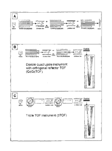
Figure 33 shows a schematic of the QitTofTm instrument.
Figures 34a, 34b and 34c shows alternative arrangements of mass spectrometers
capable
of MS/MS/MS.
The term "analyte" is not particularly limiting, and the methods according to
the present
invention may be employed to assay any type of molecule provided that it can
be
analysed by mass spectrometry, and is capable of being labelled by an isobaric
mass label
with a mass spectrornetrically distinct mass marker group. Analytes include
amino acids,
peptides, polypeptides, proteins, glycoproteins, lipoproteins, nucleic acids,
polynucleotides, oligonucleotides, DNA, RNA, peptide-nucleic acids, sugars,
starches
and complex carbohydrates, fats and complex lipids, polymers and small organic
molecules such as drugs and drug-like molecules or fragments thereof
Preferably the
analyte is a peptide, protein, nucleotide or nucleic acid.
hi relation to this invention the term analyte shall be synonymous with the
term
biomolecule.
In relation to this invention the tenn "protein" shall encompass any molecule
comprising
two or more amino acids including di-peptides, tri-peptides, peptides,
polypeptides and
proteins.
In relation to this invention the term "nucleic acid" shall encompass any
molecule
comprising two or more nucleotide bases including di-nucleotides, tri-
nucleotides,
oligonucleotides, deoxyribonucleic acids, ribonucleic acids and peptide
nucleic acids.
16
CA 02725370 2010-11-23
WO 2009/141310
PCT/EP2009/056010
The wording "set of sixplex Tandem Mass Tags (TMT)" refers to a set of six
isobaric
mass labels, wherein each label comprises a mass spectrometrically distinct
mass marker
group. An example of a set of sixplex Tandem Mass Tags are TMT6-128, TMT6-129,
TMT6-130, TMT6-131, wherein "6" represents the number of labels in the set and
the
numbers 128-131 following "TMT", represent the mass of the mass marker group.
In the
same manner, a set of duplex Tandem Mass Tags refers to a set of two isobaric
mass
labels. An examples of a set of duplex Tandem Mass Tags are TMT2-126 and TMT2-
127,
wherein "2" represents the number of labels in the set and the numbers 126 and
127
following "TMT", represent the mass of the mass marker group. A set of
fiveplex
Tandem Mass Tags refers to a set of 5 isobaric mass labels.
The term 'MS' in the context of the present invention refers to a method of
mass
spectrometry comprising producing ions from a sample and producing a mass
spectrum of
the ions.
The term `MS/MS' in the context of the present invention refers to the method
according
to the present invention comprising selecting ions of particular mass to
charge ratio,
subjecting selected ions to fragmentation, for example by Collision Induced
Dissociation
(CID), and producing a mass spectrum of the fragment ions.
The term `MS/MS/MS' in the context of the present invention refers to the
method
according to the present invention comprising steps (a) to (h).
In relation to this invention the term "mass spectrometry" shall include any
type of mass
spectrometry capable of fragmentation analysis. The mass spectrometers
suitable for use
in the present invention include instruments that comprise any form of
analyser capable
of MS/MS/MS.
In one embodiment, steps (c) to (g) of the method according to the present
invention are
carried out in separate quadrupoles in a mass spectrometer. In this
embodiment, step c)
of selecting the ions having a first mass to charge ratio is performed in the
first mass
17
CA 02725370 2010-11-23
WO 2009/141310
PCT/EP2009/056010
analyser of a serial instrument (Q1). The selected ions are then channelled
into a separate
collision cell (Q2) where they are collided with a gas or a solid surface to
produce a
plurality of fragment ions in step d). The collision products from step d) are
then
channelled into a third mass analyser (Q3) wherein ions of a second mass to
charge ratio
(MS/MS ions) are selected in step e). The selected ions from step e) are then
channelled
into a separate collision cell (Q4) wherein they are collided with a gas or a
solid surface
to produce a plurality of further fragment ions in step f). The further
fragment ions from
step f) are channelled into a further mass analyser (Q5) of a serial
instrument in step g) to
detect collision products. Typical serial instruments include five quadrupole
mass
spectrometers, tandem sector instruments and quadrupole time of flight (TOP)
mass
spectrometers.
Alternatively, steps (c) to (g) of the method according to the present
invention are carried
out sequentially in the same zone of a mass spectrometer. This may be effected
in ion
trap mass analysers and Fourier Transform Ion Cyclotron Resonance (FT-ICR)
mass
spectrometers, for example.
MS/MS/MS experiments according to the present invention can be undertaken
using
conventional 3D iontraps, hybrid geometry instruments such as a quadrupole ion
trap in
combination with a TOF analyser, as well as the larger footprint four sector
instruments.
Wu Z., Bordas-Nagy J. and FenseIau C. (1991) "Triple mass spectrometry
(MS/MS/MS)
with a floated collision cell in a four-sector tandem mass spectrometer"
Organic Mass
Spectrometry 26, 10, 908-911 describes a method for carrying out MS/MS/MS
experiments with an electrically floated collision cell in the third field-
free region on a
tandem double-focusing mass spectrometer. The experiments were performed using
a
JEOL JMS-HX110/I-IX110 four-sector mass spectrometer and although the method
involved calibration of the magnet calibration at all accelerating voltages,
it is generally
applicable at any value of the collision cell voltage,
18
CA 02725370 2010-11-23
WO 2009/141310
PCT/EP2009/056010
Quadrupole ion traps (QITs) are an effective means to accumulate and store
ions. The
combination of QIT with TOF mass spectrometry offers powerful capabilities not
available by QIT or TOF mass spectrometry alone. Syagen has already combined
these
devices into a single instrument called the QitTofrm, which is the first
commercially-
available instrument to offer the MS' advantages of QIT MS with the high-speed
data
collection rates of TOF MS. The configuration of the QitTofTm instrument is
shown in
Figure 33. Shimadzu have also subsequently developed an LCMS-QIT-TOF system.
Figure 34 shows a schematic to illustrate the geometry of the QitTofTm
instrument.
There are specific benefits of the QitTofTm geometry compared to other
instruments. The
QitTofTm configuration has the potential for higher ion transmission
efficiency and allows
effective MS" operation compared to orthogonal-extraction TOF MS. The QIT
gives the
advantage of mass-selective ejection with higher dynamic range and greater ion
trap
capacity due to the higher repetition rate because ions are pulsed out rather
than scanned
out of the QIT. The TOF provides the advantage of multichannel mass detection
leading
to efficient collection of all ions. Better ion mass accuracy is also achieved
using the TOF
analyzer.
Several other instrument geometries could be considered for MS3 experiments in
the
present invention and a selection of future possibilities are shown in Figure
34. The
performance of each design is difficult to assess at this stage and will
require further
investigation. Figure 34 A depicts a penta-quadrupole arrangement with three
scanning
quadrupoles and two collision cells. An ion multiplier detector is typically
used in
conjunction with quadrupole mass analysers. Figure 34 B depicts a double
scanning
quadrupole with an orthogonal reflectron TOF as the final stage analyser.
Figure 34 C
depicts a triple stage TOF instrument with timed ion gates permitting ions
with a user-
specified mass range to enter the first two linear TOF analysers.
In the present invention, matrix assisted laser desorption/ionisation (MAT,DT)
techniques
may be employed. MALDI requires that the biomolecule solution be embedded in a
large
molar excess of a photo-excitable 'matrix'. The application of laser light of
the
appropriate frequency results in the excitation of the matrix which in turn
leads to rapid
19
CA 02725370 2010-11-23
WO 2009/141310
PCT/EP2009/056010
evaporation of the matrix along with its entrapped biomolecule. Proton
transfer from the
acidic matrix to the biomolxule gives rise to protonated forms of the
biomolecule which
can be detected by positive ion mass spectrometry, particularly by Time-Of-
Flight (TOF)
mass spectrometry. Negative ion mass spectrometry is also possible by MALDI
TOR
This technique imparts a significant quantity of translational energy to ions,
but tends not
to induce excessive fragmentation despite this. The laser energy and the
timing of the
application of the potential difference used to accelerate the ions from the
source can be
used to control fragmentation with this technique. This technique is highly
favoured due
to its large mass range, due to the prevalence of singly charged ions in its
spectra and due
to the ability to analyse multiple peptides simultaneously. The TOF/TOF
technique may
be employed in the present invention.
The photo-excitable matrix comprises a 'dye', i.e. a compound that strongly
absorbs light
of a particular frequency, and which preferably does not radiate that energy
by
fluorescence or phosphorescence but rather dissipates the energy thermally,
i.c. through
vibrational modes. It is the vibration of the matrix caused by laser
excitation that results
in rapid sublimation of the dye, which simultaneously takes the embedded
analyte into the
gas phase.
Although MALDI techniques are useful in the context of the present invention,
the
invention is not limited to this type of technique, and other techniques
common to the art
can be employed by the skilled person in the present invention, if desired.
For example
electrospray or nanoelectrospray mass spectrometry may be employed.
The method according to the present invention may comprise a further step
prior to step
(a) of differentially labelling each sample and, when one or more calibration
samples are
present, each aliquot of the calibration sample with one or more isobaric mass
labels. In
the embodiments wherein one or more calibration samples are present the method
also
preferably comprises a further step of combining the differentially labelled
aliquots to
produce a calibration sample prior to step (a).
=
CA 02725370 2010-11-23
WO 2009/141310
PCT/EP2009/056010
The target analyte may be attached to one mass label, two mass labels or more
than two
mass labels. Preferably the target analyte or fragment thereof is attached to
two isobaric
mass labels. It is also preferable that at least one mass label is attached to
each end of the
target analyte. This is particularly preferred when the target analyte is a
protein Of nucleic
acid.
The samples may be labelled under suitable conditions to control how many
labels attach
to the target analyte. For example, an excess quantity of label may be added
to the
samples to ensure the maximum number of labels attach to each analyte. This
may be
preferable when it is advantageous to attach a mass label to each end of a
nucleic acid or
protein analyte. Alternatively, the reactive group of the mass label and/or
the conditions
for labelling may be controlled to. attach a mass label to a preferred end of
the analyte,
such as the C-terminal or N-terminal end of a protein.
If the target analyte is a protein or peptide the N-terminal and C-terminal of
each target
analyte is preferably labelled with a mass label. Preferably, the amino-
terminal amine
group and C-terminal epsilon-amine group of lysine of each analyte each
comprises a
mass label, The peptide shown in Figure 25a and Figure 26b (LVNEVTEFAK) is
attached to two labels wherein one label is attached to the N-terminal leucine
and one
label is attached to the C-terminal lysine.
In step c) in the method according to the present invention, ions having a
first mass to
charge ratio equivalent to an ion of the target analyte labelled with a
specific number of
mass labels are selected. The labelled target analytes in each sample are
selected in step
c) because they have identical masses.
In one embodiment the first mass to charge ratio is -equivalent to the mass to
charge ratio
of the unfragmented parent ion of the target analyte labelled with a specific
number of
mass labels. Alternatively, the first mass to charge ratio is equivalent to
the mass to
charge ratio of a fragment ion of the target analyte labelled with a specific
number of
mass labels.
21
CA 02725370 2010-11-23
WO 2009/141310
PCT/EP2009/056010
The specific mass to charge ratio selected for step c) depends upon the target
analyte and
the number of labels attached to the target analyte. The skilled person would
easily be
able to select a suitable first mass to charge ratio for step c). It is
preferred that the ions
selected in step c) have a 2+ Or higher charge state.
When the method according to the present invention is carried out for example
on a
sample comprising a mixture of components, such as proteins, a number of
proteins or
protein fragments may have the same mass and, therefore, a number of different
ions
having the same mass may be selected in step c).
Following step c) the selected ions having a first mass to charge ratio are
fragmented in
step d) into a plurality of fragment ions, wherein a proportion of the
plurality of fragment
ions comprise at least one intact mass label.
A proportion of the plurality of fragment ions comprising at least one intact
mass label
means that greater than 0 % of the fragment ions comprise at least one intact
mass label.
The proportion of these fragments provided in step d) is sufficient to allow
the mass
reporter groups to be detected in the mass spectrum produced in step g).
The present inventors have discovered that analytes labelled with isobaric
mass labels
fragment in step d) to produce fragment ions which comprise at least one
intact mass
label. This is an important finding in the present invention because it allows
a further
selection step to remove contaminants before cleavage of the mass reporter
group from
the labelled target analyte. This provides accurate quantification results.
The inventors
have found that it is advantageous for the target analyte to be attached to
two or more
mass labels to ensure that at least one mass label is intact after step d).
When the target analyte is a peptide, the peptides predominantly fragment into
y- and b-
ion series, with other forms also seen including a-series, c-series, x-series
and z-series.
The fragmentation conditions may be selected in step d) in order to control
the type of
22
CA 02725370 2010-11-23
WO 2009/141310
PCT/EP2009/056010
fragment ions produced. Preferably, the fragmentation conditions are selected
to ensure
b- and y-ions are the most prominent fragment ions. Preferably the collision
energy
should be chosen quite low to prevent consecutive fragmentation. For example,
an Ion
trap may be used to ensure that consecutive fragmentation does not occur.
Typically, the fragmentation is caused by Collision Induced Dissociation
(CID), Surface
Induced Dissociation (SID), Electron Capture Dissociation (ECD), Electron
Transfer
Dissociation (ETD), or Fast Atom Bombardment.
Electron capture dissociation (ECD) is a method of fragmenting multiply
charged
(protonated) peptide or proteins ions for tandem mass spectrometric analysis
(structural
elucidation). In this method multiply protonated peptide or proteins are
confined in the
Penning trap of a Fourier transform ion cyclotron resonance (FT-ICR) mass
spectrometer
and exposed to electrons with near-thermal energies. The capture of a thermal
electron by
a protonated peptide is exothermic (7.--,' 6 eV; 1 eV = 1.602 x 10⁻¹⁹ I), and
causes the
peptide backbone to fragment by a nonergodic process (i.e., a process that
does not
involve intramolecular vibrational energy redistribution).
[M [ 4,
, I- TILT+ -I- e- ----4 EM 1- nill(n-1)-1- ----) fragments
In addition, one or more protein cations can be neutralised with low energy
electrons to
cause specific cleavage of bonds to form c, z products, in contrast to b, y
products formed
by other techniques such as collisionally activated dissociation (CAD; also
known as
collision-induced dissociation, CID). Since thermal electrons introduced into
the RF
fields of RF 3D quadrupole ion trap (QIT), quadrupole time-of-flight (TOF), or
RF linear
2D quadrupole ion trap (QLT) instruments maintain their thermal energy only
for a
fraction of a microsecond and are not trapped in these devices, ECD remains a
technique
exclusively used with FTICR, the most expensive type of MS instrumentation.
23
CA 02725370 2010-11-23
WO 2009/141310
PCT/EP2009/056010
Electron transfer dissociation (ETD) is a method of fragmenting multiply
protonated
peptide or proteins ions for tandem mass spectrometric analysis (structural
elucidation).
Similar to electron capture dissociation (ECD), ETD induces fragmentation of
cations
(e.g. multiple charged peptide or proteins) by transferring electrons to them.
In contrast to
ECD, ETD does not use free electrons but employs radical anions for this
purpose (e.g.
anthracene or azobenzene anions which possess sufficiently low electron
affinities to act
as electron donors).
[M + nfi] + A- + (n-1)1 + A fragments
After the electron transfer, ETD results in a similar fragmentation pattern as
ECD, i.e. the
formation of so called c and z ions. Based on the different way of electron
transfer, ETD
can be implemented on various "lower cost" mass spectrometers like quadrupole
ion trap
(QIT) or RF linear 2D quadrupole ion trap (QLT) instruments which are not
appropriate
for ECD. For an appropriate reference see John E. P. Syka, Joshua J. Coon,
Melanie J.
Schroeder, Jeffrey Shabanowitz, and Donald F. Hunt, PNAS, Vol. 101, no. 26,
pp. 9528 ¨
9533.
Whilst the method of fragmentation is not particularly limited, the most
preferred
embodiment is where the fragmentation is caused by collision-induced
dissociation.
In one embodiment the method according to the present invention comprises a
further
step after step (d) of producing a mass spectrum of the plurality of fragment
ions from
step (d). The mass spectrum produced after step d) may be used to identify the
target
analyte by identifying one or more fragment ions characteristic of the target
analyte in the
mass spectrum. The fragment ions produced in the spectrum may be used for
database
searching, particularly for peptide analytes, to determine the identity of the
analyte.
The fragmentation in step (d) may cleave a proportion of mass marker groups
from the
mass labels and peaks representing the mass marker groups may be seen in a
mass
24
CA 02725370 2010-11-23
WO 2009/141310
PCT/EP2009/056010
spectrum if produced. However, if this mass spectrum is used to measure the
quantity of
target analyte in the samples it will produce inaccurate results due to the
presence of
labelled contaminants in step (d).
Following fragmentation in step d), ions of a second mass to charge ratio
equivalent to an
ion of a fragment of the target analyte comprising at least one intact mass
label is selected
in step e).
As discussed above, when the sample is a complex mixture step a) may select a
number
of ions including the target analyte and other contaminating ions having the
same mass.
Accordingly, analysis of the mass marker groups from the mass labels attached
to all ions
selected in step c) would provide quantitation results which do not accurately
represent
the quantity of the target analyte. To overcome this limitation step e)
provides a further
selection step of the target analyte to be passed through kr further analysis.
The mass to
charge ratio equivalent to an ion of a fragment of the target analytc
comprising at least
one intact mass label ensures that contaminating molecules selected in step c)
are
removed from the mass spectrum.
Preferably in step (e) the second mass to charge ratio is equivalent to a
fragment ion of
the target analyte comprising at least one intact mass label which fragment
ion is unique
to the target analyte.
The second mass to charge ratio selected in step e) may be any suitable
fragment ion
produced in step d) provided that the fragment ion comprises at least one
intact mass
label.
The second mass to charge ratio may be equivalent to an a-series ion, a b-
series ion, a c-
series ion, an x-series ion, a y-series ion or a z-series ion. The type of ion
selected in step
e) may be chosen depending upon the amount of each ion produced. For example,
a
peptide may predominantly fragment into b-series ions and the bl ion may be
the most
CA 02725370 2010-11-23
WO 2009/141310
PCT/EP2009/056010
prevalent ion, The most prevalent ion will ensure that a good signal of mass
reporter
groups is produced in the mass spectrum in step h).
The type of ion selected in step e) may also be chosen depending upon the
degree of
selectivity required. A larger fragment ion selected in step e) will provide
better
selectivity for target analyte. For example, selection of a b 1 ion will
differentiate between
peptides having different amino acids at the N-terminus. However, if greater
selectivity
is required to differentiate between peptides having the same bl ion, a larger
ion such as a
b2 or b3 ion may be selected. It may also be preferable to select larger ions
if
fragmentation in step d) produces different series ions having the same mass.
The best type of ion to select in step e) may be determined separately to the
method of the
present invention, for example using MS-data results or in silico methods.
In one embodiment according to the present invention, a second mass to charge
ratio is
selected in step e), such as a hl ion Or yl ion, and steps f) to h) are
carried out on the
selected fragment ion. Steps e) to h) may then be repeated and the second mass
to charge
ratio selected in step e) ensures a larger ion is selected, such as b2 or y2.
The results from
the larger ion may then be compared to the results from the smaller ion as a
check to
ensure that the results accurately reflect the quantity of the target analyte
in the sample.
Preferably, the second mass to charge ratio is equivalent to a y-series ion
comprising an
intact mass label. For example, the y-series ion may be a y I ion, y2 ion, y3
ion etc.
provided that the ion comprises at least one intact mass label.
In an alternative preferred embodiment the second mass to charge ratio is
equivalent to a
b-series ion comprising an intact mass label. For example, the b-series ion
may be a IA
ion, b2 ion, b3 ion etc. provided that the ion comprises at least one intact
mass label.
Preferably the ion, such as the y-series ion or b-series ion, has a higher
mass to charge
ratio compared to the first mass to charge ratio selected in step (c). It is
also preferably
26
CA 02725370 2010-11-23
WO 2009/141310
PCT/EP2009/056010
that the ion selected in step e) has a charge state which is one less compared
to the charge
state of the ion selected in step c) but with a higher mass to charge ratio
compared to the
charge state of the ion selected in step c). This ensures that the selected
ion appears in a
very clean part of the mass spectrum without any contaminating ions, which
provides an
excellent signal to noise ratio.
The number and positioning of the mass labels attached to the target analyte
may be
controlled depending upon which fragment ion is preferred for selection in
step e). For
example, when the analyte is a peptide and it is preferable to select a b-
series ion labelling
can be controlled to ensure that the peptide is attached to a mass label at
the N-terminal
end. If it is preferable to select a y-series ion labelling can be controlled
to ensure that the
peptide is attached to a mass label at the C-terminal end.
It may be preferable to select a b-series ion in step e) and repeat the method
selecting a y-
series ion in step e). In this embodiment, the labelling can be controlled to
ensure the
peptide is attached to a mass label at the C-terminal end and the N-terminal
end. For
example, if the target analyte is a peptide and the amino-terminal amine
function and the
C-terminal epsilon-amine function of lysine are attached to mass labels, y-
ions are
generated having one intact mass label on the lysine, or b-ions are generated
having one
intact n-terminal mass label.
The fragmentation step d) may produce pseudo y-ions which represent the full
length
peptide with the loss of one mass marker group plus, for example, the
neighbouring
carhouyl group, and appearing at a charge state -1. These ions are not useful
for selection
in step e) since they will contain contaminants of the same ink and charge
state as the
target analyte which also have lost only one mass reporter group and if the
analyte is only
attached to one mass label then this ion would not produce fragments
comprising an intact
mass label.
27
CA 02725370 2010-11-23
WO 2009/141310
PCT/EP2009/056010
Following selection of ions having the second mass to charge ratio in step e),
these ions
are then fragmented into a plurality of further fragment ions in step 0,
wherein a
proportion of the further fragment ions are ions of the mass marker group.
Due to the selection in step e), which allows ions of the target analyte to be
passed
through for further analysis, the mass marker groups released from the
fragmentation step
f) are only from the target analyte and the resulting mass spectrum will
provide accurate
quantification results for the target analyte.
A proportion of the further fragment ions are ions of the mass marker group
means that
greater than 0 % of the fragment ions are ions of the mass marker group. In
step g) a
mass spectrum of the further fragment ions is produced and, therefore, the
proportion of
ions of the mass marker group is sufficient to allow determination of the
quantity of the
target analyte in each sample from the mass spectrum.
The fragmentation in step 0 may be carried out by any of the methods as
discussed above
with respect to step d). The energy used in the fragmentation step 0 is
preferably higher
compared to the energy used in step d) in order to ensure that mass marker
groups are
cleaved from the rest of the mass label, It is preferably to use a collision
cell in step 0 not
an Ian trap because in this step it is preferably to promote consecutive
fragmentation.
In one embodiment the method according to the present invention comprises a
further
step after step (f) of selecting ions of a range of mass to charge ratios
equivalent to the
range of mass to charge ratios of the mass reporter groups. This third
selection step
ensures that only the ions of the mass reporter groups are entered into the
mass spectrum
produced in step g), thereby removing any contaminants.
Following fragmentation in step 0, a mass spectrum is produced in step g) of
the further
fragment ions.
In step h) the quantity of the target analyte in each sample is determined
from the mass
spectrum produced in step g). This step preferably comprises identifying the
fragment
28
CA 02725370 2010-11-23
WO 2009/141310
PCT/EP2009/056010
ions corresponding to the mass marker groups of the mass labels in the mass
spectrum
and determining the quantity of the analytc in each sample on the basis of the
quantity of
their mass marker groups in the mass spectrum. In the embodiment, wherein one
or more
calibration samples are analysed step h) comprises determining the quantity of
the analyte
in the test sample on the basis of the quantity of the mass marker groups in
the related
mass spectrum relative to the quantities of the mass marker groups from the
aliquots of
the calibration sample in the same mass spectrum. As discussed above, the
determined
quantity of analyte may be the absolute quantity, or a qualitative quantity of
the analyte.
The test sample may be from a natural source or may be produced synthetically.
An
example of a synthetic sample is a mixture of recombinant proteins. In one
embodiment,
the test sample is a complex mixture, for example a sample from a plant or an
animal. In a
preferred embodiment the sample is from a human.
Examples of test samples assayed in the present invention include: mammalian
tissue,
fluids such as blood, plasma, serum, cerebrospinal fluid, synovial fluid,
ocular fluid,
urine, tears and tear duct exudate, lung aspirates including bronchoalveolar
lavage fluid,
saliva, sputum, breast milk, nipple aspirate, semen, lavage fluids, cell
extracts, cell lines
and sub-cellular organelles, tissues such as solid organ tissues, cell culture
supernatants or
preparations derived from mammals, fish, birds, insects, annelids, protozoa
and bacteria,
tissue culture extracts, plant tissues, plant fluids, plant cell culture
extracts, bacteria,
viruses, fungi, fermentation broths, foodstuffs, pharmaceuticals and any
intermediary
products.
In a preferred embodiment the test sample is plasma from blood. In a
particularly
preferred embodiment the test sample is depleted plasma. This is plasma which
has been
purified to remove the most abundant plasma proteins, such as albumin, so as
to reduce
the protein load in the sample, hence reducing the number of analytes and the
total protein
content in the sample.
29
CA 02725370 2010-11-23
WO 2009/141310
PCT/EP2009/056010
The calibration sample may be a natural sample such as a body fluid Or a
tissue extract or
may be synthetic, as for the sample to be assayed. The calibration sample may
comprise a
recombinantly expressed protein, synthetically manufactured peptide or
oligonucleotide.
In addition it is possible to produce a number of different peptides by
recombinant protein
expression in a concatenated sequence. European patent application EP 1736480
discloses methods of producing multiple reference peptides as a concatenated
recombinant protein for use in multiple reaction monitoring experiments in a
manner
analogous to the AQUA methodology. Such methods of production may be combined
with isobaric mass labels to provide the calibration samples according to any
of the
various aspects of this invention.
The calibration sample may be a standardised fonn of the sample to be assayed.
The
calibration sample may comprise all of the components of the sample to be
assayed but in
particular quantities. For example, the calibration sample may comprise a
standardised
preparation of mammalian tissue, fluids such as blood, plasma, serum
cerebrospinal fluid,
synovial fluid, ocular fluid, urine, tears and tear duct exudate, lung
aspirates including
bronchoalveolar lavage fluid, saliva, sputum, breast milk, nipple aspirate,
semen, Iavage
fluids, cell extracts, cell lines and sub-cellular organelles, tissues such as
solid organ
tissues, cell culture supernatants or preparations derived from mammals, fish,
birds,
insects, annelids, protozoa and bacteria, tissue culture extracts, plant
tissues, plant fluids,
plant cell culture extracts, bacteria, viruses, fungi, fermentation broths,
foodstuffs,
pharmaceuticals and any intermediary products. If the analytes of interest are
proteins,
since all proteins in the calibration sample are labelled, the entire proteome
of such a
sample may be used as a reference for all proteins of the study sample.
Alternatively, the calibration sample may comprise only analytes to be assayed
in the
sample, and not any other components of the sample. The calibration sample
comprising
one or more analytes may be produced and isobarically labelled exogenously and
added
to the complex mixture containing the analyte. For example, if the sample is a
plasma
sample, but only a particular protein is to be assayed in the plasma sample, a
calibration
CA 02725370 2010-11-23
WO 2009/141310
PCT/EP2009/056010
sample can be prepared which comprises different aliquots of the recombinant
form of the
protein.
In an alternative method, the absolute quantity of an analyte in each aliquot
in the
calibration sample is not known. In this embodiment, the quantity of analyte
in each
aliquot in the calibration sample is a known qualitative quantity. The
calibrating step
comprises calibrating the quantity of the analyte in the test sample against
the qualitative
and determined quantities of the analytes in the aliquots of the calibration
sample. In a
particular embodiment, the qualitative quantity is an expected range of
quantities of
analyte in a subject having a particular state, such as a healthy or diseased
state. Assays
which provide such calibration samples for relative quantitation have wide
range of
applications including biomarker discovery, industrial microbiology,
pharmaceutical and
food manufacture and the diagnosis and management of human and veterinary
disease.
Relative quantitation experiments are often useful when analysing complex
biological
samples such as blood plasma. hi a specific embodiment, a large amount of
entire human
blood plasma is split into several (i.e. four) aliquots and individually
labelled with
different isobaric mass labels. For instance, one could utilise the TMTsixplex
to produce
four labelled aliquots of blood plasma. TMT6-128, TMT6-129, TMT6-130, TMT6-131
would be used for labelling. All individual samples of a blood plasma study
are labelled
with one further different version of this isobaric mass tag, i.e. TMT6-126.
The aliquots of
blood plasma can now be used to generate a calibration curve, for instance by
mixing the
four aliquots in a 0.5 to 1 to 2 to 5 p,L ratio to produce a calibration
sample, and then
adding ll of the study sample. By combining the sample with the calibration
sample
comprising four differentially labelled aliquots, virtually all experiments
performed with
this material will result in groups of five marker-ions ¨ four from the
calibration sample
and one from the test sample. Thus, the entire proteome can be used in a four-
point
calibration curve. If all test samples of the study are spiked with the
identical amount of
the calibration sample, relative quantification across all study samples
becomes possible.
Since the calibration sample can be used by multiple laboratories, cross-study
and cross-
laboratory comparisons are possible.
31
CA 02725370 2010-11-23
WO 2009/141310
PCT/EP2009/056010
Whereas the absolute quantity of an analyte might not be known, the % change
in
quantity can be calculated from the calibration curve. Depending on the
application, the
ratio and width of the calibration curve can be adjusted.
In a preferred embodiment, the quantity of analyte in each different aliquot
of the
calibration sample is selected to reflect the known or suspected variation of
the analyte in
the test sample. In a still further preferred embodiment, aliquots are
provided which
comprise the analyte in quantities which correspond to the upper and lower
limits, and
optionally intermediate points within a range of the known or suspected
quantities of the
analyte found in test samples of healthy or diseased subjects.
Because each analyte is quantified independently of all other analytes in the
sample it is
conceivable to prepare multiple sets of calibration samples each at extremely
different
concentrations to all other calibration samples, so enhancing the dynamic
range of the
experiment. It is also possible to prepare a number of reference biomolecules
for each
analyte wherein each biomolecule is provided in a range of overlapping
quantities thereby
extending the total range of the standard curve for a given analyte. As an
example a
number of different tryptic peptides from a target protein may be selected for
use as
reference standards based on their performance in a tandem mass spectrometer.
The
reference peptides may be selected on the basis of the ion intensity of the
ion
corresponding to the peptide in a mass spectrum Or on the basis of the signal-
to-noise
ratio in the area of the spectrum in which the ion corresponding to the
peptide appears.
Alternatively the reference peptides may be selected so as to avoid peptides
which have
isobaric species. The selection of proteotypic peptides, i.e. peptides which
are only
present in a particular protein is particularly favoured.
If each standard peptide is independently labelled with up to five different
members of a
sixplex set of isobaric mass tags these may be mixed in different ratios to
provide a five-
point standard curve. The same isobaric mass labels may be used to label
second, third,
fourth or more standard peptides each of which may be mixed in different
ratios covering
32
CA 02725370 2010-11-23
WO 2009/141310
PCT/EP2009/056010
a range of concentrations different to that covered by each of the other
reference peptides
for the same analyte.
A different calibration curve is produced for each peptide derived from the
target protein,
each calibration curve covering a different range of concentrations. The
concentration of
each peptide is then determined from their respective calibration curve, and
this can be
related back to the concentration of the target protein. For some of the
calibration curves,
the quantity of the peptide in the test sample may fall in the middle of the
calibration
curve, providing an accurate determination of its actual quantity in the
sample. For other
calibration curves covering a different range in concentrations, the quantity
of the peptide
in the test sample may fall outside the range of the calibration curve. By
using multiple
peptides which are each derived from a single analyte of interest, we can
produce
multiple calibration curves which can be related to the same analyte and then
choose the
most accurate calibration to determine the concentration of the analyte in the
test sample
from the concentration of one or more of the peptides. In this way a broad
dynamic range
may be covered without compromising assay sensitivity.
The calibration sample may comprise a normal quantity of an analytc. The
quantity of the
analyte in the calibration sample may indicate that a plant, animal, or
preferably a human
is healthy. Alternatively, the calibration sample may comprise an analyte in a
quantity
that indicates the presence and/or stage of a particular disease. In a further
embodiment,
the calibration sample comprises an analyte in a quantity that indicates the
efficacy and/or
toxicity of a therapy. Standard panels of known markers of a particular trait
such as
presence and/or stage of disease, response to therapy, and/or toxicity are
prepared.
Calibration samples comprising body fluids or tissue extracts labelled with an
isobaric
mass tag could be prepared from patients with well characterised disease
including but
not limited to tumours, neurodegeneration, cardiovascular, renal, hepatic,
respiratory,
metabolic, inflammatory, and infectious diseases. Known amounts of such
samples are
added to multiple test samples in such a manner that for a series of analyses
ion intensities
in the mass spectrum can be non-nalised based on the ion intensity of the
common
33
CA 02725370 2010-11-23
WO 2009/141310 PCT/EP2009/056010
calibration sample, thereby providing more accurate comparisons between the
separate
analyses, reducing the analytical variability of the study.
In the case of coronary medicine a series of peptides derived from the tryptic
digestion of
known heart disease markers such as myoglobin, troponin-I, CK-MB, BNP, pro-BNP
and
NT-pro-BNP are produced synthetically and split into three aliquots. Each
aliquot of each
reference peptide is, for example, labelled with one of three isobaric mass
tags from a set
of such isobaric mass tags wherein all tags in the set have substantially the
same
aggregate mass as determined by mass spectrometry and wherein each tag in the
set
releases a mass marker ion of unique mass on collision induced dissociation in
a mass
spectrometer. Each unique reference peptide-mass tag molecule is then added to
a carrier
solution such as a mass spectrometric-compatible buffer at a known
concentration such
that the concentration of the three differentially labelled aliquots of the
same reference
peptide are different, and that the differences span the normal biological
concentrations of
the parent protein in patients with cardiac disease. The resultant reference
peptide panel is
added at a defined volumetric ratio with a test sample that has been labelled
with a fourth
isobaric mass tag from the same set of isobaric mass tags used to label the
reference
peptides. The spiked sample is then subjected to tandem mass spectrometry
wherein the
survey scan is performed in a directed manner to only identify those precursor
ions of
characteristic retention time and mass correlating to each of the isobarically
labelled
reference peptides. For each selected ion the mass spectrum will contain
marker ions
derived from the high, medium and low concentration reference peptides and the
test
sample.
A simple. standard curve is easily constructed from the reference peptide
marker ion
intensities and the fourth marker ion from the same peptide in the test sample
can be read
against the calibration curve. By this means the absolute concentration of
multiple
= biologically relevant proteins can be determined in a single experiment.
The skilled
artisan will be aware that the number of different proteins for which
reference peptides
are prepared need not be particularly limited and will be in the range of 1 ¨
100 and most
preferably 1 - 50. Similarly the number of representative peptides may be in
the range of
34
CA 02725370 2015-11-23
WO 2009/141310
PCT/EP2009/056010
- 20, preferably 1 ¨ 10, more preferably 1 ¨ 5 and most preferably 1 ¨ 3. It
would be
understood by the skilled artisan that the example described above is a
general example
and the principles described therein may be applied to known markers of any
disease and
applied for disease diagnosis, monitoring of disease progression or monitoring
the
response of a patient to treatment.
A further application is in the use of these calibration samples in time
course experiments.
The "Status" of a sample with respect to time course can be established if the
different
aliquots (four) are from four different time points, such as time zero, 1
hour, 8 hours, and
24 hours into an experiment (drug challenge in mice and man, induction of
feimentation
in E.coli and yeast), also on a longer time scale of weeks and months for
development or
treatment response of chronic diseases.
The skilled artisan will understand that the nature of the isobaric mass label
is not
particularly limiting. Various suitable isobaric mass labels are luiown in the
art such as
Tandem Mass Tags (Thompson et al., 2003, Anal. Chem. 75(8): 1895 ¨ 1904
disclosed in WO 01/68664
and WO 03/025576 iPROT tags
disclosed
in US 6824981 and iTRAQ tags
(Peppin et al., 2004,
Methods in Clinical Proteomics Manuscript M400129-MCP200).
Any of these isobaric mass labels are suitable for preparation of the samples
and calibration samples and performing the methods of the ement invention.
Although the structure of the mass labels used in the present invention is not
especially
limited, providing that they are isobaric and have mass spectrometrically
distinct mass
marker groups (moieties), in preferred embodiments the mass label comprises
the
following structure:
X-L-M
wherein X is a mass marker moiety, L is a cleavable linker and M is a mass
normalisation
moiety. L may be a single bond, or part of X, or part of M. These mass labels
may be
CA 02725370 2010-11-23
WO 2009/141310
PCT/EP2009/056010
attached at any point to the analyte in the test or calibration samples, e.g.
through M, L or
X. Preferably, they are attached through M, e.g. the label would comprise the
structure:
(X-L-M)-
This is typically effected by including a reactive functionality in the mass
label to allow it
to bind to the analyte, e.g.:
X-L-M-reactive functionality
When the labels comprise a reactive functionality these are termed reactive
mass labels.
The reactive functionality for attaching the mass label to the analyte is not
especially
limited and may comprise any appropriate reactive group.
The term mass label used in the present context is intended to refer to a
moiety suitable to
label an analyte for determination. The term label is synonymous with the term
tag.
The term mass marker moiety used in the present context is intended to refer
to a moiety
that is to be detected by mass spectrometry. The term mass marker moiety is
synonymous
with the term mass marker group or the term reporter group. The components of
the mass
marker moiety of this invention are preferably fragmentation resistant so that
the site of
fragmentation of the markers can be controlled by the introduction of a
linkage that is
easily broken by Collision Induced Dissociation (CID), Surface Induced
Dissociation,
Electron Capture Dissociation (ECD), Electron Transfer Dissociation (ETD), or
Fast
Atom Bombardment, In the most preferred embodiment, the linkage is easily
broken by
CID.
The term mass normalisation moiety used in the present context is intended to
refer to a
moiety that is not necessarily to be detected by mass spectrometry, but is
present to
ensure that a mass label has a desired aggregate mass. The mass normalisation
moiety is
not particularly limited structurally, but merely serves to vary the overall
mass of the
mass label.
36
CA 02725370 2010-11-23
WO 2009/141310
PCT/EP2009/056010
In a preferred embodiment the aggregate molecular weight of the mass label is
600
Daltons or less, more preferably 500 Daltons or less, still more preferably
400 Daltons or
less, most preferably from 300 to 400 Daltons. Particularly preferred
molecular weights
of the mass labels are 324, 338, 339 and 380 Daltons. These preferred
embodiments are
particularly advantageous because the small size of the mass labels means that
the size of
the peptide to be detected is minimally increased when labelled with the mass
label.
In a preferred embodiment, the molecular weight of the mass marker moiety is
300
Daltons or less, preferably 250 Daltons or less, more preferably 100 to 250
Daltons, most
preferably 100-200 Daltons. These preferred embodiments are particularly
advantageous
because the small size of the mass marker moiety means that it produces a peak
in the
silent region of a mass spectrum, which allows the mass marker to be easily
identified
from the mass spectrum and also allows sensitive quantification.
Particularly preferred molecular weights of the mass marker moiety are 125,
126, 153 and
154 Daltons, or weights in which one or more or all of the 12C atoms are
replaced by 13C
atoms, e.g. for a non-substituted mass marker moiety having a weight of 125,
masses for
its substituted counterparts would be 126, 127, 128, 129, 130 and 131 Daltons
for
substitution with 1, 2, 3, 4, 5 and 6 13C atoms respectively and/or one or
more or all of
the 14N atoms are replaced by 15N atoms.
The term silent region of a mass spectrum used in the present context is
intended to refer
to the region of a mass spectrum with low background "noise" caused by peaks
relating to
the presence of fragments generated by fragmentation of the labelled peptides.
Thus, the
term silent region is intended to refer to the region of the mass spectrum
with low "noise"
caused by peaks relating to the peptide to be detected. For a peptide or
protein, the silent
region of the mass spectrum is less than 200 Daltons.
The present inventors have also discovered that the reactive mass labels
defined above are
easily and quickly reacted with a protein to form a labelled protein.
37
CA 02725370 2010-11-23
WO 2009/141310
PCT/EP2009/056010
In the present invention a set of two or more mass labels is employed. The
labels in the
sets are isobaric mass labels each having a mass marker of a different mass.
Thus, each
label in the set is as defined above and wherein each mass normalisation
moiety ensures
that a mass label has a desired aggregate mass, and wherein the set comprises
mass labels
having a mass marker moiety, each mass marker moiety having a mass different
from that
of all other mass marker groups in the set, and each label in the set having a
common
aggregate mass; and wherein all the mass labels in the set are distinguishable
from each
other by mass spectroscopy.
The term "isobaric" means that the mass labels have substantially the same
aggregate
mass as determined by mass spectrometry. Typically, the average molecular
masses of
the isobaric mass labels will fall within a range of 0.5 Da of each other.
The term
"labels" shall be synonymous with the term "tags". In the context of the
present
invention, the skilled addressee will understand that the term "mass marker
moiety" and
the term "reporter group" can be used interchangeably.
The number of labels in the set is not especially limited, provided that the
set comprises a
plurality of labels. However, it is preferred if the set comprises two or
more, three or
more, four or more, Or five or more labels, more preferably six or more
labels, most
preferably eight or more labels.
The term aggregate mass in the present context refers to the total mass of the
mass label,
i.e. the sum of the masses of the mass marker moiety, the cleavable linker,
the mass
normalisation moiety and any other components of the mass label.
The mass of the mass normalisation moiety will be different in each mass label
in the set,
The mass of the mass nonnalisation moiety in each individual mass label will
be equal to
the common aggregate mass minus the mass of the particular mass marker moiety
in that
mass label and minus the mass of the cleavable linker.
38
CA 02725370 2010-11-23
WO 2009/141310
PCT/EP2009/056010
All mass labels in the set are distinguishable from each other by mass
spectroscopy.
Therefore, a mass spectrometer can discriminate between the mass labels, i.e.
the peaks
derived from individual mass labels can be clearly separated from one another.
The
difference in mass between the mass marker groups means that a mass
spectrometer can
discriminate between ions derived from different mass labels or mass marker
groups.
The present invention may also employ an array of mass labels, comprising two
or more
sets of mass labels as defined above, wherein the aggregate mass of each of
the mass
labels in any one set is different from the aggregate mass of each of the mass
labels in
every other set in the array.
In preferred embodiments of the invention, the array of mass labels are
preferably all
chemically identical (substantially chemically identical). The term
"substantially
chemically identical" means that the mass labels have the same chemical
structure, into
which particular isotopic substitutions may be introduced or to which
particular
substituents may be attached.
In further preferred embodiments of this invention, the mass labels may
comprise a
sensitivity enhancing group. The mass labels are preferably of the form:
sensitivity enhancing group - X-L-M - reactive functionality
In this example the sensitivity enhancing group is usually attached to the
mass marker
moiety, since it is intended to increase the sensitivity of die detection of
this moiety in the
mass spectrometer. The reactive functionality is shown as being present and
attached to a
different moiety than the sensitivity enhancing group. However, the mass
labels need not
be limited in this way and in some cases the sensitivity enhancing group may
be attached
to the same moiety as the reactive functionality.
Preferred structures of mass labels employed to tag the analytes in the
present invention
will now be described in more detail.
39
CA 02725370 2010-11-23
WO 2009/141310
PCT/EP2009/056010
In preferred embodiments X is a mass marker moiety comprising the following
group:
R1
X (c Ri 2)y __
R1
wherein the cyclic unit is aromatic or aliphatic and comprises from 0-3 double
bonds
independently between any two adjacent atoms; each Z is independently N,
N(R1), C(R1),
CO, CO(R1) (i.e. ¨0-C(R1)- or ¨C(RI)-0-), C(R1)2, 0 or S; X is N, C or C(R1);
each R1 is
independently H, a substituted or unsubstituted straight or branched C1-C6
alkyl group, a
substituted or unsubstituted aliphatic cyclic group, a substituted or
unsubstituted aromatic
group or a substituted or unsubstituted heterocyclic group; and y is an
integer from 0-10.
In the above general formula, when Z is C(R1)2, each R1 on the carbon atom may
be the
same or different (i.e. each R1 is independent). Thus the C(R1)2 group
includes groups
such as CH(R1), wherein one RI is H and the other RI is another group selected
from the
above definition of RI.
In the above general formula, the bond between X and the non-cyclic Z may be
single
bond or a double bond depending upon the selected X and Z groups in this
position. For
example, when X is N or C(R1) the bond from X to the non-cyclic Z must be a
single
bond. When X is C, the bond from X to the non-cyclic Z may be a single bond or
a
double bond depending upon the selected non-cyclic Z group and cyclic Z
groups. When
the non-cyclic Z group is N or C(R1) the bond from non-cyclic Z to X is a
single bond or
if y is 0 may be a double bond depending on the selected X group and the group
to which
the non-cyclic Z is attached. When the non-cyclic Z is N(R1), CO(111), CO,
C(RI)2, 0 or
S the bond to X must be a single bond. The person skilled in the art may
easily select
CA 02725370 2010-11-23
WO 2009/141310
PCT/EP2009/056010
suitable X, Z and (CRI2)y groups with the correct valencies (single or double
bond links)
according to the above formula.
The substituents of the mass marker moiety are not particularly limited and
may comprise
any organic group and/or one or more atoms from any of groups lilA, IVA, VA,
VIA or
VIIA of the Periodic Table, such as a B, Si, N, P, 0, or S atom or a halogen
atom (e.g. F,
Cl, Br Or I).
When the substituent comprises an organic group, the organic group preferably
comprises
a hydrocarbon group. The hydrocarbon group may comprise a straight chain, a
branched
chain or a cyclic group. Independently, the hydrocarbon group may comprise an
aliphatic
or an aromatic group. Also independently, the hydrocarbon group may comprise a
saturated or unsaturated group.
When the hydrocarbon comprises an unsaturated group, it may comprise one or
more
alkene functionalities and/or one or more alkyne functionalities. When the
hydrocarbon
comprises a straight or branched chain group, it may comprise one or more
primary,
secondary and/or tertiary alkyl groups. When the hydrocarbon comprises a
cyclic group it
may comprise an aromatic ring, an aliphatic ring, a heterocyclic group, and/or
fused ring
derivatives of these groups. The cyclic group may thus comprise a benzene,
naphthalene,
anthracene, indene, fluorene, pyridine, quinoline, thiophene, benzothiophene,
furan,
benzofuran, pyrrole, indole, imidazole, thiazole, and/or an oxazole group, as
well as
regioisomers of the above groups.
The number of carbon atoms in the hydrocarbon group is not especially limited,
but
preferably the hydrocarbon group comprises from 1-40 C atoms. The hydrocarbon
group
may thus be a lower hydrocarbon (1-6 C atoms) or a higher hydrocarbon (7 C
atoms or
more, e.g. 7-40 C atoms). The number of atoms in the ring of the cyclic group
is not
especially limited, but preferably the ring of the cyclic group comprises from
3-10 atoms,
such as 3, 4, 5, 6 or 7 atoms.
41
CA 02725370 2010-11-23
WO 2009/141310
PCT/EP2009/056010
The groups comprising heteroatorns described above, as well as any of the
other groups
defined above, may comprise one or more heteroatorns from any of groups IIIA,
IVA,
VA, VIA or VITA of the Periodic Table, such as a B, Si, N, P, 0, or S atom or
a halogen
atom (e.g. F, Cl, Br or I). Thus the substituent may comprise one or more of
any of the
common functional groups in organic chemistry, such as hydroxy groups,
carboxylic acid
groups, ester groups, ether groups, aldehyde groups, ketone groups, amine
groups, amide
groups, Urine groups, thiol groups, thioether groups, sulphate groups,
sulphonic acid
groups, and phosphate groups etc. The sub stituent may also comprise
derivatives of these
groups, such as carboxylic acid anhydrydes and carboxylic acid halides.
In addition, any sub stituent may comprise a combination of two or more of the
substituents and/or functional groups defined above.
The cleavable linker of the mass label used in the present invention is not
especially
limited. Preferably, the cleavable linker is a linker cleavable by Collision
Induced
Dissociation, Surface Induced Dissociation, Electron Capture Dissociation
(ECD),
Electron Transfer Dissociation (ETD), or Fast Atom Bombardment. In the most
preferred
embodiment, the linkage is cleavable by CID. The linker may comprise an amide
bond.
In the discussion above and below reference is made to linker groups which may
be used
to connect molecules of interest to the mass label compounds used in this
invention. A
variety of linkers is known in the art which may be introduced between the
mass labels of
this invention and their covalcntly attached biological molecule. Some of
these tinkers
may he cleavable. Oligo- or poly-ethylene glycols or their derivatives may be
used as
linkers, such as those disclosed in Maskos, U. & Southern, E.M. Nucleic Acids
Research
20: 1679 -1684, 1992. Succinic acid based linkers are also widely used,
although these
are less preferred for applications involving the labelling of
oligonucleotides as they are
generally base labile and are thus incompatible with the base mediated de-
protection steps
used in a number of oligonucleotide synthesisers.
42
CA 02725370 2016-12-15
=
Propargylic alcohol is a bifunctional linker that provides a linkage that is
stable under the
conditions of oligonucleotide synthesis and is a preferred linker for use with
this
invention in relation to oligonucleotide applications. Similarly 6-
aminohexanol is a
useful bifunctional reagent to link appropriately functionalised molecules and
is also a
preferred linker.
A variety of known cleavable linker groups may be used in conjunction with the
compounds employed in this invention, such as photocleavable linkers. Ortho-
nitrobenzyl groups are known as photocleavable linkers, particularly 2-
nitrobenzyl esters
and 2-nitrobenzylarnines, which cleave at the benzylamine bond. For a review
on
cleavable linkers see Lloyd-Williams et al., Tetrahedron 49, 11065-11133,
1993, which
covers a variety of photocleavable and chemically cleavable linkers.
WO 00/02895 discloses the vinyl sulphone compounds as cleavable linkers, which
are
also applicable for use with this invention, particularly in applications
involving the
labelling of polypeptides, peptides and amino acids.
WO 00/02895 discloses the use of silicon compounds as linkers that are
cleavable by base
in the gas phase. These linkers are also applicable for use with this
invention, particularly
in applications involving the labelling of oligonucleotides.
=
The structure of the mass normalization moiety of the mass label used in the
present
invention is not particularly limited provided that it is suitable for
ensuring that the mass
label has a desired aggregate mass. However, the mass normalization moiety
preferably
comprises a straight or branched C[-C20 substituted or unsubstituted aliphatic
group
and/or one or more substituted or unsubstituted amino acids.
Preferably, the mass noimalisation moiety comprises a C1-C6 substituted or
unsubstituted
aliphatic group, more preferably a C1, C2, C3, C4, C5 substituted or
unsubstituted aliphatic
43
CA 02725370 2010-11-23
WO 2009/141310
PCT/EP2009/056010
group, still more preferably a C1, C2, or C5 substituted or unsubstituted
aliphatic group or
a C1 methyl substituted group.
The one or more substituted or unsubstituted amino acids may be any essential
or non-
essential naturally occurring amino acids or non-naturally occuning amino
acids.
Preferred amino acids are alanine, P-alanine and glycine.
The substituents of the mass normalisation moiety are not particularly limited
and may
comprise any organic group and/or one or more atoms from any of groups 111A,
IVA, VA,
VIA or VITA of the Periodic Table, such as a B, Si, N, P, 0, or S atom or a
halogen atom
(e.g, F, Cl, Br or I).
When the substituent comprises an organic group, the organic group preferably
comprises
a hydrocarbon group. The hydrocarbon group may comprise a straight chain, a
branched
chain or a cyclic group. Independently, the hydrocarbon group may comprise an
aliphatic
or an aromatic group. Also independently, the hydrocarbon group may comprise a
saturated or unsaturated group.
When the hydrocarbon comprises an unsaturated group, it may comprise one or
more
alkene functionalities and/or one or more alkyne functionalities. When the
hydrocarbon
comprises a straight or branched chain group, it may comprise one Or more
primary,
secondary and/or tertiary alkyl groups. When the hydrocarbon comprises a
cyclic group it
may comprise an aromatic ring, an aliphatic ring, a heterocyclic group, and/or
fused ring
derivatives of these groups. The cyclic group may thus comprise a benzene,
naphthalene,
anthracene, indene, fluorene, pyridine, quinoline, thiophene, benzothiophene,
furan,
benzofuran, pyrrole, indole, imidazole, thiazole, and/or an oxazole group, as
well as
regioisomers of the above groups.
The number of carbon atoms in the hydrocarbon group is not especially limited,
but
preferably the hydrocarbon group comprises from 1-40 C atoms. The hydrocarbon
group
may thus be a lower hydrocarbon (1-6 C atoms) or a higher hydrocarbon (7 C
atoms or
44
CA 02725370 2010-11-23
WO 2009/141310
PCT/EP2009/056010
more, e.g. 7-40 C atoms). The number of atoms in the ring of the cyclic group
is not
especially limited, but preferably the ring of the cyclic group comprises from
3-10 atoms,
such as 3, 4, 5, 6 or 7 atoms.
The groups comprising hetero atoms described above, as well as any of the
other groups
defined above, may comprise one or more heteroatoms from any of groups lilA,
IVA,
VA, VIA or VI1A of the Periodic Table, such as a B, Si, N, P, 0, or S atom or
a halogen
atom (e.g. F, Cl, Br or I). Thus the substituent may comprise one or more of
any of the
common functional groups in organic chemistry, such as hydroxy groups,
carboxylic acid
groups, ester groups, ether groups, aldehyde groups, ketone groups, amine
groups, amide
groups, imine groups, thioI groups, thioether groups, sulphate groups,
sulphonic acid
groups, and phosphate groups etc. The substituent may also comprise
derivatives of these
groups, such as carboxylic acid anhydrydes and carboxylic acid halides.
In addition, any substituent may comprise a combination of two or more of the
substituents and/or functional groups defined above.
In one embodiment of the present invention wherein the method comprises a step
of
labelling the samples, the labelling step comprises a step of reacting the
analyte with a
reactive mass label, wherein the reactive mass label comprises a mass label
and a reactive
functionality.
The reactive mass labels typically used in the present invention for labelling
and detecting
a biological molecule by mass spectroscopy comprise a reactive functionality
for
facilitating attachment of or for attaching the mass label to a biological
molecule and a
mass label as defined above. In preferred embodiments of the present
invention, the
reactive functionality allows the mass label to be reacted covalently to an
analyte,
preferably an amino acid, peptide or polypeptide. The reactive functionality
may be
attached to the mass labels via a linker which may or may not be cleavable.
The reactive
functionality may be attached to the mass marker moiety of the mass label or
the mass
normalization moiety of the mass label.
CA 02725370 2010-11-23
WO 2009/141310
PCT/EP2009/056010
A variety of reactive functionalities may be introduced into the mass labels
used in this
invention. The structure of the reactive functionality is not particularly
limited provided
that it is capable of reacting with one or more reactive sites on the
biological molecule to
be labelled. The reactive functionality is preferably a nucleophile or an
electrophi Ie.
In the simplest embodiments this may be an N-hydroxysuccinimide ester. An
N-hydroxysuccinimide activated mass label could also be reacted with hydrazine
to give a
hydrazide reactive functionality, which can be used to label periodate
oxidised sugar
moieties, for example. Amino-groups or thiols can be used as reactive
functionalities in
some applications. Lysine can be used to couple mass labels to free carboxyl
functionalities using a carbodiimide as a coupling reagent. Lysinc can also be
used as the
starting point for the introduction of other reactive functionalities into the
mass labels of
this invention. The thiol-reactive maleimide functionality can be introduced
by reaction
of the lysine epsilon amino group with maleic anhydride. The cysteine thiol
group can be
used as the starting point for the synthesis of a variety of alkenyl sulphone
compounds,
which are useful protein labelling reagents that react with thiols and amines.
Compounds
such as aminohexanoic acid can be used to provide a spacer between the mass
modified
mass marker moiety or mass normalisation moiety and the reactive
functionality.
Table 1 below lists some reactive functionalities that may be reacted with
nucleophilic
functionalities which are found in biomolecules to generate a covalent linkage
between
the two entities. Any of the functionalities listed below could be introduced
into the
compounds of this invention to permit the mass markers to be attached to a
biological
molecule of interest. A reactive functionality can be used to introduce a
further linker
groups with a further reactive functionality if that is desired. Table 1 is
not intended to be
exhaustive and the present invention is not limited to the use of only the
listed
functionalities.
46
CA 02725370 2010-11-23
WO 2009/141310
PCT/EP2009/056010
Nucleophilic Functionality Reactive Functionality Resultant Linking Group
-SH -S02-CH=CR2 -S-CR2-CH2-S 02-
-NH2 -S 02-CH= CR2 -N(CR2-CH2-S 02-
)2 or
-NH-CR2-CH2-502-
-NH2 0 -CO-NH-
0
¨C¨O¨N
0
_N.2 0 ________________ -CO-NH-
II
¨C¨O¨N NN
-NH2 -NCO -NH-CO-NH-
-NH2 -NCS
-NH2 -CHO -CH2-NH-
-NH2 -S02C1 -502-NH-
-NH2 -CH=CH- -NH-CH2-CH2-
-OH -0P(NCH(CH3)2)2 -0P(=0)(0)0-
Table I
In a preferred embodiment of the present invention the reactive functionality
comprises
the following group:
47
CA 02725370 2010-11-23
WO 2009/141310
PCT/EP2009/056010
0
0
R2
0 N
R2
0
wherein each R2 is independently H., a substituted or unsubstituted straight
or branched
C1-C6 alkyl group, a substituted or unsubstituted aliphatic cyclic group, a
substituted or
unsubstituted aromatic group or a substituted or unsubstituted heterocyclic
group.
The substituents of the reactive functionality are not particularly limited
and may
comprise any organic group and/or one or more atoms from any of groups MA,
IVA, VA,
VIA or VIIA of the Periodic Table, such as a B, Si, N, P, 0, or S atom or a
halogen atom
(e.g. F, CI, Br or I).
When the substituent comprises an organic group, the organic group preferably
comprises
a hydrocarbon group. The hydrocarbon group may comprise a straight chain, a
branched
chain or a cyclic group. Independently, the hydrocarbon group may comprise an
aliphatic
or an aromatic group. Also independently, the hydrocarbon group may comprise a
saturated or unsaturated group.
When the hydrocarbon comprises an unsaturated group, it may comprise one or
more
alkene functionalities and/or one or more alkyne functionalities. When the
hydrocarbon
comprises a straight or branched chain group, it may comprise one or more
primary,
secondary and/or tertiary alkyl groups. When the hydrocarbon comprises a
cyclic group it
may comprise an aromatic ring, an aliphatic ring, a heterocyclic group, and/or
thsed ring
derivatives of these groups. The cyclic group may thus comprise a benzene,
naphthalene,
anthracene, indene, fluorene, pyridine, quinoline, thiopbene, benzothiophene,
furan,
benzofuran, pyifole, indole, imidazole, thiazole, and/or an oxazole group, as
well as
regioisomers of the above groups.
48
CA 02725370 2010-11-23
WO 2009/141310
PCT/EP2009/056010
The number of carbon atoms in the hydrocarbon group is not especially limited,
but
preferably the hydrocarbon group comprises from 1-40 C atoms. The hydrocarbon
group
may thus be a lower hydrocarbon (1-6 C atoms) or a higher hydrocarbon (7 C
atoms or
more, e.g. 7-40 C atoms). The number of atoms in the ring of the cyclic group
is not
especially limited, but preferably the ring of the cyclic group comprises from
3-10 atoms,
such as 3, 4, 5, 6 or 7 atoms.
The groups comprising heteroatoms described above, as well as any of the other
groups
defined above, may comprise one or more hetcroatoms from any of groups IIIA,
IVA,
VA, VIA or VITA. of the Periodic Table, such as a B, Si, N, P, 0, or S atom or
a halogen
atom (e.g. F, Cl, Br or I). Thus the substituent may comprise one or more of
any of the
common functional groups in organic chemistry, such as hydroxy groups,
carboxylic acid
groups, ester groups, ether groups, aldehyde groups, ketone groups, amine
groups, amide
groups, iminc groups, thiol groups, thioether groups, sulphate groups,
sulphonic acid
groups, and phosphate groups etc. The substituent may also comprise
derivatives of these
groups, such as carboxylic acid anhydrydes and carboxylic acid halides.
In addition, any substituent may comprise a combination of two or more of the
substituents and/or functional groups defined above.
In a more preferred embodiment the reactive functionality comprises the
following group:
0
0
0
49
CA 02725370 2010-11-23
WO 2009/141310
PCT/EP2009/056010
In a preferred embodiment of the present invention the reactive mass label has
one of the
following structures:
0 0 0
N Th-N
0
3-[2-(2,6-Dimethyl-piperidin-1-y1)-acetylamino]-propanoic acid-(2,5-dioxo-
pyrrolidine-
1-y1)-ester (DMPip-13Ala-OSu)
0
N
0 0
0
3 - [2-(Pyrimidin-2- ylsulfany1)-acetylamino]-propanoic acid-(2,5-dioxo-
pyrroli di ne- 1 -y1)-
ester (Pyrm-13Ala-OSu)
CA 02725370 2010-11-23
WO 2009/141310
PCT/EP2009/056010
N 0 0
0 ¨N
0
0
6-[( Pyrimidin-2-ylsulfany1)-acetylanainoj-hexanoic acid-(2,5-dioxo-
pyrrolidine- 1-y1)-
ester (Pyrin-C6-0Su)
0
0
0
0
2-[2-(2,6-Dimethyl-piperidin-1-y1)-acetylamino]-propanoic acid-(2,5-dioxo-
pyrro1i1ine-
1-y1)-ester (DMPip-Al a-OS u)
0
0
0
0
[2-(2,6-Dimethyl-piperidin-1-y1)-acetylaminol-acetic acid-(2,5-dioxo-
pyrrolidine- 1-yI)-
ester (Pyrtn-Gly-OSu),
In the method according to the present invention, each label in the set has a
common
aggregate mass and each label in the set has a mass marker moiety of a unique
mass.
51
CA 02725370 2010-11-23
WO 2009/141310
PCT/EP2009/056010
It is preferred that, each mass marker moiety in the set has a common basic
structure and
each mass normalisation moiety in the set has a common basic structure, and
each mass
label in the set comprises one or more mass adjuster moieties, the mass
adjuster moieties
being attached to or situated within the basic structure of the mass marker
moiety and/or
the basic structure of the mass normalisation moiety. In this embodiment,
every mass
marker moiety in the set comprises a different number of mass adjuster
moieties and
every mass label in thc set has the same number of mass adjuster moieties.
Throughout this description, by common basic structure, it is meant that two
or more
moieties share a structure which has substantially the same structural
skeleton, backbone
or core. The skeleton comprises the mass marker moiety of the formula given
above or
the mass normalisation moiety as defined above. The skeleton may additionally
comprise
a number of amino acids linked by amide bonds. Other units such as aryl ether
units may
also be present. The skeleton or backbone may comprise substituents pendent
from it, or
atomic or isotopic replacements within it, without changing the common basic
structure.
In a preferred embodiment the set of mass labels or reactive mass labels
according to the
invention comprise mass labels having the following structure:
M(A)y-L-X(A)z
wherein M is a mass normalisation moiety, X is a mass marker moiety, A is a
mass
adjuster moiety, L is a cleavable linker, y and z are integers of 0 or
greater, and y+z is an
integer of 1 or greater. Preferably M is a fragmentation resistant group, L is
a linker that
is susceptible to fragmentation on collision with another molecule or atom and
X is
preferably a pre-ionised, fragmentation resistant group.
The sum of the masses of M and X is the same for all members of the set.
Preferably M
and X have the same basic structure or core structure, this structure being
modified by the
mass adjuster moieties. The mass adjuster moiety ensures that the sum of the
masses of
52
CA 02725370 2010-11-23
WO 2009/141310
PCT/EP2009/056010
M and X is the same for all mass labels in a set, but ensures that each X has
a distinct
(unique) mass.
The mass adjuster moiety (A) is preferably selected from:
(a) an isotopic substituent situated within the mass marker moiety and/or
within the mass normalisation moiety, and
(b) substituent atoms or groups attached to the mass marker moiety and/or
attached to the mass normalisation moiety.
Preferably the mass adjuster moiety is selected from a halogen atom
substituent, a methyl
group substituent, and 2H, 15N, 180, or I 3C isotopic substituents.
In one preferred embodiment the present invention, each mass label in the set
of mass
labels as defined above has the following structure:
X(*)"-L-Mnn
wherein X is the mass marker moiety, L is the cleavable linker, M is the mass
normalisation moiety, * is an isotopic mass adjuster moiety, and n and in are
integers of 0
or greater such that each label in the set comprises a mass marker moiety
having a unique
mass and each label in the set has a common aggregate mass.
It is preferred that X comprises the following group:
R1*
Ri
A*
Q1 ___________________________________________
RI
53
CA 02725370 2010-11-23
WO 2009/141310
PCT/EP2009/056010
wherein RE, Z, X and y are as defined above and each label in the set
comprises 0, 1 or
more * such that each label in the set comprises a mass marker moiety having a
unique
mass and each label in the set has a common aggregate mass.
In a further preferred embodiment, the reactive mass labels of the present
invention
comprise the following reactive functionality group:
0 0 *2
* *
O-N
R2
0*
wherein R2 is as defined above and the set comprises 0, 1 or more * such that
each label
in the set comprises a mass marker moiety having a unique mass and each label
in the set
has a common aggregate mass.
In all of the above preferred formulae, it is particularly preferred that the
isotopic species
* is situated within the mass marker moiety and/or the linker and/or the mass
adjuster
moiety, rather than on any reactive moiety that is present to facilitate
attaching the label
to an analyte. The number of isotopic substituents is not especially limited
and can be
determined depending on the number of labels in the set. Typically, the number
of *
species is from 0-20, more preferably from 0-15 and most preferably from 1-10,
e.g. 1, 2,
3, 4, 5, 6, 7 or R. In a set of two labels, it is preferred that the number of
species * is 1,
whilst in a set of 5 labels, it is preferred that the number is 4, whilst in a
set of 6 labels it
is preferred that the number is S. However, the number may be varied depending
upon the
chemical structure of the label.
If desired, isotopic variants of S may also be employed as mass adjuster
moieties, if the
labels contain one or more sulphur atoms.
54
CA 02725370 2010-11-23
WO 2009/141310
PCT/EP2009/056010
In a particularly preferred embodiment wherein the mass adjuster moiety is 15N
or 13C the
set of reactive mass labels comprises two mass labels having the following
structures:
0
0 0
0-N
0
0 0
N150¨N
0
The above mass labels form an example of a set of duplex Tandem Mass Tags.
In an alternative particularly preferred embodiment wherein the mass adjuster
moiety is
15N or 13C the set of reactive mass labels comprises the set comprises five
mass labels
having the following structures;
0
o
13
N 5 O-N
0
0
CA 02725370 2010-11-23
WO 2009/141310 PCT/EP2009/056010
0,-------
0 13
1N5..,, N 7_,,,,,,,i3 0 N
\k-----
H
0
0 ,13
N 0-N
''''--,/15'"--...,,------'''''-=N ----13\/-
13 H
)------
0
,
0
13
\/1N5N () __ . Ny
13 H
0
0
0
0
Y-----
./=1N5N V,/' _______________________________ N)r_
13 13 H
0
0
'
The above mass labels form an example of a set of fiveplex Tandem Mass Tags.
56
CA 02725370 2010-11-23
WO 2009/141310
PCT/EP2009/056010
In an alternative particularly preferred embodiment wherein the mass adjuster
moiety is
15N or 13C the set of reactive mass labels comprises six mass labels I-VT
having the
following structures, or stereoisomers of these structures:
o o
II o
II o o
H H
0 0
I II
H3 H,
"C o õ..õ,.......õõ)c 0 o
o
,all o
N "C. "C j
=--y- ,....õ,-- ,,N.--
H H
o "cH, o
III IV
11-13 H3
0 "C 0
'13CI)C 0 13C? 0
0 0
, I
"1\1''''N''''''''''')'-"0?N
1 H a i H
o i CH, o
V VI
The above mass labels form an example of a set of sixplex Tandem Mass Tags.
The method according to the present invention may comprise a further step of
separating
the target analyte or a fragment thereof from other components in the samples.
This step
of separating may be carried out prior to step (a), after step (a) but before
step (b) or
during step (b).
The method may also comprise a stcp of digesting each sample with at least one
enzyme
to digest components of the samples. This step of digesting may be canied out
prior to
step (a), after step (a) but before step (b) or during step (b). In one
embodiment the
samples arc labeled with the isobaric mass labels prior to digestion. In
another
embodiment the samples are labeled after digestion.
57
CA 02725370 2010-11-23
WO 2009/141310
PCT/EP2009/056010
In a further embodiment, the mass labels used in the method further comprise
an affinity
capture ligand. The affinity capture ligand of the mass label binds to a
counter-ligand so
as to separate the isobarically labeled analytes from the unlabelled analytes
prior to step
(a), after step (a) but before step (b) or during step (b). The affinity
capture ligand
provides a means of enrichment of the analytes of interest, thereby increasing
analytical
sensitivity.
Affinity capture ligands are ligands which have highly specific binding
partners. These
binding partners allow molecules tagged with the ligand to be selectively
captured by the
binding partner. Preferably a solid support is derivitised with the binding
partner so that
affinity ligand tagged molecules can be selectively captured onto the solid
phase support.
A preferred affinity capture ligand is biotin, which can be introduced into
the mass labels
of this invention by standard methods known in the art. In particular a lysine
residue may
be incorporated after the mass marker moiety or mass normalization moiety
through
which an amine-reactive biotin can be linked to the mass labels ( see for
example Goahlen
R.L. et al., Anal Mother 202(1): 68-67, "A general method for preparation of
peptides
biotinylated at the carboxy terminus." 1992; Sawutz D.G. et al., Peptides
12(5): 1019-
1012, "Synthesis and molecular characterization of a biotinylatcd analogue of
[Lys]bradykinin." 1991; Natarajan S. at al., hit J Pept Protein Res 40(6): 567-
567, "Site-
specific biotinylation. A novel approach and its application to endothelin-1
analogues and
PTH-analogue.", 1992). Iminobiotin is also applicable. A variety of avidin
counter-
ligands for biotin are available, which include monomeric and tetrameric
avidin and
streptavidin, all of which are available on a number of solid supports.
Other affinity capture ligands include digoxigenin, fluorescein, nitrophenyl
moieties and a
number of peptide epitopes, such as the c-myc epitope, for which selective
monoclonal
antibodies exist as counter-ligands. Alternatively, an antibody or other
binding agent
with specificity for the mass label structure may be produced by methods known
to one
skilled in the art. An affinity matrix may then be constructed by attaching
such binding
agent onto a solid support such as a bead, well, or planar surface in a
lateral flow device.
Labelled analytes are then purified by contacting them with the affinity
matrix in
58
CA 02725370 2010-11-23
WO 2009/141310
PCT/EP2009/056010
conditions whereby the mass labeled analytes are bound by the binding agents
and
retained whilst all unlabeled materials are removed, e.g. by washing. Finally,
the captured
analytes may be recovered by adjusting the conditions to those favouring
release of the
captured mass labeled analyte such as low pH or high salt. Preferably,
conditions of low
pH are used to avoid the need for subsequent removal of salt ions that may
interfere with
MS. Metal ion binding ligands such as hexahistidine, which readily binds Ni2+
ions, are
also applicable, Chromatographic resins, which present irninodiacetic acid
chelated Ni24
ions arc commercially available, for example. These immobilised nickel columns
may be
used to capture mass labels. As a further alternative, an affinity capture
functionality may
be selectively reactive with an appropriately derivatised solid phase support.
Boronic
acid, for example, is known to selectively react with vicinal cis-diols and
chemically
similar ligands, such as salicylhydroxamic acid.
The method according to the invention may further include the step of
separating the
isobarically labeled analytes electrophoretically or clirornatographieally
prior to step (a),
after step (a) but before step (b) Or during step (b). In a preferred
embodiment, strong
cation exchange chromatography is used.
In a further aspect of the invention, one of the samples comprises a trigger
aliquot which
comprises a trigger analyte. The trigger analyte is preferably labelled with a
non-isobaric
mass label and the method comprises a further step after step (b) and prior to
step (c) of
detecting ions having a mass to charge ratio equivalent to the mass to charge
ratio of the
trigger analyte, wherein when ions having a mass to charge ratio equivalent to
the mass to
charge ratio of the trigger analyte are detected step (c) is initiated at the
rniz of the first
mass to charge ratio. The quantity of trigger analyte in the trigger aliquot
is sufficient to
serve as a trigger during the detection step. Preferably one or the aliquots
of the
calibration sample is the trigger aliquot.
The step of detecting ions having the mass to charge ratio equivalent to the
mass to
charge ratio of the trigger analyte may comprise precursor ion scanning
Typically, this
involves allowing all ions to pass from a first mass analyser into a collision
cell, where
59
CA 02725370 2010-11-23
WO 2009/141310
PCT/EP2009/056010
CID occurs on all of the analytes in the sample instead of a particular
selected ion as in
conventional MS/MS. The final mass analyser is set to detect only the reporter
ion from
the trigger, which can be used as an indicator that an analyte of interest is
entering the
mass spectrometer at any particular point in time. When the reporter ion from
the trigger
is detected the mass spectrometer is then set to perform the method of the
present
invention on the target analyte(s) of interest including acquisition across
the full muz
range of mass marker groups present.
In a preferred embodiment, the presence of a reporter ion from the trigger
indicates that
an analyte of interest is eluting from an LC column, This would "trigger" the
execution of
a pre-defined MS/MS/MS experiment according to the present invention.
The trigger aliquot may be labelled with an isobaric mass label. Alternatively
the trigger
may not be an analyte labelled with an isobaric mass label. The trigger may be
any other
labelled analyte which co-elutes, or substantially co-elutes with the labelled
analyte of
interest during LC. The label of the trigger analyte may have a different mass
to that of
the isobaric mass labels of the calibration sample. For example, in one
embodiment, the
calibration sample comprises aliquots of an analyte differentially labelled
with isobaric
mass labels, and further comprises an aliquot of the analyte which is labelled
with a
chemically identical but isotopically distinct mass label, preferably with a
mass difference
of 5 Da from that of the isobaric mass labels. The isotopically distinct mass
label could
then serve as the "trigger". During the MS phase of the analysis each analyte
present in
the calibration sample bearing the isotopically distinct and isobaric labels
will appear as a
pair of peaks separated by the mass difference between the isobaric and
isotopically
distinct labels and wherein the analyte bearing the isotopically distinct
label is present in a
readily detectable amount. The mass spectrometer is programmed to perform a
dedicated
MS/MS/MS experiment on the isobarically labelled analyte in such pairs,
thereby
triggering the quantitative analysis of the analytes of interest.
In a preferred embodiment, the isotopically distinct mass label trigger
comprises no
isotopic substituents, and the isobaric mass labels comprise a plurality of
isotopic
substituents, preferably 2H, 15N, 180, or 13C isotopic substituents. This
provides a mass
difference between the analytes of the calibration sample labelled with
isobaric mass
labels and the analyte labelled with the trigger label, Since the trigger
label comprises no
isotopic substituents, this label can be used in large quantities if required
without the need
for costly isotope labelling.
The quantity analyte in the trigger aliquot is preferably larger than the
quantity of analyte
present in the other samples including test samples and calibration samples. A
larger
quantity of trigger analyte compared to analytes in the other samples ensures
that the
trigger analyte will be detected first and thereby trigger a scan for
selection of ions having
the first mass to charge ratio in step (c) according to the method of the
present invention.
Preferably the ratio of the quantity of the analyte in the trigger aliquot
compared to the
quantity of analyte in the other samples is 2:1 or more, more preferably 3:1
or more, more
preferably 9:1 or more and most preferably 27:1 or more. A higher quantity of
the
analyte in the trigger aliquot compared to the analyte in the other samples is
advantageous
because the detection of the trigger analyte is facilitated. For example, as
shown in
Figures 31a to 31d, the higher the ratio of trigger aliquot (TMT ) compared to
TMT6
labelled plasma, the larger the time period between the detection of the
trigger and the
following leading edges of each peak shown in the chromatogram.
Also described is a mass spectrometric device for assaying one or more target
analytes,
wherein the device comprises:
(i) a means for introducing two or more samples which may comprise the one
or more target analytes, wherein each sample is differentially labelled with a
mass label
or a combination of mass labels, wherein each mass label is an isobaric mass
label
comprising a mass spectrometrically distinct mass marker group;
(ii) a means for selecting ions having a first mass to charge ratio
equivalent to a
target analyte labelled with a specific number of the mass labels;
(iii) a means for fragmenting ions of the first mass to charge ratio into a
plurality of fragment ions, wherein a proportion of the plurality of fragment
ions comprise
at least one intact mass label;
61
CA 2725370 2017-12-20
(iv) a means for selecting ions of a second mass to charge ratio equivalent
to a
fragment of the target analyte comprising at least one intact mass label;
(v) a means for fragmenting ions of the second mass to charge ratio into a
plurality of further fragment ions, wherein a proportion of the further
fragment ions are
ions of mass marker groups of the mass labels; and
(vi) a means suitable for selecting the ions of a range of mass to charge
ratios
equivalent to the range of mass to charge ratios of the mass marker groups and
suitable
for producing a mass spectrum of the mass marker groups.
The device described is advantageous because the means for selecting ions
(ii), (iv) and
(vi) are only required to select a specific mass to charge ratio or small
range of mass to
charge ratios. This allows the device to be simple, easy to manufacture and of
a relatively
small size. The device may be manufactured for analysis of a specific target
analyte and,
therefore the means for selecting ions in steps (ii) and (iv) need only be
capable of
selecting the first mass to charge ratio and second mass to charge ratio for
the specific
target analyte. Therefore, the device may be suitable for example to be used
at the point
of care to for diagnostic purposes and will eliminate the need for samples to
be sent away
to labs and reduce the time it takes for diagnosis.
The device described is suitable for carrying out the method according to the
present
invention wherein the method comprises a third step of selecting ions
equivalent to the
range of mass marker groups. Accordingly, the discussion above regarding the
method of
the present invention including the samples, analytes, mass labels, steps of
selecting, steps
of fragmenting, producing a mass spectrum and quantifying the target analyte
also applies
to the device according to the present invention.
The means suitable for selecting the ions of the mass marker groups selects a
range of
mass to charge ratios which is dependent upon the mass range of the mass
marker groups
in the mass labels used to label the one or more target analytes. Accordingly
this range is
not particularly limited. The means suitable for selecting the ions of the
mass marker
groups may for example selects a 15 Th range of mass to charge ratios, a 8 Th
range of
62
CA 2725370 2017-12-20
mass to charge ratios, a 5 Th range of mass to charge ratios or a 2 Th range
of mass to
charge ratios.
When the means suitable for selecting the ions of the mass marker groups
selects an 8 Th
range, the range may be from 124 to 131.
When the means suitable for selecting the ions of the mass marker groups
selects a 6 Th
range, the range may be from 126 to 131 Th, which corresponds to the masses of
the mass
marker groups of TMTsixplex set of mass labels discussed above.
When the means suitable for selecting the ions of the mass marker groups
selects a 5 Th
range, the range may be from 126 to 130 Th, which corresponds to the masses of
the mass
marker groups of five-plex set of mass labels discussed above.
When the means suitable for selecting the ions of the mass marker groups
selects a 2 Th
range, the range may be from 126 to 127 Th, which corresponds to the masses of
the mass
marker groups of the TMTduplex set of mass labels discussed above.
The means for selecting ions having a first mass to charge ratio is set to
select ions
equivalent to a target analyte labelled with a specific number of the mass
labels, as
discussed above with respect to the method according to the present invention.
The first
mass to charge ratio depends upon the mass of target analyte and the mass
labels or
combination of mass labels attached. As discussed above with respect to the
method
according to the present invention, the first mass to charge ratio may be
equivalent to the
mass to charge ratio of the unfragmented parent ion of the target analyte
labelled with a
specific number of mass labels. Alternatively, in one embodiment the first
mass to charge
ratio is equivalent to the mass to charge ratio of a fragment ion of the
target analyte
labelled with a specific number of mass labels.
The means for selecting ions having a first mass to charge ratio is preferably
capable of
selecting ions less than or equal to 1500 m/z. The width of the resulting ion
beam is
preferably adjustable (tunable) to a certain extent to enable the selected ion
beam to span
a selected mass range, for example 50 Dalton range, 20 Dalton range or a 5
Dalton range.
63
CA 2725370 2017-12-20
More preferably, the selected ion beam has a unit resolution and only spans 1
Dalton.
Most preferably, the width of the selected ions is tuned to less than 0,1
Dalton.
The means for selecting ions having a second mass to charge ratio is set to
select ions
equivalent to a fragment of the target analyte comprising at least one intact
mass label.
The second mass to charge ratio depends upon the mass of target analyte and
the selected
fragment ion which comprises a least one intact mass label. Preferably, the
second mass
to charge ratio is equivalent to a fragment of the target analyte comprising
at least one
intact mass label which is unique to the target analyte. As discussed above
with respect
to the method according to the present invention the second mass to charge
ratio may be
equivalent to an a-series ion, a b-series ion, a c-series ion, an x-series
ion, a y-series ion or
a z-series ion. Preferably second mass to charge ratio is a y-ion or b-ion
having a higher
mass to charge ratio compared to first mass to charge ratio.
The means for selecting ions having a second mass to charge ratio is
preferably only
capable of selecting ions less than or equal to 1500 Daltons. The means for
selecting ions
having a second mass to charge ratio is preferably only suitable for selecting
ions over a
50 Dalton range, more preferably over a 20 Dalton range and most preferably
ion have
one mass.
The described device may comprise a further means of producing a mass spectrum
of the
plurality of fragment ions from the ions of the first mass to charge ratio. As
discussed
above with respect to the method according to the present invention, in one
described
device the means (ii), (iii), (iv), (v) and (vi) are separate quadrupoles in
the mass
spectrometer. In an alternative device, the means (ii), (iii), (iv), (v) and
(vi) are in a single
zone or multiple zones of a mass spectrometer.
The device may comprise ion traps, including linear ion traps such as ABI 4000
QTRAP,
Orbitraps, QIT-Tof from Kratos (Quadrupol-Iontrap-Tot). The types of devices
discussed
above which may be used to carry out the method of the present invention may
also be
used in the device according to the present invention.
The Invention is described by the following non-limiting examples.
64
CA 2725370 2017-12-20
Example 1 ¨ MS/MS and msimsrms analysis of labelled peptide VATVSLPR
To demonstrate the principle of the invention including the generation of mass
reporter
groups from mass labels during MS/MS and MS/MS/MS two samples of a peptide
VATVSLPR were prepared. One sample was labelled with TMT2-126 and the other
sample was labelled with TMT2-127.
0 0 0
\/N15,0
0
TMT2-126 (mass of reporter group)
0 0 0
0
TMT2-127 (mass of reporter group)
The two samples were then mixed together in a 2:1 ratio of TMT2-126:TMT2-127.
CA 2725370 2017-12-20
CA 02725370 2010-11-23
WO 2009/141310
PCT/EP2009/056010
The samples mixture was analysed by MS/MS using LCQ deca (Thermo). Figure la
shows the MS/MS profile, wherein the bl ion (325) represents the bl ion of the
peptide
(V) attached to an intact mass label and b2 ion (396) represents the b2 ion of
the peptide
(VA) attached to an intact mass label. Figure lb shows a zoom of the mass
marker
groups (126 and 127) which have been cleaved from the rest of the mass label.
The peaks
for the mass marker groups in Figure lb show the correct ratio for the
quantity of each
sample 2:1
The bl-ion was then selected and subjected to fragmentation using LCQ deca
(Thermo)
(MS/MS/MS). Figure 2a shows the MS/MS/MS spectrum of the fragmented hi-ion
wherein the peak at 226 is of an intact mass label and the peak at 297 is the
al-ion.
Figure 2b shows a zoom of the mass marker groups (126 and 127) from the
MS/MS/MS
spectrum which show that the correct ratio of 2:1 is conserved. This shows
that a
proportion of the fragment ion produced in MS/MS comprised intact mass labels,
which
could then be selected in MS/MS/MS for further fragmentation to release mass
marker
groups
Example 2 - MS/MS and MS/MS/MS analysis of mixtures of labelled isobaric
pep tides
To demonstrate the principle of the invention including the generation of mass
reporter
groups from mass labels during MS/MS and MS/MS/MS and how MS/MS/MS using
isobaric mass labels allows accurate quantification of analytes in a complex
mixture, the
following peptide solutions were prepared:
66
CA 02725370 2010-11-23
WO 2009/141310
PCT/EP2009/056010
Table 2
SIXPLEX
pep seq 126 127 128 129 130 131
1 VAFSLR 1 3 5 5 3 1
2 AVFSLR 1 1 1 4 4 4
3 FAVSLR 4 4 4 1 1 1
6 LAFSVR 5 3 1= 1 3 5
Separate samples of each peptide 1, 2, 3 and 6 were prepared and each divided
into six
aliquots. Each aliquot comprised a predetermined amount of the peptide and the
relative
ratio of the peptide in each aliquot is shown in the table above. For example,
peptide 1
was split into six aliquots having a relative ratio of peptide of 1:3:5:5:3:1.
Each of the six
aliquots for each peptide was labelled with a different T1VITsixplex mass
label. The
structures of the mass labels used to the label the peptide aliquots are shown
below:
TMT6-126 (I), TMT6-127 (II), TMT6-128 (Ill), TMT6-129 (IV), TMT6-130 (V) and
TMT6-131 (VI).
Each peptide is attached to a mass label at the N-terminus and at each lysine
residue.
67
CA 02725370 2010-11-23
WO 2009/141310
PCT/EP2009/056010
0
0
" " -1
0
0 N
0 0
1-13 H,
13c 0
0
o,N 19.1, 415
y EN,
III
IV
H,
13c3 0 15c 0
".7Thie 0 0 i"C 0
164
o/N
13CH, 0 "CH,
V VI
The six aliquots of peptide 1 were then mixed together (hereinafter referred
to as peptide
1 mixture) and analysed firstly by MS, then by MS/MS and then the bl -ion was
selected
for MS/MS/MS. This was repeated for each individual peptide mixture. The MS,
MS/MS and MS/MS/MS spectra for each peptide mixture is discussed below:
Peptide I
Figure 3 shows a MS spectrum of peptide 1 mixture, wherein the precursor 2+
ion at irt/z
461 and the precursor 1+ ion at iniz 921 is peptide 1 attached to a mass
label.. The peaks
at m/z 461 and rn/z 921 show the peptide in two different charge states.
Figure 4 shows a MS/MS spectrum of peptide 1 mixture, wherein the bl-ion at
in/z 329 is
TMT6-130 (V) attached to an intact mass label.
Figure 5a shows a zoom of the MS/MS spectrum of Figure 4 showing the peaks of
the six
different mass marker groups. The con-ect ratio of 1:3:5:5:3:1 is shown by the
peak
heights.
68
CA 02725370 2010-11-23
WO 2009/141310
PCT/EP2009/056010
Figure 5b shows a zoom of the MS/MS/MS spectrum after selection of the b-1 ion
attached to an intact mass label and further fragmentation. The conect ratio
of
1:3:5:5:3:1 is also shown by the peak heights for the mass marker groups in
MS/MS/MS.
Peptide 2
Figure 6 shows a MS spectrum of peptide 2 mixture, wherein the precursor 2.-"
ion at m/z
461 and the precursor 1+ ion at m/z 921 is peptide 2 attached to a mass label.
Figure 7 shows a MS/MS spectrum of peptide 2 mixture, wherein the bl-ion at
m/z 301 is
A attached to an intact mass label.
Figure 8a shows a zoom of the MS/MS spectrum of Figure 7 showing the peaks of
the six
different mass marker groups. The correct ratio of 1:1:1:4:4:4 is shown by the
peak
heights.
Figure 8b shows a zoom of the MS/MS/MS spectrum after selection of the b-1 ion
attached to an intact mass label and further fragmentation. The correct ratio
of
1:1:1:4:4:4 is also shown by the peak heights for the mass marker groups in
MS/MS/MS.
Peptide 3
Figure 9 shows a MS spectrum of peptide 3 mixture, wherein the precursor 2.-"
ion at ink
461 and the precursor 1+ ion at m/z921 is peptide 3 attached to a mass label.
Figure 10 shows a MS/MS spectrum of peptide 3 mixture, wherein the bl-ion at
in/z 377
is F attached to an intact mass label.
Figure lla shows a zoom of the MS/MS spectrum of Figure 10 showing the peaks
of the
six different mass marker groups. The cornet ratio of 4:4:4:1:1:1 is shown by
the peak
heights.
69
CA 02725370 2010-11-23
WO 2009/141310
PCT/EP2009/056010
Figure 1 lb shows a zoom of the MS/MS/MS spectrum after selection of the b-1
ion
attached to an intact mass label and further fragmentation. The correct ratio
of
4:4:4:1:1:1 is also shown by the peak heights for the mass marker groups in
MS/MS/MS.
Peptide 6
Figure 12 shows a MS spectrum of peptide 6 mixture, wherein the precursor 24
ion at in/z
461 and the precursor 1+ ion at rn/z 921 is peptide 3 attached to a mass
label.
Figure 13 shows a MS/MS spectrum of peptide 6 mixture, wherein the bl -ion at
ni/z 343
is L attached to an intact mass label.
Figure 14a shows a zoom of the MS/MS spectrum of Figure 13 showing the peaks
of the
six different mass marker groups. The conect ratio of 5:3:1:1:3:5 is shown by
the peak
heights.
Figure 14b shows a zoom of the MS/MS/MS spectrum after selection of the b-1
ion
attached to an intact mass label and further fragmentation. The correct ratio
of
5:3:1:1:3:5 is also shown by the peak heights for the mass marker groups in
MS/MS/MS.
The above analysis by MS, MS/MS and MS/MS/MS of peptide mixtures 1, 2, 3 and 6
shows that in MS common precursor ions are shown because both the peptides
themselves and the mass labels are all isobaric. In MS/MS, different bl-ion
fragments are
produced after one fragmentation step, wherein the bl-ions are attached to an
intact mass
label. Each peptide produces a different bl -ion. In MS/MS/MS, selection of
the mass to
charge ratio of the b1-ion attached to an intact mass label followed by
fragmentation
generates peaks corresponding to the mass marker groups, wherein the peaks of
the six
different mass marker groups correspond to the correct ratio of the labelled
peptide
mixture shown in Table 2 above.
MS, MS/MS and MS/MS/MS was then carried out on the following peptide mixtures:
CA 02725370 2010-11-23
WO 2009/141310
PCT/EP2009/056010
Peptide 1 and Peptide 6
The six aliquot mixture of peptide lwas mixed with the six aliquot mixture of
peptide 6
and analysed as above.
Figure 15 shows the MS spectrum of the peptide 1 and peptide 6 mixture,
wherein both
peptides have the same precursor 2+ ion at rn/z 461 and the precursor 1+ ion
at m/z 921
represents each peptide attached to a mass label.
Figure 16 shows a MS/MS spectrum of the peptide 1 and peptide 6 mixture,
wherein the
bl-ion at in/z 329 is from peptide 1 and the bl-ion at m/z 343 is from peptide
6.
Figure 17 shows a zoom of the MS/MS spectrum of Figure 16 showing the peaks of
the
six different mass marker groups from both peptide 1 and peptide 6. The height
of the six
peaks for each mass marker group does not correspond to the correct ratio for
either
peptide 1 or peptide 6. This is because in MS/MS both isobaric peptides were
selected
and, therefore, mass reporter groups are from both labelled peptides.
Figure 18a shows a zoom of the MS/MS/MS spectrum after selection of the U-1
ion for
peptide 1 and further fragmentation, The correct ratio of 1:3:5:5:3:1 is shown
by the peak
heights for the mass marker groups because only peptide 1 has a hi-ion of mass
129 and
therefore the mass marker groups are only from peptide 1.
Figure 18 b shows a zoom of the MS/MS/MS spectrum after selection of the b-1
ion for
peptide 6 and further fragmentation. The correct ratio of 5:3:1:1:3:5 is shown
by the peak
heights for the mass marker groups because only peptide 6 has a hi-ion of mass
343 and
therefore the mass marker groups are only from peptide 6,
Peptide 2 and Peptide 3
The six aliquot mixture of peptide 2 was mixed with the six aliquot mixture of
peptide 3
and analysed as above.
71
CA 02725370 2010-11-23
WO 2009/141310
PCT/EP2009/056010
Figure 19 shows the MS spectrum of peptide 2 and peptide 3 mixture, wherein
both
peptides have the same precursor 2+ ion at m/z 461 and the precursor 1+ ion at
m/z 921
represents each peptide attached to a mass label.
Figure 20 shows a MS/MS spectrum of peptide 2 and peptide 3 mixture, wherein
the bl-
ion at nVz 301 is from peptide 2 and the bl-ion at in/z 377 is from peptide 3.
Figure 21 shows a zoom of the MS/MS spectrum of Figure 20 showing the peaks of
the
six different mass marker groups from both peptide 2 and peptide 3. The height
of the six
peaks for each mass marker group does not correspond to the correct ratio for
either
peptide 2 or peptide 3. This is because in MS/MS both isobaric peptides were
selected
and, therefore, mass reporter groups are from both labelled peptides.
Figure 22a shows a zoom of the MS/MS/MS spectrum after selection of the b-1
ion for
peptide 2 and further fragmentation. The correct ratio of 1:1:1:4:4:4 is shown
by the peak
heights for the mass marker groups because only peptide 2 has a bl -ion of
mass 301 and
therefore the mass marker groups are only from peptide 2.
Figure 22 b shows a zoom of the MS/MS/MS spectrum after selection of the b-1
ion for
peptide 3 and further fragmentation. The correct ratio of 4:4:4:1:1:1 is shown
by the peak
heights for the mass marker groups because only peptide 3 has a hi-ion of mass
377 and
therefore the mass marker groups are only from peptide 3.
The above analysis by MS, MS/MS and MS/MS/MS of peptide mixture 1 and 6 and
peptide mixture 2 and 3 shows that quantitation by MS/MS is inaccurate when a
complex
mixture of peptide is analysed comprising isobaric peptides. However, this
problem is
overcome by selection of the hi-ion for one peptide, wherein the bl-ion
comprises an
intact mass label, and subjecting the bl-ion to fragmentation to release the
mass marker
groups. The peaks heights of the mass marker groups after MS/MS/MS show the
correct
ratio of each label representing each aliquot of the peptide. The step of
selection of bl-
ion and fragmentation can then be repeated for each peptide in the mixture.
72
CA 02725370 2010-11-23
WO 2009/141310
PCT/EP2009/056010
Example 3 ¨ MS/MS analysis of labelled peptide AEFAEVSK
Peptide AEFAEVSK was labelled with mass label TMTzero, (the structure of this
label is
shown in Figure 23), and analysed by MS/MS. The peptide is labelled with two
labels,
one at the N-tenninus and one at the C-terminal lysine. Figure 23 shows the
full MS/MS
spectrum of the labelled peptide. The peak labelled A (126) represents the
in/z of the
mass marker group. The peak labelled B (225) represents the rn/z of the whole
mass
label. The peak labelled C (1175.7 and charge state 1), is a pseudo-y-ion
which
represents the tn/z of the peptide and a portion of the mass label, a charge
loss of 1 and a
mass loss of 153 Da, resulting from the loss of one mass marker group and the
neighbouring carbonyl group. Generally, this loss may have occurred on the
aminoterminal tag as well as on the lysine tag. Since the peptide has one mass
label on the
N-terminus and one mass label on the C-tenninal lysine, one intact mass label
is still
present on the pseudo y.-ion after MS/MS.
Example 4 ¨ MS/MS analysis of labelled peptide VLEPTLK
Peptide VLEPTLK was labelled with TMTduplex, (TMT2-126 and TMT2-127, as shown
in Figure 24), and analysed by MS/MS. The peptide is labelled with two labels,
one at
the N-terminus and one at the C-terminal lysine. Figure 24 shows the full
MS/MS
spectrum of the labelled peptide. The peaks labelled A (126 and 127)
represents the irdz
of the mass marker groups. The peak labelled B (226) represents the ink of the
whole
mass label. The peak labelled C (1096.7) and the peak labelled D (1095.7)
represent the
pseudo-y-ions for each different labelled peptide, as shown in Figure 24, by
loss of 153
Da (TMT2-126) and loss of 154 Da (TMT2-127). Generally, this loss may have
occurred
on the aminoterminal tag as well as on the lysine tag. Since the peptide has
one mass label
on the N-terminus and one mass label on the C-terminal lysine, one intact mass
label is
still present on the peptide after MS/MS.
73
CA 02725370 2010-11-23
WO 2009/141310
PCT/EP2009/056010
Example 5¨ MS/MS analysis of labelled peptide LVNEVTEFAK
Peptide LVNEVTEFAK was labelled with two mass labels. One aliquot was labelled
with TMTzero (mass reporter group of 126 Da and structure shown in Figure 23)
and one
aliquot was labelled with TMTsixplex (TMT6-131). In each aliquot, the peptide
is
labelled with two labels, one at the N-terminus and one at the C-terminal
lysine.
Figure 25 a shows the MS/MS spectrum of the peptide labelled with TMTzero and
Figure
25 b shows the MS/MS spectrum of the peptide labelled with TMTsixplex (TMT6-
131).
There is a mass difference of 10 Th due to the attachment of two mass labels
on each
peptide.
Figure 26 a shows a zoom of the y3 -ion from Figure 25 a and Figure 26 b shows
a zoom
of the y3 -ion from Figure 25b. Each y3 -ion comprises one intact mass label,
as shown
by the 5 Th difference between y3 -ionsin Figure 26a and Figure 26b.
Figure 27 a shows a zoom of the y5-ion from Figure 25 a and Figure 27 b shows
a zoom
of the y5 -ion from Figure 25b. Each y5 -ion comprises one intact mass label,
as shown
by the 5 Th difference between the y5 -ion in Figure 27a and the y5 -ion in
Figure 27b.
Figure 28 a shows a zoom of the b7 -ion from Figure 25 a and Figure 28 b shows
a zoom
of the b7 -ion from Figure 25b. Each b7 -ion comprises one intact mass label,
as shown
by the 5 Th difference between the b7 -ion in Figure 28a and the b7 -ion in
Figure 28b.
Figures 26 to 28 show that after MS/MS a number of different fragment ions of
the
peptide still comprise one intact mass label and, therefore, any of these-
ions may be
selected in MS/MS/MS and will fragment to provide mass reporter groups
suitable for
accurate quantification of the peptide.
74
CA 02725370 2010-11-23
WO 2009/141310
PCT/EP2009/056010
Example 6 - MS/MS analysis of labelled peptide LVTDLTK
Peptide LVTDLTK was labelled with two mass labels. One aliquot of the peptide
was
labelled with mass label TMTzero (total mass 224 Da) and one aliquot was
labelled with
TMTsixplex(TMT6-128; total mass 229 Da). The TMTzero and TMT6-128 labelled
peptides have two mass labels attached, one at the N-terminus and one at the
lysine giving
a mass difference of 10,Da between the two labelled peptides.
0 0 0 0
TMT - Tag mass 224 Da TMT6128 - Tag mass 229 Da
The differently labelled aliquots were then mixed together and analysed by MS.
Figure
29a shows a mass spectrum of the doubly charged precursor ions at In/z 619.4
(TMTzero
labelled peptide) and rniz 624.4 (TMT6-128 labelled peptide). A mass
difference of 10 Da
between the two tagged peptides gives a difference in m/z of 5 Th between the
doubly
charged precursors.
Example 7 - MS/MS analysis of labelled peptide HPDYSVVLLLR
Peptide I-IPDYSVVLLLR was labelled with two mass labels TMTzero and TMT6-128,
as
described in example 6. The TMTzero and TMT6-128 labelled peptides have one
tag
attached at the N-terminus, giving a mass difference of 5 Da between the two
tagged
peptides.
CA 02725370 2010-11-23
WO 2009/141310
PCT/EP2009/056010
Figure 29 b shows the MS spectrum of the triply charged precursor ions at miz
512.62
(TMTzero labelled peptide) and m/z 514.30 (TMT6-128 labelled peptide). A mass
difference of 5 Da between the two tagged peptides gives a difference in m/z
of 1.66 Th
between the triply charged precursors.
Example 8 ¨ Analysis of labelled plasma peptides by chromatography and MRIVI
Plasma peptides A to M, as shown in table 3 below, were labelled with mass
labels
TMTzero and TMT6-127. The labelled peptide samples were mixed in a 1:1 ratio
and, in
the first instance, analysed by independent data acquisition (ida) on a 4000
QTRAP to
obtain MS/MS fragment ion information. This was to determine the optimal
Ql(precursor
ion selected in step c) of the method according to the present invention) and
Q3 (MS/MS
fragment ion selected in step e) transitions for TMTzero and TMT6-127 labelled
versions
of the selected peptides for subsequent analysis in steps f) to h) (MS/MS/MS).
The
collision energy to fragment the peptide for the optimal detection of the Q3
transition was
also determined.
Prior to analysis by mass spectrometry the labelled peptide sample is resolved
by
reversed-phase chromatography interfaced to the mass spectrometer. The
chromatographic properties (retention time) were defined from the ida
analysis.
Table 3 lists the different Q1 and Q3 transitions for a set of TMTzero and
TMT6-127
labelled peptides, the charge state of labelled precursor ions and the
retention time for
each peptide are also given. Q1 and Q3 transitions vary between the TMTzero
and TMT6-
127 labelled versions of the peptide; this is dependent on the number of tags
attached to
the precursor ion and its charge state (Q1 transition), and the number of tags
attached to
the fragment ion (Q3 transition). In all cases the Q3 transitions were singly
charged. The
information listed in Table 3 was required for the detection of the selected
peptides by the
method according to the present invention.
76
CA 02725370 2010-11-23
WO 2009/141310
PCT/EP2009/056010
Figure 30 shows the MRM ion chromatogram for the TMTzero and TMT6-127 labelled
peptides of Table 3, wherein the labelled peptides were run over a 30 minute
gradient
with 1 ttg protein load o/c (50Ong of each aliquot of labelled peptide). It
can be seen from
Figure 30 that the labelled peptides, mixed in a 1:1 ratio, co-elute.
77
316724GB/CMH/AV FILED
.
o
Protein Peptide sequence Charge RetentionTime
CE Ql. Q3 Q1 Q3 IJ
C
C
state (mins) TNITzero TMTzero
TMI6-127 TMT6-127 c
1-,
.1
1..,
A Albumin fINTDLTIelviT 2 31.9 34 619.38
585.40 (y3) 624.38 590.40 (y3) ca
1--,
c
13 Albumin 1 i"LVNEVTEFAICTI'll 2 35_3 43 799.48
819_46 (y5) 804.48 824.46 (y5)
C Albumin 'IvITLVNEVTEFAKimr--2 35.3 43 799.48 589.38 (y3)
804.48 594.38 (y3)
D IgG 1.1'ITALPAPIEK'1''' 2 29.3 35 643.90
878.55 (y6) 648.90 883.55(y6)
E Sercitransferin 'im TSASDLWDNLIevrr 2
34.1 46 849.47 598.40 (y3) 854.47 603.40 (y3) a
F Haptoglobulin '114'11_,GGHLDAK2 MT 3 10.5
32 457.94 670.42 (y4) 461.31 675.42 (y4) o
tv
.-.1
IV
G Complement C3 ' 'mTSLSVPYVINTPLI(Tmi 2 20.7
60 925.55 581.41 (y3) 930.55 586.41(y3) cri
La
...]
o
H a2 Macroglobulin 'TlvITLVIIVEEPHTETVR 3
6.8 38 590.66 839.44(y7) 592.33 839.44(y7) tv
o
I Albumin 'ImfFQNALLR , 13.9 43 592.90
1031.76(pseudo y) 595.90 1034.7(pseudo y) o
1
i-i
r
J Albumin 'IcAlFQNALLR 2 13.9 43 592.90
798.50 (b5) 595.40 803.50 (b5) 1
N.)
La
K IgG -ImTLTVDKTMT 2 9.0 29 512.29 653.39 (b4)
517.29 658.39 (b4)
L IgG ilvaDTLMISR 2 1Ø0 40 530.30 906.60(pseudo
y) 532.80 909.30(pseudo y)
M IgG 'Im'DTLMISR 2 10.0 40 530.30 685.40 (b4)
532.80 -690.40 (b4)
n
1-
Table 3
oci
t..,
=
=
,=
---.
un
cn
c
1--,
c
78
CA 02725370 2010-11-23
WO 2009/141310
PCT/EP2009/056010
Example 9 -
To demonstrate the accuracy and reproducibility of quantitation using TMTzero
and TMTsixplex (TMT6-127) labelled peptides combined with MRM, TMTzero
and TMTG-127 labelled plasma peptides were mixed in different ratios and the
MRM transitions for the labelled peptide transitions A-M (Table 3) assessed.
TMTzero:TMT6-127 labelled samples were combined in 1:1, 3:1, 9:1 and 27:1
ratios; each ratio was analysed in triplicate. Figures 31a to d show the MRM
ion
chromatograms for a selected peptide K over the different ratios measured.
Ratios were compared for all peptide transitions A-M (Table 3) by extracting
peak
areas for each TMTzero and TMT6-127 MRM transition using BioAnalystO
automated peak integration tool. Table 4 shows the observed mean ratios
(average
of three measurements) for all selected peptides and the coefficient of
variance for
each. The peptides are listed in order of retention time on the reversed phase
column (1-13 numbered in parentheses), It can be seen that the observed mean
ratios correlate very well with the expected ratios. Additionally, 82% of the
observed ratios have coefficients of variation less than 5% (triplicate
measurements). Observed ratios which showed the greatest deviations from the
expected ratios and had higher coefficients of variation could be explained as
these
measurements were from the more hydrophobic peptides (highest retention
times).
For these peptides a deterioration in peak shape is observed due to the
stronger
binding of these peptides to the reversed phase resin and the higher
concentrations
of acetonitrile, the eluting solvent, required for their elution causing
instability of
the electrospray. Therefore peptides with earlier retention times are optimal
for the
approach.
Shown in Figure 32, taking peptide K as an example, observed ratios correlated
very well with observed ratios. A linear relationship was shown over peak
areas
ranging from 2 x 1064 to 8 x 10e5 (R2=0.9998). A low coefficient of variation
was
also seen across replicate analyses (Table 4).
79
316724GB/CMH/AV FILED
o
PeptideExpected Ratio Observed Mean Ratio CV Peptide
Expected Ratio Observed Mean Ratio TMTzero: CV 1,)
C
C
TMTzero: TMT 6-127 TMTzero: TMT 6-127 (o/o) TMTzero: TMT 6427
TWIT 6-127 (%)
1--,
I..,
H(1) 1:1 1.0:1 0.81 F(S) 1:1 0.8:1
2.85 w
,--,
=
3:1 2.9:1 2.16 3:1 2.1:1
0.68
9:1 8.7:1 5.90 9:1 7.4:1
1.52
27:1 23.1:1 1.53 27:1 23.7:1
3.88
K(2) 1:1 0.9:1 0.72 D(6) 1:1 0.8:1
2.01
a
3:1 2.5:1 0.24 3:1 23:1
2.18 0
i.)
-..3
9:1 8.6:1 0.56 9:1 8.0:1
2.56 I.)
0-,
u.)
27:1 26.5:1 0.85 27:1 25.7:1
2.16 -..3
0
1.)
L(3) 1:1 0.9:1 171 A(7) 1:1 1.0:1
1.32 0
H
0
I
3:1 2.6:1 0.86 3:1 2.6:1
0.42 1--,
I-.
I
Is.)
9:1 8.4:1 1.47 9:1 8.5:1
2.90 u.)
27:1 26.0:1 1.67 27:1 27.4:1
2.49
M(4) 1:1 0.8:1 0.60 J(8) 1:1 -0.9:1
0.58
3:1 2.2:1 0.93 3:1 2.6:1
1.11
9:1 8.6:1 0.56 9:1 8.4:1
2.50 c-1
1-
27:1 24.5:1 0.85 27:1 27.9:1
2.69 *a
t..)
=
=
Table 4
c"
=
,--,
=
316724GB/CMH/AV FILED
0
PeptideExpected Ratio Observed Mean Ratio TMTzero: CV Peptide Expected Ratio
Observed Mean CV 1,)
C
C
TMTzero: TMT 6-127 TMT 6 -127 (o/o) TMTzero: TMT -127
Rath) TMTzero: TMT 6-127 (Vo) 1--,
.1
1..,
w
E(9) 1:1 1.0:1 -0.81 C(12) 1:1 0.9:1
4.19 ,--,
=
3:1 2.8:1 2.16 3:1 2.5:1
9.05
9:1 9.5:1 5.90 9:1 8.7:1
3.04
27:1 32.6:1 1.53 27:1
26.8:1 7.20
1(10) 1:1 0.8:1 1.32 0(13) 1:1 0.9:1
2.34
a
3:1 2.2:1 1.87 3:1 2.7:1
13.19 0
i.)
-.1
9:1 7.5:1 3.34 9:1 9.2:1
21.94 "
0-,
(.,.)
27:1 23.8:1 1.51 27:1
13.1:1 8.06 -..3
0
1.) ,
_ -
0
B(11) 1:1 0.9:1 4.23
H'
0
1
1-
3:1 2.5:1 7.19
I
NJ
9:1 8.4:1 4.98
(..)
27:1 25.5:1 5.33
Table 4 continued
Iv
JI
c-1
1-
Iv
t..)
=
=
--
c,
=
,--,
=
81