Note: Descriptions are shown in the official language in which they were submitted.
CA 02725544 2015-11-02
A RADIO-FREQUENCY-FREE
HYBRID ELECTROSTATIC/MAGNETOSTATIC CELL
FOR TRANSPORTING, TRAPPING, AND DISSOCIATING IONS
IN MASS SPECTROMETERS
FIELD
The disclosure pertains to devices for trapping charge particles in mass
spectrometers.
BACKGROUND
Mass spectrometry comprises a broad range of instruments and methodologies
used
to elucidate the structural and chemical properties of molecules, to identify
the atoms and
molecules that compose samples of physical and biological matter, and to
quantify the
atoms and molecules identified in such samples. Mass spectrometers can detect
minute
quantities of pure substances (on the order of or less than 10-15 g) and, as a
consequence,
can identify compounds at very low concentrations (on the order of or less
than one part in
1012) in chemically complex mixtures. The power of this analytical technique
is evidenced
by the fact that mass spectrometry has become a necessary adjunct to research
in every
division of natural and biological science and provides valuable information
to a wide
range of technologically based professions (e.g., medicine, law enforcement,
process
control engineering, chemical manufacturing, pharmacy, biotechnology, food
processing
- 1 -
CA 02725544 2010-11-23
WO 2009/155082
PCT/US2009/045591
and testing, and environmental engineering). In these applications, mass
spectrometry is
used to identify structures of biomolecules (such as carbohydrates, nucleic
acids and
steroids); to sequence biopolymers (such as proteins and oligosaccharides); to
diagnose
disease; to determine how drugs are used by the body; to perform forensic
analyses (e.g.,
determine the presence and quantities of drugs of abuse); to assay
environmental samples
for pollutants; to determine the age and origins of geochemical and
archaeological
specimens; to identify and quantify components of complex organic mixtures;
and to
perform elemental analyses of inorganic materials (e.g., minerals, metal
alloys, and
semiconductors).
A mass spectrometer typically comprises an ion source, a mass analyzer, a
detector,
and a data handling system. The ion source's task is to convert atoms and
molecules into
gas-phase ions so they can be transported through the instrument under the
action of
electric and magnetic forces. Ions are transferred from the ion source into
the mass
analyzer where they are dispersed according to their mass-to-charge (m/z)
ratios or a related
mechanical property, such as velocity, momentum, or energy. At present, the
most widely
used types of mass analyzers are magnetic sectors, quadrupole mass filters,
quadrupole ion-
traps, time-of-flight tubes, and Fourier transform ion cyclotron resonance (FT
ICR) cells.
After the mass analyzer separates the ions, they interact with the detector to
generate
current or voltage signals, either of which has a magnitude proportional to
the number of
ions that produced it. These electrical signals, whatever their form, can be
continuously
processed, stored, and displayed on a monitor over the course of an analysis
by a
computerized data system; at the end of the analysis, they can be printed out
on paper as a
graph of signal intensity versus m/z, i.e. as a mass spectrum. In principle,
the pattern of
ion-signals that appears in the mass spectrum of a pure molecular substance
constitutes a
unique fingerprint from which the molecule's mass and various features of its
structure can
be deduced.
Mass spectrometry can be performed on a molecular sample in multiple, tandem
stages to probe incisively into the complexities of molecular structure and to
markedly
increase specificity and sensitivity in analyses of complex mixtures of
molecules. If the
sample is a pure compound, a product-ion tandem analysis (FIG. 1A) can provide
much
- 2 -
CA 02725544 2010-11-23
WO 2009/155082
PCT/US2009/045591
additional information about the analyte's structure. If the sample is a
mixture of
compounds, a precursor-ion tandem analysis (FIG. 1B) can be used to uniquely
identify a
number of the mixture's molecular components; in this latter application, the
procedure
substantially increases signal-to-background ratios (and, thus, reduces limits
of detection)
by eliminating interferences from compounds of noninterest.
A tandem mass spectrometric unit, commonly designated as MS/MS or MS2,
comprises two transmission mass analyzers (e.g., magnetic sectors, quadrupole
mass filters,
time-of-flight tubes, or a hybrid combination of such analyzers) arranged to
perform
spatially separated mass analyses in sequence (FIG. 1C), a single three-
dimensional (3D)
trapping mass analyzer (e.g., quadrupole ion-trap or FT ICR cell) that can
perform two or
more temporally separated mass analyses in sequence (FIG. 1D), or a hybrid
arrangement
of both transmitting and 3D trapping analyzers. In the first phase of a
product-ion tandem
mass analysis (precursor selection), a packet of ions of a particular m/z
value, which are
called precursor ions or precursors, is selected from among all the ions of
various masses
formed in the source as shown in FIG. 1A. In a transmission instrument, the
first analyzer
performs this operation, and in a 3D trapping instrument, the analyzer itself
performs it. In
the first phase of a precursor-ion tandem mass analysis (precursor scan), the
precursors are
spatially resolved from one another by the first analyzer of a transmission
instrument. A
precursor-ion analysis cannot be performed on a 3D trapping instrument. In the
second
phase (fragmentation), the precursor ions are induced to dissociate by a
physicochemical
process (FIGS. lA and 1B). In a transmission instrument, this induced
fragmentation takes
place in a cell located between the two analyzers (FIG. 1B), and in a 3D
trapping
instrument, it takes place in the mass analyzer itself (FIG. 1C). In the third
phase of a
product-ion analysis (product-ion selection), the ionic fragments resulting
from the
dissociation process are resolved into a product-ion mass spectrum (FIG. 1A).
In a
transmission instrument, the second analyzer performs this operation, and in a
3D trapping
instrument, the analyzer itself performs it. In the third phase of a precursor-
ion analysis
(FIG. 1B), only a certain ionic fragment from the dissociation of a particular
precursor is
transmitted by the second analyzer of the transmission instrument on which the
analysis is
being performed. The MS2 sequence can be extended to an MS3 sequence by using
the
- 3 -
CA 02725544 2010-11-23
WO 2009/155082
PCT/US2009/045591
second mass analyzer in a transmission instrument or the second round of mass
dispersion
in a 3D trapping instrument to select a packet of particular product ions from
the preceding
fragmentation stage as the precursors for a second level of fragmentation and
product-ion
analysis. This pattern can be repeated for yet higher orders of tandem
analysis (MS') so
long as the number of product ions from a given stage of fragmentation is
sufficient to
produce an interpretable mass spectrum in the subsequent stage of mass
analysis.
A gaseous molecular ion can be decomposed into fragments if its internal
energy
can be raised sufficiently during an interaction with a physical or chemical
agent. The
physicochemical processes most commonly used in MS/MS to fragment precursor
ions are
photon-induced dissociation (PID), low-energy collision-induced dissociation
(CID), high-
energy CID, electron impact excitation of ions from organic (EIEIO), electron
transfer
dissociation (ETD), electron capture dissociation (ECD), and electron
detachment
dissociation (EDD). In current practice, PID, low-energy CID, and high-energy
CID are
used universally to analyze all types of molecules whereas ETD, ECD, and EDD
are used
almost exclusively in the analysis of peptides and proteins. ECD, EDD, and ETD
exhibit
little selectivity for particular amino acids (proline and amino acids
associated with
disulphide bonds are exceptions); in addition, all three preserve labile post-
translational
modifications (PTMs), e.g., phosphorylation, o-glycosylation, and n-
glycosylation.
Consequently, these three dissociation processes are particularly suitable for
analyzing
peptides having as many as 20-25 amino acids and for determining the sites and
nature of
PTMs.
Each disassociation process induces fragmentation by forcing transitions in
the
precursor ions from bonding energy states to antibonding energy states. In
PID, infrared
photons induce nonpredetermined bonds to break by exciting various rotational
and
vibrational states, and ultraviolet photons of a specific wavelength induce
predetermined
bonds to break by exciting particular electronic states. PID requires an
arrangement by
which the precursor ions can be irradiated with an intense beam of photons;
using a laser
as the light source and an arrangement of common optical components, PID can
(with little
difficulty) be made to take place in any type of transmission dissociation
cell or 3D
analyzer. In CID, gas-phase collisions between precursors and inert atoms
(like helium) or
- 4 -
CA 02725544 2010-11-23
WO 2009/155082
PCT/US2009/045591
molecules (like nitrogen) induce nonpredetermined bonds to break by exiting
various
rotational, vibrational and electronic states. Low-energy CID and high-energy
CID alike
require that the precursor ions be intimately confined with the collision gas
at a relatively
high pressure. In current practice, low-energy CID is carried out most
efficiently in 2D RF-
multipole (e.g., quadrupole, hexapole, or octapole) ion-guides or 3D RF-
trapping analyzers
(e.g. quadrupole ion-traps or FT ICR cells), and high-energy CID is carried
out in electric
and magnetic field-free transmission cells designed to differentially maintain
the collision
gas at a relatively high pressure.
In ETD, exothermic single-electron-transfers from anions (which function both
as
bases and one-electron reducing agents) to multiply protonated peptidic
precursors induce
cleavage almost exclusively of the peptides' N¨Ca (amine) backbone-bonds by
exciting
electronic states associated with the latter. ETD requires that the cationic
precursors be
intimately confined in space and time with anionic reagent molecules; this
condition can be
achieved in the 2D RF field of a linear multipole ion guide by applying a
secondary RF-
voltage to the multipole's end lenses. In ECD, exothermic single-electron-
captures of free,
low-energy (on the order of 1 eV for "normal" ECD and 20 eV for "hot" ECD)
electrons by
multiply protonated (cationic) peptidic precursors induce the peptides' N¨Ca
backbone-
bonds to break by almost exclusively exciting electronic states associated
with the latter. In
EDD (the negative-ion counterpart to ECD), single-electron-captures of free,
moderately
low-energy (on the order of 20 eV) electrons (which in each anion results in
the creation of
a positive-radical or hole that exothermically recombines with one of the
anion's negative
charges) induce the peptides' inter-residue bonds to break by almost
exclusively exciting
electronic states associated with the latter. ECD and EDD require that the
precursor ions be
forced to mingle with a dense population of low-energy electrons. Since the
reagent
electrons and the multiply protonated precursor ions have opposite polarities
and masses
that differ by more than six orders of magnitude, the conditions for
simultaneously
confining them in the same volume of space cannot be satisfied in a purely
electrostatic
cell, and can only be minimally satisfied in an RF cell. To date, the only
instrument in
which it has been possible to achieve this condition to any practical degree
has been the FT
ICR mass spectrometer.
- 5 -
CA 02725544 2010-11-23
WO 2009/155082
PCT/US2009/045591
Since its advent in 1998, electron capture dissociation (ECD) has come to be
regarded as a potentially powerful tool for elucidating protein structure.
Numerous efforts
to optimize ECD for protein analysis have been reported over the past decade.
Less
publicized has been a small number of recent attempts to overcome the
limitation of ECD's
original implementation, namely, the necessity for practical purposes of
having to perform
it on FT ICR instruments. Several researchers have independently succeeded in
observing
ECD in a linear ion trap, a three dimensional (3D) ion trap, and a digital 3D
ion trap. (Baba
et al., Anal. Chem. 2004, 76 : 4263; Satake et al., Anal. Chem. 2007,79 :
8755; Silivra et
al., J. Am. Soc. Mass Spectrom. 2005, 16 : 22; Ding et al., Anal. Chem. 2006,
78 : 1995.)
In the first two of these demonstrations, magnetic fields were used for
electron
confinement, and in the last one, a digitally generated, rectangular-trapping,
electric-field
waveform was used for this purpose. In all three approaches, it was necessary
to use a
moderating gas (He) either to convert some of the electrons' translational
energy into
rotational energy about the magnetic field lines, to compensate for the
unavoidable transfer
of energy from the RF field to the electrons, or both. In the two 3D ion-trap
demonstrations, ECD occurred in the analyzer itself, whereas in the linear ion-
trap
demonstration, it took place in a custom-designed cell. By virtue of being
analyzer-
independent, the linear multipole would seem to be a more promising platform
than the 3D
ion trap.
In any of the configurations described above, ions are vulnerable to losses in
a mass
spectrometer as they are transported from the ion source to the mass analyzer
or between
two mass analyzers.
Electrostatic lenses, radio-frequency (RF) multipoles, and
combinations of both are typically used to avoid or mitigate such losses.
Unfortunately, the
devices are complex, expensive, and frequently can be configured for only a
limited range
of applications. For example, conventional devices typically cannot be
conveniently
reconfigured to use a different dissociation process. In addition, in RF-field
based devices,
beam energy control is difficult because of beam interaction with the RF
field. Beam losses
are also high due to the dependence of beam propagation on the phase of the
applied RF
field. Thus, improved devices are needed to transport, trap, and dissociate
electrically
charged, gas-phase molecules (ions).
- 6 -
CA 02725544 2010-11-23
WO 2009/155082
PCT/US2009/045591
SUMMARY
An exemplary mass spectrometry apparatus in accordance with the present
invention comprises, from a first end to a second end along an axis, a first
conductive
aperture coupled to receive a first electrical potential, a first
magnetostatic lens, and a
second conductive aperture coupled to receive a second electrical potential,
wherein the
first and second conductive apertures and the magnetostatic lens define a
charged particle
interaction cavity that extends along the axis.
According to some exemplary
configurations, the first magnetostatic lens comprises, from the first end to
the second end
along the axis, a first pole piece, a magnet, and a second pole piece, wherein
the first pole
piece and the second pole piece are magnetically coupled to the magnet. In
additional
configurations, the first conductive aperture and the second conductive
aperture are defined
by the first pole piece and the second pole piece, respectively. In some
configurations, the
first magnet is a permanent magnet or an electromagnet. In other
configurations, the first
and second conductive apertures are circular. In additional configurations,
the first and
second conductive apertures are non-circular. In other embodiments, the axis
includes a
straight line portion and/or a curved portion. In
still further configurations, a second
magnetostatic lens is situated adjacent the second conductive aperture and a
third
conductive aperture is configured to receive a third electrostatic potential.
In other embodiments, mass spectrometry apparatus of the present invention
comprises a plurality of magnetostatic lenses situated along an axis and a
plurality of
electrostatic lenses interleaved with the magnetostatic lenses.
The plurality of
magnetostatic lenses and the plurality of electrostatic lenses define an
interaction cavity
situated along the axis in which charged particles are in at least at some
regions of the
interaction cavity simultaneously responsive to both a magnetic flux produced
by at least
one of the magnetostatic lenses and an electric field produced by at least one
of the
electrostatic lenses. In other configurations, the plurality of magnetostatic
lens further
comprises respective magnets and pole pieces, and the electrostatic lenses are
defined at
least in part by the magnets or the pole pieces of the plurality of
magnetostatic lenses.
In yet a further aspect of the present invention, exemplary electrostatic/
magnetostatic charged particle guides are provided which comprise a first
magnetostatic
- 7 -
CA 02725544 2010-11-23
WO 2009/155082
PCT/US2009/045591
lens including a first pole piece, a first insulator, a first magnet, a second
insulator, and a
second pole piece, wherein the first magnetostatic lens defines a first lens
aperture situated
on the axis and wherein the first and second insulators are configured to
electrically insulate
the first magnet from the first and second pole pieces. A first electrical
connector is
coupled to the first pole piece. In other configurations, charged particle
guides include a
second magnetostatic lens situated adjacent the first magnetostatic lens and
that includes a
third insulator, a third pole piece, a second magnet, a fourth insulator, and
a fourth pole
piece. The third insulator is configured to electrically insulate the second
magnet from the
third pole piece, and the fourth insulator is configured to electrically
insulate the fourth pole
piece from the second magnet. In some configurations, the first magnet and the
second
magnet are magnetic rings. In further configurations, the second pole piece
and the third
pole piece are formed as a common pole piece situated between and magnetically
coupled
to the first magnet and the second magnet.
Still further the present invention provides an exemplary apparatus that
comprises a
plurality of magnetostatic lenses periodically situated along an axis and one
or more
conductive aperture plates situated on the axis and associated with the
plurality of
magnetostatic lenses, wherein the conductive aperture plates are electrically
isolated from
each other. As used herein, the term "conductive aperture plate" refers to
both the
aforementioned pole pieces and to components comprising a non-magnetic,
conducting
material but otherwise similar to the pole pieces. A plurality of electrical
connectors is
provided that are independently electrically coupled to respective conductive
aperture
plates. Thus, the term "conductive apertures" include apertures that are
defined by at least
portions of magnets of the plurality of magnetostatic lenses, pole pieces, or
non-conductive
plates having at least partial conductive coatings. In further configurations,
the conductive
apertures are defined by an electrically conductive coating on an insulating
substrate.
In another of its aspects, the present invention provides an apparatus that
comprises
a plurality of magnetostatic lenses having alternate polarities as situated
along an axis and a
plurality of conductive aperture plates situated along the axis and
interleaved with the
magnetostatic lenses, wherein each of the conductive aperture plates is
configured to be
coupled to a respective voltage. In additional configurations, each of the
plurality of the
- 8 -
1
CA 02725544 2015-11-02
conductive aperture plates is formed of a ferromagnetic material and is
magnetically
coupled to a respective magnet.
Exemplary methods of the present invention include providing a charged
particle
beam to at least one magnetostatic lens interleaved with at least one
electrostatic lens,
wherein the magnetostatic lens is configured to produce a static magnetic
field that directs
the charged particle beam along an axis. At least first and second electrical
potentials
configured to trap, accelerate, decelerate, or focus the charged particle beam
with the
electrostatic lens are selected and applied. In other configurations, the
static magnetic field
is selected to focus the charged particle beam along the axis or to direct the
charged particle
beam along a sinusoidal path along the axis. In further configurations, the
first and second
electrical potentials are selected to substantially trap at least a portion of
the charged
particle beam. In other representative configurations, the charged particle
beam is provided
to a plurality of magnetostatic lenses interleaved with at least one
electrostatic lens or a
plurality of electrostatic lenses.
Still further the present invention provides an exemplary radio-frequency-free
cell,
comprising, from a first end to a second end along a longitudinal axis: an
electron source; a
first conductive aperture coupled to a first electrical potential; a first
magnetostatic lens
comprising only a single magnet; and a second conductive aperture coupled to a
second
electrical potential, wherein the first and second conductive apertures are
disposed
externally to and on opposing sides of the first magnetostatic lens, and
wherein the first and
second conductive apertures and the first magnetostatic lens define a radio-
frequency-free
cavity for charged particle interaction that extends along the axis.
Still further the present invention provides an exemplary radio-frequency-free
cell,
comprising, from a first end to a second end along a longitudinal axis: an
electron source; a
first conductive aperture coupled to a first electrical potential; a first
magnetostatic lens;
and a second conductive aperture coupled to a second electrical potential,
wherein the first
and second conductive apertures and the first magnetostatic lens define a
radio-frequency-
free cavity for charged particle interaction that extends along the axis, and
wherein the first
magnetostatic lens comprises, from the first end to the second end along the
axis, a first
pole piece, a magnet, and a second pole piece, wherein the first pole piece
and the second
pole piece are magnetically coupled to the magnet, and wherein the first
conductive
- 9 -
CA 02725544 2015-11-02
aperture and the second conductive aperture are defined by the first pole
piece and the
second pole piece, respectively.
Still further the present invention provides an exemplary radio-frequency-free
cell,
comprising, from a first end to a second end along a longitudinal axis: an
electron source; a
first conductive aperture coupled to a first electrical potential; a first
magnetostatic lens; a
second conductive aperture coupled to a second electrical potential, wherein
the first and
second conductive apertures and the first magnetostatic lens define a radio-
frequency-free
cavity for charged particle interaction that extends along the axis; a second
magnetostatic
lens situated adjacent the second conductive aperture; and a third conductive
aperture
coupled to a third electrostatic potential.
These and other features and aspects of the disclosed technology are set forth
below
with reference to the accompanying drawings.
BRIEF DESCRIPTION OF THE DRAWINGS
FIGS. 1A-1B schematically illustrate two modes of tandem mass spectrometry.
FIGS. 1C-1D schematically illustrate representative tandem mass spectrometer
configurations in which the disclosed devices can be used or substituted for
conventional
devices used as fragmentation or dissociation cells.
FIGS. 2A-2B schematically illustrate cross-sectional side-views of
representative
magnetostatic lenses for use in the present invention configured for axial and
radial
focusing, respectively, having electrically isolated pole pieces.
FIG. 2C schematically illustrates, in partial cross-section, an exemplary
dissociation cell of the present invention based on a single magnet and a
single applied
potential.
FIGS. 2D-2E schematically illustrate cross-sectional side-views of exemplary
magnetic lens arrays configured to provide axial and radial focusing,
respectively, that are
based on the single magnetic lens configurations of FIGS. 2A-2B.
- 9a -
CA 02725544 2010-11-23
WO 2009/155082
PCT/US2009/045591
FIGS. 3A-3B schematically illustrate end and cross-sectional side views,
respectively, of an exemplary hybrid (radio-frequency-free)
electrostatic/magnetostatic
charged particle guide of the present invention that includes two magnetic
lenses and in
which accelerating/decelerating/trapping electrical potentials are applied to
magnetic lens
pole pieces.
FIG. 4 schematically illustrates a cross-sectional side-view of an exemplary
hybrid
electrostatic/magnetostatic charged particle guide of the present invention
that includes five
magnetic lenses in which accelerating/decelerating/trapping electrical
potentials are applied
to magnetic lens pole pieces.
FIG. 5 schematically illustrates a representative tandem mass spectrometry
method
of the present invention.
FIGS. 6A-6B illustrate ECD spectra of doubly protonated Substance P using the
hybrid (radio-frequency-free) electrostatic/magnetostatic charged particle
guide of FIG. 4
with a total flight time through dissociation cell ¨25 ps and total flight
time through
dissociation cell ¨12 ps, respectively.
FIG. 7A illustrates a product-ion spectrum of doubly protonated gramicidin S
dissociated by ECD in the flow-through five-lens electrostatic/magnetostatic
cell of FIG. 4.
FIGS. 7B-7C illustrate product-ion spectra of doubly protonated gramicidin S
dissociated by ECD and double-resonance ECD, respectively, in an FT ICR cell.
FIG. 8A illustrates electrosprayed mass spectra of neurotensin produced by
selecting the triply protonated peptide ion (m/z 558) as sole precursor and
performing ECD
in the flow-through five-lens electrostatic/magnetostatic cell of FIG. 4
(left), and by ECD
in an FT ICR cell (right).
FIG. 8B illustrates an electrosprayed mass spectrum of neurotensin produced by
selecting the doubly protonated peptide ion (m/z 837) as sole precursor and
then performing
ECD in the flow-through five-lens electrostatic/magnetostatic cell of FIG. 4.
FIG. 8C illustrates an electrosprayed mass spectrum of neurotensin produced by
selecting no precursor ion and performing ECD in the flow-through five-lens
electrostatic/magnetostatic cell of FIG. 4.
- 10 -
CA 02725544 2010-11-23
WO 2009/155082
PCT/US2009/045591
FIG. 8D illustrates an electrosprayed mass spectrum of neurotensin produced by
selecting no precursor ion and performing no ECD.
FIG. 9A-9C schematically illustrate an exemplary hybrid
electrostatic/magnetostatic
cell of the present invention, in side view, cross-sectional view, and three-
dimensional
view, respectively, that includes thermal electron sources alongside the cell.
FIGS. 10 schematically illustrates a three-dimensional view in partial cross-
section
of an exemplary hybrid electrostatic/magnetostatic cell of the present
invention that
includes a thermal electron source inside the cavity.
FIGS. 11A-11C schematically illustrate side views of exemplary configurations
of
internal electronic sources.
FIG. 12A illustrates a spectrum of doubly protonated Glu-fibrinopeptide
produced
by CID in the two-lens hybrid electrostatic/magnetostatic cell of FIG. 3.
FIG. 12B illustrates a spectrum of doubly protonated Glu-fibrinopeptide
produced
by CID using an Applied Biosystems Q-STAR XL hybrid quadrupole-TOF mass
spectrometer.
FIG. 13A illustrates a spectrum of doubly protonated substance P produced by
CID
in the two-lens hybrid electrostatic/magnetostatic cell of FIG. 3.
FIG. 13B illustrates a spectrum of doubly protonated substance P produced by
simultaneous ECD and CID in the two-lens electrostatic/magnetostatic cell of
FIG. 3, with
the ion signals labeled with b's and a's correspond to fragments produced by
CID, and the
ion signals labeled with c's correspond to fragments produced by ECD.
FIGS. 14A-14B schematically illustrate a three-dimensional view AND cross-
sectional view of an exemplary hybrid electrostatic/magnetostatic cell of the
present
invention similar to that of FIG. 10 but having a central non-magnetic
conductive aperture
plates.
DETAILED DESCRIPTION
As used in this application and in the claims, the singular forms "a," "an,"
and "the"
include the plural forms unless the context clearly dictates otherwise.
Additionally, the
term "includes" means "comprises."
- 11 -
CA 02725544 2010-11-23
WO 2009/155082
PCT/US2009/045591
The systems, apparatus, and methods described herein should not be construed
as
limiting in any way. Instead, the present disclosure is directed toward all
novel and non-
obvious features and aspects of the various disclosed embodiments, alone and
in various
combinations and sub-combinations with one another. The disclosed systems,
methods, and
apparatus are not limited to any specific aspect or feature or combinations
thereof, nor do
the disclosed systems, methods, and apparatus require that any one or more
specific
advantages be present or problems be solved.
Although the operations of some of the disclosed methods are described in a
particular, sequential order for convenient presentation, it should be
understood that this
manner of description encompasses rearrangement, unless a particular ordering
is required
by specific language set forth below. For example, operations described
sequentially may in
some cases be rearranged or performed concurrently. Moreover, for the sake of
simplicity,
the attached figures may not show the various ways in which the disclosed
systems,
methods, and apparatus can be used in conjunction with other systems, methods,
and
apparatus. Additionally, the description sometimes uses terms like "produce"
and
"provide" to describe the disclosed methods. These terms are high-level
abstractions of the
actual operations that are performed. The actual operations that correspond to
these terms
will vary depending on the particular implementation and are readily
discernible by one of
ordinary skill in the art.
Theories of operation, scientific principles, or other theoretical
descriptions
presented herein in reference to the apparatus or methods of this disclosure
have been
provided for the purposes of better understanding and are not intended to be
limiting in
scope. The apparatus and methods in the appended claims are not limited to
those
apparatus and methods which function in the manner described by such theories
of
operation.
Magnetostatic lenses can have high transmission efficiencies and are routinely
employed in (for example) electron microscopes, linear accelerators, and
traveling wave
tubes, but have not been adapted for mass spectrometry, largely because they
have and
continue to be viewed as unsuitable for this application. Surprisingly, as
disclosed herein,
contrary to this conventional wisdom, the present inventors have discovered
and
- 12 -
CA 02725544 2010-11-23
WO 2009/155082
PCT/US2009/045591
demonstrated that permanent magnet based systems (and other systems using
static
magnetic fields) provide numerous unexpected advantages. For example,
conventional
electrostatic and RF-driven devices for transporting, trapping, and
dissociating ions in mass
spectrometers must generally be precisely configured based on the type of mass
analyzer
used. Thus, while conventional devices limit the types of analyses that can be
performed,
often requiring substantial instrumental reconfigurations to change the nature
of an
analysis, the disclosed devices can be simply reconfigured or, in some cases,
be
preconfigured to accommodate a variety of analyses.
In contrast to conventional devices, ion beams with kinetic energies up to 5
keV or
larger are focused along a magnetic lens axis and can be transported with low
loss.
Because electrostatic and magnetostatic fields have no phases, particle beams
entering such
fields suffer almost no losses. Thus, the disclosed devices permit higher
transmission
efficiencies and lower detection limits than conventional devices.
In the exemplary configurations disclosed herein, magnetostatic devices
include
permanent magnets (e.g., magnets 208, 250, 610-616, 750 of FIGS. 2A-2E) that
provide
static magnetic flux densities that are generally between about 0.01 T and 1.0
T, but smaller
or larger flux densities can be used. For a given geometry, flux densities can
be selected to
provide suitable charge particle trapping and/or transport, and in some cases,
to maximize
trapping and/or transport such that a substantial portion of a charged
particle beam can be
available for gas phase reactions, delivered to an analyzer, or otherwise
retained or
delivered for analysis or additional reactions. Magnetic flux densities can be
selected based
on, for example, the availability, cost, and mechanical characteristics of
permanent
magnets. Permanent magnets having magnetic flux densities of between about
0.01 T and
1 T are readily available in a variety of sizes and shapes at moderate costs.
Disclosed herein are representative components of mass spectrometers that are
configured to transport, trap, and/or fragment ions based on a series of
superimposed
electrostatic lenses (e.g., lenses 312-316, 410-415 of FIGS. 3B, 4) and
magnetostatic lenses
(e.g., lenses 302, 304, 460-464 of FIGS. 3B, 4) that are generally situated
along a linear
axis or a curved axis such as a section of a circle, ellipse, or other curve,
or combinations of
line segments and curved arcs. In some disclosed exemplary configurations, the
- 13 -
CA 02725544 2010-11-23
WO 2009/155082
PCT/US2009/045591
superimposed electrostatic lenses (e.g., lenses 410-415 of Fig. 4) and
magnetostatic lenses
(e.g., lenses 460-464 of Fig. 4) are periodically arranged with a fixed period
along an axis
408, but in other exemplary configurations, a variable period such as a period
that
increases, decreases, or alternately increases and decreases along the axis is
used. In
typical exemplary configurations, the superimposed electrostatic lenses 410-
415 and
magnetostatic lenses 460-464 are interleaved or otherwise associated with a
series of
permanent magnets (e.g., magnets 402-406) or electromagnets, and magnetic pole
pieces
(e.g., pole pieces 410-415) associated with the magnets are electrically
insulated from the
magnets in order to serve as electrostatic lens elements 410-415. It will be
appreciated that
the disclosed embodiments are illustrative and not to be taken as limiting the
scope of the
disclosure or the claimed subject matter. For example, in each of the
configurations
presented herein, some or all of the magnetic poles pieces may be replaced by
pieces
formed of a non-magnetic, conductive materials (conductive aperture plates) to
provide the
electrostatic lens elements.
In convenient configurations, the disclosed cells (e.g., cells 600, 700 of
FIGS. 2D,
2E) are based solely on a periodic arrangement of magnetostatic lenses (e.g.,
lenses 640,
740) and, in these configurations, radiofrequency fields are not needed.
However,
conventional devices based on RF fields can be used in concert with the
disclosed devices.
The disclosed devices can be configured to transport, trap, or transport and
trap ions,
electrons, or both in mass spectrometers, regardless of type, by electrically
insulating iron
pole pieces (e.g., pole pieces 602-609, 754-756) that separate the
periodically arranged
magnets (e.g., magnets 610-616, 750) and connecting each or some pole pieces
to suitable
electrical potentials using one or more power supplies or a resistive voltage
divider. In
other configurations, the disclosed devices can be configured to transport,
trap, or transport
and trap ions, electrons, or both in mass spectrometers, regardless of type,
with a series of
magnetic lens elements with different bore sizes and shapes such as a
triangle, rectangle,
oval or other shapes that include linear and/or curved portions.
In other configurations, the disclosed devices can be configured to transport,
trap, or
transport and trap molecular ions in conjunction with fragmentation in tandem
mass
spectrometers, regardless of type. For photon-assisted dissociation (PID),
this can be
- 14 -
CA 02725544 2010-11-23
WO 2009/155082
PCT/US2009/045591
accomplished by providing one or more apertures to introduce a dissociating
light beam,
typically a laser beam, to a common location with at least a portion of an ion
beam. The
apertures can be provided as one or more bores in one or more of the
conductive aperture
plates and/or soft iron pole pieces. Alternatively, such apertures can be
provided in
magnets, or other components. Lenses, prisms, and mirrors, or combinations
thereof can be
arranged to deliver the light beam to the common location. The disclosed
devices can be
adapted for low- or high-energy collision-induced dissociation (CID) by
providing a
conduit for introduction of a neutral gas. Such a conduit can be provided by
drilling a hole
or holes through one or more of soft iron spacers, directing a gas line or gas
lines of any
sort into either or both ends of the cavity, or by any combination thereof.
Electron-transfer dissociation (ETD) or other charge-transfer-induced
dissociation
processes can be implemented by providing for anions or cations to be
introduced into the
cavity by, for example, situating a chemical ionization source or other type
of ion source at
either end or both ends of a device. Electron-capture dissociation (ECD),
electron-
detachment dissociation (EDD), electron impact excitation of ions from organic
(EIEIO), or
other electron-induced processes can be implemented by providing for electrons
to be
introduced by one or more electron sources situated at either end or both ends
of a device
cavity.
In this description, devices that provide combinations of electric fields and
magnetic
fields that are configured to trap or transport charged particles such as
ions, electrons, or
other charged particles, or charged-particle beams are referred to, for
convenience, as "ion
guide" apparatus. In some configurations, such ion guides can include features
for
production of charged particles by one or more dissociation techniques, or can
include one
or more assemblies configured to produce dissociation. Representative
configurations that
are substantially cylindrical are described, but the ion guide can have
square, ovoid, or
other cross-sections and circular cross-sections are selected for convenient
illustration. For
simplicity, the disclosed exemplary configurations are based on ring-shaped
permanent
magnets, but other shapes can be used. In other exemplary configurations,
electromagnets
could be used.
- 15 -
CA 02725544 2010-11-23
WO 2009/155082
PCT/US2009/045591
Referring to FIG. 2A, a magnetic lens 200 comprises soft iron pole pieces 202,
204
situated on either side of a hole 206 in a permanent ring magnet 208 and along
an axis 212.
With a magnet having a first surface 214 corresponding to a south pole, and an
opposite
surface 215 corresponding to a north pole, axial focusing is provided.
Electric insulators
220, 222 are provided so that electrical potentials can be applied
independently to the soft
iron pole pieces 202, 204, without applying a potential directly to the magnet
208. The
pole pieces 202, 204 may be provided in the form of a generally circular plate
having a
central aperture that coincides with the central aperture of the ring magnet
208. The pole
pieces 202, 204 desirably include cylindrical flanges 223, 225 that extend
into the aperture
of the ring magnet 208, with each flange 223, 225 extending into the magnet
aperture a
distance of one third of the thickness, T, of the ring magnet 208.
In the configuration shown in FIG. 2B, a ring magnet 250 is radially segmented
about an axis 264 and comprises a first segment 251 polarized with a first
polarity and a
second segment 252 polarized with an opposite polarity, though more than two
segments
may be used, so as to provide a magnetic lens 240 that provides radial
focusing. The
magnetic lens of FIG. 2B also includes electric insulators 260, 262 that
electrically insulate
pole pieces 254, 256 from the magnet 250. Periodic arrangements of devices
such as
shown in FIGS. 2A-2B are illustrated in FIGS. 2D-2E. Alternatively, the
magnets 250, 750
may be provided in the form of Halbach array. In the configuration of FIG. 2D,
focusing is
axial, and charged particles tend to be directed toward an axis 270 within
cavity 619. In the
configuration of FIG. 2E, focusing is radial, and charged particles tend to
follow sinusoidal
paths about an axis 272 within cavity 716.
The periodic focusing arrangements of FIGS. 2D-2E are illustrated along linear
axes
270, 272, but in other configurations can be arranged along curved axes.
Magnets 610-616,
750 that provide magnetic flux densities of between about 0.01-1.5 T can be
used in most
applications, and voltages of up to at least 5 kV can be applied to the pole
pieces 602-609,
754-756 to realize hybrid segmented-electrostatic-focusing/strong-periodic-
magnetostatic-
focusing devices that can transport and trap ions and electrons that have
kinetic energies
commonly found in mass spectrometers. For applications that require or might
benefit
from nonlinear electrostatic/magnetostatic focusing (e.g., collisional
cooling, ion mobility
- 16 -
CA 02725544 2010-11-23
WO 2009/155082
PCT/US2009/045591
spectrometry, or gas-phase chemistry), magnetic lens elements 640, 740 with
different bore
sizes can be provided and arrayed symmetrically or asymmetrically to define a
charged
particle propagation cavity that is conical, hour-glass shaped, or has some
other shape.
Finally for multi-stage tandem mass spectrometry (MS') experiments, provisions
for
exposing the ion beam in the cavity 616, 716 to fragment-inducing agents
(e.g., photon
beams, electrons, fast ions, or fast atoms, gases of neutral atoms, reagent
ions) can be added
to either a linear or curvilinear hybrid electrostatic/magnetostatic
structure.
For most MS experiments, electric field strengths less than about 5,000 V/cm
(the
highest likely to be used) and magnetic flux densities on the order of 5 T do
not affect
either photons or electrically neutral gases. Thus, for PID and CID
experiments, photons or
neutral gases can be readily introduced into cells that use such field
strengths to trap and
transport charged particles.
In the following, two representative configurations of such structures are
described.
In these configurations, components are situated along linear axes 306, 408
and magnets
308, 310, 402-406 are oriented so as to provide axial focusing, FIGS. 3B, 4.
As noted
previously, other configurations can be used and the particular configurations
described
below are selected for convenient illustration.
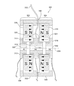
With reference to FIGS. 3A-3B, a representative ion guide or cell 300
comprises
magnetic lenses 302, 304 that are situated along an axis 306. The magnetic
lenses 302, 304
comprise magnets 308, 310, respectively, that are arranged with like poles
facing each other
to provide axial focusing, although in other configurations, different
arrangements can be
used. The pole pieces 312, 314, 316 are electrically separated from the
magnets 308, 310
by electric insulators 318-321 so that the pole pieces 312, 314, 316 can be
coupled to
different electrical potentials V1, V2, V3, (or V1_6, of Fig. 4), for example.
(Alternatively or
in additional to the insulators 318-321, the magnets 308, 310 may comprise a
non-
conductive material, such as a ceramic, for example.) Typically, these
voltages V1, V2, V3
(or V1_6) are static, but time-varying voltages V1, V2, V3 (or V1_6) can be
applied to retard,
accelerate, capture, or otherwise manipulate charged particles in an inner
cavity 342
defined by the inner bores of the magnets 302, 304 and the pole pieces 312,
314, 316. The
pole pieces 312, 314, 316 are generally formed of soft iron or other magnetic
material and
- 17 -
CA 02725544 2010-11-23
WO 2009/155082
PCT/US2009/045591
provide conductive apertures A1, A2, A3. As shown in FIGS. 3A-3B, the magnetic
lenses
302, 304 share the pole piece 314, but in other configurations, separate pole
pieces can be
provided.
In the configuration of FIGS. 3A-3B, the magnets 308, 310 are formed as rings
that
include a central bore 322 that is aligned with the axis 306. In one
configuration, the
magnets 308, 310 are axially polarized N425H-grade Nd-Fe-B ring-magnets
(SuperMagnetMan, Birmingham, AL USA) that are about 3.0" in diameter, 0.5"
thick, and
have a 0.375" bore. The magnets 308, 310 are arranged in an alternating-
polarity-structure
similar to that of an axial traveling wave tube (TWT). The magnets 302, 304
are fixed in
position with aluminum casing members 332, 334 that can be secured to each
other with
screws or other fasteners. An end plate 336 is provided for the magnet 302. As
shown in
FIG. 3B, an inlet 340 is provided for introduction of a gas to the inner
cavity 342 so that the
ion guide 300 can be configured for CID. Pole pieces can also be provided and
electrically
insulated from one or both of the magnets 308, 310.
With reference to FIG. 4, an ion guide 400 includes magnets 402-406 that are
situated along an axis 408. Soft iron rings 410-413 are situated between the
magnets, and
soft iron rings 414-415 are situated at ends of a housing 420 that retains the
magnets 402-
406. Electric insulator rings 432-441 are situated between the magnets 402-406
and the
soft iron rings 410-415 so that electrical potentials can be independently
established on one
or more or all of the soft iron rings 410-415. (Alternatively or in additional
to the insulator
rings 432-441, the magnets 402-406 may comprise a non-conductive material,
such as a
ceramic, for example.) In one configuration, the insulator rings 432-441 are
made of a
poly(tetrafluoroethene) or poly(tetrafluoroethylene) (PTFE) and have a
thickness of about
0.010". Each of the soft iron rings 410-415 can be connected to an
independently
adjustable, floating power supply that can supply a voltage in a range of 0 up
to 5000 V,
or other bipolar or unipolar voltage range. In some configurations, time
varying voltages
are provided. In the configuration of FIG. 4, a ring-shaped filament 443 of
tungsten-
rhenium wire is located concentrically on the axis 408 near a surface 450 at
which ions
enter the ion guide 400. In this configuration, the soft iron rings 410-415
serve both as pole
pieces for the magnets 402-406 and, depending on the applied voltages, as one
or more
- 18 -
CA 02725544 2010-11-23
WO 2009/155082
PCT/US2009/045591
electrostatic lenses 410-415. The apparatus of FIG. 4 can also be provided
with an aperture
such as hole 451 drilled radially into one or more of the soft iron rings for
introduction of a
neutral gas for CID through a pipe or tube 452. A hole through one or more of
the soft iron
rings 410-415 and the housing 420 can also be provided for introduction of an
optical beam
such as a laser beam for laser assisted dissociation.
In addition the present invention provides configurations of ion guides/cells
with
differing locations of the source of electrons. Such configurations can
increase the
population of low-energy electrons sufficiently to raise the reaction
efficiencies of ECD,
EDD, or any other electron capture process by one or more orders of magnitude
and,
thereby, enable users to conduct more comprehensive proteomics experiments.
In this regard, the present invention provides devices that locate the source
of
electrons, (FIG. 10), such as filament 843 within the cavity 806 of a radio-
frequency-free
(RFF) hybrid electrostatic/magnetostatic cell or trap 800 for purposes of
performing ECD,
EDD, or any other electron capture process. As with the cell 300 of FIG. 3B,
the cell 800
may include two permanent ring magnets 808, 810 and three soft iron pole
pieces 812, 814,
816 arranged in a similar manner to corresponding components of the cell 300.
For
purposes of illustration, the magnets 808, 810 and pole pieces 812, 814, 816
are shown as
being electrically isolated from one another by means of an air gap, however,
electrical
insulators, such as 0.010" thick poly(tetrafluoroethylene), may be used in
place of the air
gap and/or the magnets 808, 810 may comprise a non-conductive material, such
as a
ceramic, for example. The filament 843 may terminate in a circular loop
disposed within
the cavity 806 of the cell 800 proximate the central pole piece 814. In this
regard, a
ceramic insulator 809 may be provided on the central pole piece 814 to prevent
electrical
contact between the filament lead 844 and the pole piece 814.
A similar exemplary configuration, is also provided utilizing an internal
filament 893,
but using two (non-magnetic) conductive aperture plates 864, 865, which may be
comprise
titanium for instance, in place of the central pole piece 814, to provide
conductive
apertures, FIG. 14A-14B. Two permanent ring magnets 858, 860, soft iron pole
pieces
862, 866 may be arranged in a similar manner to corresponding components of
the cell 800.
- 19 -
CA 02725544 2010-11-23
WO 2009/155082
PCT/US2009/045591
Again, for purposes of illustration, the magnets 858, 860 and pole pieces 862,
866 are
shown as being electrically isolated from one another by means of an air gap,
however,
electrical insulators, such as 0.010" thick poly(tetrafluoroethylene), may be
used in place of
the air gap and/or the magnets 858, 860 may comprise a non-conductive
material, such as a
ceramic, for example. The filament 843 may terminate in a circular loop
disposed within
the cavity 806 of the cell 800 proximate the central pole piece 814. Computer
simulation of
trajectories of electrons emitted from a ring-filament 893 located inside cell
indicates that
essentially all of the electrons would be trapped in the magnetic bottle.
The term "source of electrons" can include any embodiment of an individual
electron-source, e.g. thermal source 845 (FIG. 11A) or photoelectric source
846 (FIG.
11B), or multiple electron-sources 847 (FIG. 11C), placed in any geometric
orientation or
arrangement, i.e. radial or axial, within one or more segments of the cavity
of the radio-
frequency-free hybrid electrostatic/magnetostatic cells, e.g., cells 300, 400,
of the present
invention. Locating intense sources of low-energy electrons in the cavity 806
of an
electrostatic/magnetostatic cell 800, (FIG. 10), will significantly increase
product-ion yields
from electron capture reactions to levels that are impossible to attain in RF-
based and
digital-based cells. This in turn will make it possible to obtain much more
information
from studies of the energetics and kinetics of electron capture reactions and
from tandem
mass spectrometric analyses of proteins and peptides.
In addition, in accordance with the present invention, the source of reagent
electrons
may be located at a position or positions along the side (as opposed to at an
end or at both
ends) of the hybrid electrostatic/magnetostatic cell to provide greater
flexibility in the
design and construction of an ECD/EDD cell and, further, to allow an electron
monochromator to be used as the source of electrons in order to increase the
selectivity of
ECD, EDD, or any other electron capture process. Precise control over electron
energy
used in an ECD experiment makes it possible to exercise a degree of
selectivity over how
some polypeptides fragment. In those cases where this applies, this phenomenon
can be
exploited to increase sensitivity. An electron monochromator is any device
that can select
nearly monoenergetic electrons from the population emitted by a hot metal
filament and
- 20 -
CA 02725544 2010-11-23
WO 2009/155082
PCT/US2009/045591
tune the energy of the selected electrons so that it matches the resonant
electron capture
energy of any negative ion of interest with an accuracy of better than 0.1 eV.
In this regard, the present invention provides devices that locate the
electron sources
943 alongside the hybrid electrostatic/magnetostatic cell or trap 900 (FIGS.
9A-9C). As
with the cell 300 of FIG. 3B, the cell 900 may include two permanent ring
magnets 908,
910 and two soft iron pole pieces 912, 914 disposed on opposing ends of the
cell 900 in a
similar manner to corresponding components of the cell 300. The pole pieces
912, 914 are
electrically isolated from the magnets 908, 910 electrical insulators 911,
913, such as
0.010" thick poly(tetrafluoroethylene). In this configuration, electrons may
be admitted
from the external source 943 into the hybrid electrostatic/magnetostatic cell
900 through a
radial port in the wall of the cell 900, FIGS. 9C-9C. The source(s) 943 may be
mounted
either outside or inside the periphery of the hybrid cell 900 along any radius
that passes
between two magnets 908, 910.
The electron source 943 can include any
nonmonochromatic, or monochromatic, embodiment of a thermal electron-source or
electron-sources, placed in any geometric orientation or arrangement about the
periphery
within one or more segments of the cavity of any of the configurations of the
hybrid
electrostatic/magnetostatic cell of the present invention.
Locating intense sources 943 of low-energy electrons on the periphery of an
electrostatic/magnetostatic cell 900 will provide greater flexibility in the
design and
construction of an ECD/EDD cell and, further, will allow an electron
monochromator to be
used as the source of electrons in order to increase the selectivity of ECD,
EDD, or any
other electron capture process. This capability, which is impossible to
implement in RF-
based and digital-based cells, will in turn make it possible to obtain much
more information
from studies of the energetics and kinetics of electron capture reactions as
well as from
tandem mass spectrometric analyses of proteins and peptides.
Referring to FIG. 5, a representative mass spectrometer 500 includes a first
quadrupole mass filter 502 situated to receive a charged particle beam to be
analyzed. The
first filter 502 is controlled so as to select some portion of the input
charged particle beam
that is then delivered to a hybrid ion guide 504 such as those illustrated in
FIGS. 3A-3B
and 4. In this configuration, the ion guide 504 is coupled to receive
electrons from a ring
- 21 -
CA 02725544 2010-11-23
WO 2009/155082
PCT/US2009/045591
filament electron source 508 as well as an ion beam after ion selection by the
first filter
502. Electrons from the ring-filament electron source 508 merge with the ion
beam in the
ion guide 504 producing a charged particle beam that is analyzed by a second
quadrupole
mass filter 506 or other mass analyzer.
Example 1
In one example in which a commercial quadrupole-mass-filter/octapole-CID-
cell/quadrupole-mass-filter (QqQ) mass spectrometer (Finnigan TSQ700: Thermo
Fisher
Scientific, Inc., Waltham, MA USA) was modified by replacing the RF octapole
CID cell
with the ion-guide apparatus 504 configured as the ECD/CID-cell 400 in FIG. 4,
ECD
spectra of doubly protonated gramicidin S (Sigma Chem. Co., St.Louis, MO USA),
doubly
protonated substance P, doubly protonated neurotensin, and triply protonated
neurotensin
(all three from American Peptide Co, Sunnyvale, CA USA), were obtained without
the use
of either RF fields or an energy-moderating gas. Sample solutions were
prepared by
dissolving standards of substance P, neurotensin, and gramicidin S in H20/Me0H
(50:50,
v/v) to a final concentration of 10-5 M.
The cell magnets 402-406 were the afore-mentioned N425H-grade Nd-Fe-B ring-
magnets (SuperMagnetMan, Birmingham, AL USA), the insulators 432-440 comprised
0.010" thick poly(tetrafluoroethylene), and the pole pieces 410-415 comprised
soft iron.
Each of the pole pieces 410-415 and the magnet's aluminum housing 420 were
connected
to an independently adjustable 100-V channel of a 7-channel power supply V1-
6, VH
(which could be floated up to 8 kV) so that the pole pieces 410-415 could
function as
electrostatic lenses as well as a pole pieces for the magnetostatic lenses 460-
464. A ring-
shaped, floating filament 443 of tungsten-rhenium wire of 0.07" (1.78 mm)
diameter,
located concentric with the cell's axis 408 at the ion-entrance, served as the
source of
electrons. Two titanium lenses disposed between the filament 443 and ion guide
cell 400
were used to guide electrons into the cell 400.
The peptide solutions were separately electrosprayed at a flow rate of 0.2
pL/min,
and doubly protonated substance P, doubly protonated gramicidin S, doubly
protonated
neurotensin, and triply protonated neurotensin were respectively selected as
precursors. By
adjusting the potentials V1,6 on the cell's electrostatic lenses 410-415,
settings were easily
- 22 -
CA 02725544 2010-11-23
WO 2009/155082
PCT/US2009/045591
found that allowed the electrons emitted from the ring-filament 443 to merge
in sufficient
numbers with the ion beam to produce ECD spectra of doubly protonated
Substance P that
appear in all respects (except, obviously, in resolution) the same as those
produced on FT
ICR instruments, FIGS. 6A. For this study, electron emission from the tungsten-
rhenium
filament 443 was set at 5 pA, the filament and EMS cell potentials at -120 V,
the potential
on the first Titanium lens Til at V1 = -115 V, the potential on the second
Titanium lens Ti2
at V2 = -20 V, and the potentials on all of the other lenses 410-415 at V1_6= -
80V.
The segmented design of the ECD cell 400 provides additional opportunities for
controlling electron-ion interactions and dissociation of precursor ions. For
instance, by
appropriately setting the potentials V1,6 on the electrostatic lenses 410-415,
the electron
capture events can be forced to take place in the early entry side segments of
the cell 400,
and decomposition of the radical precursor ions can be observed as a function
of time after
electron capture. To demonstrate this possibility, the total flight time of
[M+2H] +. radical
ions through the cell 400 was decreased (by changing the cell potential from -
80 V to
-300 V) from ¨25 ps to ¨12 ps to produce spectra within which the relative
strengths of the
fragment signals are markedly different (FIG. 6B). Since no changes in the
relative
intensities of the fragment ions were observed when the electron energy was
varied, it
would seem that the majority of the decrease of the intensities of the shorter
c-type ions is
most likely due to the decreased residence time of the radical ions, [M+2H]+
*, inside the
cell 400 before they enter the second analyzer. It is clear that the new cell
400 makes it
possible to investigate the mechanisms of ECD from previously unavailable
vantage points.
In addition, analytical quality ECD product-ion spectra of doubly protonated
gramicidin S (FIG. 7A), triply protonated neurotensin (FIG. 8A-left), and
doubly
protonated neurotensin (FIG. 8B) were readily produced in the RFF
electrostatic/
magnetostatic cell 400. These spectra were obtained without recourse to an
buffering gas,
as was necessary in previous efforts to perform ECD MS/MS in non-FT ICR
instruments,
or synchronizing electron injection with a specific phase of an RF field as
was necessary in
previous attempts to attain ECD in ion-traps. The cell 400 used in this study
was installed
in the Finnigan TSQ700 (which is a 20-year-old, low-resolution mass
spectrometer that is
well suited to testing prototypes but cannot produce mass spectra that yield
all of the
- 23 -
CA 02725544 2010-11-23
WO 2009/155082
PCT/US2009/045591
inherent information available); nevertheless, the mass spectra produced with
this modified
instrument incorporating the cell 400 of the present invention appear in all
respects (other
than the obvious exceptions of resolution and mass accuracy) to be at least as
good for
purposes of peptide identification as those produced by FT ICR instruments
(FIGS. 7B-7C,
8A-right). (FIG. 7B-7C reproduced with permission from Elsevier from Lin et
al., J. Am.
Soc. Mass Spectrom. 2006, 17, 1605-1615, copyright 2006, and FIG. 8A-right
reproduced
with permission from American Chemical Society from Hakansson et al, Anal.
Chem.
2001, 73, 3605-10, copyright 2001.) The effort and time to produce these mass
spectra,
however, were much less than required to produce their FT ICR counterparts.
Product-ion mass spectra of doubly protonated cyclic peptides are considerably
more complex than those of linear peptides. The initial ring-opening, which
statistically
can occur anywhere in the backbone of the peptide, creates a mixture of linear
peptides any
one of which can dissociate further to produce a secondary family of
fragments. The ECD
product-ion spectra of cyclic peptides are no exception to this tendency. An
ECD product-
ion spectrum of the repetitive cyclic peptide gramicidin S recorded during
this experiment
(FIG. 7A) is shown for purposes of comparison with mass spectra produced on an
FT ICR
instrument via ECD (FIG. 7B) and double-resonance ECD (FIG. 7C). Examination
of
these three mass spectra and other published mass spectra of gramicidin S
indicates that
ECD in the RFF electrostatic/magnetostatic cell 400 produces, with comparable
signal-to-
background, fragment-ions corresponding to the same losses of small molecules,
amino
acid residues, and side chains that are generally observed in ECD product-ion
spectra of
gramicidin S.
ECD of triply protonated neurotensin in the RFF electrostatic/magnetostatic
cell
400 produced a product-ion spectrum of both singly and doubly charged fragment
ions
(FIG. 8A - left) that is qualitatively identical to that produced in an FT ICR
cell (FIG. 8A -
right). Specifically, the RFF cell's spectrum exhibits the same six c-type and
seven Z-type
ions as well as the charge-reduced species [M+3H+e-]2 . observed in the FT ICR
spectrum
¨ only the bonds on the N-terminal side of the two prolines remained, as
expected, intact.
ECD in an FT ICR cell is generally not commensurate with the time scale of
liquid
chromatography. By contrast, ECD in the RFF electrostatic/magnetostatic cell
400 of the
- 24 -
CA 02725544 2010-11-23
WO 2009/155082
PCT/US2009/045591
present invention takes place in-flight through the device on a microsecond
time scale (the
time range for a singly protonated peptide of mass 1000 Da to travel through
the 70-mm
ECD cell 400). It should eventually be possible, therefore, to carry out ECD
in the RFF
cell in time with the elution of peptides off an HPLC column.
In order to perform ECD efficiently, the precursor ions must be forced to
mingle
with a dense population of low-energy electrons. Since the reagent electrons
and the
multiply protonated precursor ions have opposite polarities and masses that
differ by more
than six orders of magnitude, the conditions for simultaneously confining them
in the same
volume of space cannot be satisfied in a purely electrostatic cell, and can
only be minimally
satisfied in a cell in which an RF field is present. As the number of charged
particles of a
given polarity increases in an RF device, space-charge forces (i.e.,
repulsions between
particles of the same polarity) result in lost particles (2D RF ion-traps) or
degradation in
analyzer-performance (3D ion-traps and FT ICR cells). In principle, a
segmented-
electrostatic-focusing/strong-periodic-magnetostatic-focusing device, e.g.
cell 400, has a
substantially greater charged-particle capacity than any RF-based device.
Magnetic fluxes
on the order of 1 T are more than strong enough to confine high volume-
densities of ions
and electrons with kinetic energies typically involved in electron capture
reactions. This
capability should make it possible to perform experiments in the RFF
electrostatic/magnetostatic cell that would be at best difficult and at worst
impossible in an
FT ICR cell.
An example of this was demonstrated using neurotensin as the sample. A regular
mass spectrum of the electrosprayed neurotensin sample was recorded (FIG. 8D)
by
operating the modified Finnigan mass spectrometer strictly in the Q3-mode
(i.e., setting the
first analyzer Q1 in a transmission only mode and the second analyzer Q3 in a
scanning
mode). In addition to the peaks corresponding respectively to singly, doubly,
and triply
protonated neurotensin nominally at m/z 1673, 837, and 558, peaks
corresponding to a
number of other species appear in the spectrum. The latter are presumably due
to
impurities in the sample. When electrons are introduced into the dissociation
cell 400, all
of the impurity peaks disappear, and peaks distinctly corresponding to the ECD
product
ions of doubly and triply protonated neurotensin appear in their place (FIG.
8C). This
- 25 -
CA 02725544 2010-11-23
WO 2009/155082
PCT/US2009/045591
becomes unequivocally evident when the composite ECD spectrum (FIG. 8C) is
compared
with the individually produced ECD product-ion spectra of triply (FIG. 8A) and
doubly
(FIG. 8B) protonated neurotensin. Clearly, recombination with electrons was
sufficiently
high in the RFF electrostatic/magnetostatic cell 400 to neutralize all of the
impurity ions,
which presumably but not necessarily were singly charged, recorded in the
electrosprayed
spectrum (FIG. 8D) while efficiently producing fragment ions from the doubly
and triply
charged neurotensin ions.
In an RFF electrostatic/magnetostatic cell, such as cell 400, the reagent
electrons
cannot acquire kinetic energy from the magnetic field; however, their average
energy can
be controlled by the potentials Vi_6 applied to the electrostatic lenses 410-
415. By
abandoning RF-fields altogether in favor of segmented-electrostatic focusing
in conjunction
with strong-magnetostatic focusing, it should be possible to conduct ECD
experiments on
less costly instruments in which the average kinetic energies of the ions and
electrons can
be controlled with minimal loss of ions or electrons in the absence of an
energy-moderating
bath gas. This, in turn, could make it possible to increase the product-ion
yields and, thus,
the information to be gained from ECD reactions to levels that are much higher
than
possible in any RF-based cell. The strong magnetostatic focusing provided by
the cell's
traveling wave tube configuration together with the capability for moving and
trapping ions
provided by the cell's electrostatic segments 410-415 could enable regular
collision
induced dissociation over a much broader range of collision energies than
those typically
possible in ion trap or quadrupole instruments. Moreover, the cell's design
and compact
construction allow it to be incorporated into virtually any type of tandem
mass
spectrometer, e.g., triple quadrupole, hybrid quadrupole ion trap, hybrid
quadrupole time-
of-flight, or even FT-ICR.
The segmentation of the RFF electrostatic/magnetostatic cell 400 makes it
possible
to study the energetics and kinetics of ECD reactions as well as to exploit
them in MS/MS
analyses. For instance, decompositions of the radical precursor ions can be
observed as a
function of time by limiting electron capture events to the first entry-side
lens 460 of the
cell 400 and adjusting the potentials on the subsequent lenses 461-464 to
regulate the flight
times of the product ions. This was easily demonstrated by producing an ECD
product-ion
- 26 -
CA 02725544 2010-11-23
WO 2009/155082
PCT/US2009/045591
spectrum of doubly protonated substance P ion at the front end of cell 400 and
setting the
potentials of the rest cell's electrostatic elements 410-415 for ion
transport. This
experimental capability could be used, for example, to investigate mechanisms
like the
recently proposed sequential formation of diagnostic c-type ions.
Example 2
In Example 1, it was noticed that ECD was occurring in the lens segment 460
closest to the filament 443. As a result of this observation, the size of the
original cell 400
was reduced to two segments (i.e. two magnets) only, resulting in the cell 300
of FIGS. 3A-
3B. The initial set of experiments with the two-segment cell 300 showed that
it indeed had
the same ECD efficiency as the original five-segment one. The magnets 308, 310
of the ion
guide cell 300 were the afore-mentioned N425H-grade Nd-Fe-B ring-magnets
(SuperMagnetMan, Birmingham, AL USA), the insulators 318-321 comprised 0.010"
thick
poly(tetrafluoroethylene), and the pole pieces 312, 314, 316 comprised soft
iron. The
working embodiment included a gas line (pipe) 352 providing collision gas
(e.g., Argon)
for CID into the cell 300 through the iron pole piece 314 separating magnets
308, 310. For
ECD, electron emission from the tungsten-rhenium filament was set at 10 pA,
the filament
and EMS cell potentials at -120 V, the potential on the first Titanium lens
Til at V1 =
-115 V, the potential on the second Titanium lens Ti2 at V2 = -20 V, and the
potentials on
all of the other lenses 410-415 at Vi_6 = -80 V.
The two-segment cell 300 was tested in the CID mode by using Ar as the
collision
gas, setting the cell's potential so that the ion energy (laboratory frame of
reference) was
200 eV, and recording a CID product-ion spectrum of doubly protonated Glu-
fibrinopeptide, FIG. 12A. Prior to introduction of the gas, the vacuum inside
the
instrument analyzer manifold was 1.8 mTorr (1.8 x 10⁻⁵ mmHg). When collisonal
gas was
added, it became 2.1 mTorr (2.1 x 10⁻⁵ mmHg). Comparison of this spectrum with
a
published spectrum, FIG. 12B (Wang B. et al., "Isotopologue Distributions of
Peptide
Product Ions by Tandem Mass Spectrometry: Quantitation of Low Levels of
Deuterium
Incorporation", Anal Biochem. 2007, 367(1), 40-48. Reprinted with permission
from
Elsevier.) shows that both spectra exhibit the same series of y-type ions, but
that the
distributions of their respective peak intensities have distinctly different
envelopes.
- 27 -
CA 02725544 2010-11-23
WO 2009/155082
PCT/US2009/045591
After having demonstrated that the two-segment cell 300 could be operated in
both
ECD and CID modes independently, simultaneous ECD and CID was attempted in the
cell
300 on doubly protonated substance P. This was done by first recording a CID
product-ion
spectrum of the peptide, FIG. 13A, and subsequently turning on the electron
filament to
record its combined ECD/CID spectrum, FIG. 13B.
In the CID product-ion spectrum of substance P, FIG. 13B, a relatively
complete
series of b-type fragment ions accompanied by a less intense series of a-type
fragment ions
is observed, as is generally the case for a peptide that has an arginine on
its N-terminus. In
the combined CID/ECD production ion spectrum, the CID series of b-type and a-
type ions
is virtually unchanged; however, superimposed on this CID series of fragment-
ion peaks is
a series of the same six c-type ions (i.e., c4-cio) typically observed in an
ECD product-ion
spectrum of substance P. This result demonstrates the "golden complementary
pairs"
(actually, the presence of a-, b-, and c-type ion signals in a single product-
ion mass
spectrum constitutes triplets in this particular example) being recorded in a
single,
simultaneous (i.e., non-tandem), in-flight, ECD/CID experiment.
If the filament were left on to produce electrons for ECD and the gas valve
left open
to provide collision gas for CID, any combination of ECD, CID, or ECD/CID
experiments
could be interchangeably carried out in the cell hybrid
electrostatic/magnetostatic cell 300.
Reducing the filament's potential would stop the ECD process and, increasing
the cell's
potential (i.e., making it less negative) would stop the CID process. Since
voltages can
easily be switched in nanoseconds, changing from one dissociation mode to
another can
easily be done on a time-scale commensurate with the ions' flight times
through the mass
spectrometer (i.e. microseconds). Rapidly switching the ECD mode off and on
while
recording a product ion spectrum could, for example, be used to confirm the
presence of
golden complementary pair or triplets. Use of a fast, automated, alternating
dissociation
mode with the hybrid electrostatic/ magnetostatic cell of the present
invention also might,
when used in conjunction with Walsh-Hadamard transforms, be a means for
increasing
signal-to-noise ratio or for decreasing the duty cycle in time-of-flight
measurements of
product ions.
- 28 -
CA 02725544 2010-11-23
WO 2009/155082
PCT/US2009/045591
By this experiment, a-, b-, and c-type ion signals have been recorded in a
single
product-ion mass spectrum by simultaneously performing ECD and CID in a hybrid
electrostatic/magnetostatic cell 300 of the present invention. Use of this
technique in
MS/MS analyses of peptides could significantly increase the number of peptides
(and ergo
proteins) that can be accurately matched to sequence entries in genomic and
proteomic
data-bases and even sequenced de novo.
The ion guides disclosed herein can be adapted to accommodate one or many
dissociation processes. The interleaved, periodic focusing of a hybrid
electrostatic/
magnetostatic structure permits PID, low- and high-energy CID, ETD, ECD, EDD,
and
EIEIO either individually or in sequential combinations. Any one of several
possible
embodiments of the disclosed dissociation cells can be inserted between two
analyzers of
any transmission/transmission or transmission/trapping tandem mass
spectrometer.
Furthermore, if a mass spectrometer comprises two or more tandem units, any
one of
several possible embodiments of the disclosed device can be incorporated in
each unit.
As shown above, the disclosed hybrid electrostatic/magnetostatic ion guides
300,
400 are segmented so that precursors, reagent ions, and electrons can be
segregated,
trapped, and combined. For example, in an ECD or EDD experiment, limiting
electron
capture events to the first one or two entry-side segments of the cell, e.g.
at electrostatic
lenses 414, 410, and appropriately adjusting the potentials on the subsequent
lenses, e.g.,
lenses 412-415, would make it possible to observe decompositions of the
radical precursor
ions as a function of time. If the magnets in an electrostatic/magnetostatic
hybrid cell's
magnetic lens-elements were situated to provide radial focusing, ions would
propagate
through the cell along sinusoidal paths and the added path length created by
this extra
motion might be used to engineer more compact spectrometers, more selective
spectrometers, or both. Ion-mobility spectrometers incorporate funnel and
hourglass ion-
guides to collect and concentrate ions both before entering and after exiting
the ion-
mobility tube; engineering these auxiliary components as
electrostatic/magnetostatic
structures could result in higher transmission efficiencies and, thereby,
lower detection
limits.
- 29 -
CA 02725544 2010-11-23
WO 2009/155082
PCT/US2009/045591
In some configurations, hybrid cells 300, 400 as disclosed herein are
configured to
perform high energy CID. In such procedures, electrical potentials applied to
pole pieces
312-316, 410-415 or to conductive aperture plates are selected to accelerate
precursors to
kinetic energies greater than, for example, about 1 keV. In other
configurations, charged
particles associated with different arrival times can be trapped in respective
sections of a
hybrid cell 300, 400, and released for subsequent analysis based on time
varying potentials
applied to the pole pieces 312-316, 410-415.
In typical configurations, ion guides comprise at least one magnetic lens that
includes a magnet and a pair of ferromagnetic pole pieces. In configurations
that include
two or more such magnetic lenses, each magnetic lens can include two pole
pieces, but in
some configurations, magnetic lenses share a pole piece that is situated
between magnets of
adjacent lenses. In other configurations, separate pole pieces can be
provided.
With reference to FIG. 2C, a representative fragmentation cell 230 comprises a
ring
magnet 232. The cell is situated on an axis 238 and defines a fragmentation
volume 236
that extends along the axis 238. In other configurations, a non-conductive
magnet provided
with a conductive layer is used, and an electrical connector is provided to
apply a voltage to
the magnet that is different than an instrument ground or other instrument
voltage. In other
configurations, the magnet 232 is an electromagnet or a combination of an
electromagnet
and a permanent magnet. Insulator layers or insulating coatings can be
provided if desired
for a particular application. As shown in FIG. 2C, a filament 240 is situated
to produce
electrons for coupling into the fragmentation volume 236.
It will be apparent that the disclosed exemplary configurations are
representative
only, and the disclosure is not to be limited to the particular exemplary
configurations used
for illustration. For example, magnets can be segmented into one or more
pieces and/or can
be polarized in any technically possible manner to conveniently provide
suitable magnetic
field polarities, and electromagnets can be used instead of permanent magnets,
or
combinations of electromagnets and permanent magnets can be used. Magnets can
be
made from electrically non-conductive materials such as ceramics. Using such
magnets,
insulators configured to electrically isolate magnets from pole pieces or
conductive aperture
plates are unnecessary. In other examples, magnets can be covered or partially
covered by
- 30 -
CA 02725544 2010-11-23
WO 2009/155082
PCT/US2009/045591
one or more electrically insulating layers such as an epoxy layer. In some
embodiments, a
separate housing is provided to secure the magnets and pole pieces, but in
other examples,
some or all magnets and/or pole pieces can be secured with an adhesive, and a
housing can
be omitted. In the examples described above, two or more magnets and
associated pole
pieces are provided. In other examples, a single magnet and two associated
pole pieces
configured to be maintained at different electrical potentials can be
provided. In view of
these and other variations, we claim all that is encompassed by the appended
claims.
- 31 -