Note: Descriptions are shown in the official language in which they were submitted.
CA 02725555 2010-11-23
WO 2009/144723 PCT/IL2009/000530
METHOD AND APPARATUS FOR CO2 EVALUATION
FIELD OF THE INVENTION
The invention relates to evaluation of CO2 level in the blood of a patient.
Some
embodiments of the invention relate to deriving an evaluation of CO2 level
based on
non-invasive detection of one or more signals related to haemodynamic
parameters.
BACKGROUND OF THE INVENTION
The level of CO2 (Carbon Dioxide) in the blood of humans and other beings has
several significant biologic functions such as in respiratory rate and depth
control,
muscle contraction or dilatation of arterioles where, typically, higher
resistance is due to
vessels constriction and lower resistance is due to vessels dilation.
Clearly, the ability to measure and monitor CO2 levels are of significant
clinical
value. Indeed, different methods and devices have been developed for measuring
this
parameter. Known devices include laboratory tests measuring CO2 levels in a
blood
sample, devices testing CO2 levels directly from an arterial line catheter,
capnographs or
capnometers that measure CO2 levels in the exhaled air (generally being in
good
correlation with blood CO2 levels) or transcutaneous CO2 monitors which use
heated
electrodes attached to the skin, measuring the local carbon dioxide gas
tension of the
tissue. While these devices may provide valuable information, they are, in
general,
costly and require disposable elements and some of these devices, such as
intra-arterial
sensors, are invasive.
While CO2 monitoring is a major parameter for assessment of breathing, yet
under certain clinical circumstances, such as emergency conditions, CO2
monitoring
may be cumbersome. For example, a capnograph cannula attached to the patient's
nose
may dislodge and fail to provide reliable values.
Methods and apparatus for measurement of CO2 in patients are disclosed in
prior
publications, some of which are cited below as examples.
US patent 6,741,876 relates to measurement of blood constituents, including
CO2, by spectroscopy; US application 2007/0129645 relates to invasively
measuring
respiration waveform and deducing CO2 level from the respiratory, waveform
parameters; US patent 6,819,950 relates to non-invasive measurement of blood
absorption at two locations and deducing CO2 levels from a pH parameter; US
patent
7,405,055 relates to determination of a blood constituent, including C02,
using a single
1
CA 02725555 2010-11-23
WO 2009/144723 PCT/IL2009/000530
device by a particular formula; US application 2007/0027375 relates to non-
invasive
measurement of blood flow at two locations and deducing CO2 levels from an
average
of the measurements; US patent 5,766,127 relates to simultaneous spectroscopic
measurements at about the same location to deduce blood perfusion; US patent
7,341,560 relates to monitoring blood parameters by a plurality of light
sources and
detectors positioned on a single body part; US patent 6,942,622 relates to
monitoring
the effects of blood/ haemodynamic parameters including CO2 on autonomic tone;
US
patent 6,501,975 relates to correlating two blood signals from a single
location for
deriving blood gas concentration; US patent 6,826,419 relates to correlating
two blood
signals from a single location for deriving blood gas concentration; US
application
2004/0204638 relates to correlating two blood signals from a single location
for
deriving blood constituent concentration; US patent 7,351,203 relates to
covariate
monitoring at a single location, including monitoring C02; US application
2005/0076909 relates to covariate monitoring including CO2 but no derivation
of CO2;
US application 2004/0236240 relates to monitoring respiratory conditions based
on
blood parameters including CO2 but no derivation of C02; US patent 7,225,013
relates
to using CO2 signal for predicting change in a patient; US patent 7,195,013
relates to
modulating autonomous function using CO2 signal; and US patent 6,896,660
relates to
covariate monitoring, including CO2 as single parameter for estimation of
tissue
perfusion.
SUMMARY OF THE INVENTION
Generally, the invention relates to deriving an evaluation of CO2 level in the
blood of a patient by processing of one or more detected signals related to
one or more
haemodynamic parameters of the patient. Preferably the signals are detected
non-invasively.
For brevity and clarity, without limiting and unless otherwise specified, a
signal
or part thereof related to a haemodynamic parameter, or a signal or part
thereof of the
haemodynamic parameter, are denoted herein interchangeably as 'haemodynamic
signal'
or 'haemodynamic waveform'.
Accordingly, a general aspect of the invention relates to a method and
apparatus
for evaluating CO2 level of a patient by detecting at the patient's body at
least one
haemodynamic signal from an at least one tissue (such as an organ or part
thereof),
processing (employing) the at least one haemodynamic signal to derive a value
related
2
CA 02725555 2010-11-23
WO 2009/144723 PCT/IL2009/000530
to the CO2 level of the patient, and based on a relation of the derived value
to CO2
determining an evaluation of CO2 level of the patient, wherein in some
embodiments
the derived value constitutes the evaluation of CO2 level.
An aspect of the invention relates to a method and apparatus for detecting at
a
site of the patient's body a haemodynamic signal from a tissue, processing the
waveform
and deriving a value functionally related to the CO2 level of the patient. In
some
embodiments of the invention, the CO2 level of the patient is linearly
determined from
the derived value.
Another related aspect of the invention relates to a method and apparatus for
simultaneously detecting haemodynamic signals from a plurality or tissues,
processing
the signals and deriving a value functionally related to the CO2 level of the
patient based
on interrelation between the signals.
In some embodiments of the invention, one site of the patient is used for
detection in a plurality of underlying tissues. Optionally and alternatively,
a plurality of
sites is used for detection in underlying tissues.
In some embodiments of the invention, the interrelation between the signals is
due to the physiological differences in the response of vascular beds in
different body
organs or tissues. While variations of CO2 levels in most of the blood vessels
affect
changes of haemodynamic parameters in a certain direction, variations of
sympathetic
nervous system activity affect changes in opposite directions in different
organs (such
as muscle versus skin) and changes of a different magnitude in other organs
(such as
brain).
In some embodiments of the invention, evaluation of CO2 level based on the
simultaneous correlation between haemodynamic parameters may provide a better
performance in terms such as precision and/or repeatability and/or consistency
between
patients and/or reliance on calibration relative to an evaluation based on a
single
parameter, while the interrelation between the simultaneously detected signals
can be
used to assess the activity of the autonomic nervous system..
In some embodiments of the invention, the CO2 level is evaluated periodically,
optionally providing continuous monitoring of the CO2 level of a patient.
In some embodiments, the detectors are connected to or integrated with other
components providing a system (apparatus) for evaluation and/or monitoring of
CO2
levels of a patient and optionally for performing other activities such as
derivation and
3
CA 02725555 2010-11-23
WO 2009/144723 PCT/IL2009/000530
calculations of other parameters of the patient, archiving, trending,
correlation and
linkage with other systems.
In some embodiments of the invention, the system comprises or is linked with a
processor and comprises or is linked with a medium comprising or storing a
program
that implements an algorithm for processing the acquired signals and
performing the
computations to obtain a value of the CO2 level of the patient. Typically and
optionally,
the system comprises or is linked with a medium comprising or storing a
program that
controls the signal detection and/or operation interface or any designed
activity.
Any adequate new or customized or other equipment suitable for detecting and
acquiring haemodynamic signals may be used. Some detectors for acquiring
haemodynamic signals are known in the art, including standard (off-the-shelf)
devices
and including non-invasive devices. For example, non-invasive detectors such
as
transcranial Doppler ultrasound probes (TCD) for detecting flow in brain
vessels or IR/
visible light Photoplethysmography (PPG) probes or oximeters, wherein the
standard
equipments is, optionally, modified or adjusted.
In some embodiments, the detected signals are optionally used to obtain other
values in addition to and as complementary values to CO2 evaluation, whether
by
known methods and/or devices of the art or modifications thereof or by new
methods
and/or devices. For example, other haemodynamic measurements, heart rate,
blood
oxygen saturation (SpO2), respiratory depth, respiratory rate and variability,
blood
pressure and variations thereof, or heart rate and variability thereof. The
other values
may also be used for assessment of the patient condition and/or adjusting or
correction
of the CO2 evaluation.
In the specification and claims the following terms and derivatives and
inflections thereof imply the respective non-limiting characterizations below.
Patient - humans and other non-human mammals.
CO2 level in the blood (of a patient) - CO2 partial pressure in the blood or
an
approximation thereof sufficiently close to indicate a clinical state or a
physiological
state. For example, as a correlation with EtCO2 of a capnometer or with direct
measurement of blood samples such as by intra-arterial CO2 analyzer.
Haemodynamic (signal, parameter) - relating to blood flow in a blood vessel or
vessels of an organ or tissue or part thereof. For example, resistance to
blood flow or
mathematical indices correlated with resistance (e.g. pulsatility index (PI),
resistivity
4
CA 02725555 2010-11-23
WO 2009/144723 PCT/IL2009/000530
index (RI), S/D systolic to diastolic ratio (S/D), blood flow velocities), or
other
mathematical indices correlated with flow or resistance or derivation and/or
combination thereof.
Tissue - a tissue or part thereof of the patient's body or some organ or part
thereof.
Site (of a patient) - location in or on the body of the patient, such as a
patch or
region of skin or a portion of muscles.
Waveform/curve - representation of variations of a signal or data, or part
thereof
(not precluding intervals with constant signal or data).
io Signal - values representing some physical or physiological phenomenon,
typically in a digital form as a series of numerical values.
Acquisition/detection (of signal) - obtaining a signal via a detector (sensor)
in a
form suitable for processing, typically as a series of numerical readings
accessible to a
processor. For example, an analog signal from a sensor, subsequently converted
to
digital form (ADC).
Detector/sensor - a device or other equipment used to acquire biological
signal
or signals. Unless otherwise specified or evident from the context, the terms
'detector'
and 'sensor' may be used interchangeably and irrespective if a basic component
or a sub-
unit of a system is referred to.
According to the context and without limiting, an acquired signal or part
thereof
(e.g. for a certain time span) is denoted as 'signal'.
According to the context and unless otherwise specified, a cardiac cycle or a
signal of a cardiac cycle or a representation thereof is denoted as 'cycle'.
Unless particularly indicated, the terms 'resistance' and 'compliance' are
used
herein interchangeably denoting blood flow parameters.
According to an aspect of some embodiments of the present invention there is
provided a method for evaluating CO2 level of a patient, comprising:
(a) detecting on the patient's body at least one haemodynamic signal from at
least one tissue or part thereof;
(b) processing the at least one haemodynamic signal to derive a value related
to
the CO2 level of the patient; and
(c) determining an evaluation of CO2 level of the patient based on a relation
of
the derived value to CO2 level of the patient.
5
CA 02725555 2010-11-23
WO 2009/144723 PCT/IL2009/000530
In some embodiments, detecting is performed non-invasively.
In some embodiments, the at least one haemodynamic signal from at least one
tissue or part thereof constitute one signal from one tissue or part thereof.
In some embodiments, the at least one haemodynamic signal from at least one
tissue or part thereof constitute a plurality of signals from a plurality of
similar tissues
or parts thereof.
In some embodiments, the plurality of signals are detected substantially
simultaneously.
In some embodiments, the similar tissues are disjoint skin regions.
In some embodiments, the at least one haemodynamic signal from at least one
tissue or part thereof constitutes a plurality of signals from one tissue or
part thereof.
In some embodiments, the plurality of signals are detected substantially
simultaneously.
In some embodiments, the one tissue or part thereof is a skin region.
In some embodiments, the at least one haemodynamic signal from at least one
tissue or part thereof constitutes a plurality of signals from a plurality of
different
tissues or parts thereof.
In some embodiments, the plurality of signals are detected simultaneously.
In some embodiments, the plurality of different tissues comprises at least one
tissue selected from skin, muscle or brain.
In some embodiments, the plurality of different tissues comprises at least two
tissues selected from skin, muscle or brain.
In some embodiments, processing comprises identifying a region on the at least
one signal, or a derivation thereof, by which a value functionally related to
CO2 level of
the patient is derived.
In some embodiments, identifying a region comprises analyzing a temporal
derivative, or a combination thereof, of the at least one signal or a
derivation thereof.
In some embodiments, a value functionally related to CO2 level of the patient
is
derived by integrating the temporal derivate, or a combination thereof, about
the region.
In some embodiments, a value functionally related to CO2 level of the patient
is
linearly related to CO2 level of the patient.
In some embodiments, wherein processing comprises:
(a) defining a model of a haemodynamic parameter based on a plurality of
signals from a plurality of different tissue of part thereof; and
6
CA 02725555 2010-11-23
WO 2009/144723 PCT/IL2009/000530
(b) substituting in the model at least one separately acquired haemodynamic
parameter thereby deriving a value related to the CO2 level of the patient.
In some embodiments, a value related to the CO2 level of the patient
constitutes
the evaluation of CO2 level of the patient.
According to an aspect of some embodiments of the present invention there is
provided an apparatus for evaluating CO2 level of a patient, comprising:
(a) at least one detector at the patient's body for detecting at least one
haemodynamic signal from an at least one tissue or part thereof; and
(b) a processor and a program for deriving an evaluation of the CO2 level of
the
patient based on the at least one haemodynamic signal.
In some embodiments, the apparatus further comprises apparatus for providing
at least the evaluation of the CO2 level of the patient.
In some embodiments, the evaluation of the CO2 level is provided continuously
in real-time.
In some embodiments, the at least one detector is non-invasive to the patient.
In some embodiments, the apparatus is sufficiently small and lightweight for
wearing by the patient. In some embodiments the apparatus is sufficiently
mobile to be
worn by an ambulatory patient.
In some embodiments, the apparatus is configured to implement the methods
described above.
BRIEF DESCRIPTION OF THE DRAWINGS
Some non-limiting exemplary embodiments of the invention are illustrated in
the
following drawings.
Identical or duplicate or equivalent or similar structures, elements, or parts
that
appear in one or more drawings are generally labeled with the same reference
numeral,
optionally with an additional letter or letters to distinguish between similar
objects or
variants of objects, and may not be repeatedly labeled and/or described.
Dimensions of components and features shown in the figures are chosen for
convenience or clarity of presentation and are not necessarily shown to scale
or true
perspective. For convenience or clarity, some elements or structures are not
shown or
shown only partially and/or with different perspective or from different point
of views.
7
CA 02725555 2010-11-23
WO 2009/144723 PCT/IL2009/000530
Fig. 1 illustrates a chart of a waveform of variations of skin blood vessels
pulsatility.
Fig. 2 illustrates a flowchart schematically outlining actions for deriving
CO2
levels from a haemodynamic waveform, according to exemplary embodiments of the
invention;
Fig. 3 illustrates a flowchart outlining actions for deriving CO2 levels from
a
haemodynamic waveform, according to exemplary embodiments of the invention;
Fig. 4 illustrates aligned and superimposed normalized heart cycles derived
from
the waveform such as of Fig. 1, according to exemplary embodiments of the
invention;
Fig. 5 illustrates the aligned and superimposed first temporal derivatives of
normalized heart cycles of a waveform such as of Fig. 1, according to
exemplary
embodiments of the invention;
Fig. 6 illustrates a representative first temporal derivate of normalized
heart
cycles of a waveform such as of Fig. 1, according to exemplary embodiments of
the
invention;
Fig. 7 illustrates a chart of correlated waveforms of evaluated CO2 levels,
EtCO2
from a capnograph and respiration rate from a capnograph, according to
exemplary
embodiments of the invention;
Fig. 8 illustrates a chart of statistical correlation between evaluated CO2
levels
and EtCO2 from a capnograph, according to exemplary embodiments of the
invention;
Fig. 9 illustrates a chart of a Bland-Altman agreement analysis between
evaluated CO2 levels and EtCO2 from a capnograph, according to exemplary
embodiments of the invention;
Fig. 10 schematically illustrates a diagram describing how CO2 levels
correlate
with skin resistance and muscle resistance, according to exemplary embodiments
of the
invention;
Fig. 11 illustrates a flowchart schematically outlining actions for deriving
CO2
levels from a plurality of haemodynamic signals, according to exemplary
embodiments
of the invention;
Fig. 12 schematically illustrates a diagram of CO2 evaluation system,
according
to exemplary embodiments of the invention; and
Fig. 13 illustrates a flowchart outlining actions for user operation involved
in
evaluating CO2 level of a patient, according to exemplary embodiments of the
invention.
8
CA 02725555 2010-11-23
WO 2009/144723 PCT/IL2009/000530
DESCRIPTION OF EMBODIMENTS OF THE INVENTION
The following description relates to one or more non-limiting examples of
embodiments of the invention. The invention is not limited by the described
embodiments or drawings, and may be practiced in various manners or
configurations
or variations. The terminology used herein should not be understood as
limiting unless
otherwise specified.
The non-limiting section headings used herein are intended for convenience
only
and should not be construed as limiting the scope of the invention.
Single signal
Fig. 1 illustrates a chart 100 of a waveform 102 of variations of blood flow
phenomena acquired at a particular tissue (for example, skin) by a detector
(for
example, PPG), generally representing other haemodynamic signals of a patient.
The horizontal axis 112 denotes a time scale (in seconds) and the vertical
axis
114 denotes a scale of the pulsatile phenomena, such as voltage or current at
the
detector.
Waveform 102 follows (possibly with some delay) the heart cycle (beats) and is
modulated by the respiration as exemplified by an envelope of the extremum
points of
waveform 102 with upper part 104 (maximums) and lower part 106 (minimums).
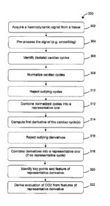
Fig. 2 illustrates a flowchart 200 schematically outlining actions for
deriving
CO2 levels from haemodynamic waveforms, such as 102, according to exemplary
embodiments of the invention.
A haemodynamic signal such as waveform 102 is acquired (202), for example
via a PPG probe on the skin. In some embodiments, a limited time span of the
signal is
stored in a memory for subsequent processing.
The acquired signal is analyzed to isolate separate cardiac cycles (204). A
plurality of cardiac cycles may be combined (e.g. by averaging), possibly
after
normalization to a common scale, to represent a typical cycle or cycles of the
signal.
The cardiac cycles, or combined cycles as a representative cycle, are
processed
(206) to obtain CO2 levels. In some embodiments of the invention,
characteristics of the
cardiac cycle shape are determined and processed to derive a value
functionally related
to the CO2 level, and the CO2 level is obtained by applying the appropriate
formula.
9
CA 02725555 2010-11-23
WO 2009/144723 PCT/IL2009/000530
Typically the function is a linear formula where, optionally, the coefficients
are preset
or predefined or obtained by a calibration procedure.
Until otherwise stated, the following discussions below refer also to Fig. 3
that
illustrates a flowchart 300 outlining actions for deriving CO2 levels from a
haemodynamic waveform, according to exemplary embodiments of the invention.
Signal acquisition
A signal is acquired (302) for a time span comprising a series of several
consecutive cardiac cycles, typically but not necessarily covering a
respiratory cycle
(typically of about 6 seconds). In some embodiments, the cardiac cycles are
distinguished, for example, by rough detection of peaks and/or valleys, or by
estimated
or measured heart rate or by other methods such as estimation based on a
previous
acquisition. In some embodiments, the acquisition time span is, about 6 or
more seconds
(e.g. 8 or 12 seconds).
In some embodiments of the invention, the signal, or part thereof, is
preprocessed (304) such as by smoothing (e.g. by a low pass filter) to remove
noise or
other high-frequencies (e.g. spikes) relative to what is expected. Optionally,
other signal
conditioning is used such as known in the art, for example, exponential
filter.
Cycle separation
The signal is analyzed to identify and separate the cycles (306), such as by
identifying maximum (peaks) and minimum (valleys) regions or points and/or
minimal
rise and/or descent rates and/or by using signal analysis algorithms of the
art.
The separated cycles, or sub-set of the cycles, are normalized (308) to a
common
scale such as by scaling them so that the peaks share a common value (e.g. 1)
and the
valleys share a common value (e.g. 0) and, optionally, all the cycles start at
a common
virtual time such as t=0. Optionally the cycles' widths are adjusted to share
a common
or approximate common width such as to compensate for varying heart rate.
For example, referring to waveform 102 of Fig. 1, the envelope of extremum
points (104 and 106) may be evaluated or approximated by a function or series
of
functions such as spline or splines and/or a polynomial formula or formulas
(e.g. of the
3`d degree or higher), optionally taking into account a full breathing cycle
(or cycles)
and effects thereof on the cardiac pulse signal. In some cases a sufficient
approximation
is a series of lines connecting the extremum points.
CA 02725555 2010-11-23
WO 2009/144723 PCT/IL2009/000530
For each cycle the respective lower envelope 106 is subtracted and the result
is
divided by the resultant maximal values, providing cycles in a 0-1 range.
Before or after the normalization, the cycles are analyzed to reject (ignore
or
discard) outliers (310) , such as cycles that do not fit the expected and/or
predefined or
determined (e.g. learned) constraints and/or the general shape of the majority
of the
cycles, such as artifacts or distorted shapes due to the patient condition or
movements.
In some embodiments, the rejection is based on median filter or properties of
the cycles
such as area or height or width or rate of change, or the rejection may be
based on other
methods of the art.
Having ignored the rejected cycles, in some embodiments of the invention the
cycles are used to obtain a representative cycle or cycles of the time span
(312). For
example, a typical cycle or resembling cycles are selected or a combination of
the
cycles is used as a representative cycle (see more below).
Fig. 4 illustrates aligned normalized heart cycles 402 derived from a waveform
such as waveform 102 of Fig. 1. At the vertical scale 414 the cycles' peaks
are set at a
level of 1, the bases at a level of 0 and the cycles are aligned and
superimposed on each
other and with respect to time scale 412 such that the maximum points of the
first
derivate vs. time (temporal derivative) or the peaks of the cycles are set at
t=0.
Optionally or alternatively, in some embodiments, the cycles' peaks or
derivatives
maximal points are aligned at a common arbitrary virtual time.
In some embodiments of the invention, the aligned cycles, having a common
scale and time (and optionally approximately common width) are added up and
divided
by the number of cycles to obtain a representative cycle (simple average).
Optionally or
additionally, a weighted average is performed where cycles that deviate from
the
majority of the cycles and/or from the simple average such as by area
difference are
given lower weight relative to cycles that deviate less, optionally
functionally related to
the difference. Optionally or alternatively, other methods are used to obtain
representative cycle or cycles such as by picking cycles that have the largest
correlations between the cycles.
In some embodiments of the invention, the assemblage of normalized cycles, or
alternatively one or more representative cycles are further processed.
For brevity and clarity, relating to the cycles in the discussions below
implies
either an assemblage of the normalized cycles or one or more representative
cycles
thereof, unless otherwise specified or evident from the context.
11
CA 02725555 2010-11-23
WO 2009/144723 PCT/IL2009/000530
Shape analysis
In some embodiments, the shapes of the cycles are further analyzed by taking
the first temporal derivate of the cycles ('the derivative') (314).
Fig. 5 illustrates the aligned and superimposed first temporal derivatives 502
of
normalized heart cycles of a waveform such as waveform 102 of Fig. 1. With
respect to
magnitude scale 514 the maximal points (peaks) of the derivates are aligned a
at virtual
time t=0 of time scale 512.
Typically, several zones are discerned in the derivative shape, as listed in
Table
1 below (and with respect to Fig. 5 that shows corresponding numerals):
Numeral Approximate typical
Zone
label time (ms)
1 First maximum point (global maximum) 0
2 First Minimum point 50
Second maximum
3 80
(alternatively as an inflection point)
4 Second minimum 125
5 Third maximum point 150
6 Third minimum point 220
Table 1
In some embodiments, before further analysis, the derivates are pre-processed
including, without limiting, the following steps:
-Rejection (ignoring or discarding) of outliers (316), such as derivative
signals
that do not fit the expected and/or the general shape of the majority of the
cycles. In
some embodiments, the rejection is based on median filter of properties of the
signals
such as area or height or width of the derivatives signals 502 that do not
conform to a
predefined or determined (e.g. learned from pervious or other measurement) set
of
constraints. Optionally, in some embodiments, the rejection is based on the
values
and/or separation in time of the points in derivates 502 as listed in Table 1,
such as first
maximal (global) maximum (1) or third minimum (6). For example, if the
separation is
more or less by 30% of the expected separation. Optionally or additionally,
the rejection
may be based on other methods of the art. In case of a single representative
cycle this
instant step is immaterial.
12
CA 02725555 2010-11-23
WO 2009/144723 PCT/IL2009/000530
- Smoothing the retained (non-rejected) derivates, such as by a low pass
filter to
remove noise such as due to derivative properties or to remove residual
effects of
breathing.
The shapes of derivatives 502, or selected typical derivatives shapes, are
s combined (e.g. average, weighted average, median selection) to form a
representative
derivative shape (318) (unless a single representative shape was previously
obtained and
the derivate of which was taken). In order to reduce sensitivity to variations
and
possible distortions in the signals, in some embodiments derivates 502 are
selected
within a significantly longer time span than a typical respiration cycle (e.g.
several
respirations cycles such as 30 or 60 seconds) or from several acquisitions.
Fig. 6 illustrates a representative first temporal derivate 602 of normalized
heart
cycles of a waveform such as waveform 102 of Fig. 1 (hereinafter, also
'ShapeD'). The
illustration is with respect to relative magnitude scale 614 and time axis
scale 612
(similar to time scale 512 of Fig. 5), wherein the maximal value ('l' in Fig.
5) is taken
as 100%. Fig. 6 also illustrates auxiliary lines and features (e.g. 'pl', 'w')
to further
clarify the discussion below and reference to Fig. 6 is accordingly implied.
Representative first temporal derivate ShapeD is further analyzed to obtain
key
points and features in ShapeD (320) as follows:
- Determine the points in ShapeD where the initial (temporal, time-wise)
ascent
and descent are at 50% of the peak (100%), namely, p 1 and p2, respectively.
Optionally
or alternatively, instead of using the 50% level, the inflection point level
of the rise or
fall, or combination thereof is used (such as by averaging or time-wise
distance between
the inflection points).
- Calculate the time-wise distance between points pl and p2 (hereinafter,
'wid'
equivalent to 'w' in Fig. 6).
- Determine the tangent 604 to the initial temporal descent at point p2.
- Determine the intersection of tangent 604 with the time axis 612 to obtain
intersection point p3.
- Compute the integral between ShapeD and time axis 612 between intersection
point p3 and p3+wid (timewise), shown as striped region 606 and 606a
(collectively
606). Since ShapeD is a representative first derivate of the normalized heart
cycles,
integral 606 is equivalent to the difference between the normalized cycle
between
corresponding point p3 and p3+wid (corresponding on the time axis 412 with
respect to
one or combined curves in Fig. 4).
13
CA 02725555 2010-11-23
WO 2009/144723 PCT/IL2009/000530
A possible rationale behind the above procedure is to calculate a normalized
value from a cycle, where this value represents the decay of the heart cycle
signal, from
the "expected maximum point" represented as point p3.
It was unexpectedly found that the value of integral 606 (hereinafter also
'AreaD') tracks, at least approximately, the CO2 level, (and may be regarded
also as
haemodynamic parameter or index)
C02 evaluation derivation
In some embodiments of the invention, CO2 level ('C02L'), at least with an
approximate relation to a capnograph, is derived from AreaD (322) as follows.
The functional expression for obtaining CO2L is expressed as:
CO2L = M x Areal) + N (1)
In some embodiments, a sufficiently (such as of clinical significance)
approximation is achieved by setting coefficient 'M' as M = 80. Optionally,
other values
are used, optionally or additionally, by determining or adjusting coefficient
'M'
according to previous measurements or other references such as blood samples.
In some embodiments, coefficient 'N' can be derived by calibration of CO2L
relative to a reference such as a capnograph or according to blood samples or
intra-
arterial CO2 analyzer. Optionally or alternatively, CO2L is calibrated
assuming a normal
physiology and/or condition of the patient which can be monitored and assessed
according to the signals (such as 402 of Fig. 4 or 502 of Fig. 5). Normal
physiology
and/or condition, which may also be obtained by using the same detection
apparatus or
an auxiliary detection apparatus, are, for example, normal breathing (e.g.
about 6
seconds per cycle), normal heart rate (e.g. about 60-70 bps) or normal Sp02,
or
combinations thereof. Assuming CO2L in normal conditions to be about 38mmHg,
coefficient 'N' is obtained from formula (1) by:
N = CO2L - M x Areal) (2)
In some embodiments of the invention, coefficient 'N' is adjusted or
determined
periodically or responsive to perceived (detected) change of the patient
condition, and
some previously determined values of CO2L may be used as in formula (2) above.
In some embodiments of the invention, one or more of the coefficients 'M' and
N' may be obtained by comparing and/or correlating the detected signal (such
as
waveform 102) to a typical or representative corresponding detected signal, or
by
comparing and/or correlating ShapeD to a typical or representative derivative
of CO2
14
CA 02725555 2010-11-23
WO 2009/144723 PCT/IL2009/000530
signal in a normal or typical patient. See also discussion on using templates
and limits
below.
In some embodiments of the invention, a better accuracy of and/or sensitivity
to
CO2 levels are achieved by non-linear formulas or other methods (e.g. fuzzy
logic) and
s the parameters of the formulas (e.g. polynomial or exponent) or settings of
the methods
are calibrated and adjusted similarly as described for formulas (1)-(2). The
non-linear
computation is, in some embodiments, beneficial relative to the linear
computations in
cases of seemingly non-realistic high and/or low CO2 levels that were derived
linearly
such as by formulas (1)-(2) above.
Experimental results example
Fig. 7 illustrates a chart, with vertical scale 714 of CO2 level in mmHg and
with
horizontal scale 712 in virtual time in seconds, of correlated waveforms of
evaluated
CO2 levels 702, EtCO2 from a capnograph 704 and respiration rate from a
capnograph
706, according to exemplary embodiments of the invention.
As can be seen in Fig. 7, evaluated CO2 level 702 approximately corresponds to
EtCO2 level 704, with maximal deviation of less than about 8mmHg.
Fig. 8 illustrates a chart, with vertical scale 814 of CO2 level valuation 814
in
mmHg and with horizontal scale 812 of capnograph EtCO2 in mmHg, of statistical
agreement between evaluated CO2 levels and EtCO2 from a capnograph, according
to
exemplary embodiments of the invention.
Fig. 9 illustrates a chart of a Bland-Altman correlation between evaluated CO2
levels and EtCO2 from a capnograph, according to exemplary embodiments of the
invention.
The average difference between linearly derivedCO2 as described above and
CO2 from a capnograph is 0.29 which is clinically sufficiently small positive
bias, and
the Standard deviation of the differences is 3.09. In interpreting Bland-
Altman plots, it
is expected that the majority of data points would fall between the lines
denoting 2StD
above and below the zero line as Fig. 9 indeed illustrates.
Unless otherwise stated, no further reference to Fig. 3 is implied.
Enhancements
In some embodiments of the invention, the derived CO2L is correlated with
other
measurements, such as PPG at muscle sensor, respiration rate, respiration
depth, heart
rate variability or heart rate to validate and/or adjust the CO2L derivation.
CA 02725555 2010-11-23
WO 2009/144723 PCT/IL2009/000530
In some embodiments of the invention, the method described above for
obtaining CO2L level based on AreaD, or a similar method to that effect, can
be
simultaneously applied to another similar tissue or tissues (e.g. other skin
regions/patches) to obtain additional simultaneous CO2L values. Subsequently
the
plurality of Areal) values and/or CO2L values may be manipulated (e.g.
combined,
averaged) to obtain CO2 evaluation of the patient with higher fidelity
relative to a single
tissue. See also discussion below with respect to a plurality of tissue. In
some
embodiments, different sensors are applied simultaneously to the same tissue
(e.g.
particular skin patch or region such as a finger tip) and the signals and/or
derived values
are manipulated or combined such as by correlation or averaging or by other
methods
such as weighted average to obtain CO2 evaluation with higher fidelity
relative to a
single sensor.
It should be noted that using Areal) is an example of obtaining a quantity
related
to CO2 level based on analysis of the signal or derivative or other derivation
thereof,
and other methods may be used to obtain quantities related to CO2 levels,
possibly
correlated with physiological activities.
Plurality of signals
In some embodiments of the invention, in order to improve the accuracy of the
evaluation of C02, notably under some particular physiological or clinical
conditions,
a plurality of tissues are detected simultaneously for a plurality of signals
related to
haemodynamic parameters and the interrelations between the signals (or
derivations
thereof) is used to derive an evaluation of CO2 level in a patient.
The interrelations between the signals is based on the physiological
differences
in reactions of vascular beds in different body organs to CO2 levels vs.
reactions to
other effectors, such as autonomic nervous system activity. While changes in
CO2 levels
cause changes in same direction in most body blood vessels, changes of
sympathetic
nervous system activity cause changes in opposite directions and different
magnitudes
in different organs (such as muscle versus skin) and changes of a different
magnitude in
other organs (such as the brain).
Possible mechanisms
A possible explanation to the different haemodynamic behavior of different
tissues is that the diameters of arteries change in response to some of the
following
stimuli:
16
CA 02725555 2010-11-23
WO 2009/144723 PCT/IL2009/000530
Neural - Activity of the autonomic nervous system (Sympathetic and
Parasympathetic divisions) that respond to a number of external and internal
changes,
epinephrine for example.
Chemical - response to changes in blood levels of several chemicals, including
CO2 in particular and others such as lactic acid, angiotensin, oxygen and NO.
Some stimuli are systemic (autonomic activation, blood CO2 levels, blood
pressure changes or endocrine control) while others may be local such as local
release
of endothelial factors due to various events possibly including exercise, with
possible
further downstream effects, or local neurogenic reflexes and para-endocrine
control.
Generally, the hemodynamic changes are not specific to the type of stimulus,
and they sum-up to constriction/dilatation of the blood vessel thereby
raising/lowering
resistance to blood flow, changing blood pressure, and/or
decreasing/increasing blood
flow. A complex interaction may occur between the stimuli. For example, while
CO2
levels rise, the blood vessel dilates yet rising CO2 levels beyond a certain
threshold may
also act on the vasomotor center in the brainstem to activate the sympathetic
system,
which in turn will counteract the vasodilation and constrict the vessel (such
as in the
skin) or may further dilate it (such as in a muscle). Sympathetic activity
also acts on the
heart to increase heart rate, stroke volume and cardiac output, and the
increased blood
flow may affect blood flow waveforms in arteries.
Based on recognition of the different response to stimuli (e.g., autonomic
system
and CO2 levels) as described above, in some embodiments of the invention, the
simultaneous changes in different vessels is processed and, based on
mathematical
equations, the level of blood CO2 is evaluated.
For simplicity and clarity, the descriptions below provide examples in linear
terms which are valid for certain inter-relationships or conditions. Yet, it
should be
understood that for complex interactions such as described above, the overall
behavior
should be described in more elaborate terms such as non-linear formulas.
Some embodiments of the invention are based on the understanding that during
most cases of clinical patient monitoring, the patient has to remain
quiescent.
Consequently, it is expected that the major impact on blood flow are due to
CO2 and
autonomic function while other factors are estimated to be either of
negligible impact or
affect the vascular system in the same direction and magnitude, such that the
signals and
derived evaluation of CO2 are not detrimentally affected. For example, while a
CO2 rise
brings about vasodilatation in most of the human body arteries (except for
pulmonary
17
CA 02725555 2010-11-23
WO 2009/144723 PCT/IL2009/000530
arteries at certain situations), activation due to stimuli of the sympathetic
system will
produce vasodilation in muscle arteries, and at the same time constriction of
blood
vessels to the skin, kidneys and other organs while having a minimal influence
on brain
blood vessels. The following Table 2 summarizes a simplified representation of
changes described above:
Para-Sympathetic Sympathetic C02Increase
activation activation
Skeletal muscle Constrict Dilate Dilate
Skin Dilate Constrict Dilate
Brain Minor effect Minor effect Dilate
Table 2
It should be noted that Table 2 merely shows a simplified representation of
the
physiological effects. For example, when CO2 levels go above or below a known
threshold level, reflex sympathetic activity may occur. However, this
sympathetic
activity might have effects in the same direction noted in the table while the
change in
CO2 levels may maintain effects attributed to CO2. Therefore, for blood
vessels in some
organs the sympathetic reflex may diminish the effects of C02, while in others
the same
reflex may enhance the CO2 effect.
It should also be noted that some of the changes outlined above are immediate
and are subsequently compensated by tissue auto-regulation mechanisms. The
compensation mechanism implies that initial flow changes are compensated
quickly and
flow may return to normal within a very short time after a change in
sympathetic
activation. The compensatory change, however, involves a change in the overall
resistance and compliance of the local vasculature, a change that is
manifested in the
haemodynamic indices, as measured and calculated by the methods described
herein.
The quick variations noted above are with respect to duration of one or few
heart beats
or a respiration cycle.
Exemplary arbitrary units
For simplicity and clarity, the impacts on the autonomic system will
hereinafter
be referred to as the combined sum of activities thereof (sympathetic and
parasympathetic). A maximal arterial dilatation (loss of smooth muscle tone)
will
receive the value of -10, while maximal constriction will receive the value of
+10. Each
division of the autonomic system will receive a number from 0 to 10 to
represent the
activity of the respective division. The Table 3 below represents the arterial
smooth
muscle tone, on a scale from -10 to +10, as a result of different combinations
of
18
CA 02725555 2010-11-23
WO 2009/144723 PCT/IL2009/000530
sympathetic and parasympathetic activations in a theoretical physiology where
CO2
effect is non-existent and wherein Arterial Tone is equal to Autonomic Tone.
Sympathetic Tone Parasympathetic tone Arterial Autonomic' Tone
0 10
10 5 5
10 10 0
5 0 5
5 5 0
5 10 -5
0 0 0
0 5 -5
0 10 -10
Table 3
Having a scale for autonomic activity on blood vessel diameter/resistance
5 arbitrarily defined between +10 (complete dilatation in skeletal muscle
arteries) and -10
(complete constriction in skeletal muscle arteries), similarly the effect of
CO2 on blood
vessels is herein defined using a similar scale, from +10 (complete dilatation
effect
when CO2 levels are maximal) to -10 (complete constriction effect when CO2
levels are
minimal).
10 C02 derivation overview
Fig. 11 illustrates a flowchart 1100 schematically outlining actions for
deriving
CO2 levels from a plurality of haemodynamic signals, according to exemplary
embodiments of the invention.
Haemodynamic signals from a plurality of tissues, such as skin, muscle or
brain,
are acquired (1102).
Haemodynamic parameters of the tissues, such as PI, RI, V or S/D are derived
from the signals (1104). A haemodynamic parameter can also be derived as
described,
for example, for Areal) above, or other haemodynamic parameters may likewise
be
derived. For different tissues the same or different haemodynamic parameters
can be
used, as well as combinations of different parameters.
Resistances of the tissues are derived ' from the haemodynamic parameters
according to methods such as known in the art (1106).
The derived resistances of the tissues are substituted in the equations of
factors
related to the tissues that affect the resistances (interaction model),
including CO2 factor
and autonomous system factor (1108).
19
CA 02725555 2010-11-23
WO 2009/144723 PCT/IL2009/000530
Exemplary model
An exemplary, simplified for clarity, non limiting mathematical model that
portrays how both factors, namely, autonomic and CO2 level, interact on the
blood
vessel and affect the total resistance of the vessels to blood flow is
formulated below
(formulas (3)-)(5)). It should be noted that other, possibly more elaborate,
models, may
be used.
RES(muscle) = F (A(mcl)xC02 + B(mcl)xAut + C(mcl)xOth + D(mcl)) (3)
RES(skin) = F (A(skin)xC02 + B(skin)xAut + C(skin)xOth + D(skin)) (4)
RES(brain) = F (A(bm)xC02 + B(brn)xAuT + C(srn)xOth + D(brn)) (5)
Where:
F is a function of the arguments;
RES(organ) is the total combined resistance/compliance of blood vessels in the
respective organ;
A(organ) is a coefficient describing the relationship between CO2 level
(denoted
in the model as 'CO2') and the effect thereof on the respective organ;
B(organ) is a coefficient describing the relationship between Autonomic
activity
level ('Aut') and the effect thereof on the respective organ;
C(organ) is a coefficient describing the relationship between levels of other
additional factors or stimuli ('0th') in addition to CO2 and Autonomic
activity, and the
effect thereof on the respective organ. C(organ) may be replaced by particular
coefficients related to specific factors.
D(organ) is a constant factor related to intrinsic features of the blood
vessels in
the respective organ without external effect.
For brevity and clarity, 'muscle' is abbreviated to 'mcl' and 'brain' to
'brn'.
At least for an approximation, the function 'F' is considered to be a unity,
namely, formulas (3)-(5) are linear formulas.
The equations and coefficients may be defined differently at different ranges
of
physiological parameters. For example, A(organ) may have a value A, in a range
of
0-30mmHg C02, a value A2 in a range of 30-45mmHg and a value A3 above 45mmHg,
yet within a specified range, a set of constant coefficients applies.
A likely underlying assumption in some embodiments of the invention is that
besides autonomic function and CO2 levels, the effects of other factors are
maintained
constant, at least approximately, under monitoring conditions. As patients
usually
remain at rest or are required to do so, and as many of the other factors
change due to
CA 02725555 2010-11-23
WO 2009/144723 PCT/IL2009/000530
physical activity or to local circulatory conditions, the assumption is likely
to be valid
under most clinical conditions. It is also assumed that other effects (in
addition to CO2
and autonomic activation) either change in the same magnitude and direction,
or are of
negligible magnitude, so the effects are cancelled in formulas (3)-(5). The
existence of
other factors in more complex situations does not rule out the use of this
method. For
example, if monitoring is performed during exercise, the equations will
include factors
such as C1 (local effects of exercise on the organ), C2 (systemic effects of
exercise), etc.
Solution of equations can be achieved by applying more detectors to a variety
of sites.
Table 4 below exemplifies hypothetical values for the coefficients used in the
model of formulas (3)-(5) above. Optionally or alternatively, other values,
scales or
coefficients may be used.
Organ A B
(muscle) -1 -1
(skin) -1 +1
(brain) -1 +0.01 (-0, negligible)
Table 4
Table 4 exemplifies the different effects of different types of organs,
namely,
while the 'A' coefficients (CO2 factor) for the three listed organs are of the
same
direction and magnitude (-1), the 'B' coefficients (Autonomous system) is the
same for
muscle and opposite for skin, and negligible for the brain.
A plausible interpretation is that a negative coefficient signifies the fact
that
resistance is inversely proportional to dilatation, where factors which
produce dilatation
(high CO2, sympathetic activity on muscle) increase vessels diameter, thereby
increasing flow and decreasing resistance, and vice versa, factors which
produce
constriction of blood vessels (low CO2, sympathetic activity on other organs)
decrease
vessels diameter thereby reducing flow and increasing resistance.
Resistance of blood vessels is related to other haemodynamic parameters that
can be measured and evaluated by equipment and methods of the art. For
example, PI
(Pulsatility Index), RI (Resistivity Index), S/D (Systolic over Diastolic
Ratio), or V
(blood flow velocities) such as maximal, minimal, mean, and combinations
thereof, or
other values such as Areal) described above.
Generally, the resistance can be schematically expressed as:
Resistance = g (PI, RI, V, AreaD...) (6)
Where 'g' is a function of the haemodynamic parameter or parameters.
21
CA 02725555 2010-11-23
WO 2009/144723 PCT/IL2009/000530
For example:
RES(organ) = k(organ) x RI (7)
Where the notation is of the model of formulas (3)-(5) above.
Accordingly, by simultaneously measuring (acquiring) on several sites
(tissues)
hemodynamic parameters (same parameters or different parameter or combinations
thereof) the relative resistance can be calculated such as by formula (7)
where the
coefficient is obtained by calibration or correlation with two or more organs
or tissues.
Having independent values for resistance in organs (e.g. muscle, skin, brain),
substituting the independent value into the formulas (3)-(5) above form
equations that
can be solved and the respective contributions of CO2 and Autonomic activity
factors
can be calculated, thereby deriving an evaluation of CO2 levels.
Substituting in the formulas (3)-(5) above the independently obtained RES
values and the coefficients from Table 4, one obtains:
RES(muscle) = (-1)xCO2 + (-1)xAut + C(muscle)xOth + D(muscle) (8)
RES(skin) = (-1)xCO2 + (+l)xAut + C(skin)xOth + D(skin) (9)
RES(brain) = (-1) xCO2 + OxAut + C(brain)xOth + D(brain) (10)
Table 5 below presents a hypothetical analysis of how different conditions,
such
as listed in Table 3 above, affect the mathematical model of formulas (3)-(5)
and
respective substituted equations (8)-(9), assuming that the effects of other
factors (in
addition to CO2 and Autonomous system) substantially cancel each other as
discussed
above so that coefficients 'C' and 'D' do not participate in equations (8)-
(9).
RES
CO2 AUT Muscle Skin Brain
-10 Max +10 (10)+(-10)= 0 (l0)+(10)=20 (10)+(0)=(10)
max constriction Avg 0 (10)+(0)=(10) (l0)+(0)=(10) (10)+(0)=10
low CO2 (-20mmHg) Min (10)+(-1 *-10) (10)+(- 10)=0 (10)+(0)=10
(-10) =20
0 Max +10 (0)+(-10)=(-10) 0+10=10 0+0=0
mid diameter Avg 0 0+0=0 0+0=0 0+0=0
average CO2 Min 0+10=10 0+(- 1 0)=(- 10) 0+0=0
(--40mmHg) (-10)
+10 Max +10 (-10)+(-10)=(-20) (-10)+10=0 (-10)+0=(-10)
max dilation Avg 0 (-10)+0=(- 10) (-10)+0=(- 10) (-10)+0=(- 10)
high CO2 (-60mmHg) Min (-10)+10=0 (-10)+ (-10)+0=(-10)
(-10) (-10)=(-20)
Table 5
22
CA 02725555 2010-11-23
WO 2009/144723 PCT/IL2009/000530
As based on values in Table 3, Table 5 provides arbitrary sample values for
the
range of resistance values in different organs. In muscle and skin, the
resistance varies
between (-20) for lowest resistance (complete dilation) and (+20) for highest
resistance
(maximal constriction). In the brain, the resistance varies between (-10) for
lowest
resistance (complete dilation) and (+10) for highest resistance (maximal
constriction).
Based on the arbitrary exemplary conditions and results listed in Table 5
above,
CO2 levels can be deduced from RES values using equations (8)-(10), as
exemplified in
Table 6 below that show muscle and skin resistance parameters and the
corresponding
CO2 levels and autonomic activity levels.
In Table 6 only muscle and skin values are exemplified, though it should be
noted that using brain values and/or other values may facilitate greater
precision than
using muscle and skin only.
Skin Muscle CO2 level CO2 level AUT activity
-20 0 High 10 -10
-10 -10 High 10 0
-10 10 Normal 0 -10
0 -20 High 10 10
0 0 Normal 0 0
0 20 Low -10 -10
10 -10 Normal 0 10
10 10 Low -10 0
0 Low -10 10
Table 6
15 As can be realized from Table 6, distinctive combinations of skin and
muscle
resistance parameters correlate with distinctive CO2 and Autonomic activity
levels,
allowing the calculation of CO2 levels.
Based on Table 6, Fig. 10 schematically illustrates a diagram describing how
CO2 levels correlate with skin resistance and muscle resistance, according to
exemplary
20 embodiments of the invention, where the vertical axis scale 1014 represents
the muscle
resistance and horizontal axis scales 1012 represents the skin resistance, and
where both
scales are in a range between (-20) and (+20) in the arbitrary exemplary
values
discussed above. Line 1002 depicts high level of CO2 (60mmHg), line 1004
depicts
23
CA 02725555 2010-11-23
WO 2009/144723 PCT/IL2009/000530
medium (normal) level of CO2 (40mmHg) and line 1006 depicts low level of CO2
(20mmHg).
As can be realized from Fig. 10, when skin vascular resistance is in the
middle
range (0), muscle vascular resistance is inversely proportional to CO2 which
can be
directly calculated therefrom. A lowest skin vascular resistance (complete
dilatation,
(-20)) results from high CO2 levels with unbalanced autonomic activity, that
is,
maximal parasympathetic and no sympathetic activity. A maximal skin vascular
resistance (maximal constriction, (+20)) results from low CO2 with unbalanced
autonomic activity, that is, maximal sympathetic and no parasympathetic
activity.
When the skin vasculature is partly constricted (relative to the middle range
of
(+10)), a partly constricted muscle vasculature (+10) results from low CO2
with
unbalanced autonomic activity, that is, maximal sympathetic and no
parasympathetic
activity. A partly dilated muscle vasculature (-10) results from normal CO2
with
balanced autonomic activity. A partly constricted muscle vasculature (+10)
results from
normal CO2, and a partly dilated muscle vasculature (-10) results from high
CO2. Other
CO2 levels and/or resistance levels, based on other data may be used.
Using three organs such as muscle, skin and brain as employed in formulas (3)-
(5) are used as examples, and a sub-set or larger set of organs or other
organs may be
used, possibly using a plurality of organs for high fidelity of CO2 evaluation
(e.g. with
respect to other methods such a blood sampling) or possibly trading simplicity
or
convenience (e.g. in emergency) with the fidelity of CO2 evaluations,
Special cases
In some cases the effect of the CO2 factor is much larger than that of the
autonomous system, as well as larger than the other factors, namely:
A(organ) >> B(organ) (11)
A(organ) >> C(organ) (12)
Consequently, formulas (3)-(5) may be represented by one formula of an organ,
e.g. skin:
RES(skin) = A(skin)xCO2 + D (14)
Substituting an independent resistance measure equation, such as (7) provides
an
evaluation of CO2 level as:
A(skin) = k(skin)* RI (15)
24
CA 02725555 2010-11-23
WO 2009/144723 PCT/IL2009/000530
Where 'RI' is a resistivity index (or another haemodynamic measure) and the
proportionality factor 'k' can be calibrated or otherwise determined.
Therefore, in certain cases the multi-signal method can be reduced and
simplified to a single signal method.
Detectors
Standard or specialized sensors may be used for acquiring haemodynamic or
related signals from a patient. Following are some viable examples.
1MHz or 2MHz PW TCD probes for detecting flow in brain vessels, through
skull.
2MHz or 4MHz PW probes for detecting flow in Internal Carotid Artery.
4MHz or 8MHz PW/CW probes for detecting flow in peripheral arteries,
including arteries supplying skeletal muscle.
Photoplethysmography (PPG) probes using IR or NIR (Near Infra-Red) or
visible light for detecting flow in skin vasculature (560nM - green, or 660nM -
Red)
and/or muscle vasculature (880nM - IR).
NIR devices that measure changes (for oxygen saturation) in both skin and
brain.
Bioimpedance electrodes for detecting fluid changes that usually reflect blood
flow changes in the short term in a variety of organs that may be adapted for
skin,
muscle and brain.
Laser Doppler probes usually used for evaluation of skin blood flow, also when
placed directly on a tissue such as muscle or brain.
Pulse Oximetry sensors (a specific type of PPG) or oxygen saturation (SPO2)
sensors that can provide complementary information for calculation accuracy in
extreme
values of the C02/02 range. The raw plethysmographic waveforms generated by
these
devices, before calculation of Sp02, can also be used for the general
estimation of CO2
by using the methods as described above.
Pulse oximetry sensors, and/or bioimpedance sensors, specifically adapted for
non-invasively measuring blood flow signals of brain tissue.
Tonometric sensors, used for deriving blood pressure changes when placed non-
invasively on the skin over representative arteries (or possibly by invasive
methods).
ECG, though not a haemodynamic signal per se, can still give information on
heart rate which can be used as part of the equations for autonomic activity
level.
CA 02725555 2010-11-23
WO 2009/144723 PCT/IL2009/000530
Other adequate new or customized detectors or other equipment suitable for
detecting and acquiring haemodynamic signals or related signals can be used,
optionally
with some modifications or adjustments, preferably as non-invasive sensors.
System (apparatus)
In some embodiments of the invention the detector or detectors are connected
to
or integrated with electronic and/or electrical and/or mechanical components
and/or
other components (e.g. chemicals such that change color due to heat),
providing a
system for evaluation and/or monitoring of CO2 levels of a patient by
implementing one
or more of the methods such as described above or variation and/or part
thereof.
In some embodiments of the invention, the system performs additional
activities
such as derivation and calculations of other parameters of the patient (e.g.
heart rate,
respiration rate), archiving, trending, correlations with past measurements of
the patient
or other patients, or linkage with other systems.
In some embodiments of the invention the system comprises or is linked with
one or more processors. In some embodiments, the system comprises or is
integrated
with or linked with a medium comprising or storing a program or programs,
optionally
with auxiliary data, that implements one or more algorithms and/or procedures
and
optionally with a medium for storing data. The tasks performed by the system
with the
processor and program comprise acquiring and processing the acquired signals,
performing the computations to obtain a value of the CO2 level of the patient,
and
optionally other tasks such as calibration or control and supervision of
components of
the system (e.g. of a sensor), or interaction with the user (operator) or
obtaining some
other parameters of the patient.
Typically, in some embodiments, the system operates continuously and monitors
CO2 level in real-time (at least relative to the approximate respiration rate
of the
patient).
In some embodiments of the invention, the system comprises built-in (or
remote)
display and/or a printer to provide readout of CO2 level or other parameters
and
optionally of waveform of the acquired or conditioned signals (e.g. for system
checking). Optionally or additionally, the system comprises other apparatus to
provide
the evaluation of CO2 level or other values, such as a voice-generation
apparatus as a
readout medium. Optionally or additionally, the system comprises user
interface
comprising elements such as buttons or sliders and/or indicators (e.g. LEDs)
and/or
26
CA 02725555 2010-11-23
WO 2009/144723 PCT/IL2009/000530
graphical interface. The user interface is used for tasks such as calibration,
control (e.g.
on/off), or setting operation modes. Optionally, the system comprises buzzer
or other
alarm equipment (e.g. vibrations) to notify about physiological conditions
and/or system
malfunction or bad contact or connection of the sensor to the patient.
In some embodiments of the invention, the system comprises components (e.g.
readout with limits or zones indications or alarm buzzer) such as to provide
feedback to
the patient, optionally assisting the patient to regulate the respiration
and/or CO2 level.
In some embodiments of the invention, the system comprises components (to
provide linkage or feedback to another device, such as an artificial
ventilator, optionally
assisting the second device to regulate the respiration and/or CO2 level. In
some
embodiments, the linkage is by a communication link (e.g. cable or wireless)
or the
linkage can be a visual and/or audible indication that alerts personnel to
activate the
second device.
In some embodiments of the invention, the system is a portable system,
optionally sufficiently small and light for wearing on the body of the patient
(e.g. an
ambulatory patient), such as on a belt or a wrist and is, optionally, battery
operated.
It should be noted that attaching electrodes or other external sensors to or
proximate to the skin, as may be used in conjunction with the system described
above,
can provide an effective method of monitoring patients in, for example,
emergency or
ambulatory situations.
It is generally assumed herein that an appropriate power supply is used for
the
system operation.
Fig. 12 schematically illustrates a diagram of a system 1200 for CO2
evaluation
illustrating with arrows the main control linkages between the components
thereof,
according to exemplary embodiments of the invention.
System 1200 comprises or is connected to a sensor 1202 which is attached to
the
patient (1304) being monitored. Optionally, system 1200 comprises or is
connected to
additional sensors exemplified as 1202a and 1202b and marked with dashed
outline
(collectively sensor 1202) wherein the additional sensors are attached to
other tissues or
organs of the patient. Typically and preferably, sensors 1202 are attached on
the skin of
the patient or approximate to the skin (non-invasive detection), while in -
some
embodiments one or more of sensors 1202 are used subcutaneously or in a vein
or
artery.
27
CA 02725555 2010-11-23
WO 2009/144723 PCT/IL2009/000530
The system operation is carried out by a processor (or processors) 1206
according to a program or programs and data stored in memory 1210 under the
control
of a user interface 1208. Memory 1210 typically comprises read-only memory
and/or
read/write memory. The output of sensor 1202 is collected (acquired) via input
ports of
the processor (or other ports) into a buffer 1204 for storing the raw data
that is further
processed. Optionally, buffer 1204 is comprised in memory 1210 or in a module
of
processor 1206. System 1200 optionally comprises a buzzer 1214 representing
also any
other alarm equipment or mechanism.
Operation overview
Fig. 13 illustrates a flowchart 1300 outlining actions for user operation
involved
in evaluating CO2 level of a patient, according to exemplary embodiments of
the
invention. In the following discussion reference to system 1200 of Fig. 12 is
implied as
a non-limiting example.
Suitable tissue or tissues of the patient for using sensor or sensors 1202 are
located (1302) and optionally prepared, for example, a patch or region of skin
to be used
is located and cleaned.
Sensor (or sensors) 1202 are attached to the patient, optionally mechanically
secured to ensure sufficient and stable contact, for example, by an elastic
band or strap
with a fastener such as buckle or hooks-and-loops pair.
Using user interface 1208 (or as a default operation upon connecting sensor
1202), system 1200 begins to acquire signals which are verified for
acceptability
(1306). For example, the signals are visually verified by showing on display
1212 the
signal with lower and/or lower acceptable limits and if the signal is outside
the limits, or
the signal is noisy or irregular, the sensor and/or contact thereof to the
patient should be
checked. Optionally or additionally, in some embodiments, the signals stored
in buffer
1204 are compared by processor 1206 to a template or templates of an
appropriate
signal stored in memory 1210 (e.g. typical template and/or upper and lower
limits
templates) and/or the quality of the signal is assessed for regularity and
noise, and
processor 1206 alarms the operator by display 1212 and/or buzzer 1214 in case
of
non-acceptable signals.
Having acquisition of appropriate signals, system 1200 is calibrated (1308) if
necessary (e.g. system 1200 may be already calibrated, or possesses automatic
calibration capability). Calibration may be carried out by acquiring CO2 level
from
28
CA 02725555 2010-11-23
WO 2009/144723 PCT/IL2009/000530
another source, for example, capnograph or using kit for blood sample CO2
evaluation
or intra-arterial CO2 analyzer. Optionally or alternatively, the calibration
may be carried
out by processor 1206 optionally with data in memory 1210 using matching or
convergence procedures to reach plausible CO2 values.
When the signals are acceptable and the system 1200 is calibrated, system 1200
is set, typically by user interface 1208, to start monitoring (1310).
Optionally, by user
interface 1208 an operation mode is set, such as continuous evaluation,
periodic
evaluation, what to display, whether other parameters are obtained and
displayed, etc.
Optionally, using user interface 1208 operational limits are set so that
system
1200 activates buzzer 1214 and/or displays notification on display 1212 if the
limits are
breached.
In some embodiments, system 1200 supervises the acquired signals for
acceptability (see also above) and in case of insufficient signal quality
system 1200
activates buzzer 1214 and/or displays notification on display 1212
Advantages
Possible and/or probable advantages of monitoring CO2 level, particularly
non-invasively and more particularly with portable light-weight apparatus, is
a fast and
simple operation which can be important in emergency cases or for long-term
monitoring of CO2 akin to Holter recorder.
Another possible advantage is evaluating CO2 levels directly correlated with
arterial CO2 and that in a non-invasive manner. Current measurements using a
capnograph measure End-Tidal-CO2 values which reflect CO2 values within the
lungs
so that when there is a pause in breathing (apnea), for example, the
capnograph cannot
measure and provide CO2 values. On the other hand, by using the methods and
equipment such as described above CO2 and evaluation based on the heart and
vascular
activity can be continuously provided.
General
The following non-limiting characterizations of terms are applicable in the
specification and claim unless otherwise specified or indicated in or
evidently implied
by the context, and wherein a term denotes also variations, derivatives,
inflections and
conjugates thereof.
The terms 'processor' or 'computer', beyond the ordinary context of the art,
denote any deterministic apparatus capable to carry out a provided or an
incorporated
29
CA 02725555 2010-11-23
WO 2009/144723 PCT/IL2009/000530
program and/or access and/or control data storage apparatus and/or other
apparatus such
as input and output ports.
The terms 'software', 'program', 'software procedure' ('procedure') or
'software
code' ('code') may be used interchangeably, and denote one or more
instructions or
directives or circuitry for performing a sequence of operations that generally
represent
an algorithm and/or other process or method. The program is stored in or on .a
medium
(e.g. RAM, ROM, disk, etc.) accessible and executable by an apparatus such as
a
processor or other circuitry.
The processor and program may constitute the same apparatus, at least
partially,
such as an array of electronic gates (e.g. FPGA, ASIC) designed to perform a
programmed sequence of operations, optionally comprising or linked with a
processor
or other circuitry.
The terms 'about', `close', 'approximate', 'practically' and 'comparable'
denote a
respective relation or measure or amount or quantity or degree yielding an
effect that
has no adverse consequence or effect relative to the referenced term or
embodiment or
operation or the scope of the invention.
The terms 'substantial', `considerable', 'significant', 'appreciable' (or
synonyms
thereof) denotes a measure or extent or amount or degree which encompass most
or
whole of a referenced entity, or is sufficiently large or close or effective
or important
relative to a referenced entity or with respect the referenced subject matter.
The terms 'negligible', 'slight' and 'insignificant' (or synonyms thereof)
denote, a
sufficiently small respective relation or measure or amount or quantity or
degree to have
practical consequences relative to the referenced term and on the scope of the
invention.
The terms 'similar', 'resemble', 'like' and the suffix '-like' denote shapes
and/or
structures and/or operations that look or proceed as, or approximately as the
referenced
object.
The terms 'constant', 'uniform', 'continuous', 'simultaneous' and other
seemingly
definite terms denote also close or approximate respective terms.
The terms 'vertical', 'perpendicular', 'parallel', 'opposite', 'straight' and
other
angular and geometrical relationships denote also approximate yet functional
and/or
practical, respective relationships.
The terms 'preferred', 'preferably', 'typical' or 'typically' do not limit the
scope of
the invention or embodiments thereof.
CA 02725555 2010-11-23
WO 2009/144723 PCT/IL2009/000530
The terms 'comprises', 'comprising', 'includes', 'including', 'having' and
their
inflections and conjugates denote 'including but not limited to'.
The term 'may' denotes an option which is either or not included and/or used
and/or implemented, yet the option comprises a part of the invention.
Unless the context indicates otherwise, referring to an object in the singular
form
(e.g. 'a thing" or "the thing") does not preclude the plural form (e.g. "the
things").
The present invention has been described using descriptions of embodiments
thereof that are provided by way of example and are not intended to limit the
scope of
the invention or to preclude other embodiments. The described embodiments
comprise
various features, not all of which are necessarily required in all embodiments
of the
invention. Some embodiments of the invention utilize only some of the features
or
possible combinations of the features. Alternatively and additionally,
portions of the
invention described or depicted as a single unit may reside in two or more
separate
entities that act in concert or otherwise to perform the described or depicted
function.
Alternatively and additionally, portions of the invention described or
depicted as two or
more separate physical entities may be integrated into a single entity to
perform the
described/depicted function. Variations related to one or more embodiments may
be
combined in all possible combinations with other embodiments.
When a range of values is recited, it is merely for convenience or brevity and
includes all the possible sub-ranges as well as individual numerical values
within that
range. Any numeric value, unless otherwise specified, includes also practical
close
values enabling an embodiment or a method, and integral values do not exclude
fractional values. A sub-range values and practical close values should be
considered as
specifically disclosed values.
In the specifications and claims, unless particularly specified otherwise,
when
operations or actions or steps are recited in some order, the order may be
varied in any
practical manner.
Terms in the claims that follow should be interpreted, without limiting, as
characterized or described in the specification.
31