Note: Descriptions are shown in the official language in which they were submitted.
CA 02725857 2010-11-25
WO 2009/144008 PCT/EP2009/003735
Generation of induced pluripotent stem (iPS) cells
The present invention relates to a method of generating an induced pluripotent
stem
(iPS) cell comprising the step of introducing into a target cell one or two
coding
sequences each giving rise upon transcription to a factor that contributes to
the
reprogramming of said target cell into an induced pluripotent stem cell and
selected
from Oct3/4 or a factor belonging to the Myc, Klf and Sox families of factors,
wherein
the target cell endogenously expresses at least the factors that are not
encoded by
the coding sequences to be introduced and selected from Oct3/4 or factors
belonging to the Myc, KIf and Sox families of factors, and wherein the cell
resulting
from the introduction of the one or two coding sequences expresses the
combination of factor Oct3/4 and at least one factor of each family of factors
selected from the group of Myc, KIf and Sox. Furthermore, the present
invention
relates to an induced pluripotent stem cell generated by the method of the
invention
and a method of identifying a compound that contributes to the reprogramming
of a
target cell into an induced pluripotent stem cell. Also, a method of
generating a
transgenic non-human animal and a composition comprising an iPS cell generated
by the method of the present invention for gene therapy, regenerative
medicine, cell
therapy or drug screening are envisaged.
Several documents are cited throughout the text of this specification. The
disclosure
content of the documents cited herein (including manufacturer's
specifications,
instructions, etc.) is herewith incorporated by reference.
Pluripotent stem cells like embryonic stem (ES) cells are hallmarked by their
ability
to self-renew and differentiate into a wide variety of cell types. ES cells
can be
differentiated in vitro into specialized cell lineages of all three embryonic
germ layers
- ectodermal, mesodermal and endodermal - in the presence of physical inducing
and biological inducing factors. So far, many promising studies have shown the
therapeutic potential of differentiated derivatives of ESCs in ameliorating a
range of
disease in animal models. As a result, pluripotent stem cells have enormous
CA 02725857 2010-11-25
WO 2009/144008 PCT/EP2009/003735
2
potential for use in tissue engineering and transplantation therapy. If these
cells can
be induced to differentiate into a particular cell type, they may provide an
almost
unlimited source of cells for transplantation for the treatment of many
devastating
degenerative diseases such as diabetes, Parkinson's disease and Alzheimer's
disease (Biswas et al., 2007; Kim et al., 2007; Zimmermann et al., 2007).
Only recently, it has been shown that somatic cells may be genetically
modified to
redifferentiate into a state that is in terms of pheno- and genotype as well
as
pluripotency similar to ES cells (Takahashi and Yamanaka, 2006; Okita et al.,
2007;
Wernig et al., 2007). The so-called "reprogramming" of somatic cells is a
valuable
tool to understand the mechanisms of regaining pluripotency and further opens
up
the possibility to generate patient-specific pluripotent stem. cells.
Reprogramming of
mouse and human somatic cells into pluripotent stem cells, designated as
induced
pluripotent stem (iPS) cells, has been possible with the expression of the
transcription factor quartet Oct4, Sox2, c-Myc, and KIf4.
Presently, although it is widely acknowledged that iPS cells have a great
potential
for medical applications such as, e.g., patient-specific regenerative cell
therapy, the
currently employed methods to generate iPS cells prevent their use in the
medical
field. Specifically, the retroviral vectors used to introduce and express the
combination of several reprogramming factors randomly integrate into the
genome
in multiple copies, preferably into the vicinity or into active endogenous
genes and
hence may cause activating or inactivating mutations of cancer or tumor
suppressor
genes, respectively. Thus, the generation of iPS cells using a method that
minimizes
the degree of modification of the target cell's genome may boost the
clinically safe
application of this approach.
Accordingly, the present invention relates in a first embodiment to a method
of
generating an induced pluripotent stem (iPS) cell comprising the step of
introducing
into a target cell one or two coding sequences each giving rise upon
transcription to
a factor that contributes to the reprogramming of said target cell into an
induced
pluripotent stem cell and selected from Oct3/4 or a factor belonging to the
Myc, KIf
and Sox families of factors, wherein the target cell endogenously expresses at
least
the factors that are not encoded by the coding sequences to be introduced and
CA 02725857 2010-11-25
WO 2009/144008 PCT/EP2009/003735
3
selected from Oct3/4 or factors belonging to the Myc, Klf and Sox families of
factors,
and wherein the cell resulting from the introduction of the one or two coding
sequences expresses the combination of factor Oct3/4 and at least one factor
of
each family of factors selected from the group of Myc, Klf and Sox.
An "induced pluripotent stem (iPS) cell" is a cell that exhibits
characteristics similar
to embryonic stem cells (ESCs). Said characteristics include, for example,
unlimited
self renewal in vitro, a normal karyotype, a characteristic gene expression
pattern
including stem cell marker genes like Oct3/4, Sox2, Nanog, alkaline
phosphatase
(ALP) and stem cell-specific antigen 3 and 4 (SSEA3/4), and the capacity to
differentiate into specialized cell types (Hanna, J., et al. (2007). Science
318(5858):
1920-3; Meissner, A., et al. (2007). Nat Biotechnol 25(10): 1177-81; Nakagawa,
M.,
et al. (2007). Nat Biotechnol.; Okita, K., et al. (2007). Nature 448(7151):
313-7;
Takahashi, K., et al. (2007Cell 131(5): 861-72; Wernig, M., et al. (2007).
Nature
448(7151): 318-24; Yu, J., et al. (2007). Science 318(5858): 1917-20; Park, I.
H., et
al. (2008). Nature 451(7175): 141-6). The state of the art generation of iPS
cells
from fibroblast cultures has been described in Takahashi, Okita, Nakagawa,
Yamanaka (2007) Nature Protocols 2(12). The pluripotency of murine iPS cells
can
tested, e.g., by in vitro differentiation into neural, glia and cardiac cells
and the
production of germline chimaeric mice through blastocyst injection. Human iPS
cells
lines can be analyzed through in vitro differentiation into neural, glia and
cardiac
cells and their in vivo differentiation capacity can be tested by injection
into
immunodeficient SCID mice and the characterisation of resulting tumors as
teratomas.
iPS cells can generally be evaluated and classified according to the following
cellular biological properties:
Morphology: iPS cells are morphologically similar to embryonic stem cells
(ESCs).
Each cell has a round shape, large nucleolus and scant cytoplasm. Colonies of
iPS
cells are also similar to that of ESCs. Human iPS cells form sharp-edged,
flat,
tightly-packed colonies similar to hESCs whereas mouse iPS cells form the
colonies
similar to mESCs, less flatter and more aggregated colonies than that of
hESCs.
CA 02725857 2010-11-25
WO 2009/144008 PCT/EP2009/003735
4
Growth properties: Doubling time and mitotic activity are cornerstones of
ESCs, as
stem cells must self-renew as part of their definition. iPS cells are
mitotically active,
actively self-renewing, proliferating, and dividing at a rate equal to ESCs.
Stem cell markers: iPS cells express cell surface antigenic markers expressed
on
ESCs. Human iPSCs express the markers specific to hESC, including SSEA-3,
SSEA-4, TRA-1-60, TRA-1-81, TRA-2-49/6E, and Nanog. Mouse iPS cells express
SSEA-1 but not SSEA-3 nor SSEA-4, similarly to mESCs.
Stem Cell Genes: iPS cells express genes expressed in undifferentiated ESCs,
including, e.g., Oct3/4, Sox2, Nanog, GDF3, REX1, FGF4, ESG1, DPPA2, DPPA4,
and hTERT.
Telomerase activity: Telomerases are necessary to sustain cell division
unrestricted
by the Hayflick limit of -50 cell divisions. hESCs express high telomerase
activity to
sustain self-renewal and proliferation, and iPS cells also demonstrate high
telomerase activity and express hTERT (human telomerase reverse
transcriptase),
a necessary component in the telomerase protein complex.
Pluripotency: iPS cells are capable of differentiation in a fashion similar to
ESCs into
fully differentiated tissues. For example, iPS cells injected into
immunodeficient mice
spontaneously form teratomas after nine weeks. Teratomas are tumors of
multiple
lineages containing tissue derived from the three germ layers endoderm,
mesoderm
and ectoderm; this is unlike other tumors, which typically are of only one
cell type.
Teratoma formation is a landmark test for pluripotency. Further, hESCs in
culture
spontaneously form ball-like embryo-like structures termed "embryoid bodies",
which
consist of a core of mitotically active and differentiating hESCs and a
periphery of
fully differentiated cells from all three germ layers. iPS cells also form
embryoid
bodies and have peripheral differentiated cells. Blastocyst Injection: hESCs
naturally
reside within the inner cell mass (embryoblast) of blastocysts, and in the
embryoblast, differentiate into the embryo while the blastocyst's shell
(trophoblast)
differentiates into extraembryonic tissues. The hollow trophoblast is unable
to form a
living embryo, and thus it is necessary for the embryonic stem cells within
the
embryoblast to differentiate and form the embryo. iPS cells can be injected by
micropipette into a trophoblast, and the blastocyst is transferred to
recipient
females. Chimeric living mouse pups can thus be created, i.e. mice with iPS
cell
derivatives incorporated all across their bodies with a varying degree of
chimerism.
CA 02725857 2010-11-25
WO 2009/144008 PCT/EP2009/003735
5 Promoter demethylation: Methylation is the transfer of a methyl group to a
DNA
base, typically the transfer of a methyl group to a cytosine molecule in a CpG
site
(adjacent cytosine/guanine sequence). Widespread methylation of a gene
interferes
with expression by preventing the activity of expression proteins or
recruiting
enzymes that interfere with expression. Thus, methylation of a gene
effectively
silences it by preventing transcription. Promoters of pluripotency-associated
genes,
including for example Oct3/4, Rex1, and Nanog, are demethylated in iPS cells,
demonstrating their promoter activity and the active promotion and expression
of
pluripotency-associated genes in iPSCs.
Histone demethylation: Histones are compacting proteins that are structurally
localized to DNA sequences that can effect their activity through various
chromatin-
related modifications. H3 histones associated with, e.g., Oct3/4, Sox2, and
Nanog
are demethylated, indicating the expression of Oct3/4, Sox2, and Nanog.
The term "introducing" as used in accordance with the present invention
relates to
the process of bringing the coding sequences into the target cell and
subsequently
incorporation of said coding sequences into the genomic DNA of the target
cell. This
process is generally known as stable transfection and methods for stable
transfection are well-known to the person skilled in the art and described,
e.g., in
Bonetta, L.,(2005), Nature Methods 2, 875-883. Due to the low rate of
reprogramming events taking place in transfected cells it is advantageous to
rely on
an efficient stable transfection method. Hence, the coding sequences are
preferably
introduced into a target cell by a method achieving high transfection /
infection
efficiency. For example, transfection / infection efficiencies of at least 30
%, at least
50 %, or at least 80 % are preferred. Suitable methods include, for example,
lipofection, electroporation, nucleofection, magnetofection or viral vector
infection.
Preferably, retroviral vectors are used to achieve stable transfection of the
target
cells as said vectors not only mediate efficient entry of the coding sequences
into
the target cell but also their integration into the genomic DNA of the target
cell.
Retroviral vectors have shown to be able to transduce a wide range of cell
types
from different animal species, to integrate genetic material carried by the
vector into
target cells, to express the transduced coding sequences at high levels, and,
advantageously, retroviral vectors do not spread or produce viral proteins
after
infection. Suitable retroviral vector systems are well-known to the person
skilled in
CA 02725857 2010-11-25
WO 2009/144008 PCT/EP2009/003735
6
the art such as, e.g., retroviral vectors with the MoMuLV LTR, the MESV LTR,
lentiviral vectors with various internal promoters like the CMV promoter,
preferably
with enhancer / promoter combinations that show silencing of transgene
expression
in embryonic / pluripotent cells. Episomal vector systems like adenovirus
vectors,
other non-integrating vectors, episomally replicating plasmids could also be
used.
Preferably, the retroviral MX vector system is used in the method of the
invention
(Kitamura et al., (2003), Exp Hematol., 31(11):1007-1014).
Target cells to be used in the method of the invention can be derived from
existing
cells lines or obtained by various methods including, for example, obtaining
tissue
samples in order to establish a primary cell line. Methods to obtain samples
from
various tissues and methods to establish primary cell lines are well-known in
the art
(see e.g. Jones and Wise, Methods Mol Biol. 1997). Suitable somatic cell lines
may
also be purchased from a number of suppliers such as, for example, the
American
tissue culture collection (ATCC), the German Collection of Microorganisms and
Cell
Cultures (DSMZ) or PromoCell GmbH, Sickingenstr. 63/65, D-69126 Heidelberg. In
accordance with the method of the invention, a suitable target cell
endogenously
expresses factors selected from Oct3/4 or factors belonging to the Myc, KIf
and Sox
families of factors, wherein said factors in combination with exogenously
introduced
factors selected from the complementary set of factors, i.e. Oct3/4 or factors
belonging to the Myc, Klf and Sox families of factors, are capable to
reprogram a
non-pluripotent target cell into an iPS cell. The cell resulting from the
introduction of
the one or two coding sequences expresses the combination of factor Oct3/4 and
at
least one factor of each family of factors selected from the group of Myc, KIf
and
Sox. The person skilled in the art is well-aware of methods to determine
whether at
least two of the above-described factors are endogenously expressed in a
target
cell. Such methods include, e.g., western blotting, realtime-PCR or
intercellular
stainings. The skilled person is further capable to realize without further
ado which
exogenous factor(s) are needed to complement the set of endogenously expressed
factors in order to generate a cell that expresses the combination of Oct3/4
and at
least one factor of each family of factors selected from the group of Myc, KIf
and
Sox to initiate reprogramming of the target cell into an iPS cell. The cell
into which
the coding sequence(s) in expressible form have been introduced thus expresses
a
set of factors consisting of Oct3/4 and at least one factor of each family of
factors
selected from the group of Myc, KIf and Sox.
CA 02725857 2010-11-25
WO 2009/144008 PCT/EP2009/003735
7
The invention also encompasses embodiments where a coding sequence is
introduced that is already endogenously present in the target cell. This may
be
effected, e.g., in cases where the endogenous coding sequence is expressed
only
at a low level with the effect that the corresponding factor does not or not
sufficiently
contribute to the reprogramming of the target cell.
The term "coding sequence" . relates to a nucleotide sequence that upon
transcription gives rise to the encoded product. The transcription of the
coding
sequence in accordance with the present invention can readily be effected in
connection with a suitable promoter. Preferably, the coding sequence
corresponds
to the cDNA sequence of a gene that gives rise upon transcription to a factor
that
contributes to the reprogramming of a target cell into an induced pluripotent
stem
cell, wherein the reprogramming factors in accordance with the method of the
invention are selected from Oct3/4 or factors belonging to the Myc, Klf and
Sox
families of factors.
A "factor that contributes to the reprogramming of a target cell into an
induced
pluripotent stem cell" relates to a factor that is capable of contributing to
the
induction of the reprogramming of target cells into induced pluripotent stem
cells,
wherein the factor is selected from Oct3/4 and factors belonging to the Myc,
Klf and
Sox families of factors. Such reprogramming factors include, for example,
Oct3/4,
Sox2, Sox1, Sox3, c-Myc, n-Myc, I-Myc, KIf1, Klf2, KIf4, Klf5, and the like,
or
mutants thereof with retained reprogramming capabilities. Said contribution to
the
reprogramming may be in the form of, for example, changing the methylation
pattern
of a cell to one similar to an embryonic stem cell, shifting the expression
profile of a
cell towards the expression profile of an embryonic stem cell or affecting
conformation of the aggregated nuclear DNA by modulating the histone binding
similar to that observed in an embryonic stem cell wherein each of said
changes
may be effected either alone or in combination by a suitable reprogramming
factor.
Apart from the above-recited factors, the skilled person is aware of methods
to
identify further suitable reprogramming factors such as, e.g., bisulphite
genomic
sequencing, RT-PCR, real-time PCR, microarray analysis, karyotype analysis,
teratoma formation, alkaline phosphatase staining, all of which are well-known
to the
person skilled in the art and are, for example described in Okita, K., et al.
(2007),
CA 02725857 2010-11-25
WO 2009/144008 PCT/EP2009/003735
8
Nature 448(7151): 313-7; Park, I. H., et al. (2008), Nature 451(7175): 141-6;
Takahashi, K., et al. (2007), Cell 131(5): 861-72; Wernig, M., et al. (2007),
Nature
448(7151): 318-24; Takahashi, K. et al. (2007), Nat Protoc 2(12): 3081-9; or
Hogan,
B., et al. (1994), "Manipulating the Mouse Embryo: A Laboratory Manual", Cold
Spring Harbour Press.
Oct3/4 belongs to the family of octamer ("Oct") transcription factors, and
plays a role
in maintaining pluripotency. The absence of Oct3/4 in cells normally
expressing
Oct3/4, such as blastomeres and embryonic stem cells, leads to spontaneous
trophoblast differentiation. Thus, the presence of Oct3/4 contributes to the
pluripotency and differentiation potential of embryonic stem cells. Various
other
genes in the "Oct" family, including Oct1 and Oct6, fail to elicit induction,
thus
demonstrating the exclusiveness of Oct3/4 to the induction process. The term
"Oct4"
is used herein interchangeably with the term "Oct3/4".
The Sox family of genes is associated with maintaining pluripotency similar to
Oct3/4, although it is associated with multipotent and unipotent stem cells in
contrast to Oct3/4, which is exclusively expressed in pluripotent stem cells.
KIf4 of the Kif family of genes was initially identified as a factor for the
generation of
mouse iPS cells and was demonstrated as a factor for generation of human iPS
cells.
The genes belonging to the Myc family are proto-oncogenes implicated in
cancer. It
was demonstrated that c-Myc is a factor implicated in the generation of mouse
iPS
cells and that it was also a factor implicated in the generation of human iPS
cells.
Introduction of the "Myc" family of genes into target cells for the generation
of iPS
cells is troubling for the eventuality of iPS cells as clinical therapies, as
25% of mice
transplanted with c-Myc-induced iPS cells developed lethal teratomas. N-Myc
and I-
Myc have been identified to replace c-myc with similar efficiency.
The term "reprogramming" as used in accordance with the present invention
relates
to the process of changing the geno- and phenotypical profile of a cell that
results in
a cell that is geno- and/or phenotypically similar to an embryonic stem cell.
Said
changes comprise, for example, changes in the methylation pattern, shifts in
the
expression profile or conformational changes of the aggregated nuclear DNA as
described herein above.
CA 02725857 2010-11-25
WO 2009/144008 PCT/EP2009/003735
9
The above applies mutatis mutandis to other embodiments described herein
below.
The method of the invention is based upon the surprising finding that it is
possible to
obtain iPS cells by the introduction of only two reprogramming factors. Prior
to this
finding the dogma of the prior art was that viable iPS cells which are
functional in in
vivo experiments, i.e. capable of contributing to the three germlayers, could
only
successfully be generated by introducing at least three, but more effectively
by
introducing a combination of four reprogramming factors.
Exemplarily, it was demonstrated that murine neural stem cells (NSCs) could be
reprogrammed by introducing a combination of four (4F), three (3F) and only
two
(2F) reprogramming factors as well as only one reprogramming factor using the
retroviral MX vector system. The NSCs were established from adult OG2/Rosa26
heterozygous transgenic mice brain (Ryan, A. K. & Rosenfeld, M. G., Genes Dev
11, 1207-25 (1997); Do, J. T. & Scholer, H. R., Stem Cells 22, 941-9 (2004);
Pollard,
S. M., Conti, L., Sun, Y., Goffredo, D. & Smith, A., Cereb Cortex 16 Suppl 1,
i112-20
(2006)), expressing GFP under the control of the Oct4 promoter (Oct4-GFP) and
the
lacZ transgene from the constitutive Rosa26 locus.
First observed were GFP+ colonies in NSC cultures infected with Oct4 and
Klf4 (2F OK) and 1-2 weeks later in those infected with Oct4 and c-Myc (2F OM)
(Table 1).
Transfected factors Timing of GFP-positive Establishment of iPS cell
colonies line
OK 2-3 weeks +
OM 3-4 weeks +
Table 1: Overview of the applied combinations of reprogramming factors,
timing of GFP colony formation, and establishment of iPS cell lines
CA 02725857 2010-11-25
WO 2009/144008 PCT/EP2009/003735
5 The 2F OM iPS cells were further analyzed and showed an ESC-like expression
pattern as well as contributing to the three germ layers in teratomas.
2F OK iPS cells were compared with 4F (generated using standard approach of
introducing 4 reprogramming factors to generate iPS cells) iPS cells and ESCs.
On
10 day 14 post-infection, 5 GFP+ colonies were dissociated and propagated
under
ESC culture conditions (Fig. 1c, f), yielding 3 (i.e. 60%) 2F OK iPS cell
clones (B-2,
D-7 and F-4) that were morphologically indistinguishable from ESCs (Fig. 1d,
g). No
colonies had formed from NSCs infected with control virus (MX) (Fig. le, h).
The
reprogramming efficiencies were estimated from the number of Oct4-GFP+
colonies
and transduction rates with MX-GFP control virus on NSCs for the 2F OK iPS and
4F iPS by time course (Fig. 1 i, j). Thereby a reprogramming efficiency of
3.6% for
4F reprogramming of NSCs and 0.11 % for the two factors approach was
calculated,
what is comparable to reprogramming of fibroblasts with selection (below
0.08%,
Takahashi, K. & Yamanaka, S., Cell 126, 663-76 (2006); Okita, K., Ichisaka, T.
&
Yamanaka, S., Nature 448, 313-7 (2007); Wernig, M. et al., Nature 448, 318-24
(2007)) and without selection (0.5%; Meissner, A., Wernig, M. & Jaenisch, R.,
Nat
Biotechnol 25, 1177-81 (2007)) (Fig. 1j). Transduction with all 4 factors had
a
positive impact on the timing and number of GFP+ colonies. Integration of the
viral
transgenes was confirmed by genotyping PCR. The viral transgenes of all 4
factors
were detected in 4F iPS cells, while 2F OK iPS cells only contained the Oct4
and
KIf4 transgenes.
2F OK iPS cells stained positive for SSEA-1 and alkaline phosphatase, and
exhibited ES cell marker genes expression patterns similar to 4F iPS cells and
ESCs (Fig. 2a). qRT-PCR results demonstrated that expression of endogenous
Oct4, Sox2, c-Myc, and KIf4 in 2F OK iPS cells was comparable to ESCs, and the
silencing of the viral transcripts in 2F OK iPS cells with a 1000-fold
reduction after
30 days. 2F iPS global gene expression also clusters close to ESCs and 4F iPS
(Fig. 2b). Scatter plots of DNA microarray analyses demonstrated a higher
similarity
between 2F iPS cells and ESCs than between 2F iPS cells and NSCs (Fig. 2c, d).
Thus, 2F iPS cells (clone F-4) seemed to be very similar to mouse ESCs at the
global transcription level.
CA 02725857 2010-11-25
WO 2009/144008 PCT/EP2009/003735
11
The differentiation ability of 2F OK iPS cells was confirmed by in vitro
differentiation
into embryoid bodies (EBs). These cells expressed the ectoderm (Tuj1),
endoderm
(a-fetoprotein), and mesoderm marker FIk1 (expressed by beating cells
mimicking
cardiomyocytes) (Fig. 3a). Teratomas contained derivatives of all three germ
layers
(Fig. 3b), and expressed markers of the three germ layer. No teratoma had
formed
from donor cells (NSCs). These data demonstrate that 2F OK iPS cells exhibit a
pluripotent phenotype in in vitro and in vivo.
To investigate their developmental potential, 2F OK PS cells were aggregated
with
8-cell-stage embryos. IFS cells had contributed to the formation of the inner
cell
mass in developing blastocysts (Fig. 4a). After transferring aggregated
blastocysts
into pseudopregnant females, 16 live embryos were obtained on E13.5, of which
2
embryos showed germ cell contribution in the foetal gonads, judged from Oct4-
GFP
expression (Fig. 4b). X-gal staining (visualising the NSC donor cells that
carry the
Rosa 13-geo26 (lacZ) transgene) of embryonic tissue from whole embryos
revealed
that in the resulting chimeras, 2F OK iPS cells contributed to the development
of all
three germ layers (Fig. 4c, e). The strictest test for developmental potency
tetraploid
(4N) embryo aggregation (n=122) resulted in 2 dead (arrested) embryos at E13.5
(Fig. 4d). This is within the normal rate for 4N embryo aggregation and was
not
related to deficient pluripotency of the introduced cells. These data
demonstrate that
PS cells can give rise to all of the tissues of a late-stage embryo. In
diploid (2N)
aggregation, PCR genotyping showed that 2 out of 13 chimeras were positive for
the Oct4-GFP allele of the donor cell (Fig. 4f and g (top panel)). To assess
whether
2F OK PS cells can contribute to the germline, chimeras were mated with CD-1
females. Two out of 12 pups had a Oct4-GFP allele and 1 out of 12 mice had a
/acZ
allele. Since the donor cells are derived from a heterogeneous mouse (Oct4+/-
Rosa26+/-), they also have the Oct4 and KIf4 transgenes (Fig. 4g (bottom
panel)).
No tumour formation was observed from adult chimeras and F1 mice by the age of
17 weeks and 3 weeks respectively. This finding indicates that 2F OK iPS cells
can
contribute the full term development of chimera, resulting in a next
generation (Fl)
of viable pups and thus suggests that the iPS cells have a similar
developmental
property like ESCs.
CA 02725857 2010-11-25
WO 2009/144008 PCT/EP2009/003735
12
As described in detail in Example 9 below, the inventors were able to also
demonstrate conversion of human cells into pluripotent stem cells by the
introduction of two or only one reprogramming factor. Said reprogramming
factors
were Oct4 or Oct4 and KIf4.
In conclusion, the above findings demonstrate the successful generation of iPS
cells
using two reprogramming factors or only one reprogramming factor. The
advantage
of the method of the invention lies in the use of only two or even only one
retroviral
vector for stable transfection of one or two reprogramming factors. The
possibility of
inducing iPS cells with a reduced number of retroviral vectors as compared to
prior
art approaches presents a major step towards the minimization of genetic
modulation of the initial cell population to be reprogrammed. Accordingly, the
risk of
formation of aberrant and tumourigenic cells is significantly decreased, hence
allowing the generation of iPS cells suitable for therapeutic purposes, inter
alia.
In a preferred embodiment of the method of the invention, the factors
belonging to
the factor families of Myc, KIf and Sox and endogenously expressed by or
encoded
by the coding sequences to be introduced into the target cell are selected
from the
group consisting of I-Myc, n-Myc, c-Myc, KIf1, KIf2, KIf4, Klf15, Sox1, Sox2,
Sox3,
Sox15 and Sox18.
The coding sequence of, for example, murine Oct3/4, Sox2, c-Myc, and KIf4 can
be
found in SEQ ID NOs: 1, 5, 9 and 13, respectively. The protein sequence of
murine
Oct3/4, Sox2, c-Myc and KIf4 can be found in SEQ ID NOs: 2, 6, 10 and 14,
respectively. The coding sequence of human Oct3/4, Sox2, c-Myc and KIf4 can be
found in SEQ ID NOs: 3, 7, 11 and 15, respectively. The protein sequence of
human
Oct3/4, Sox2, c-Myc and KIf4 can be found in SEQ ID NOs: 4, 8, 12 and 16,
respectively. The skilled person is in the position to determine the coding
sequences
of reprogramming factors for any target species using methods well-known in
the
art. For example, he can retrieve data relating to sequence and function from
databases such as, for example, the databases maintained by the National
Center
for Biotechnology Information (NCBI) and accessible via the World Wide Web
under
http://www.ncbi.nlm.nih.gov/. Further, databases for comparative genomics
include
without limitation, a database maintained also by the NCBI at
http://www.dcode.org/,
CA 02725857 2010-11-25
WO 2009/144008 PCT/EP2009/003735
13
a database for protein annotations for all completely sequenced organisms
accessible at http://supfam.org/SUPERFAMILY/, a database comprising genome
information for various species accessible at
http://www.cbs.dtu.dk/services/GenomeAtlas/, or a database comprising gene
clusters accessible at http://phigs.jgi-psf.org/. Said databases allow the
skilled
person to identify coding sequences for reprogramming factors in other species
starting from the sequences known for mice and humans by, for example,
performing cross-species sequence alignments to identify homologous genes.
Several, only recently published scientific articles (Hanna, J., et al.
(2007). Science
318(5858): 1920-3; Meissner, A., et al. (2007). Nat Biotechnol 25(10): 1177-
81;
Nakagawa, M., et al. (2007). Nat Biotechnol.; Okita, K., et al. (2007), Nature
448(7151): 313-7; Takahashi, K., et al. (2007), Cell 131(5): 861-72; Wernig,
M., et
al. (2007). Nature 448(7151): 318-24; Yu, J., et al. (2007). Science
318(5858):
1917-20; Park, I. H., et al. (2008). Nature 451(7175): 141-6) have shown that
transcription factors belonging to the Oct, Sox, KIf and Myc families are
capable of
contributing to the induction of reprogramming in murine as well as human
somatic
cells.
In another preferred embodiment, the target cell does not endogenously express
one of the factors encoded by the one or two coding sequences to be introduced
into said target cell.
Methods of assessing endogenous expression of factors are well-known to the
skilled person and described elsewhere in this specification. In order to
generate
iPS cells in accordance with the method of the invention the target cell may
not
endogenously express one of the factors encoded by the one or two coding
sequences that are to be introduced into the target cell. For example, it
could be
demonstrated that Oct3/4 was not expressed in murine neural stem cells as
target
cells, whereas Sox2, KIf4 and c-Myc were endogenously expressed. Exogenous
introduction of Oct3/4 and subsequent expression was sufficient to complement
the
quartet of reprogramming factors and induce generation of iPS cells.
In another preferred embodiment, the target cell is a multipotent stem cell.
CA 02725857 2010-11-25
WO 2009/144008 PCT/EP2009/003735
14
Multipotent stem cells can give rise to several other cell types, but those
types are
limited in number. This is in stark contrast to pluripotent stem cells being
capable of
differentiating into any cell type. An example of a multipotent stem cell is a
hematopoietic cell, found e.g. in bone marrow, cord blood or circulation, that
can
develop into several types of blood cells, but cannot develop into other types
of
cells. Another example of multipotent cells are neural stem cells. Multipotent
cells
are particularly suitable as reprogramming target cells, since they already
have
reprogramming factors upregulated.
In more preferred embodiment, the multipotent stem cell is an ectodermal cell.
The ectoderm is the outermost of the three primary germ cell layers (the other
two
being the mesoderm and endoderm) that make up the very early embryo. It
differentiates to give rise to many important tissues and structures including
the
outer layer of the skin and its appendages (the sweat glands, hair, and
nails), the
teeth, the lens of the eye, parts of the inner ear, neural tissue, brain, and
spinal
cord. Ectodermal cells as multipotent stem cells are particularly suitable as
target
cells, since ectodermal cells like neural stem cells already endogenously
express
reprogramming factors.
In another--preferred embodiment, the target cell is a neural stem cell (NSC).
Neural stem cells exist not only in the developing mammalian nervous system
but
also in the adult nervous system of all mammalian organisms, including humans.
Neural stem cells can also be derived from more primitive embryonic stem
cells.
The location of the adult stem cells and the brain regions to which their
progeny
migrate in order to differentiate remain unresolved, although the number of
viable
locations is limited in the adult (for a review see Gage, 2000). Neural stem
cells are
particularly suitable as target cells as they already endogenously express
reprogramming factors.
In a more preferred embodiment, the coding sequence to be introduced encodes
the
factor Oct3/4.
CA 02725857 2010-11-25
WO 2009/144008 PCT/EP2009/003735
5 As outlined herein above and demonstrated in the Examples below, the
introduction
of Oct3/4 alone into a neural stem cell was sufficient to generate iPS cells.
As c-Myc
increases tumourigenicity in chimera pups (Okita, K., Ichisaka, T. & Yamanaka,
S.,
Nature 448, 313-7 (2007)), the recent studies demonstrating iPS cell
generation
without the c-Myc retroviral integration (Nakagawa, M. et al., Nat Biotechnol
26,
10 101-106 (2008); Wernig, M., Meissner, A., Cassady, J. P. & Jaenisch, R.,
Cell Stem
Cells 2, 11-12 (2008)) present a significant improvement. However, the
possibility of
inducing iPS cells without c-Myc as presented in this embodiment in
combination
with the reduced number of retroviral vectors is a major step towards the
minimization of genetic modulation of the initial cell population to be
reprogrammed.
The same target cell could also be reprogrammed by the introduction of only
two
factors. Accordingly, in a different more preferred embodiment, the two coding
sequences to be introduced encode factors Oct3/4 and c-Myc or Oct3/4 and KIf4.
In an even more preferred embodiment, the target cell endogenously expresses
the
factors c-Myc, KIf4 and Sox2.
It could be shown that the target cell when endogenously expressing the above
combination of reprogramming factors was amenable to reprogramming upon
introduction of one or two exogenous reprogramming factors, such as Oct3/4
alone
or Oct3/4 and c-Myc or Oct3/4 and KIf4.
In an even more preferred embodiment, the target cell endogenously expresses
the
factors c-Myc, KIf4 and Sox2 at levels at least 10-fold lower or at most 10-
fold higher
as compared to the corresponding expression levels in embryonic stem cells of
the
same genus as the target cell.
It is advantageous in accordance with the method of the invention when the
expression levels of the endogenous reprogramming factors are in a certain
range
as compared to the expression levels in ESCs of the same genus as the target
cell.
Preferably, the target cell endogenously expresses the reprogramming factors c-
Myc, KIf4 and Sox2 at levels at least 10-fold lower or at most 10-fold higher
as
compared to the corresponding expression levels of said factors in ESCs. More
CA 02725857 2010-11-25
WO 2009/144008 PCT/EP2009/003735
16
preferred is the expression of Sox2 about two-fold higher, c-Myc about 10-fold
higher and/or KIf4 about 8-fold lower than in ESCs belonging to the same genus
as
the target cells. The term "about" as used in the context of the present
invention
refers to an average deviation of maximum +/- 20 %, preferably +/- 10 %. Also
envisaged is the expression at levels at least 8-, 6-, 5-, 4-, 3- or 2-fold
lower or at
most 8-, 6-, 5-, 4-, 3- or 2-fold higher or any arbitrary number in-between as
compared to said ESCs.
In a more preferred embodiment, the target cell is a murine or a human neural
stem
cell.
Furthermore, the invention relates to an induced pluripotent stem cell
generated by
the method of the invention.
Pluripotent stem cells generated by the method of the invention may be useful
in a
variety of experimental as well as therapeutic settings. For example, the use
of the
iPS cells, of cells derived therefrom by differentiation or tissues generated
from said
iPS cells or cells derived therefrom as a therapeuticum or diagnosticum,
within gene
or cell transplantation treatments, for the identification and validation of
genomic
targets as well as Drug screening approaches are envisaged.
The culture conditions for iPS cells are the same as established for embryonic
stem
cells of the corresponding species and are well-known to the person skilled in
the
art. Generally, cell culture methods, such as, for example, media
constituents,
marker choice and selection, cell quantification and isolation, are methods
well-
known in the art and described, for example, in "Practical Cell Culture
Techniques",
Boulton et Baker (eds), Humana Press (1992), ISBN 0896032140; "Human Cell
Culture Protocols", Gareth E. Jones, Humana Press (1996), ISBN 089603335X and
exemplarily in the example section. Methods for culturing and maintaining
cells in
culture are well-known in the art; growth media and other cell culture related
material as well as instructions and methods for successful culturing of cells
can, for
example, be obtained at Sigma-Aldrich or Invitrogen.
Further, the invention relates to a method of identifying a compound that
contributes
CA 02725857 2010-11-25
WO 2009/144008 PCT/EP2009/003735
17
to the reprogramming of a target cell into an induced pluripotent stem cell
comprising the steps of : (a) reprogramming a target cell according to the
method of
the invention, wherein one coding sequence to be introduced is replaced by the
compound to be tested; and (b) assessing whether iPS cells are formed in the
presence and absence of the compound to be tested, wherein the formation of
iPS
cells from target cells in which the compound to be tested has been introduced
is
indicative of the compound contributing to the reprogramming of a target cell
into an
induced pluripotent stem cell.
In accordance with the invention the compound to be tested may be one or more
nucleic acids, such as DNA, cDNA, RNA, dsRNA, siRNA, shRNA, miRNA, proteins,
peptides, small molecules (organic or inorganic), chemicals or any combination
thereof.
Reprogramming a target cell in accordance with the method of the invention has
been described herein-above. Depending on the nature of the compound to be
tested the method of the invention may need to be modified as regards the
introduction step of the compound into the target cell. For example, if other
transcription factors are to be evaluated the corresponding coding sequences
may
be introduced as described above without modification. In contrast, chemicals
or
small molecules may be introduced by exogenously adding the respective
compound to the cell medium and taking advantage of passive or active cellular
uptake mechanisms. The skilled person is well-aware of methods that allow the
introduction of any compound to be tested into the cell, preferably into the
nucleus,
in order to test whether the compound can indeed substitute the factor it
replaces
and accordingly induce reprogramming of the target cell. Nucleic acids, such
as
DNA, cDNA, RNA, dsRNA, siRNA, shRNA, miRNA can be introduced by
transfection or infection, small molecules (organic or inorganic), chemicals
just be
penetration throughout the membrane.
The skilled person is well aware of methods to assess whether iPS cells are
formed
in the presence and absence of the compound to be tested. Criteria for the
classification of an iPS cell are known to the skilled person and have been
described herein above. Depending on the criteria to be assessed the methods
vary
and may include, e.g., visual control by microscopy, expression analysis of
markers,
teratoma formation alone or in combination.
CA 02725857 2010-11-25
WO 2009/144008 PCT/EP2009/003735
18
The finding of the invention that cells endogenously expressing a set of
factors
contributing to the reprogramming of said cell may be complemented by the
exogenous addition of further factors resulting in a cell expressing a quartet
of
reprogramming factors, i.e. Oct3/4 and a factor of each family of factors Myc,
Klf
and Sox, leading to the induction of reprogramming of the target cell,
significantly
simplifies the identification of compounds that can replace a factor in the
reprogramming process. As only one or two factors have to be introduced
instead of
three or the entire set of four factors known in the art to generate cells
suitable for
screening, a considerable reduction of time, costs and experimental
difficulties is
achieved. Also high throughput screening approaches for novel reprogramming
factors will evidently be improved as regards time and efficiency with a
reduced set
of factors necessary to be introduced.
Also, the invention relates to a method of generating a transgenic non-human
animal comprising the steps of: (a) introducing the induced pluripotent stem
cell of
the invention or generated by the method of the invention into a non-human
preimplantation embryo; (b) transferring the embryo of step (a) into the
uterus of a
female non-human animal; and (c) allowing the embryo to develop and to be
born.
The term "transgenic non-human animal" as used in accordance with the
invention
relates to an animal in which there has been effected a deliberate
modification of its
genome by methods described herein.
The method of the invention of generating a transgenic non-human animal is
preferably carried out according to methods that have been established for
generating transgenic non-human animals by the use of embryonic stem cells,
however, replacing the embryonic stem cells with iPS cells of the invention.
Said
methods are well-known in the art (Hogan, B., R. Beddington, et al. (1994),
"Manipulating the Mouse Embryo: A Laboratory Manual", Cold Spring Harbour
Press; Hanna, J., et al. (2007), Science 318(5858): 1920-3; Meissner, A., et
al.
(2007), Nat Biotechnol 25(10): 1177-81; Nakagawa, M., et al. (2007), Nat
Biotechnol.; Okita, K., et al. (2007), Nature 448(7151): 313-7; Takahashi, K.,
et at.
(2007), Cell 131(5): 861-72; Wernig, M., et al. (2007), Nature 448(7151): 318-
24;
Yu, J., et al. (2007), Science 318(5858): 1917-20; Park, I. H., et al. (2008),
Nature
CA 02725857 2010-11-25
WO 2009/144008 PCT/EP2009/003735
19
451(7175): 141-6). In brief, introduction of the iPS cell into a non-human
preimplantation embryo, like a morula or a blastocyst, is preferably effected
by
microinjection into a morula or blastocyst or by aggregation of iPS cells with
8-cell or
morula embryos. Said chimaeric embryo is then transferred into the uterus of a
pseudopregnant non-human female where it develops into an embryo that is
finally
born (cf. Example 8).
Generating a transgenic non-human animal line from iPS cells is based on the
pluripotence of said iPS cells (i. e., their ability, once injected into a
host developing
embryo, such as a blastocyst or morula, to participate in embryogenesis and to
contribute to the germ cells of the resulting animal). As outlined above, the
blastocysts containing the injected iPS cells are allowed to develop in the
uteri of
pseudopregnant non-human females and are born as chimeras. The resultant
transgenic non-human animals are chimeric for cells originating from iPS cells
and
are backcrossed to wildtype non-human animals and screened for animals
carrying
only the genetic content of an iPS cell so as to identify transgenic animals
homozygous for the combination of DNA segments.
The transgenic non-human animals may, for example, be transgenic mice, rats,
hamsters, dogs, monkeys, rabbits, pigs, or cows. Preferably, said transgenic
non-
human animal is a mouse.
Accordingly, the invention also relates to a transgenic non-human animal
generated
by the method of the invention.
Finally, the invention relates to a composition comprising an iPS cell
generated by
the method of the invention for gene therapy, regenerative medicine, cell
therapy or
drug screening.
A composition as used herein relates to a composition that comprises iPS cells
and
preferably further constituents that maintain cell viability of said cell.
Such
constituents are well-known to the skilled person and comprise, for example,
cell
media constituents. Further, depending on the intended application the
composition
CA 02725857 2010-11-25
WO 2009/144008 PCT/EP2009/003735
5 may comprise additional constituents, for example, constituents facilitating
administration to a patient.
A composition comprising the iPS cells of the invention (as well as the iPS
cells of
the invention per se) can be used in a variety of experimental as well as
therapeutic
10 scenarios. The PS cell of the invention having a comparatively low number
of
transgenic expression elements and an overall reduced risk of developing into
cancerous cells are expected to be beneficial in gene therapy, regenerative
medicine, cell therapy or drug screening.
Gene therapy, which is based on introducing therapeutic DNA constructs for
15 correcting a genetic defect into germ line cells by ex vivo or in vivo
techniques, is
one of the most important applications of gene transfer. Suitable vectors and
methods for in vitro or in vivo gene therapy are described in the literature
and are
known to the person skilled in the art (Davis PB, Cooper MJ., AAPS J. (2007),
19;9(1):E11-7; Li S, Ma Z., Curr Gene Ther. (2001),1(2):201-26). In accordance
with
20 the invention, cells obtained from a patient could, for example, be
genetically
corrected by methods known in the art and subsequently be reprogrammed into
iPS
cells having the pheno- and genotype of ES cells, by the method of the
invention.
This evidences the applicability of iPS cells in gene therapy and/or cell
therapy.
Regenerative medicine can be used to potentially cure any disease that results
from
malfunctioning, damaged or failing tissue by either regenerating the damaged
tissues in vivo or by growing the tissues and organs in vitro and subsequently
implanting them into the patient. The iPS cells of the invention being capable
of
differentiating into virtually any tissue (ectoderm, mesoderm, endoderm cells)
can
be used in any aspect of regenerative medicine and hence drastically reduce
the
need for ES cells.
The iPS cells of the invention can also be used to identify drug targets and
test
potential therapeutics hence reducing the need for ES cells and in vivo
studies.
Experimental setups and methods to identify and/or assess effects of a
potential
drug including, for example, target-site and -specificity, toxicity,
bioavailability, are
well-known to the person skilled in the art.
Further, the PS cells may be used to study the prevention and treatment of
birth
defects or study cell differentiation.
Also, the iPS cells of the invention may be useful in an experimental setting -
CA 02725857 2010-11-25
WO 2009/144008 PCT/EP2009/003735
21
besides therapeutic applications - to study a variety of aspects related to
dedifferentiation when inducing reprogramming of a target cell such as, e.g.,
spatiotemporal shifts in the expression pattern of genes or of methylation
patterns,
or the morphological changes leading to changes in aggregation behaviour. The
iPS
cells can further be subject to studies relating to, e.g., gene therapy, gene
targeting,
differentiation studies, tests for safety and efficacy of drugs,
transplantation of
autologous or allogeneic regenerated tissue, tissue repair (e.g., nervous
system,
heart muscle), diseases like, e.g., Parkinson's disease, heart attack,
diabetes,
cancer, leukemia or spinal cord injury, embryonal gene expression, genetic
manipulation of embryonal genes, early embryology and fetal development,
identification of embryonic cell markers, cell migration or apoptosis.
The figures show:
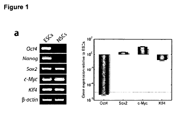
Figure 1: Generation of 2F Oct4/KIf4 (OK) iPS cells from adult NSCs of
OG2/Rosa26 transgenic mice.
a. RT-PCR and qRT-PCR analyses of Oct4, Nanog, KIf4, Sox2, and c-Myc in ESCs
and NSCs. 13-actin was used as loading control. b. Western blot analyses of
the four
factors in ESCs and NSCs. Anti-actin antibody was used as loading control. c.
Morphology of 2F OK iPS cell colony on day 14 post-infection. An ESC-like
colony
expressing Oct4-GFP (f). d. Morphology of an established 2F OK iPS cells
(clone F-
4) on day 30 post-infection, grown on irradiated MEFs. Phase contrast and Oct4-
GFP (g) are shown. e. Morphology of NSCs and mock infection on day 30 post-
infection (h). i. Generation of GFP-positive colonies at day 7, 14, and 21
after 2F OK
and 4F infection (n = 3; error bars indicated s.d.). j. Reprogramming
efficiency of
generating 2F and 4F iPS cells (n = 3). Indicated are the total numbers of
GFP+
colonies per 50,000 plated NSCs at day 7, 14, and 21 after infection.
Figure 2: Gene expression profile of iPS cells.
a. RT-PCR analysis of ES cell marker gene expression in ESCs, 4F iPS cells
(clone
A-2c), 2F OK iPS cells (clones B-2, D-7 and F-4), and NSCs. Primers are
specific
for transcripts from the respective endogenous locus. R-actin was used as
loading
control. b. The heatmap of the different expressed genes among the NSC, 2F
(OK)
iPS, 4F iPS and ESC. The gene hierarchical cluster was performed with a
cityblock
CA 02725857 2010-11-25
WO 2009/144008 PCT/EP2009/003735
22
distance and an average linkage. c. Global gene expression patterns were
compared between 2F iPS cells (clone F-4) and ESCs, and between 2F iPS cells
(clone F-4) and NSCs with DNA microarrays. d. Black lines indicate two-fold
changes in gene expression levels between the paired cell types. Genes
overexpressed in 2F iPS cells (clone F-4) compared with NSCs or ESCs are shown
in blue; those underexpressed are shown in red. Positions of pluripotency
genes
Oct4, Nanog, Sox2, c-Myc, KIf4 and Lin28 in scatter plots are indicated. The
gene
expression level is scaled in log2.
Figure 3: 2F Oct4/KIf4 (OK) iPS cells (clone F-4) are pluripotent and
differentiate in vitro and in vivo.
a. In vitro differentiation into all three germ layers. After embryoid body
formation,
aggregates were transferred onto gelatine-coated plates and allowed to
differentiate
for another 10 days. Cells were stained with anti-Tujl, a nti-a-fetop rote in
(AFP), or
anti-FIkl. Nuclei were stained with DAPI. b. Teratomas of F-4 iPS cells
containing
all three germ layers. F-4 iPS cells (1.5 x 10⁶ cells) were subcutaneously
inoculated
into nude mice. After 4 weeks, teratomas were stained with haematoxylin and
eosin
dyes. Shown is a teratoma containing a neural rosette (ectoderm), muscle
(mesoderm), and columnar epithelium (endoderm).
Figure 4: In vivo developmental potential of 2F Oct4/KIf4 (OK) iPS cells
(clone
F-4).
a. The chimeric embryos of F-4 iPS cells developed to blastocysts after 24 hrs
of
aggregation. Fluorescence optics show Oct4-GFP cells located in the inner cell
mass of blastocysts. b. Germline contribution of F-4 iPS cells to mouse
embryonic
development as shown by the expression of Oct4-GFP. Embryos were analyzed
with a fluorescence microscope at E13.5. c, d. The 13.5 dpc chimeric embryos
(control, 2N, and 4N) were stained with X-gal solution. e. Histological
analysis of
/acZ-stained 13.5 dpc chimeric embryo (2N). f. Chimeric mouse (8-week-old)
generated by F-4 iPS cells. Agouti coat colour originated from F-4 iPS cells.
g. PCR
genotyping of chimeras derived from F-4 iPS cell. PCR analyses were performed
for
Oct4-GFP (top panel). Germline transmission of F-4 iPS cells. Genotyping of
offspring from chimeric males mated with CD-1 females demonstrated the
presence
CA 02725857 2010-11-25
WO 2009/144008 PCT/EP2009/003735
23
of Oct4-GFP and IacZ allele and Oct4 and KIf4 virus integrations (bottom
panel).
Abbreviation: Gastroint. tract.: gastrointestinal tract.
Figure 5: One-factor hNSC-derived iPS (IF hNiPS) cell colony formation and
cell line characterization.
(A) Morphology of hNSCs grown in NSC medium. (B) Colony formation of hOCT4-
infected cells 10 weeks post-infection. (C) The colony grows hESC-like
morphology
but center of colony still remain unreprogrammed neural rosettes. (D) Typical
hESC-
like iPS colony growing on feeder after mechanical isolation at passage 1 (1 F
hNiPS
clone C). (E) High magnification of iPS colony at passage 10. (F) 1 F hNiPS
colonies
were stained for AP. Scale bars, 250 pm. (G) Immunocytochemical analysis of
pluripotency markers (OCT4, SSEA4, TRA-1-60 and TRA-1-81) in 2F hNiPS (clone
A) and 1F hNiPS (clone C) cells. Nuclei are stained with DAPI (blue). Scale
bars,
250 pm.
Figure 6: Expression level of pluripotent markers and DNA methylation
analysis in hNSC-derived iPS (hNiPS) cells.
(A) Quantitative PCR analysis for pluripotent markers in H1 hESCs, hNSCs, 2F
hNiPS clones (A, B and C) and 1 F hNiPS clones (A and C). Data are shown
relative
expression to H9 hESCs using primers specific for endogenous transcripts. RNA
expression levels are shown on logarithmic scale. Transcripts levels were
normalized to R-actin levels. Error bars indicate the s.d. from triplicates.
(B) Bisulfite
sequecing analysis of OCT4 and NANOG promoter regions in H9 hESCs, hNSCs,
2F hNiPS clones (A, B and C) and 1 F hNiPS clones (A and C). Each row of
circles
for a given amplicon represents the methylation status of each CpG in one
bacterial
clone for that region. Open circles represent unmethylated CpGs, and closed
circles
represent methylated CpGs. Bottom numbers of each column indicate CpG
dinucleotide locations, relative to the transcriptional start site (TSS; +1).
Figure 7: In vitro differentiation of hNSC-derived iPS (hNiPS) cells into all
three germ layers.
(A) Immunofluorescence analysis shows differentiation of 2F and 1 F hNiPS
cells
into all three germ layers: endoderm (alpha-fetoprotein; AFP), mesoderm (alpha-
smooth muscle actin; a-SMA) and ectoderm (13-tublin Illb; Tuj1). Nuclei are
stained
CA 02725857 2010-11-25
WO 2009/144008 PCT/EP2009/003735
24
with DAPI (blue). Scale bars, 100 pm. (B) Quantitative PCR analyses of one-
month
embryoid bodies (EBs) differentiation derived from 2F hNiPS (clone A) and 1F
hNiPS (clone C) cells. Endoderm (AFP, GATA6 and Sox17), mesoderm (FOXF1
and HAND1) and ectoderm (NCAM1, PAX6 and Sox1). Data are shown relative
expression to each undifferentiated parental hNiPS cells. RNA expression
levels are
shown on logarithmic scale. Transcripts levels were normalized to (3-actin
levels.
Figure 8: In vivo pluripotency and global gene expression profile of hNSC-
derived iPS (hNiPS) cells.
(A) Teratoma formation after transplantation of 2F hNiPS (clone A) and 1 F
hNiPS
(clone C) cells into SCID mice, and teratomas were sectioned and stained with
hematoxylin and eosin at 6-8 weeks. Histological section of identified cells
representing all three germ layers: endoderm (respiratory epithelium; r),
mesoderm
(skeletal muscle; m, cartilage; c) and ectoderm (neural epithelium; n).
Enlargements
of sections showing respiratory epithelium, muscle and neural epithelium
indicated
by arrows. Scale bars, 100 pm. (B) Heat map (left panel) and hierarchical
cluster
analysis (right panel) of global gene expression from hNSCs, 1 F hNiPS (clone
C),
2F hNiPS (clone A) H9 hESCs and H1 hESCs (left). (C) Scatter plots comparing
global gene expression profiles between 1 F hNiPS (clone C) and H9 hESCs (left
panel), 2F hNiPS (clone A) and H9 hESCs (middle panel), and hNSCs and 1 F
hNiPS (clone C) (right panel). The black lines indicate twofold difference in
gene
expression levels between the paired cell populations. The transcript
expression
levels are on the log2 scale.
The examples illustrate the invention:
Example 1: Generation of OG2 mice
The OG2 strain was crossed with the ROSA26 transgenic strain (Do, J. T. &
Scholer, H. R., Stem Cells 22, 941-9 (2004); Szabo, P. E., Hubner, K.,
Scholer, H. &
Mann, J. R., Mech Dev 115, 157-60 (2002)) over several generations to produce
compound homozygous mice for the neo/IacZ and Oct4-GFP transgenes. To derive
NSCs, homozygous OG2 x ROSA26 male mice were crossed with ICR females to
produce heterozygous pups. Brain tissue was collected from 5-day-old OG2 x
ROSA26 heterozygous mice.
CA 02725857 2010-11-25
WO 2009/144008 PCT/EP2009/003735
5 Example 2: Generation of induced pluripotent stem cells
iPS cells and ESCs were grown on irradiated MEFs and in ESC medium (DMEM
supplemented with 15% FBS, nonessential amino acids, L-glutamine,
penicillin/streptomycin, 13-mercaptoethanol, and 1,000 U/ml leukemia
inhibitory
factor (LIF)). pMX-based retroviral vectors encoding the mouse cDNAs of Oct4,
10 Sox2, KIf4, and c-Myc were separately cotransfected with packaging-
defective
helper plasmids into 293 cells using Fugene 6 transfection reagent (Roche). 48
his
later, virus supernatants were collected as previously described (Zaehres, H.
&
Daley, G. Q., (2006), Methods Enzymol 420, 49-64). NSCs derived from
OG2/Rosa26 transgenic mice were seeded at a density of 5 x 10⁴ cells per 6-
well
15 plate and incubated with virus-containing supernatants for the four factors
(1:1:1:1)
or for Oct4 and KIf4 (1:1) supplemented with 6 pg/ml protamine sulfate (Sigma)
for
24 hrs. Transduction efficiencies were calculated with pMX-GFP control virus.
Cells
were replated in fresh neural expansion medium. Two days after infection, the
cells
were further subcultured on irradiated MEFs in ESC medium containing LIF
without
20 any further selection. Oct4-GFP-positive colonies were mechanically
isolated, and
individual cells were dissociated and subsequently replated onto MEFs. The
colonies were selected for expansion.
Example 3: qRT-PCR analysis
25 Total RNA was extracted from cells using the MiniRNeasy Kit (Qiagen GmbH,
Hilden, Germany; http://www.qiagen.com) according to the manufacturer's
instructions. Complementary DNA synthesis was performed with the High Capacity
cDNA Archive Kit (Applied Biosystems GmbH, Darmstadt, Germany;
http://www.appliedbiosystems.com) following the manufacturer's instructions
with a
down-scaled reaction volume of 20 pl. Transcript levels were determined using
the
ABI PRISM Sequence Detection System 7900 (Applied BioSystems) and the ready-
to-use 5'-nuclease Assays-on-Demand. For each real-time amplification, the
template was equivalent to 5 ng of total RNA. Measurements were done in
triplicate;
a RT- blank of each sample and a no-template blank served as negative
controls.
Amplification curves and gene expression were normalized to the housekeeping
gene Hprt, used as internal standard.
Oligonucleotides were designed by the Taqman Assay-on-Demand for the detection
of the following genes: Pou5fl (Oct3/4) (Mm00658129_gH), Sox2
CA 02725857 2010-11-25
WO 2009/144008 PCT/EP2009/003735
26
(Mm00488369_sl ), c-Myc (Mm00487803_ml ), KIf4 (Mm00516104_ml) B-Act
(Mm00607939_sl), and Hprtl (Mm00446968_ml). Oligos for the detection of
Nanog and the viral sequences were custom-designed. Quantification was
normalized on the endogenous Hprt gene within the log-linear phase of the
amplification curve obtained for each probe/primers set using the AACt method
(ABI
PRISM 7700 Sequence Detection System, user bulletin #2).
Primer sequences for viral-specific qRT-PCR
pMXs-Oct4 PF: 5'-TGGTACGGGAAATCACAAGTTTG (SEQ ID NO: 17), PR: 5'-
GTCATAGTTCCTGTTGGTGAAGTTCA (SEQ ID NO: 18), Probe: 5'-6FAM-
CTTCACCATGCCCCTCA-MGB (SEQ ID NO: 19)
pMXs-Sox2 PF: 5'-GTGTGGTGGTACGGGAAATCAC (SEQ ID NO: 20), PR: 5'-
TTCAGCTCCGTCTCCATCATG (SEQ ID NO: 21), Probe: 5'-6FAM-
TGTACAAAAAAGCAGGCTTGT-MGB (SEQ ID NO: 22)
pMXs-KIf4 PF: 5'-GTGTGGTGGTACGGGAAATCA (SEQ ID NO: 23), PR: 5'-
CGCGAACGTGGAGAAGGA (SEQ ID NO: 24), Probe: 5'-6FAM-
CTTCACCATGGCTGTCAG-MGB (SEQ ID NO: 25)
pMXs-cMyc PF: 5'-TGGTACGGGAAATCACAAGTTTG (SEQ ID NO: 26), PR: 5'-
GTCATAGTTCCTGTTGGTGAAGTTCA (SEQ ID NO: 27), Probe: 5'-6FAM-
CTTCACCATGCCCCTCA-MGB (SEQ ID NO: 28)
Nanog PF: 5'-AACCAGTGGTTGAATACTAGCAATG (SEQ ID NO: 29), PR: 5'-
CTGCAATGGAT GCTG GGATACT (SEQ ID NO: 30), Probe: 5'-6FAM-TTCAGAA
GGGCTCAGCAC-MGB (SEQ ID NO: 31)
Example 4: Microarray analysis
The microarray study was carried out using Affymetrix Mouse Genome 430 2.0
GeneChip arrays (Affymetrix, Santa Clara, CA) essentially as described before
(Ruau, D. et al., (2008), Stem Cells). Briefly, total RNA was extracted from
cells with
RNAeasy kit including DNAse digestion (Qiagen, Hilden, Germany). Biotin-
labelled
cRNA was obtained from 3 pg of total RNA with the GeneChip One-Cycle labelling
CA 02725857 2010-11-25
WO 2009/144008 PCT/EP2009/003735
27
kit (Affymetrix). Fifteen micrograms of cRNA were fragmented and hybridized to
Affymetrix 430 2.0 GeneChip arrays at 45 C for 16 hrs. DNA chips were washed,
stained and scanned using an Affymetrix Fluidics device and GCS3000 scanner,
and the images obtained were analyzed using the GCOS software. The experiment
was performed in triplicates for the ESCs and iPS cells and in duplicates for
the
NSCs. Normalization was calculated with RMA algorithm (Irizarry, R. A. et al.,
(2003), Nucleic Acids Res 31, el 5) implemented in BioConductor.
Example 5: In vitro differentiation of PS cells
Oct4-GFP cells were harvested by FACS analysis and used for in-vitro
differentiation in embryoid bodies (EBs), which was performed with hanging
drop in
ESC medium without LIF. After 3 days, EBs were plated onto gelatine-coated 4-
well
dishes for another 10 days. The cells were stained with anti-Tuj1 antibody
(1:100;
Chemicon), anti-a-fetoprotein (AFP) antibody (1:100; R&D Systems), or anti-
FIk1
antibody (1:100; R&D Systems).
Example 6: Western blot analysis, SSEA-1 and AP staining
Total cell lysates (2 x 10⁶) prepared from the ESC and NSC were subjected to
western blot analysis for expression of Oct4 (Santa Cruz), Sox2 (Santa Cruz),
KIf4
(Abcam), and c-Myc (Abcam). (3-actin expression levels in all the samples were
used as loading control (Abcam).
SSEA-1 and alkaline phosphatase (AP) staining was performed with the ES Cell
Characterization Kit (Chemicon) according to the manufacturer's protocol.
Example 7: Teratoma formation
iPS cells and NSCs cells (1.5 x 10⁶ cells/mice) were injected subcutaneously
into
the dorsal flank of nude mice. Four weeks after the injection, teratomas that
had
formed were fixed overnight in 4% PFA and embedded in paraffin. Sections were
stained with haematoxylin and eosin dyes.
Example 8: Chimera formation
iPS cells were aggregated and cultured with denuded post-compacted 8-cell-
stage
mouse embryos. Briefly, 2-cell-stage embryos were flushed from mice [(C57BU6 x
CA 02725857 2010-11-25
WO 2009/144008 PCT/EP2009/003735
28
C3H) F1 females x CD1 males] at 1.5 dpc and placed in M2 medium and cultured
overnight in KSOM medium with 0.1% BSA overnight to 8-cell stage. Clumps of
loosely connected iPS'cells (10-20 cells) from short trypsin-treated day-2
cultures
were selected and transferred into microdrops of KSOM medium with 10% FCS
under mineral oil; each clump was placed in a depression in the microdrop.
Meanwhile, batches of 30 to 40 embryos were briefly incubated with acidified
Tyrode's solution until the zona pellucida had disintegrated. A single embryo
was
place onto the clump. All aggregates were assembled in this manner, and
cultured
at 37 C in an atmosphere of 5% CO2 in air. After 24 hours of culture, the
majority of
the aggregates had formed blastocysts. A total of 64 aggregated blastocysts
(2.5
dpc) were transferred into the uterine horns of five pseudopregnant mice (CD-1
background).
Example 9: Reprogramming of human neural stem cells by Oct4
hNSCs that derived from human fetal brain tissue were expanded in serum-free
NSC medium as described previously (cf. Fig. 5A) (Kim et al., Exp Neurol 199,
222
(2006); Park et al., Nat Biotechnol 20, 1111 (2002)). hNSCs were first
infected with
pMXs encoding human OCT4 and KLF4 (2F) or OCT4 (1F). Then, infected hNSCs
were maintained in NSC medium (Kim et al., Exp Neurol 199, 222 (2006)) for up
to
7 days. Day 8 post-infection, the cells were replated onto feeder cell layers
in hESC
medium containing 10ng/ml bFGF and MEF-conditioned medium (CM) in a 1:1 ratio
which culture continued to grow until the hESC-like colonies appeared. Within
10-11
weeks post-infection, the hESC-like iPS colonies were identified but the
centre of
the colonies still appears like a neural rosette (cf. Fig. 5B). The colony
grew larger
exhibiting typical hESC-like morphology within another 5-6 days but still the
neural
rosettes remain in the center of the colony (cf. Fig. 5C). The neural rosettes
are
removed from the colony. Then, a piece of the colony was transferred on a
feeder
cell layer by mechanical isolation (cf. Fig. 5D). We successfully established
two
clones out of three hESC-like colonies by picking from OCT4 infected hNSCs (1
F
hNiPS clone A and C, reprogramming efficiency 0.02%). Otherwise, we also
established 3 clones out of five hESC-like colonies in 2F-infected hNSCs (2F
hNiPS
A, B and C, reprogramming efficiency, 0.15%) within 7-8 weeks post-infection.
All of
which could be expanded in hESC culture condition. The 1 F hNiPS cells were
morphologically similar to hESCs and stained positive for alkaline phosphatase
(cf.
CA 02725857 2010-11-25
WO 2009/144008 PCT/EP2009/003735
29
Fig. 5E and F). Immunofluorescence staining confirmed that 2F and 1 F hNiPS
cells
uniformly expressed the pluripotency markers, including OCT4, SSEA4, TRA-1-60
and TRA-1-81 (cf. Fig. 5G). These results demonstrate that human iPS cells can
be
generated from hNSCs by OCT4 and KLF4 as well as OCT4 alone.
Next, we tested mRNA expression levels of pluripotency marker genes in these
PS
cells at molecular level by quantitative RT-PCR analysis. 2F and 1F hNiPS
cells
endogenously expressed the hESCs-specific markers, were similar to H9 and H1
hESCs and were markedly up-regulated compared with parental hNSCs (cf. Fig.
6A). Genotyping PCR showed 1 F hNiPS clones have an OCT4 transgene only and
2F hNiPS clones have OCT4 and KLF4 transgenes in the genome. We also
confirmed that the expression level of transgenic OCT4 or KLF4 was
significantly
silenced in 2F and 1F hNiPS clones, except the OCT4 expression from 2F hNiPS
clone B. Southern blot analysis confirmed the integration of the OCT4
transgene in
2F and 1F hNiPS clones. To exclude the possibility that iPS clones arose
through
contamination from hESCs in the laboratory, DNA fingerprinting analysis was
performed and confirmed that hNiPS cells precisely correlate to the donor
hNSCs
(cf. Table 2).
To confirm epigenetic remodelling of the OCT4 and NANOG promoters from
reprogrammed cells, we performed bisulfite sequencing to determine the
demethylation of both promoters. OCT4 and NANOG promoter regions were
demethylatd in 2F and 1 F hNiPS cells relative to the donor hNSCs and were
similar
to hESCs. Taken together, hNSCs can be reprogrammed into iPS cells that
similar
to hESCs at molecular level by transduction of OCT4 alone.
Next, we tested in vitro pluripotency of 2F and 1F hNiPS cells by embryoid
body
(EB) differentiation and direct differentiation. hNiPS cells readily
differentiated into
endoderm (AFP), medoderm (a-SMA) and ectoderm (Tuj1) by EB differentiation
(cf.
Fig. 5A) and we confirmed the expression of all three germ layer makers from
direct
differentiation by quantitative RT-PCR analysis (cf. Fig. 7B). To confirm in
vivo
pluripotency of these human iPS cells, the cells were subcutaneously
transplanted
into severe combined immunodeficient (SCID) mice. After 6-8 weeks injection,
2F
and 1 F hNiPS cells gave rise to teratomas containing all three germ layers,
including respiratory tract, skeletal muscle, cartilage and neural epithelium
(cf. Fig.
8A). These results indicate that 2F and 1 F hNiPS cells have a pluripotency in
vitro
and in vivo alike hESCs.
CA 02725857 2010-11-25
WO 2009/144008 PCT/EP2009/003735
5 Finally, we performed global gene expression analysis on hNSC, 2F and 1 F
hNiPS
cells derived from hNSCs, H9 and H1 hESCs by cDNA microarrays. Heat map
showed that 2F and 1 F hNiPS cells similar to hESCs, otherwise parental hNSCs
are
isolated from pluripotent polulations (cf. Fig. 8B, left panel) and
hierarchical
clustering analysis showed that hNiPS cells clustered with hESCs and were
distinct
10 from parental hNSCs (cf. Fig. 8B, right panel). Scatter plots analysis
showed that
hNiPS cells are significantly more similar to hESCs as like between different
hESCs
than parental hNSCs (cf. Fig. 8C). 1 F and 2F hNiPS cells also show similarity
with
H1 hESCs. These data indicate that hNiPS cells are similar to hESCs in global
gene
expression profiles. Our results demonstrated 1F and 2F hNiPS cells closely
15 resemble hESCs in molecular level and pluripotency.
Table 2: STR analysis of hNSCs and hNiPS cells
2F NhiPS IF NhiPS
Genomic loci H9 hESCs hNSCs A B C A C
Amelogenin X;X X;Y X;Y X;Y X;Y X;Y X;Y
CSF1PO 11:11 11;13 11;13 11;13 11;13 11;13 11;13
D13S317 9;9 8;11 8;11 8;11 8;11 8;11 8;11
D16S539 12;13 9;9 9;9 9;9 9;9 9;9 9;9
D 18S51 13;13 15;16 15;16 15;16 15;16 15;16 15;16
D21S11 30;30 31;32 31;32 31;32 31;32 31;32 31;32
D3S1358 = 13;16 16;16 16;16 16;16 16;16 16;16 16;16
D5S818 11;12 7;12 7;12 7;12 7;12 7;12 7;12
D7S820 9;11 11;11 11;11 11;11 11;11 11;11 1`1;11
D8S1179 8;14 12;14 12;14 12;14 12;14 12;14 12;14
FGA 26;28 23;24 23;24 23;24 23;24 23;24 23;24
Penta D 9;13 11;12 11;12 11;12 11;12 11;12 11;12
Penta E 11;14 11;18 11;18 11;18 11;18 11;18 11;18
TH01 9;9 7;7 7;7 7:7 7;7 7;7 7;7
TPOX 10;11 8;8 8;8 8;8 8;8 8;8 8;8
vWA 17;17 17;17 17;17 17;17 17;17 17;17 17;17
20 Material and methods:
Cell culture
Human NSCs were derived from the telencephalon (HFT13), established as
previously described (Kim et al., Exp Neurol 199, 222 (2006)). Briefly,
Telencephalon tissue was freshly dissected, dissociated in 0.1% trypsin for 30
min
25 and seeded into 10cm plates at a density of 200,000 cells/ml in NSC medium.
These cells were cultured in NSC medium as previously described (Kim et at.,
Exp
Neurol 199, 222 (2006); Park et al., Nat Biotechnol 20, 1111 (2002)). Human ES
CA 02725857 2010-11-25
WO 2009/144008 PCT/EP2009/003735
31
and iPS cells were maintatined on mitomycin C-treated CF1 mouse feeder layers
(Millipore) in human ESC medium, which contains knockout DMEM (Invitrogen)
supplemented with 20% knockout serum replacement (Invitrogen), 1mM L-
glutamine, 1% non-essential amino acids, 0.1 mM 3- mercaptoethanol,
penicillin/streptomycin and 10 ng/ml human basic fibroblast growth factor
(bFGF)
(Invitrogen) as previously described (Takahashi et al., Cell 131, 861 (2007)).
Induction of IF hNiPS and 2F hNiPs cells
The pMX-based retroviral vectors encoding the human cDNAs of OCT4 and KLF4
(Takahashi et Yamanaka, Cell 126, 663 (2006)) were cotransfected with
packaging-
defective helper plasmids into 293 cells using Fugene transfection reagent
(Roche)
to produce vesicular stomatitis virus (VSV) G protein pseudotyped virus as
previously described (Zaehres et Daley, Methods Enzymol 420, 49 (2006)). Viral
supernatants were collected and concentrated by ultracentrifugation 48 h post-
transfection to infect human NSCs. For generation of iPS cells, human NSCs
were
seeded at a density of 5 x 10⁴ cells per 6-well plate and incubated with virus-
containing supernatants for OCT4 or OCT4 and KLF4 supplemented with 6 pg/ml
protamine sulfate (Sigma) for 24 h. On the next day, the medium was replaced
with
fresh NSC medium at 1d post-infection and maintained up to 7 d post-infection.
Cells were further cultured in human ESC medium from 8 d post-infection. The
iPS
colonies were mechanically isolated at 2 month or 2.5 month post-infection and
were subsequently replated and maintained on CF1 mouse feeder layers
(Millipore)
in human ESC medium.
Quantitative RT-PCR
Total RNA was isolated from bulk cell culture samples or hand-picked
undifferentiated colonies using RNeasy columns (Qiagen) with on-column DNA
digestion. cDNA was produced using oligo-dT15 priming and M-MLV reverse
transcriptase (USB) according to the manufacturer's instructions at 42 C for 1
h.
About 50 ng of total RNA equivalent was typically used as template in 20 pi
SYBR
Green PCR reactions (40 cycles of 15" 95 C / 60" 60 C on Applied Biosystems
7300 instrumentation) that additionally contained 0.375 pM of each primer and
10pl
of SYBR Green PCR mix (ABI). All primers used were confirmed to amplify the
predicted product at close-to-optimal efficiency without side products. Primer
CA 02725857 2010-11-25
WO 2009/144008 PCT/EP2009/003735
32
sequences are given in Table 3. Relative expression levels were calculated
using
the comparative. Ct method, based on biological control samples and two
housekeeping genes for normalization. Error bars reflect standard errors
arising
from biological replicates (marker gene expression data) or from using
independent
housekeeping genes for normalization (transgene silencing data).
Global gene expression analysis
For transcriptional analysis, 400 ng of total DNA-free RNA was used as input
for
labelled cRNA synthesis (Illumina TotalPrep RNA Amplification Kit - Ambion)
following the manufacturer's instructions (IVT: 10h). Quality-checked cRNA
samples
were hybridized as biological or technical duplicates for 18 h onto HumanRef-8
v3
expression BeadChips (Illumina), washed, stained, and scanned following
guidelines and using materials / instrumentation supplied / suggested by the
manufacturer. The microarray data are available from the GEO (Gene Expression
Omnibus) website under accession number GSE GSE15355.
Bisulfite sequencing
Genomic DNA was isolated from bulk cell culture samples or hand-picked
undifferentiated colonies using DNeasy columns (Qiagen). 300 ng was used as
input for bisulfite conversion (EpiTect Bisulfite Kit - Qiagen). 50 ng of
converted DNA
was used as template for conventional nested PCRs amplifying 467 and 336bp
regions of the OCT4 and NANOG promoters, respectively. Primers were specific
for
conversion of the sense DNA strand and are given in Table 3. Purified PCRs
were
TA-cloned into pCR2.1-TOPO (Invitrogen). Insert sequences of randomly picked
clones were analyzed using the BiQ Analyzer program, following its quality
check-
based suggestions to drop individual clones if appropriate. Data from one CpG
site
at position +20 relative to the OCT4 translation start codon is not shown as
it was
uninformative.
Short tandem repeat (STR) analysis
Genomic DNA was isolated from cultured cell samples using DNeasy columns
(Qiagen). This was used as template for STR analysis employing the PowerPlex
16
System (Promega) and ABI PRISM instrumentation. Numbers shown denote bp
lengths of the 15 autosomal fragments. The analysis was carried out at
Eurofins
Medigenomix, Martinsried, Germany.
CA 02725857 2010-11-25
WO 2009/144008 PCT/EP2009/003735
33
Teratoma formation
hNiPS cells and hNSCs (3-5 x 10⁶ cells/mice) were injected subcutaneously into
the
dorsal flank of SCID mice. Teratomas were fixed in 4% PFA overnight and
embedded in paraffin after 6-8 weeks injection. Sections were stained with
haematoxylin and eosin dyes.
Alkaline phosphatase (AP) and immunofluorescence staining
Alkaline phosphatase (AP) staining was performed with the ES Cell
Characterization
Kit (Chemicon) according to the manufacturer's protocol. Immunofluorescence
staining was performed using the following primary antibodies: AFP (Sigma,
1:100),
a-SMA (Sigma, 1:50), TuJ1 (Chemicon, 1:500), OCT4 (Santa Cruz, 1:200), SSEA4
(Chemicon, 1:200), TRA-1-60 (Chemicon, 1:200), TRA-1-81 (Chemicon, 1:200).
Table 3. Primers for Real-time PCR and Bisulfite sequencing.
Real-time PCR Primers
Gene Forward primer 5'-3' Reverse primer 5'-3'
ACTB TCAAGATCATTGCTCCTCCTGAG ACATCTGCTGGAAGGTGGACA
AFP AGCAGCTTGGTGGTGGATGA CCTGAGCTTGGCACAGATCCT
CDHI (E-CAD) TTGAGGCCAAGCAGCAGTACA ATCCAGCACATCCACGGTGA
CDX2 TCACTACAGTCGCTACATCACCATC TTAACCTGCCTCTCAGAGAGCC
DNMT3B GCTCACAGGGCCCGATACTT GCAGTCCTGCAGCTCGAGTTTA
DPPA4 TGGTGTCAGGTGGTGTGTGG CCAGGCTTGACCAGCATGAA
FGF2 GGCAAGATGCAGGAGAGAGGA GCCACGTGAGAGCAGAGCAT
FOXF1 AAAGGAGCCACGAAGCAAGC AGGCTGAAGCGAAGGAAGAGG
GAPDH CTGGTAAAGTGGATATTGTTGCCAT TGGAATCATATTGGAACATGTAAACC
GATA6 TGTGCGTTCATGGAGAAGATCA TTTGATAAGAGACCTCATGAACCGACT
GDF3 TTGGCACAAGTGGATCATTGC TTGGCACAAGTGGATCATTGC
HAND1 TCCCTTTTCCGCTTGCTCTC CATCGCCTACCTGATGGACG
KLF4 endo ACAGTCTGTTATGCACTGTGGTTTCA CATTTGTTCTGCTTAAGGCATACTTGG
KLF4 viral GTCGGACCACCTCGCCTTAC TTTATCGTCGACCACTGTGCTG
LIN28 GGAGGCCAAGAAAGGGAATATGA AACAATCTTGTGGCCACTTTGACA
MYC CCAGCAGCGACTCTGAGGA GAGCCTGCCTCTTTTCCACAG
NANOG CCTGTGATTTGTGGGCCTG GACAGTCTCCGTGTGAGGCAT
NCAMI TCATGTGCATTGCGGTCAAC ACGATGGGCTCCTTGGACTC
OCT4 endo GGAAGGAATTGGGAACACAAAGG AACTTCACCTTCCCTCCAACCA
OCT4 viral GGCTCTCCCATGCATTCAAAC TTTATCGTCGACCACTGTGCTG
SOX17 TTCGTGTGCAAGCCTGAGATG GTCGGACACCACCGAGGAA
SOX2 TGGCGAACCATCTCTGTGGT CCAACGGTGTCAACCTGCAT
TDGFI Cri to GGGATACAGCACAGTAAGGAGCTAA CACAAAAGGACCCCAGCATG
ZNF206 TCACCATGGCCAGAGGAGAG GCAGGCCACGCCTTATTCTC
ZNF589 TCGGGTGGCTAAATTACATCCAG CCCAAGGGAGTAAGGCAAACTG
Primers for bisulfite sequencing
Gene Forward primer 5'-3' Reverse primer 5'-3'
OCT4 outer GAGGATAGGAATTTAAGATTAGTTTGGGTA AAATCCCCCACACCTCAAAACCTAACCCAA
OCT4 inner GAGGTTGGAGTAGAAGGATTGTTTTGGTTT CCCCCCTAACCCATCACCTCCACCACCTAA
OCT4 inner unconverted GAGGCTGGAGCAGAAGGATTGCTTTGGCCC
CCCCCCTGGCCCATCACCTCCACCACCTGG
NANOG outer TTAGTTTTTAGAGTAGTTGGGATTATAGA ATAATAACATAAAACAACCAACTCAATCCA
NANOG inner TGGTTAGGTTGGTTTTAAATTTTTG AACCCACCCTTGTAAATTCTCAATTA
NANOG inner unconverted TGGCCAGGCTGGTTTCAAACTCCT GGACCCACCCTTGTGAATTCTCAGTTA
CA 02725857 2010-11-25
WO 2009/144008 PCT/EP2009/003735
34
Southern blot analysis
BamHl digested genomic DNA from 1F hNiPS, hNSC and 2F hNiPS cells were
separated on a 0.8% agarose gel and transferred to Biodyne B nylon membrane
(PALL Life Sciences). DNA was hybridized with a 32P labeled fragment of OCT4
(Eco81l (Saul) human OCT4 cDNA fragment) using the DecaLabelTM DNA
Labeling Kit (Fermentas). Labeled Lambda Hindlll digested DNA served as a
marker.
In vitro differentiation of human iPS cells
For immunocytochemistry, embryoid bodies (EBs) were generated from iPS cells
with the hanging drop method in MEF-conditioned medium. After 5 days, EBs were
transferred to gelatin-coated plate and subsequent culturing for another 14
days in
knockout DMEM (Invitrogen) supplemented with 20% FBS, 1mM L-glutamine, 1%
non-essential amino acids, 0.1 mM P- mercaptoethanol, and
penicillin/streptomycin.
For qRT-PCR, iPS colonies were mechanically isolated and replated on Matrigel-
coated plate in MEF-conditioned medium. After 2 d, medium replaced with each
three germ layer differentiation medium. For endoderm differentiation, the
cells
maintained in RPM11640 medium supplemented with 2% FBS, 100 ng/ml Activin A
(R&D Systems), L-glutamine, and penicillin/streptomycin for 3 weeks (Huangfu
et
al., Nat Biotechnol 26, 1269 (2008)). For mesoderm differentiation, knockout
DMEM
supplemented with 100 uM ascorbic acid (Sigma), 20%.FBS, 1mM L-glutamine, 1%
non-essential amino acids, 0.1 mM P- mercaptoethanol, and penicillin/
streptomycin
for 3 weeks (Aasen et al., Nat Biotechnol 26, 1276 (2008)). For ectoderm
differentiation, the cells maintained in N2B27 medium for 7 days and the
medium
replaced with N2 medium supplemented with 10 ng/ml bFGF2 (peprotech), 100
ng/ml Sonic Hedgehog (R&D Systems), 10 ng/ml PDFG (R&D Systems), L-
glutamine, and penicillin/streptomycin for 2 weeks. The medium was changed
every
other day. Primer sequences are given in Table 3.
CA 02725857 2010-11-25
WO 2009/144008 PCT/EP2009/003735
5 References
Biswas, A. and Hutchins, R. (2007). Embryonic stem cells. Stem Cells Dev
16(2):213-22.
Davis, P.B., Cooper, M.J. (2007). Vectors for airway gene delivery. AAPS J
10 19;9(1):E11-7.
Do, J. T. & Scholer, H. R. (2004). Nuclei of embryonic stem cells reprogram
somatic
cells. Stem Cells 22:941-9.
Gage, F.H. (2000). Mammalian neural stem cells. Science 287(5457):1433-8.
Hanna, J., Wernig, M., Markoulaki, S., Sun, C.W., Meissner, A., Cassady, J.P.,
15 Beard, C., Brambrink, T., Wu, L.C., Townes, T.M. and Jaenisch, R. (2007).
Treatment of sickle cell anemia mouse model with iPS cells generated from
autologous skin. Science 318(5858):1920-3.
Hogan, B., R. Beddington, et al. (1994), "Manipulating the Mouse Embryo: A
Laboratory Manual", Cold Spring Harbour Press.
20 Jones, G.E., Wise, C.J. (1997). Establishment, maintenance, and cloning of
human
dermal fibroblasts. Methods Mol Biol. 75:13-21.
Kim, D.S., Kim, J.Y., Kang, M., Cho, M.S., Kim, D.W. (2007). Derivation of
functional
dopamine neurons from embryonic stem cells. Cell Transplant 16(2):117-23.
Li, S. and Ma, Z. (2001). Nonviral gene therapy. Curr Gene Ther.,1(2):201-26.
25 Meissner, A., Wernig, M. and Jaenisch, R. (2007). Direct reprogramming of
genetically unmodified fibroblasts into pluripotent stem cells. Nat Biotechnol
25(10):
1177-81.
Nakagawa, M., Koyanagi, M., Tanabe, K., Takahashi, K., Ichisaka, T., Aoi, T.,
Okita,
K., Mochiduki, Y., Takizawa, N. and Yamanaka, S. (2008). Generation of induced
30 pluripotent stem cells without Myc from mouse and human fibroblasts. Nat
Biotechnol. 26(1):101-6.
Okita, K., Ichisaka, T. and Yamanaka, S. (2007). Generation of germ line-
competent
induced pluripotent stem cells. Nature 448(7151):313-7.
Park, I. H., Zhao, R., West, J.A., Yabuuchi, A., -Huo, H., Ince, T.A., Lerou,
P.H.,
35 Lensch, M.W., Daley, G.Q. (2008). Reprogramming of human somatic cells to
pluripotency with defined factors. Nature 451(7175):141-6.
Pollard, S. M., Conti, L., Sun, Y., Goffredo, D. & Smith, A. (2006). Adherent
neural
stem (NS) cells from fetal and adult forebrain. Cereb Cortex 16 Suppl 1, it 12-
20.
Ryan, A. K. and Rosenfeld, M. G. (1997). POU domain family values:
flexibility,
partnerships, and developmental codes. Genes Dev 11:1207-25.
Takahashi, K. and Yamanaka, S. (2006). Induction of pluripotent stem cells
from
mouse embryonic and adult fibroblast cultures by defined factors. Cell 126:663-
76.
Takahashi, K., Okita, K., Nakagawa, M., and Yamanaka, S. (2007). Induction of
pluripotent stem cells from fibroblast cultures. Nat Protoc 2(12):3081-9.
Takahashi, K., Tanabe, K., Ohnuki, M., Narita, M., Ichisaka, T., Tomoda, K.
and
Yamanaka, S. (2007). Induction of pluripotent stem cells from adult human
fibroblasts by defined factors. Cell 131(5):861-72.
Wernig, M., Meissner, A., Foreman, R., Brambrink, T., Ku, M., Hochedlinger,
K.,
Bernstein, B.E. and Jaenisch, R. (2007). In vitro reprogramming of fibroblasts
into a
pluripotent ES-cell-like state. Nature 448:318-24.
Wernig, M., Meissner, A., Cassady, J. P. and Jaenisch, R. (2008). c-Myc is
dispensable for direct reprogramming of mouse fibroblasts. Cell Stem Cells
2:10-12.
Yu, J., Vodyanik, M.A., Smuga-Otto, K., Antosiewicz-Bourget, J., Frane, J.L.,
Tian,
S., Nie, J., Jonsdottir, G.A., Ruotti, V., Stewart, R., Slukvin, I.I. and
Thomson, J.A.
CA 02725857 2010-11-25
WO 2009/144008 PCT/EP2009/003735
36
(2007). Induced pluripotent stem cell lines derived from human somatic cells.
Science 318(5858):1917-20.
Zimmermann, W. H. and Eschenhagen, T. (2007). Embryonic stem cells for cardiac
muscle engineering. Trends Cardiovasc Med 17(4):134-40.
H. T. Kim et al., Exp Neurol 199, 222 (May, 2006)
K. I. Park, Y. D. Teng, E. Y. Snyder, Nat Biotechnol 20, 1111 (Nov, 2002)
H. Zaehres, G. Q. Daley, Methods Enzymol 420, 49 (2006)
D. Huangfu et al., Nat Biotechnol 26, 1269 (Nov, 2008)
T. Aasen et al., Nat Biotechnol 26, 1276 (Nov, 2008)