Note: Descriptions are shown in the official language in which they were submitted.
Estimation and Mapping of Ablation Volume
BACKGROUND OF THE INVENTION
Field of the Invention
[0001] This
invention relates to relates generally to
minimally invasive treatment of organs inside the body. More
particularly, this invention relates to methods and devices for
prediction and assessment of ablation treatments applied to car-
diac tissue.
Description of the Related Art
[0002] Intracardiac
radio-frequency (RF) ablation is a
well known method for treating cardiac arrhythmias. Typically, a
catheter having an electrode at its distal tip is inserted
through the patient's vascular system into a chamber of the
heart. The electrode is brought into contact with a site (or
sites) on the endocardium, and RE energy is applied through the
catheter to the electrode in order to ablate the heart tissue at
the site. It is important to ensure proper contact between the
electrode and the endocardium during ablation in order to
achieve the desired therapeutic effect without excessive damage
to the tissue.
[0003]
Various techniques have been suggested for veri-
fying electrode contact with the tissue. For example, U.S. Pa-
tent No. 6,695,808 describes apparatus for treating a selected
patient tissue or organ region. A probe has a contact surface
that may be urged against the region, thereby creating contact
pressure. A pressure transducer measures the contact pressure.
This arrangement is said to meet the needs of procedures in
which a medical instrument must be placed in firm but not exces-
sive contact with an anatomical surface, by providing infor-
mation to the user of the instrument that is indicative of the
existence and magnitude of the contact force.
CA 2725865 2017-11-09
[0004] As
another example, U.S. Patent No. 6,241,724
describes methods for creating lesions in body tissue using seg-
mented electrode assemblies. In one embodiment, an electrode as-
sembly on a catheter carries pressure transducers, which sense
contact with tissue and convey signals to a pressure contact
module. The module identifies the electrode elements that are
associated with the pressure transducer signals and directs an
energy generator to convey RF energy to these elements, and not
to other elements that are in contact only with blood.
[0005] A further
example is presented in U.S. Patent
No. 6,915,149. This patent describes a method for mapping a
heart using a catheter having a tip electrode for measuring lo-
cal electrical activity. In order to avoid artifacts that may
arise from poor tip contact with the tissue, the contact pres-
sure between the tip and the tissue is measured using a pressure
sensor to ensure stable contact.
[0006] U.S.
Patent Application Publication 2007/0100332
describes systems and methods for assessing electrode-tissue
contact for tissue ablation. An electro-mechanical sensor within
the catheter shaft generates electrical signals corresponding to
the amount of movement of the electrode within a distal portion
of the catheter shaft. An output device receives the electrical
signals for assessing a level of contact between the electrode
and a tissue.
[0007] Visualization
of ablation lesions in real time is
important in enabling the physician to ensure that each point
along the treatment path has been sufficiently ablated to inter-
rupt conduction, while avoiding the dangers of excessive ablation.
U.S. Patent No. 7,306,593, issued to Keidar et a/., describes a
method for ablating tissue in an organ by contacting a probe
inside the body with the tissue to be ablated, and measuring one
2
CA 2725865 2017-11-09
CA 02725865 2010-12-17
, .
or more local parameters at the position using the probe prior
to ablating the tissue. A map of the organ is displayed, show-
ing, based on the one or more local parameters, a predicted ex-
tent of ablation of the tissue to be achieved for a given dosage
of energy applied at the position using the probe. The given do-
sage of energy is applied to ablate the tissue using the probe,
and an actual extent of the ablation at the position is measured
using the probe subsequent to ablating the tissue. The measured
actual extent of the ablation is displayed on the map for corn-
parison with the predicted extent.
SUMMARY OF THE INVENTION
[0008] It
has been found experimentally that the volume
of heart tissue ablated when RF energy is applied by a catheter
electrode in contact with the tissue at a given point is roughly
proportional to the RF power (P) and roughly proportional to the
mechanical force (F) between the catheter and the tissue. Thus,
the P*F product gives a good indication of the rate of ablation
of the tissue and may be used in real-time mapping of the volume
of tissue ablated.
[0009] An embodiment
of the invention provides a method
of ablation, which is carried out by inserting a probe into a
body of a living subject, urging the probe into contact with a
tissue in the body, determining a mechanical force that is ex-
erted by the probe against the tissue, and applying a specified
dosage of energy to the tissue for ablation thereof, wherein at
least one of the application time of the dosage and the power
level depend on the mechanical force.
[0010] An
aspect of the method is performed prior to ap-
plying the specified dosage of energy by reporting an indication
of an expected ablation volume at the power level, the applica-
tion time and the mechanical force.
[0011] A
further aspect of the method includes display-
ing a visual indication of the ablation volume, and responsively
to the visual indication controlling the ablation volume by va-
3
,
rying at least one of the power level, the mechanical force and
the application time.
[0012]
Another aspect of the method includes calculating
a rate of ablation as a function of the power level and the me-
chanical force, and controlling the rate of ablation by varying
at least one of the power level and the mechanical force.
[0013]
Still another aspect of the method includes moni-
toring tissue temperature of the tissue and controlling the rate
of ablation is performed responsively to the temperature.
[0014] Other
embodiments of the invention provide appa-
ratus for carrying out the above-described method.
[0014a] In
one embodiment, there is provided an ablation
apparatus, comprising: a flexible catheter, having a proximal
segment, a distal end configured to be inserted into a body cav-
ity of a living subject and a distal tip, which is disposed at
the distal end of the catheter and is configured to be brought
into contact with a tissue in the body cavity; a pressure detec-
tor configured to sense a mechanical force against the distal
tip when the distal tip engages the tissue; an ablator, config-
ured to apply a given dosage of energy to the tissue so as to
ablate the tissue; a controller, configured to be responsive to
a signal produced by the pressure detector to determine a magni-
tude of the mechanical force, and to compute an ablation volume
using the given dosage of energy and the magnitude of the me-
chanical force; and a monitor linked to the controller, which is
operative to display a map of the tissue and an indication of
the computed ablation volume in the tissue, wherein the pressure
detector comprises: a resilient member configured to couple the
distal tip to the proximal segment of the catheter and config-
ured to deform in response to engagement of the distal tip with
the tissue; and a position sensor within the catheter configured
to sense a position of the distal tip relative to the proximal
segment of the catheter, and generate signals responsively to
changes in response to deformation and orientation of the resil-
4
CA 2725865 2017-11-09
ient member; and wherein the controller is operative to deter-
mine deformation and orientation coordinates of the distal end
of the catheter using the signals generated by the position sen-
sor, the controller further operative to determine, responsively
to the signals the magnitude of the mechanical force.
BRIEF DESCRIPTION OF THE SEVERAL VIEWS OF THE DRAWINGS
[0015] For
a better understanding of the present inven-
tion, reference is made to the detailed description of the in-
vention, by way of example, which is to be read in conjunction
with the following drawings, wherein like elements are given
like reference numerals, and wherein:
[0016] Fig.
1 is a pictorial illustration of a sys-
tem for performing ablative procedures on a heart of a living
subject, which is constructed and operative in accordance with
an embodiment of the invention;
[0017] Fig.
2 is a cutaway view of the distal end of a
catheter used in the system shown in Fig. 1, in accordance with
an embodiment of the present invention;
[0018] Fig. 3 is a
pictorial view of the distal end of
the catheter shown in Fig. 2 in contact with endocardial tissue,
in accordance with an embodiment of the invention;
[0019] Fig.
4 is a composite map of the heart, illus-
trating aspects of a cardiac ablation procedure in accordance
with an embodiment of the invention;
[0020] Fig.
5 is a flow chart of a method of estimation
and mapping tissue ablation volume, in accordance with a dis-
closed embodiment of the invention; and
4a
CA 2725865 2017-11-09
[0021]
Fig. 6 is a cutaway view of a catheter used in
the system shown in Fig. 1, which is constructed and operative
in accordance with an alternate embodiment of the invention.
DETAILED DESCRIPTION OF THE INVENTION
[0022] In
the following description, numerous specific
details are set forth in order to provide a thorough understand-
ing of the various principles of the present invention. It will
be apparent to one skilled in the art, however, that not all
these details are necessarily always needed for practicing the
present invention. In this instance, well-known circuits, con-
trol logic, and the details of computer program instructions for
conventional algorithms and processes have not been shown in de-
tail in order not to obscure the general concepts unnecessarily.
[0023] Turning now
to the drawings, reference is ini-
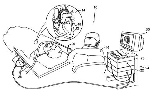
tially made to Fig. 1, which is a pictorial illustration of a
system 10 for performing ablative procedures on a heart 12 of a
living subject, which is constructed and operative in accordance
with a disclosed embodiment of the invention. The system corn-
prises a catheter 14, which is percutaneously inserted by an op-
erator 16 through the patient's vascular system into a chamber
or vascular structure of the heart. The operator 16, who is typ-
ically a physician, brings the catheter's distal tip 18 into
contact with the heart wall at an ablation target site. Electri-
cal activation maps may then be prepared, according to the meth-
ods disclosed in U.S. Patent Nos. 6,226,542, and 6,301,496, and
in commonly assigned U.S. Patent No. 6,892,091. Although the em-
bodiment described with respect to Fig. 1 is concerned primarily
with cardiac ablation, the principles of the invention may be
applied, mutatis mutandis, to other catheters and probes and to
body tissues other than the heart.
[0024]
Areas determined to be abnormal by evaluation of
the electrical activation maps can be ablated by application of
thermal energy, e.g., by passage of radiofrequency electrical
5
CA 2725865 2017-11-09
current through wires in the catheter to one or more electrodes
at the distal tip 18, which apply the radiofrequency energy to
the myocardium. The energy is absorbed in the tissue, heating it
to a point (typically about 5000) at which it permanently loses
its electrical excitability. When successful, this procedure
creates non-conducting lesions in the cardiac tissue, which dis-
rupt the abnormal electrical pathway causing the arrhythmia. Al-
ternatively, other known methods of applying ablative energy can
be used, e.g., ultrasound energy, as disclosed in U.S. Patent
Application Publication No. 2004/0102769. The principles of the
invention can be applied to different heart chambers, and to
mapping in sinus rhythm, and when many different cardiac ar-
rhythmias are present.
[0025] The
catheter 14 typically comprises a handle 20,
having suitable controls on the handle to enable the operator 16
to steer, position and orient the distal end of the catheter as
desired for the ablation. To aid the operator 16, the distal
portion of the catheter 14 contains position sensors (not shown)
that provide signals to a positioning processor 22, located in a
console 24. The console 24 typically contains an ablation power
generator 25. The catheter 14 may be adapted to conduct ablative
energy to the heart using any known ablation technique, e.g.,
radiofrequency energy, ultrasound energy, and laser energy. Such
methods are disclosed in commonly assigned U.S. Patent
Nos. 6,814,733, 6,997,924, and 7,156,816.
[0026] The
positioning processor 22 is an element of a
positioning sub-system of the system 10 that measures location
and orientation coordinates of the catheter 14.
[0027] In
one embodiment, the positioning sub-system
comprises a magnetic position tracking arrangement that deter-
mines the position and orientation of the catheter 14 by gener-
ating magnetic fields in a predefined working volume its vicini-
ty and sensing these fields at the catheter. The magnetic posi-
tion tracking arrangement typically comprises a set of external
6
CA 2725865 2017-11-09
radiators, such as field generating coils 28, which are located
in fixed, known positions external to the patient. The field
generating coils 28 are driven by field generators (not shown),
which are typically located in the console 24, and generate
fields, typically electromagnetic fields, in the vicinity of the
heart 12.
[0028] In
an alternative embodiment, a radiator in the
catheter 14, such as a coil, generates electromagnetic fields,
which are received by sensors (not shown) outside the patient's
body.
[0029] Some
position tracking techniques that may be
used for this purpose are described, for example, in the above-
noted U.S. Patents 6,690,963, and in commonly assigned U.S. Pa-
tent Nos. 6,618,612 and 6,332,089, and U.S. Patent Application
Publications 2004/0147920, and 2004/0068178. Although the posi-
tioning sub-system shown in Fig. 1 uses magnetic fields, the
methods described below may be implemented using any other suit-
able positioning system, such as systems based on electromagnet-
ic fields, acoustic or ultrasonic measurements. A suitable corn-
mercial positioning sub-system is the CARTO XP EP Navigation and
Ablation System, available from Biosense Webster, Inc., 3333 Di-
amond Canyon Road, Diamond Bar, CA 91765.
[0030] As
noted above, the catheter 14 is coupled to the
console 24, which enables the operator 16 to observe and regu-
late the functions of the catheter 14. Console 24 includes a
processor, preferably a computer with appropriate signal pro-
cessing circuits. The processor is coupled to drive a moni-
tor 30. The signal processing circuits typically receive, ampli-
fy, filter and digitize signals from the catheter 14, including
signals generated by the above-noted sensors and a plurality of
sensing electrodes (not shown) located distally in the cathe-
ter 14. The digitized signals are received and used by the con-
sole 24 to compute the position and orientation of the cathe-
ter 14 and to analyze the electrical signals from the elec-
7
CA 2725865 2017-11-09
trodes. The information derived from this analysis may be used
to generate an electrophysiological map of at least a portion of
the heart 12 or structures such as the pulmonary venous ostia,
for diagnostic purposes such as locating an arrhythmogenic area
in the heart or to facilitate therapeutic ablation.
[0031]
Typically, the system 10 includes other elements,
which are not shown in the figures for the sake of simplicity.
For example, the system 10 may include an electrocardiogram
(ECG) monitor, coupled to receive signals from one or more body
surface electrodes, so as to provide an ECG synchronization sig-
nal to the console 24. As mentioned above, the system 10 typi-
cally also includes a reference position sensor, either on an
externally-applied reference patch attached to the exterior of
the subject's body, or on an internally-placed catheter, which
is inserted into the heart 12 maintained in a fixed position
relative to the heart 12. By comparing the position of the cath-
eter 14 to that of the reference catheter, the coordinates of
catheter 14 are determined relative to the heart 12, irrespec-
tive of heart motion. Alternatively, any other suitable method
may be used to compensate for heart motion. Nevertheless, the
positioning sub-system cannot guarantee that an energy-conveying
component of the catheter 14 is in actual contact with the tis-
sue to be ablated.
[0032]
Reference is now made to Fig. 2, which is a cuta-
way view of distal end 33 of catheter 14 (Fig. 1), showing de-
tails of the structure of the catheter in accordance with an em-
bodiment of the present invention. The catheter shown in Fig. 2
includes a pressure transducer, which is more fully disclosed in
commonly assigned U.S. Patent Application Publication
No. 2009/0093806. Other known types of pressure transducers can
be substituted for the pressure transducer described with refer-
ence to Fig. 2.
[0033] Catheter 14 comprises a flexible insertion
tube 54, with a distal section 72 connected to the remainder of
8
CA 2725865 2017-11-09
CA 02725865 2010-12-17
= .
the insertion tube 54 at a joint 56. The insertion tube is cov-
ered by a flexible, insulating material 60, such as CelconTm or
TeflonTm. The area of joint 56 is covered, as well, by a flexi-
ble, insulating material, which may be the same as material 60
or may be specially adapted to permit unimpeded bending and com-
pression of the joint, (This material is cut away in Fig. 2 in
order to expose the internal structure of the catheter.) Distal
tip 18 may be covered, at least in part, by an electrode 50,
which is typically made of a metallic material, such as a plati-
num/iridium alloy. Alternatively, other suitable materials may
be used, as will be apparent to those skilled in the art. The
distal section 72 is typically relatively rigid, by comparison
with a proximal section 74.
[0034] The
distal section 72 is connected to the proxi-
mal section 74 by a resilient member 58. In Fig. 2, the resil-
ient member 58 has the form of a coil spring, but other types of
resilient components may alternatively be used for this purpose.
For example, resilient member 58 may comprise a polymer, such as
silicone, polyurethane, or other plastics, with the desired
flexibility and strength characteristics. Resilient member 58
permits a limited range of relative movement between distal sec-
tion 72 and the proximal section 74 in response to forces ex-
erted on the distal section 72 or directly against the distal
tip 18. Such forces are encountered when the distal tip is
pressed against the endocardium during an ablation procedure.
The desired pressure for good electrical contact between the
distal tip and the endocardium during ablation is on the order
of 20-30 grams. The spring serving as the resilient member 58 in
this embodiment may be configured, for example, to permit axial
displacement (i.e., lateral movement along the axis of catheter
14) of the distal end 33 by about 1-2 mm and angular deflection
of the distal section 72 with respect to the proximal section 74
by up to about 30 degrees in response to a desired pressure.
[0035] As
noted above, distal section 72 contains a mag-
netic position sensor 62. Position sensor 62 may comprise one or
9
CA 02725865 2010-12-17
. .
more miniature coils, and typically comprises multiple coils
oriented along different axes. Alternatively, position sensor 62
may comprise another type of magnetic sensor, such as a Hall ef-
fect or magnetoresistive sensor, for example. The magnetic
fields created by the field generating coils 28 (Fig. 1) cause
the position sensor 62 to generate electrical signals, with am-
plitudes that are indicative of the position and orientation of
position sensor 62 relative to the fixed frame of reference of
field generating coils 28. Positioning processor 22 (Fig. 1) re-
ceives these signals via wires (not shown in the figures) run-
ning through catheter 14, and processes the signals in order to
derive the location and orientation coordinates of distal tip 18
in this fixed frame of reference, as described in the patents
and patent applications cited above. Some of the position sens-
ing and mapping features of the catheter 14 are implemented in
the NOGA-STAR catheter and the and CARTOTm systems, marketed by
Biosense Webster, Inc.
[0036] In
addition, catheter 14 contains a miniature
magnetic field generator 64 near the distal tip 18, which is
driven by a current conveyed through catheter 14 from console 24
(Fig. 1). The current is generated so as to create magnetic
fields that are distinguishable in time and/or frequency from
the fields of field generating coils 28 (Fig. 1). For example,
the current supplied to field generator 64 may be generated at a
selected frequency in the range between about 16 kHz and 25 kHz,
while field generating coils 28 are driven at different frequen-
cies. Additionally or alternatively, the operation of field gen-
erating coils 28 and field generator 64 may be time-multiplexed.
[0037] The
magnetic field created by field generator 64
causes one or more coils in position sensor 62 to generate elec-
trical signals at the drive frequency of field generator 64. The
amplitudes of these signals vary depending upon the location and
orientation of distal tip 18 relative to proximal section 74.
Positioning processor 22 (Fig. 1) processes these signals in or-
der to determine the axial displacement and the magnitude of the
CA 02725865 2010-12-17
. .
angular deflection of the distal tip 18 relative to the proximal
section 74. Position sensor 62 may determine six position and
orientation coordinates (X, Y, Z directions and pitch, yaw and
roll orientations) of the distal end and distal tip of cathe-
ter 14. For this purpose, at least two sensing coils are typi-
cally required in the position sensor. In the present embodi-
ment, three sensing coils 76 are used, in order to improve the
accuracy and reliability of the position measurement. Alterna-
tively, if only a single sensing coil is used, system 10 may be
able to determine only five position and orientation coordinates
(X, Y, Z directions and pitch and yaw orientations). As the
readings of displacement and deflection should be accurate to
within a few tenths of a millimeter and about one degree, re-
spectively, it is desirable to include three coils 76 in posi-
tion sensor 62, preferably mutually orthogonal, as shown in
Fig. 2.
[0038] As
the position of the position sensor 62 with
reference to some fixed frame of reference (not shown) can be
determined, it is possible to compute the relative movement of
the distal tip 18 relative to the proximal section 74. This
gives a measure of the deformation and angular deviation of re-
silient member 58. Generally speaking, the deformation is pro-
portional to the mechanical force that is exerted on the resil-
ient member 58, which is roughly equal to the force that is ex-
erted on the distal tip 18 by the heart tissue with which the
distal tip 18 is in contact. Thus, the combination of field gen-
erator 64 with position sensor 62 serves as a pressure sensing
system for determining the approximate pressure exerted by the
endocardial tissue on the distal tip 18 of the catheter 14 (or
equivalently, the pressure exerted by electrode 50 against the
endocardial tissue).
[0039]
Reference is now made to Fig. 3, which is a pic-
torial view of the distal end 33 of the catheter 14 in contact
with endocardium 70 of the heart 12, in accordance with an em-
bodiment of the invention. Pressure exerted by the distal tip 18
11
CA 02725865 2010-12-17
, .
against the endocardium 70 deforms the endocardial tissue
slightly, so that the electrode 50 contacts the tissue over a
relatively large area. Since the electrode 50 engages the endo-
cardium 70 at an angle 82, rather than head-on, distal section
72 bends at joint 56 forming a bend angle 84 relative to the in-
sertion tube of the catheter. The bend facilitates optimal con-
tact between the electrode and the endocardial tissue.
[0040]
Reverting to Fig. 2, positioning processor 22
(Fig. 1) receives and processes the signals generated by posi-
tion sensor 62 in response to the magnetic field of genera-
tor 64, in order to derive an indication of the pressure exerted
by distal tip 18 on endocardium 70 (Fig. 3). As noted earlier,
for good ablation, pressure of about 20-30 grams is desirable.
Lower pressure means that there may be inadequate contact be-
tween electrode 50 and the endocardial tissue. As a result, much
or all of the thermal energy may be carried away by the blood
inside the heart, and the tissue will be ablated inadequately or
not at all. Higher pressure means that the electrode is pressing
too hard against the endocardial tissue. Excessive pressure of
this sort may cause cavitation in the tissue, leading to exten-
sive tissue damage and possibly even perforation of the heart
wall.
[0041] It is
possible to determine the coordinates of
the position sensor 62 with respect to some fixed frame of ref-
erence. In embodiments in which the field generator 64 has at
least two coils it is also possible to determine the directional
orientations of the axes of the position sensor 62 with respect
to one another, and thereby compute the bend angle 84 (Fig. 3).
[0042] By
virtue of the combined sensing of displacement
and deflection, this pressure sensing system reads the pressure
correctly regardless of whether the electrode engages the endo-
cardium head-on or at an angle. The pressure reading is insensi-
tive to temperature variations and free of drift, unlike piezo-
electric sensors, for example.
12
[0043] The
magnitudes of the displacement and deflection
may be combined by vector addition to give a total magnitude of
the movement of distal tip 18 relative to the proximal section
74. When there are three coils, the system can determine the
sition of the distal section 72 and the distal tip 18 with six
degrees of freedom. Force vectors 78, 80 can then be computed,
the vector 80 representing the magnitude of the component that
is normal to the wall of the heart 12. The relationships between
force and deflection may be pre-calibrated for each catheter and
a calibration table constructed and used subsequently in force
measurements.
[0044]
Referring again to Fig. 1, console 24 outputs an
indication of the pressure measured to the operator 16, and may
issue an alarm if the pressure is too low or too high. Optional-
ly, ablation power generator 25 may be interlocked, so as to
supply power to electrode 50 (Fig. 2) only when the pressure
against the endocardium 70 (Fig. 3) is in the desired range. Al-
ternatively or additionally, the pressure indication may be used
in closed-loop control of an automated mechanism for maneuvering
and operating catheter 14, as described more fully in the above
noted U.S. Patent Application Publication No. 2004/0102769, in
order to ensure that the mechanism causes the distal section 72
to engage the endocardium 70 (Fig. 3) in the proper location and
with the appropriate contact pressure.
[0045] While RF power
is discussed with respect to the
methods and systems herein, in embodiments of the system 10
(Fig. 1), other forms of energy may be delivered to the tissue,
i.e., laser and microwave techniques, and high intensity focused
ultrasound energy, as described in commonly assigned U.S. Patent
Application Publication No. 2006/0287648.
[0046] The
product P * F gives a good indication of the
rate of ablation of the tissue, where P represents RF power and
F represents the magnitude of the force vector exerted by the
catheter against the endocardial surface of the heart. The op-
13
CA 2725865 2017-11-09
CA 02725865 2010-12-17
erator may increase or decrease either or both of the component
parameters, P and F in order to control the ablation rate. The
total volume V of tissue ablated, up to a maximum dictated by
tissue characteristics and safety considerations, is roughly
proportional to the product
V = k(P * F * T) (1),
wherein T is the time duration of RF power application, and k is
a proportionality constant.
[0047]
Reference is now made to Fig. 4, which is a corn-
posite map 86 of the heart illustrating aspects of a cardiac ab-
lation procedure in accordance with a disclosed embodiment of
the invention. The procedure may be actual or simulated, for
purposes of predicting the necessary force to be applied by a
cardiac catheter 88 in an operating position within a chamber of
heart 90. Arrows 92, 94 represent two different force vectors,
the length of the arrows corresponding to the magnitudes of the
forces being. A dosage of energy, e.g., RF ablation current is
to be applied at a predetermined power level for a time suffi-
cient to produce an ablation lesion. Predicted small and large
circular ablation zones 96, 98 correspond to the short and long
arrows 92, 94, respectively.
[0048]
Additionally or alternatively, when the force be-
ing applied and the RF power are known, the size of the ablation
zone can be predicted and dynamically displayed. The complete-
ness of the ablation can be calculated as time varies, and pro-
gress displayed during the procedure as by changing the visual
characteristics of the ablation zones 96, 98. The ablation vol-
ume grows over time in proportion to the product P*F.
[0049]
Similarly, by fixing the desired size of the ab-
lation zone, the required force can be computed at a given RF
power and application time or for a given total energy dosage at
different combinations of application time and RF power.
[0050] Using
the map 86, a simple, clear measure of es-
timated ablation volume is provided to the operator, which can
be measured easily and accurately in near real-time.
14
[0051]
Reference is now made to Fig. 5, which is a flow
chart of a method of estimation and mapping of tissue ablation
volume, in accordance with a disclosed embodiment of the inven-
tion. The method requires a determination of mechanical force
developed by contact between a probe and the tissue site to be
ablated. The method can be performed by the system 10 (Fig. 1)
using catheter 14. However, other methods that are capable of
measuring the pressure can be applied, for example impedance-
based measurements, such as disclosed in commonly assigned U.S.
Patent Application Publication No. 2007/0060832. Alternatively,
suitable optical or ultrasound techniques may be used to deter-
mine the mechanical force.
[0052] The
process begins at initial step 100. The heart
is catheterized conventionally and the catheter navigated to a
desired location at which tissue ablation is required.
[0053] Next, at step 102, the
cardiac catheter is
brought into contact with the endocardial surface, generally at
an angle of incidence other than perpendicular as shown in
Fig. 3.
[0054] Next, at step
104, The mechanical force or a de-
sired force vector applied to the endocardium by the catheter is
determined. The deflection angle, e.g., angle 82 (Fig. 3) may be
determined automatically, using the information provided by lo-
cation sensors in the catheter.
[0055] Next, at step
106, ablation power, e.g., RF pow-
er, is determined for the current medical procedure.
[0056]
Then, at step 108, an estimated ablation time is
tentatively chosen, which establishes the energy dosage to be
applied. Alternatively, steps 106, 108 can be modified to set
the ablation time, and estimate power levels, respectively. The
operator may he assisted at this step in that a controller may
report an indication of an expected ablation volume at the ener-
gy dosage and the mechanical force.
CA 2725865 2017-11-09
CA 02725865 2010-12-17
. .
[0057] Next,
at step 110 the size of the lesion to be
created by ablating is computed, according to the conditions es-
tablished in step 104 and step 108.
[0058]
Control now proceeds to decision step 112, where
it is determined if the current lesion size is acceptable. If
the determination at decision step 112 is affirmative, then con-
trol proceeds to final step 114, where power, typically RF pow-
er, is applied, and ablation is performed. During the ablation
the currently ablated tissue volume is dynamically displayed as
shown in Fig. 4 until the computed ablation volume has been
achieved. The operator may vary the power to control the time of
application. Additionally or alternatively the operator may ad-
just the position of the catheter to vary the mechanical force
applied to the endocardial tissues.
[0059] If the
determination at decision step 112 is neg-
ative, then control returns to step 108, where the ablation time
is re-estimated.
[0060]
Typically, the size of the lesion to be created
by ablation is known. In such cases, the loop defined by
step 108, step 110 and decision step 112 can be iterated auto-
matically until an acceptable size has been determined.
[0061]
Alternatively the lesion size may be computed di-
rectly at optional step 116 using the relationship of Equa-
tion 1, and then perform ablation at final step 114. In this
case, step 108, step 110 and decision step 112 can be omitted.
[0062] In
alternate embodiments of the method, proposed
power and proposed application time data can be received as in-
put and ablation volumes computed at different mechanical forces
of contact with the tissue.
Alternate Embodiment.
[0063]
Reference is now made to Fig. 6, which is a cut-
away view of distal end 33 of catheter 14 (Fig. 1), which is
constructed and operative in accordance with a disclosed embodi-
ment of the invention. This embodiment is similar to Fig. 2, ex-
16
CA 02725865 2010-12-17
. .
cept now the distal section 72 includes a conventional tempera-
ture sensor 118 that is capable of detecting abnormal rise in
temperature of the tissues at the operating site. By displaying
the output of the temperature sensor 118 on the monitor 30
(Fig. 1) or providing a suitable audible alert, the rate of ab-
lation may be controlled by responsively to the temperature of
the tissues in order to prevent charring or dangerous tempera-
ture elevation outside the computed ablation volume.
[0064]
Equation 1 can be modified to account for the
temperature such that only actual ablation time, rather than to-
tal elapsed time is taken into consideration. Ablation time can
be defined to run only when contact force exceeding a predeter-
mined force threshold is ascertained and the temperature exceeds
a predetermined temperature threshold. Alternatively, ablation
time can be defined to run only when contact force exceeding a
predetermined force threshold is ascertained or the temperature
exceeds a predetermined temperature threshold.
[0065]
Equation 1 may be modified in several ways to ac-
count for ablation time. The following examples are practical
approximations, in which various first and second order correc-
tions are not shown for clarity of presentation. The threshold
values given below are suitable:
V ' k * (P * F * T (F > Fthreshold)) (2)
V P-- k * (P * F * T (F > Fthreshold/ t > tthreshold) ) (3)
V ez-- k * (P * F * T (t > tthreshoid)) (4)
wherein Fthreshold - 5gr, and t threshold = 47 C. The ablation power
is applied only during time intervals when the conditions shown
are met.
[0066] It
will be appreciated by persons skilled in the
art that the present invention is not limited to what has been
particularly shown and described hereinabove. Rather, the scope
17
CA 02725865 2010-12-17
of the present invention includes both combinations and sub-
combinations of the various features described hereinabove, as
well as variations and modifications thereof that are not in the
prior art, which would occur to persons skilled in the art upon
reading the foregoing description.
18