Note: Descriptions are shown in the official language in which they were submitted.
CA 02726452 2010-11-29
WO 2009/158170 PCT/US2009/046308
DOCKING APPARATUS AND METHODS OF USE
BACKGROUND OF THE INVENTION
[0001] 1. Field of the Invention. The present invention relates generally to
medical
systems and methods for treatment. More particularly, the present invention
relates to
systems and methods for treating aneurysms.
[0002] Aneurysms are enlargements or "bulges" in blood vessels which are often
prone to
rupture and which therefore present a serious risk to the patient. Aneurysms
may occur in
any blood vessel but are of particular concern when they occur in the cerebral
vasculature or
the patient's aorta.
[0003] The present invention is particularly concerned with aneurysms
occurring in the
aorta, particularly those referred to as aortic aneurysms. Abdominal aortic
aneurysms
(AAA's) are classified based on their location within the aorta as well as
their shape and
complexity. Aneurysms which are found below the renal arteries are referred to
as infrarenal
abdominal aortic aneurysms. Suprarenal abdominal aortic aneurysms occur above
the renal
arteries, while thoracic aortic aneurysms (TAA's) occur in the ascending,
transverse, or
descending part of the upper aorta.
[0004] Infrarenal aneurysms are the most common, representing about eighty
percent
(80%) of all aortic aneurysms. Suprarenal aneurysms are less common,
representing about
20% of the aortic aneurysms. Thoracic aortic aneurysms are the least common
and often the
most difficult to treat.
[0005] The most common form of aneurysm is "fusiform," where the enlargement
extends
about the entire aortic circumference. Less commonly, the aneurysms may be
characterized
by a bulge on one side of the blood vessel attached at a narrow neck. Thoracic
aortic
aneurysms are often dissecting aneurysms caused by hemorrhagic separation in
the aortic
wall, usually within the medial layer. The most common treatment for each of
these types
and forms of aneurysm is open surgical repair. Open surgical repair is quite
successful in
patients who are otherwise reasonably healthy and free from significant co-
morbidities. Such
open surgical procedures may be problematic, however, since access to the
abdominal and
thoracic aortas is difficult to obtain and because the aorta must be clamped
off, placing
significant strain on the patient's heart.
1
CA 02726452 2010-11-29
WO 2009/158170 PCT/US2009/046308
[0006] Over the past decade, endoluminal grafts have come into widespread use
for the
treatment of aortic aneurysm in patients who cannot undergo open surgical
procedures. In
general, endoluminal repairs access the aneurysm "endoluminally" through
either or both
iliac arteries in the groin. The grafts, which typically have been fabric or
membrane tubes
supported and attached by various stent structures, are then implanted,
typically requiring
several pieces or modules to be assembled in situ. Successful endoluminal
procedures have a
much shorter recovery period than open surgical procedures.
[0007] Present endoluminal aortic aneurysm repairs, however, suffer from a
number of
limitations. For example, a significant number of endoluminal repair patients
experience
leakage at the proximal juncture (attachment point closest to the heart)
within two years of
the initial repair procedure. While such leaks can often be fixed by further
endoluminal
procedures, the need to have such follow-up treatments significantly increases
cost and is
certainly undesirable for the patient. A less common but more serious problem
has been graft
migration. In instances where the graft migrates or slips from its intended
position, open
surgical repair is required. This is a particular problem since the patients
receiving the
endoluminal grafts are often those who are not considered to be good surgical
candidates.
[0008] Further shortcomings of the present endoluminal graft systems relate to
both
deployment and configuration. For example, many of the commercially available
endovascular systems are too large (above 12F) for percutaneous introduction.
Moreover,
current devices often have an annular support frame that is stiff and
difficult to deliver as well
as unsuitable for treating many geometrically complex aneurysms, particularly
infrarenal
aneurysms with little space between the renal arteries and the upper end of
the aneurysm,
referred to as short-neck or no-neck aneurysms. Aneurysms having torturous
geometries, are
also difficult to treat.
[0009] For these reasons, it would be desirable to provide improved methods
and systems
for the endoluminal and minimally invasive treatment of aortic aneurysms. In
particular, it
would be desirable to provide prostheses with better sealing and minimal or no
endoleaks. It
would also be desirable to provide prostheses which resist migration, which
are flexible,
relatively easy to deploy, use standardize components and/or a modular design
that can treat
many if not all aneurismal configurations, including short-neck and no-neck
aneurysms as
well as those with highly irregular and asymmetric geometries. It would be
further desirable
to provide systems and methods which are compatible with current designs for
endoluminal
2
CA 02726452 2010-11-29
WO 2009/158170 PCT/US2009/046308
stents and grafts, including single lumen stents and grafts, bifurcated stents
and grafts,
parallel stents and grafts, as well as with double-walled filling structures
which are the
subject of the commonly owned, copending applications described below. The
systems and
methods would preferably be deployable with the stents and grafts at the time
the stents and
grafts are initially placed. Additionally, it would be desirable to provide
systems and
methods for repairing previously implanted aortic stents and grafts, either
endoluminally or
percutaneously. At least some of these objectives will be met by the
inventions described
hereinbelow.
[0010] 2. Description of the Background Art. U.S. Patent Publication No.
2006/0025853 describes a double-walled filling structure for treating aortic
and other
aneurysms. Copending, commonly owned U.S. Patent Publication No. 2006/0212112,
describes the use of liners and extenders to anchor and seal such double-
walled filling
structures within the aorta. The full disclosures of both these publications
are incorporated
herein by reference. PCT Publication No. WO 01/21108 describes expandable
implants
attached to a central graft for filling aortic aneurysms. See also U.S. Patent
Nos. 5,330,528;
5,534,024; 5,843,160; 6,168,592; 6,190,402; 6,312,462; 6,312,463; U.S. Patent
Publications
2002/0045848; 2003/0014075; 2004/0204755; 2005/0004660; and PCT Publication
No.
WO 02/102282.
BRIEF SUMMARY OF THE INVENTION
[0011] The present invention provides systems and methods for the treatment of
aneurysms, particularly aortic aneurysms including both abdominal aortic
aneurysms (AAA)
and thoracic aortic aneurysms (TAA).
[0012] In a first aspect of the present invention a system for treating an
aneurysm in a
blood vessel comprises a docking scaffold radially expandable from a
contracted
configuration to an expanded configuration and having an upstream end, a
downstream end
and a central passageway therebetween. In the expanded configuration the
upstream end
engages a portion of the blood vessel upstream of the aneurysm. The system
also comprises
a first leg scaffold that is radially expandable from a contracted
configuration to an expanded
configuration and a portion of the first leg scaffold is slidably received in
the central
passageway such that an outside surface of the first leg scaffold in the
expanded
configuration engages an inside surface of the docking scaffold. The system
also comprises a
second leg scaffold radially expandable from a contracted configuration to an
expanded
CA 02726452 2010-11-29
WO 2009/158170 PCT/US2009/046308
configuration, and a portion of the second leg scaffold is slidably received
in the central
passageway such that an outside surface of the second leg scaffold in the
expanded
configuration engages an inside surface of the docking scaffold. A first
double-walled filling
structure is coupled with at least one of the leg scaffolds in the expanded
configuration. The
filling structure has an outer wall and an inner wall, and the filling
structure is adapted to be
filled with a hardenable fluid filling medium so that the outer wall conforms
to an inside
surface of the aneurysm and the inner wall forms a first substantially tubular
lumen to
provide a path for blood flow therethrough.
[0013] The hardenable filling material may comprise a polymer and the blood
vessel may
be an aorta. Often, the aneurysm is an abdominal aortic aneurysm. The system
may further
comprise an expandable member such as a balloon and the balloon may be
tapered.
[0014] In some embodiments, the outer surface of the first leg scaffold in the
expanded
configuration engages the outer surface of the expanded second leg scaffold
thereby defining
a mating region. The mating region may be disposed at least partially within
the central
passageway. The mating region may form a generally double D-shaped cross
section.
[0015] The first leg and second leg scaffolds may traverse the aneurysm in a
direction
substantially parallel to one another or in some cases, they may cross each
other. The
downstream end of the first leg or second leg scaffold may be disposed
downstream of the
aneurysm or it may be disposed in an iliac artery. The downstream end of the
docking
scaffold may be disposed in a number of positions including upstream of the
aneurysm, in the
aneurismal sac, below the aneurysm or disposed in the blood vessel so as to
traverse a renal
artery bifurcation without inhibiting blood flow. The docking scaffold may
comprise an
expandable region that is adapted to linearly expand and contract in order to
accommodate
aneurysms of varying length. The docking scaffold may comprise a self-
expanding region
and a balloon expandable region as well as also including an external flange.
[0016] When the first double-walled filling structure is coupled with the
first leg scaffold,
the first double-walled filling structure at least partially fills the
aneurysm when filled with
the hardenable filling material. Some embodiments may further comprise a
second double-
walled filling structure having an outer wall and an inner wall, wherein the
second filling
structure is adapted to be filled with a hardenable fluid filling medium so
that the outer wall
conforms to an inside surface of the aneurysm and the inner wall forms a
second substantially
tubular lumen to provide a path for blood flow therethrough. The second double-
walled
4
CA 02726452 2010-11-29
WO 2009/158170 PCT/US2009/046308
filling structure may be coupled with the second leg scaffold in the expanded
configuration.
When the second double-walled filling structure is coupled with the second leg
scaffold, the
second double-walled filling structure at least partially fills the aneurysm
when filled with the
hardenable filling material. Some embodiments may also further comprise a
third double-
walled filling structure having an outer wall and an inner wall, wherein the
third filling
structure is adapted to be filled with a hardenable fluid filling medium so
that the outer wall
conforms to an inside surface of the aneurysm and the inner wall forms a third
substantially
tubular lumen to provide a path for blood flow therethrough. The third double-
walled filling
structure is disposed at least partially over the docking scaffold in the
expanded
configuration. When the third double-walled filling structure is coupled with
the docking
scaffold, the third double-walled filling structure often at least partially
fills the aneurysm
when filled with the hardenable filling material.
[0017] In some embodiments, the third double-walled filling structure is
coupled with the
docking scaffold and an upstream portion of the docking scaffold remains
uncovered by the
first double-walled filling structure in the expanded configuration. The
uncovered upstream
portion may be disposed upstream of the aneurysm. The uncovered upstream
portion may
also engage the blood vessel in the expanded configuration. When filled with
filling medium,
the third double-walled filling structure may seal an upper portion of the
aneurysm thereby
preventing blood flow between the outer wall of the third double-walled
filling structure and
an inner wall of the blood vessel. The third double-walled filling structure
may be coupled
with the docking scaffold and a downstream portion of the docking scaffold may
remain
uncovered by the third double-walled filling structure in the expanded
configuration.
[0018] The docking scaffold may comprise a restraining element that limits
expansion of at
least a portion of the docking scaffold to a target diameter. The restraining
element may be
expandable. The restraining element may comprise a band that is disposed
around the
docking scaffold. Sometimes the restraining element may form a tapered region
on one end
of the docking scaffold in the expanded configuration.
[0019] In some embodiments, an upstream portion of the first leg scaffold
remains
uncovered in the expanded configuration and a downstream portion of the first
leg scaffold
may remain uncovered in the expanded configuration. The downstream portion of
the first
leg scaffold may be disposed in an iliac artery. The second leg scaffold may
comprise an
upstream portion that remains uncovered in the expanded configuration and a
downstream
5
CA 02726452 2010-11-29
WO 2009/158170 PCT/US2009/046308
portion of the second leg scaffold may also remain uncovered in the expanded
configuration.
The downstream portion of the second leg scaffold may be disposed in an iliac
artery. The
first and second leg scaffolds may be fixedly coupled together and either may
comprise an
external flange. Sometimes, the first or second leg scaffolds may comprise a
self-expanding
region and a balloon expandable region.
[0020] In still other embodiments, the first leg scaffold or second leg
scaffold may
comprise a sealing element disposed at least partially along the portion of
the respective
scaffold that is slidably received in the central passageway. The sealing
element forms a seal
between the outside surface of the first leg or second leg scaffold in the
expanded
configuration and the inside surface of the docking scaffold. The sealing
element may be
expandable and may have a chamfered surface.
[0021] In some embodiments, the system further comprise a third leg scaffold.
The third
leg scaffold is radially expandable from a contracted configuration to an
expanded
configuration. A portion of the third leg scaffold may be slidably received by
the first or
second leg scaffold such that a surface of the third leg scaffold in the
expanded configuration
engages a surface of the first or second leg scaffold. For example, the
outside surface of the
third leg scaffold may engage an inside surface of the first or second leg
scaffold, or vice
versa; the inside surface of the third leg scaffold may engage an outside
surface of the first or
second leg scaffold. An upstream end of the third leg scaffold may be disposed
downstream
of the aneurysm, for example in an iliac artery. Some embodiments may further
comprise a
fourth double-walled filling structure. The fourth filling structure has an
outer wall and an
inner wall and is adapted to be filled with a hardenable fluid filling medium
so that the outer
wall conforms to an inner surface of the aneurysm and the inner wall forms a
fourth
substantially tubular lumen to provide a path for blood flow therethrough. The
fourth double-
walled filling structure may be coupled with the third leg scaffold. When
filled with the
hardenable filling material, the fourth double-walled filling structure may at
least partially fill
an aneurysm in the iliac artery.
[0022] The system may also further comprise a fourth leg scaffold. The fourth
leg scaffold
is radially expandable from a contracted configuration to an expanded
configuration. A
portion of the fourth leg scaffold may be slidably received by the second leg
scaffold such
that a surface of the fourth leg scaffold in the expanded configuration
engages a surface of the
second leg scaffold. For example, the outside surface of the fourth leg
scaffold may engage
6
CA 02726452 2010-11-29
WO 2009/158170 PCT/US2009/046308
an inside surface of the second leg scaffold, or vice versa, the inside
surface of the fourth leg
scaffold may engage an outside surface of the second leg scaffold. An upstream
end of the
fourth leg scaffold may be disposed downstream of the aneurysm, for example in
an iliac
artery. Still some other embodiments may further comprise a fifth double-
walled filling
structure. The fifth filling structure has an outer wall and an inner wall.
The fifth filling
structure is adapted to be filled with a hardenable fluid filling medium so
that the outer wall
conforms to an inner surface of the aneurysm and the inner wall forms a fifth
substantially
tubular lumen to provide a path for blood flow therethrough. The fifth double-
walled filling
structure is coupled with the fourth leg scaffold. When filled with the
hardenable filling
material, the fourth double-walled filling structure at least partially fills
an aneurysm in the
iliac artery.
[0023] In some embodiments, the system may comprise a crown scaffold radially
expandable from a contracted configuration to an expanded configuration. The
crown
scaffold has an upstream portion and a downstream portion. In the expanded
configuration,
the downstream portion is slidably received by the upstream end of the docking
scaffold. The
downstream portion may be slidably received in the central passageway such
that an outside
surface of the crown scaffold engages an inside surface of the docking
scaffold. The
upstream portion of the crown scaffold may engage a portion of the blood
vessel upstream of
the aneurysm. The crown scaffold may be self-expanding, balloon expandable or
a
combination thereof.
[0024] Sometimes, the docking scaffold comprises a divider disposed within the
docking
scaffold and adapted to separate the slidably received portion of the first
leg scaffold from the
slidably received portion of the second leg scaffold. The divider is often
integrally formed
with the docking scaffold. The divider may split the cross-section of the
docking scaffold
into two D-shaped cross-sections. The divider may be adapted to limit the
length of the
portion of the first leg scaffold and the portion of the second leg scaffold
that are slidably
received in the central passageway. Sometimes, the divider comprises an
expandable
structure, such as a double-walled filling structure, expandable from a
contracted
configuration to an expanded configuration. The expandable structure is
configured to secure
the slidably received portions of the first and second leg scaffolds when the
expandable
structure is expanded to the expanded configuration. This also helps form a
seal to prevent
blood flow past the expandable structure.
7
CA 02726452 2010-11-29
WO 2009/158170 PCT/US2009/046308
[0025] In some embodiments, the downstream end of the docking scaffold is
bifurcated, for
example, into a first portion and a second portion, wherein the first portion
is adapted to
slidably receive the first leg and the second portion is adapted to slideably
receive the second
leg. The docking scaffold may optionally be at least partially covered with a
material.
[0026] In another aspect of the present invention, a method for treating an
aneurysm in a
blood vessel comprises advancing a docking scaffold through the blood vessel
to a position
upstream of the aneurysm and radially expanding the docking scaffold from a
contracted
configuration to an expanded configuration, wherein in the expanded
configuration the
docking scaffold engages a portion of the blood vessel upstream of the
aneurysm. Advancing
a first leg scaffold through the blood vessel toward the docking scaffold
allows the first leg
scaffold to be slidably received by the docking scaffold and radially
expanding the first leg
scaffold from a contracted configuration to an expanded configuration engages
the first leg
scaffold with at least a portion of an inner surface of the docking scaffold.
Advancing a
second leg scaffold through the blood vessel toward the docking scaffold
allows the second
leg scaffold to be slidably received by the docking scaffold and radially
expanding the second
leg scaffold from a contracted configuration to an expanded configuration
engages the second
leg scaffold with at least a portion of the inner surface of the docking
scaffold. Advancing a
first double-walled filling structure through the blood vessel moves the
double-walled filling
structure toward the aneurysm and filling the first double-walled filling
structure with a fluid
filling medium allows an outer wall of the first filling structure to conform
to an inside
surface of the aneurysm and an inner wall of the first filling structure forms
a first
substantially tubular lumen to provide a first blood flow path across the
aneurysm. The first
filling structure is coupled with at least one of the leg scaffolds in the
expanded
configuration.
[0027] Advancing the docking scaffold may comprise positioning at least a
portion of the
docking scaffold upstream of the aneurysm, across the aneurysm, downstream of
the
aneurysm or across a renal artery bifurcation without obstructing blood flow
into the renal
artery. The method may also comprise restraining a portion of the docking
scaffold during
radial expansion which may form a region of the docking scaffold having a
constant
predetermined diameter or a tapered region. Sometimes, restraining comprises
limiting radial
expansion of the docking scaffold with a band disposed circumferentially
therearound.
8
CA 02726452 2010-11-29
WO 2009/158170 PCT/US2009/046308
[0028] Radially expanding the first leg and second leg scaffolds to the
expanded
configuration may comprise engaging the first leg scaffold with the second leg
scaffold and
advancing the first leg and second leg scaffolds may comprise crossing the
first leg scaffold
with the second leg scaffold.
[0029] The first filling structure may be disposed at least partially over the
first leg scaffold
in the expanded configuration. The method may also further comprise
polymerizing the fluid
filling medium in the first filling structure.
[0030] The method may further comprise advancing a second double-walled
filling
structure through the blood vessel toward the aneurysm. The method may also
comprise
filling the second double-walled filling structure with a fluid filling medium
so that an outer
wall of the second filling structure conforms to an inside surface of the
aneurysm and an
inner wall of the second filling structure forms a second substantially
tubular lumen to
provide a second blood flow path across the aneurysm. The second filling
structure may be
disposed at least partially over the second leg scaffold in the expanded
configuration. The
fluid filling medium may be polymerized in the second filling structure.
[0031] The method may also comprise advancing a third double-walled filling
structure
through the blood vessel toward the aneurysm and filling the third double-
walled filling
structure with a fluid filling medium so that an outer wall of the third
filling structure
conforms to an inside surface of the aneurysm and an inner wall of the third
filling structure
forms a third substantially tubular lumen to provide a third blood flow path
across the
aneurysm. The third filling structure may be disposed at least partially over
the docking
scaffold in the expanded configuration, and the method may comprise
polymerizing the fluid
filling medium in the third filling structure.
[0032] The method may also comprise polymerizing the fluid filling medium in
the third
filling structure. Filling the third double-walled filling structure may
comprise sealing an
upper portion of the aneurysm to prevent blood flow between an inner wall of
the aneurysm
and an outer wall of the third double walled filling structure. Radially
expanding the docking
scaffold comprises radially expanding an expandable member which may include
inflating a
balloon. In some embodiments, filling the first double-walled filling
structure comprises
filling the first filling structure while the balloon is inflated.
[0033] Sometimes, advancing the first or second leg scaffold may comprises
positioning a
portion of the scaffold in an iliac artery. Often, the method may further
comprise sealing the
9
CA 02726452 2010-11-29
WO 2009/158170 PCT/US2009/046308
first or second leg scaffolds within the docking scaffold to prevent blood
flow between an
outer surface of the first or second leg scaffolds and an inner surface of the
docking scaffold.
Sealing may include inflating a sealing element.
[0034] The method may also comprise advancing a third leg scaffold through the
blood
vessel toward the first or second leg scaffold and radially expanding the
third leg scaffold.
The third leg scaffold is advanced so that the third leg scaffold is slidably
received by the first
or second leg scaffold. The third leg scaffold is radially expanded from a
contracted
configuration to an expanded configuration. In the expanded configuration, the
third leg
scaffold engages at least a portion of a surface of the first or second leg
scaffold, for example,
the inside surface or the outside surface. Sometimes, a fourth double-walled
filling structure
with a fluid filling medium may also be advanced. The fourth filling structure
is advanced so
that an outer wall of the fourth filling structure conforms to an inside
surface of the aneurysm
and an inner wall of the fourth filling structure forms a fourth substantially
tubular lumen to
provide a fourth blood flow path. The fourth filling structure is disposed at
least partially
over the third leg scaffold in the expanded configuration. The fluid filling
medium in the
fourth filling structure may be polymerized. When the fluid filling medium is
polymerized,
the fourth filling structure may at least partially fill an aneurysm in the
iliac artery.
[0035] Sometimes, a fourth leg scaffold is advanced through the blood vessel
toward the
second leg scaffold and radially expanded from a contracted configuration to
an expanded
configuration. The fourth leg scaffold is advanced so that the fourth leg
scaffold is slidably
received by the second leg scaffold. In the expanded configuration, the fourth
leg scaffold
engages at least a portion of the surface of the second leg scaffold, for
example, the inside
surface or the outside surface. A fifth double-walled filling structure with a
fluid filling
medium may be advanced. The fifth filling structure is advanced so that an
outer wall of the
fifth filling structure forms a fifth substantially tubular lumen to provide a
fifth blood flow
path. The fifth filling structure is disposed at least partially over the
fourth leg scaffold in the
expanded configuration. The fluid filling medium in the fifth filling
structure may be
polymerized. When the fluid filling medium is polymerized, the fifth filling
structure may at
least partially fill an aneurysm in the iliac artery.
[0036] The method may also comprise advancing a crown scaffold through the
blood
vessel to a position upstream of the aneurysm and radially expanding the crown
scaffold from
a contracted configuration to an expanded configuration. In the expanded
configuration, the
CA 02726452 2010-11-29
WO 2009/158170 PCT/US2009/046308
crown scaffold engages the upstream end of the docking scaffold. The crown
scaffold may
be slidably received in the central passageway such that an outside surface of
the crown
scaffold engages an inside surface of the docking scaffold. The upstream
portion of the
crown scaffold may engage a portion of the blood vessel upstream of the
aneurysm.
[0037] These and other embodiments are described in further detail in the
following
description related to the appended drawing figures.
BRIEF DESCRIPTION OF THE DRAWINGS
[0038] Fig. 1 illustrates the anatomy of an abdominal aortic aneurysm.
[0039] Figs. 2A-2I show an exemplary method of treating an aneurysm with a
docking
station.
[0040] Figs. 3A-3C illustrate how guidewires and scaffolds will often cross
each other as
they traverse the aneurysm.
[0041] Fig. 4A-4L illustrate another exemplary embodiment of a method for
treating an
aneurysm using double-walled filling structures and a docking station.
[0042] Figs. 5A-5D show various configurations of a docking station scaffold
relative to an
abdominal aortic aneurysm.
[0043] Figs. 6A-6C illustrate the use of a restraining element to control
expansion of a
scaffold.
[0044] Figs. 7A-7C illustrate an embodiment of a sealing element.
[0045] Figs. 8A-8D illustrate another embodiment of a sealing element.
[0046] Fig. 9 illustrates use of sealing elements.
[0047] Fig. 10 illustrates another use of sealing elements.
[0048] Figs. 1 l A-11 B illustrate yet another use of sealing elements.
[0049] Figs. 12A-12C illustrate an inflatable sealing element.
[0050] Fig. 13 illustrates a configuration of scaffolds for treating
aneurysms.
[0051] Fig. 14A-14B illustrate a configuration of a docking station scaffold
with a crown
scaffold relative to an abdominal aortic aneurysm.
11
CA 02726452 2010-11-29
WO 2009/158170 PCT/US2009/046308
[0052] Figs. 15A-C illustrate configurations of a docking station scaffold
with a divider
element.
[0053] Figs. 16A-C illustrate configurations of a docking station scaffold
with a fillable
divider element.
[0054] Figs. 17A-B illustrate configurations of a docking station scaffold
that is bifurcated.
[0055] Fig. 18 shows an embodiment of an iliac extension coupled with a
docking scaffold.
[0056] Figs. 19A-19C illustrate an embodiment of a variable length endograft.
[0057] Fig. 20 illustrates the use of a flexible docking scaffold in an
aneurysm.
[0058] Fig. 21 illustrates the use of an external flange to help fix the
endograft into
position.
[0059] Fig. 22 shows a hybrid scaffold comprising a balloon expandable region
and self-
expanding region.
[0060] Figs. 23A-23B illustrate various expandable members.
DETAILED DESCRIPTION OF THE INVENTION
[0061] Fig. 1 illustrates the anatomy of an infrarenal abdominal aortic
aneurysm
comprising the thoracic aorta (TA) having renal arteries (RA) at an end above
the iliac
arteries (IA). The abdominal aortic aneurysm (AAA) typically forms between the
renal
arteries (RA) and the iliac arteries (IA) and may have regions of mural
thrombus (T) over
portions of its inner surface (S).
[0062] Figs. 2A-21 show an exemplary method of treating an aneurysm using a
docking
station scaffold. Fig. 2A shows an infrarenal abdominal aortic aneurysm AAA
similar to that
in Fig. 1. In Fig. 2B, a guidewire GW is introduced using standard
percutaneous or cutdown
procedures into an iliac artery and the guidewire is advanced across the
aneurysm toward the
renal arteries RA. A docking station delivery system 102 is then advanced over
the
guidewire GW in Fig. 2C. The delivery system 102 includes a flexible catheter
shaft 103
having a balloon 104 near its distal end and a docking station scaffold or
scaffolding 106
positioned over the balloon 104. In some embodiments, the scaffolding 106 may
be a bare
metal stent-like scaffold, while in other embodiments the scaffolding 106 may
be a covered
12
CA 02726452 2010-11-29
WO 2009/158170 PCT/US2009/046308
stent-like scaffold. The covering may be a material such as DacronTM or ePTFE,
for
example, materials that are commonly used in grafts and stent-grafts. An
optional retractable
outer sheath (not illustrated) may be positioned over the scaffolding 106 and
balloon 104 in
order to provide protection during delivery. The delivery catheter is advanced
across the
aneurysm so that approximately one-third of the docking station is disposed in
the neck of the
aneurysm with approximately two-thirds of the remaining scaffolding extending
into the sac
of the aneurysm AAA after expansion. One of ordinary skill in the art will
appreciate that the
position of the scaffold 106 may be adjusted in order to accommodate various
anatomies.
[0063] In Fig. 2D, the balloon 104 is radially expanded so as to
correspondingly expand
scaffold 106 into engagement with the neck of the aneurysm. If scaffold 106
includes a
covering (not illustrated), the covering material will also be expanded with
the scaffold 106.
In this embodiment, scaffolding 106 is a balloon expandable stent-like
structure that may
have numerous geometries such as disclosed in U.S. Patents 4,733,665 to
Palmaz, 5,733,303
to Israel et al. and 5,292,331 to Boneau. Many other geometries of stent-like
structures are
well reported in the patent and medical literature. In alternative
embodiments, scaffolding
106 may also be a self-expanding stent-like structure, often fabricated from
an alloy of nickel
and titanium, such as Nitinol. After proper expansion and positioning of the
scaffold 106 has
been verified using fluoroscopy or other known techniques, the balloon 104 may
be deflated
and delivery catheter 102 removed from the patient, thus only expanded
scaffold 106 and
guidewire GW are left, as seen in Fig. 2D.
[0064] Referring now to Fig. 2E, a second guidewire GW is introduced using
standard
percutaneous or cutdown procedures from the contralateral leg, across the
aneurysm AAA
toward the renal arteries RA. In this exemplary embodiment, both guidewires
are illustrated
traversing the aneurysm AAA more or less parallel to one another, as seen in
Fig. 2E.
However, often the guidewires GW will cross and this will be discussed below.
After both
guidewires GW are properly positioned, a scaffolding delivery system 108 is
advanced over
the first guidewire GW, across the aneurysm AAA into the docking station 106.
Delivery
system 108 includes a catheter shaft 109 having a balloon 110 disposed near a
distal end of
the shaft 109 and a long scaffolding 112 disposed over the balloon 110.
Scaffolding 112 may
also optionally be covered with a material such as DacronTM or ePTFE, as
described above
with respect to docking station 106, or it may be a bare metal or polymer
scaffold. An
optional outer sheath (not illustrated) may also be used to protect and/or
constrain the balloon
110 and scaffolding 112 during delivery. The scaffolding 112 is balloon
expandable although
13
CA 02726452 2010-11-29
WO 2009/158170 PCT/US2009/046308
it may also be self-expanding and generally takes the same form as the docking
station 106
with the major difference being its length. Scaffolding 112 is long enough the
traverse the
aneurysm AAA while still providing long enough proximal and distal ends that
can expand
into and engage the docking station 106 and the iliac arteries. Scaffolding
112 is advanced
into the docking station 106 approximately one-third of the way, although
clearly this may be
modified as required.
[0065] Fig. 2F also shows another scaffolding delivery system 114 advanced
over the
second guidewire GW. Delivery system 114 is similar to delivery system 108 and
includes a
catheter shaft 115 having a balloon 118 disposed near the distal end of shaft
115 and
scaffolding 116 is disposed over the balloon 118. Scaffolding 116 may also be
covered with
a material similar to that described above with respect to scaffolding 112 or
it may remain
uncovered. An optional outer sheath (not illustrated) may also be used to
protect and/or
constrain the balloon 118 and scaffolding 116 during delivery. Scaffolding 116
is balloon
expandable, but may be self-expanding and generally takes the same form as
scaffolding 112.
Scaffolding 116 is advanced into docking station 106 approximately one-third
of the way,
although this may be adjusted as required. Fig. 2F shows both scaffolds 112,
116 traversing
the aneurysm AAA parallel to one another, yet as previously discussed, often
guidewires GW
will cross, thus scaffolds 112 and 116 would also cross as they traverse the
aneurysm.
[0066] Referring now to Fig. 2G, once both scaffolds 112, 116 have been
positioned across
the aneurysm and into docking station 106, balloons 110, 118 are inflated so
as to radially
expand scaffolds 112, 116 such that one end of each scaffold engages an iliac
artery while the
opposite end of each scaffold engages at least a portion of the inner surface
of docking station
106. If the scaffolds 112, 116 are covered, the covering material (not
illustrated) will also
expand with the scaffold. Each balloon 110, 118 may be inflated independently
of one
another, or in preferred embodiments, both balloons 110, 118 are inflated
simultaneously,
thereby also expanding both scaffolds 112, 116 simultaneously. This helps to
ensure that
both scaffolds expand symmetrically with respect to one another and against
one another so
that the ends of each scaffold expand into the preferred double D-shaped
configuration within
the docking station 106, as seen in Fig. 21. Other geometries of the mating
ends of scaffolds
112 and 116 are possible, such as circular, elliptical, etc. and ideally the
region where the two
scaffolds meet should have minimal impact on disrupting blood flow
thereacross. Balloons
110 and 118 are then deflated and delivery catheters 108 and 114 are removed
from the
treatment site.
14
CA 02726452 2010-11-29
WO 2009/158170 PCT/US2009/046308
[0067] The docking station 106 and two scaffold legs 112, 116 now form the
basis of a
blood pathway that will exclude aneurysm AAA. In the embodiment where
scaffolds 112,
116 include a covering material such as Dacron TM or ePTFE, the lumens are
fully formed and
blood will flow from the thoracic aorta TA into docking station 106 and then
flow is
bifurcated across aneurysm AAA into both iliac arteries IA. In the embodiment
where the
scaffolds 112, 116 do not have a covering material and are bare metal or bare
material
scaffolds, blood can still flow through the sidewall apertures of the expanded
scaffolds 112,
116. Thus, as shown in Fig. 2H, a filling material 120 may be used to fill the
aneurismal sac
so that blood flow remains within the lumens created by scaffolds 112, 116. An
intravascular
catheter (not illustrated) may be advanced into one or both expanded scaffolds
112, 116 and
either placed against an aperture in one of the scaffold sidewalls, or the
catheter may be
advanced through one of the sidewall apertures. A hardenable filling material
120 may then
be delivered to fill the aneurismal space. The filling material 120 may be
viscous enough or
its size may be large enough to prevent backflow into the scaffold 112, 116 or
a balloon
catheter may be expanded within the scaffolds to prevent backflow. Once the
filling material
120 has hardened, a bifurcated lumen for blood flow across the aneurysm is
formed.
Furthermore, the hardening material may help lock the scaffolds in position
relative to the
aneurysm thereby preventing future migration. Fig. 21 shows a cross section of
the scaffolds
taken across line 21-21 in Fig. 2H. The docking station 106 will generally
take a round shape
while the two iliac scaffolds 112, 116 will preferably form opposed double D-
shapes. Filling
material 120 will fill any gaps between the stents and aneurismal wall.
Further information
on using a hardening material to fill an aneurysm around scaffolding
structures may be found
in U.S. Patent Application No. 11/444,603 (Attorney Docket No. 025925-
00181OUS), the
entire contents of which are fully incorporated herein by reference.
[00681 As previously mentioned, Figs. 2A-21 show both guidewires GW and
scaffolds 112,
116 traversing the aneurysm AAA in a generally parallel fashion. However,
often times, due
to the bias of the guidewires, the guidewires GW will cross each other as they
traverse the
aneurysm AAA, as seen in Fig. 3A. Thus, as scaffolds 112, 116 are advanced
over the
guidewires GW across the aneurysm AAA, they too will cross, as seen in Fig.
3B. Fig. 3C
shows how both scaffolds 112, 116 will cross each other in the expanded
configuration as
well.
[00691 A preferred embodiment for treating an abdominal aortic aneurysm is
illustrated in
Figs. 4A-4L. The major difference between this embodiment and the previous
embodiment
CA 02726452 2010-11-29
WO 2009/158170 PCT/US2009/046308
of Figs. 2A-21 is the use of double-walled filling structures to help anchor
the scaffolds in
position and to seal the aneurismal sac, as will be described below.
[0070] Referring now to Fig. 4A, an abdominal aortic aneurysm AAA is located
below the
thoracic aortic TA, between the renal arteries RA and the iliac arteries IA.
Sometimes, the
aneurysm AAA may have mural thrombus T on an inner surface S of the aneurysm
AAA. In
Fig. 4B, a guidewire GW is introduced using standard percutaneous or cutdown
procedures
through an iliac artery, across the aneurysm AAA and toward the renal arteries
RA. An
endograft delivery system 202 is then advanced over the guidewire GW towards
the renal
arteries RA in Fig. 4C. Delivery system 202 includes a catheter shaft 204
having a balloon
206 near its distal end. A radially expandable scaffolding 210 is positioned
over the balloon
206 and a double-walled filling structure 208 is disposed over the scaffolding
210. The
filling structure 208 covers most of scaffolding 210, but in preferred
embodiments
scaffolding 210 has a region on both ends that is not covered by filling
structure 208. The
scaffolding 210 is a stent-like support structure, similar to those discussed
with respect to
Figs. 2A-21 above. The double-walled filling structure is an ePTFE sealed bag
coated on the
inside with polyurethane that is wrapped around scaffold 210 so that it may be
filled with a
hardenable filling material to help seal the scaffolding around the aneurysm
and create a
lumen for blood flow. Further details on the double-walled filling structure
are disclosed in
U.S. Patent Publication No. 2006/0212112 (Attorney Docket No. 025925-
001610US), the
entire contents of which are fully incorporated herein by reference.
[0071] In Fig. 4D, balloon 206 is radially expanded, often by inflating the
balloon 206 with
saline and/or contrast media and this correspondingly expands the filling
structure 208 and
scaffold 210 such that the filling structure 208 and the scaffold 206 engage a
wall of the
blood vessel above the aneurysm AAA. In this embodiment, an exposed, uncovered
region
of scaffold 210 will expand directly into engagement with the blood vessel
wall and a portion
of filling structure 208 will also directly engage the blood vessel wall. In
preferred
embodiments, approximately one-third of the scaffold 210 will be positioned
above the
aneurysm AAA and approximately two-thirds of the scaffold 210 will be
positioned in the
aneurismal sac, although one will appreciate that different positions are
possible depending
on physician preference and patient anatomy. Additionally, in other
embodiments, more or
less of scaffold 210 may be covered by the filling structure 208.
16
CA 02726452 2010-11-29
WO 2009/158170 PCT/US2009/046308
[0072] In Fig. 4E, filling structure 208 is filled with a hardenable filling
material such as
PEG or another polymer that may be polymerized in situ. In Fig. 4E, the
filling structure 208
is filled via a filling tube (not shown) that may run along side the delivery
catheter shaft 204
or via a lumen in the delivery catheter shaft 204. The filling tube is
discussed in greater
detail in U.S. Patent Application No. 12/429,474 (Attorney Docket No. 025925-
002610US),
the entire contents of which are incorporated herein by reference.
Additionally, the filling
structure 208 is filled preferably while balloon 206 is still inflated. This
helps to maintain a
lumen for blood flow and also helps to prevent collapsing of the scaffold 210
as the filling
structure 208 is filled. In some embodiments, the filling structure 208 may be
filled after the
balloon 206 has been deflated. In either case, it may be desirable to monitor
pressure of the
filling material as it fills the filling structure 206 and/or the volume of
filling material
introduced into the filling structure 208. Additional information on pressure
and volume
monitoring of filling structures is disclosed in U.S. Patent Application No.
12/429,474
(Attorney Docket No. 025925-002610US), previously incorporated by reference.
Filling
status may also be monitored by observing the filling structure 208 under
fluoroscopy or
ultrasound as it is filled. Fig. 4E shows the filling structure 208 filled
while balloon 206 is
still expanded. Once filled, filling structure 208 partially fills the
aneurismal sac and seals off
the top portion of aneurysm AAA from blood flow. A lumen is therefore created
for blood
flow through the inside of scaffold 210, which is also further anchored into
position not only
by the expanded scaffold 210 but also by the filled filling structure 208.
After the filling
structure has been filled and hardened, delivery catheter 204 is removed,
leaving only the
scaffold 210, filled filling structure 208 and guidewire GW in place, as seen
in Fig. 4F. In
some embodiments, a pre-filling of filling structure 208 may be used prior to
filling with the
hardenable material. This is performed to help unfurl the filling structure
208 and pre-filling
the filling structure 208 with a fluid such as carbon dioxide, saline or
contrast media helps the
operator estimate the volume of hardenable filling material to be used during
the final filling
of the filling structure 208.
[0073] Once docking scaffold 210 is expanded into position, it will serve as a
docking
station for two additional endografts which will form the legs of the system
and provide
lumens for blood flow across the aneurysm AAA into the iliac arteries IA. In
Fig. 4G, a
second guidewire GW is percutaneously introduced and advanced from the
contralateral limb
across the aneurysm AAA, through the scaffold 210 upstream toward the renal
arteries. In
Fig. 4G, the guidewires GW are shown crossing each other which often occurs,
although as
17
CA 02726452 2010-11-29
WO 2009/158170 PCT/US2009/046308
previously indicated above, the guidewires may also traverse the aneurysm in a
generally
parallel fashion. In Fig. 4H, two additional endograft systems are advanced
over the
guidewires GW. A first endograft delivery system 212 comprises a catheter
shaft 214 having
a balloon 220 coupled to the shaft 214 near the distal end. A scaffold 216 is
positioned over
the balloon 220 and a filling structure 218 is positioned over most of the
scaffold 216 while
still leaving the ends of scaffold 216 exposed. The scaffold 216 and filling
structure 218
generally take the same form as scaffolding 210 and filling structure 208
described above,
with the major differences being their lengths and diameters. A second
endograft delivery
system 222 also comprises a catheter shaft 224 having a balloon 226 coupled to
the shaft 224
near the distal end. Also, a scaffold 228 is positioned over the balloon 226
and a filling
structure 230 is positioned over most of the scaffold 228 while still leaving
the ends of
scaffold 228 exposed. The scaffold 228 and filling structure 230 generally
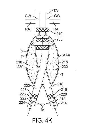
take the same
form as scaffolding 216 and filling structure 218.
[0074] In Fig. 41, both endograft delivery systems 212, 222 are advanced such
that the
docking scaffold 210 with filled filling structure 208 slidably receives an
end of both
scaffolds 216, 228 and optionally a portion of both filling structures 218,
230. In this
embodiment, the scaffolds 216, 228 are advanced approximately one-third of the
way into the
docking scaffold 210 although one of skill in the art will appreciate that
this distance may be
adjusted as required in order to accommodate different anatomies.
[0075] In Fig. 4J, both balloons 220, 226 are inflated thereby expanding both
scaffolds 216,
228 along with their respective filling structure 218, 230. The balloons 220,
226 in this
embodiment are inflated simultaneously in order to help ensure symmetric
expansion of both
scaffolds 216, 228 and both filling structures 218, 230. However, in some
embodiments,
inflation may be sequentially performed. The balloons 220, 226 are expanded so
as to ensure
that one end of each scaffold expands into engagement with the docking
scaffold 210 while
the other end of each scaffold expands into engagement with an iliac artery
IA. In this
embodiment, the scaffolds 216, 228 are balloon expandable, however, they may
also be self-
expanding.
[0076] After expansion of the balloons 220, 226 the filling structures are
filled with a
hardenable filling material such as PEG which can be polymerized in situ. This
is seen in
Fig. 4K. As discussed above, in some embodiments, prior to filling the filling
structures 218,
230 with the hardenable filling material, they may be pre-filled with carbon
dioxide, contrast
18
CA 02726452 2010-11-29
WO 2009/158170 PCT/US2009/046308
media, saline or a combination thereof in order to help unfurl each filling
structure and also to
give a preliminary indication of volume and/or pressure to use to fill the
structures. Also, in
this embodiment, the filling structures 218, 230 are filled while balloons
220, 226 are inflated
in order to help prevent crushing of the underlying scaffolds 216, 228
although in other
embodiments, the balloons need not be inflated during this step. Fig. 4L
illustrates the final
configuration of the endograft system after the delivery catheters and
guidewires have been
removed from the patient. A docking scaffold 210 is upstream of the aneurysm
AAA and
two scaffolds 216, 228 are expanded with one end in the docking scaffold 210
and the other
end in the iliac arteries IA. Each scaffold 210, 216 and 228 has a filling
structure 208, 218,
230 which is filled with a hardenable material to help anchor each scaffold in
position and to
help seal the aneurismal sac off from blood flow thereby forcing blood to flow
through the
lumens created by the scaffolds and their respective filling structures. While
this
embodiment shows one filling structure associated with each scaffold, in other
embodiments
some scaffolds may have a corresponding filling structure while others will
not.
[0077] The balloons used to deploy the scaffolds and filling structures are
often similar to
balloons used for angioplasty and stenting. However, in some cases, it may be
helpful to use
alternatively shaped balloons to help ensure proper deployment of the filling
structures. For
example, in Fig. 23A, a balloon 904 having a lower flange region may be used
to help ensure
that expansion of the filling structures 902 is limited to a defined region.
Or, for example, in
Fig. 23B, a tapered balloon 906 is used to shape the filling structures 902 so
that an internal
chamfer is formed, thereby helping to ensure a smooth transition for receipt
of the iliac
extension legs.
[00781 Now referring to Fig. 21, an optional external flange on the docking
scaffold and/or
the iliac leg scaffolds may further secure each scaffold into position. In
Fig. 21, the docking
scaffold 850 includes an outer annular ring or flange 856. This flange may be
fabricated
from a metal or polymer and it expands with the scaffold during deployment.
Because it has
a larger profile than the scaffold body, the filling structure 862 will expand
around it and
once the filling medium has hardened, the flange will be locked into position.
Similarly, an
optional flange 858 may be included in one or both of the iliac leg scaffolds
852, 854 to
provide an area for filling structures 860 to expand around and capture.
[00791 In the embodiment discussed above with respect to Figs. 4A-41, the
filling structure
is shown disposed over the scaffold. Other configurations are possible. For
example, the
19
CA 02726452 2010-11-29
WO 2009/158170 PCT/US2009/046308
scaffold may be disposed axially separated from the filling structure in order
to reduce overall
delivery profile. Additional disclosure on delivery system configurations may
be found in
U.S. Patent Application No. 12/429,474 (Attorney Docket No. 025925-002610US),
previously incorporate herein by reference. Additionally, the docking scaffold
210 is shown
positioned with approximately one-third of its length positioned in the aorta
upstream of the
aneurysm while the remainder of the scaffold is positioned in the aneurismal
sac. One of
ordinary skill in the art will appreciate that different configurations of the
docking scaffold
210 may be utilized. For example, Fig. 5A shows a docking scaffold 210 with
optional
filling structure 208 positioned in the aorta upstream of the aneurysm and
below the renal
arteries RA. Fig. 5B shows yet another variation where the docking scaffold
208 is
positioned with an upper portion in the aorta upstream of the aneurysm, a main
section
traverses the aneurysm and a lower portion is positioned below the aneurysm
just before iliac
bifurcation. Figs. 5C shows still another variation where the docking scaffold
210 is placed
in the aorta above the aneurysm and across the renal arteries RA. In this
embodiment, the
scaffold 210 and optional filling structure 208 have windows or lateral
apertures that permit
blood flow from the aorta to the renal arteries without significantly
obstructing flow. Fig. 5D
illustrates yet another variation where the docking scaffold 210 is placed
partially in the aorta
above the aneurysm and a downstream portion is in the aneurismal sac. Any of
the
embodiments shown in Figs. 5A-5D may also optionally include a filling
structure 208 which
generally takes the same form as filling structures previously described.
[0080] Any of the docking scaffolds may be coupled with two iliac leg
extensions as
described herein. Most of the embodiments disclosed use two discrete iliac leg
extensions
delivered separately from both iliac arteries. However, in some embodiments,
the iliac leg
extensions may be of integral construction rather than discrete. For example,
in Fig. 18, a
docking scaffold 804 having a filling structure 802 is disposed across the
aneurysm AAA
such that one end is upstream of the aneurysm and the opposite end is
downstream of the
aneurysm. An iliac leg extension of unitary construction having two iliac legs
806, 808
coupled together is then slidably received and radially expanded in the
downstream portion of
the docking scaffold 804 such that blood flow is bifurcated to each iliac
artery. The iliac leg
extension may be a stent-like scaffold only, it may be a covered graft or it
may be a graft with
scaffolds only at its ends such as the embodiment in Fig. 18 which has
scaffolds 814, 812 and
810 at its ends. One or more optional filling structures may also be coupled
with the iliac
extension.
CA 02726452 2010-11-29
WO 2009/158170 PCT/US2009/046308
[0081] Often the docking scaffold is a fixed length. While some foreshortening
may occur
during radial expansion, the docking scaffold generally does not change length
significantly.
This requires the physician to accurately determine the required length prior
to deployment
and also requires a number of different length to be inventoried. An accordion-
like docking
scaffold allows a single scaffold to accommodate a number of aneurysm lengths.
Figs. 19A-
19C illustrate an exemplary embodiment of a variable length docking scaffold.
In Fig. 19A,
the docking scaffold 820 includes an accordion-like main body 824 and stent-
like ends 822,
826. The main body 824 may be a graft alone or it may also be supported by a
scaffold
structure such as a stent. The graft material may be Dacron woven to allow
axial extension
and compression or it may be ePTFE which will also stretch and compress
depending on the
material properties such as internodal distance. Other materials may also be
used. Both ends,
822, 826 may include balloon expandable or self-expanding stents to help
anchor the docking
scaffold in position. Fig. 19B shows the docking scaffold in a compression
configuration so
that it may accommodate a shorter aneurysm and Fig. 19C shows the docking
scaffold in an
elongated configuration for a longer aneurysm. In addition to providing a
scaffolding that
can accommodate varying lengths, this embodiment is also more flexible and
thus may
accommodate bends and other tortuosity often seen in aneurysms, such as in
Fig. 20. While
this embodiment is described with respect to the docking scaffold, one of
skill in the art will
appreciate that this embodiment may also be used in the iliac legs or other
portions of the
system.
[0082] Figs. 6A-6C illustrate another feature of the docking scaffold which
may optionally
be included in any of the embodiments disclosed herein. Fig. 6A illustrates
the standard
docking scaffold 300 which is generally cylindrically shaped with a constant
diameter. In
some cases, it may be desirable to expand the docking scaffold 300 so that a
lower end
expands to a constant diameter every time. This standardizes the docking
region of scaffold
300 and allows more consistency in mating the docking scaffold with the two
legs.
Additionally, this allows the upper portion of the scaffold to accommodate a
variety of vessel
anatomies and sizes without interfering with the docking aspect of the
scaffold. Fig. 6B
illustrates an exemplary embodiment of a docking scaffold 300 having a
restraining member
302 disposed over a lower portion of the scaffold 300. The restraining member
302 may be a
corset like band of material that limits expansion of the scaffold, or the
scaffold itself may
have shorter struts that expand less than other regions of the scaffold. The
restraining
member 302 or shorter struts allow the lower portion of scaffold 300 to expand
to a
21
CA 02726452 2010-11-29
WO 2009/158170 PCT/US2009/046308
predetermined diameter 306 which is sized so as to mate with the two endograft
legs. In still
other embodiments, a restraining member 304 or the scaffold design itself may
be used to
limit expansion of the docking scaffold to create a tapered or flared region
such as seen in
Fig. 6C. The tapered or flared region may be used to help guide the endograft
legs into the
docking scaffold 300 during assembly of the endograft system in situ.
[0083] Figs. 7A-7C illustrate still another feature of the docking scaffold
system which
may optionally be included in any of the embodiments disclosed herein. In
order to help
ensure sealing between the docking scaffold and the two legs, a sealing
element may be
disposed around one or both of the leg scaffolds. The sealing element may be
used to fill
gaps as well as cause thrombus formation. Fig. 7A illustrates a scaffold 320
having such a
sealing element 322. Fig. 7B is a perspective view showing the sealing
element. The sealing
element 322 may be a foam-like plug or a spongy, material that can be
compressed to
minimize profile during delivery. Exemplary materials for the sealing elements
may include,
but are not limited to polyurethane, polycarbonate, polyester, ePTFE,
polyolefins, parylene,
gelatin, silicone and the like. A sheath may be used to constrain the sealing
element 322
during delivery. Upon retraction of the sheath the sealing element expands to
fill any gaps.
In addition to sealing any gaps, the sealing element may be fabricated from a
material or
contain a therapeutic agent which causes thrombosis thereby providing
additional sealing
ability. Fig. 7C shows an exemplary cross-sectional view of the docking
scaffold 324 having
two leg scaffolds 320 expanded and engaged therein. Sealing elements 322 on
both leg
scaffolds 320 fill the gaps between the docking scaffold 324 and the two leg
scaffolds 320 to
prevent blood flow therethrough.
[0084] Shaped sealing elements may also facilitate blood or fluid flow across
a sealed
region. For example, Fig. 8A illustrates a side view of a scaffold 320 having
a sealing
element 322 disposed on one end. An internal chamfer 323 provides a smoother
transition
for fluids to enter the scaffold 320. Fig. 8B illustrates a perspective view
of Fig. 8A. Fig. 8C
shows a perspective view of an exemplary embodiment where two sealing elements
322 are
disposed against one another, thereby forming a double D-shaped region. Again,
the chamfer
323 provides a smooth transition. Fig. 8D shows a side view of Fig. 8C.
[0085] Additionally, Figs. 9 and 10 illustrate how the sealing elements may be
used in
alternative embodiments. For example, in Fig. 9 two scaffolds 325 are placed
side-by-side in
an aneurysm AAA. An upper portion of each scaffold 325 is positioned upstream
of the
22
CA 02726452 2010-11-29
WO 2009/158170 PCT/US2009/046308
aneurysm AAA and sealing elements 328 form a seal between the scaffolds 328
and blood
vessel wall. Both scaffolds 325 traverse the aneurysm AAA and an opposite end
of each
scaffold 325 is positioned in an iliac artery IA. In the embodiment of Fig. 9,
the scaffolds
325 are preferably covered with a cover such as ePTFE or Dacron so that blood
flow follows
the lumen created by the scaffolds 325 into the iliac arteries, IA, thereby
excluding the
aneurysm AAA. Fig. 10 illustrates another embodiment where the sealing
elements 326 are
used to form a seal. In Fig. 10, a docking scaffold 330 with double-walled
filling structure
332 is positioned with an upper portion in the neck of the aneurysm AAA and
the main body
traversing the aneurysm AAA. Iliac leg scaffolds 324 dock with the docking
scaffold 330
and sealing elements 326 seal the system to ensure blood flow only through the
endograft
lumens. In the embodiment of Fig. 10, the docking scaffold 330 may optionally
be covered
along with the iliac leg scaffolds 324 with a cover such as ePTFE or Dacron
328. Figs. 11A-
11B illustrate such an embodiment. In Fig. 11 A, a docking scaffold 330 is
positioned
partially upstream of the aneurysm AAA and a filled filling structure 332
partially fills the
aneurismal space. Two iliac scaffolds 324 dock with docking scaffold 330 and
their opposite
ends are positioned in the two iliac arteries IA. Sealing elements 326 on the
upstream portion
of scaffolds 324 help form a seal and a covering material such as ePTFE or
Dacron cover the
iliac scaffolds 328 to restrict blood flow to the lumen created by the iliac
scaffolds 324. Fig.
11B shows the two iliac scaffolds 324 adjacent one another and having sealing
elements 326
at one end, a covered middle portion and an uncovered scaffold portion on the
opposite end.
[00861 In still other embodiments, the sealing elements may be expandable or
inflatable
members. Figs. 12A-I2C illustrate an exemplary embodiment. In Fig. 12A, a
docking
scaffold 330 is placed in the vessel and partially across the aneurysm AAA. A
filling
structure 332 is filled with hardenable filling material such as PEG and iliac
scaffold legs 328
are docked into the docking scaffold 330. The iliac scaffold legs 328 may be
grafts alone or
they may be supported by a stent-like scaffold structure. Expandable sealing
elements 326 on
each iliac scaffold leg 328 form a seal. Fig. 12B shows a cross section along
the line 12B-
12B in Fig. 12A and shows how the expandable sealing elements 326 fill the
gaps between
the docking scaffold 330 and the two iliac scaffold legs 328. Fig. 12C shows
how an inflator
330 coupled to an inflation tube 332 may be used to expand or inflate the
sealing elements
326 to help form or adjust the seal.
[00871 In some embodiments, additional scaffolding legs may be provided. Fig.
13 shows
a docking scaffold system similar to those previously described. Docking
station 402 is
23
CA 02726452 2010-11-29
WO 2009/158170 PCT/US2009/046308
generally similar to scaffolds 106, 210, and 330 as described above. Leg
scaffolding 404,
406, as well as additional leg scaffolding 410 and 412 may be generally
similar to any of
scaffoldings 112, 116, 218, 228, 325, and 328 as described above. As shown in
Fig. 13, two
additional leg scaffolds 410, 412 are be provided. Additional leg scaffolds
410 and 412,
traverse the iliac arteries and couple to leg scaffolds 404 and 406
respectively. Additional leg
scaffolds 410 and 412 are delivered via guidewire and subsequently expanded,
for example,
by self-expansion or balloon expansion. Additional leg scaffolds 410, 412 may
be delivered
and expanded into position before or after leg scaffolds 404, 406 are
delivered. When
additional leg scaffolds are delivered and expanded before leg scaffolds 404,
406, a
downstream portion of the outside surface of leg scaffolds 404, 406 engages
the upstream
portion of the inside surface of additional leg scaffolds 410, 412. When
additional leg
scaffolds are delivered and expanded after leg scaffolds 404, 406, a
downstream portion of
the inside surface of leg scaffolds 404, 406 engages the upstream portion of
the outside
surface of additional leg scaffolds 410, 412. Additional leg scaffolds 410,
412 may be used
to treat an iliac artery aneurysm IAA. Additional leg scaffolds 410, 412 may
include a
covering material such as Dacron TM or ePTFE so as to fully form a blood flow
lumen through
iliac arteries IA. The iliac artery aneurysm may then be filled with a
hardenable filling
material as described above. The hardening material may also help lock the
scaffolds in
position relative to the aneurysm thereby preventing future migration.
Alternatively,
additional leg scaffolds may include a filling structure which is filled with
a hardenable
material to help anchor the additional leg scaffolds in position and to help
seal the aneurismal
sac off from blood flow thereby forcing blood to flow through the lumens
created by the
scaffolds and their respective filling structures. While the embodiment of
Fig. 13 shows one
iliac artery aneurysm and two additional leg scaffolds, in other embodiments
more than one
iliac artery aneurysm may be present and different numbers of additional leg
scaffolds may
be provided.
[00881 In some embodiments, a crown scaffold 501 may be provided. As shown in
Figs.
14A and 14B, crown scaffold 501 is a bare metal stent. Crown 501 is guidewire
delivered to
a site upstream of an aneurysm AAA and may be self-expandable or balloon
expanded.
Crown 501 is often a standard, generic part while docking scaffold 502 and leg
scaffolds 504,
506 may be customized for the patient. Crown 501 is often delivered and
expanded after
docking scaffold 502 is such that the surface of the downstream portion of
crown 501 is
engaged with the surface of the upstream portion of docking scaffold 502.
Docking scaffold
24
CA 02726452 2010-11-29
WO 2009/158170 PCT/US2009/046308
502 and leg scaffolds 504 and 506 are generally similar to the scaffolds
previously described.
In some cases, a filling structure may be provided for the crown scaffold to
help anchor it in
position relative to an aneurysm. Fig. 14A shows the crown scaffold 501,
docking scaffold
502, and leg scaffolds 504 and 506 delivered and expanded in position relative
to the
aneurysm AAA. For clarity, Fig. 14B shows an exploded view of the expanded
scaffolds.
[0089] In some instances, a docking scaffold 602 may include a divider 604.
Divider 604
is often integrally formed with docking scaffold 602, which is a stent-like
scaffold. As
shown in Fig. 15A, 602 is shown shaded. Divider 604 splits the inside volume
of docking
scaffold 602 into an upstream portion 610 with a circular cross section, and
two downstream
portions 606 and 608 with D-shaped cross sections as shown in Fig. 15B. When
leg scaffolds
are delivered and expanded within the downstream portions of scaffold 602,
divider 604
keeps the leg scaffolds from taking more cross-sectional area than allotted.
Divider 604 also
prevents the leg scaffolds from intruding too far upstream into the central
passageway of
docking scaffold 602. For clarity, divider 604 is shown without the rest of
docking scaffold
602 in Fig. 15C.
[0090] An internal double-walled filling structure 621 may also be used as a
divider. As
seen in Fig. 16A, filling structure or divider 621 splits the inside volume of
docking scaffold
621 into upstream portion 625 with a circular cross section and two downstream
portions 627
and 629. After leg scaffolds are delivered and expanded within the downstream
portions 627
and 629, divider 621 can be filled and expanded such that it holds the leg
scaffolds in place.
Fig. 16A and 16B show divider 621 unfilled. Fig. 16C shows divider 621 when
filled.
[0091] The docking scaffold may also be formed so that the leg scaffolds are
prevented
from intruding on one another. As seen in Fig. 17A and 17B, the downstream
portion of
docking scaffold 710 bifurcates into a first portion 713 and a second portion
716. Each
portion 713, 716 has its own, generally circular lumen for receiving a leg
scaffold. Double-
layered filling structures may also be provided for docking scaffold 710,
docking scaffold
portion 713, and/or docking scaffold 716 to hold the docking scaffold in place
relative to an
aneurysm and/or attached leg scaffolds.
[0092] While typical scaffold structures are often either balloon expandable
or self-
expanding, in some embodiments it may be advantageous to provide a scaffold
having a
balloon expandable region and a self-expanding region. For example, Fig. 22
illustrates a
scaffold 875 having an upper portion that is balloon expandable 876 and a
lower portion that
CA 02726452 2010-11-29
WO 2009/158170 PCT/US2009/046308
is self-expanding 878. In this embodiment, the two regions are illustrated as
being
approximately the same length, although one will appreciate that region length
may be
adjusted as required. In this embodiment, the self-expanding region is
advantageous since it
will expand until it engages the vessel wall or docking scaffold or it can
expand to a
predetermined shape, such as a D-shape. This is particularly desirable in
situations where a
physician wishes to avoid using a balloon to expand aneurismal tissue which
may be
damaged or significantly weakened or where it is difficult to form the desired
shape by
balloon expansion. A balloon expandable region is desirable when a fixed
diameter is needed
unlike the self-expanding scaffolds which may continue to radially expand. The
balloon
expandable portion 876 may be integrally formed with the self-expanding
region, for
example by laser cutting the stent from a Nitinol tube and then differentially
heat treating the
two sections, or two discrete sections may be joined together by welding,
suturing, bonding,
etc.
[0093] While the above is a complete description of the preferred embodiments
of the
invention, various alternatives, modifications, and equivalents may be used.
Therefore, the
above description should not be taken as limiting in scope of the invention
which is defined
by the appended claims.
26