Note: Descriptions are shown in the official language in which they were submitted.
CA 02726585 2010-12-01
WO 2009/149074 PCT/US2009/045958
CONTROLLED DEPLOYMENT HANDLES FOR BONE STABILIZATION
DEVICES
CROSS REFERENCE TO RELATED APPLICATIONS
[0001] This application claims priority to U.S. Provisional Patent Application
Serial No.
61/058,157, filed June 2, 2008 and titled "CONTROLLED DEPLOYMENT HANDLE FOR
BONE STABILIZATION DEVICES", and U.S. Provisional Patent Application Serial
No.
61/142,552, filed January 5, 2009, and titled "CONTROLLED DEPLOYMENT HANDLE
FOR
BONE STABILIZATION DEVICES."
[0002] This application is related to U.S. Patent Application Serial No.
11/468,759, filed on
August 30, 2006, entitled "IMPLANTABLE DEVICES AND METHODS FOR TREATING
MICRO-ARCHITECTURE DETERIORATION OF BONE TISSUE", which claims the benefit
of U.S. Provisional Application No. 60/713,259, filed on August 31, 2005,
entitled
"IMPLANTABLE DEVICE FOR TREATING VCF, TOOLS AND METHODS". This
application is also related to U.S. Serial No. 12/041,607 (titled " FRACTURE
FIXATION
SYSTEM AND METHOD") filed 3/3/2008; US Serial No. 12/044,884 (titled "
TRANSDISCAL
INTERBODY FUSION DEVICE AND METHOD") filed 3/7/2008; U.S. Serial No.
12/044,880
(titled " SYSTEMS, METHODS AND DEVICES FOR SOFT TISSUE ATTACHMENT TO
BONE") filed 3/7/2008; U.S. Serial No. 12/024,938 (titled " SYSTEMS, DEVICES
AND
METHODS FOR STABILIZING BONE") filed 2/1/2008; and U.S. Serial No. 12/025,537
(titled
" METHODS AND DEVICES FOR STABILIZING BONE COMPATIBLE FOR USE WITH
BONE SCREWS") filed 2/4/2008.
[0003] All of these patent applications are incorporated herein by reference
in their entirety.
INCORPORATION BY REFERENCE
[0004] All publications and patent applications mentioned in this
specification are herein
incorporated by reference in their entirety to the same extent as if each
individual publication or
patent application was specifically and individually indicated to be
incorporated by reference.
FIELD OF THE INVENTION
[0005] The invention relates to devices, systems and methods for treating and
supporting
bone, including bone within vertebral bodies suffering from a vertebral
compression fracture
(VCF). More particularly, the devices, methods and systems described herein
relate to rotary
-1-
CA 02726585 2010-12-01
WO 2009/149074 PCT/US2009/045958
handles and applicator systems and controls for inserting self-expanding bone
support implants.
BACKGROUND OF THE INVENTION
[0006] Deterioration of bone tissue, and particularly micro-architecture
deterioration, can
result from a variety of factors including disease, aging, stress and use. For
example,
osteoporosis is a disease characterized by low bone mass and micro-
architecture deterioration of
bone tissue. Osteoporosis leads to bone fragility and an increase fracture
risk. While
osteoporosis affects the entire skeleton, it commonly causes fractures in the
spine and hip.
Spinal or vertebral fractures have serious consequences, with patients
suffering from loss of
height, deformity, and persistent pain that can significantly impair mobility
and quality of life.
Vertebral compression fractures (VCFs) and hip fractures are particularly
debilitating and
difficult to effectively treat.
[0007] Devices for supporting and repairing bone, including implants for
repairing spinal
compressions including VCFs have been described. One particularly useful type
of implant for
support and/or treatment of bone are self-expanding implants that may be
deployed within bone
to cut through the bone with little or any compression, and may be filled with
one or more bone
fillers (e.g., cement) in the regions within and around the implant for added
support. Such
implants may also act as supports or anchors for additional implants.
[0008] These bone implants (which are described in greater detail below) may
be inserted
using a controller (e.g., applicator system) that must provide support for the
implant during and
before implantation. For example, the implant may be released to self-expand
within the bone,
and must be manipulated into position and released while maintaining force on
the implant to
maintain it in a compressed (delivery) configuration. The inserter must allow
precise control of
the release of the implant into the bone. It may also be beneficial to allow
the implant to be
removed using the inserter.
[0009] It may be beneficial to have the inserter be modular, so that one or
more portions
could be reused, saving cost and time. For example, a handle portion may be re-
used by
connecting to various elongate (e.g., cannula) portions of the applicator.
[00010] It may also be helpful to provide a device having a minimum of
components, and
devices that are configured to include one or more failsafe mechanisms that
permit the implant to
be removed even in case the implant or applicator becomes jammed or otherwise
disrupted.
[00011] Related U.S. Application Serial No. 12/024,938 (filed on 2/1/08),
titled "SYSTEMS,
DEVICES AND METHODS FOR STABILIZING BONE") describes bone stabilization
devices
and methods for inserting them using a delivery device. The delivery device
may be configured
to include a cannula (or multiple cannula) and one or more trocars. As
mentioned above, it
-2-
CA 02726585 2010-12-01
WO 2009/149074 PCT/US2009/045958
would be extremely beneficial to have a delivery device including a handle
that can be used to
control the delivery and/or expansion of an implant device.
[00012] Examples of controllers, inserters, handles and devices forming such
an improved
handle are provided herein.
SUMMARY OF THE INVENTION
[00013] Described herein are handles and applicator systems including handles
for engaging
delivery (and/or retrieval) of a bone stabilization device, as well as systems
or kits including
handles, and methods for using them.
[00014] An applicator (or applicator system) may include a handle region and
an elongate
linkage member that couples with the handle. In particular, described herein
are rotary
applicator handles that are configured to couple with the proximal end of the
elongate linkage
member and drive the axial motion (e.g., in the direction of the long axis of
the elongate linkage
member) of a portion of the elongate linkage member. An implant such as a bone
stabilizing
implant may be coupled to the distal end of the elongate linkage member, and
axial movement of
a portion of the elongate linkage member may result in expansion or
contraction of the implant.
As used herein, "axial" motion of a component of the elongate linkage member
refers to motion
in the direction of the long axis of the elongate linkage member. For example,
an elongate
linkage member may include a first elongate member that may move relative to a
second
elongate member. In some variations the first elongate member is an outer
(e.g., cannula)
member and the second elongate member is an inner (e.g., rod) member. The
outer cannula and
the inner rod may coaxially slide relative to each other, which is one type of
"axial" movement.
Axial movement of the elongate linkage member is translated into force across
an implant that is
coupled to the distal end of the elongate linkage member, causing the implant
to collapse (e.g.,
into a narrow-diameter delivery configuration) or expand (e.g., into an
expanded-diameter
deployed configuration in which a plurality of struts bow out from the body of
the implant).
[00015] In general, the handles described herein are rotary applicator handles
that are
activated by rotating a control on the handle (e.g., a knob, a rotating grip,
etc.). Rotating the
control drives rotation of a rotary gear within the handle, and the rotary
gear drives axial
movement of a portion of an elongate linkage member when an elongate linkage
member is
coupled to the handle. In variations in which the elongate linkage member
includes a first
elongate member and a second elongate member that are movable relative to each
other, the
proximal ends of the first and second elongate members are held in separate
seats in the handle.
By holding the proximal ends of the first and second elongate members, these
members may be
moved relative to each other, thereby controlling the motion of the implant
coupled to the distal
-3-
CA 02726585 2010-12-01
WO 2009/149074 PCT/US2009/045958
end of the elongate linkage member. Typically the implant is coupled to the
distal end of the
elongate linkage member so that the proximal end is connected to one of the
elongate members
forming the elongate linkage member (e.g., the first elongate linkage member)
and the distal end
of the implant is coupled to the distal end of the other elongate linkage
member (e.g., the second
elongate linkage member).
[00016] In some variations, the rotary applicator handles described herein are
ratcheting
handles in which the rotary gear is a ratcheting gear including a pawl that
helps control the
direction of axial movement driven by the gear. A control on the handle (e.g.,
a direction switch
or a ratchet switch) may be used to select the direction of movement enabled
by the handle. This
control may be connected to the pawl. Other controls, including safety
controls for releasing the
force applied by the handle to the elongate linkage member (and therefore the
implant), or for
releasing the elongate linkage member from the handle, may also be included.
For example, the
handles described herein may include a control for regulating/controlling the
release of the
stabilization device. Stabilization devices are typically self-expanding
devices, and the control
may regulate the self-expansion so that the rate and degree of self-expansion
allowed is
regulated. The handles may be lockable, and may include a latch or other
locking structure.
These handles may also include ratcheting mechanism or other controlled
expansion/release
mechanism. In some variations the devices include a failsafe release
configured to release either
the applicator and/or the device. These devices may also include a one or more
finger controls
for controlling the handle, and the handle may be configured for gripping in
one or more of the
subject's hands.
[00017] In some variations, the handle includes indicators or sensors. For
example, the
handle may include an indicator of the orientation of the implant attached to
the distal end of a
coupled elongate linkage member. In particular, the handle may be configured
so that the
elongate linkage member is not rotated when axial motion is applied and
therefore the implant is
not rotated during delivery of the device. For example, the seats for the
proximal end of the
elongate linkage member may be keyed to prevent rotation of the implant.
[00018] The implants described herein may also be referred to as bones
stabilization devices.
These implants may include a self-expanding body that can be deployed in a
linear
configuration. The deploying configuration is typically an elongate tubular
shape that is open at
both ends. In some variations the device may have an elongate, substantially
tubular shape that
includes a plurality of struts extending along the length of the implant in
the deployed
configuration. For example, the struts maybe extended laterally in an expanded
configuration.
Expansion of the struts may foreshorten the implant. A self-reshaping (e.g.,
self-expanding)
-4-
CA 02726585 2010-12-01
WO 2009/149074 PCT/US2009/045958
device may include a preset configuration that is expanded, and may reset from
another
configuration into the preset configuration (or vice versa). For example, the
devices may include
a linear configuration (a deployed configuration) and an expanded
configuration. The linear
configuration can be stabilized by constraints that prevent self-reshaping of
the device into an
anchoring (expended) configuration. Self-reshaping to an anchoring
configuration may be
performed by two or more linear portions of the device, which (upon release
from constraint)
radially-expand into bowed struts of various configurations, while at the same
time shortening
the overall length of the device. Embodiments of the struts may include a
cutting surface on the
outwardly leading edge or surface of the strut, which cuts through cancellous
bone as it radially
expands. After implantation within a vertebral body, the bowed struts may
expand though the
cancellous bone to contact the cortical bone of the inner surfaces of superior
and inferior
endplates of the compressed vertebral body, and push the endplates outward to
restore the
vertebral body to a desired height.
[00019] In general, the implants described herein may be inserted into tissue
(e.g., bone such
as a vertebra) so that they do not foreshorten when allowed to self-expand. As
described in
greater detail below, this may be accomplished by controlling both the
proximal and distal ends
(or end regions) of the implant with the applicator. Thus, the applicator
(including the handle)
may be configured to control the relative motions of the ends of the implant.
For example, if the
distal end is held while the proximal end is allowed to foreshorten, the
device may be inserted
without distally foreshortening or otherwise moving. Movement of the distal
end of the device
may result in the implant moving undesirably from the implantation site, and
may cause damage
or inaccuracy.
[00020] The implant maybe prepared for insertion by collapsing it. An
applicator or inserter
(described below) may be used to collapse it from a pre-biased expanded
configuration, in which
the struts are bowed or otherwise expended, and a more linear collapsed or
delivery
configuration, in which the struts are collapsed towards the body. For
example, the step of
delivering the first self-expanding implant may include the step of applying a
restraining force
across the implant to hold the first implant in a collapsed configuration. In
some variations, the
method also includes the step of applying a restraining force across the first
implant by applying
force across the implant to collapse a plurality of expandable struts along
the implant.
[00021] The step of releasing restraining forces to radially expand the self-
expanding implant
within the cancellous bone may comprise allowing the proximal end of the
implant to
foreshorten. The step of releasing restraining forces to radially expand the
first and second self-
expanding implants within the cancellous bone may also (or alternatively)
comprise removing
the distal end portion of the implant for a first inserter region and removing
the proximal end
-5-
CA 02726585 2010-12-01
WO 2009/149074 PCT/US2009/045958
portion of the implant from a second inserter region.
[00022] Any of the handle devices described herein may be used with any
appropriate
elongate linkage member. In some variations, a handle and an elongate linkage
member may be
used together to form an applicator or applicator system. The handles
described herein may be
reusable or disposable. In some variations a handle is intended for use in
with multiple implants
in a single procedure; each implant may be connected to a separate elongate
linkage member.
Thus, in some variations the rotary applicator handles described herein are
configured for use
with a single size of implant; in other variations, the handle may be used or
adapted for use with
implants of different sizes. Handles may distinguish different sizes of
implants based on the
shape (e.g., the keyed shape) of the proximal end of the elongate linkage
member to which the
implant is attached distally. In some variations the handle distinguishes
different sizes of
implants based on the separation between the proximal ends of first and second
elongate
members forming the elongate linkage member.
[00023] Rotary applicator handles may be formed of any appropriate materials,
including
metals, plastics (e.g., polymeric materials), ceramics, or the like, including
any combination
thereof.
[00024] For example, described herein are rotary applicator handle for
delivery or removal of
a bone stabilizing implant that is distally coupled to an elongate linkage
member. These handles
may include: a handle grip configured to be held in the palm of a hand; a
housing at least
partially surrounding a first seat configured to hold the proximal end of a
first elongate member
of the elongate linkage member and a second seat configured to hold the
proximal end of a
second elongate member of the elongate linkage member; a rotary gear within
the housing, the
rotary gear configured to drive the axial motion of the first member of the
elongate linkage
member relative to the second member of the elongate linkage member; and a
rotatable control
coupled to rotary gear and configured to rotate the rotary gear.
[00025] The rotary gear may be a ratcheting gear comprising a pawl. In some
variations, the
rotary applicator handle includes a directional switch coupled to the rotary
gear and configured
to control direction of axial motion driven by the rotary gear.
[00026] In some variations, the rotary gear comprises a drive shaft. The
rotary or rotatable
control may be a knob that rotates the drive shaft.
[00027] The rotary applicator may also include an indicator to indicate the
orientation of the
bone stabilizing implant relative to the handle. The handle may be marked
(e.g.,
alphanumerically, etc.) to indicate the size of the implant that it is to be
used with. The rotary
applicator handle may also include a release control configured to release the
elongate linkage
member from the handle. For example, the handle may include a force release
control
-6-
CA 02726585 2010-12-01
WO 2009/149074 PCT/US2009/045958
configured to release the axial force applied to the elongate linkage member
by the handle.
[00028] The rotary applicator handle may include a mating region configured to
mate with a
shaft stabilizer on the first member of the elongate linkage member. The
mating region may be
at the distal end of the handle, and maybe a keyed fitting, maintaining the
orientation of the
elongate linkage member (and therefore the implant) when engaged with the
handle.
[00029] In some variations the rotatable control is a rotatable control grip.
This rotatable grip
may be configured for use by a second hand (e.g., separate from the hand
holding the handle
grip), or it may be a finger grip, so that it may be rotated by the thumb and
index finger, for
example.
[00030] In general, the expansion and contraction of the implant (and
particularly a self-
expanding implant) may be controlled. For example, when the implant is
converted a
(constrained) elongate, tubular delivery configuration having a small cross-
section to an
expanded configuration in which the struts extend from the body of the device,
the implant may
be foreshortened. The applicator system controls the deployment of the implant
(from the
compressed configuration to the expanded configuration) by applying axial
force to pull apart
(collapse) or draw together (expand) the proximal and distal ends of the
implant. One end of the
implant (e.g., the distal end) may be held relatively motionless while the
applicator system
moves the other end to collapse or expand the implant. Preventing the distal
end from moving
during expansion or collapse may prevent damage to the patient, and may help
maintain the
position of the implant during insertion. For example, the rotary gear may be
configured to
axially move the second seat relative to the first seat so that the proximal
end of an implant
coupled to the first member of the elongate linkage member moves while the
distal end of the
implant remains relatively stationary.
[00031] In some variations, the handle is a ratcheting applicator handle for
delivery or
removal of a bone stabilizing implant that is distally coupled to an elongate
linkage member. In
this example, the handle includes: a first handle grip region; a housing at
least partially
surrounding a first seat configured to hold the proximal end of an inner
member of the elongate
linkage member and a second seat configured to hold the proximal end of an
outer member of
the elongate linkage member; a ratcheting gear within the housing, the
ratcheting gear configured
to drive the axial motion of the outer member of the elongate linkage member
relative to the
inner member of the elongate linkage member; a rotatable grip coupled to
ratcheting gear and
configured to rotate the ratcheting gear; and a directional switch coupled to
a pawl and
configured to select the axial direction that the outer member is driven
relative to the inner
member.
[00032] As mentioned, any of these handles may be used as part of an inserter
or applicator
-7-
CA 02726585 2010-12-01
WO 2009/149074 PCT/US2009/045958
system. Thus, described herein are inserter systems for delivery or removal of
a bone stabilizing
implant that include: an elongate linkage member configured to distally couple
with the bone
stabilizing implant and a rotary handle. The elongate linkage member may
include: a first
elongate member configured to releasably couple at its distal end with the
proximal end region of
the bone stabilizing implant; and a second elongate member configured to
releasably couple at its
distal end with the distal end region of the bone stabilizing implant. The
rotary handle may
include: a handle grip region; a housing at least partially surrounding a
first seat configured to
hold the proximal end of the first elongate member and a second seat
configured to hold the
proximal end of the second elongate member; a rotary gear within the housing,
the rotary gear
configured to drive the axial motion of the first member relative to the
second elongate member;
and a rotatable control configured so that rotation of the rotatable control
moves the rotary gear.
[00033] As mentioned, the first member may comprise an outer cannula and the
second
elongate member may comprise an internal rod. These outer and inner members
may be
coaxially arranged.
[00034] The elongate linkage member may also include an end grip at the
proximal end of the
first elongate member that is keyed to fit within the first seat of the rotary
handle. The rotary
gear may be a ratcheting gear comprising a pawl. The system may also include a
directional
switch coupled to the rotary gear and configured to control the direction of
axial motion driven
by the rotary gear.
[00035] In some variations, the system also includes a self-expanding implant.
Any of the
implants described herein may be used, including implants having a plurality
of self-expanding
struts and a proximal attachment region configured to releasably attach to the
first elongate
member and a distal attachment region configured to releasably attach to the
second elongate
member.
[00036] Also described herein are methods of using the rotary handles
described. For
example, a method of collapsing and expanding a self-expanding implant is
described. This
method may include the steps of seating the proximal end of an elongate
linkage member within
a rotary applicator handle so that the proximal end of a first elongate member
of the elongate
linkage member is held within a first seat and the proximal end of a second
elongate member of
the elongate linkage member is held within a second seat; and rotating a
control on the rotary
applicator handle to drive a rotary gear that axially moves the first elongate
member relative to
the second elongate member so that the proximal end of a self-expanding
implant that is coupled
to the distal end of the first elongate member is moved relative to the distal
end of the self-
expanding implant that is coupled to the distal end of the second elongate
member.
[00037] The step of rotating the control on the rotary applicator handle may
include limiting
-8-
CA 02726585 2010-12-01
WO 2009/149074 PCT/US2009/045958
the axial motion of the first elongate member relative to the second elongate
member to prevent
damage to the self-expanding implant. A limiter may be included as a stop of
other structure
within the handle, limiting axial motion to within a specified range. This
range may be
adjustable in variations of the handle that are used for different sized
implants.
[00038] The step of rotating the control on the rotary applicator may comprise
moving the
first elongate member relative to the second elongate member without
substantially moving the
second elongate member. As mentioned above, this may prevent movement of the
distal end of
the implant.
[00039] The methods may be performed with any of the ratcheting handles
described. For
example, the step of rotating the control on the rotary applicator handle may
include driving a
ratcheting rotary gear comprising a pawl. In some variations, the method may
therefore include
the step of selecting the direction of axial motion by switching a ratchet
switch that is coupled to
apawl.
[00040] The method may also include the steps of releasing the device from the
applicator
system. For example the method may include the steps of disengaging (e.g.,
rotating) the first
and second members to release the proximal and distal ends of the implant from
the elongate
linkage member. This step may be performed in some variations while the
elongate linkage
member is attached to the handle, or after the two are decoupled. For example,
the method may
include the steps of activating a control on the rotary applicator handle to
release the axial force
applied to the elongate linkage member by the rotary applicator handle. In
some variations, the
method may also include the steps of releasing the elongate linkage member
from the handle.
BRIEF DESCRIPTION OF THE DRAWINGS
[00041] FIG. 1 shows one variation of a system including a self-expanding bone
support
implant and an applicator.
[00042] FIGS. 2A-2E are variations of stabilization devices.
[00043] FIGS. 3A and 3B are enlarged side and side perspective views
(respectively) of the
stabilization device shown in FIG. 2A.
[00044] FIGS. 4A and 4B are enlarged side and side perspective views
(respectively) of the
stabilization device shown in FIG. 2C.
[00045] FIGS. 5A and 5B are enlarged side and side perspective views
(respectively) of the
stabilization device shown in FIG. 2E.
[00046] FIG. 6A is one variation of a stabilization device having a plurality
of continuous
curvature of bending struts removably attached to an inserter.
[00047] FIG. 6B is another variation of a stabilization device removably
attached to an
-9-
CA 02726585 2010-12-01
WO 2009/149074 PCT/US2009/045958
inserter.
[00048] FIG. 7A is another variation of a stabilization device connected to an
inserter. FIGS.
7B and 7C show detail of the distal and proximal ends (respectively) of the
stabilization device
and inserter of FIG. 7A.
[00049] FIG. 8A is one variation of a handle that may be used with an
inserter.
[00050] FIGS. 8B-8E illustrate connecting an inserter to a handle such as the
handle of FIG.
8A.
[00051] FIGS. 9A-9D illustrate the operation of an inserter and handle in
converting a
stabilization device from a relaxed, deployed configuration (in FIGS. 9A and
9B) to a contracted,
delivery configuration (in FIGS. 9C and 9D).
[00052] FIG. 10 is one variation of an inserter connected to a stabilization
device within an
access cannula.
[00053] FIG. 11 shows one variation of a trocar and access cannula.
[00054] FIG. 12A- 12C shows one variation of a hand drill.
[00055] FIG. 13 shows one variation of a cement cannula and two cement filling
devices.
[00056] FIGS. 14A-14D show different variations of an access cannula that may
be used with
a stabilization device and inserter, trocar, drill, and cement cannula,
respectively.
[00057] FIGS. 15A-15G illustrate one method of treating a bone.
[00058] FIGS. 16A-16B illustrate one method of using bone cement with the
stabilization
devices described herein.
[00059] FIG. 16C shows two implanted stabilization device and pedicle screws.
[00060] FIGS. 17A - 17D show a series of lateral views of a vertebral body
with a height H1
(anterior on the left, posterior on the right) at a cross-section along a
sagittal plane near a pedicle,
showing (FIG. 17A) insertion of a deployment device into a drilled channel, an
expandable
vertebral body stabilization device contained within the deployment device.
[00061] FIG. 17B shows an early point in the deployment of a self-reshaping
vertebral
stabilization device, with expandable struts beginning to expand.
[00062] FIG. 17C shows full expansion of the expandable struts of the self-
reshaping device
and consequent restoration of vertebral body to a height H2.
[00063] FIG. 17D shows injection of a stabilizing composition into the space
within the
expanded struts of the self-reshaping device and into available space
surrounding the device.
[00064] FIGS. 18A-18C illustrates another variation of a stabilization device.
[00065] FIG. 19A shows one variation of a handle for an applicator; FIG. 19B
shows another
variation of a handle for an applicator.
-10-
CA 02726585 2010-12-01
WO 2009/149074 PCT/US2009/045958
[00066] FIG. 20A shows another variation of a handle for an applicator.
[00067] FIG. 20B shows one variation of an elongate linkage member portion of
an
applicator.
[00068] FIGS. 21 A and 21 B show front and back exploded views, respectively
of a handle
such as the handle shown in FIG. 19A.
[00069] FIGS. 22A-22C illustrate various components of a handle as described.
[00070] FIGS. 23A and 23B illustrate another variation of an applicator.
[00071] FIG. 23C shows the handle region of the applicator shown in FIG. 23A.
[00072] FIGS. 24A and 24B show isometric and side perspective views,
respectively, of a
handle portion of an applicator.
[00073] FIGS. 25A-25J show a handle such as the handle shown in FIGS. 24A and
24B in
which component parts of the handle are sequentially removed to illustrate the
connection
between the different functional components.
[00074] FIGS. 26A and 26B show front and isometric perspective views,
respectively, of
another applicator including a handle and elongate linkage member.
[00075] FIG. 27A shows a back view of the handle of the device shown in FIGS.
26A and
26B.
[00076] FIG. 27B shows a side perspective view of the handle of FIG. 27A.
[00077] FIGS. 28A and 28B illustrate one variation of an elongate linkage
member of an
applicator.
[00078] FIG. 29 illustrates interaction of the handle and elongate linkage
member of an
applicator such as the one shown in FIG. 26A.
[00079] FIG. 30 shows an exploded view of the handle of the applicator shown
in FIG. 26A.
DETAILED DESCRIPTION OF THE INVENTION
[00080] The devices, systems and methods described herein may aid in the
treatment of
fractures and microarchitetcture deterioration of bone tissue, including
vertebral compression
fractures ("VCFs"). The implantable stabilization devices described herein
(which may be
referred to as "implants," "stabilization devices," or simply "devices") may
help restore and/or
augment bone. Thus, the stabilization devices described herein may be used to
treat pathologies
or injuries. For purposes of illustration, many of the devices, systems and
methods described
-11-
CA 02726585 2010-12-01
WO 2009/149074 PCT/US2009/045958
herein are shown with reference to the spine. However, these devices, systems
and methods may
be used in any appropriate body region, particularly bony regions. For
example, the methods,
devices and systems described herein may be used to treat hip bones.
[00081] In general, the devices and systems described are rotary handles and
systems
including rotary handles for the insertion and/or removal of one or more bone
stabilization
devices. The systems may also be referred to as applicators or applicator
systems. An applicator
may include a handle and an elongate cannula region. An example of one
variation of a system
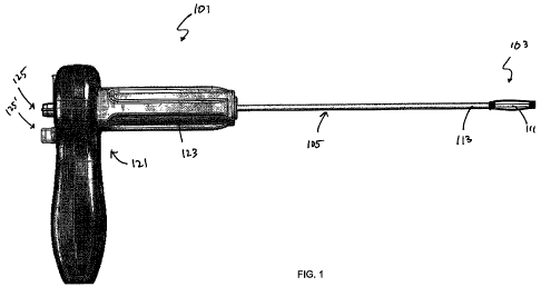
including an applicator and a bone stabilization device is shown in FIG. 1. In
FIG. 1, the
applicator 101 includes a handle portion 107 and an elongate cannula 105,
which maybe referred
to as a delivery device or as an elongate linkage member. An implant 103 is
attached to the
distal end of the applicator 101. In this example, the implant is held in a
collapsed configuration
by applying force from both ends of the implant. In this example, the elongate
linkage member
includes an inner member (rod) 111 and an outer member 113 that are movably
(slideably)
disposed relative to each other. This variation is described in greater detail
below. In FIG. 1, the
proximal end of the bone stabilization device is releasably coupled to the
outer member 113 and
the distal end of the implant is releasably coupled to the inner member 111.
The applicator 101
may separately control the relative motion of the proximal and distal end of
the implant (which is
pre-biased to self-expand to a delivery configuration) by controlling the
relative motions of the
outer cannula 113 and the inner member 111 at the handle 121. In this example,
the handle
includes a ratchet mechanism 123 (e.g., a rotary gear including a pawl, not
visible in FIG. 1) and
a number of controls 125,125' for directing the motion of the applicator.
[00082] Any of the applicators or inserters described herein may be used with
any appropriate
bone stabilization device (typically referred to as a "stabilization device"),
examples of which are
provided herein. These stabilization devices may be a self-expanding device
that expands from a
compressed profile having a relatively narrow diameter (e.g., a delivery
configuration) into an
expanded profile (e.g., a deployed configuration). Stabilization devices
generally include a shaft
region having a plurality of struts that may extend from the shaft body. The
distal and proximal
regions of a stabilization device may include one or more attachment regions
configured to
attach to an inserter for inserting (and/or removing) the stabilization device
from the body.
FIGS. 2A through 6 and 18A-C show exemplary stabilization devices.
[00083] Side profile views of five variations of stabilization devices are
shown in FIGS. 2A
through 2E. FIG. 2A shows a 10 mm asymmetric stabilization device in an
expanded
configuration. The device has four struts 201, 201', formed by cutting four
slots down the length
of the shaft. In this example, the elongate expandable shaft has a hollow
central lumen, and a
proximal end 205 and a distal end 207. By convention, the proximal end is the
end closest to the
-12-
CA 02726585 2010-12-01
WO 2009/149074 PCT/US2009/045958
person inserting the device into a subject, and the distal end is the end
furthest away from the
person inserting the device.
[00084] The struts 201, 201' of the elongate shaft is the section of the shaft
that projects from
the axial (center) of the shaft. Three struts are visible in each of figures
2A-2E. In general, each
strut has a leading exterior surface that forms a cutting surface adapted to
cut through cancellous
bone as the strut is expanded away from the body of the elongate shaft. This
cutting surface may
be shaped to help cut through the cancellous bone (e.g., it may have a tapered
region, or be
sharp, rounded, etc.). In some variations, the cutting surface is
substantially flat.
[00085] The stabilization device is typically biased so that it is relaxed in
the expanded or
deployed configuration, as shown in FIGS. 2A to 2E. In general, force may be
applied to the
stabilization device so that it assumes the narrower delivery profile,
described below (and
illustrated in FIG. 9C). Thus, the struts may elastically bend or flex from
the extended
configuration to the unextended configuration.
[00086] The struts in all of these examples are continuous curvature of
bending struts.
Continuous curvature of bending struts are struts that do not bend from the
extended to an
unextended configuration (closer to the central axis of the device shaft) at a
localized point along
the length of the shaft. Instead, the continuous curvature of bending struts
are configured so that
they translate between a delivery and a deployed configuration by bending over
the length of the
strut rather than by bending at a discrete portion (e.g., at a notch, hinge,
channel, or the like).
Bending typically occurs continuously over the length of the strut (e.g.,
continuously over the
entire length of the strut, continuously over the majority of the length of
the strut (e.g., between
100-90%, 100-80%, 100-70%, etc.), continuously over approximately half the
length of the strut
(e.g., between about 60-40%, approximately 50%, etc.).
[00087] The "curvature of bending" referred to by the continuous curvature of
bending strut is
the curvature of the change in configuration between the delivery and the
deployed
configuration. The actual curvature along the length of a continuous curvature
of bending strut
may vary (and may even have "sharp" changes in curvature). However, the change
in the
curvature of the strut between the delivery and the deployed configuration is
continuous over a
length of the strut, as described above, rather than transitioning at a hinge
point. Struts that
transition between delivery and deployed configurations in such a continuous
manner may be
stronger than hinged or notched struts, which may present a pivot point or
localized region where
more prone to structural failure.
[00088] Thus, the continuous curvature of bending struts do not include one or
more notches
or hinges along the length of the strut. Two variations of continuous
curvature of bending struts
are notchless struts and/or hingeless struts. In FIG. 2A, the strut 201 bends
in a curve that is
-13-
CA 02726585 2010-12-01
WO 2009/149074 PCT/US2009/045958
closer to the distal end of the device than the proximal end (making this an
asymmetric device).
In this example, the maximum distance between the struts along the length of
device is
approximately 10 mm in the relaxed (expanded) state. Thus, this maybe referred
to as a 10 mm
asymmetric device.
[00089] FIG. 2B shows another example of a 10 mm asymmetric device in which
the curve of
the continuous curvature of bending strut has a more gradual bend than the
devices shown in
FIG. 2A. This variation may be particularly useful when the device is used to
support non-
cancellous bone in the deployed state. For example, the flattened curved
region 209 of the
continuous curvature of bending strut may provide a contact surface to support
the non-
cancellous bone. For example, the leading edge of the strut (the cutting edge)
may expand
through the cancellous bone and abut the harder cortical bone forming the
exterior shell of the
bony structure. FIG. 2C shows a symmetric 10 mm device in which this concept
211 is even
more fully developed. FIGS. 2D and 2E are examples of 18 mm devices similar to
the 10 mm
devices shown in FIGS. 2A and 2B, respectively.
[00090] FIGS. 3A and 3B show enlarged side and side perspective views
(respectively) of the
10 mm asymmetric device shown in FIG. 2A. These figures help further
illustrate the
continuous curve of the continuous curvature of bending strut 301. The
proximal end (the end
facing to the right in FIGS. 3A and 3B), shows one variation of an attachment
region to which
the device may be attached to one portion of an introducer. In this example,
the end includes a
cut-out region 305, forming a seating area into which a complementary
attachment region of an
inserter may mate. Although not visible in FIGS. 3A and 3B, the distal region
307 of the device
may also include an attachment region. In some variations, the inner region
(and/or outer region)
of the proximal end 315 of the device may be threaded. Threads may also be
used to engage the
inserter at the proximal (and/or distal) ends of the device as part of the
attachment region.
[00091] An attachment region may be configured in any appropriate way. For
example, the
attachment region may be a cut-out region (or notched region), including an L-
shaped cut out, an
S-shaped cut out, a J-shaped cut out, or the like, into which a pin, bar, or
other structure on the
inserter may mate. In some variations, the attachment region is a threaded
region which may
mate with a pin, thread, screw or the like on the inserter. In some
variations, the attachment
region is a hook or latch. The attachment region may be a hole or pit, with
which a pin, knob, or
other structure on the inserter mates. In some variations, the attachment
region includes a
magnetic or electromagnetic attachment (or a magnetically permeable material),
which may mate
with a complementary magnetic or electromagnet region on the inserter. In each
of these
variations the attachment region on the device mates with an attachment region
on the inserter so
that the device may be removably attached to the inserter.
-14-
CA 02726585 2010-12-01
WO 2009/149074 PCT/US2009/045958
[00092] The attachment region on the implant may be formed of a material
forming the
majority of the implant (e.g., a shape memory material such as a shape memory
alloy), or it may
be formed of a different material and secured to the rest of the implant. In
particular, when the
implant attachment regions comprises threads, it may be particularly
advantageous to form the
threads in anther material (e.g., PMMA or other polymers, ceramics, or metals)
that is then
secured to the shape memory alloy forming the body of the implant. In some
variations the
attachment regions comprise an internal threaded region at the distal end of
the implant and an
external threaded region at the proximal end of the implant (counter-threaded
as described
below). It is known that shape memory materials such as Nitinol are
particularly difficult to cut
threads in and to weld to, particularly in an internal diameter such as the
distal end of the device.
Thus, in some variations the distal end of the device includes a plug formed
of PMMA or other
biocompatible material that forms threads and can be inserted into the
implants distal end.
[00093] The stabilization devices described herein generally have two or more
releasable
attachment regions for attaching to an inserter. For example, a stabilization
device may include
at least one attachment region at the proximal end of the device and another
attachment region at
the distal end of the device. This may allow the inserter to apply force
across the device (e.g., to
pull the device from the expanded deployed configuration into the narrower
delivery
configuration), as well as to hold the device at the distal end of the
inserter. However, the
stabilization devices may also have a single attachment region (e.g., at the
proximal end of the
device). In this variation, the more distal end of the device may include a
seating region against
which a portion of the inserter can press to apply force to change the
configuration of the device.
In some variations of the self-expanding stabilization devices, the force to
alter the configuration
of the device from the delivery to the deployed configuration comes from the
material of the
device itself (e.g., from a shape-memory material), and thus only a single
attachment region (or
one or more attachment region at a single end of the device) is necessary.
[00094] In variations of the stabilization device that include a proximal
releasable attachment
site and a distal releasable attachment site (which may be located at either
at the proximal and
distal ends, or spaced from the ends), the releasable attachment sites may be
configured to
operate in opposite directions. For example, when the attachment sites are
threaded regions
(e.g., FIG. 3A-3B), the threads on the proximal attachment or coupling site
may be configured to
run counterclockwise while threads on the distal attachment or coupling site
are configured to
run clockwise. Thus, each end of the implant may be coupled or de-coupled to
the applicator
may rotating in opposite directions relative to each other. In addition, the
coupling regions may
be configured so that the rotational tolerances are controlled so that there
is very little slippage
between the applicator and the implant when rotating to engage or disengage.
-15-
CA 02726585 2010-12-01
WO 2009/149074 PCT/US2009/045958
[00095] Similar to FIGS. 3A and 3B, FIGS. 4A and 4B show side and side
perspective views
of exemplary symmetric 10 mm devices, and FIGS. 5A and 5B show side and side
perspective
views of 18 mm asymmetric devices. .
[00096] The continuous curvature of bending struts described herein may be any
appropriate
dimension (e.g., thickness, length, width), and may have a uniform cross-
sectional thickness
along their length, or they may have a variable cross-sectional thickness
along their length. For
example, the region of the strut that is furthest from the tubular body of the
device when
deployed (e.g., the curved region 301 in FIGS. 3A and 3B) maybe wider than
other regions of
the strut, providing an enhanced contacting surface that abuts the non-
cancellous bone after
deployment.
[00097] The dimensions of the struts may also be adjusted to calibrate or
enhance the strength
of the device, and/or the force that the device exerts to self-expand. For
example, thicker struts
(e.g., thicker cross-sectional area) may exert more force when self-expanding
than thinner struts.
This force may also be related to the material properties of the struts.
[00098] As mentioned, in some variations, different struts on the device may
have different
widths or thicknesses. In some variations, the same strut may have different
widths of
thicknesses along its length. Controlling the width and/or thickness of the
strut may help control
the forces applied when expanding. For example, controlling the thickness may
help control
cutting by the strut as it expands.
[00099] Similarly, the width of the strut (including the width of the outward-
facing face of the
strut) may be controlled. The outward-facing face may include a cutting
element (e.g., a sharp
surface) along all or part of its width, as mentioned.
[000100] Varying the width, thickness and cutting edge of the struts of a
device may modulate
the structural and/or cutting strength of the strut. This may help vary or
control the direction of
cutting. Another way to control the direction of cutting is to modify the pre-
biased shape. For
example, the expanded (pre-set) shape of the struts may include one or more
struts having a
different shape than the other struts. For example, one strut may be
configured to expand less
than the other struts, or more than other struts. Thus, in some variations,
the shape of the
expanded implant may have an asymmetric shape, in which different struts have
different
expanded configurations.
[000101] The struts maybe made of any appropriate material. In some
variations, the struts
and other body regions are made of substantially the same material. Different
portions of the
stabilization device (including the struts) may be made of different
materials. In some
variations, the struts may be made of different materials (e.g., they may be
formed of layers,
and/or of adjacent regions of different materials, have different material
properties). The struts
-16-
CA 02726585 2010-12-01
WO 2009/149074 PCT/US2009/045958
may be formed of a biocompatible material or materials. It may be beneficial
to form struts of a
material having a sufficient spring constant so that the device may be
elastically deformed from
the deployed configuration into the delivery configuration, allowing the
device to self-expand
back to approximately the same deployed configuration. In some variation, the
strut is formed of
a shape memory material that may be reversibly and predictably converted
between the deployed
and delivery configurations. Thus, a list of exemplary materials may include
(but is not limited
to): biocompatible metals, biocompatible polymers, polymers, and other
materials known in the
orthopedic arts. Biocompatible metals may include cobalt chromium steel,
surgical steel,
titanium, titanium alloys (such as the nickel titanium alloy Nitinol),
tantalum, tantalum alloys,
aluminum, etc. Any appropriate shape memory material, including shape memory
alloys such as
Nitinol may also be used.
[000102] Other regions of the stabilization device may be made of the same
material(s) as the
struts, or they may be made of a different material. Any appropriate material
(preferably a
biocompatible material) may be used (including any of those materials
previously mentioned),
such as metals, plastics, ceramics, or combinations thereof. In variations
where the devices have
bearing surfaces (i.e. surfaces that contact another surface), the surfaces
may be reinforced. For
example, the surfaces may include a biocompatible metal. Ceramics may include
pyrolytic
carbon, and other suitable biocompatible materials known in the art. Portions
of the device can
also be formed from suitable polymers include polyesters, aromatic esters such
as polyalkylene
terephthalates, polyamides, polyalkenes, poly(vinyl) fluoride, PTFE,
polyarylethyl ketone, and
other materials. Various alternative embodiments of the devices and/or
components could
comprise a flexible polymer section (such as a biocompatible polymer) that is
rigidly or semi
rigidly fixed.
[000103] The devices (including the struts), may also include one or more
coating or other
surface treatment (embedding, etc.). Coatings may be protective coatings
(e.g., of a
biocompatible material such as a metal, plastic, ceramic, or the like), or
they may be a bioactive
coating (e.g., a drug, hormone, enzyme, or the like), or a combination
thereof. For example, the
stabilization devices may elute a bioactive substance to promote or inhibit
bone growth,
vascularization, etc. In one variation, the device includes an elutible
reservoir of bone
morphogenic protein (BMP).
[000104] As previously mentioned, the stabilization devices may be formed
about a central
elongate hollow body. In some variations, the struts are formed by cutting a
plurality of slits
long the length (distal to proximal) of the elongate body. This construction
may provide one
method of fabricating these devices, however the stabilization devices are not
limited to this
construction. If formed in this fashion, the slits maybe cut (e.g., by
drilling, laser cutting, etc.)
-17-
CA 02726585 2010-12-01
WO 2009/149074 PCT/US2009/045958
and the struts formed by setting the device into the deployed shape so that
this configuration is
the default, or relaxed, configuration in the body. For example, the struts
may be formed by
plastically deforming the material of the struts into the deployed
configuration. In general, any
of the stabilization devices may be thermally treated (e.g., annealed) so that
they retain this
deployed configuration when relaxed. Thermal treatment may be particularly
helpful when
forming a strut from a shape memory material such as Nitinol into the deployed
configuration.
[0001051 FIGS. 18A-18C illustrate another variation of a bone stabilization
device. In this
example, the bone stabilization device is pre-biased in an expanded
configuration, and an
expansion limiter is slideably coupled to the outside of the device. In
general, an expansion
limiter may be a tube, funnel, or other structure that may be fitted over one
or both ends of the
stabilization device. The stabilization device may be otherwise similar, e.g.,
pre-biased in the
expanded configuration to those described above. The minimum diameter of the
expansion
limiter (which may also be referred to as an "over tube") is typically
somewhat larger than the
outer diameter of the stabilization device in the collapsed configuration
(prior to expansion). At
least a partial length of the expansion limiter may be threaded, ratcheted, or
otherwise shaped
such that a relative position of the expansion controller relative to the
stabilization device can be
controlled and maintained. For example, at least a partial length of the
exterior of the
stabilization device may be shaped to mate with the expansion limiter. For
example, the
expansion limiter may travel on threads controlling the position of the
limiter relative to the
stabilization device. In this example, the position of the expansion limiter
relative to the
stabilization device may be changed by rotating and/or translating it. The
expansion limiter may
be moved along the length of the stabilization device to allow it to change
diameter (e.g.,
expand). In variations of the device including an expansion limiter, the
expansion limiter may be
coupled to a member of the applicator (e.g., a first elongate member or the
outer cannula
member). Thus, the outer cannula member may be coupled to the limiter while
the inner
member is coupled to the proximal or distal end of the implant. Motion of the
limiter relative to
the implant may be used to expand or collapse the implant, as illustrated in
FIGS. 18A-18C. As
the expansion limiter 1805 in FIG. 18A is moved distally in FIGS. 18B and 18C,
the implant
1801 collapses.
[0001061 As mentioned, the expansion limiter may be coupled to the applicator,
or may for a
portion of the applicator. Thus, the applicator may move the expansion limiter
relative to the
stabilization device to allow it to controllably expand (preferably while
leaving the distal end
fixed relative to the insertion site in the body). In some variation the
expansion limiter may be
an outer sleeve that fits over all or a portion of the stabilization device
and may be withdrawn to
deliver it.
-18-
CA 02726585 2010-12-01
WO 2009/149074 PCT/US2009/045958
[000107] FIG. 6A shows one variation of a stabilization device 600 having a
plurality of
continuous curvature of bending struts 601, 601' removably attached to an
elongate linkage
member (referred to here as an inserter) 611. In this example, an attachment
region 615 at the
proximal portion of the stabilization device is configured as an L-shaped
notch, as is the
attachment region 613 at the distal portion of the device. The inserter 611 in
this example does
not include a separate handle, although grips 631, 633 are integrally formed
at the proximal end.
[000108] As mentioned, an inserter may include an elongate body having a
distal end to which
the stabilization device maybe attached and a proximal end which may include a
handle or other
manipulator that coordinates converting an attached stabilization device from
a delivery and a
deployed configuration, and also allows a user to selectively release the
stabilization device from
the distal end of the inserter.
[000109] The elongate linkage member (inserter) 611 shown in FIG. 6A includes
a first
elongate member 621 that coaxially surrounds a second elongate member 623. In
this variation,
each elongate member 621, 623 includes a stabilization device attachment
region at its distal end,
to which the stabilization device is attached, as shown. In this example, the
stabilization device
attachment region includes a pin that mates with the L-shaped slots forming
the releasable
attachment regions on the stabilization device. In FIG. 6A the L-shaped
releasable attachments
on the stabilization device are oriented in opposite directions (e.g., the
foot of each "L" points in
opposite directions). Thus, the releasable attachment devices may be locked in
position
regardless of torque applied to the inserter, preventing the stabilization
device from being
accidentally disengaged.
[000110] The inserter shown in FIG. 6A also includes two grips 631, 633 at the
proximal ends
of each elongate member 621, 623. These grips can be used to move the elongate
members (the
first 621 or second 623 elongate member) relative to each other. The first and
second elongate
members of the inserter may be moved axially (e.g., may be slid along the long
axis of the
inserter) relative to each other, and/or they may be moved in rotation
relative to each other
(around the common longitudinal axis). Thus, when a stabilization device is
attached to the
distal end of the inserter, moving the first elongate member 621 axially with
respect to the
second elongate member 623 will cause the stabilization device to move between
the deployed
configuration (in which the struts are expanded) and the delivery
configuration (in which the
struts are relatively unexpanded). Furthermore, rotation of the first elongate
member of the
inserter relative to the second elongate member may also be used to disengage
one or more
releasable attachment regions of the stabilization device 613, 615 from the
complementary
attachment regions of the inserter 625, 627. Although he stabilization devices
described herein
are typically self-expanding stabilization devices, the inserter may be used
with stabilization
-19-
CA 02726585 2010-12-01
WO 2009/149074 PCT/US2009/045958
devices that do not self-expand. Even in self-expanding devices, the inserter
may be used to
apply additional force to convert the stabilization device between the
delivery and the deployed
configuration. For example, when allowed to expand in a cancellous bone, the
force applied by
the struts when self-expanding may not be sufficient to completely cut through
the cancellous
bone and/or distract the cortical bone as desired. In some variations, the
inserter may also permit
the application of force to the stabilization device to expand the struts even
beyond the deployed
configuration.
[000111] An inserter may also limit or guide the movement of the first and
second elongate
members, so as to further control the configuration and activation of the
stabilization device. For
example, the inserter may include a guide for limiting the motion of the first
and second elongate
members. A guide may be a track in either (or both) elongate member in which a
region of the
other elongate member may move. The inserter may also include one or more
stops for limiting
the motion of the first and second elongate members.
[000112] As mentioned above, the attachment regions on the inserter mate with
the
stabilization device attachments. Thus, the attachment regions of the inserter
may be
complementary attachments that are configured to mate with the stabilization
device
attachments. For example, a complimentary attachment on an inserter may be a
pin, knob, or
protrusion that mates with a slot, hole, indentation, or the like on the
stabilization device. The
complementary attachment (the attachment region) of the inserter may be
retractable. For
example, the inserter may include a button, slider, etc. to retract the
complementary attachment
so that it disconnects from the stabilization device attachment. A single
control may be used to
engage/disengage all of the complementary attachments on an inserter, or they
may be controlled
individually or in groups.
[000113] FIG. 6B is another variation of a stabilization device 600 releasably
connected to an
inserter 611, in which the attachment region 635 between the stabilization
device and the inserter
is configured as a screw or other engagement region, rather than the notch 615
shown in FIG.
6A.
[000114] In some variation the inserter includes a lock or locks that hold the
stabilization
device in a desired configuration. For example, the inserter may be locked so
that the
stabilization device is held in the delivery configuration (e.g., by applying
force between the
distal and proximal ends of the stabilization device). In an inserter such as
the one shown in
FIG. 6A, for example, a lock may secure the first elongate member to the
second elongate
member so that they may not move axially relative to each other.
[000115] FIG. 7A is another example of an inserter 711 and an attached
stabilization device
700. Similar to FIG. 6A, the stabilization device includes a first elongate
member 721 attached
-20-
CA 02726585 2010-12-01
WO 2009/149074 PCT/US2009/045958
to the proximal end of the stabilization device, and a second elongate member
723 attached to
the distal end of the stabilization device. The first 721 and the second 723
elongate members are
also configured coaxially (as a rod and shaft) that may be moved axially and
rotationally
independently of each other. The stabilization device 700 includes a plurality
of continuous
curvature of bending struts, shown in detail in FIG. 7B. The stabilization
device 700 is shown in
the deployed configuration. The distal end of the stabilization device
includes a releasable
attachment 713 that is configured as a threaded region which mates with a
threaded
complementary attachment 725 at the distal end of the structure.
[000116] The proximal ends of the coaxial first and second elongated members
721, 723 also
include grips 731, 733. These grips are shown in greater detail in FIG. 7C. As
with the grips
described in FIG. 6A, these grips may be grasped directly by a person (e.g., a
physician,
technician, etc.) using the device, or they may be connected to a handle.
Thus, in some
variations one or both grips are `keyed' to fit into a handle, so that they
can be manipulated by
the handle. An example of this is shown in FIG. 8A-8E, and described below.
The inserter of
FIG. 7A also includes a knob 741 attached to the first elongated member 721
distal to the
proximal end of the elongated member. This knob may also be used to move the
first (or outer)
elongate member of the inserter (e.g., to rotate it), or to otherwise hold it
in a desired position.
The knob maybe shaped and/or sized so that it may be comfortably handheld. In
some variations
(described in greater detail below) this knob 741 is a keyed member that is
secured to the outer
member (cannula) of the inserter 711. This keyed member may be configured to
secure within a
handle so and may help orient the device (including the implant) and the
handle, and may sever
to secure the cannula in the handle. The keyed member may have an outer shape
(e.g.,
rectangular, etc.) that locks the relative motion of all or a portion of the
handle with respect to the
outer member.
[000117] Any of the inserters described herein may include, or may be used
with, a handle. A
handle may allow a user to control and manipulate an inserter. For example, a
handle may
conform to a subject's hand, and may include other controls, such as triggers
or the like. Thus, a
handle may be used to control the relative motion of the first and second
elongate members of
the inserter, or to release the connection between the stabilization device
and the inserter, or any
of the other features of the inserter described herein.
[000118] An inserter maybe packaged or otherwise provided with a stabilization
device
attached. Thus, the inserter and stabilization device may be packaged sterile,
or may be
sterilizable. In some variations, a reusable handle is provided that may be
used with a pre-
packaged inserter stabilization device assembly. In some variations the handle
is single-use or
disposable. The handle may be made of any appropriate material. For example,
the handle may
-21-
CA 02726585 2010-12-01
WO 2009/149074 PCT/US2009/045958
be made of a polymer such as polycarbonate.
[000119] FIG. 8A illustrates one variation of a handle 800 that maybe used
with an inserter,
such as the inserter shown in FIGS. 7A-7C. The handle 800 includes a hinged
joint 803, and the
palm contacting 805 region and finger contacting 807 region of the handle 800
may be moved
relative to each other by rotating about this hinged joint 803. This variation
of a handle also
includes a thumb rest 809, which may also provide additional control when
manipulating an
inserter with the handle. The thumb rest may also include a button, trigger,
or the like.
[000120] FIGS. 8B-8E illustrate the connection of an inserter such as the
inserter described
above in FIGS. 7A-C into a handle 800. In FIG. 8B the proximal end of the
inserter is aligned
with openings 811, 811' in the handle. These openings are configures so that
the grips 731, 733
at the distal ends of the first and second elongate members of the inserter
can fit into them. In
this example, the grip 733 is shaped so that it can be held in the opening
811' of the handle in an
oriented fashion, preventing undesirable rotation. Thus, in FIG. 8C the
proximal end of the
inserter (the grips 731 and 732) are placed in the openings 811, 811'. The
inserter may then be
secured to the handle by rotating cover 833, as shown in FIGS. 8D and 8E.
[000121] By securing the proximal end of the inserter in the handle, the
handle can then be
used to controllably actuate the inserter, as illustrated in FIGS. 9A-9D. In
this example the
stabilization device is in the deployed configuration (shown in FIG. 9A) when
the handle is
"open" (shown in FIG. 9B). By squeezing the handle (rotating the finger grip
region towards the
palm region, as shown in FIG. 9D) the inserter applies force between the
proximal and distal
regions of the stabilization device, placing it in a delivery configuration,
as shown in FIG. 9C.
[000122] As mentioned above, in the delivery configuration the struts of the
stabilization
device are typically closer to the long axis of the body of the stabilization
device. Thus, the
device may be inserted into the body for delivery into a bone region. This may
be accomplished
with the help of an access cannula (which may also be referred to as an
introducer). As shown in
FIG. 10, the inserter 1015 is typically longer than the access cannula 1010,
allowing the
stabilization device to project from the distal end of the access cannula for
deployment. The
access cannula may also include a handle 1012.
[000123] Any of the devices (stabilization devices) and applicators (including
handles) maybe
included as part of a system or kit for correcting a bone defect or injury.
FIGS. 10 through 14D
illustrate different examples of tools (or variations of tools) that may be
used as part of a system
for repair bone. Any of these tools (or additional tools) may also be used to
perform the methods
of repairing bone (particularly spinal bone) described herein. For example,
FIG. 11 shows a
trocar 1105 having a handle 1107 and a cutting/obdurating tip 1109. This
trocar 1105 may also
be used with an access cannula 1111. Another example of an access cannula 1111
(or
-22-
CA 02726585 2010-12-01
WO 2009/149074 PCT/US2009/045958
introducer) is shown adjacent to the trocar 1106 in FIG. 11. This exemplary
access cannula has
an inner diameter of approximately 4.2 mm, so that the trocar 1105 will fit
snugly within it, and a
stabilization device in a delivery configuration will also fit therein. Any
appropriate length
cannula and trocar may be used, so long as it is correctly scaled for use with
the introducer and
stabilization device. For example, the access cannula may be approximately
15.5 cm long. The
trocar an introducer may be used to cut through tissue until reaching bone, so
that the introducer
can be positioned appropriately.
[000124] A bone drill, such as the hand drill shown in FIGS. 12A-12C, may then
be used to
access the cancellous bone. The twist drill 1201 shown in FIG. 12A-12C has a
handle 1203 at
the proximal end and a drill tip 1205 at the distal end. This twist drill may
be used with the same
access cannula previously described (e.g., in this example the twist drill has
an outer diameter of
4.1 mm and a length of 19.5 cm). The distal (drill) end of the twist drill may
extend from the
cannula, and be used to drill into the bone. The proximal end of the twist
drill shown in FIGS.
12A-12C is calibrated (or graduated) to help determine the distance drilled.
[000125] Any of the devices shown and described herein may also be used with a
bone cement.
For example, a bone cement may be applied after inserting the stabilization
device into the bone,
positioning and expanding the device (or allowing it to expand and distract
the bone) and
removing the inserter, leaving the device within the bone. Bone cement may be
used to provide
long-term support for the repaired bone region.
[000126] Any appropriate bone cement or filler may be used, including PMMA,
bone filler or
allograft material. Suitable bone filler material include bone material
derived from
demineralized allogenic or xenogenic bone, and can contain additional
substances, including
active substance such as bone morphogenic protein (which induce bone
regeneration at a defect
site). Thus materials suitable for use as synthetic, non-biologic or biologic
material may be used
in conjunction with the devices described herein, and may be part of a system
includes these
devices. For example, polymers, cement (including cements which comprise in
their main phase
of microcrystalline magnesium ammonium phosphate, biologically degradable
cement, calcium
phosphate cements, and any material that is suitable for application in tooth
cements) may be
used as bone replacement, as bone filler, as bone cement or as bone adhesive
with these devices
or systems. Also included are calcium phosphate cements based on
hydroxylapatite (HA) and
calcium phosphate cements based on deficient calcium hydroxylapatites (CDHA,
calcium
deficient hydroxylapatites). See, e.g., U.S. Pat. No. 5,405,390 to O'Leary et
al.; U.S. Pat. No.
5,314,476 to Prewett et al.; U.S. Pat. No. 5,284,655 to Bogdansky et al.; U.S.
Pat. No. 5,510,396
to Prewett et al.; U.S. Pat. No. 4,394,370 to Jeffries; and U.S. Pat. No.
4,472,840 to Jeffries,
which describe compositions containing demineralized bone powder. See also
U.S. Pat. No.
-23-
CA 02726585 2010-12-01
WO 2009/149074 PCT/US2009/045958
6,340,477 to Anderson which describes a bone matrix composition. Each of these
references is
herein incorporated in their entirely.
[000127] FIG. 13 shows a tapered cement cannula 1301 that maybe used to
deliver bone
cement to the insertion site of the device, and also shows two cement
obturators 1303, 1305 for
delivering the cement (piston-like). The cannula delivering cement is also
designed to be used
through the access cannula, as are all of the components described above,
including the
stabilization device and inserter, trocar, and drill. This is summarized in
FIGS. 14A-14D. FIG.
14A illustrates an access cannula 4101 with a stabilization device 1403 and
inserter inserted
through the access cannula, as shown in FIG. 10. FIG. 14B shows a trocar 1405
within the
access cannula 1401. FIG. 14C shows a hand drill 1407 within the same access
cannula 1401,
and FIG. 14D shows a cement cannula 1409 and a cement obturator 1411 within
the same access
cannula 1401. These devices may be used to repair a bone.
Exemplary Method of Repairing a Bone
[000128] As mentioned above, any of the devices described herein may be used
to repair a
bone. A method of treating a bone using the devices describe herein typically
involves
delivering a stabilization device (e.g., a self-expanding stabilization device
as described herein)
within a cancellous bone region, and allowing the device to expand within the
cancellous bone
region so that a cutting surface of the device cuts through the cancellous
bone.
[000129] For example, the stabilization devices described herein may be used
to repair a
compression fracture in spinal bone. This is illustrated schematically in
FIGS. 15A-15G. FIG.
15A shows a normal thoracic region of the spine in cross-section along the
sagital plane. The
spinal vertebras are aligned, distributing pressure across each vertebra. FIG.
15B shows a
similar cross-section through the spine in which there is a compression
fracture in the 11th
thoracic vertebra 1501. The 11thvertebra is compressed in the fractured
region. It would be
beneficial to restore the fractured vertebra to its uninjured position, by
expanding (also referred
to as distracting) the vertebra so that the shape of the cortical bone is
restored. This may be
achieved by inserting and expanding one of the stabilization devices described
herein. In order
to insert the stabilization device, the damaged region of bone must be
accessed.
[000130] As mentioned above, an introducer (or access cannula) and a trocar,
such as those
shown in FIG. 11 may be used to insert the access cannula adjacent to the
damaged bone region.
Any of the steps described herein may be aided by the use of an appropriate
visualization
technique. For example, a fluoroscope may be used to help visualize the
damaged bone region,
and to track the p of inserting the access cannula, trocar, and other tools.
Once the access
cannula is near the damaged bone region, a bone drill may be used to drill
into the bone, as
-24-
CA 02726585 2010-12-01
WO 2009/149074 PCT/US2009/045958
shown in FIG. 15C.
[000131] In FIG. 15C the drill 1503 enters the bone from the access cannula.
The drill enters
the cancellous bony region within the vertebra. After drilling into the
vertebra to provide access,
the drill is removed from the bone and the access cannula is used to provide
access to the
damaged vertebra, as shown, by leaving the access cannula in place, providing
a space into
which the stabilization device may be inserted in the bone, as shown in FIG.
15D. In FIG. 15E a
stabilization device, attached to an inserter and held in the delivery
configuration, is inserted into
the damaged vertebra.
[000132] Once in position within the vertebra, the stabilization device is
allowed to expand (by
self-expansion) within the cancellous bone of the vertebra, as shown in FIG.
15F. In some
variations, the device may fully expand, cutting through the cancellous bone
and pushing against
the cortical bone with a sufficient restoring force to correct the
compression, as shown in FIG.
15G. However, in some variations, the force generated by the device during
self-expansion is
not sufficient to distract the bone, and the inserter handle may be used
(e.g., by applying force to
the handle, or by directly applying force to the proximal end of the inserter)
to expand the
stabilization device until the cortical bone is sufficiently distracted.
[000133] Once the stabilization device has been positioned and is expanded, it
may be released
from the inserter. In some variations, it may be desirable to move or redeploy
the stabilization
device, or to replace it with a larger or smaller device. If the device has
been separated from the
inserter (e.g., by detaching the removable attachments on the stabilization
device from the
cooperating attachments on the inserter), then it may be reattached to the
inserter. Thus, the
distal end of the inserter can be coupled to the stabilization device after
implantation. The
inserter can then be used to collapse the stabilization device back down to
the delivery
configuration (e.g., by compressing the handle in the variation shown in FIGS.
9A-9D), and the
device can be withdrawn or re-positioned.
[000134] As mentioned above, a cement or additional supporting material may
also be used to
help secure the stabilization device in position and repair the bone. For
example, bone cement
maybe used to cement a stabilization device in position. FIGS. 16A-16C
illustrate one variation
of this. In FIG. 16A the stabilization device 1601 has been expanded within
the cancellous bone
1603 and is abutting the cortical bone 1605. Although in some variations the
addition of the
stabilization device may be sufficient to repair the bone, it may also be
desirable to add a cement,
or filler to help secure the repair. This may also help secure the device in
position, and may help
close the surgical site.
[000135] For example, in FIG. 16B a fluent bone cement 1609 has been added to
the
cancellous bone region around implant. This cement will flow through the
channels of
-25-
CA 02726585 2010-12-01
WO 2009/149074 PCT/US2009/045958
trebeculated (cancellous) bone, and secure the implant in position. This is
shown in greater
detail in the enlarged region. This bone cement or filler can be applied using
the delivery
cannula (e.g., through a cement cannula, as described above), and allowed to
set.
[000136] While preferred embodiments of the present invention have been shown
and
described herein, such embodiments are provided by way of example only.
Numerous variations,
changes, and substitutions are possible without departing from the invention.
Thus, alternatives
to the embodiments of the invention described herein may be employed in
practicing the
invention. The exemplary claims that follow help further define the scope of
the systems,
devices and methods (and equivalents thereof).
[000137] The devices and methods for treating vertebral bodies describes above
in detail may
be used for the implantation of a self-reshaping device through a pedicle into
the cancellous bone
interior of a vertebral body, as mentioned. The self-reshaping of embodiments
of the device
includes a coincident longitudinally shortening of the device as a whole, and
a radial expansion
of struts. Following implantation and release from constraints that maintain
the linear
configuration, the struts of device self-expand, and while expanding, they cut
through cancellous
bone so as to arrive at the inner surface of the surrounding cortical bone of
the superior (or
cephalad) and inferior (or caudal) endplates of the vertebral body. The device
may be sized and
configured such that self-expansion takes the device to an appropriate
dimension for the
vertebral body. Thus, as the device approaches its final expanded dimension,
it presses the
surface outwardly so as to restore the height and volume of the vertebral body
toward the
dimensions of the vertebral body prior to the fracture.
[000138] Figure 16C illustrates two stabilization devices 511, 511' inserted
bilaterally into a
spinal segment. A pedicle (bone) screw 513, 513' (attached through a pedicle
of a vertebral
body) has been attached into each stabilization device. Thus, in any of the
variations described,
the distal end of the device may also include a bone screw attachment region,
so that a pedicle
screw may be stabilized both at the proximal and the distal ends of the
device. A bone screw
may be inserted completely through the stabilization device, and may extend
from the distal end.
In some variations, the central region of the device includes a continuous (or
mostly continuous)
channel into which the bone screw may pass.
[000139] In one variation of the method described herein, two self-expanding
devices may be
inserted bilaterally into a compression-fractured vertebral body for the
purpose of restoring the
height of the vertebra and expanding the body of the vertebra to restore it to
its pre-fractured
configuration. A compression fracture of a vertebral body typically reduces
the height of a
vertebral body; this compressed height will generally be referred to as H1.
Upon implantation
-26-
CA 02726585 2010-12-01
WO 2009/149074 PCT/US2009/045958
and expansion of a self-reshaping vertebral body stabilization device, the
height of the vertebral
body at the side or site of implantation is increased to a height H2. The
height H2 is typically
toward or an approximation of the height of the vertebral body prior to its
state of compression.
[000140] Methods of using the implants, applicators and systems including them
may include a
step of selecting devices appropriate in form, shape, and size for each
implantation site. Thus, in
some variations the applicator or inserter devices described herein may be
configured so that
they may be used with implants of different sizes (both length and/or widths).
For example, the
devices may be configured so that the relative movement and separation of the
inner and outer
members spans a variety of sizes (e.g., lengths) of the bone stabilization
implants from expanded
to collapsed lengths. In some variations the handles include a limiter that
prevents
overexpansion of the applicator when coupled to an implant.
[000141] FIGS. 17A - 17D show a series of lateral views of a vertebral body
110 with a height
H1 (anterior on the left, posterior on the right) at a cross-section along a
sagittal plane near a
pedicle of the vertebral body. The vertebral body 110 has an outer layer of
cortical bone,
including a superior endplate 102a and an inferior endplate 102b, and an
interior region
including cancellous bone 101. FIG. 17A shows insertion of a deployment device
70 into a pre-
drilled channel, a self-reshaping vertebral body stabilization device
contained (not shown) within
the deployment device. FIG. 17B shows an early point in the deployment of a
self-reshaping
vertebral stabilization device 30, with expandable struts beginning to expand.
FIG. 17C shows
full expansion of the expandable struts of the self-reshaping device 30 and
consequent
restoration of vertebral body to a height H2. FIG. 17D shows injection of a
stabilizing material
61 into the space within the expanded struts of the self-reshaping device 30
and into available
space within bone cancellous bone 101 surrounding the device. The material
physically stabilizes
the position of the device in the bone, stabilizes local bone that has been
disrupted, and may also
provide a matrix for the in-growth of bone, which further contributes to the
stabilization of the
device.
Handles
[000142] FIGS. 8A-8E and 9B and 9D illustrated one variation of a handle of an
applicator, as
described above. Other variations of handles, and particularly removable or
reusable handles,
are shown and describe in FIGS. 19A-30.
[000143] In general, these handles include a capture mechanism for connecting
to the elongate
member (e.g., inserter) that connects to the implant. As mentioned, an
elongate member may be
referred to as a delivery device or an elongate linkage member of the
applicator. The elongate
member typically includes a first elongate member (e.g., an outer member or
cannula) that is
-27-
CA 02726585 2010-12-01
WO 2009/149074 PCT/US2009/045958
configured to removably secure or couple with one region of an implant (e.g.,
the proximal end
of the implant), and a second elongate member (e.g., an inner member, cannula
or rod) that is
configured to removably secure or couple with a second region of the implant
(e.g., the distal end
of the implant). The first and second members of the elongate member may be
configured to
couple and uncouple from the implant by rotating in opposite directions. The
proximal end of
the elongate member may include a proximal grips or couplers for grasping
and/or manipulating
the inner and outer members to control the expansion or contraction of the
implant. The linkage
portion of the applicator connects distally to the proximal and distal regions
of the implant, and
the handle engages the proximal end of the linkage portion of the applicator
by connecting to and
controlling these proximal couplers.
[000144] For example, the FIG. 19A shows a cross-section through one variation
of a handle
coupled to an elongate linkage portion 1901. The inner rod of the elongate
linkage portion 1901
is connected to a ball-shaped grip 1905, while the outer cannula of the
elongate linkage portion
1901 terminates proximally in a second grip region that is approximately
rectangular 1907. In
some variations (as described herein) one or both of these grip regions may be
keyed so that the
rotation of the inner and outer members can be controlled. In FIG. 19A, the
relative longitudinal
translation of the inner and outer elongate members of the linkage portion are
controlled. For
example, the handle includes a threaded rod or drive shaft 1921 that can be
rotated by rotation of
an adjustment knob 1923 at the proximal end. The threaded rod is a rotary gear
that moves in
rotation only, and does not translate along the longitudinal axis. The handle
includes a latch
1933 for locking the expansion position of the implant by locking the internal
moving slider
1935. This latch lock may be configured to prevent removal of the device from
the handle when
the implant is under tension by the handle (e.g., held collapsed). In some
variations, the handle
may have release to release the tension on the implant (e.g., held by the
linkage portion of the
applicator) before it may be released from the handle.
[000145] The diameter of the drive shaft, as well as the threads per inch, can
be configured to
control the mapping of the lateral movement and rotation of the adjustment
knob based on
implant size. For example, a typical implant may require a lateral change of
approximately 1.4
mm to change from a collapsed (delivery) configuration to an expanded
(deployed)
configuration. The movement of the inner member relative to the outer member
may be geared
by adjustment of the dimensions so that an exact and convenient movement
between the
adjustment knob and the implant can be created.
[000146] FIG. 19B shows another variation of a handle similar to the variation
shown in FIG.
19A. In this example, the handle includes the features described above, but is
further configured
so that it is compatible with only a single `size' of implant, based on the
separation between the
-28-
CA 02726585 2010-12-01
WO 2009/149074 PCT/US2009/045958
proximal ends of the inner and outer members of the linkage portions of the
applicator. A
different length push rod 1921 is used for each size implant, adjusting the
length of the seating
components 1951, 1953 for the grip regions of the elongate linkage portion
1961. In some
variations, the handle may be configured to work with a variety of different-
sized implants. For
example, in a variation such as the one shown in FIGS. 19A and 19B, the handle
may be
configured so that the push rod is adjustable.
[000147] FIG. 20A shows another variation of a handle similar to those shown
in FIGS. 19A
and 19B. In FIG. 20A, some of the details are omitted for clarity. In this
example, the drive shaft
can be disengaged from driving the delivery shaft (elongate linkage portion)
1985 and push rod
1981. When the drive shaft is disengaged, the user can rotate the adjustment
knob 1987
clockwise to detach one end of the implant from the delivery device and
counterclockwise to
detach the other end of the implant, in variations in which the implant
proximal and distal
coupling ends are counter-matched, as described above. In this variation, the
drive-shaft
engagement may be spring-loaded so that the default condition is that it is
engaged. As
mentioned above, the handle may include a safety lock to prevent disengaging
the drive shaft
when force is being applied to hold the implant in the delivery configuration.
[000148] In some variations the delivery device (also referred to herein as
the elongate linkage
portion 2001), including an outer member (e.g., cannula) and an inner member
(e.g., rod or
cannula), that couples to the implant distally and the handle proximally, may
also include a bias
2005 that maintains a load on the implant when it is connected. For example,
FIG. 20B
illustrates one variation of an elongate linkage portion including a bias. In
this variation, the
proximal end of the elongate linkage portion includes a bias (e.g. spring)
that tends to keep the
distal ends of the outer elongate member and the inner elongate member
separated (e.g., helping
hold an attached implant in a pre-biased delivery configuration by pulling the
proximal end of
the elongate linkage portion together. Biasing the elongate linkage portion in
this manner may
be helpful to decrease the force needed to be provided by a user to hold the
implant in the
delivery configuration.
[000149] FIGS. 21A and 21B illustrate front and back exploded views of one
variation of a
handle similar to the variation shown in FIGS. 19A-20A. For example, in FIG.
21A, the handle
includes a grip region 2101, 2101', an adjustment knob 2105 (shown enlarged in
FIG. 22B), a
drive shaft 2107 coupled to the adjustment knob 2105, a sliding receiver 2109
for the inner
member of the elongate linkage portion, and a fixed receiver 2111 for the
outer member of the
elongate linkage portion, and a latch 2113 for locking the relative positions
of the inner and outer
members, as well as a lock 2115 for the latch. The lock mechanism also
includes a spring
element 2119 for biasing the latch closed or opened until it is engaged. In
the variation shown in
-29-
CA 02726585 2010-12-01
WO 2009/149074 PCT/US2009/045958
FIG. 21A and 21B, the handle is configured so that the sliding receiver 2109
seats the grip
(which may be keyed or unkeyed) of the inner member (e.g., rod) of the
delivery device/elongate
linkage portion (not shown), and the fixed receiver 2111 seats the grip (which
may be keyed or
unkeyed) of the outer member (e.g., cannula) of the delivery device/elongate
linkage portion.
This configuration may be reversed. For example, the sliding receiver may be
configured to seat
the grip of the inner member (e.g., rod) of the elongate linkage portion and
the fixed receiver
may be configured to seat the grip of the outer member (e.g., cannula). FIG.
22A shows an
enlarged perspective view of the fixed receiver and FIG. 22C shows an enlarged
perspective
view of the movable receiver.
[000150] FIGS. 23A-23C illustrate another variation of a handle. The handle
2301 shown in
FIG. 23C is configured to couple with the proximal end of an elongate linkage
portion of an
applicator. A handle such as the one shown in FIG. 23C includes one or more
sections that are
rotatable relative to other regions of the handle (or a relative to the
coupled elongate linkage
member). Rotation of a portion of the handle may controllably move the inner
member of the
elongate linkage member relative to the outer member of the elongate linkage
member (or vice
versa), allowing the implant to be controllably self-expanded (e.g., deployed)
or alternatively
collapsed (e.g., for removal). FIGS. 23A and 23B illustrate the handle 2301
coupled to an
elongate linkage member 2304.
[000151] The exemplary handle 2101 shown in FIG. 23A-23C includes only six
components,
though more or fewer components may be used. FIGS. 25A-25K illustrate the
relationship
between each of these components (and the inserter distal end). As mentioned
above, the
handles may be configured to be reusable/durable, or they may be configured as
single-use.
[000152] In some variations the handle is permanently affixed to the elongate
linkage member
(e.g., forming a unitary applicator); in other variations the elongate linkage
member of the
inserter is separate from the handle.
[000153] In use, a handle that is detachably coupleable to an elongate linkage
member may be
attached within the handle, e.g. , by removing a handle cover (see FIG. 25B).
The cover may be
replaced to secure the proximal ends of the inserter in place. Once the
inserter is secured in
position, the handle (E.g., the proximal end) may be rotated to allow the
proximal end of the
implant to controllably move towards the distal end, allowing the implant to
expand. Rotation in
the opposite direction moves the proximal end of the implant away from the
distal end. A rotary
gear may be include within the housing and configured to advance the first
elongate member of
the elongate linkage member.
[000154] In some variations, the handle may be configured as a ratcheting
handle. A ratcheting
handle may include a lever arm can engage the rotatable region of the handle
and allow it to be
-30-
CA 02726585 2010-12-01
WO 2009/149074 PCT/US2009/045958
rotated. The lever arm may provide a further mechanical advantage for
collapsing or expanding
a stabilization device. In some variations (not pictured), a portion of the
handle may be
removable so that he handle can be ratcheted from different angles or
directions. In some
variations, the handle may include a directional control for the ratchet
mechanism, such as a
button, lever, etc. Changing the setting on the directional control may allow
the direction
rotation to be changed, while the applied direction of rotation (e.g., pushing
or pulling the level
arm) is the same.
[000155] In some variations, the distal end of the stabilization device is
connected to an inner
member of the inserter. For example, the inner member of the inserter may be a
rod that is
relatively fixed as an outer rod or cannula may be moved around it (or along
it). Thus, the shaft
(e.g., the hollow outer part) moves to expand/contract the stabilization
device.
[000156] In addition to the inserters (e.g., handles and elongate linkage
members) described
and illustrated above, other variations of inserters may also be used. An
inserter may include a
threaded outer member that is configured to secure to the proximal end of the
stabilization
device. In this example, a handle may be configured to mate with the threaded
outer portion of
the inserter, For example, this may eliminate the threading in the handle.
This threading may be
keyed to prevent rotation of the inserter. Preventing rotation, particularly
unnecessary rotation,
may prevent the device from unthreading prematurely at the distal end. In some
variations the
keying may be a channel, etc.
[000157] In any of the variations described herein, the handles (or other
portions of the
inserter) may be marked or coded to indicate the size of the implant. For
example, the handle
(which may mate with a generic handle, regardless of the size of the attached
stabilization
device) may be marked with numbering/lettering to indicate the size, and/or
color coated. In
some variations the handle is marked to indicate the orientation of the
implant (e.g., the self-
expanding struts) relative to the inserter.
[000158] FIGS. 26A-30 illustrate another variation of an inserter for
inserting a bone support
implant. This variation is similar to the device shown in FIG. 1. In this
variation, the inserter
2600 includes a handle region 2601 and an elongate linkage member 2603. The
elongate linkage
member include an inner member 2605 and an outer member 2607. The distal ends
of the inner
and outer members are threaded to couple to end regions (proximal and distal)
of an implant as
described above.
[000159] The handle 2601 shown in FIG. 26A is a ratcheting handle configured
to connect to
the elongate linkage member 2603. FIG. 26B shows a side perspective view of
the inserter
shown in FIG. 26A. This handle includes a perpendicular handle region 2611 and
a ratchet grip
region 2615.
-31-
CA 02726585 2010-12-01
WO 2009/149074 PCT/US2009/045958
[000160] FIG. 27A illustrates a back view of the handle shown in FIGS. 26A-
26B. The
operation of this device will be described in greater detail below. The device
includes a ratchet
switch 2623 that changes the direction of the ratcheting mechanism so that the
handle turns to
engage the implant in expansion or collapse (e.g., driving the distal ends of
the inner and outer
member of the elongate linkage member either apart or towards each other). The
handle shown
in this example also includes an indicator of the orientation 2628 of the
struts on the implant.
Thus, the implant may be loaded onto the elongate linkage member, and
therefore the handle, in
a manner that maintains the orientation of the implant.
[000161] The handle shown in FIG. 27A also includes a release control or
mechanism 2705
that operates as an "escape hatch" safety feature. In this example, the
release mechanism may be
unscrewed from the handle to release the elongate linkage member from the
handle in the event
that the handle fails (e.g., jams, locks, or the like). Activation of the
release mechanism releases
any force applied by the handle. Thus, the implant (connected to the elongate
linkage member)
may be removed from the handle.
[000162] In some variations the handle may also include an indicator of the
size of the implant
to be used (e.g., 10 mm, 12 mm, 16 mm, 18 mm, etc.). In some variations the
system includes
one or more sensors or connections to sensors. For example, the handle may
include a connector
to a temperature sensor or other sensor (including visualization devices) for
sensing data from
the implant or the region of implantation.
[000163] FIG. 27B illustrates a side perspective view of the handle shown in
FIG. 27A. The
ratchet handle 2615 is shown as partially transparent. In use, the ratchet
handle may be rotated
relative to the handle body 2715 to advance or withdraw the inner and outer
members of the
elongate linkage member, and thereby expand/contract the implant. The ratchet
mechanism
internal to the handle includes a limiter to prevent it from being
overextended in either direction,
protecting the device from over-expansion or over-collapsing, which may lead
to breaking of the
implant. In some configurations the handle may be preset for use with a
particular size implant.
In other variations, the handle may be configured to be switched to selected
sizes.
[000164] FIGS. 28A and 28B show one variation of an elongate linkage member
portion of an
applicator. In FIG. 28A, the elongate linkage member includes an inner member
2801 and an
outer member 2803. The outer member includes a keyed engagement member 2805,
which may
be referred to as a shaft stabilizer. The keyed engagement member is
configured to mate with
the handle (as illustrated in FIG. 29) and maintain the orientation of the
implant at the distal end
of the applicator. It may also stabilize the shaft of the elongate linkage
member (e.g., the outer
member) within the handle. Thus, the handle may include a mating region 2922
at the distal end
(e.g., opening into the handle) configured to mate with the shaft stabilizer
2805.
-32-
CA 02726585 2010-12-01
WO 2009/149074 PCT/US2009/045958
[000165] FIG. 30 shows an exploded view of the handle portion of the
applicator shown in
FIGS. 26A-29. In FIG. 30, the handle includes: a front and back handle grip
region 3001, 3003;
a ratchet grip region 3005; a shaft driver overmold element 3009; a retainer
3011; a retainer
attachment 3015; the ratchet mechanism 3007; a release switch 3019; a rod
release 3021; a rod
(inner member) stop 3025; a rod end cap 3033; a ratchet direction switch 3035
and a ratchet
direction pawl 3037.
[000166] In operation, the applicator may be connected to the proximal and
distal ends of an
implant by connecting to the elongate linkage member, as mentioned above. The
proximal end of
the implant may connect to the outer member, while the distal end of the
implant connects to the
inner member (e.g., rod). Both ends may include counter-directional threads.
The threads may
be on the outer surface of the proximal end and on the inner surface of the
distal end. The
implant may be connected to the elongate linkage member either before or after
it has been
coupled to the handle. In some variations the elongate linkage member is pre-
packaged coupled
to the implant, so that it may be opened from a sterile packaging for use. The
same handle may
be re-used for different implants, typically within the same patient.
[000167] A self-expanding implant, connected to the applicator as described
above, maybe
inserted into a patient by manipulating the handle and shaft of the
applicator. Once it is
positioned as desired (which may be visualized by florosocopy), it may be
allowed to
controllably self-expand using the applicator. As mentioned, the applicator
may include an
indicator of the orientation of the self-expanding struts. Thus, the handle
and shaft of the
applicator may be manipulated (e.g., rotated) orient the implant so that the
struts will be
positioned as desired.
[000168] The elongate linkage member may be connected to the handle by
engaging the keyed
engagement member (shaft stabilizer) on the surface of the elongate linkage
member. Inserting
the shaft stabilizer into the handle also engages the inner and outer members
of the elongate
linkage member. Thereafter, rotation of the ratcheting handle will move the
outer member, and
therefore the proximate end of the implant, relative to the inner member. The
direction of
motion may depend on the ratchet switch, which moves the pawl member to select
the engaged
motion of the ratchet mechanism.
[000169] In some variations it is helpful that the proximal end of the implant
is moving relative
to the length of the implant. By moving the proximal end, the implant may be
inserted into a
desired location and controllable allowed to self-expand into a position
without extending from
the distal implantation location. Thus, the implant will not shift position
relative to the distal
insertion site by foreshortening as the implant is controllably self-expanded
into a deployed
configuration.
-33-
CA 02726585 2010-12-01
WO 2009/149074 PCT/US2009/045958
[0001701 The ratchet direction may be selected and switched using the
ratcheting switch as
indicated. In some variations, an indicator (e.g., a symbol, color, text,
etc.) may indicate the
direction of movement enabled (e.g., expansion/deployment or
contraction/retraction of the
implant).
[0001711 Once the implant has been inserted and allowed to self-expand, the
applicator (handle
and shat of the elongate linkage member) may be removed. The force applied to
the implant by
the handle maybe released by pushing the release button (switch), on the
handle, so that the
shaft of the elongate linkage member may be removed from the handle. The
handle may be
removed from the elongate linkage member and the elongate linkage member may
then be
removed from the implant by the proximal and distal ends. In some variations
the implant may
be removed from the elongate linkage member while still attached to the
handle. In other
variations the handle is removed first. The elongate linkage member may be
decoupled from the
proximal and distal ends of the implant by rotating the inner and outer
members (e.g., counter
clockwise at the distal end and clockwise at the proximal end) in threaded
variations.
10001721 If the position of the implant is not optimal, the position may be re-
adjusted using the
handle as indicated above, e.g., by collapsing the implant using the handle
and moving the
implant.
10001731 The methods, devices and systems described herein provide only some
variations
described herein, and additional variations may be included and are
contemplated. While
embodiments of the present invention have been shown and described herein,
such embodiments
are provided by way of example only. Thus, alternatives to the embodiments of
the invention
described herein may be employed in practicing the invention. The exemplary
claims that follow
help further define the scope of the systems, devices and methods (and
equivalents thereof).
-34-