Note: Descriptions are shown in the official language in which they were submitted.
CA 02726691 2014-04-30 '
53155-6
1
Description
Title of Invention: USE OF ONCOGENE NRF2 FOR CANCER
PROGNOSIS
Technical Field
[0001] The present invention relates to the field of efficacy prediction of
a cancer drug,
prognostic prediction of cancer, and cancer treatment. More specifically, the
present
invention relates to an efficacy prediction method for an mTOR-related cancer
drug by
detecting the abnormalities of NRF2, a prediction method for prognosis of
cancer by
detecting the abnormalities of NRF2, and a cancer treatment agent that
inhibits NRF2
gene or protein.
Background Art
[0002] It is known that environmental factors, such as smoking, radiation,
chronic in-
flammation caused by virus infection, etc, and exposure to toxic chemical
substances
influences to onset and development of cancer. Previous researches have
revealed that
oxidative stress caused by factors which causes abnormalities of DNA and
protein is
with the development of cancer.
[0003] A living organism has the physiological defense mechanism against
such oxidative
stress. A transcription factor called Nuclear factor erythroid 2-related
factor 2 (NRF2)
is recognized as one of the important molecules that plays a role in molecular
mechanism of said physiological defense system. NRF2 is a DNA binding molecule
with high transcriptional induction ability, which is activated when a cell is
exposed to
oxidative stress, and induces the expression of many groups of enzymes, such
as glu-
tathione reductase, which relieve oxidative stress, to protect a cell from the
disorder
caused by the stress.
[0004] For example, NRF2 is known as an important transcription factor that
transmits a
promoting signal to an antioxidant response element (ARE) component, which is
a
DNA regulatory element controlling transcription of the gene products which
protect
cells from carcinogens, oxidants, and other toxic compounds. It has been
reported that
an enhancer via ARE having cancer inhibitory activity increases the NRF2 level
in the
nucleus (see Yuesheng et al. Molecular Cancer Therapeutics, 3 (7) 885-893,
2004). In
the oral administration model of the benzo-alfa-pyran, it has been appeared
that the
number of cancers are increased in NRF2 knockout mice compared to in wild type
(see
Ramos-Gomez et al. Proc. Natl Acad. Sci. USA, 98, 3410-3415, 2001). In
addition, the
document suggests that an anticancer agent oltipraz increases the expression
of NRF2,
and that the anticancer effect of oltipraz is not seen in NRF2 knockout mice,
and thus
enhancement of NRF2 expression may lead to the anticancer effect.
[0005] Furthermore, it has been reported that in a living organism an
existence of NRF2 is
2
WO 2010/010672 PCT/JP2009/003335
controlled by negative feedback of Kelch-like ECH-associated protein 1
(KEAP1), and
an inhibitor to KEAP1 is under development as an anticancer agent (see Ewan,
Drug
Discovery Today, 10 (14) 950-951, 2005). Thus, it is expected that drugs
targeting
NRF2 or KEAP1 which enhance the expression of NRF2 may be used as an
anticancer
agent.
[0006] On the other hand, it is reported that, in lung cancer, constant
activation of NRF2 is
observed due to the reduced activity of KEAP1 caused by KEAP1 gene mutation is
observed and that the activated NRF2, induced constant expression of an anti-
oxidant
protein. It has been reported that increased expression of NRF2 may be one of
the
reasons of the resistance of a cancer cells to cisplatin (see Ohta et al.
Cancer Res., 68,
1303-1309, 2008 and International Publication W02006/128041). It has also been
reported that administration of alkylating agents such as cisplatin, mephalan,
chlo-
rambucil, and BCNU increases the expression of a gene regulated by ARE, such
as
NRF2. It has been suggested that the increased gene expression products
regulated by
ARE can be involved in the resistance of cancer cells to the anticancer
agents. It has
also been suggested that all trans retinoic acid (ATRA), which can bind to
NRF2, may
be able to enhance the effect of a chemotherapic drug (see International
Publication
W02008/012534).
[0007] Thus, it is appeared that administration of an alkylating anticancer
agent may activate
NRF2, and the NRF2 may play a role in resistance to the alkylating anticancer
agent.
An alkylating anticancer agent interrupts proliferation by cross-linking bases
of DNA
in a cancer cell. The mechanism of activation of NRF2 upon administration of
an
alkylating agent remains to be explained. Although a part of mechanism of the
ac-
quisition of resistance due to NRF2 against the effect of an alkylating
anticancer drug
is predicted from a cytoprotective action of NRF2, the overview is not fully
understood
yet. The relation between anticancer agents other than an alkylating
anticancer agent
and NRF2 has not been reported.
[0008] As noted above, activation of NRF2 observed in a cancer cell has
been considered
mainly based on the reduced activity of KEAP1 due to KEAP1 gene mutation. The
relation between mutation and activation of NRF2 in a cancer cell, and the
relation
between the NRF2 activation due to the mutation and the malignant alteration
of
cancer have not been known. Especially, NRF2 has been thought to have an
anticancer
effect in the onset of the cancer caused by oxidative stress, etc. it has been
considered
that NRF2 rather suppresses the malignant alteration of cancer. In relation
with a
chemotherapic drug, it has suggested that NRF2 is responsive to administration
of an
alkylating agent, and, at least, an all trans retinoic acid enhances the
effect of an
alkylating agent. However, it has been completely unknown whether an effect of
an all
trans retinoic acid is based on NRF2 suppressing effect, and if so, what
mechanism
CA 02726691 2010-12-01
CA 02726691 2014-04-30
53155-6
3
underlies inhibition. Therefore, the relation between suppression of NRF2 and
an-
ticancer agents other than an alkylating anticancer agent has been wholly
unknown.
[0009] Mammalian target of rapamycin (mTOR) is a serine threonine ldnase
identified as a
target molecule of a macrolide antibiotic, rapamycin and it serves as
regulator on cell
growth, cell proliferation, cell motility, cell survival, protein synthesis
and tran-
scription. Since a rapamycin induces an apoptosis of a cancer cell lacking the
function
of p53, it is considered that an mTOR inhibitor has an anticancer activity
(see Shik
Huang et al. Molecular Cell, 11, 1491-1501, 2003). In addition, mTOR
inhibitors have
been under development as anticancer agent for, for example, renal cancer and
pancreatic duct cancer.
[0010] An mTOR is also known as an insulin receptor tyrosine kinase. A
research on the
apoptosis of cerebrovascuIar endothelial cells in a hyperglycemia patient
concludes
that an mTOR inhibitor impairs expression of insulin-inducible NRF2-mediated
Glutamate-L-cystein ligase-catalytic subunit (GCLc), oxidation reduction
balance, and
survival of a human cerebrovascular endothelial cell (see Okouchi, Masahiro et
al.
Current Neurovascular Research, 3 (4) 249-261, 2006). However, especially as
for the
field of cancer treatment, the effect of the expression of NRF2 on an action
of an
mTOR inhibitor has not been reported yet.
Summary of Invention
[0011] The present invention is directed to a method for predicting cancer
by detecting
NRF2 gene mutation. Especially the present invention is directed to a method
for
predicting efficacy of an mTOR-related cancer drug, a method for selecting a
ef-
ficacious patient by detecting NRF2 gene mutation, or a method for predicting
a
prognosis of cancer. The present invention also is directed to a method for
treating
cancer by inhibiting NRF2 gene or NRF2 protein, or a cancer treatment agent
that uses
NRF2 gene or NRF2 protein inhibitor as an active ingredient.
CA 02726691 2015-05-19
53155-6
3a
[0012] In a specific embodiment, the present invention is directed to
a method for
providing information about a selection method for efficacy prediction of an
mTOR-related
cancer drug or a method for selecting a efficacious patient by detecting NRF2
gene mutation
or protein. In addition, the present invention is directed to a method for
predicting the
effectiveness of an mTOR-related cancer drug or for selecting an efficacious
patient by
detecting NRF2 gene or protein mutation. Specifically, the present invention
is directed to a
method for predicting that an mTOR-related cancer drug is effective when NRF2
gene or
protein has mutation. The present invention also is directed to a kit that is
able to detect a
mutation in NRF2 for predicting the effectiveness of an mTOR-related cancer
drug. For
example, the present invention is directed to kit comprising an nucleic acid
capable of binding
to the NRF2 gene or a
4
WO 2010/010672 PCT/JP2009/003335
substance capable of binding to the NRF2 protein, (e.g. antibody), wherein the
nucleic
acid and the substance are capable of detecting a mutation in NRF2.
Alternatively, the
present invention is directed to a kit which can detect alteration in function
of NRF2
due to mutation. For example, a kit which detects digestion of NRF2 by KEAP1
is also
is directed to in the present invention.
[0013] In another embodiment, the present invention is directed to a method
for providing
information on a malignancy of cancer or on a prognostic quality by detecting
the
mutation of NRF2 in a cancer tissue cell from a cancer patient. Alternatively,
the
present invention is directed to a method for diagnosing malignancy of cancer
or for
predicting prognosis of cancer which comprises detecting a mutation in NRF2 in
a
cancer tissue cell from a cancer patient and diagnosing a patient who has a
mutation in
NRF2 is malignant or predicting a patient who has a mutation in NRF2 is poor
prognosis. Alternatively, the present invention is directed to a kit for
diagnosing cancer
or predicting a prognosis which is able to detect a mutation in NRF2. For
example, the
present invention is directed to a kit comprising an nucleic acid capable of
binding to
the NRF2 gene or a substance capable of binding to the NRF2 protein, such as
an
antibody, wherein the nucleic acid and the substance are able to detect a
mutation in
NRF2. In addition, the present invention is directed to a kit which is able to
detect a
mutation by using a gene amplification technology, such as PCR. Furthermore,
the
present invention is directed to a kit comprising an invader probe, an allele
probe,
triplex-specific DNase, and a universal fluorescent-labeled probe with a
quenching
probe, for example, the Invader (TM) assay kit, etc. In addition, the present
invention
contains a kit that is able to measure the metergasia of NRF2 caused by
mutation. For
example, the present invention also is directed to a kit which can detects
digestion of
NRF2 by KEAP1.
[0014] In another embodiment, the present invention is directed to a method
for treating
cancer comprising inhibiting NRF2 gene or NRF2 protein. The present invention
includes a method for treating cancer comprising suppressing NRF2 gene
expression.
In addition, the present invention is directed to a method for treating cancer
comprising
suppressing expression or activity of NRF2 protein. Moreover, the present
invention is
directed to a cancer drug containing an inhibitor of NRF2 gene or NRF2
protein.
Specifically, the present invention is directed to an agent for treating
cancer which
comprises an antisense, dsRNA, a ribozyme, an aptamer for NRF2, a fragment of
a
NRF2 binding protein, or an antibody or fragment thereof as an active
ingredient.
[0015] More specifically, the present invention is directed to the
following inventions.
(1)A method for obtaining information for predicting response of a cancer
patient to
an mTOR-related cancer drug, comprising:
(a)detecting DNA or RNA coding mutated NRF2 or mutated NRF2 protein in a
CA 02726691 2010-12-01
5
WO 2010/010672 PCT/JP2009/003335
sample originated from the patient; and
(b)associating the measured level of DNA or RNA coding mutated NRF2 or mutated
NRF2 protein with the response of the cancer of the patient to the mTOR-
related
cancer drug.
(2)A method for obtaining information for predicting response of a cancer of a
patient
to an mTOR-related cancer drug from a tumor sample originated from the
patient,
comprising:
(a) detecting DNA or RNA coding mutated NRF2 or mutated NRF2 protein in a
sample originated from the patient;
(b) classifying into one of cancer response classes according to the detected
level of
DNA or RNA coding mutated NRF2 or mutated NRF2 protein, wherein the classi-
fication result depends on the expression level of mutated NRF2 gene or
mutated
NRF2 protein; and
(c) predicting response of the cancer of the patient to a cancer drug, based
on a known
property specific to cancers which belong to the one of the cancer response
classes
classified.
(3)The method according to (2), wherein high level of DNA or RNA coding
mutated
NRF2 or mutated NRF2 protein indicates that the patient is highly responsive
to an
mTOR-related cancer drug.
(4)A kit for predicting a response of cancer patient to an mTOR-related cancer
drug,
comprising at least one of the substances selected from (i) to (iv):
(i) a substance that binds to DNA or RNA coding NRF2 and does not bind to DNA
or
RNA coding mutated NRF2;
(ii) a substance that does not bind to NRF2 gene and binds to mutated NRF2
gene;
(iii) a substance that binds to NRF2 protein and does not bind to mutated NRF2
protein; and
(iv) a substance that does not bind to NRF2 protein and binds to mutated NRF2
protein.
(5)A method for obtaining information for predicting prognosis of a cancer
patient
comprising:
(a) detecting DNA or RNA coding mutated NRF2 or mutated NRF2 protein in a
sample that is originated from the patient; and
(b) associating measured level of DNA or RNA coding mutated NRF2 or mutated
NRF2 protein with prognosis of the patient.
(6)The method according to (5), wherein high level of DNA or RNA coding
mutated
NRF2 or mutated NRF2 protein indicates poor prognosis of the patient.
(7)A cancer drug containing an NRF2 inhibitor as an active ingredient.
(8)The cancer drug according to (6), wherein the NFR2 inhibitor is an
antisense,
CA 02726691 2010-12-01
CA 02726691 2016-06-28
,- 53155-6
6
dsRNA, a ribozyme, an aptamer, an NRF2 binding-protein fragment, or an
antibody or
fragment thereof.
[0015a] The present invention as claimed relates to:
- a method for predicting response of a cancer patient to an inhibitor of mTOR
or PI3K,
comprising: (a) measuring whether DNA or RNA coding for mutated NRF2 protein
or
mutated NRF2 protein is present in a sample originated from the patient; and
(b) predicting
the response of the cancer patient to said inhibitor based on the presence of
DNA or RNA
coding for mutated NRF2 or of mutated NRF2 protein, wherein the presence of
DNA or RNA
coding for mutated NRF2 protein or of mutated NRF2 protein is indicative of an
increased
. response of the patient to the inhibitor of mTOR or PI3K, wherein said
mutated NRF2 protein
is a mutated NRF2 protein in which tryptophan at position 24 of SEQ ID NO: 2
is substituted
with cysteine or lysine, glutamine at position 26 of SEQ ID NO: 2 is
substituted with glutamic
acid, isoleucine at position 28 of SEQ ID NO: 2 is substituted with glycine,
leucine at position
30 of SEQ ID NO: 2 is substituted with phenylalanine, glycine at position 31
of SEQ ID NO:
2 is substituted with alanine, glutamine at position 75 of SEQ ID NO: 2 is
substituted with
histidine, aspartic acid at position 77 of SEQ ID NO: 2 is substituted with
valine or glycine,
glutamic acid at position 79 of SEQ ID NO: 2 is substituted with lysine,
threonine at position
80 of SEQ ID NO: 2 is substituted with lysine or proline, or glutamic acid at
position 82 of
SEQ ID NO: 2 is substituted with aspartic acid, and wherein the cancer is
selected from
esophageal cancer, lung cancer and head and neck cancer;
- a kit for predicting a response of a cancer patient to an inhibitor of mTOR
or P13 K,
comprising: a nucleic acid molecule that does not bind to DNA or RNA coding
for NRF2
protein and binds to DNA or RNA coding for mutated NRF2 protein, wherein the
presence of
DNA or RNA coding for mutated NRF2 protein or of mutated NRF2 protein is
indicative of
an increased response of the patient to the inhibitor of mTOR or PI3K, wherein
said mutated
NRF2 protein is a mutated NRF2 protein in which tryptophan at position 24 of
SEQ ID
NO: 2 is substituted with cysteine or lysine, glutamine at position 26 of SEQ
ID NO: 2 is
CA 02726691 2016-06-28
53155-6
6a
substituted with glutamic acid, isoleucine at position 28 of SEQ ID NO: 2 is
substituted with
= glycine, leucine at position 30 of SEQ ID NO: 2 is substituted with
phenylalanine, glycine at
position 31 of SEQ ID NO: 2 is substituted with alanine, glutamine at position
75 of SEQ ID
NO: 2 is substituted with histidine, aspartic acid at position 77 of SEQ ID
NO: 2 is
substituted with valine or glycine, glutamic acid at position 79 of SEQ ID NO:
2 is substituted
with lysine, threonine at position 80 of SEQ ID NO: 2 is substituted with
lysine or proline, or
glutamic acid at position 82 of SEQ ID NO: 2 is substituted with aspartic
acid, and wherein
the cancer is selected from esophageal cancer, lung cancer and head and neck
cancer; and
- a method for predicting prognosis of a esophageal cancer patient comprising:
(a) measuring
whether DNA or RNA coding for mutated NRF2 protein or of mutated NRF2 protein
is
present in a sample that is originated from the patient; and (b) predicting
the prognosis based
on the presence of DNA or RNA coding for mutated NRF2 or of mutated NRF2
protein,
wherein the presence of DNA or RNA coding for mutated NRF2 or of mutated NRF2
protein
is indicative of a negative prognosis of the patient, and wherein said mutated
NRF2 protein is
a mutated NRF2 protein in which tryptophan at position 24 of SEQ ID NO: 2 is
substituted
with cysteine or lysine, glutamine at position 26 of SEQ ID NO: 2 is
substituted with glutamic
acid, isoleucine at position 28 of SEQ ID NO: 2 is substituted with glycine,
leucine at
position 30 of SEQ ID NO: 2 is substituted with phenylalanine, glycine at
position 31 of SEQ
ID NO: 2 is substituted with alanine, glutamine at position 75 of SEQ ID NO: 2
is substituted
with histidine, aspartic acid at position 77 of SEQ ID NO: 2 is substituted
with valine or
glycine, glutamic acid at position 79 of SEQ ID NO: 2 is substituted with
lysine, threonine at
position 80 of SEQ ID NO: 2 is substituted with lysine or proline, or glutamic
acid at
position 82 of SEQ ID NO: 2 is substituted with aspartic acid.
[0016] An efficacy prediction method of the present invention is able
to predict
response of a cancer patient to an mTOR-related cancer drug or to predict
whether an mTOR-
related cancer drug achieve an effect to the cancer patient before
administration. Therefore,
the present invention enables to choose a drug expected to be effective for a
cancer patient
and to avoid unnecessary cancer drug administration to a patient who is not
expected to
effectively respond, hence, to relieve the patient from pain of unnecessary
side effects. The
CA 02726691 2016-06-28
, 53155-6
6b
present invention is able to provide information for selecting a cancer drug.
In addition, the
present invention can provide meaningful information for deciding a therapy
regimen strategy
for a cancer patient by predicting prognosis of the cancer patient.
Furthermore, the method of
the present invention for inhibiting NRF2 gene or NRF2 protein or a treatment
drug of the
present invention comprising an NRF2 gene or NRF2 protein inhibitor can be
used as a novel
treatment method or a novel treatment drug for cancer.
CA 02726691 2015-05-19
53155-6
6c
Brief Description of Drawings
[0017] [fig.11FIG. 1 shows biological pathway related to mTOR.
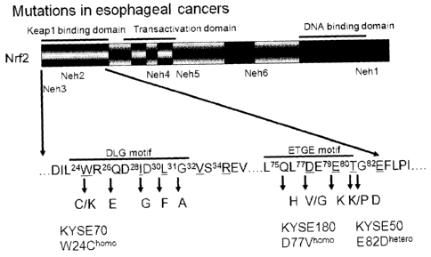
[fig.2]FIG. 2 shows the positions of the mutations and amino acid
substitutions caused
thereby in the NRF2 gene in a clinical sample of esophageal cancer and
esophageal
cancer cell lines (KYSE-50, KYSE-70, KYSE-180).
[fig.3]FIG. 3 shows the result of detection of NRF2 expression in normal
esophagus (A
and B) and esophageal cancer (C) by immunohistochemical staining using
antibody
against NRF2. In the figure, an arrow represents a cell expressing NRF2. FIG.
3B is an
enlarged view of a part of FIG. 3A.
[fig.4]FIG. 4 shows the result of a statistical analysis (Kaplan-Meier
analysis) on rela-
tionship between postoperative survival time and presence or absence of gene
mutation
for esophageal cancer cases screened by NRF2 gene mutation. In the figure, the
vertical axis represents a cumulative survival rate, and the horizontal axis
represents
the elapsed days after surgery.
[fig.5]FIG. 5 shows changes in cell proliferation upon administration or non-
administration of dsRNA to the esophageal cancer cell lines that have NRF2
gene
mutation (KYSE-50 and KYSE-180). In the figure, the vertical axis is the ratio
number
of cells administered NRF2 dsRNA to number of cells administered control
dsRNA.
[fig.6]FIG. 6 shows changes in proliferation due to rapamycin treatments to
the
esophageal cancer cell lines without abnormalities in NRF2 gene (KYSE-30, KYSE-
140, KYSE-170, KYSE-270) and to the cancer cell lines with abnormalities in
NRF2
gene (KYSE-50, KYSE-70, KYSE-180). In the figure, the vertical axis shows a
ratio
CA 02726691 2015-05-19
53155-6
7
of number of cells of each cell line, when number of cells with 0 nM rapamycin
(without drug) is set as 100%. In the figure, horizontal axis shows dosage of
rapamycin.
[fig.7]FiG. 7 shows changes in proliferation due to rapamycin treatments to
the lung
cancer cell lines without abnormalities in NRF2 gene (SQ-5. QG-56) and to the
cancer
cell lines with abnormalities in NRF2 gene (LK-2, EBC-1). In the figure, the
vertical
axis shows a ratio of number of cells of each cell line, when number of cells
with 0 nM
rapamycin (without drug) is set as 100%. In the figure, horizontal axis shows
dosage
of rapamycin.
[fig.8]FIG. 8 shows changes in proliferation due to rapamycin treatments to
the head
and neck cancer cell lines without abnormalities in NRF2 gene (H0-1-N-1, HSC2)
and
to the cancer cell lines with abnormalities in NRF2 gene (H0-1-u-1). In the
figure, the
vertical axis shows a ratio of number of cells of each cell line, when number
of cells
with 0 nM rapamycin (without drug) is set as 100%. In the figure, horizontal
axis
shows dosage of rapamycin.
Best Mode for Carrying out the Invention
[0018] A. Prediction of mTOR-related cancer drug
In one aspect, the present invention relates to a method or a kit for
predicting
response of cancer patient to an mTOR-related cancer drug, or a method for
obtaining
information for predicting response of cancer patient to an mTOR-related
cancer drug.
As used herein, "mTOR-related cancer drug" is not limited as long as an agent
inhibits expression or activity of an mTOR or a substance that is involved in
upstream
or downstream pathway of mTOR and is effective for cancer treatment. An mTOR-
related cancer drug, includes an agent that directly inhibits an mTOR (mTOR
inhibitor), for example, a chemotherapic drug such as sirolimus (also known as
rapamycin), everolimus, temsirolimus and deferolimus; protein such as an
antibody;
peptides such as an antibody fragment; and nucleic acid such as an aptamer, an
antisense, and dsRNA. Since an NRF2 inhibitor inhibits mTOR pathway, NRF2
inhibitor of the present invention may also be included as an mTOR inhibitor.
It is
known that an mTOR pathway is involved in a plurality of pathways as shown in
FIG.
1. However, particularly preferable agents that is involved in an mTOR pathway
and
targeted by an mTOR-related cancer drug in the present appication are, for
example,
type I phosphoinositide 3-kinase (hereinafter, abbreviated as "PI3K"),
phosphoinositide-
dependent protein kinase 1 (hereinafter abbreviated as "PDK1"), FK506 binding
protein
(FKBP12), Akt (also known as protein kinase B (PKB)), p70 ribosomal protein S6
kinase 1 (hereinafter, abbreviated as "S6K1"), c-Jun N-terminal kinase
(hereinafter, ab-
breviated as "JNK"), and hypoxic inducible factor 1 alfa (hereinafter,
abbreviated as
8
WO 2010/010672
PCT/JP2009/003335
"HIFI alfa"). Therefore, PI3K inhibitors such as TG100115, TCN-P, LY294002,
wortmannin, BFZ235, and SF1126; PDK1 inhibitors such as UCN-01, BX912, B-
3012, and 05U030313; FKBP12 inhibitors such as AP1903 and tacrolimus; Akt in-
hibitors such as XL418, LY294002, wortmannin, TCN-P, BV-1701-1, FPA-124,
KP372-1, and GSK690693; S6K1 inhibitors such as XL418 and H-89; JNK inhibitors
such as AM111, 5P600125, a compound described in U.S. Patent No. 7,199,124,
and
AS601245; and HIFI alfa inhibitors such as PX478 and SF1126, are also included
in
the mTOR-related cancer drug in the present application.
[0019] As used herein, "response to an mTOR-related cancer drug" means
an effect to at
least one of indicators representing condition of a cancer patient when the
cancer
patient is administered with the mTOR-related cancer drug, wherein the effect
is
caused by administration of an mTOR-related cancer drug. The indicators
include
reduction of tumor size, supression of tumor growth, metastasis, prognostic
quality, re-
cidivation, or recurrence, etc. As used herein, "good response to an mTOR-
related
cancer drug" means showing effectiveness of at least one of the indicators
indicating
the condition of disease in cancer patient a cancer patient who is receiving
an mTOR-
related cancer drug, compared to not receiving the mTOR-related cancer drug,
and
includes, for example, reduction of tumor size, repression of tumor growth,
repression
of metastasis or no metastasis, improvement of prognosis, no recidivation, or
no re-
currence, etc.
[0020] As used herein, "DNA or RNA coding mutated NRF2 gene" is a DNA or RNA
coding having mutation(s) in any part of nucleotides or ribonucleotides
sequence
encoding normal NRF2, for example DNA having mutation(s) in part of sequence
of
SEQ ID NO.1. As used herein "mutated NRF2 protein" is a protein having
mutation(s)
in a part of amino acid sequence (SEQ ID NO 2) constituting normal NRF2. Par-
ticularly, DNA or RNA coding mutated NRF2 or mutated NRF2 protein is DNA or
RNA coding mutated NRF2 or protein that enhances expression level of NRF2
protein
by the mutation. In a more particular aspect, the mutated NRF2 protein
includes
protein in which tryptophan at a position 24 of NRF2 protein is substituted
with
cysteine or lysine, glutamine at a position 26 of NRF2 protein is substituted
with
glutamic acid, isoleucine at a position 28 of NRF2 protein is substituted with
glycine,
leucine at a position 30 of NRF2 protein is substituted with phenylalanine ,
glycine at a
position 31 of NRF2 protein is substituted with alanine, glutamine at a
position 75 of
NRF2 protein is substituted with histidine, aspartic acid at a position 77 of
NRF2
protein is substituted with valine or glycine, glutamic acid at a position 79
of NRF2
protein is substituted with lysine, threonine at a position 80 of NRF2 protein
is sub-
stituted with lysine or proline, and/or glutamic acid at a position 82 of NRF2
protein is
substituted with aspartic acid. The mutated NRF2 gene includes a gene that
encodes
CA 02726691 2010-12-01
9
WO 2010/010672 PCT/JP2009/003335
mutated NRF2 protein in which tryptophan at a position 24 of NRF2 protein is
sub-
stituted with cysteine or lysine, glutamine at a position 26 of NRF2 protein
is sub-
stituted with glutamic acid, isoleucine at a position 28 of NRF2 protein is
substituted
with glycine, leucine at a position 30 of NRF2 protein is substituted with
phenylalanine
, glycine at a position 31 of NRF2 protein is substituted with alanine,
glutamine at a
position 75 of NRF2 protein is substituted with histidine, aspartic acid at a
position 77
of NRF2 protein is substituted with valine or glycine, glutamic acid at a
position 79 of
NRF2 protein is substituted with lysine, threonine at a position 80 of NRF2
protein is
substituted with lysine or proline, and/or glutamic acid at a position 82 of
NRF2
protein is substituted with aspartic acid. A level of a DNA or RNA coding
mutated
NRF refers to a level of a DNA or RNA coding mutated NRF2 measured by a
procedure which can detect mutated gene including a level measured by a method
using hybridization such as Southern blotting, Northern blotting, and the ASO
method,
or by a method using PCR such as the PCR-SSCP, the ARMS, and direct gel assay,
or
a level given as a value calculated by software suitable for each measuring
method.
[0021] As used herein, a term "to associate with" used with the
relationship between
measured level of DNA or RNA coding mutated NRF2 or mutated NRF2 protein and
response of a patient to an mTOR-related cancer drug in order to determine
response of
a patient to the mTOR-related cancer drug, means to compare a presence or
level of
DNA or RNA coding mutated NRF2 or mutated NRF2 protein in a subject with a
level
of the DNA or RNA coding mutated NRF2 or the mutated NRF2 protein in a patient
whose response to the mTOR-related cancer drug was poor or a patient whose
response to the mTOR-related cancer drug is known to be poor, or a patient
whose
response to the mTOR-related cancer drug was not poor or a patient whose
response to
the mTOR-related cancer drug is predicted to be not poor. The level of DNA or
RNA
coding mutated NRF2 or mutated NRF2 protein in a patient for comparison may be
obtained, for example, based on the disclosure of the present invention, by
measuring
the level of mutated NRF2 gene or mutated NRF2 protein in a sample originated
from
a patient whose response to the mTOR-related cancer drug is previously found,
or by
evaluating in combination with other evaluation method using other indicator
for
response to mTOR-related cancer drug. A possibility that a patient responds to
an
mTOR-related cancer drug can be determined by using the level of DNA or RNA
coding mutated NRF2 or mutated NRF2 protein. The level of DNA or RNA coding
mutated NRF2 or mutated NRF2 protein can be associated with response to an
mTOR-
related cancer drug by using statistical analysis. Statistical significance is
determined
by comparing of two or more groups, and determining a confidence interval
and/or a p-
value (Dowdy and Wearden, Statistics for Research, John Wiely & Sons, NewYord,
1983). A confidence interval of the present invention may be 90%, 95%, 98%,
99%,
CA 02726691 2010-12-01
10
WO 2010/010672 PCT/JP2009/003335
99.5%, 99.9%, or 99.99%, for example. In addition, a p value of the present
invention
may be 0.1, 0.05, 0.025, 0.02, 0.01, 0.005, 0.001, 0.0005, 0.0002, or 0.0001,
for
example.
[0022] Preferably, DNA or RNA coding mutated NRF2 or mutated NRF2 protein can
be as-
sociated with response of a patient to an mTOR-related cancer drug by presence
or
absence thereof. For another example, a level of DNA or RNA coding mutated
NRF2
or mutated NRF2 protein in a sample originated from a patient may be
associated with
response of a patient to an mTOR-related cancer drug by comparing with a
threshold
level established as a response indicator to an mTOR-related cancer drug for
DNA or
RNA coding mutated NRF2 or mutated NRF2 protein. Such a threshold level may be
determined, for example, with sensitivity of not less than 50, 55, 60, 65, 70,
75, 80, 85,
90, 91, 92, 93, 94, 95, 96, 97, than 98%. The threshold level may be
determined, for
example, with specificity of not less than 40, 45, 50, 55, 60, 65, 70, 75, 80,
85, 90, 91,
92, 93, 94, 95, 96, 97, or 98%.
[0023] A determination of response to an mTOR-related cancer drug means
predicting the
course or the outcome of condition of a patient by administration of mTOR-
related
cancer drug, and does not mean that the course or outcome of the condition of
a patient
by administration can be predicted with 100% accuracy. The determination of
response
to an mTOR-related cancer drug means to determine whether the likelihood of a
certain course or outcome increases by administration of the anticancer agent,
and it
does not mean to determine the likelihood of the certain course or outcome
happens by
comparing to the case where the course or outcome does not happen. Namely, a
result
of the determination of response to an mTOR-related cancer drug shows that, by
ad-
ministration of an mTOR-related cancer drug, a specific course or outcome is
more
likely to be observed in a patient whose level of DNA or RNA coding mutated
NRF2
or mutated NRF2 protein is increased, compared with a patient who does not
show
such feature.
[0024] As used herein, a "cancer response class" is a cancer group
providing a similar
property. More particularly, cancer response class is a cancer group showing a
similar
expression pattern of a specific gene expression or showing a similar clinical
condition. Members who belong to a certain cancer response class show the same
or
similar response to an mTOR-related cancer drug. The gene expression or the
clinical
condition of the members belonging to the response class is, preferably,
different and
distinguishable from gene expression or clinical condition of members not
belonging
to the same response class. As such gene expression, mutated NRF2 gene
expression is
preferable. At least two classes, "response to mTOR is high" and "response to
mTOR
is low", can be included in the cancer response classes. In addition, larger
number of
classes may be included as well.
CA 02726691 2010-12-01
11
WO 2010/010672 PCT/JP2009/003335
[0025] As used herein, "classifying into one class among the cancer
response classes" means
grouping a cancer patient according to the level of DNA or RNA coding mutated
NRF2 or NRF2 protein in a sample originated from the patient. The level
normally
means the amount of the DNA, RNA or protein such as expression level, but may
mean the level of mutation such as number or extent of the mutation. The
grouping
may be done according to an absolute or a relative indicator. For example, the
grouping may be carried out by classifying a target patient into a group with
a prede-
termined level of DNA or RNA coding mutated NRF2 or mutated NRF2 protein,
according to the level of DNA or RNA coding mutated NRF2 or mutated NRF2
protein of the patient. Alternatively, after determining an level of DNA or
RNA coding
mutated NRF2 or mutated NRF2 protein in a group of unspecified patients
including
the target patient, the patients may be classified into two or more groups
according to
the difference in the relative level of DNA or RNA coding mutated NRF2 or
mutated
NRF2 protein. In addition, classification may be done using degree of
difference
compared to the level of DNA or RNA coding mutated NRF2 or mutated NRF2
protein in healthy individuals as an indicator. Preferably, on comparison, a
subject is
classified into a class with higher response to mTOR when a level of DNA or
RNA
coding mutated NRF2 or mutated NRF2 protein is higher.
[0026] In one embodiment, a method or a kit of the present invention for
predicting response
of cancer patient to an mTOR-related cancer drug can be carried out based on a
known
method that uses a nucleic acid molecule, such as Southern hybridization,
Northern hy-
bridization, dot hybridization, fluorescence in situ hybridization (FISH), a
DNA mi-
croarray, the ASO method, etc. may be included in such a method. Using the
prediction kit, analysis can be performed qualitatively, quantitatively, or
semi-
quantitatively.
[0027] Specifically, a method of the present invention for predicting
response of cancer
patient to an mTOR-related cancer drug can be carried out, for example, with
the
following steps of:
(a)preparing a sample originated from a patient;
(b)contacting the sample with at least one nucleic acid, wherein the nucleic
acid is
selected from (i) and (ii):
(i) nucleic acid that specifically binds to DNA or RNA coding normal NRF2 and
does not bind to DNA or RNA coding mutated NRF2, and nucleic acid that binds
to
DNA or RNA coding normal NRF2 and DNA or RNA coding mutated NRF2, and
(ii) nucleic acid that does not bind to DNA or RNA coding normal NRF2 and
specifically bind to DNA or RNA coding mutated NRF2;
(c)detecting binding of the DNA or RNA to the nucleic acid and measuring the
level
of DNA or RNA coding mutated NRF2; and
CA 02726691 2010-12-01
12
WO 2010/010672 PCT/JP2009/003335
(d)predicting response of a cancer patient to an mTOR-related cancer drug from
the
level of DNA or RNA coding mutated NRF2, wherein existence or increase of DNA
or
RNA coding mutated NRF2 indicates that the patient highly likely responses to
the
mTOR-related cancer drug.
[0028] A kit of the present invention for predicting response of cancer
patient to an mTOR-
related cancer drug based on a known method that uses a nucleic acid molecule
includes nucleic acids that specifically binds to a specific gene (for
example, DNA or
RNA coding normal NRF2, DNA or RNA coding mutated NRF2, or both).
Specifically a kit of the present invention for predicting a response of
cancer patient to
an mTOR-related cancer drug comprises at least one of the substances selected
from (i)
to (iv):
(i) a substance that binds to DNA or RNA coding NRF2 and does not bind to DNA
or RNA coding mutated NRF2;
(ii) a substance that does not bind to NRF2 gene and binds to mutated NRF2
gene;
(iii) a substance that binds to NRF2 protein and does not bind to mutated NRF2
protein; and
(iv) a substance that does not bind to NRF2 protein and binds to mutated NRF2
protein.
The nucleic acid used for the kit can be obtained by chemical synthesis, or by
preparing a gene containing desired nucleic acid from a biomaterial and then
am-
plifying it using the primer designed to amplify the desired nucleic acid.
[0029] In another embodiment, a method or a kit of the present invention
may be based on a
known method using PCR. For example, ARMS (Amplification Refractory Mutation
System) method, the RT-PCR (Reverse transcriptase-PCR) method, Nested PCR
method, etc. may be included in such a method. The amplified nucleic acid, may
be
detected by using dot blot hybridization method, surface plasmon resonance
method
(SPR method), PCR-RFLP method, In situ RT-PCR method, PCR-SSO (sequence
specific Oligonucleotide) method, PCR-SSP method, the AMPFLP (Amplifiable
fragment length polymorphism) method, MVR-PCR method, and the PCR-SSCP
(single strand conformation polymorphism) method. Analysis of the kit can be
performed qualitatively, quantitatively, or semi-quantitatively.
[0030] Specifically, a method of the present invention for predicting
response of a cancer
patient to an mTOR-related cancer drug can be carried out, for example, with
the
following steps of:
(a)preparing a sample originated from a patient;
(b)amplifying at least one nucleic acid selected from DNA or RNA coding normal
NRF2 and DNA or RNA coding mutated NRF2;
(c)detecting an amplification level of nucleic acid and measuring the level of
DNA or
CA 02726691 2010-12-01
13
WO 2010/010672 PCT/JP2009/003335
RNA coding mutated NRF2; and
(d)predicting response of a cancer patient to an mTOR-related cancer drug from
the
level of DNA or RNA coding mutated NRF2, wherein existence or increase of DNA
or
RNA coding mutated NRF2 indicates that the patient highly responses to an mTOR-
related cancer drug.
[0031] A kit of the present invention for predicting response of cancer
patient to an mTOR-
related cancer based on a known method using PCR includes a primer that
specifically
binds to a part of specific gene (for example, DNA or RNA coding normal NRF2,
DNA or RNA coding mutated NRF2, or both). The primer used in the kit can be
prepared by chemical synthesis, properly designed by using method known to
those
skilled in the art with referring the disclosure of the present specification
and known
information, and prepared by chemical synthesis.
[0032] In the other embodiment, a method of the present invention for
predicting response
of cancer patient to an mTOR-related cancer drug can be carried out, for
example,
using a known method as the Invader (TM) method (see, for example,
Kwiatkowski,
R.W. et al.: "Clinical genetic, and pharmacogenetic applications of the
Invader assay."
Mol. Diagn., 4: 353-364, 1999).
For example, the method for predicting response of cancer patient to an mTOR-
related cancer drug, according to the present specification, can be carried
out, by the
following steps of:
(a)preparing a sample originated from a patient;
(b)forming a triplex with DNA which is complementary to allele probe by
contacting
the samples with nucleic acid as described in the following (i) and (ii):
(i) an allele-specific probe comprising a sequence complementary to a part of
DNA
coding normal NRF2 and a sequence complementary to a part of quenching probes
(flap) and/or a sequence complementary to a part of DNA coding mutated NRF2
and a
sequence complementary to a part of quenching probes (flap) , and
(ii) invader probe comprising a sequence complementary to a part of DNA coding
normal NRF2 and/or DNA coding mutated NRF2;
(c)releasing a flap from the nucleic acid formed triplex by contacting triplex-
specific
DNase to the sample of the nucleic acid obtained from (b);
(d)contacting the released flaps to a universal fluorescent-labeled probe
comprising a
sequence complementary to the flaps and the quenching probes;
(e)generating fluorescence by releasing the fluorescent-labeled probe by
contacting
triplex-specific DNase to the sample of the nucleic acid obtained from (d);
and
(f)measuring the level of DNA coding mutated NRF2 by detecting the generated
flu-
orescence, wherein existence or increase of DNA coding mutated NRF2 indicates
that
the patient highly likely responses to an mTOR.
CA 02726691 2010-12-01
14
WO 2010/010672 PCT/JP2009/003335
[0033] In one embodiment, a kit of the present invention for predicting
response of cancer
patient to an mTOR-related cancer drug may be a kit suitable for the above
Invader
(TM) method. For example, the kit of the present invention for predicting
response of
cancer patient to an mTOR-related cancer drug may comprise an allele-specific
probe
comprising a sequence complementary to a part of DNA coding normal NRF2 and a
sequence complementary to a part of quenching probes (flap) and/or an allele-
specific
probe comprising a sequence complementary to a part of DNA coding mutated NRF2
and a sequence complementary to a part of quenching probes (flap), an invader
probe
comprising a sequence complementary to a part of DNA coding normal NRF2 and/or
DNA coding mutated NRF2, triplex-specific DNase, and a universal fluorescent-
labeled probe provided with a quenching probe. The flaps are preferably
different
among allele-specific probes. The fluorescent labels may be suitably selected
from
which is known to those skilled in the art, and preferably are different among
universal
fluorescent-labeled probes. For example, FAM and VIC can be used as a
fluorescent-
label.
[0034] A probe included in the above kit of present invention for
predicting response of
cancer patient to an mTOR-related cancer drug can be prepared by chemical
synthesis,
properly designed by a method known to those skilled in the art with referring
the
disclosure of the present specification and known information, and prepared by
chemical synthesis, or can be prepared by preparing gene containing desired
nucleic
acid sequences from a biomaterial and amplifying it using the primer designed
to
amplify the desired nucleic acid sequence. Triplex-specific DNase included in
a kit of
the present invention is commercially available (for example, Cleavase, Third
Wave
Japan, inc).
[0035] In the other embodiment, a method or a kit of the present invention
for predicting
response of cancer patient to an mTOR-related cancer drug may be based on a
known
method that uses an antibody molecule. For example, ELISA (Catty, Raykundalia,
1989), radioimmunoas say (Catty, Murphy, 1989), immunohistochemical methods
(Heider et al., 1993), Western blotting, etc. may be included as such a
method.
[0036] In another embodiment, a method of the present invention for
predicting response of
cancer patient to an mTOR-related cancer drug can be comprising of:
(a) preparing a sample originated from a patient;
(b) contacting at least one antibody with the sample, wherein the antibody is
selected
from following (i) and (ii):
(i) antibody that specifically binds to NRF2 protein and does not bind to
mutated
NRF2 protein, and antibody that binds to NRF2 protein and mutated NRF2
protein,
and
(ii) antibody that does not bind to NRF2 protein and specifically bind to
mutated
CA 02726691 2010-12-01
15
WO 2010/010672 PCT/JP2009/003335
NRF2 protein;
(c)detecting binding of the protein to the antibody and measuring the
expression level
of mutated NRF2 protein; and
(d)predicting a response of cancer patient to an mTOR-related cancer drug from
the
expression level of mutated NRF2 protein, wherein expression or increased
expression
of mutated NRF2 protein indicates that the patient highly likely responses to
the
mTOR.
[0037] A kit of the present invention includes an antibody or fragment
thereof that
specifically binds to specific protein (for example, NRF2 protein, mutated
NRF2
protein, or both). As long as it binds to a target protein, any structure,
size, im-
munoglobulin class, origin, etc. of the antibody or the fragment thereof can
be used.
The antibody or the fragment thereof included in the kit of the present
invention may
be monoclonal or polyclonal. A fragment of the antibody is a part of the
antibody
(partial fragment) or a peptide containing a part of the antibody that retains
the binding
activity to the antigen of the antibody. The fragment of antibody may include
F(aN)2 ,
Fab', Fab, single chain Fv (scFv), disulfide-bonded Fv (dsFv) or a polymer
thereof, a
dimerized V region (Diabody), or a peptide containing CDR. As used herein, CDR
is
defined by Kabat et al., "Sequences of Proteins of Immunological Interest",
U.S. De-
partment of Health and Human Services, 1983, or Chothia et al., J. Mol. Biol.,
196,
901-917, 1987. The kit of the present invention may include isolated nucleic
acid
encoding the antibody included in the kit of the present invention or encoding
an
amino acid sequence of fragment of the antibody, a vector including the
nucleic acid,
and a cell carrying the vector.
[0038] An antibody can be obtained by a method which is well known to those
skilled in the
art. For example, a polypeptide retaining all or a part of the target
proteins, or an ex-
pression vector for mammalian cells integrating a polynucleotide that encodes
them is
prepared as an antigen. After immunization of an animal using the antigen, an
immune
cell obtained from the immunized animal and a myeloma cell are fused to obtain
a
hybridoma. Then an antibody is collected from the culture of the hybridoma.
Finally a
monoclonal antibody against NRF2 protein or mutated NRF2 protein can be
obtained
by performing antigen-specific purification of the obtained antibody using
NRF2
protein or mutated NRF2 protein or the part thereof, which was used for the
antigen. A
polyclonal antibody may be prepared by immunizing an animal with the same
antigen
as the above, collecting a blood sample from the immunized animal, separating
serum
from the blood, and then performing antigen specific purification of the
serum, using
the above-mentioned antigen. A fragment of the antibody can be obtained by
treating
the obtained antibody with enzyme or by using sequence information of the
obtained
antibody.
CA 02726691 2010-12-01
16
WO 2010/010672 PCT/JP2009/003335
[0039] Binding of a label to an antibody or its fragment can be performed
by a method
generally known in the art. For example, a protein or a peptide may be
fluorescent-
labeled, by washing the protein or the peptide with a phosphate buffer, adding
dye
prepared with DMSO, buffer, etc., and then standing for 10 minutes at room tem-
perature after mixing the solution. In addition, a commercially available
labeling kit
such as, a biotin labeling kit such as Biotin Labeling Kit-NH2, Biotin
Labeling Kit-SH
(Dojindo Laboratories); an alkaline phosphatase labeling kit such as Alkaline
Phosphatase Labeling Kit-NH2, Alkaline Phosphatase Labeling Kit-SH (Dojindo
Lab-
oratories); a peroxidase labeling kit such as Peroxidase Labeling Kit-NH2,
Peroxidase
Labeling Kit-NH2 (Dojindo Laboratories); a phycobiliprotein labeling kit such
as Allo-
phycocyanin Labeling Kit-NH2, Allophycocyanin Labeling Kit-SH, B-Phycoerythrin
Labeling Kit-NH2, B-Phycoerythrin Labeling Kit-SH, R-Phycoerythrin Labeling
Kit-
NH2, R-Phycoerythrin Labeling Kit-SH (Dojindo Laboratories); a fluorescent
labeling
kit such as Fluorescein Labeling Kit-NH2 HiLyte Fluor (TM) 555 Labeling Kit-
NH2,
HiLyte Fluor (TM) 647 Labeling Kit-NH2 (Dojindo Laboratories); and DyLight 547
and DyLight 647 (Techno Chemical Corp.), Zenon (TM), Alexa Fluor (TM) antibody
labeling kit, Qdot (TM) antibody labeling kit (Invitrogen Corporation) and EZ-
Label
Protein Labeling Kit (Funakoshi Corporation) may be used for labeling. The
labeled
antibody or its fragment can be detected by using an adequate instrument for
proper
labeling.
[0040] As a sample for a prediction method and a prediction kit, according
to the present
specification, tissue sample or fluid that is obtained from a subject for a
biopsy can be
used, for example. The sample is not particularly limited as long as it is
adequate for
immunological determination of the present invention; for example, it may be
included
tissue, blood, plasma, serum, lymph fluid, urine, serous fluid, spinal fluid,
synovial
fluid, aqueous humor, lacrimal fluid, saliva, or their fraction or treated
material.
Preferably tissue, especially cancer tissue, is used as a sample. For a method
of the
present invention for predicting response of cancer patient to an mTOR-related
cancer
drug, a cancer type of interest is not particularly limited, and preferably is
a solid
cancer, such as lung cancer, head and neck cancer, esophageal cancer, cervical
cancer,
biliary cancer, breast cancer, and malignant melanoma. Analysis of the kit can
be
performed qualitatively, quantitatively, or semi-quantitatively.
[0041] B. Prognosis of cancer
In the other aspect, the present invention is a method or kit for predicting a
prognosis
of cancer patient, or a method for obtaining information for predicting a
prognosis of
cancer patient. In the present invention, the prognostic prediction may be
determining a
risk of recurrence, metastasis or especially death of the patient as an
outcome of
cancer.
CA 02726691 2010-12-01
17
WO 2010/010672 PCT/JP2009/003335
As used herein, "prognosis" means a course or an outcome of a cancer patient
after in-
hibition or mitigation of tumor growth by surgical treatment, etc (for
example,
presence or absence of metastasis, vital status, etc.). In the present
specification,
prognosis may be a vital status at the time of 1, 2, 3, 4, 5, 6, 7, 8, 9, 10,
15, 20 years or
more after the inhibition or mitigation of tumor growth by surgical treatment.
A
prognosis may be predicted by examining a biomarker, mutated NRF2 protein or
gene
coding the mutated NRF2 protein. Prognostic prediction can be made by
determining
whether prognosis of a patient is good or poor or determining probability of
good
prognosis or poor prognosis by presence or absence, or increase or decrease of
the
biomarker. As used herein, "prognostic determination" and "prognostic
evaluation" is
used synonymously with "prognostic prediction".
[0042] As used herein, "good prognosis" means that a condition of a patient
has not been
critical for a long period of time (for example, 3, 5, 6, 7, 8, 9, 10, 15, 20
years or more)
after the inhibition or mitigation of tumor growth by surgical treatment for
patients,
etc. Alternatively, good prognosis may mean survival, non-metastasis, non-
recurrence,
or non-recidivation for such a long period. For example, good prognosis may
mean
surviving, preferably without metastasis or recurrence, for at least three
years or es-
pecially at least five years. The most preferable status for good prognosis is
long-term
disease-free survival. "Good prognosis" as used herein may also include any
state,
wherein diseases such as metastasis may be found but low-grade malignancy and
not
seriously affect survivability.
[0043] As used herein, "poor prognosis" means that a condition of a patient
becomes fatal in
a short period of time (for example, 1, 2, 3, 4, 5 year(s) or less) after the
inhibition or
mitigation of tumor growth by surgical treatment, etc. Alternatively poor
prognosis
may mean that death, metastasis, recurrence, or recidivation in such a short
period. For
example, poor prognosis may mean recurring, metastasis, or death within at
least three
years or especially at least five years.
[0044] As used herein, a term "to associate with" used with the
relationship between
measured expression level of DNA or RNA coding mutated NRF2 or mutated NRF2
protein and response of a patient in order to determine response of a patient,
means to
compare a presence or level of DNA or RNA coding mutated NRF2 or mutated NRF2
protein in a subject with an level of the DNA or RNA coding mutated NRF2 or
the
mutated NRF2 protein in a patient whose response was poor or a patient whose
response is known to be poor, or a patient whose response was not poor or a
patient
whose response is predicted to be not poor. Also, "to associate with" is used
comparing
a presence or level of DNA or RNA coding mutated NRF2 or mutated NRF2 protein
in
a subject with a level of the DNA or RNA coding mutated NRF2 or the mutated
NRF2
protein in a healthy subject who is not developed cancer. The level of DNA or
RNA
CA 02726691 2010-12-01
18
WO 2010/010672 PCT/JP2009/003335
coding mutated NRF2 or mutated NRF2 protein in a patient for comparison may be
obtained, for example, based on the disclosure of the present invention, by
measuring
the level of DNA or RNA coding mutated NRF2 or mutated NRF2 protein in a
sample
originated from a patient whose response is previously found, or by evaluating
in com-
bination with other evaluation method using other response indicator. The
level of
DNA or RNA coding mutated NRF2 or mutated NRF2 protein can be used to predict
possible death, recurrence or metastasis for the patient. A prognostic factor
can be as-
sociated with prognosis by using statistical analysis. Statistical
significance is de-
termined by comparing of two or more groups, and determining a confidence
interval
and/or a p-value (Dowdy and Wearden, Statistics for Research, John Wiely &
Sons,
NewYord, 1983). A confidence interval of the present invention may be 90%,
95%,
98%, 99%, 99.5%, 99.9%, or 99.99%, for example. In addition, a p value of the
present
invention may be 0.1, 0.05, 0.025, 0.02, 0.01, 0.005, 0.001, 0.0005, 0.0002,
or 0.0001,
for example.
[0045] For example, DNA or RNA coding mutated NRF2 or mutated NRF2 protein of
the
present invention can be associated with response of a patient by presence or
absence
thereof. For another example, a level of a DNA or RNA coding mutated NRF2 or
mutated NRF2 protein in a sample originated from a patient may be associated
with
response of a patient by comparing with a threshold level established as a
prognosis
indicator for DNA or RNA coding mutated NRF2 or mutated NRF2 protein of the
present invention. Such a threshold level may be determined, for example, with
sen-
sitivity of not less than 50, 55, 60, 65, 70, 75, 80, 85, 90, 91, 92, 93, 94,
95, 96, 97, or
98%. The threshold level may be determined, for example, with specificity of
not less
than 40, 45, 50, 55, 60, 65, 70, 75, 80, 85, 90, 91, 92, 93, 94, 95, 96, 97,
or 98%.
[0046] A prognostic determination means predicting the course or the
outcome of condition
of a patient, and does not mean that the course or outcome of the condition of
a patient
can be predicted with 100% accuracy. The prognostic determination means to
determine whether the likelihood of a certain course or outcome increases, and
it does
not mean to determine the likelihood of the certain course or outcome happens
by
comparing to the case where the course or outcome does not happen. Namely, a
prognostic determination result shows that a specific course or outcome is
more likely
to be observed in a patient whose level of mutated NRF2 gene or mutated NRF2
protein of the present invention is increased or decreased, compared with a
patient who
does not show such feature.
[0047] In one embodiment, a method or a kit of the present invention for
predicting
prognosis of cancer patient can be based on a known method that uses a nucleic
acid
molecule. For example, Southern hybridization, Northern hybridization, dot hy-
bridization, fluorescence in situ hybridization (FISH), a DNA microarray, etc.
may be
CA 02726691 2010-12-01
19
WO 2010/010672 PCT/JP2009/003335
included as such a method. As a sample of the kit, tissue sample or fluid
obtained from
a subject for a biopsy can be used, for example. The sample is not
particularly limited
as long as it is adequate for immunological determination of the present
invention, and
may include tissue, blood, plasma, serum, lymph fluid, urine, serous fluid,
spinal fluid,
synovial fluid, aqueous humor, lacrimal fluid, saliva, or their fraction or
treated
sample. Preferably, sample for the kit is tissue, especially cancer tissue.
Analysis can
be performed qualitatively, quantitatively, or semi-quantitatively.
[0048] A method of the present invention for predicting prognosis of cancer
patient can be
carried out, for example, with the following steps of:
(a)preparing a sample originated from the patient;
(b)contacting at least one nucleic acid with the sample, wherein the nucleic
acid is
selected from (i) and (ii):
(i) nucleic acid that specifically binds to DNA or RNA coding normal NRF2 and
does not bind to DNA or RNA coding mutated NRF2, and nucleic acid that binds
to
DNA or RNA coding normal NRF2 and DNA or RNA coding mutated NRF2, and
(ii) nucleic acid that does not bind to DNA or RNA coding normal NRF2 and
specifically bind to DNA or RNA coding mutated NRF2e;
(c)detecting binding of the nucleic acid to the DNA or RNA and measuring the
level
of DNA or RNA coding mutated NRF2; and
(d)predicting a prognosis of cancer patient from the expression level of DNA
or RNA
coding mutated NRF2, wherein expression or increased expression of DNA or RNA
coding mutated NRF2 indicates poor prognosis of the cancer patient.
[0049] A kit of the present invention for predicting prognosis of cancer
patient includes
nucleic acid that specifically binds to a specific DNA or RNA (for example,
DNA or
RNA coding normal NRF2, DNA or RNA coding mutated NRF2, or both).
Specifically a kit of the present invention for predicting prognosis of cancer
patient
comprises at least one of the substances selected from (i) to (iv):
(i) a substance that binds to DNA or RNA coding NRF2 and does not bind to DNA
or RNA coding mutated NRF2;
(ii) a substance that does not bind to NRF2 gene and binds to mutated NRF2
gene;
(iii) a substance that binds to NRF2 protein and does not bind to mutated NRF2
protein; and
(iv) a substance that does not bind to NRF2 protein and binds to mutated NRF2
protein.
The nucleic acid used for the kit can be obtained by chemical synthesis or by
preparing a gene containing desired nucleic acid sequence from a biomaterial,
and am-
plifying it using the primer designed to amplify the desired nucleic acid
sequence.
[0050] In the other embodiment, a method of the present invention for
predicting prognosis
CA 02726691 2010-12-01
20
WO 2010/010672 PCT/JP2009/003335
of cancer patient can be carried out, for example, using a known method as the
Invader
(TM) method (see, for example, Kwiatkowski, R.W. et al.: "Clinical genetic,
and phar-
macogenetic applications of the Invader assay." Mol. Diagn., 4: 353-364,
1999).
For example, the method for predicting prognosis of cancer patient can be
carried out,
by the following steps of:
(a)preparing a sample originated from a patient;
(b)forming a triplex with DNA which is complementary to allele probe by
contacting
the samples with nucleic acid as described in the following (i) and (ii):
(i) an allele-specific probe comprising a sequence complementary to a part of
DNA
coding normal NRF2 and a sequence complementary to a part of quenching probes
(flap) and/or a sequence complementary to a part of DNA coding mutated NRF2
and a
sequence complementary to a part of quenching probes (flap) , and
(ii) invader probe comprising a sequence complementary to a part of DNA coding
normal NRF2 and/or DNA coding mutated NRF2;
(c)releasing a flap from the nucleic acid formed triplex by contacting triplex-
specific
DNase to the sample of the nucleic acid obtained from (b);
(d)contacting the released flaps to a universal fluorescent-labeled probe
comprising a
sequence complementary to the flaps and the quenching probes;
(e)generating fluorescence by releasing the fluorescent-labeled probe by
contacting
triplex-specific DNase to the sample of the nucleic acid obtained from (d);
and
(f)measuring the level of DNA coding mutated NRF2 by detecting the generated
fluo-
rescence, wherein existence or increase of DNA coding mutated NRF2 indicates
poor
prognosis of the cancer patient.
[0051] In one embodiment, a kit of the present invention for predicting
prognosis of cancer
patient may be a kit suitable for the above Invader (TM) method. For example,
the kit
of the present invention for predicting prognosis of cancer patient may
comprise an
allele-specific probe comprising a sequence complementary to a part of DNA
coding
normal NRF2 and a sequence complementary to a part of quenching probes (flap)
and/
or an allele-specific probe comprising a sequence complementary to a part of
DNA
coding mutated NRF2 and a sequence complementary to a part of quenching probes
(flap), an invader probe comprising a sequence complementary to a part of DNA
coding normal NRF2 and/or DNA coding mutated NRF2, triplex-specific DNase, and
a universal fluorescent-labeled probe provided with a quenching probe. The
flaps are
preferably different among allele-specific probes. The fluorescent labels may
be
suitably selected from which is known to those skilled in the art, and
preferably are
different among universal fluorescent-labeled probes. For example, FAM and VIC
can
be used as a fluorescent-label.
[0052] A probe included in a kit of present invention for predicting
prognosis of cancer
CA 02726691 2010-12-01
21
WO 2010/010672 PCT/JP2009/003335
patient can be prepared by chemical synthesis, properly designed by a method
known
to those skilled in the art with referring the disclosure of the present
specification and
known information, and prepared by chemical synthesis, or can be prepared by
preparing gene containing desired nucleic acid sequences from a biomaterial
and am-
plifying it using the primer designed to amplify the desired nucleic acid
sequence.
Triplex-specific DNase included in a kit of the present invention is
commercially
available (for example, Cleavase, Third Wave Japan, inc).
[0053] In another embodiment, a method or a kit of the present invention
for predicting
prognosis of cancer patient may be based on a known method that uses an
antibody
molecule. For example, ELISA (Catty, Raykundalia, 1989), radioimmunoassay
(Catty,
Murphy, 1989), immunohistochemical methods (Heider et al., 1993), Western
blotting,
etc. may be included as such a method.
[0054] A method of the present invention for predicting prognosis of cancer
patient can be
carried out, for example, with the following steps of:
(a)preparing a sample originated from the patient;
(b)contacting at least one antibody with the sample, wherein the antibody is
selected
from (i) and (ii):
(i) antibody that specifically binds to NRF2 protein and does not bind to
mutated
NRF2 protein, and antibody that binds to NRF2 protein and mutated NRF2
protein,
(ii) antibody that does not bind to NRF2 protein and specifically bind to
mutated
NRF2 protein;
(c)detecting binding of the protein to the antibody and measuring the
expression level
of mutated NRF2 protein; and
(d)predicting a prognosis of cancer patient from the expression level of
mutated
NRF2 protein, wherein expression or increased expression of mutated NRF2
protein
indicates poor prognosis of cancer patient.
[0055] A kit of the present invention for predicting prognosis of cancer
patient includes an
antibody or fragment thereof that binds to specific protein (for example, NRF2
protein,
mutated NRF2 protein or both). As long as it binds to a target protein, any
structure,
size, an immunoglobulin class, origin, etc. of the antibody or fragment
thereof can be
used. The antibody or the fragment thereof in the kit of the present invention
may be
monoclonal or polyclonal. A fragment of the antibody is a part of the antibody
(partial
fragment) or a peptide containing a part of the antibody that retains the
binding activity
to the antigen of the antibody. The fragment of an antibody may include F(aN)2
, Fab',
Fab, single chain Fv (scFv), disulfide-bonded Fv (dsFv) or a polymer thereof,
a
dimerized V region (Diabody), or a peptide containing CDR. As used herein, CDR
is
defined by Kabat et al., "Sequences of Proteins of Immunological Interest",
U.S. De-
partment of Health and Human Services, 1983, or Chothia et al., J. Mol. Biol.,
196,
CA 02726691 2010-12-01
22
WO 2010/010672 PCT/JP2009/003335
901-917, 1987. The kit of the present invention may include isolated nucleic
acid
encoding the antibody included in the kit of the present invention or encoding
an
amino acid sequence of fragment of the antibody, a vector including the
nucleic acid,
and a cell carrying the vector.
[0056] An antibody used for a kit of the present invention for predicting
prognosis of cancer
patient can be obtained by a method described above. Also, Binding of a label
to an
antibody or its fragment can be performed by a method as shown above. In
addition, a
commercially available labeling kit such as listed above may be used for
labeling. The
labeled antibody or its fragment can be detected by using an adequate
instrument for
proper labeling.
[0057] As a sample of the kit for predicting prognosis of cancer patient,
tissue sample or
fluid obtained from a subject for a biopsy can be used, for example. The
sample is not
particularly limited as long as it is adequate for immunological determination
of the
present invention, and may include tissue, blood, plasma, serum, lymph fluid,
urine,
serous fluid, spinal fluid, synovial fluid, aqueous humor, lacrimal fluid,
saliva, or their
fraction or treated sample. Preferably, sample for the kit is tissue,
especially cancer
tissue. Analysis can be performed qualitatively, quantitatively, or semi-
quantitatively.
In the above methods of the present invention for predicting prognosis of
cancer
patient, a cancer type of interest is not particularly limited and preferably
is a solid
cancer, such as lung cancer, head and neck cancer, esophageal cancer, cervical
cancer,
biliary cancer, breast cancer, and malignant melanoma.
[0058] C. Screening of cancer drug
In the other aspect, the present invention is directed to a method for
screening cancer
drug. Since DNA or RNA coding mutated NRF2 or mutated NRF2 protein may be
used as an indicator of a prognosis of cancer patient, DNA or RNA coding
mutated
NRF2 or mutated NRF2 protein may also be used in screening of a cancer drug as
an
indicator for improved prognosis of a patient. For example, effect of the
cancer drug to
improvement of cancer prognosis can be determined by measuring the level of
DNA or
RNA coding mutated NRF2 or mutated NRF2 protein at a certain period of time
after
adding a test drug to a cancer cell or after administration of a test drug to
a cancer
model animal. More specifically, when a level of DNA or RNA coding mutated
NRF2
or mutated NRF2 protein is decreased or not observed after addition or
administration
of a test drug, that drag can be selected as a treatment drug that improves
cancer
prognosis.
[0059] D. Cancer drug
In the other aspect, the present invention is directed to a cancer drug that
contains
NRF2 inhibitor as an active ingredient. As used herein, a "NRF2 inhibitor" is
not
limited as long as it inhibits function or expression of NRF2 protein or
expression of
CA 02726691 2010-12-01
23
WO 2010/010672 PCT/JP2009/003335
NRF2 gene, and may include such as, an antisense, dsRNA, a ribozyme, an
aptamer, a
fragment of a NRF2-binding protein, NRF2 antibody or its fragment, or binding
protein.
[0060] An "antisense" refers to nucleic acid containing a sequence
complementary to mRNA
that encodes NRF2. The antisense may be consisted of DNA, RNA or both. The
antisense does not need to be 100% complementary to mRNA of target NRF2. As
long
as it is able to specifically hybridize under stringent conditions (Sambrook
et al.1989),
the antisense may contain non-complementary base. When the antisense is
introduced
into a cell, it binds to a target polynucleotide and inhibits transcription,
RNA
processing, translation or stability. The antisense includes, in addition to
an antisense
polynucleotide, polynucleotide mimetics, one containing modified back bone,
and
3'and 5' terminal portions. Such antisense can be properly designed from NRF2
sequence information and produced using a method that is well known to those
skilled
in the art (for example, chemical synthesis).
[0061] A "dsRNA", refers to RNA containing double stranded RNA structure
that inhibits
gene expression by RNA interference (RNAi), and includes siRNA (short
interfering
RNA) and shRNA (short hairpin RNA). The dsRNA does not need to have a 100%
homology to a target gene sequence so far as it inhibits expression of the
target gene. A
part of the dsRNA may be substituted with DNA for stabilization or other
purpose(s).
Preferably, the siRNA is double stranded RNA of 21 to 23 bases. The siRNA can
be
prepared by a method which is well known to those skilled in the art, for
example, by
chemical synthesis or as an analog of naturally occurring RNA. An shRNA is a
short
chain of RNA that has a hairpin turn structure. The shRNA can be prepared by a
method that is well known to those skilled in the art, for example, by
chemical
synthesis or by introducing a DNA encoding shRNA into a cell and expressing
the
DNA.
[0062] A "ribozyme" is RNA possessing catalytic activity, and it is capable
of cleaving,
pasting, inserting, and transferring RNA. A structure of a ribozyme may be
included
hammerhead, hairpin, etc.
[0063] An "aptamer" is nucleic acids that bind to substance, such as
protein. An aptamer
may be RNA or DNA. The form of nucleic acids may be double stranded or single
stranded. The length of an aptamer is not limited as far as it is able to
specifically bind
to a target molecule, and may be consisted of, for example, 10 to 200
nucleotides,
preferably 10 to 100 nucleotides, more preferably 15 to 80 nucleotides, and
further
more preferably 15 to 50 nucleotides. An aptamer can be selected using a
method that
is well known to those skilled in the art. For example, SELEX (Systematic
Evolution
of Ligands by Exponential Enrichment) (Tuerk, C. and Gold, L., 1990, Science,
249,
505-510) may be employed.
CA 02726691 2010-12-01
24
WO 2010/010672 PCT/JP2009/003335
[0064] A "fragment of a NRF2-binding protein" is a fragment of protein
which binds to
NRF2 and inhibits NRF2 to perform an original function. A NRF2 binding protein
may
include, for example, ferritin, light polypeptide (FTL), jun oncogene (JUN),
cathepsin
Li (CTSL1), interleukin enhancer binding factor 3 (ILF3), KEAP1, pleckstrin
homology domain interacting protein (PHIP), nuclear factor erythroid-derived 2
(NFE2), v-maf musculoaponeurotic fibrosarcoma oncogene homolog G (MAFG), and
V-maf musculoaponeurotic fibrosarcoma oncogene family, protein K (MAFK). For
example, a fragment of a NRF2-binding protein can be obtained by preparing
partial
peptide of such protein and selecting peptide that binds to NRF2. In addition,
in order
to improve its stability or enhance its inhibitory activity, the peptide may
be properly
modified, and an amino acid mutation may be introduced into a part of the
peptide.
[0065] The number of amino acid that is recognized by anti-NRF2 antibody or
its fragment
used in a kit or treatment drug of the present invention is not particularly
limited as
long as the antibody is able to bind to NRF2. When an antibody is used as a
treatment
drug, it is preferable to recognize amino acid as many as it is able to
inhibit the
function of NRF2. The number of the amino acid that an antibody or its
fragment
recognizes is at least one and more preferably at least three. An
immunoglobulin class
of the antibody is not limited, and may be either IgG, IgM, IgA, IgE, IgD, or
IgY, and
is preferably IgG. The antibody of the present application may include any
antibody
isotypes.
[0066] As used herein, "fragment of an antibody" is a part of the antibody
(partial fragment)
or a peptide containing a part of the antibody retaining an activity for an
antigen of the
antibody. A fragment of antibody may includes F(aN)2 , Fab', Fab, single chain
Fv
(hereinafter, abbreviated as "scFv"), disulfide bonded Fv (hereinafter,
abbreviated as
"dsFv") or a polymer thereof, a dimerized V region (hereinafter, abbreviated
as
"Diabody"), or a peptide containing CDR.
F(aN)2 is a fragment obtained by processing IgG with proteolytic enzyme pepsin
as
an antibody fragment of a molecular weight of about 100,000 with antigen
avidity. A
Fab' is an antibody fragment produced by cleavage of disulfide bonds on hinge
region
of the F(ab'), and it has a molecular weight of about 50,000 and antigen
avidity. An
sdFv is a polypeptide in which one VH and one VL are joined with a peptide
linker,
and it has antigen avidity. A dsFy is a fragment having antigen avidity in
which amino
acid residues substituted with cystein in VH and VL are joined via a disulfide
bond. A
Diabody is a fragment of dimerized scFvs. The Diabody of the present invention
may
be monospecific or bispecific (multispecific antibody). The dimerized scFv may
be
identical or different. A peptide containing CDR is a peptide containing at
least one
CDR amino acid sequence selected from CDR1, CDR2, and CDR3 of variable region
of a heavy chain and CDR1, CDR2, and CDR3 of variable region of a light chain.
CA 02726691 2010-12-01
25
WO 2010/010672 PCT/JP2009/003335
[0067] An antibody of the present invention can be produced by immunizing a
nonhuman
mammal or a bird with a peptide containing NRF2 or a part of NRF2, using an
adjuvant(for example, a mineral oil or an aluminum precipitation and heat-
killed
bacterium or lipopolysaccharide, Freund's complete adjuvant, Freund's
incomplete
adjuvant, etc.) as necessary.
[0068] A NRF2 used as an immunogen is not limited as long as it is mammalian
NRF2, such
as a mouse, a rabbit, and a human, and is preferably human NRF2. An immunogen
used for preparation of the antibody of the present invention can be obtained
by in-
troducing an expression vector containing cDNA encoding NRF2 into Escherichia
coli,
yeast, an insect cell, an animal cell, etc. and expressing it. When a peptide
containing a
part of NRF2 is used as an immunogen, it can be prepared by introducing an ex-
pression vector including cDNA which encodes such peptide into Escherichia
coli,
yeast, an insect cell, an animal cell, etc. and expressing it. When a peptide
containing a
part of NRF2 is used as an immunogen, a peptide containing a part of NRF2 or
combined peptides in which one or more kind of parts of NRF2 is joined via a
linker
may be used.
[0069] A peptide containing NRF2 or a part of NRF2 can be produced by
chemical synthesis
using the Fmoc method, the Boc method, or the like. For example, a peptide
that
contains an desired amino acid sequence can be obtained by immobilizing C
terminus
amino acid of a peptide containing NRF2 or a part of NRF2 onto polystyrene
resin,
reacting an amino acid protected with a 9-fluorenylmethyloxycarbonyl group
(Fmoc
group) or a tert-butoxycarbonyl group (Boc group) using a condensing agent
such as
diisopropylcarbodiimide (DIC) to attach the deprotected amino acid to the C
terminus
amino acid, and repeating the process of wash and deprotection.
[0070] A peptide that contains NRF2 or a part of NRF2 can be also
synthesized using an
automated peptide synthesizer. Such a peptide synthesizer may include, for
example,
PSSM-8 (Shimazu Corporation), Model 433A Peptide Synthesizer (Applied
Biosystems, Inc.), ACT 396 Apex (Advanced ChemTech Inc.), etc.
[0071] An animal to be immunized is not limited as long as a hybridoma can
be produced,
and such as a mouse, a rat, a hamster, a rabbit, a chicken and a duck, etc.
can be used.
Preferably a mouse or a rat, more preferably a mouse, and most preferably a
NRF2
knockout mouse are used for immunization. An immunogen can be administrated,
for
example, by a subcutaneous injection, an intraperitoneal injection, an
intravenous
injection, intradermal injection, an intramuscular injection, or a plantar
injection, and
preferably by a subcutaneous injection or an intraperitoneal injection. The
amount of
the immunogen is not limited as long as it is enough amount to produce an
antibody,
and preferably 0.1 to 1000 microgram, more preferably 1 to 500 microgram and
further
more preferably 10 to 100 microgram. An immunization can be performed once or
CA 02726691 2010-12-01
26
WO 2010/010672 PCT/JP2009/003335
several times with an adequate interval. The immunization is preferably
performed 2 to
times with 1 to 5 weeks interval and more preferably performed 3 times with 3
weeks
interval. One to two weeks after the last immunization, blood sample is
collected from
eye socket or caudal vein of an immunized animal, and antibody titer is
measured
using its serum. Measurement of antibody titer can be performed by a method
that is
well known to those skilled in the art, for example, radioisotope immunoassay
(RIA),
solid-phase enzyme-linked immunosorbent assay (ELISA), fluorescent antibody
technique, and passive hemagglutination assay, and preferably performed by
ELISA.
An antibody of the present invention can be obtained by purification from the
serum of
an animal which shows a sufficient antibody titer.
[0072] A monoclonal antibody of the present invention can be produced by
culturing a
hybridoma that is obtained by fusing a myeloma cell with an antibody producing
cell
obtained from an animal that is immunized following the above mentioned
method.
Such the fusion method may be, for example, the method of Milstein et al.
(Galfre, G.
& Milstein, C., Methods Enzymol. 73:3-46, 1981). The antibody producing cell
to be
used can be collected from spleen, pancreas, lymph node, and peripheral blood,
preferably spleen, of a mouse or a rat that has been immunized with the above-
mentioned method and showed sufficient antibody titer in serum. A myeloma cell
to be
used is not limited as long as a cell is derived from a mammal, such as a
mouse, a rat, a
guinea pig, a hamster, a rabbit, or a human, and can be proliferated in vitro.
Such a cell
may include, for example, P3-X63Ag8 (X63) (Nature, 256, 495, 1975),
P3/NS1/1-Ag4-1 (NS1) (Eur. J. Immunol., 6, 292, 1976), P3X63Ag8U1 (P3U1)
(Curr.
Top. Microbiol. Immunol., 81, 1, 1978), P3X63Ag8.653 (653) (J. Immunol., 123,
1548, 1979), 5p2/0-Ag14 (Sp2/0) (Nature, 276, 269, 1978), 5p2/0/F0-2 (FO-2)
(J.
Immunol. Methods, 35, 1, 1980), and is preferably P3U1.
[0073] The antibody producing cell and myeloma cell obtained by following
the above
mentioned method are washed with a medium, PBS (Phosphate Buffered Saline),
etc.,
and then fused by adding cell agglutination medium, such as a polyethylene
glycol
(hereinafter, abbreviated as "PEG") (Elsevier Publishing, 1988). The ratios of
the
antibody producing cells and the myeloma cells to be fused may be in a range
from 2:1
to 1:2, for example. After cell fusion has been performed, hybridoma is
cultured in
culture medium, such as HAT (hypoxanthine-aminopterin-thymidine) medium to
allow
selective proliferation. After the culture, culture supernatant is collected,
and a sample
that binds to an antigen protein, but does not bind to a non-antigen protein
is selected
by ELISA, etc. The sample is single-celled by the limiting dilution method,
and a cell
that stably shows high antibody titer is selected.
[0074] A monoclonal antibody can be obtained by culturing the hybridoma
obtained by the
abovementioned method in vitro followed by purification of the culture.
Alternatively,
CA 02726691 2010-12-01
27
WO 2010/010672 PCT/JP2009/003335
the monoclonal antibody of the present invention can be obtained by preparing
ascites
after transplanting a hybridoma to an isogenic animal or an immunodeficiency
animal
to which pristane is previously administrated into the abdominal cavity, and
then
purifying the collected ascites. Purification of a monoclonal antibody can be
achieved,
after centrifugal separation, by collecting IgG fractions using a protein A
column, a
protein G column, etc. When the antibody class is IgY and IgM, purification
can be
carried out on a column using mercaptopyridine as a ligand. Purification can
be carried
out, regardless of antibody class, using a NRF2 immobilized column, ion
exchange
chromatography, hydrophobic interaction chromatography, etc.
[0075] When an antibody is used as a treatment drug, it is preferable to
use a humanized
chimeric antibody, a humanized antibody or a human antibody as the antibody. A
humanized chimeric antibody can be obtained by constructing DNA encoding VH
and
VL of a nonhuman animal-derived monoclonal antibody that binds to NRF2 to
inhibit
the function of NRF2, incorporating the constructed DNA into cDNA of constant
region of a human-derived immunoglobulin and introducing the incorporated DNA
into an expression vector, and introducing the vector into an adequate host
cell to
express it (Morrison, S.L. et al., Proc. Natl. Acad. Sci. USA, 81, 6851-6855,
1984).
[0076] A humanized antibody can be obtained by constructing DNA encoding V
region in
which an amino acid sequence that encodes CDR of VH and VL of a nonhuman
animal-derived monoclonal antibody that binds to NRF2 to inhibit the function
of
NRF2 is transplanted into FRs of VH and VL of a human antibody, incorporating
the
constructed DNA into cDNA of constant region of a human-derived immunoglobulin
and introducing the incorporated DNA into an expression vector,and introducing
the
vector into an adequate host cell to express it (see L. Rieohmann et al.,
Nature, 332,
323, 1988; Kettleborough, C.A. et al., Protein Eng., 4, 773-783, 1991; Clark
M.,
Immunol. Today., 21, 397-402, 2000).
[0077] A human antibody can be obtained by using a human antibody phage
library or a
human antibody producing transgenic mouse, for example (Tomizuka et al.,
Nature
Genet., 15, 146-156 (1997)). A human antibody phage library is a library of
phages
displaying Fab or scFv of a human antibody, etc. on the surface as a fusion
protein by
introducing VH gene and VL gene from an antibody gene-pool consisting of
various
sequences derived from human B cells, into a phage gene. Such a human antibody
phage library may include a naive (non-immune) library, which is created by am-
plifying VH gene and VL gene of an antibody in a normal human from a
peripheral
blood lymphocyte, etc. using RT-PCR and generating a library thereof,
(Cambridge
Antibody Technology; Medical Research Council; Dyax Corp., etc.), a
synthesized
library, which is created by selecting a specific functional antibody gene in
a human B
cell, substituting a part of antigen binding regions in a V gene fragment such
as a
CA 02726691 2010-12-01
28
WO 2010/010672 PCT/JP2009/003335
CDR3 region, with an oligonucleotide encoding an adequate length of a
randomized
amino acid sequence, and generating a library thereof, (BioInvent
International AB.;
Crucell; and MorphoSys AG), and an immune library, which is created form lym-
phocytes of a patient of cancer, autoimmune disease or infectious disease, or
a person
who is vaccinated with an antigen of interest as a vaccine.
[0078] A fragment of an antibody, such as F(aN)2 , Fab', Fab, scFv, dsFy or
a polymer
thereof, Diabody, or a peptide containing CDR, can be produced by following
manners. The F(aN)2 fragment can be obtained as an antibody fragment with
antigen
avidity that has a molecular weight of approximately 100,000 by treating an
IgG
antibody of the present invention that binds to NRF2 with proteolytic enzyme
pepsin,
and cleaving at amino acid residue 234 of H chain. Alternatively, a F(aN)2
fragment of
the present invention can be obtained by linking Fab's, which will be
described below,
via a thioether bond or a disulfide bond. A Fab' fragment of the present
invention can
be obtained by treating F(abi)2 which binds to NRF2 obtained by the
abovementioned
manner, with a reducing agent dithiothreitol. Alternatively, the Fab' fragment
of the
present invention can be obtained by inserting DNA that encodes Fab' of an
antibody
that binds to NRF2 into an expression vector, introducing the vector into a
host cell,
and expressing it. A Fab fragment can be obtained as an antibody fragment with
antigen avidity that has a molecular weight of approximately 50,000 in which
about a
half of the region in the N terminal side of H chain and the entire region of
L chain are
linked via a disulfide bond, by treating the antibody that binds to NRF2 with
pro-
teolytic enzyme papain and cleaving at amino acid residue 224 of H chain. In
addition,
the Fab fragment can be obtained by inserting DNA that encodes Fab of an
antibody
that binds to NRF2 into an expression vector, introducing the vector into a
host cell,
and expressing the DNA. A scFv can be obtained by constructing DNA that
encodes
scFv by acquiring cDNA that encodes VH and VL of a NRF2-binding antibody,
inserting DNA that encodes a linker sequence between these cDNAs, inserting
the
DNA into an expression vector, introducing the vector into a host cell, and
expressing
the DNA. The length of a linker is not limited as far as the linker allows
association
between VH and VL, and is preferably 10 to 20 residues and more preferably 15
residues. A sequence of a linker is not limited as long as it does not inhibit
the folding
of polypeptide chains of two domains, VH and VL. The linker is preferably
consisted
of glycine and/or serine and more preferably of GGGGS (G: a glycine, S:serine)
sequence or its repeated sequence. A dsFy can be obtained by substituting one
amino
acid residue in each of VH and VL with a cysteine residue by site specific
mutation,
and linking VH and VL via a disulfide bond between the cysteine residues. The
amino
acid to be substituted is not limitied as long as the amino acid residue does
not affect
the antigen binding on the basis of the conformation. Diabody can be obtained
by con-
CA 02726691 2010-12-01
29
WO 2010/010672 PCT/JP2009/003335
structing DNA encoding the above scFv so that an amino acid sequence of the
linker
may be 8 residues or less (preferably five residues), inserting the DNA into
an ex-
pression vector, introducing the vector into a host cell, and expressing it.
Bispecific
Diabody can be obtained by combining DNA of VH and VL from two different types
of scFvs. A peptide containing CDR can be obtained by constructing DNA
including
DNA which encodes an amino acid sequence of CDR of VH or VL of an antibody
that
binds to NRF2 , inserting the DNA into an expression vector, introducing the
vector
into a host cell, and expressing it.
[0079] A drug of the present invention may be administered as a medical
drug either alone
or with other drug. As another drug which can be administered with a drug of
the
present invention is not limited as long as it does not impair the effect of a
treatment or
a preventive drug of the present invention, and preferably a drug for
treatment or
prevention of cancer can be included, for example, an alkylating agent, such
as
ifosfamide, cyclophosphamide, dacarbazine, temozolomide, nimustine, busulfan,
pro-
carbazine, melphalan, and ranimustine; an antimetabolite, such as,
enocitabine,
capecitabine, carmofur, cladribine, gemcitabine, cytarabine, cytarabine
ocfosfate,
tegafur, tegafur-uracil, tegafur gimeracil oteracil potassium, doxifluridine,
hydroxy-
carbamide, fluorouracil, fludarabine, pemetrexed, pentostatin, mercaptopurine,
and
methotrexate; a plant alkaloid, such as irinotecan, etoposide, sobuzoxane,
docetaxel,
nogitecan, paclitaxel, vinorelbine, vindesine, and vinblastine; an
anticancerous an-
tibiotic, such as actinomycin D, aclarubicin, amrubicin, idarubicin,
epirubicin, zi-
nostatin stimalamer, daunorubicin, doxorubicin, pirarubicin, bleomycin,
peplomycin,
mitomycin C, and mitoxantrone; a platinum based drug, such as oxaliplatin, car-
boplatin, cisplatin, and nedaplatin; a hormone drug, such as anastrozole,
exemestane,
estramustine, ethinylestradiol, chlormadinone, goserelin, tamoxifen,
dexamethasone,
toremifene, bicalutamide, flutamide, prednisolone, fosfestrol, mitotane,
methyl-
testosterone, medroxyprogesterone, mepitiostane, leuprorelin, and letrozole; a
bi-
ological response modifier, such as interferon alfa, interferon b, interferon
g, in-
terleukin, ubenimex, dried BCG, and lentinan; and a molecular target drug,
such as
imatinib, gefitinib, gemtuzumab ozogamicin, tamibarotene, trastuzumab,
tretinoin,
bortezomib, and rituximab, etc.
[0080] A formulation of a drug of the present invention is not limited as
far as it can be ad-
ministered to a patient, and preferably a formation for an injection. A
formulation of a
drug of the present invention may be included a liquid formulation or a freeze
dried
formulation, for example. A drug of the present invention can include
injectable form,
an additive, for example, a solubilizing agent, such as propylene glycol, and
ethylenediamine, etc.; a buffering agent, such as phosphate; a tonicity agent,
for
example, sodium chloride and glycerin, a stabilizing agent, such as sulfite; a
preserving
CA 02726691 2010-12-01
CA 02726691 2014-04-30
53155-6
agent, such as phenol; and a soothing agent, such as lidocaine, (see "Iyakuhin
Tenkabutsu Jiten"
published on July 1, 2007 by Yakuji Nippo, LTD, and "Handbook of
Pharmaceutical Excipients
(Fifth edition)" edited by Raymond C Rowe et al., published in 2005 by
Pharmaceutical Press).
When a drug of the present invention is used as an injectable form, an
ampule, a vial, a prefilled syringe, a pen injector-cartridge, an intravenous
bag, etc.
may be used as a storage container.
[0081] An administration route of a drug of the present invention is not
limited as long as it
exert desired curative effect or preventive effect, and preferably
intmvascular admin-
istration. Specifically, it can be administered into a blood vessel, for
example, in-
travenous or intra-coronary arterial. An administration method of a drug of
the present
invention may include an intravenous administration by injection or
intravenous drip
infusion, and an intramuscular administration by intramuscular injection. The
drug of
the present invention may be administered by single, continuous, or
intermittent ad-
ministration. For example, a drug of the present invention may be continuously
ad-
ministered for 1 minute to 2 weeks. A drug of the present invention is
preferably ad-
ministered continuously for 5 minutes to 1 hour, and more preferably it is
administered
continuously for 5 minutes to 15 minutes.
[0082] A dosage of a drug of the present invention is not limited as long
as a desired
curative effect or preventive effect is obtained, and can be properly
determined in ac-
cordance with symptom, gender, age, etc. The dosage of a curative drug or a
preventive drug of the present invention can be determined, using, for
example, the
curative effect or preventive effect for cancer as an indicator. The dosage of
a curative
drug or a preventive drug of the present invention is preferably 1 ng/kg to 10
mg/kg,
more preferably 10 ng/kg to 1 mg/kg, further preferably 50 rig/kg to 500
microgram/
kg, further more preferably 50 ng/kg to 100 microgram/kg, further more
preferably 50
ng/kg to 50 microgram/kg, and most preferably 50 ng/kg to 5 microgram/kg.
Mode for the Invention
[0083] Detailed examples of the present invention are described below.
However, the
present invention is not in any way limited to the aspects described in the
examples.
[0084] Example 1
NRF2 gene mutation in esophageal cancer
After extracting DNA from a clinical sample of esophageal cancer (a specimen
excised by surgery) and esophageal cancer cell lines (KYSE-50, KYSE-70, KYSE-
180), NRF2 genes were amplified by PCR, and then the sequence was determined
by
sequencing analysis. The primers used and their sequences are shown in Table
1. After
amplifying each exon by PCR, a sequencing reaction was carried out using the
same
primers, and the sequences were decoded with a full automatic capillary
sequencer
31
WO 2010/010672 PCT/JP2009/003335
(ABI 3130). PCR conditions were 30 seconds at 94 degrees C, 30 cycles of (30
seconds at 94 degrees C, 30 seconds at 55 degrees C, and 90 seconds at 72
degrees C),
and 5 minutes at 72 degrees C.
[0085] Table 1
Primers Sequences Sequence Id
No
NRF2-EX1 forward primer 5' GCCGCCACCAGAGCCGCCCTGTC 3' 3
NRF2-EX1 forward primer (internal) 5' AGCCCCAACACACGGTCCACAGCT 3' 4
NRF2-EX1 reverse primer 5' GAAGCCGGTTGCGGCTGTCCCTC 3' 5
NRF2-EX2 forward primer 5' ACCATCAACAGTGGCATAATGTG 3' 6
NRF2-EX2 reverse primer 5' GGCAAAGCTGGAACTCAAATCCAG 3' 7
NRF2-EX3 forward primer 5' TGAATATTTAGCTTGGCAATGTGA 3' 8
NRF2-EX3 reverse primer 5' GGAGATTCATTGACGGGACTTAC 3' 9
NRF2-EX4 forward primer 5' GTTTTGTAGTGGTGCCTTAGAGC 3' 10
NRF2-EX4 reverse primer 5' TAATAGCACCCTCCAATCCTTCC 3' 11
NRF2-EX5-1 forward primer 5' CTGAAGATAATGTGGGTAGGGAG 3' 12
NRF2-EX5-1 reverse primer 5 TAGAAGTTCAGAGAGTGAATGGC 3' 13
,
NRF2-EX5-2 forward primer 5' TCTGCTTICATAGCTGAGCCCAG 3' 14
NRF2-EX5-2 reverse primer 5' CAGGCAATTCTITCTCTGGIGTG 3' 15
NRF2-EX5-3 forward primer 5' ACCCTTGICACCATCTCAGGGGC 3' 16
NRF2-EX5-3 reverse primer 5' CATCTTCATCACGTAGCATGCTG 3' 17
NRF2-EX5-4 forward primer 5' AAATGACAAAAGCCTTCACCTAC 3' 18
NRF2-EX5-4 reverse primer 5' GCATTTCACATCACAGTAGGAGC 3' 19
[0086] The positions of the mutations in the NRF2 gene and the amino acid
substitutions
caused in clinical samples of esophageal cancer and esophageal cancer cell
lines
(KYSE-50, KYSE-70, KYSE-180) are shown in FIG. 2. A mutation was detected in
advanced cancer (18/82, 22%), and no mutation was detected in early cancer
(0/36).
[0087] Example 2
NRF2 gene expression in normal esophagus epithelium and in esophagus cancer
NRF2 expression in normal esophagus (A and B) and in esophageal cancer (C) was
detected by immunohistochemical staining using an antibody against NRF2. A
specimen with a thickness of 3 micron was prepared from a sample of esophageal
cancer that was formalin-fixed and paraffin-embedded, and it was reacted with
a
polyclonal anti-NRF2 antibody (C-20, Santa Cruz Biotechnology, diluted 100-
fold).
Then the expression was visualized using an immunohistological approach
(stained
brown).
The result is shown in FIG. 3. In the figure, an arrow represents a cell with
ex-
pression. FIG. 3B is an enlarged view of a part of FIG. 3A. NRF2 expression
confined
to the bottom layer of basal cells was observed in the normal esophagus
epithelium,
and increased expression of NRF2 was observed in the esophageal cancer cell.
[0088] Example 3
Correlation between NRF2 gene abnormality and a vital prognosis of a cancer
patient
Correlation between postoperative survival time and presence or absence of
gene
mutation was statistically analyzed (Kaplan-Meier analysis) for esophageal
cancer
CA 02726691 2010-12-01
32
WO 2010/010672 PCT/JP2009/003335
cases where NRF2 gene mutation was screened.
[0089] The result is shown in FIG. 4. There was a significant correlation
between them with
a statistic value of 0.005. A patient having cancer with an abnormality in
NRF2 gene
had poor vital prognosis compared to a patient having cancer without a NRF2
gene ab-
normality. This results suggest that a patient having an abnormality in NRF2
gene may
need an active treatment after surgery.
[0090] Example 4
A growth-suppressive effect on cancer cells due to dsRNA against NRF2
NRF2 gene expression was suppressed by dsRNA for the esophageal cancer cell
lines that have NRF2 gene mutation (KYSE-50 and KYSE-180). Esophagus cancer
cell lines were seeded into 96 well plates at 5000 cells/well, and control
dsRNA
(ON-TARGET plus Non-targeting Pool, Dharmacon, D-001810-10) or dsRNA against
NRF2 (NRF2 dsRNA) (5'-UAAAGUGGCUGCUCAGAAUUU-3' (SEQ ID NO 20)
and 5'-pAUUCUGAGCAGCCACUUUAUU-3' (SEQ ID NO 21)) were introduced
using Lipofectamine (Lipofectamine RNAiMax, Invitrogen). Then they were
incubated
at 37 degrees C for 72 hours. Then, the number of viable cells was determined
by a
MTS assay for the NADH activity (CellTiter 96 Aqueous One Solution Cell Pro-
liferation Assay, Promega).
[0091] The result is shown in FIG. 5. In the figure, the ratio of cell
counts for NRF2 dsRNA
to cell counts for control dsRNA is shown. When NRF2 gene expression was
suppressed by dsRNA in the esophageal cancer cell lines which have NRF2 gene
mutation (KYSE-50 and KYSE-180), 26% (KYSE-50) or 38% (KYSE-180) of sup-
pressive effect on proliferation was found compared with the control.
[0092] Example 5
Identification of a molecule pathway associated with NRF2 activation by biosta-
tistical analysis
In order to investigate changes in gene expression due to NRF2 mutation,
expression
levels were compared between the genes expressed in the cells in which the
mutated
NRF2 gene was introduced and those expressed in the control cells, and a set
of genes
that were changed due to NRF2 mutation was analyzed. Two differently mutated
NRF2 genes (NRF2-TK and NRF2-LF) were introduced into 293 cells, and clones
that
constantly express the mutated NRF2 were established. RNA was extracted from
the
established cells and the control cells (cells in which only a vector was
introduced).
After labeling 0.5 mg of total RNAs with Cy3-CPT, the expression levels were
measured for approximately 30,000 genes using an Agilent gene expression mi-
croarray.
[0093] The gene expression in the two microarrays of NRF2 variants and the
gene ex-
pression in the two microarrays of the control cells were compared using the
IBMT
CA 02726691 2010-12-01
33
WO 2010/010672 PCT/JP2009/003335
method (Sartor M.A, Tomlinson C.R., Wesselkamper S.C., Sivaganesan S., Leikauf
G.D., Medvedovic. Intensity-based hierarchical Bayes method improves testing
for dif-
ferentially expressed genes in microarray experiments. BMC Bioinformatics.
7:538-54,
2006). By the analysis, a p value that indicates the significant difference in
expression
between the genes was obtained. The obtained p value was corrected using
Benjamini
& Hochberg method (Benjamini, Y., and Hochberg, Y. (1995). Controlling the
false
discovery rate: a practical and powerful approach to multiple testing. Journal
of the
Royal Statistical Society Series B, 57, 289-300), and designated as a
corrected p value.
A gene having a p value of less than 0.05 was considered to be a statistically
sig-
nificant gene, and 2290 appropriate genes were obtained. In order to examine
whether
the 2290 genes were concentrated in a specific pathway, a statistical test
based on hy-
pergeometric distribution (Boyle, E.I., Weng, S., Gollub, J., Jin, H.,
Botstein, D.,
Cherry, J.M., Sherlock, G. (2004) GO: TermFinder-open source software for
accessing
Gene Ontology information and finding significantly enriched Gene Ontology
terms
associated with a list of genes, Bioinformatics 20, 3710-3715) was carried out
against
the gene set database published by BROAD institute (MsigDB, C2) (Subramanian
A,
Tamayo P, Mootha VK, Mukherjee S, Ebert BL, Gillette MA, Paulovich A, Pomeroy
SL, Golub TR, Lander ES, Mesirov JP (2005). "Gene set enrichment analysis: a
knowledge-based approach for interpreting genome-wide expression profiles".
Proc.
Natl. Acad. Sci. U.S.A. 102 (43): 15545-50). Then the obtained p value was
corrected
by the Benjamini & Hochberg method, which is mentioned above. A gene set
having a
p value of less than 0.05 was considered to be a statistically significant
gene set.
[0094] As a result, PENG RAPAMYCIN DN was found from the gene sets which were
statistically significant. The p value of PENG RAPAMYCIN DN is shown in Table
2.
Using a biostatistics technique, PENG RAPAMYCIN DN and others were identified
as molecule pathways that were significantly activated by activation of NRF2
gene.
PENG RAPAMYCIN DN is a molecule pathway where the expression is decreased
when treated with rapamycin, which is an mTOR pathway inhibitor. In other
words, it
is a pathway that can serve as an indicator of activation of mTOR pathway
(Peng et al.,
Mol. Cell Biol. 2002 Aug; 22 (15): 5575-5584).
[0095] Table 2
PATINA,/ x n M N p value corrected
p value
PENG_RAPAMYCIN_DN x:48 n:1909 M:229 N16510 2.88E-05 1.94E-03
[0096] Example 6
Response of cancer cells having NRF2 gene abnormality to an mTOR inhibitor
(rapamycin)
Suppression of proliferation by rapamycin treatment was examined for the
CA 02726691 2010-12-01
CA 02726691 2014-04-30 =
53155-6
34
esophageal cancer cell lines without NRF2 gene abnormality (KYSE-30, KYSE-140,
KYSE-170, KYSE-270) and for the cell lines with NRF2 gene abnormality (KYSE-
50,
KYSE-70, KYSE-180). The 7 esophageal cancer cell lines were seeded onto 96
well
plates at 5000 cells / well, and 0, 1, 5, and 10 nM of rapamycin were added.
After
= incubated at 37 degrees C for 72 hours, viable cells were counted based
on the NADH
activity (MTS assay, CellTiter 96 Aqueous One Solution Cell Proliferation
Assay,
= Promega).
In addition, growth suppression effect of rapamycin treatment to the lung
cancer cell
lines without NRF2 gene abnormality (SQ-5, QG-56) or with NRF2 gene
abnormality
(LK-2, EBC-1) and to the head and neck cancer cell lines without NRF2 gene ab-
normality (H0-1-N-1, HSC2) or with NRF2 gene abnormality (H0-1-u-1) were also
examined by similar method for the above esophageal cancer cell lines.
[0097] The result is shown in FIGs. 6, 7 and 8. The graph shows
the change in a ratio of cell
counts for each cell line at each of the concentrations, when 0 nM (no drug
added) is
set as 100%. As shown in FIG.6, cell proliferation of esophageal cancer cell
lines with
NRF2 gene abnormality was significantly suppressed by rapamycin treatment
(decreased down to about 70% of the control). Also, as shown in FIGs. 7 and 8,
cell
proliferation of lung cancer cell lines and head and neck cancer cell lines
with NRF2
gene abnormality was significantly suppressed by rapamycin treatment to about
62-70% and 70% of the control, respectively.
[0098]
Industrial Applicability
[0099] The efficacy prediction method in accordance with the
present invention can be used
as a method for predicting the response of a cancer patient to an mTOR-related
cancer
drug or predicting whether an mTOR-related cancer drug is effective for the
cancer
patient before administration. In addition, the prognostic prediction method
in ac-
cordance with the present invention is able to provide important information
for de-
veloping a therapy regimen strategy for a cancer patient by predicting the
patient's
prognosis. Furthermore, the method of the present invention for inhibiting the
NRF2
gene or NRF2 protein and the treatment drug of the present invention
comprising a
NRF2 gene or NRF2 protein inhibitor can be used as a treatment method or a
treatment
drug for cancer.
CA 02726691 2011-01-14
34a
SEQUENCE LISTING IN ELECTRONIC FORM
In accordance with Section 111(1) of the Patent Rules, this description
contains a sequence listing in electronic form in ASCII text format
(file: 53155-6 Seq 12-12-10 vl.txt).
A copy of the sequence listing in electronic form is available from the
Canadian Intellectual Property Office.
The sequences in the sequence listing in electronic form are reproduced
in the following table.
SEQUENCE TABLE
<110> INFOCOM CORPORATION
NATIONAL CANCER CENTER
<120> NOVEL ONCOGENE NRF2
<130> PW09002IF
<150> JP2008-192876
<151> 2008-07-25
<160> 21
<170> PatentIn version 3.1
<210> 1
<211> 1818
<212> DNA
<213> Homo sapiens
<400> 1
atgatggact tggagctgcc gccgccggga ctcccgtccc agcaggacat ggatttgatt 60
gacatacttt ggaggcaaga tatagatctt ggagtaagtc gagaagtatt tgacttcagt 120
cagcgacgga aagagtatga gctggaaaaa cagaaaaaac ttgaaaagga aagacaagaa 180
caactccaaa aggagcaaga gaaagccttt ttcgctcagt tacaactaga tgaagagaca 240
ggtgaatttc tcccaattca gccagcccag cacatccagt cagaaaccag tggatctgcc 300
aactactccc aggttgccca cattcccaaa tcagatgctt tgtactttga tgactgcatg 360
cagcttttgg cgcagacatt cccgtttgta gatgacaatg aggtttcttc ggctacgttt 420
cagtcacttg ttcctgatat tcccggtcac atcgagagcc cagtcttcat tgctactaat 480
caggctcagt cacctgaaac ttctgttgct caggtagccc ctgttgattt agacggtatg 540
caacaggaca ttgagcaagt ttgggaggag ctattatcca ttcctgagtt acagtgtctt 600
aatattgaaa atgacaagct ggttgagact accatggttc caagtccaga agccaaactg 660
acagaagttg acaattatca tttttactca tctataccct caatggaaaa agaagtaggt 720
aactgtagtc cacattttct taatgctttt gaggattcct tcagcagcat cctctccaca 780
gaagacccca accagttgac agtgaactca ttaaattcag atgccacagt caacacagat 840
tttggtgatg aattttattc tgctttcata gctgagccca gtatcagcaa cagcatgccc 900
tcacctgcta ctttaagcca ttcactctct gaacttctaa atgggcccat tgatgtttct 960
gatctatcac tttgcaaagc tttcaaccaa aaccaccctg aaagcacagc agaattcaat 1020
gattctgact ccggcatttc actaaacaca agtcccagtg tggcatcacc agaacactca 1080
gtggaatctt ccagctatgg agacacacta cttggcctca gtgattctga agtggaagag 1140
ctagatagtg cccctggaag tgtcaaacag aatggtccta aaacaccagt acattcttct 1200
ggggatatgg tacaaccctt gtcaccatct caggggcaga gcactcacgt gcatgatgcc 1260
caatgtgaga acacaccaga gaaagaattg cctgtaagtc ctggtcatcg gaaaacccca 1320
CA 02726691 2011-01-14
34b
ttcacaaaag acaaacattc aagccgcttg gaggctcatc tcacaagaga tgaacttagg 1380
gcaaaagctc tccatatccc attccctgta gaaaaaatca ttaacctccc tgttgttgac 1440
ttcaacgaaa tgatgtccaa agagcagttc aatgaagctc aacttgcatt aattcgggat 1500
atacgtagga ggggtaagaa taaagtggct gctcagaatt gcagaaaaag aaaactggaa 1560
aatatagtag aactagagca agatttagat catttgaaag atgaaaaaga aaaattgctc 1620
aaagaaaaag gagaaaatga caaaagcctt cacctactga aaaaacaact cagcacctta 1680
tatctcgaag ttttcagcat gctacgtgat gaagatggaa aaccttattc tcctagtgaa 1740
tactccctgc agcaaacaag agatggcaat gttttccttg ttcccaaaag taagaagcca 1800
gatgttaaga aaaactag 1818
<210> 2
<211> 605
<212> PRT
<213> Homo sapiens
<400> 2
Met Met Asp Leu Glu Leu Pro Pro Pro Gly Leu Pro Ser Gln Gin Asp
1 5 10 15
Met Asp Leu Ile Asp Ile Leu Trp Arg Gin Asp Ile Asp Leu Gly Val
20 25 30
Ser Arg Glu Val Phe Asp Phe Ser Gin Arg Arg Lys Glu Tyr Glu Leu
35 40 45
Glu Lys Gin Lys Lys Leu Glu Lys Glu Arg Gin Glu Gin Leu Gin Lys
50 55 60
Glu Gin Glu Lys Ala Phe Phe Ala Gin Leu Gln Leu Asp Glu Glu Thr
65 70 75 80
Gly Glu Phe Leu Pro Ile Gin Pro Ala Gin His Ile Gin Ser Glu Thr
85 90 95
Ser Gly Ser Ala Asn Tyr Ser Gin Val Ala His Ile Pro Lys Ser Asp
100 105 110
Ala Leu Tyr Phe Asp Asp Cys Met Gin Leu Leu Ala Gin Thr Phe Pro
115 120 125
Phe Val Asp Asp Asn Glu Val Ser Ser Ala Thr Phe Gin Ser Leu Val
130 135 140
Pro Asp Ile Pro Gly His Ile Glu Ser Pro Val Phe Ile Ala Thr Asn
145 150 155 160
Gin Ala Gin Ser Pro Glu Thr Ser Val Ala Gin Val Ala Pro Val Asp
165 170 175
Leu Asp Gly Met Gin Gin Asp Ile Glu Gin Val Trp Glu Glu Leu Leu
180 185 190
Ser Ile Pro Glu Leu Gin Cys Leu Asn Ile Glu Asn Asp Lys Leu Val
195 200 205
Glu Thr Thr Met Val Pro Ser Pro Glu Ala Lys Leu Thr Glu Val Asp
210 215 220
Asn Tyr His Phe Tyr Ser Ser Ile Pro Ser Met Glu Lys Glu Val Gly
225 230 235 240
Asn Cys Ser Pro His Phe Leu Asn Ala Phe Glu Asp Ser Phe Ser Ser
245 250 255
Ile Leu Ser Thr Glu Asp Pro Asn Gin Leu Thr Val Am Ser Leu Asn
260 265 270
Ser Asp Ala Thr Val Asn Thr Asp Phe Gly Asp Glu Phe Tyr Ser Ala
275 280 285
Phe Ile Ala Glu Pro Ser Ile Ser Asn Ser Met Pro Ser Pro Ala Thr
290 295 300
Leu Ser His Ser Leu Ser Glu Leu Leu Asn Gly Pro Ile Asp Val Ser
305 310 315 320
Asp Leu Ser Leu Cys Lys Ala Phe Asn Gin Asn His Pro Glu Ser Thr
325 330 335
CA 02726691 2011-01-14
34c
Ala Glu Phe Asn Asp Ser Asp Ser Gly Ile Ser Leu Asn Thr Ser Pro
340 345 350
Ser Val Ala Ser Pro Glu His Ser Val Glu Ser Ser Ser Tyr Gly Asp
355 360 365
Thr Leu Leu Gly Leu Ser Asp Ser Glu Val Glu Glu Leu Asp Ser Ala
370 375 380
Pro Gly Ser Val Lys Gln Asn Gly Pro Lys Thr Pro Val His Ser Ser
385 390 395 400
Gly Asp Met Val Gln Pro Leu Ser Pro Ser Gln Gly Gln Ser Thr His
405 410 415
Val His Asp Ala Gln Cys Glu Asn Thr Pro Glu Lys Glu Leu Pro Val
420 425 430
Ser Pro Gly His Arg Lys Thr Pro Phe Thr Lys Asp Lys His Ser Ser
435 440 445
Arg Leu Glu Ala His Leu Thr Arg Asp Glu Leu Arg Ala Lys Ala Leu
450 455 460
His Ile Pro Phe Pro Val Glu Lys Ile Ile Asn Leu Pro Val Val Asp
465 470 475 480
Phe Asn Glu Met Met Ser Lys Glu Gln Phe Asn Glu Ala Gln Leu Ala
485 490 495
Leu Ile Arg Asp Ile Arg Arg Arg Gly Lys Asn Lys Val Ala Ala Gln
500 505 510
Asn Cys Arg Lys Arg Lys Leu Glu Asn Ile Val Glu Leu Glu Gln Asp
515 520 525
Leu Asp His Leu Lys Asp Glu Lys Glu Lys Leu Leu Lys Glu Lys Gly
530 535 540
Glu Asn Asp Lys Ser Leu His Leu Leu Lys Lys Gln Leu Ser Thr Leu
545 550 555 560
Tyr Leu Glu Val Phe Ser Met Leu Arg Asp Glu Asp Gly Lys Pro Tyr
565 570 575
Ser Pro Ser Glu Tyr Ser Leu Gln Gln Thr Arg Asp Gly Asn Val Phe
580 585 590
Leu Val Pro Lys Ser Lys Lys Pro Asp Val Lys Lys Asn
595 600 605
<210> 3
<211> 23
<212> DNA
<213> Artificial sequence
<220>
<223> primer sequence
<400> 3
gccgccacca gagccgccct gtc 23
<210> 4
<211> 24
<212> DNA
<213> Artificial sequence
<220>
<223> primer sequence
<400> 4
agccccaaca cacggtccac agct 24
CA 02726691 2011-01-14
34d
<210> 5
<211> 23
<212> DNA
<213> Artificial sequence
<220>
<223> primer sequence
<400> 5
gaagccggtt gcggctgtcc ctc 23
<210> 6
<211> 23
<212> DNA
<213> Artificial sequence
<220>
<223> primer sequence
<400> 6
accatcaaca gtggcataat gtg 23
<210> 7
<211> 24
<212> DNA
<213> Artificial sequence
<220>
<223> primer sequence
<400> 7
ggcaaagctg gaactcaaat ccag 24
<210> 8
<211> 24
<212> DNA
<213> Artificial sequence
<220>
<223> primer sequence
<400> 8
tgaatattta gcttggcaat gtga 24
<210> 9
<211> 23
<212> DNA
<213> Artificial sequence
<220>
<223> primer sequence
<400> 9
ggagattcat tgacgggact tac 23
CA 02726691 2011-01-14
34e
<210> 10
<211> 23
<212> DNA
<213> Artificial sequence
<220>
<223> primer sequence
<400> 10
gttttgtagt ggtgccttag agc 23
<210> 11
<211> 23
<212> DNA
<213> Artificial sequence
<220>
<223> primer sequence
<400> 11
taatagcacc ctccaatcct tcc 23
<210> 12
<211> 23
<212> DNA
<213> Artificial sequence
<220>
<223> primer sequence
<400> 12
ctgaagataa tgtgggtagg gag 23
<210> 13
<211> 23
<212> DNA
<213> Artificial sequence
<220>
<223> primer sequence
<400> 13
tagaagttca gagagtgaat ggc 23
<210> 14
<211> 23
<212> DNA
<213> Artificial sequence
<220>
<223> primer sequence
<400> 14
tctgctttca tagctgagcc cag 23
CA 02726691 2011-01-14
34f
<210> 15
<211> 23
<212> DNA
<213> Artificial sequence
<220>
<223> primer sequence
<400> 15
caggcaattc tttctctggt gtg 23
<210> 16
<211> 23
<212> DNA
<213> Artificial sequence
<220>
<223> primer sequence
<400> 16
acccttgtca ccatctcagg ggc 23
<210> 17
<211> 23
<212> DNA
<213> Artificial sequence
<220>
<223> primer sequence
<400> 17
catcttcatc acgtagcatg ctg 23
<210> 18
<211> 23
<212> DNA
<213> Artificial sequence
<220>
<223> primer sequence
<400> 18
aaatgacaaa agccttcacc tac 23
<210> 19
<211> 23
<212> DNA
<213> Artificial sequence
<220>
<223> primer sequence
<400> 19
gcatttcaca tcacagtagg age 23
CA 02726691 2011-01-14
34g
<210> 20
<211> 21
<212> RNA
<213> Artificial sequence
<220>
<223> siRNA sequence against NRF2
<400> 20
uaaaguggcu gcucagaauu u 21
<210> 21
<211> 21
<212> RNA
<213> Artificial sequence
<220>
<223> siRNA sequence against NRF2
<400> 21
auucugagca gccacuuuau u 21