Note: Descriptions are shown in the official language in which they were submitted.
CA 02726801 2010-12-02
WO 2009/149336 PCT/US2009/046388
[0001] ADAPTIVE TEMPERATURE SENSOR FOR
BREATH MONITORING DEVICE
[0002] FIELD OF THE INVENTION
[0003] The present invention relates to a sleep monitoring and diagnosing
system including a temperature sensing and pressure sensing device suitable
for both nasal and oral breath monitoring for measuring respiratory air wave
and
airflow information during a sleep apnea diagnostic session and processing the
acquired air wave and airflow breathing information for input to conventional
polysomnography equipment. The temperature and pressure sensing devices
can be used individually or concurrently and, when utilized together, have a
structural and signal based relationship which facilitates obtaining a
verified
output representative of the patient's breathing patterns.
[0004] BACKGROUND OF THE INVENTION
[0005] Sleep apnea (SA) is a common disorder observed in the practice of sleep
medicine and is responsible for more mortality and morbidity than any other
sleep disorder. Sleep apnea is characterized by recurrent failures to breathe
adequately during sleep (termed apneas or hypopneas) as a result of
obstructions in the upper airway.
[0006] Apnea is typically defined as a complete cessation of airflow. Hypopnea
is typically defined as a reduction in airflow disproportionate to the amount
of
respiratory effort expended and/or insufficient to meet the individual's
metabolic
needs. During an apnea or hypopnea-commonly referred to as a respiratory
event-oxygen levels in the brain decrease while the carbon dioxide (CO2)
levels
rise, causing the person sleeping to awaken. The heart beats rapidly and blood
pressure rises to levels (up to 300 mm Hg). The brief arousals to breathe are
followed by a return to sleep, but the apneas may recur over 60 times per hour
in severe cases.
[0007] Sleep apnea is a serious, yet treatable health problem for individuals
worldwide. Published reports indicate that untreated sleep apnea patients are
three to five times more likely to be involved in industrial and motor vehicle
accidents that have impaired vigilance and memory. Studies show that more
than 15% of men and 5% of women over the age of 30 and up to 30% of men
and women over the age of 65 suffer from sleep apnea. Sleep apnea during
pregnancy is associated with hypertension and a risk of growth retardation in
the
fetus. Current estimates reveal that over 90% of individuals with moderate to
severe sleep apnea remain undiagnosed.
[0008] The current standard for the diagnosis of sleep apnea is called
polysomnography (PSG), which is administered and analyzed by a trained
CA 02726801 2010-12-02
WO 2009/149336 PCT/US2009/046388
technician and reviewed by a Board Certified Sleep Specialist. The limited
availability of sleep centers coupled with the high capital expense, in order
to
add capacity for diagnosis of sleep disorders, has resulted in a growing
number
of patients awaiting analysis by polysomnography.
[0009] A conventional full overnight PSG includes recording of the following
signals: electroencephalogram (EEG), sub-mental electromyogram (EMG),
electroculogram (EOG), respiratory airflow (oronasal flow monitors),
respiratory
effort (plethysmography), oxygen saturation (oximetry), electrocardiography
(ECG), snoring sounds and body position. These signals are considered the
"gold standard" for the diagnosis of sleep disorders in that they offer a
relatively
complete collection of parameters from which respiratory events may be
identified and sleep apnea may be reliably diagnosed. The RR interval,
commonly referred to as beats per minute, is derived from the ECG. The body
position is normally classified as: right side, left side, supine, prone, or
up (e.g.,
sitting erect). Typically, a microphone is taped over the pharynx and the body
position sensor is attached over the sternum of the patient's chest. Each
signal
provides some information to assist with the visual observation and
recognition
of the respiratory events.
[0010] A collapse of the upper airway is identified when the amplitude of the
respiratory airflow and the effort signals decrease by at least 50%, snoring
sounds either crescendo or cease, and oxygen desaturation occurs. A
respiratory event is confirmed (i.e., desaturation not a result of artifact)
by the
recognition of an arousal (i.e., the person awakens to breathe), typically
identified by an increase in the frequency of the EEG, an increase in the
heart
rate or changing in snoring patter. The remaining signals assist in
determining
specific types of respiratory events. For example, the EEG and EOG signals are
used to determine if a respiratory event occurred in non-rapid eye movement
(NREM) or rapid eye movement (REM) sleep. The position sensor is used to
determine if an airway collapse occurs only, or mostly, in just one position
(typically supine).
[0011] A reduction or absence of airflow at the airway opening defines sleep-
disordered breathing. Absent of airflowfor 10 seconds in an adult is apnea,
and
airflow reduced below a certain amount is a hypopnea. Ideally one would
measure actual flow with a pneumotachygraph of some sort, but in clinical
practice this is impractical, and devices that are comfortable and easy to use
are
substituted. The most widely used are thermistors which are placed in front of
the nose and mouth to detect heating (due to expired gas) and cooling (due to
inspired air) of a thermally sensitive resistor. They provide recordings of
-2-
CA 02726801 2010-12-02
WO 2009/149336 PCT/US2009/046388
changes in airflow, but as typically employed are not quantitative
instruments.
Currently available thermistors are sensitive, but frequently lag or have a
delay
in response time relative to pressure sensors and pressure transducers. Also,
if they touch the skin, they cease being flow sensors. Measurement of end
tidal CO2 is used in some laboratories to detect expiration to produce both
qualitative and quantitative measures of a patient's breath.
[0012] An alternative method is to measure changes in pressure in the nasal
airway that occur during breathing. This approach provides an excellent
reflection of true nasal flow. A simple nasal cannula attached to a pressure
transducer can be used to generate a signal that resembles one obtained with
a pheumatachygraph. It allows detection of the characteristic plateau of
pressure due to inspiratory flow limitation that occurs in subtle obstructive
hypopneas.
[0013] An obstructive apnea or hypopnea is defined as an absence or reduction
in airflow, in spite of continued effort to breathe, due to obstruction in the
upper
airway. Typical polysomnography includes some recording of respiratory effort.
The most accurate measure of the effort is a change in pleural pressure as
reflected by an esophageal pressure monitor. Progressively more negative
pleural pressure swings, leading to an arousal, have been used to define a
"Respiratory Effort Related Arousal" (RERA), the event associated with the so-
called "upper Airway Resistance Syndrome". However the technology of
measuring esophageal pressure is uncomfortable and expensive, and rarely
used clinically. Most estimates of respiratory effort during polysomnography
depend on measures of rib cage and/or abdominal motion. The methods include
inductance or impedance plethysmography, or simple strain gages. Properly
used and calibrated, any of these devices can provide quantitative estimates
of
lung volume and abdominal-rib cage paradox. However, calibrating during an
overnight recording is very difficult and, as a practical matter, is almost
never
done. The signals provided by respiratory system motion monitors are typically
just qualitative estimates of respiratory effort.
[0014] Pressure sensing devices are currently available and used during a
sleep
diagnostic session to detect changes in respiratory air pressure and/or
airflow
to confirm whether or not a patient is breathing and to gather other breathing
information from the patient. Accurate modeling of the patient's breathing
cycle
is limited by the use of only pressure sensors as the placement of sensors and
system failures can cause false readings or pressure offsets that must be
adjusted to properly model the breathing cycle.
-3-
CA 02726801 2010-12-02
WO 2009/149336 PCT/US2009/046388
-4-
[0015] Combining pressure sensor measurements with temperature sensor
measurements can improve breath monitoring and modeling thereby leading to
a more accurate diagnosis and more quickly determine a patient's breathing
failure by utilizing temperature monitors directly positioned at the nasal and
oral
breathing passages of the patient. Additionally, in using a temperature sensor
for breath monitoring, it is generally necessary to test the electrical leads
and
circuit components of the temperature sensing device to insure that all of the
electrical leads and components are, in fact, operational and not faulty.
[0016] In addition, conventional test circuitry typically is completely
separate from
the temperature sensing device and this leads to further difficulties such as
the
test circuitry being either misplaced, lost, having insufficient electrical
power,
etc., thereby rendering it difficult to test the pressure sensing device prior
or
during use.
[0017] SUMMARY OF THE INVENTION
[0018] It is an object of the invention to provide a system including an
apparatus
and method for monitoring patient breathing through a temperature sensor and
pressure sensor adapted for use with a nasal and oral cannula.
[0019] It is a further object of the invention to provide a method of securing
a
temperature sensor to a nasal and oral cannula such that the temperature
sensor can be positioned directly at the outlet of the nares of the patient's
nose
and adjusted to properly position the sensors directly in the air flow from
the
patient's mouth and nose and out of contact with the patients skin.
[0020] Another object of the invention is to provide an electronic circuit for
the
temperature sensors that includes a test circuit for determining the
continuity of
the temperature sensor circuit as a whole. The electronic circuit also has
connections to an external microprocessor or controller to measure and
accurately model a patient's breathing patterns based on the temperature and
pressure data so as to provide a diagnosis for sleep apnea or, alternatively,
to
provided a basis for a determining proper gas and oxygen delivery to a
patient.
[0021] Another object of the present invention is to facilitate ease of use of
a
coupled nasal cannula and temperature sensing device whereby the
temperature sensing device mounts securely to a portion of the cannula and the
structure of the mount and temperature sensing device permits relative
adjustment of the sensors into position to properly align with the patient's
nasal
and oral expiration and inspiration, i.e., air flow.
-4-
CA 02726801 2010-12-02
WO 2009/149336 PCT/US2009/046388
-5-
[0022] Another object of the present invention is to provide test circuitry
which
is integrated directly into the signal temperature sensing device and readily
allows the temperature sensing device to be quickly and conveniently tested,
prior to and during use of the temperature sensing device, and includes a
visual
or audible indicator which indicates the continuity of the circuit the test
circuit but
does not continuously use power except when actuated by a user to test the
circuit.
[0023] Yet another object of the present invention is to provide test
circuitry in
which the integrity of all of the internal circuitry of the temperature
sensing device
can be quickly and conveniently checked, by utilizing an internal battery
powered
circuit, to insure that there is adequate electrical conductivity for all of
the internal
circuitry and that none of the internal circuits are open, e.g., no electrical
short
is contained within any of the internal circuits.
[0024] The present invention relates to an airflow and temperature sensing
device adaptive to a cannula for receiving respiratory breathing information
from
a patient to be monitored, the temperature sensing device comprising: a nasal
breath monitor and an oral breath monitor configured as a series of
thermistors
inserted within an insulating sleeve and arranged in a T-shape form so as to
adapt to connection with the rounded tubular surface of a nasal and oral
cannula. Each thermistor is a temperature sensing device and is connected to
wire leads that exit the insulating sleeve at each extension of a nares
support
frame within the nasal breath monitor. The T-shaped sensor configuration
includes a right frame branch and a left frame branch that each extend from
opposing sides of a central point to form an adjustable nares bridge. The
nares
bridge is flexible and allows movement of each of the branches in essentially
a
360 degree freedom of movement range to facilitate proper alignment of the
thermistors, mounted within each branch, with the nasal air flow of the
patient
for proper monitoring.
[0025] An oral support branch extends from the central point to form the oral
breath monitor. An oral temperature sensor is mounted within the oral support
branch but spaced from the adjustable nares bridge. Manipulating the
adjustable oral branch the oral sensor can be moved axially or laterally,
i.e., 360
degrees to properly align the oral temperature sensor with the oral breath of
the
patient for proper monitoring.
[0026] In one embodiment of the invention, each temperature sensor is a
thermistor with negative temperature coefficient characteristics that exhibits
a
-5-
CA 02726801 2010-12-02
WO 2009/149336 PCT/US2009/046388
-6-
decrease in electrical resistance as temperature increases and increase in
electrical resistance as temperature decreases. Changes in temperature within
a range of 1 C to 2 C, and more preferably within a 1 C, will change the
resistance of the thermistor sensor and cause an increase or decrease in
current
within an external temperature sensor or respiratory airflow detection
circuit. By
attaching the temperature sensor to a nasal and oral cannula with the use of a
special mounting holster integrated within the cannula, the breathing cycle of
a
patient can be monitored. On exhalation by the patient there will be an
increase
temperature of the air immediately at the base of the nasal outlet or nares
and
at the oral outlet of the mouth. This increase in temperature will decrease
the
resistance of the temperature sensor thermistors causing an electrical change
within the respiratory airflow detection circuit. According to one embodiment,
this electrical change creates a change in frequency within a capacitive
filter
circuit generating a signal emission that is read by a microprocessor that
tracks
the amplitude and frequency of each thermistor resistance change. Each
exhalation and inhalation of the patient is directly tracked by the close
proximity
of the temperature sensor to the nares and oral cavity of the patient.
[0027] Temperature modeling of the breathing cycle could supplement the
commonly used pressure sensor breath cycle modeling to better indicate
aberrations within the cycle and more reliably track changes that are related
specifically to the breathing physiology of the patient and not external
limitations
of the monitoring system. Temperature sensors directly at the patient's nose
and mouth more accurately detect changes and more quickly detect any
stoppage of breathing by the patient providing for the use of the external
resistance change for activating an alarm signal to indicate the patient is in
distress.
[0028] The use of sensors for monitoring breathing of a patient requires that
the
circuitry within the system be operational and free from faults prior and
during
use. The present invention includes test circuitry that identifies faults in
the
thermistors, the thermistor leads and the internal circuit components of the
respiratory airflow detection circuit. No external test equipment is required
to
safely and easily test if the leads are free from any short(s) or open(s) and
to
determine that the thermistors and other circuitry components are operational.
In one embodiment, the external leads from the thermistors and nares support
frame are connected to test circuitry that can be activated to test continuity
and
powered operation within the system by pressing a test button and visually
-6-
CA 02726801 2010-12-02
WO 2009/149336 PCT/US2009/046388
-7-
acknowledging an LED indicator to confirm that the circuit operation is
properly
functioning. The failure of the LED to illuminate indicates a system fault
that
must be investigated prior to use of the temperature sensing device.
[0029] The present invention relates to a temperature sensing device for
coupling to a cannula and receiving respiratory breathing information from a
patient to be monitored. The temperature sensing device has an internal test
circuit for testing an integrity of all electrical leads and circuit
components prior
to use for ensuring that the temperature sensing device is operational.
[0030] The present invention also relates to a method of using a cannula to
receive respiratory breathing information from a patient to be monitored, the
method comprising the steps of: using a temperature sensing device comprising
a support frame with adjustable bride supports and temperature sensors
mounting along to support frame for receiving the respiratory breathing
information from the patient to be monitored; processing the received
respiratory
breathing information from the patient and outputting, a signal indicative of
the
sensed breathing cycle of the patient; accommodating a respiratory airflow
detection circuit within an exterior housing for processing the received
respiratory
breathing information from the patient and outputting, a signal indicative of
sensed airflow of the patient; and testing an integrity of the electrical
leads,
temperature sensors and circuit components via an internal test circuit, prior
to
use of the temperature sensing device, to ensure that the temperature sensors
for breath monitoring are operational.
[0031] BRIEF DESCRIPTION OF THE DRAWINGS
[0032] The invention will now be described, by way of example, with reference
to the accompanying drawings in which:
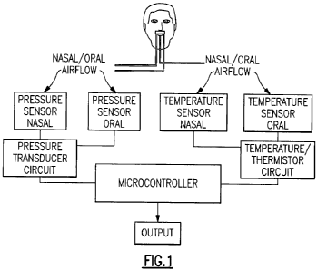
[0033] FIG. I is a flow diagram representation of the present invention within
a
breath monitoring system;
[0034] FIG. 2 is a graph illustrating a flow rate profile of the breathing
cycle of a
patient combining pressure sensor and temperature sensor data;
[0035] FIG. 3A is a diagrammatic representation of the temperature sensor of
the
present invention;
[0036] FIG. 3B is a perspective view of an embodiment of the pressure and
temperature sensor mounted together without an oral pressure sensing prong;
[0037] FIG. 3C is a perspective view of an embodiment of the pressure and
temperature sensor mounted together with an oral pressure sensing prong;
-7-
CA 02726801 2010-12-02
WO 2009/149336 PCT/US2009/046388
-8-
[0038] FIG. 3D is an enlarged perspective view of a free end a lower branch a
T-shaped sensor which is provided with an exterior surface which tends to be
ejected or forced out of or away from the mouth, gums and/or teeth of a young
child or infant in the event that such patient attempts to suck, bite or chew
on the
same;
[0039] FIG. 4A is representation of the cannula and temperature sensor and
associated initial arm angle of the appertaining arms of the cannula;
[0040] FIGS. 4B and 4C are representations of the cannula and temperature
sensor and associated adjacent angles of the appertaining arms of the cannula;
[0041] FIG. 5 is a circuit schematic diagram of the respiratory airflow
detection
circuit with test circuitry for testing the operational functionality of the
temperature sensor;
[0042] FIG. 6 is a front perspective view of a cannula of a first embodiment
of the
present invention used to support the temperature sensor;
[0043] FIG. 7 is a rear view of the cannula of the first embodiment used to
support the temperature sensor;
[0044] FIG. 8 is a side view of the cannula of the first embodiment used to
support the temperature sensor via the holster of the cannula;
[0045] FIG. 9 is a perspective view of a second described embodiment of the
cannula having an oral pressure sensing prong extending therefrom;
[0046] FIG. 10 is a side view of the cannula of the second embodiment of the
invention used to support the temperature sensor shown therewith;
[0047] FIG. 11 is a bottom perspective view of the cannula of the second
embodiment used to support the temperature sensor;
[0048] FIG. 12 is a rear view of the cannula of the second embodiment used to
support the temperature sensor;
[0049] FIG. 13A is a diagrammatic front elevational view of a third embodiment
of a cannula supporting a temperature sensor;
[0050] FIG. 13B is a diagrammatic top plan view of FIG. 13A showing
engagement of the cannula with the temperature sensor;
[0051] FIG. 13C is a diagrammatic cross-sectional view of the cannula and the
temperature sensor of FIG. 13A along section line 13C-13C;
(0052] FIG. 14 is a diagrammatic perspective view of the cannula of FIG. 13A
prior to be assembled with the temperature sensor; and
[0053] FIG. 15 is a perspective view of the temperature sensor of FIG. 13A
with
the overmolded material barrier prior to be assembled with the cannula.
-8-
CA 02726801 2010-12-02
WO 2009/149336 PCT/US2009/046388
-9-
[0054] DETAILED DESCRIPTION OF THE INVENTION
[0055] The present invention is directed to an apparatus and method for
monitoring and modeling a patient's breathing according to both pressure and
temperature measurements. As seen in Figure 1, from oral and nasal airflow of
a patient oral and nasal temperature measurements are obtained according to
temperature changes measured by a thermistor during the exhalation and
inhalation interval of a patient during a sleep diagnostic session. A
temperature
sensor-generally a thermistor although other types of thermocouples and
temperature sensors could be used as well-is positioned adjacent the nares
(nostrils) of the patient's nose (nasal temperature sensing) and adjacent the
patient's mouth (oral temperature sensing). An output signal, from the
temperature sensor(s), is conditioned by a thermistor circuit and sent to a
micro
controller to be processed into acquired air wave and airflow breathing data
for
input to conventional polysomnography equipment which produces an output
representation of the patient's breathing cycle generally as a qualitative,
viewable waveform.
[0056] A pressure sensor is also used in the system in conjunction with the
temperature sensor. The pressure sensor-like the thermistor-is a non-invasive
alternative for measuring nasal and oral airflow of a patient during the
diagnostic
study. A pressure sensor is generally the preferred method of determining
nasal
air flow since the nasal prongs of the cannula are situated essentially inside
the
nares of the patient's nose and directly in the flow path of nasal inspiration
and
expiration. It follows that nasal pressure sensing, often achieved with a
pressure
transducer, is generally a more accurate method of assessing hypopneas in real
time, which is critical to the accurate diagnosing of a patient.
[0057] If a patient breaths through his or her mouth, on the other hand, it is
more
difficult to obtain an accurate pressure measurement based on inspiration and
expiration through the mouth. Because of the size of a patient's mouth in
general, it is difficult to align an oral prong or cannula opening at an
appropriate
position to obtain the oral inspiration and expiration. For example, a person
may
breath out the side of their mouth and thus an oral prong, located in the
center
of the mouth for pressure sensing, may not receive adequate breathing flow to
properly determine pressure. In the case of a mouth breather like this, the
temperature sensor with an oral thermistor may provide the best response using
-9-
CA 02726801 2010-12-02
WO 2009/149336 PCT/US2009/046388
-10-
the temperature differential between the ambient air and whatever portion of
the
patient's breathing is obtained.
[0058] To determine an accurate wave form of the patient's breathing, a nasal
cannula is generally used by the patient which is then connected to a pressure
sensor, for example, a sensitive pressure transducer. The pressure transducer
emits a signal which is proportional to the flow and this signal is processed,
by
the micro controller, to generate a respiratory waveform signal which
indicates
the fluctuations in pressure caused by inspiration and expiration of the
patient.
In the present system, a temperature sensor may also be used with the cannula,
or mask in the case of titration, to provide further accuracy in determining
breathing cycle data and an accurate wave form.
[0059] In general, and as discussed in further detail below, in order to most
effectively determine an actual accurate wave form including the most accurate
amplitude as well as frequency, i.e., breaths per minute, the present
embodiment of the system includes a thermistor(s), as the temperature sensor
for obtaining the oral and nasal temperature changes of a patient's
inspiration
and expiration, which is adapted to be affixed to a nasal and oral cannula.
The
cannula is used, as described above, to obtain the nasal and oral airflow and
derived pressure changes in the patient's breathing which, along with the data
obtained by the thermistor, can then be compared to obtain the most accurate
waveform and most precise monitoring and diagnosis of a patient's respiratory
airflow and breathing cycles including confirmation of distress signals from
hypopneas or apnea events.
[0060] FIG. 1 is a basic flow chart of an embodiment of a temperature and
pressure sensor breath monitoring system for providing conformational data of
changes or aberrations within a patient's breathing cycle from a nasal
pressure
sensor and oral pressure sensor as well as a nasal temperature sensor and oral
temperature sensor. The attachment of the temperature sensor and thermistors
to the cannula ensures that the thermistors are located adjacent the oral and
nasal passages of the patient to obtain an accurate temperature change in
concurrence with the nasal and oral inlets of the cannula which receive the
air
flow indicative of pressure changes which effect the pressure sensor. The
nasal
pressure sensor is provided in conjunction with the oral pressure sensor, via
the
cannula, to provide a pressure signal to the microcontroller, and the nasal
temperature sensor along with an oral temperature sensor, via a thermistor, is
connected to the microcontroller to supply a further temperature change signal
-10-
CA 02726801 2010-12-02
WO 2009/149336 PCT/US2009/046388
-11-
to the microcontroller. This system therefore provides a pressure and
temperature signal from each breathing cycle to the microprocessor or
controller
and can be accumulated, processed and provided as a breathing pattern output
for diagnosis and treatment purposes.
[0061] FIG. 2 shows an example of a breathing pattern output derived from the
acquired temperature and pressure data of the patient's breathing cycle.
Pressure data is collected from the cannula and the pressure sensor, on the
one
hand, and temperature data, on the other, is also collected from the oral and
nasal temperature sensors over a period of time to track the patient's
breathing
cycle. When both the pressure and temperature sensors are plotted together,
as shown in FIG. 2, it becomes apparent, despite any lag time in the
temperature
measurement and response, where potential anomalies or errors may exist in
the respective temperature and pressure sensors and signals, and also that the
system can more reliably detect apnea, hyopopnea and other subtle flow
limitations where both pressure and temperature signal outputs can be
concurrently determined from a patient's baseline oral and nasal breathing
pattern.
[0062] Turning now to FIGS. 3A, 3B and 3C, the temperature sensor 1 of the
embodiment shown here is a triad, i.e., three thermistors 3, 5 and 7
comprising
a first nasal thermistor 3 in series with a second nasal thermistor 5 on a
nasal
circuit, and an oral thermistor 7 that is positioned along an oral circuit
connected
in parallel and structurally aligned perpendicular to the nasal circuit of the
first
and the second nasal thermistors 3 and 5. First and second leads 9 and 11 are
connected to the respective circuit junctions of the nasal and oral circuits
to send
the resistivity change to a conditioning circuit C, described in further
detail below.
[0063] The temperature sensor 1, including the thermistors, is formed in a
T-shaped configuration with the first nasal thermistor 3 located in a left
branch 13 of the sensor 1. The second thermistor 5 positioned in the right
branch 15 of the sensor 1, and the oral thermistor 7 located in the lower
branch
of the T-shaped sensor. When properly positioned on the cannula and on the
face of a patient, the left and right branches 13, 15 extend in each lateral
direction under the nasal septum of the patient's nose toward respective free
ends 17, 19 so that each of the nasal thermistors 3, 5 are positioned directly
adjacent the opening to each respective left and right nares of the patient's
nose
[0064] The left and right branches 13, 15 form a rigid but flexible bridge
that
provides structurally stable and flexible support to allow for each of the
left and
-11-
CA 02726801 2010-12-02
WO 2009/149336 PCT/US2009/046388
-12-
the right branches 13, 15 to be adjusted, i.e., bent, manipulated, curved or
articulated into a desired position relative to one another and relative to
the oral
thermistor 7. Although the branches are shown here as being linearly aligned,
the flexibility of the branches 13,15 permits non-linear alignment as can be
seen
in subsequent figures. This non-linear flexibility facilitates aligning and
maintaining the respective right and left nasal thermistors 3, 5 with the
patient's
right and left nares and does so in conjunction with the nasal prongs of the
cannula supporting the temperature sensing device inlets. It is also to be
appreciated that there does not necessarily have to be two thermistors 3, 5 in
the
bridge, e.g., that there could only be a single thermistor located in the
bridge
which could be aligned with one of nostrils of the patient or possibly at a
location
between the nostrils of the patient or could be aligned with one of the nares
of
the cannula or possibly between the nares of the cannula.
[0065] Similarly, a lower branch of the T-shaped sensor extends
perpendicularly
downwardly relative to the flexible bridge and is also adjustable, flexible
and
manipulatable such that the lower branch 21, which includes the oral
temperature circuit and oral thermistor 7, provides the same rigidity and
maleability to structurally support the oral thermistor at a desired
orientation or
position adjacent the patient's mouth. In the case of each branch 13, 15 and
21,
the branches can independently arranged with respect to one another about the
center joint 23. In otherwords, each branch is radially flexible in a 360
rotational
manner about the center joint 23, and each branch is also axially flexible,
i.e.,
bendable along its longitudinal axis to ensure that the oral thermistor 7 is
not
only placed in an appropriate position adjacent the patient's mouth so that it
is
fully located in the path of inspiration and expiration, but also can be
adjusted so
as not to touch any part of the patient's mouth, tongue, skin or face,
[0066] The T-shape configuration of the temperature sensor 1 is important
because, by its very nature, the T-shape defines three (3) independent
branches 13, 15 and 21 which extend from a center joint 23 to three (3)
free ends. The left and right upper branches each define a left and right free
end 17, 19 and the depending prong 29 also defines its own respective lower
free end. With each branch extending from the center joint 23 in this manner
to
the respective free ends 17, 19 and 25, each branch 13, 15, and 21 along with
the associated thermistor 3, 5 and 7 can consequently be independently
adjusted, bent and/or configured to a desired shape or configuration
independent of one another. By way of example, the left and right
-12-
CA 02726801 2010-12-02
WO 2009/149336 PCT/US2009/046388
-13-
branches 13, 15 may be bent in a manner to curve laterally in cooperation with
the curved shape of the cannula or the curved skin and face surface of the
patient, as can be seen in FIG. 3B and 3C. This allows each thermistor in the
sensor to be directly aligned in the flow path of the nasal airflow passing
through
the patient's nares. Similarly, but independently of the left and right
branches 13, 15, the lower branch 21 may be curved, bent or manipulated so as
to most effectively position the oral thermistor 7 in the most advantageous
position to receive the oral temperature change from the patient's oral
airflow.
Also, by appropriately arranging the lower branch 21 independent of the left
and
the right branches 13, 15, it can be assured that the lower branch 21 and the
oral thermistor 7 does not contact the patient's skin or mouth and thereby
adversely influence the response of the thermistor to the oral airflow of the
patient.
[0067] This independent flexibility of the lower branch 21 is critical because
if the
oral thermistor 4 touches the skin or face of the patient, the thermistor will
be
effected by the body and skin temperature in addition to any temperature
changes caused by the patient's breathing. Also, the ability to bend and
manipulate the lower branch 21 in what is essentially a 360 degree manner
ensures that the oral thermistor 7 can be placed in the most direct path of
the
patient's inspiration and expiration airflow. While the flow path of
inspiration and
expiration generally does not vary significantly through the nares or nostrils
of
the nose, because of the relative smaller size of the nare openings as
compared
to the mouth and the flow rate of a patient's breathing, the mouth is much
larger
than the nares and a patient may breath out the side, top or bottom of his or
her
mouth. Thus, the ability to radially and axially articulate and maintain the
lower
branch 21, and hence the oral thermistor 7, in a region where the patient's
most
direct oral inspiration and expiration is occurring is critical to obtaining
an
appropriate and accurate reading and response of oral expiration and
inspiration.
This rigid flexibility of the temperature sensor and adjustments thereof
relative
to the nares and mouth permits proper positioning and configuring of the
temperature sensor to align and match the proper physical characteristics of
patients independently of the nasal and oral prongs of the cannula to which
the
sensor 1 is attached.
[0068] In the arrangement shown in FIG. 3D, it is to be appreciated that the
two
thermistors 3, 5 are located adjacent the nasal prongs 33, 33 while the third
thermistor 7 is supported by the lower branch 21 and can be suitably
positioned
-13-
CA 02726801 2010-12-02
WO 2009/149336 PCT/US2009/046388
-14-
or adjusted to a location so that the third thermistor 7 can be located
directly in
the flow path of the air being exhausted from the mouth of a patient. In the
event that the patient is a young child or an infant, for example, such child
or
infant may be prone to either suck, bite and/or chew on the remote free end of
the lower branch 21 and/or the thermistor 7 since the free end of the lower
branch 21 is positioned adjacent the mouth of such patient. To avoid or
minimize as much as possible this tendency, the free or leading end of the
lower
branch 21 generally terminates in teardrop shaped end or, alternatively, the
free
end of the lower branch 21 may be provided with an end cap EC which has an
exterior surface which is teardrop shaped, or suitably shaped or contoured, so
that the free or leading end of the lower branch 21 tends to be ejected or
forced
out of or away from the mouth, gums and/or teeth of a young child or infant in
the event that such patient attempts to suck, bite and/or chew on the same.
Due
to the modified exterior shape or contour of the free or leading end of the
lower
branch 21, such shape does not provide any flat or cylindrical or other
surfaces
or areas which can be easily and readily be grasped by the gums, teeth and/or
mouth of a young child or infant and this minimizes the possibility that the
young
child or infant will be able to captively retain the free end of the lower
branch 21
in his/her mouth for an extended period of time during use. That is, the
exterior
profile or contour of the cap EC or the free or leading end of the lower
branch 21
is designed, e.g., is generally rounded, curved or tapered, to bias the
thermistor
7 away from the mouth of such young child or infant so that the thermistor 7
can
remain in the path of the air flow being exhausted from the mouth of such
young
child or infant and still effectively operate to sense temperature of the air
being
exhaled.
[0069] It is to be appreciated that the end cap EC can either be integrally
formed
with the free end of the lower branch 21 or be affixed thereto following
manufacture of the T-shaped sensor by a conventional adhesive.
[0070] The ability to independently position the branches 13, 15 and 21
relative
to the fixed orientation in which the center joint 23 of the temperature
sensor I
is held with respect to the cannula is also important in regards to the shape
of
the cannula 31 and the cannula body 32. In an embodiment of the present
invention, the cannula body 35 extends for a portion of its length along a
main
x-axis, as can best be seen in FIGS. 4A, 4B and 4C. Elbows 37 are formed at
either end bending in a 3-dimensional sense to define opposed arms 39
extending along a y-axis. As explained more fully in U.S. Patent No.
4,106,505,
-14-
CA 02726801 2010-12-02
WO 2009/149336 PCT/US2009/046388
-15-
the teaching of which is incorporated herein by reference, the y-axis
extending
along the length of each arm 39 intersects a horizontal plane defined by the x-
axis of the main body 35 at an acute angle A from above the horizontal plane,
as can be seen in FIG. 4A. In FIGS. 4B and 4C, the forward extension of the
arms 39 (towards the patient's face) defines an acute angle B of intersection
between the y-axis and horizontal x-axis. The independent flexibility of each
branch 13, 15 and 21 of the temperature sensor 1 ensures that the branches
may be suitably positioned, and retained in such a position, where the
branches
not only conform to this described shape of the cannula body 35 but also where
the nasal and oral thermistors 3, 5 and 7 can be best positioned relative to
the
cannula to receive the necessary airflow while still avoid touching the
patient's
face.
[0071] It is to be appreciated that not all the branches 13, 15 and 21 are
necessarily the same length. For example as discussed in further detail below,
the temperature sensor 1 may be offset from a centerline of the cannula so
that
the left and the right branches 13, 15 might have different lengths relative
to the
center joint 23 of the sensor 1 to properly position the respective
thermistors 3
or 5 adjacent the nasal prongs 33 and in the patient's nasal airflow.
Alternatively, where the branches 13, 15 are the same length, the thermistors
may be spaced different distances from the center joint 23 of the sensor 1 so
that they are aligned adjacent the nasal prongs 33 and in the nasal air flow
of the
patient. Typically, the lower branch 21 is longer than the upper branches 13,
15
to extend from the center joint 23 to an appropriate position in the oral
airflow of
the patient.
[0072] The nasal and oral thermistors 3, 5 and 7 and their respective circuits
and
wire leads 9, 11, shown in FIG. 3A, may be joined in any manner known in the
art for example by soldering, taping, brazing or welding and may be protected
and insulated by applying an inner layer of heat-shrink tubing 27 to protect
and
insulate these joints and connections from the external environment. An outer
layer of heat shrink material 29 may be applied over the circuits, joints,
leads and
thermistors as well to provide some level of insulation from the environment,
without degrading the response of thermistors and circuits. Also, any
portion(s)
of the temperature sensor circuit not covered by the heat shrink material may
be
sealed with a non-conductive sealant or fixative, for example, a silicone
polymer
generally depicted as layer 28, or some such similar non-conductive material
to
entirely seal the temperature sensor circuit from contact with ambient air.
The
-15-
CA 02726801 2010-12-02
WO 2009/149336 PCT/US2009/046388
-16-
center joint 23 of the T-shaped temperature sensor 1 may, for example, be
sealed with the layer 28 to provide not only sealing and insulation of the
circuit,
but also define a relatively rigid reference point from which each of the left
and
the right branches 13, 15 and the lower branch 21 extend and can be
independently adjusted relative thereto.
[0073] The airflow temperature sensor I can be a negative temperature
coefficient (NTC) thermistor which exhibits decreasing electrical resistance
with
an increase in environmental temperature and increasing electrical resistance
with a decrease in environmental temperature. By way of example, the
thermistors 3 and 5 of the nasal temperature circuit shown in FIG. 3A may have
a resistance of 5k each, while the oral thermistor 7, arranged in parallel,
may
have a 10k resistance. In another embodiment, all the thermistors could be
arranged in series as 10k resistance, particularly where a more substantial
power supply is provided besides a small DC battery, discussed with respect to
FIG. 5 below. A larger power supply would permit higher resistance to be used
through the circuit and thus a greater range of responsiveness for any
temperature differential.
[0074] As discussed above, the left external lead 9 and the right external
lead 11
of the temperature sensor 1 are connected to a respiratory temperature
detection circuit C having a test circuit as shown in FIG. 5. The respiratory
airflow detection circuit C determines the change in temperature across the
thermistor(s) based on the proportional change of a voltage divider in the
circuit.
The test circuit T ensures that the continuity of the circuit is maintained
and can
be monitored and readily ascertained, at any desired time, by merely
depressing
a button and without maintaining a diode or indicating light on at all times.
[0075] As can be seen in FIG. 5, which is a schematic of the respiratory
temperature detection circuit, the left external lead 9 is coupled as an input
at J 1
and the right external lead 11 is coupled as an input at J2. Power is applied
to
the circuit via a battery, for example a 3 volt coin cell connected to J5
(Pos)
and J6 (Neg). Thermally equilibriating a change in temperature across the
thermistors in the temperature sensor I will cause the voltage divider voltage
to
change proportionally with temperature at the junction of R2 and the
thermistor
lead terminal J1. If the rate of change in temperature is within a passband,
then
the voltage can be measured at the head box leads.
[0076] The resistors and capacitors form a band pass filter with the
combination
of R2 and C2 forming a low pass filter with a cutoff frequency of around 42 Hz
-16-
CA 02726801 2010-12-02
WO 2009/149336 PCT/US2009/046388
-17-
and the combination of C5 plus C6 and R1 form the high pass filter with a
cutoff
frequency of around 0.066 Hz.
[0077] The capacitors C5 and C6 with resistor R1 and the resistive inputs of
the temperature sensors through J1 and J2 form a filter capacitive circuit
that generates frequency changes as the resistance changes within the
thermistors of the temperature sensors on each inhalation and exhalation of
the
patient's breathing cycle. An output analog signal is generated and fed, via
connections J3 and J4, to a microprocessor or other controller to model the
patient's breathing cycle or to compare the signal to other breath monitors
such
as a pressure sensor output of oral or nasal breath, as shown in FIGS. 1 and
2.
[0078] Figure 5 also includes the test circuit T that tests the integrity of
lead
lines 9 and 11, connected to J1 and J2, and the internal circuit components of
the respiratory airflow detection circuit. The test circuit T includes a
switch S1
that, when closed, creates a closed circuit for all components. Power is
applied
to the transistors circuits when the switch S1 is temporarily closed. A first
LED D1 will illuminate if a white or black head box lead is plugged into the
J7
lead tester jack and S1 is closed verifying the integrity of the head box
lead. A
second LED D2 will illuminate when S1 is closed verifying the integrity of the
thermistor leads. Any failure within the leads, the connections or the circuit
components will fail to illuminate at least one of the test indicators, D1 or
D2, and
this identifies to the operator a problem within the circuit.
[0079] FIGS. 6, 7 and 8 show details a cannula 31 for use in the presently
described system in conjunction with the above described temperature sensor 1
and the circuit C. The cannula 31 includes a main cannula body 32 which is
hollow and has first and second ends defining respective openings through
which air and/or gas are delivered or received generally through a pair of
nasal
prongs 33, as are well known in the art, for receiving exhalation gases and/or
supplying oxygen to the patient. The cannula 31 of this embodiment is further
provided with an integral receiving holster 41 and stop portion 43 which
defines
a receiving notch 45 therebetween. The holster 41 is integrally connected or
formed with the body 32 of the cannula 31 and provided with a sensor
passage 47. The sensor passage 47 may be of any desired shape, and does
not even have to be entirely enclosed, i.e., formed as a cylinder, but is
sized so
as to receive a portion of the temperature sensor 1, namely, the lower branch
21
which is located within the passage 47 and is generally frictionally retained
therein.
-17-
CA 02726801 2010-12-02
WO 2009/149336 PCT/US2009/046388
-18-
[0080] During assembly, the lower branch 21 is pushed into the sensor
passage 47 so that the oral thermistor 7 passes into and through the passage
47
and extends out a bottom end of the passage 47 (see Fig. 8). The lower
branch 21 is pushed through the passage 47 until the extension for the left
and
the right branches 13, 15 of the pressure sensor 1 abut a top end of the
passage 47 and accordingly situate the center joint 23 of the T-shaped sensor
snugly in the receiving notch 45 between the stop portion 43 and a top surface
of the holster 41. The stop portion 43, which is also integrally connected
with the
body of the cannula 31, extends outward therefrom to approximately the same
dimensions as the holster 41. The receiving space or notch 45, defined between
the stop portion 43 and the top surface of the holster 41, thus closely
receives
and holds the center joint 23 but is sufficiently flexible to facilitate the
insertion
and removal of the pressure sensor 1 into the passage 47 of the holster 41.
[0081] Once the T-shaped temperature sensor 1, as can be seen in FIG. 3,
is inserted into the sensor passage 47, the branches 13, 15 and 21 may be
independently manipulated in order to provide the appropriate positioning,
alignment and/or curvature to these branches and their free ends as necessary
in order to facilitate the most reliable data collection position, as
previously
described.
[0082) With reference now to FIGS. 9-12, a further embodiment of a cannula
31',
according to the present invention, will be described. This embodiment also
includes a holster 41 and a stop portion 43 in combination with an oral
airflow
pressure sensing tube 51 which communicates, in addition to the nasal
prongs 33, with the main body of the cannula 31'. An oral pressure sensing
tube 51 is provided to be substantially centered on, or even slightly offset,
relative to a centerline A of the cannula body and the nasal prongs 33 on the
cannula 31' (see FIG. 11). In order to ensure that the oral sensing thermistor
7
is not blocked or obstructed by the oral pressure sensing tube 51 in any
manner,
the holster 41 and the stop 43, in this embodiment, are radially offset from
both
the cannula centerline A as well as a centerline of the oral pressure sensing
tube 51. This offset separation ensures that when the lower branch 21 of the
sensor I is inserted through and into the holster 41, the lower branch 21
extends
along the side of the oral pressure sensing tube 51 and thus can be directly
aligned adjacent the patient's oral airflow without being blocked or otherwise
obstructed by the pressure sensing tube 51.
-18-
CA 02726801 2010-12-02
WO 2009/149336 PCT/US2009/046388
-19-
[0083] Similar to the description of the first embodiment described with
reference
to FIGS. 5, 6 and 7, the holster 41 of FIGS. 9, 10 and 11 is provided with the
passage 47 and the stop portion 43 to define the receiving notch 45
therebetween into which the center joint of the pressure sensor 1 is located
when the temperature sensor 1 is attached with the oral and nasal pressure
sensing cannula 31' to create the diagnostic system, as shown and described
herein.
[0084] For the apparatus and system as described above, the temperature
sensor 1 and the pressure sensing cannula 31, 31' can be used together
and facilitate obtaining similar but differently processed signals which are
indicative of the patient's breathing patterns. The malleability and
adjustability
of the T-shaped pressure sensor ensures that the left and the right upper
branches 13, 15 can be adjusted, in any desired manner, so that they
essentially
align with the nasal prongs 33 and the nares of the patient's nostrils.
The relative flexibility allows the left and the right upper branches 13, 15
as well
as the lower branch 21 to be bent inwards or outwards so as to conform to a
bend in the cannula body, for instance, as can be seen in Figures 5 and 9
while,
as can be seen in FIG. 10, the lower branch 21 may be bent so as to achieve an
entirely different axial and radial curvature and/or alignment than the oral
sensing tube. For example, the free end of the lower branch 21 may be moved
in a 360 range of movement, relative to a free end of the pressure sensing
tube, and be more accurately placed in the direct airflow of the patient's
mouth,
relative to the pressure sensing tube, and therefore potentially provide a
more
accurate data from the patient's respiratory airflow.
[0085] With reference now to FIGS. 13A, 13B, 13C, 14 and 15, a detailed
description concerning a further embodiment of the present invention will now
be provided. As this embodiment is somewhat similar to the previous
embodiments, only the differences between this embodiment and the previous
embodiments will be discussed in detail.
[0086] As with the previous embodiments, the cannula 31" generally comprises
a main body 32 which is open at opposed ends thereof (not shown in detail) and
has an internal chamber 52 communicating with both open ends of the main
body 32. The main body 32 also supports first and second spaced apart nasal
prongs 33, 33 which facilitate communication with a respective one of the
nostrils
of the patient. Each opposed open end of the cannula 31" can be connected,
by conventional tubing 54, to suitably detection equipment 56, such as a
-19-
CA 02726801 2010-12-02
WO 2009/149336 PCT/US2009/046388
-20-
pressure transducer, for example, and each one of the nares or nasal prongs
33,
33 has an internal passageway 58 which communicates with the internal
chamber 52 of the main body 32. According to this embodiment, the internal
chamber 52 of the cannula is undivided, that is, the passageway 58 of the
first
nasal prong 33 communicates with the passageway 58 of the second nasal
prong 33 and vice versa, via the internal chamber 52 of the cannula 31 ". It
is to
be appreciated that, if desired, the internal chamber 52 of the cannula 31"
may
be divided, e.g., by a partitioning or dividing wall or septum (not shown),
into two
completely separate internal chambers such that the dividing wall prevents the
passageway 58 of the first nasal prong 33 from communicating, via the internal
chamber 52 of the cannula, with the passageway 58 of the second nasal
prong 33.
[0087] The first and the second nasal prongs 33, 33, as described above, are
used to detect breathing of the patient. To facilitate attachment of a desired
temperature sensing device, such as a thermistor 60, to the cannula 31"
adjacent the first and the second nares or nasal prongs 33, 33, the cannula
31"
is provided with a pair of holsters 41 which are spaced apart by a distance of
between about 0.125 inches and about 0.5 inches, for example, but aligned with
one another, to facilitate receiving and positioning a thermistor at a
location
precisely between the first and the second nares or nasal prongs 33, 33 of the
cannula 31 ". Each of the aligned holsters 41 have a sensor passage 47 formed
therein which extends through the respective holsters 41 to facilitate
receiving
and supporting the desired temperature sensor 60 therein, such as a
thermistor.
Each one of the two aligned holsters 41 is typically cylindrical in shape and
has
a length of between about 0.4 and about 0.5 inches, a through bore of between
about 0.08 and about 0.10 inches and a diameter of between about 0.15 and
about 0.19 inches. It is to be appreciated that one or both of the holsters 41
may
have an elongate cut, slot or opening formed therein (not shown), extending
the
entire axial length of the side wall of the holster 41, which facilitates the
holster(s) 41 expanding somewhat in diameter to allow accommodation of
different diameter and/or sized temperature sensors 60, e.g., slightly larger
thermistors.
[0088] The lead lines 9, 11 and the internal circuitry of the thermistor 60 is
typically covered with a plastic overmolded material, or some other protective
barrier 28, which protects the internal component of the thermistor 60 and
also
provides some rigidity to the thermistor 60 to assists with "feeding" or
"threading"
-20-
CA 02726801 2010-12-02
WO 2009/149336 PCT/US2009/046388
-21-
a leading end of the thermistor 60 through the first and the second aligned
sensor passages 47 of the respective first and second holsters 41, 41 so as to
be captively retained by the cannula 31". The plastic overmolded material or
barrier 28 typically includes a stop feature 62, e.g., an enlarge diameter
section
or some other stop feature of the plastic overmolded material or barrier 28,
that
is designed to abut against an end face 64 of the first holster 41 and prevent
further or over insertion of the thermistor/plastic overmolded material
assembly
relative to the first and the second holsters 41, 41.
[0089] Following insertion and engagement of the thermistor 60 with the first
and
second holsters 41, the thermistor 60 is correctly located and positioned
between the first and the second nares or nasal prongs 33, 33 of the
cannula 31". As a result of such positioning, the thermistor 60 is precisely
located between the first and the second nares or nasal prongs 33, 33 so that
the airflow being inspired and expired by the patient will contact the
thermistor
60 and facilitate detection of the temperature of the inspired and expired
airflow.
As with the previous embodiments, the lead lines 9, 11 are coupled to the
respiratory airflow detection circuit C for determining the change in
temperature
across the thermistor 60.
[0090] An important aspect of this embodiment of the present invention is to
sufficiently space the exterior surface 66 of the thermistor 60 from the
exterior
surface of the main body 32 of the cannula 31" so as to avoid any contact
between those surfaces (see FIG. 13C). It is to be appreciated that if the
cannula 31 ", or any other surface, is located too close to or contacts the
exterior
surface 66 of the thermistor 60, this can disrupt accurate temperature sensing
by the thermistor 60. Preferably the exterior surface of the thermistor 60 is
spaced from the exterior surface of the cannula 31" by a distance of between
about 0.040 and 0.080 inches or so.
[0091] It is to be appreciated that although the embodiment shown in FIGS.
I3A,
13B, 13C 14 and 15 of the drawings may be utilized with adults, this
embodiment
is particularly suited for use with smaller patients such as young adults,
children
and infants.
[0092] Since certain changes may be made in the above described improved
sleep apnea diagnosing apparatus and method, without departing from the spirit
and scope of the invention herein involved, it is intended that all of the
subject
matter of the above description or shown in the accompanying drawings shall be
-21-
CA 02726801 2010-12-02
WO 2009/149336 PCT/US2009/046388
-22-
interpreted merely as examples illustrating the inventive concept herein and
shall
not be construed as limiting the invention.
-22-