Note: Descriptions are shown in the official language in which they were submitted.
CA 02727171 2010-12-06
WO 2009/149370 PCT/US2009/046441
TITLE OF THE INVENTION
METHODS FOR THE TREATMENT OF RHEUMATOID ARTHRITIS
FIELD OF INVENTION
[0001] The present disclosure relates to methods for the treatment and/or
prevention
of rheumatoid arthritis. Such methods may be used to treat a subject suffering
from or to
prevent occurrence of the same in an at risk subject.
BACKGROUND OF THE INVENTION
[0002] The present disclosure is directed to methods for the treatment and/or
prevention of rheumatoid arthritis (RA) in a subject. Such methods may be used
to treat a
mammalian subject, such as for example a human subject, suffering from
rheumatoid arthritis
or to prevent occurrence of the same in an at risk subject. RA is a chronic
multi system
autoimmune disease which can be debilitating and reduces life expectancy. The
hallmark of
the disease is a persistent inflammatory arthritis, usually of peripheral
joints, which can lead
to cartilage destruction, bone erosion, and loss of joint integrity. In
addition to the joint
findings in RA, patients often present with extraarticular manifestations of
the disease
including subcutaneous rheumatoid nodules, muscle weakness and atrophy,
vasculitis, and
pleuritis. The course of RA is quite variable in that some patients experience
its destructive
potential and show a marked functional impairment, whereas others experience a
mild illness
of brief duration.
[0003] Microscopic examination of the synovial lining from the joints of RA
patients
reveals an acute and chronic inflammatory state characterized by infiltration
with
predominantly CD4+ T lymphocytes. Depletion of T-cells by thoracic duct
drainage,
lymphoid irradiation or cytotoxic drugs has been claimed to have efficacy in
treating RA.
The T-cells produce a number of cytokines that promote B-cell proliferation
and
differentiation into antibody-forming/antigen-presenting cells. A common,
although not
specific, finding in two thirds of patients is the presence of rheumatoid
factors, autoantibodies
directed against IgG. The production of rheumatoid factors can lead to immune-
complex
formation, consequent complement activation, and exacerbation of the
inflammatory process
(Lipsky, 1998). In RA, the mechanism of disease seems to involve activation of
T
lymphocytes by yet unknown antigens. These antigens may be infectious agents
or other
endogenous molecules that are no longer recognized as self-molecules. Antigen-
activated
CD4+ T-cells stimulate monocytes, macrophages, and synovial fibroblasts to
produce the
1
CA 02727171 2010-12-06
WO 2009/149370 PCT/US2009/046441
cytokines IL-1, IL-6, and TNF-a. These proinflammatory cytokines are
considered
responsible for the perpetuation of the inflammatory process within the joint,
including
cartilage destruction and erosion of periarticular bone. Inhibition of both
TNF-a and IL-1 has
been shown to reduce the inflammation and to slow the joint destruction
(Keystone and
Strond, 2005).
[0004] IL-1 is a pro-inflammatory cytokine secreted by a number of different
cell
types including monocytes and macrophages. The IL-1 gene family comprises the
agonist
cytokines IL 1 alpha (IL-la and IL-10, and the natural receptor antagonist (IL-
1Ra). IL-la
and IL-10 are produced as precursors. ProIL-la is functionally active and,
because of a lack
of the leader peptide, it remains in the cytoplasm, while prolL-10 is inactive
and is secreted
and becomes active after cleavage by a specific intracellular protease. In
disease, IL-10 is
found in circulation whereas IL-la is rarely detected; it is released only in
severe disease
states, most likely as a consequence of cell death (Dinarello, 1996). When
released as part of
an inflammatory reaction, IL-10 produces a range of biological effects, mainly
through the
induction of other inflammatory mediators such as corticotrophin, platelet
factor 4,
prostaglandin E2 (PGE-2), IL-6, and IL-8. IL-10 induces both local and
systemic
inflammatory effects through the activation of the IL-1 receptor found on
almost all nucleated
cell types (Dinarello, 2005). IL 10 binds the IL-1 receptor (IL-1R), inducing
a
conformational change that allows binding of the accessory protein to the IL-
1(3/IL-1R
complex. Thus, the formation of this complex induces intracellular signaling
through the IL
1R (Dinarello, 1996).
[0005] IL-1 is present in the synovial tissue and fluids of patients with
rheumatoid
arthritis and stimulates the production of mediators such as prostaglandin
E(2), nitric oxide,
cytokines, chemokines, and adhesion molecules that are involved in articular
inflammation.
Furthermore, IL-1 stimulates the synthesis and activity of matrix
metalloproteinases and other
enzymes involved in cartilage destruction in rheumatoid arthritis and
osteoarthritis. The
effects of IL-1 are inhibited in vitro and in vivo by natural inhibitors such
as IL-1 receptor
antagonist (IL-1Ra) and soluble receptors. IL-1Ra belongs to the IL-1 family
of cytokines
and binds to IL-1 receptors but does not induce any intracellular response. IL
lRa inhibits
the effect of IL-1 by blocking its interaction with cell surface receptors. IL-
1 inhibitors have
been used in experimental models of rheumatoid arthritis supporting the role
of IL-1 in the
pathogenesis of the disease.
[0006] Anakinra is a recombinant form of the naturally-occurring IL-1 blocker,
IL-
1Ra. Anakinra has been used extensively for the approved indication of RA, and
also shown
2
CA 02727171 2010-12-06
WO 2009/149370 PCT/US2009/046441
significant clinical activity in systemic juvenile idiopathic arthritis (sJIA)
and, to a lesser
extent, the other subtypes of juvenile idiopathic arthritis (JIA). Recent
literature has also
shown activity of anakinra in other systemic IL-1-mediated disorders (Neonatal-
Onset
Multisystem Inflammatory Disease [NOMID], Muckle-Wells Disease, and Familial
Cold
Auto-Inflammatory Syndrome), as well as knee osteoarthritis (Goupille, et al.,
2003).
However, the frequent dosing of injectable medications, such as Anakinra, is
generally
undesirable and may result in problems with patient compliance, thereby
further decreasing
effectiveness of this treatment modality/ or limiting its desirability. Thus,
there remains a
need for effective means to treat RA, particularly treatment compositions and
methods that do
not require frequent (e.g., daily) injections.
[0007] The current disease-modifying anti-rheumatic drugs (DMARDs) are
effective
in controlling inflammatory symptoms. The newer biologic response modifiers
(BRMs) offer
improvements in disease control but with added safety risks. These risks
increase with
extended use, and some of the BRMs lose their effectiveness over time. A
recent survey of
500 RA patients conducted by the Arthritis Foundation found that two-thirds of
RA patients
still suffer daily pain, stiffness, or fatigue despite treatment with DMARDs
or BRMs (Gruver,
2004). There continues to be an unmet need for RA therapies that are safe and
provide long-
term control of the disease.
[0008] The present disclosure provides compositions and methods for the
treatment of
rheumatoid arthritis. The methods disclosed herein comprise, for example,
administering a
high affinity anti-IL-10 antibody or fragment thereof under dosing regimens as
provided
herein. Methods that directly target the IL-1(3 ligand with an antibody,
particularly antibodies
that exhibit high affinity, may provide advantages over other potential
methods of treatment,
such as IL-10 receptor antagonists (e.g., Anakinra). A challenge for IL-1
receptor antagonist-
based therapeutics is the need for such therapeutics to occupy a large number
of receptors,
which is a formidable task since these receptors are widely expressed on all
cells except red
blood cells (Dinarello, Curr. Opin. Pharmacol. 4:378-385, 2004). In most
immune-mediated
diseases, such as the diseases disclosed herein, the amount of IL-10 cytokine
that is
measurable in body fluids or associated with activated cells is relatively
low. Thus, a method
of treatment and/or prevention that directly targets the IL-1(3 ligand should
provide a superior
strategy, particularly when administering an IL-1(3 antibody with high
affinity.
3
CA 02727171 2010-12-06
WO 2009/149370 PCT/US2009/046441
SUMMARY OF THE INVENTION
[0009] The present disclosure is directed to methods and related articles of
manufacture for the treatment and/or prevention of rheumatoid arthritis in a
subject. Such
methods may be used to treat a mammalian subject (e.g., human) suffering from
or at risk for
rheumatoid arthritis. The methods also may be used to prevent the occurrence
of rheumatoid
arthritis in an at risk subject. The disclosure further contemplates the use
of such methods for
additional diseases or conditions, such as for example systemic juvenile
idiopathic arthritis.
As illustrated in Examples below, we have surprisingly found that antibodies,
such as those
disclosed herein, can be used to achieve the desired level of activity over a
broad range of
doses, including at very low doses.
[0010] In one aspect of the disclosure, a method is provided for treating
rheumatoid
arthritis in a subject, the method comprising administering an anti-IL-10
antibody or
fragment thereof to the subject, wherein administration of an initial dose of
the antibody or
antibody fragment is followed by the administration of one or more subsequent
doses, and
wherein the initial dose and each one or more subsequent doses are
administered at an
interval of once a week to once every six months. In one embodiment, the
initial dose and
each one or more subsequent doses are administered at an interval of once
every two weeks
to once every three months. In another embodiment, the initial dose and each
one or more
subsequent doses are administered at an interval of once every two weeks to
once every two
months. In another embodiment, the initial dose and each one or more
subsequent doses are
administered at an interval of once every month to once every three months. In
another
embodiment, the initial dose and each one or more subsequent doses are
administered at an
interval of once every month to once every two months. In yet another
embodiment, the
initial dose and each one or more subsequent doses are administered at an
interval of once
every month to once every three months or more.
[0011] The anti-IL-1R antibodies or antibody fragments (e.g., binding
fragments) used
in the methods of the present disclosure generally bind to IL-10 with high
affinity. In
preferred embodiments, the antibody or antibody fragment binds to IL-1(3 with
a dissociation
constant of about 10 nM or less, about 5 nM or less, about 1 nM or less, about
500 pM or
less, about 250 pM or less, about 100 pM or less, about 50 pM or less, or
about 25 pM or less.
In particularly preferred embodiments, the antibody or antibody fragment binds
to human IL-
with a dissociation constant of about 500 pM or less, about 250 pM or less,
about 100 pM
or less, about 50 pM or less, about 10 pM or less, about 5 pM or less, about 3
pM or less,
4
CA 02727171 2010-12-06
WO 2009/149370 PCT/US2009/046441
about 1 pM or less, about 0.75 pM or less, about 0.5 pM or less, about 0.3 pM
or less, about
0.2 pM or less, or about 0.1 pM or less.
[0012] In another aspect of the disclosure, the anti-IL-10 antibody or
antibody
fragment is a neutralizing antibody. In another aspect, the anti-IL-10
antibody or antibody
fragment binds to an IL-10 epitope such that the bound antibody or fragment
substantially
permits the binding of IL-10 to IL-1 receptor I (IL-1RI). In another aspect,
the anti-IL-10
antibody or antibody fragment binds to IL-10, but does not substantially
prevent the bound
IL-10 from binding to IL-1 receptor I (IL-1RI). In another aspect, the
antibody or antibody
fragment does not detestably bind to IL-la, IL-1R or IL-1Ra. In yet another
aspect of the
disclosure, the antibody or antibody fragment binds to an epitope contained in
the sequence
ESVDPKNYPKKKMEKRFVFNKIE (SEQ ID NO: 1). In another aspect, the antibody or
fragment thereof competes with the binding of an antibody having the light
chain variable
region of SEQ ID NO: 5 and the heavy chain variable region of SEQ ID NO: 6.
[0013] In one embodiment, the antibody or antibody fragment comprises 1, 2 or
3 of
the CDR sequences of SEQ ID NO. 3, SEQ ID NO. 4, SEQ ID NO. 5 or SEQ ID NO. 6.
In
another embodiment, the antibody or antibody fragment comprises a light chain
variable
region sequence of SEQ ID NO. 3, or SEQ ID NO. 5. In another embodiment, the
antibody
or antibody fragment comprises a heavy chain variable region sequence of SEQ
ID NO. 4, or
SEQ ID NO. 6. In one embodiment, the antibody or fragment thereof comprises a
light chain
variable region of SEQ ID NO: 5 and the heavy chain variable region of SEQ ID
NO: 6. In
yet another aspect of the disclosure, the antibody or antibody fragment binds
to an epitope
incorporating G1u64 of IL-10. In yet another aspect of the disclosure, the
antibody or
antibody fragment binds to amino acids 1-34 of the N terminus of IL-10.
Preferably, the
antibody or antibody fragment is human engineered, humanized or human.
[0014] In another aspect of the disclosure, a method is provided for treating
rheumatoid arthritis in a subject (e.g., mammal, human), the method comprising
administering an anti-IL-10 antibody or fragment thereof to the human, wherein
administration of an initial dose of the IL-10 antibody or antibody fragment
is followed by
the administration of one or more subsequent doses. In one embodiment,
administration of
an initial dose of the antibody or antibody fragment is followed by the
administration of two
or more subsequent doses. In another embodiment, administration of an initial
dose of the
antibody or antibody fragment is followed by the administration of one or more
subsequent
doses, and wherein said one or more subsequent doses are in an amount that is
approximately
the same or less than the initial dose. In another embodiment, administration
of an initial
CA 02727171 2010-12-06
WO 2009/149370 PCT/US2009/046441
dose of the antibody or antibody fragment is followed by the administration of
one or more
subsequent doses, and wherein at least one of the subsequent doses is in an
amount that is
more than the initial dose.
[0015] In one embodiment, two or more, three or more, four or more, five or
more,
six or more, seven or more, eight or more, nine or more, ten or more or eleven
or more
subsequent doses of the antibody are administered. In another embodiment
administration of
the initial dose and each one or more subsequent doses are separated from each
other by an
interval of at least about two weeks, at least about three weeks, at least
about one month, at
least about two months, at least about three months, at least about four
months, at least about
five months, at least about six months, at least about seven months, at least
about eight
months, at least about nine months, at least about ten months, at least about
eleven months, or
at least about twelve months.
[0016] In another embodiment, the antibody or fragment is administered in one
or
more doses of 5 mg/kg or less of antibody or fragment, 3 mg/kg or less of
antibody or
fragment, 2 mg/kg or less of antibody or fragment, 1 mg/kg or less of antibody
or fragment,
0.75 mg/kg or less of antibody or fragment, 0.5 mg/kg or less of antibody or
fragment, 0.3
mg/kg or less of antibody or fragment, 0.1 mg/kg or less of antibody or
fragment, 0.03 mg/kg
or less of antibody or fragment, 0.01 mg/kg or less of antibody or fragment,
0.003 mg/kg or
less of antibody or fragment or 0.001 mg/kg or less of antibody or fragment.
Preferably, in
each of the aforementioned embodiments, the antibody or fragment is
administered in one or
more doses of at least 0.01 mg/kg of antibody or fragment, at least 0.01 mg/kg
of antibody or
fragment, or at least 0.03 mg/kg of antibody or fragment. Preferably, the
antibody or
fragment is administered in one or more doses of 0.001 mg/kg to 1 mg/kg, 0.001
mg/kg to 0.3
mg/kg, 0.003 mg/kg to 1 mg/kg, 0.003 mg/kg to 0.3 mg/kg. . The above dosage
amounts
refer to mg (antibody or fragment)/kg (weight of the individual to be
treated).
[0017] In another embodiment, the initial dose and one or more subsequent
doses of
antibody or fragment are each from about 0.01 mg/kg to about 10 mg/kg of
antibody, from
about 0.03 to about 1 mg/kg of antibody, from about 0.03 to about 0.3 mg/kg of
antibody,
from about 0.05 to about 5 mg/kg of antibody, from about 0.05 mg/kg to about 3
mg/kg of
antibody, from about 0.1 mg/kg to about 3 mg/kg of antibody, from about 0.1
mg/kg to about
1 mg/kg of antibody, from about 0.1 mg/kg to about 0.5 mg/kg of antibody, from
about 0.3
mg/kg to about 5 mg/kg of antibody, from about 0.3 mg/kg to about 3 mg/kg of
antibody,
from about 0.3 mg/kg to about 1 mg/kg of antibody, from about 0.5 mg/kg to
about 5 mg/kg
of antibody, from about 0.5 mg/kg to about 3 mg/kg of antibody, from about 0.5
mg/kg to
6
CA 02727171 2010-12-06
WO 2009/149370 PCT/US2009/046441
about 1 mg/kg of antibody, from about 1 mg/kg to about 5 mg/kg of antibody, or
from about
1 mg/kg to about 3 mg/kg of antibody. In certain embodiments, two or more,
three or more,
four or more, five or more, six or more, seven or more, eight or more, nine or
more, ten or
more or eleven or more subsequent doses of the antibody are administered. The
above dosage
amounts refer to mg (antibody or fragment)/kg (weight of the individual to be
treated). The
same applies hereinafter if a dosage amount is mentioned.
[0018] In another aspect, the disclosure provides a method of treating
rheumatoid
arthritis in a subject (e.g., human), the method comprising administering a
therapeutically
effective amount of an anti-IL-10 antibody or fragment thereof to the subject
as an initial
dose of about 5 mg/kg or less of antibody or fragment, 3 mg/kg or less of
antibody or
fragment, 2 mg/kg or less of antibody or fragment, 1 mg/kg or less of antibody
or fragment,
0.75 mg/kg or less of antibody or fragment, 0.5 mg/kg or less of antibody or
fragment, 0.3
mg/kg or less of antibody or fragment, 0.1 mg/kg or less of antibody or
fragment, or 0.03
mg/kg or less of antibody or fragment, and a plurality of subsequent doses of
antibody or
fragment in an amount about the same or less than the initial dose.
[0019] Preferably, in the aforementioned embodiments wherein the antibody or
fragment is administered as an initial dose and a plurality of subsequent
doses, the dose of
antibody or fragment is at least 0.001 mg/kg of antibody or fragment, at least
0.003 mg/kg of
antibody or fragment, at least 0.01 mg/kg of antibody or fragment, at least,
0.03 mg/kg of
antibody or fragment, at least 0.05 mg/kg of antibody or fragment, or at least
0.09 mg/kg of
antibody or fragment.
[0020] In yet another aspect of the present disclosure, the antibody or
fragment is
administered as a fixed dose, independent of a dose per subject weight ratio.
In one
embodiment, the antibody or fragment is administered in one or more fixed
doses of 1000 mg
or less of antibody or fragment, 750 mg or less of antibody or fragment, 500
mg or less of
antibody or fragment, 250 mg or less of antibody or fragment, 100 mg or less
of antibody or
fragment, about 25 mg or less of antibody or fragment, about 10 mg or less of
antibody or
fragment or about 1.0 mg or less of antibody or fragment. In another
embodiment, the
antibody or fragment is administered in one or more fixed doses of at least
about 0.lmg of
antibody or fragment, at least aboutlmg of antibody or fragment, at least
about 5 mg of
antibody or fragment, or at least about 10 mg of antibody or fragment.
[0021] In certain embodiments, the fixed dose is from about 1 mg to about 10
mg,
about 1 mg to about 25 mg, about 10 mg to about 25 mg, about 10 mg to about 50
mg, about
mg to about 100 mg, about 25 mg to about 50 mg, about 25 mg to about 100 mg,
about 50
7
CA 02727171 2010-12-06
WO 2009/149370 PCT/US2009/046441
mg to about 100 mg, about 50 mg to about 150 mg, about 100 mg to about 150 mg,
about 100
mg to about 200 mg, about 150 mg to about 200 mg, about 150 mg to about 250
mg, about
200 mg to about 250 mg, about 200 mg to about 300 mg, about 250 mg to about
300 mg,
about 250 mg to about 500 mg, about 300 mg to about 400 mg, about 400 mg to
about 500
mg, about 400 mg to about 600 mg, about 500 mg to about 750 mg, about 600 mg
to about
750 mg, about 700 mg to about 800 mg, about 750 mg to about 1000 mg. In a
preferred
embodiment, the fixed dose is administered in one or more doses of about 0.1mg
to about 100
mg, about 1.0 mg to about 100 mg or about 1.0 mg to about 50 mg. In another
preferred
embodiment, the fixed dose is selected from the group consisting of about 1 mg
to about 10
mg, about 1 mg to about 25 mg, about 10 mg to about 25 mg, about 10 mg to
about 100 mg,
about 25 mg to about 50 mg, about 50 mg to about 100 mg, about 100 mg to about
150 mg,
about 150 mg to about 200 mg, about 200 mg to about 250 mg.
[0022] In one aspect of the disclosure, an aforementioned method of treating
rheumatoid arthritis in a subject is provided, wherein the dose of the
antibody or fragment is
sufficient to achieve an improvement in one or more ACR core response
criteria. In another
aspect, the dose of the antibody or fragment is sufficient to achieve at least
a 50% reduction
in joint pain, at least a 60% reduction in joint pain, at least a 70%
reduction in joint pain, at
least a 80% reduction in joint pain, at least a 90% reduction in joint pain,
at least a 95%
reduction in joint pain or a 100% reduction in joint pain.
[0023] In another aspect of the disclosure, the dose of the antibody or
fragment is
sufficient to achieve at least a 20% improvement in ACR 50 scoring, at least a
30%
improvement in ACR 50 scoring, at least a 40% improvement in ACR 50 scoring or
at least a
50% improvement in ACR 50 scoring.
[0024] In one embodiment the aforementioned improvements are at 3 months or
longer, 4 months or longer, 5 months or longer, 6 months or longer 9 months or
longer or 12
months or longer.
[0025] In another aspect, the dose of antibody or fragment is sufficient to
achieve a
any of the aforementioned improvements and at least one of the following:
decrease in
inflammatory infiltration, decrease in loss of cartilage, decrease in bone
resorption,
improvement in radiographic scoring. In one embodiment, the improvement in
radiographic
scoring is determined by X-ray. In another embodiment, the improvement is a
slower rate of
deterioration. In another embodiment, the improvement is no detectable
deterioration.
[0026] In another embodiment, the dose of the antibody or fragment is
sufficient to
achieve at least a 20% decrease in CRP levels, at least a 30% decrease in CRP
levels, at least
8
CA 02727171 2010-12-06
WO 2009/149370 PCT/US2009/046441
a 40% decrease in CRP levels, at least a 50% decrease in CRP levels, at least
a 60% decrease
in CRP levels, at least a 70% decrease in CRP levels, at least a 80% decrease
in CRP levels,
at least a 90% decrease in CRP levels. In another embodiment, the dose of the
antibody or
fragment is sufficient to achieve at least a 20% decrease in ESR, at least a
40% decrease in
ESR, at least a 60% decrease in ESR, at least a 70% decrease in ESR, at least
a 80% decrease
in ESR, at least a 90% decrease in ESR.
[0027] In another aspect, the disclosure provides a method of treating
rheumatoid
arthritis in a subject, the method comprising administering an anti-IL-10
antibody or
fragment thereof to the subject, wherein the dose of the antibody or fragment
is sufficient to
achieve an improvement in ACR scoring, at least a 20% decrease in CRP and at
least a 20%
decrease in ESR. In one embodiment, the dose of the antibody or fragment is
sufficient to
achieve an improvement in ACR scoring, at least a 30% decrease in CRP and a
30% decrease
in ESR. In another embodiment, the dose of the antibody or fragment is
sufficient to achieve
an improvement in ACR scoring, at least a 40% decrease in CRP and a 40%
decrease in ESR.
[0028] In another aspect, the disclosure provides a method of treating
rheumatoid
arthritis in a subject, the method comprising administering a therapeutically
effective amount
of an anti-IL-10 antibody or fragment thereof to the subject, wherein
administration of an
initial dose of the antibody or antibody fragment is followed by the
administration of one or
more subsequent doses, and wherein the plasma concentration of said antibody
or antibody
fragment in the human is permitted to decrease below a level of about 0.1
ug/mL for a period
of time greater than about 1 week and less than about 6 months between
administrations
during a course of treatment with said initial dose and one or more subsequent
doses. In one
embodiment, the plasma concentration of said antibody or antibody fragment is
permitted to
decrease below a level of about 0.07 ug/mL, about 0.05 ug/mL, about 0.03 ug/mL
or about
0.01 ug/mL for a period of time greater than about 1 week and less than about
5 months,
about 4 months, about 3 months, about 2 months, about 1 month, about 3 weeks,
or about 2
weeks between administrations. In one embodiment, these plasma values refer to
values
obtained for an individual that is treated with the antibody of fragment in
accordance with the
disclosure. In one embodiment, such an individual may be a patient suffering
from
rheumatoid arthritis.
[0029] The present disclosure contemplates that an anti-IL-10 antibody or
fragment
used in accordance with the methods herein may be administered in any of the
aforementioned dose amounts, numbers of subsequent administrations, and dosing
intervals
between administrations, and that any of the disclosed dose amounts, numbers
of subsequent
9
CA 02727171 2010-12-06
WO 2009/149370 PCT/US2009/046441
administrations, and dosing intervals between administrations may be combined
with each
other in alternative regimens to modulate the therapeutic benefit. In certain
embodiments, the
one or more subsequent doses are in an amount that is approximately the same
or less than
the first dose administered. In another embodiment, the one or more subsequent
doses are in
an amount that is approximately more than the first dose administered.
Preferably the anti-IL-
antibody or fragment is administered by subcutaneous, intramuscular or
intravenous
injection. The disclosure contemplates that each dose of antibody or fragment
may be
administered at one or more sites.
[0030] In one embodiment, the anti-IL-10 antibody or fragment is administered
in
combination with at least one other medically accepted treatment for the
disease, condition or
complication. In another embodiment, the at least one other medically accepted
treatment for
the disease, condition or complication is reduced or discontinued, while
treatment with the
anti-IL-10 antibody or fragment is maintained at a constant dosing regimen. In
another
embodiment, the at least one other medically accepted treatment for the
disease, condition or
complication is reduced or discontinued, and treatment with the anti-IL-10
antibody or
fragment is reduced. In another embodiment, the at least one other medically
accepted
treatment for the disease, condition or complication is reduced or
discontinued, and treatment
with the anti-IL-10 antibody or fragment is increased. In yet another
embodiment, the at least
one other medically accepted treatment for the disease, condition or
complication is
maintained and treatment with the anti-IL-10 antibody or fragment is reduced
or
discontinued. In yet another embodiment, the at least one other medically
accepted treatment
for the disease, condition or complication and treatment with the anti-IL-10
antibody or
fragment are reduced or discontinued.
[0031] In another aspect, methods provided herein are in conjunction with at
least one
additional treatment method, said additional treatment method comprising
administering at
least one pharmaceutical composition comprising an active agent other than an
IL-10
antibody or fragment. In yet another aspect, the methods prevent or delay the
need for at
least one additional treatment method, said additional treatment method
comprising
administering at least one pharmaceutical composition comprising an active
agent other than
an IL-10 antibody or fragment. In still another aspect, the methods reduce the
amount,
frequency or duration of at least one additional treatment method, said
additional treatment
method comprising administering at least one pharmaceutical composition
comprising an
active agent other than an IL-10 antibody or fragment. In yet another
embodiment, treatment
with the at least one active agent is maintained. In another embodiment,
treatment with the at
CA 02727171 2010-12-06
WO 2009/149370 PCT/US2009/046441
least one active agent is reduced or discontinued, while treatment with the
anti-IL-10
antibody or fragment is maintained at a constant dosing regimen. In another
embodiment,
treatment with the at least one active agent is reduced or discontinued and
treatment with the
anti-IL-10 antibody or fragment is reduced. In another embodiment, treatment
with the at
least one active agent is is reduced or discontinued, and treatment with the
anti-IL-10
antibody or fragment is increased. In yet another embodiment, treatment with
the at least one
active agent is maintained and treatment with the anti-IL-1(3 antibody or
fragment is reduced
or discontinued. In yet another embodiment, treatment with the at least one
active agent and
treatment with the anti-IL-1(3 antibody or fragment are reduced or
discontinued.
[0032] In another aspect, the disclosure provides a method of treating
rheumatoid
arthritis in a subject, the method comprising administering a therapeutically
effective amount
of an anti-IL-10 antibody or fragment thereof to the subject, wherein
administration of an
initial dose of the antibody or antibody fragment is followed by the
administration of one or
more subsequent doses, and wherein the plasma concentration of said antibody
or antibody
fragment in the human is maintained at a level of at least about 0.03 ug/mL,
at least about
0.05 ug/mL, at least about 0.08 ug/mL, at least about 0.1 ug/mL, at least
about 0.15 ug/mL, at
least about 0.2 ug/mL, at least about 0.25 ug/mL, at least about 0.3 ug/mL, at
least about 0.4
ug/mL, at least about 0.5 ug/mL, at least about 0.6 ug/mL, at least about 0.8
ug/mL, at least
about 1 ug/mL, at least about 1.5 ug/mL, at least about 2 ug/mL, at least
about 3 ug/mL, at
least about 4 ug/mL, or at least about 5 ug/mL during a course of treatment
with said initial
dose and one or more subsequent doses. In one embodiment, these plasma values
refer to
values obtained for an individual that is treated with the antibody of
fragment in accordance
with the disclosure. In one embodiment, such an individual may be a patient
suffering from
rheumatoid arthritis.
[0033] In another aspect, the disclosure provides a method of treating
rheumatoid
arthritis in a subject, the method comprising administering a therapeutically
effective amount
of an anti-IL-10 antibody or fragment thereof to the subject, wherein the
antibody or
fragment thereof has a lower IC50 than an IL-1(3 receptor antagonist in a
human whole blood
IL-1(3 inhibition assay that measures IL-1(3 induced production of IL-8. In
one embodiment,
the antibody or fragment has an IC50 that is less than about 90%, 80%, 70%,
60%, 50% of the
IC50 of an IL-10 receptor antagonist in a human whole blood IL-10 inhibition
assay that
measures IL-10 induced production of IL-8. In a further embodiment, the
antibody or
fragment has an IC50 that is less than about 40%, 30%, 20%, 10% of the IC50 of
an IL-10
receptor antagonist in a human whole blood IL-10 inhibition assay that
measures IL-10
11
CA 02727171 2010-12-06
WO 2009/149370 PCT/US2009/046441
induced production of IL-8. In a preferred embodiment, the antibody or
fragment has an IC50
that is less than about 8%, 5%, 4%, 3%, 2%, 1% of the IC50 of an IL-1(3
receptor antagonist in
a human whole blood IL-1 0 inhibition assay that measures IL-10 induced
production of IL-8.
In one embodiment, the IL-1(3 receptor antagonist is anakinra (i.e., Kineret
).
[0034] In another aspect, the disclosure provides a method of treating
rheumatoid
arthritis in a subject, the method comprising administering a therapeutically
effective amount
of an anti-IL-10 antibody or fragment thereof to the subject, wherein the
antibody or
fragment thereof provides in vivo inhibition of IL-1B stimulated release of IL-
6 in mice
compared to a control antibody using an assay that is described by Economides
et al., Nature
Med., 9:47-52 (2003) which is incorporated by reference. In one embodiment the
antibody or
fragment provides in vivo inhibition of IL-1B stimulated release of IL-6 in
mice of at least
about 10%, 20%, 30%, 40%, 50% compared to the control antibody. In a further
embodiment, the antibody or fragment provides in vivo inhibition of IL-1B
stimulated release
of IL-6 in mice of at least about 60%, 70%, 80%, 90%, 95% compared to the
control
antibody. In one embodiment, the control antibody is an isotype control
antibody.
[0035] In another aspect, the disclosure provides a method of treating
rheumatoid
arthritis in a subject, the method comprising administering a therapeutically
effective amount
of an anti-IL-10 antibody or fragment thereof to the subject, wherein the
antibody or
fragment thereof inhibits Staphylococcus epidermidis induced cytokine
production in human
whole blood compared to a control where no antibody is used. In one embodiment
the
antibody or fragment provides a greater level of inhibition of Staphylococcus
epidermidis
induced cytokine production in human whole blood by at least about 10%, 20%,
30%, 40%,
50% compared to the control. In a further embodiment, the antibody or fragment
provides a
greater level of inhibition of Staphylococcus epidermidis induced cytokine
production in
human whole blood by at least about 60%, 70%, 80%, 90%, 95% compared to the
control. In
one embodiment, the inhibited cytokines are IL-1B, IL-la, IL-6, IL-8, IL-1Ra,
TNFa or IFNy.
[0036] In another aspect, the disclosure discloses the use of an anti-IL-10
antibody or
fragment thereof which as a lower IC50 than an IL-1(3 receptor antagonist in a
human whole
blood IL-10 inhibition assay that measures IL-10 induced production of IL-8,
in the
manufacture of a composition for use in the treatment of rheumatoid arthritis.
In one
embodiment, the IL-1(3 receptor antagonist is anakinra (i.e., Kineret )
[0037] In another aspect of the disclosure, the use of the IL-10 antibodies or
binding
fragments is contemplated in the manufacture of a medicament for treating or
preventing a
disease or condition as disclosed herein. In any of the uses, the medicament
can be
12
CA 02727171 2010-12-06
WO 2009/149370 PCT/US2009/046441
coordinated with treatment using a second active agent. In another embodiment
of the
disclosure, the use of a synergistic combination of an antibody of the present
disclosure for
preparation of a medicament for treating a patient exhibiting symptoms of at
risk for
developing a disease or condition as disclosed herein, wherein the medicament
is coordinated
with treatment using a second active agent is contemplated. Embodiments of any
of the
aforementioned uses are contemplated wherein the amount of the IL-1(3 binding
antibody or
fragment in the medicament is at a dose effective to reduce the dosage of
second active agent
required to achieve a therapeutic effect.
[0038] In yet another aspect of the disclosure, an article of manufacture is
provided,
comprising a container, a composition within the container comprising an anti-
IL-1(3 antibody
or fragment thereof, and a package insert containing instructions to
administer the antibody or
fragment to a human in need of treatment according to the aforementioned
methods of the
disclosure. In one embodiment, the container further comprises a
pharmaceutically suitable
carrier, excipient or diluent. In a related embodiment, the composition within
the container
further comprises a second active agent.
[0039] Kits are also contemplated by the present disclosure. In one
embodiment, a kit
comprises a therapeutically or prophylactically effective amount of an anti-IL-
1(3 antibody or
fragment, packaged in a container, such as a vial or bottle, and further
comprising a label
attached to or packaged with the container, the label describing the contents
of the container
and providing indications and/or instructions regarding use of the contents of
the container
for treatment or prevention of a disease or condition according to the
aforementioned
methods of the disclosure. In one embodiment, the container further comprises
a
pharmaceutically suitable carrier, excipient or diluent. In a related
embodiment, the container
further contains a second active agent.
[0040] In one embodiment, the article of manufacture, kit or medicament is for
the
treatment or prevention of rheumatoid arthritis in a subject. In another
embodiment, the
instructions of a package insert of an article of manufacture or label of a
kit comprise
instructions for administration of the antibody or fragment according to any
of the
aforementioned dose amounts, numbers of subsequent administrations, and dosing
intervals
between administrations, as well as any combination of dose amounts numbers of
subsequent
administrations, and dosing intervals between administrations described
herein. In yet
another embodiment, the container of kit or article of manufacture is a pre-
filled syringe.
[0041 ] It is to be understood that where the present specification mentions
methods of
treatments making use of antibodies or fragments thereof with certain
properties (such as Kd
13
CA 02727171 2010-12-06
WO 2009/149370 PCT/US2009/046441
values or IC50 values), this also means to embody the use of such antibodies
or fragments
thereof in the manufacture of a medicament for use in these methods. Further,
the disclosure
also encompasses antibodies or fragments thereof having these properties as
well as
pharmaceutical compositions comprising these antibodies or fragments thereof
for use in the
methods of treatment discussed hereinafter.
BRIEF DESCRIPTION OF THE DRAWINGS
[0042] Fig. 1 is a graph showing the results of an in vitro IL-1(3 inhibition
experiment
for the antibody designated AB7 and for Kineret involving IL-1 induced
production of IL-8.
[0043] Fig. 2A is a graph showing the results of an in vivo IL-10 inhibition
experiment for the antibodies designated AB5 and AB7 involving IL-1 stimulated
release of
IL-6.
[0044] Fig. 2B is a graph showing the results of an in vivo IL-10 inhibition
experiment for the antibodies designated AB7 involving IL-1 stimulated release
of IL-6, and
comparing inhibition of human (panel A) versus mouse (panel B) IL-1(3.
[0045] Fig. 3 is a graph showing serum concentrations following administration
0.1, 1
or 10 mg/kg of an anti-IL-10 antibody.
[0046] Fig. 4 is a graph modeling plasma concentration profiles of an anti-IL-
10
antibody following five monthly doses of 0.1, 0.3, 1 or 3 mg/kg.
[0047] Fig. 5 is a table showing reduction of Staphyloccus epidermidis-induced
cytokine production in human whole blood by treatment with an anti-IL-10
antibody.
[0048] Fig. 6 is a graph showing the pharmacokinetics of AB7 in humans
following
administration of a dose of 0.01 mg/kg of antibody.
[0049] Fig. 7A is a graph showing serum concentrations following IV
administration
of 0.01, 0.03, 0.1, 0.3, or 1.0 mg/kg of an anti-IL-10 antibody in human
subjects.
[0050] Fig. 7B is a graph showing serum concentrations following SC
administration
of 0.03, 0.1 and 0.3 mg/kg of an anti-IL-10 antibody in human subjects
[0051 ] Fig. 8 is a graph showing median percent change in CRP at day 28
following
administration of 0.01, 0.03, 0.1, 0.3, or 1.0 mg/kg of an anti-IL-10 antibody
to human
subjects.
[0052] Fig. 9 is a graph showing the protection of mice from collagen-induced
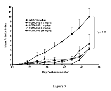
arthritis by using the XOMA 052 antibody.
[0053] Fig. 10 is a graph showing the protection of mice from collagen-induced
arthritis by using the XOMA 052 antibody.
14
CA 02727171 2010-12-06
WO 2009/149370 PCT/US2009/046441
[0054] Fig. 11A is a graph showing the protection of mice from collagen-
induced
arthritis after disease onset by using the XOMA 052 antibody.
[0055] Fig. 1lB is a graph showing the protection of mice from collagen-
induced
arthritis after disease onset by using the XOMA 052 antibody.
[0056] Fig. 12 is a graph showing that the antibody XOMA 052 prevents bone
pathology in a mouse model of collagen-induced arthritis.
[0057] Fig. 13 are images showing that the antibody XOMA 052 prevents
inflammation and bone and cartilage destruction in a mouse model of collagen-
induced
arthritis.
DETAILED DESCRIPTION
[0058] IL-1(3 is a pro-inflammatory cytokine secreted by a number of different
cell
types including monocytes and macrophages. When released as part of an
inflammatory
reaction, IL-1 (3 produces a range of biological effects, mainly mediated
through induction of
other inflammatory mediators such as corticotrophin, platelet factor-4,
prostaglandin E2
(PGE2), IL-6, and IL-8. IL-1(3 induces both local and systemic inflammatory
effects through
the activation of the IL-1 receptor found on almost all cell types.
[0059] The interleukin-1 (IL-1) family of cytokines has been implicated in
several
disease states such as rheumatoid arthritis (RA), osteoarthritis, Crohn's
disease, ulcerative
colitis (UC), septic shock, chronic obstructive pulmonary disease (COPD),
asthma, graft
versus host disease, atherosclerosis, adult T-cell leukemia, multiple myeloma,
multiple
sclerosis, stroke, and Alzheimer's disease. IL-1 family members include IL-la,
IL-1(3, and
IL-1Ra. Although related by their ability to bind to IL-1 receptors (IL-1R1,
IL-1R2), each of
these cytokines is expressed by a different gene and has a different primary
amino acid
sequence. Furthermore, the physiological activities of these cytokines can be
distinguished
from each other.
[0060] Compounds that disrupt IL-1 receptor signaling have been investigated
as
therapeutic agents to treat IL-1 mediated diseases, such as for example some
of the
aforementioned diseases. These compounds include recombinant IL-1Ra (Amgen
Inc.,
Thousand Oaks, CA), IL-1 receptor "trap" peptide (Regeneron Inc., Tarrytown,
NY), as well
as animal-derived IL-1 (3 antibodies and recombinant IL-1 (3 antibodies and
fragments thereof.
[0061] As noted above, IL-1 receptor antagonist (IL-1Ra) polypeptide has been
used
in the treatment of rheumatoid arthritis (RA), but there remains a need for
more effective
CA 02727171 2010-12-06
WO 2009/149370 PCT/US2009/046441
means to treat RA, particularly those that do not require frequent (e.g.,
daily), repeated
injections. An additional challenge for IL-1 receptor antagonist-based
therapeutics is the need
for such therapeutics to occupy a large number of receptors, which is a
formidable task since
these receptors are widely expressed on all cells except red blood cells
(Dinarello, Curr.
Opin. Pharmacol. 4:378-385, 2004). In most immune-mediated diseases, the
amount of IL-
cytokine that is measurable in body fluids or associated with activated cells
is relatively
low. Thus, a method of treatment and/or prevention that directly targets the
IL-1(3 ligand is a
superior strategy, particularly when administering an IL-1(3 antibody with
high affinity.
[0062] The present disclosure provides methods and related compositions and
articles
of manufacture for the treatment and/or prevention of rheumatoid arthritis in
a subject (e.g.,
mammalian, human), using an antibody or fragment thereof specific for IL-1 R.
As provided
below, antibodies with high affinity can be far more potent an inhibitor of
the IL-1 pathway
than is IL-Ra (e.g., Anakinra, Kineret ), and used to achieve a therapeutic
effect at a lower
dose and/or with less frequent administration than necessary for other drugs,
such as
recombinant IL-1Ra.
[0063] Such methods as described herein with an IL-10 antibody or fragment may
include the treatment of a subject suffering from RA. The methods also may
include
preventing the occurrence of RA in an at risk subject.
Antibodies, Humanized Antibodies, and Human Engineered Antibodies
[0064] The IL-1 (e.g., IL-1(3) binding antibodies or fragments thereof of the
present
disclosure may be provided as polyclonal antibodies, monoclonal antibodies
(mAbs),
recombinant antibodies, chimeric antibodies, CDR-grafted antibodies, fully
human
antibodies, single chain antibodies, and/or bispecific antibodies, as well as
fragments,
including variants and derivatives thereof, provided by known techniques,
including, but not
limited to enzymatic cleavage, peptide synthesis or recombinant techniques.
[0065] Antibodies generally comprise two heavy chain polypeptides and two
light
chain polypeptides, though single domain antibodies having one heavy chain and
one light
chain, and heavy chain antibodies devoid of light chains are also
contemplated. There are
five types of heavy chains, called alpha, delta, epsilon, gamma and mu, based
on the amino
acid sequence of the heavy chain constant domain. These different types of
heavy chains give
rise to five classes of antibodies, IgA (including IgAi and IgA2), IgD, IgE,
IgG and IgM,
respectively, including four subclasses of IgG, namely IgGi, IgG2, IgG3 and
IgG4. There are
also two types of light chains, called kappa (K) or lambda (X) based on the
amino acid
sequence of the constant domains. A full-length antibody includes a constant
domain and a
16
CA 02727171 2010-12-06
WO 2009/149370 PCT/US2009/046441
variable domain. The constant region need not be present in an antigen binding
fragment of
an antibody. Antigen binding fragments of an antibody disclosed herein can
include Fab,
Fab', F(ab')2, and F(v) antibody fragments. As discussed in more detail below,
IL-10
binding fragments encompass antibody fragments and antigen-binding
polypeptides that will
bind IL-1(3.
[0066] Each of the heavy chain and light chain sequences of an antibody, or
antigen
binding fragment thereof, includes a variable region with three
complementarity determining
regions (CDRs) as well as non-CDR framework regions (FRs). The terms "heavy
chain" and
"light chain," as used herein, mean the heavy chain variable region and the
light chain
variable region, respectively, unless otherwise noted. Heavy chain CDRs are
referred to
herein as CDR-H1, CDR-H2, and CDR-H3. Light chain CDRs are referred to herein
as
CDR-L1, CDR-L2, and CDR-L3. Variable regions and CDRs in an antibody sequence
can
be identified (i) according to general rules that have been developed in the
art or (ii) by
aligning the sequences against a database of known variable regions. Methods
for identifying
these regions are described in Kontermann and Dubel, eds., Antibody
Engineering, Springer,
New York, NY, 2001, and Dinarello et al., Current Protocols in Immunology,
John Wiley
and Sons Inc., Hoboken, NJ, 2000. Databases of antibody sequences are
described in and can
be accessed through "The Kabatman" database at www.bioinf.org.uk/abs
(maintained by
A.C. Martin in the Department of Biochemistry & Molecular Biology University
College
London, London, England) and VBASE2 at www.vbase2.org, as described in Retter
et al.,
Nucl. Acids Res., 33(Database issue): D671-D674 (2005). The "Kabatman"
database web
site also includes general rules of thumb for identifying CDRs. The term
"CDR," as used
herein, is as defined in Kabat et al., Sequences of Immunological Interest,
5a' ed., U.S.
Department of Health and Human Services, 1991, unless otherwise indicated.
[0067] Polyclonal antibodies are preferably raised in animals by multiple
subcutaneous (sc) or intraperitoneal (ip) injections of the relevant antigen
and an adjuvant.
An improved antibody response may be obtained by conjugating the relevant
antigen to a
protein that is immunogenic in the species to be immunized, e.g., keyhole
limpet
hemocyanin, serum albumin, bovine thyroglobulin, or soybean trypsin inhibitor
using a
bifunctional or derivatizing agent, for example, maleimidobenzoyl
sulfosuccinimide ester
(conjugation through cysteine residues), N-hydroxysuccinimide (through lysine
residues),
glutaraldehyde, succinic anhydride or other agents known in the art.
[0068] Animals are immunized against the antigen, immunogenic conjugates, or
derivatives by combining, e.g., 100 g or 5 g of the protein or conjugate
(for rabbits or mice,
17
CA 02727171 2010-12-06
WO 2009/149370 PCT/US2009/046441
respectively) with 3 volumes of Freund's complete adjuvant and injecting the
solution
intradermally at multiple sites. One month later, the animals are boosted with
1/5 to {fraction
(1/10)} the original amount of peptide or conjugate in Freund's complete
adjuvant by
subcutaneous injection at multiple sites. At 7-14 days post-booster injection,
the animals are
bled and the serum is assayed for antibody titer. Animals are boosted until
the titer plateaus.
Preferably, the animal is boosted with the conjugate of the same antigen, but
conjugated to a
different protein and/or through a different cross-linking reagent. Conjugates
also can be
made in recombinant cell culture as protein fusions. Also, aggregating agents
such as alum
are suitably used to enhance the immune response.
[0069] Monoclonal antibody refers to an antibody obtained from a population of
substantially homogeneous antibodies. Monoclonal antibodies are generally
highly specific,
and may be directed against a single antigenic site, in contrast to
conventional (polyclonal)
antibody preparations that typically include different antibodies directed
against different
determinants (epitopes). In addition to their specificity, the monoclonal
antibodies are
advantageous in that they are synthesized by the homogeneous culture,
uncontaminated by
other immunoglobulins with different specificities and characteristics.
[0070] Monoclonal antibodies to be used in accordance with the present
disclosure
may be made by the hybridoma method first described by Kohler et al., (Nature,
256:495-7,
1975), or may be made by recombinant DNA methods (see, e.g., U.S. Patent No.
4,816,567).
The monoclonal antibodies may also be isolated from phage antibody libraries
using the
techniques described in, for example, Clackson et al., (Nature 352:624-628,
1991) and Marks
et al., (J. Mol. Biol. 222:581-597, 1991).
[0071] In the hybridoma method, a mouse or other appropriate host animal, such
as a
hamster or macaque monkey, is immunized as herein described to elicit
lymphocytes that
produce or are capable of producing antibodies that will specifically bind to
the protein used
for immunization. Alternatively, lymphocytes may be immunized in vitro.
Lymphocytes
then are fused with myeloma cells using a suitable fusing agent, such as
polyethylene glycol,
to form a hybridoma cell (Goding, Monoclonal Antibodies: Principles and
Practice, pp. 59-
103 (Academic Press, 1986)).
[0072] The hybridoma cells thus prepared are seeded and grown in a suitable
culture
medium that preferably contains one or more substances that inhibit the growth
or survival of
the unfused, parental myeloma cells. For example, if the parental myeloma
cells lack the
enzyme hypoxanthine guanine phosphoribosyl transferase (HGPRT or HPRT), the
culture
18
CA 02727171 2010-12-06
WO 2009/149370 PCT/US2009/046441
medium for the hybridomas typically will include hypoxanthine, aminopterin,
and thymidine
(HAT medium), which substances prevent the growth of HGPRT-deficient cells.
[0073] Preferred myeloma cells are those that fuse efficiently, support stable
high-
level production of antibody by the selected antibody-producing cells, and are
sensitive to a
medium. Human myeloma and mouse-human heteromyeloma cell lines also have been
described for the production of human monoclonal antibodies (Kozbor, J.
Immunol., 133:
3001 (1984); Brodeur et al., Monoclonal Antibody Production Techniques and
Applications,
pp. 51-63 (Marcel Dekker, Inc., New York, 1987)). Exemplary murine myeloma
lines
include those derived from MOP-21 and M.C.-11 mouse tumors available from the
Salk
Institute Cell Distribution Center, San Diego, Calif. USA, and SP-2 or X63-Ag8-
653 cells
available from the American Type Culture Collection, Rockville, Md. USA.
[0074] Culture medium in which hybridoma cells are growing is assayed for
production of monoclonal antibodies directed against the antigen. Preferably,
the binding
specificity of monoclonal antibodies produced by hybridoma cells is determined
by
immunoprecipitation or by an in vitro binding assay, such as radioimmunoassay
(RIA) or
enzyme-linked immunoabsorbent assay (ELISA). The binding affinity of the
monoclonal
antibody can, for example, be determined by Scatchard analysis (Munson et al.,
Anal.
Biochem., 107:220 (1980)).
[0075] After hybridoma cells are identified that produce antibodies of the
desired
specificity, affinity, and/or activity, the clones may be subcloned by
limiting dilution
procedures and grown by standard methods (Goding, Monoclonal Antibodies:
Principles and
Practice, pp. 59-103 (Academic Press, 1986)). Suitable culture media for this
purpose
include, for example, DMEM or RPMI-1640 medium. In addition, the hybridoma
cells may
be grown in vivo as ascites tumors in an animal. The monoclonal antibodies
secreted by the
subclones are suitably separated from the culture medium, ascites fluid, or
serum by
conventional immunoglobulin purification procedures such as, for example,
protein A-
Sepharose, hydroxylapatite chromatography, gel electrophoresis, dialysis, or
affinity
chromatography.
[0076] It is further contemplated that antibodies of the disclosure may be
used as
smaller antigen binding fragments of the antibody well-known in the art and
described herein.
[0077] The present disclosure encompasses IL-1 (e.g., IL-1(3) binding
antibodies that
include two full length heavy chains and two full length light chains.
Alternatively, the IL-1(3
binding antibodies can be constructs such as single chain antibodies or "mini"
antibodies that
retain binding activity to IL-10. Such constructs can be prepared by methods
known in the
19
CA 02727171 2010-12-06
WO 2009/149370 PCT/US2009/046441
art such as, for example, the PCR mediated cloning and assembly of single
chain antibodies
for expression in E. coli (as described in Antibody Engineering, The practical
approach
series, J. McCafferty, H. R. Hoogenboom, and D. J. Chiswell, editors, Oxford
University
Press, 1996). In this type of construct, the variable portions of the heavy
and light chains of
an antibody molecule are PCR amplified from cDNA. The resulting amplicons are
then
assembled, for example, in a second PCR step, through a linker DNA that
encodes a flexible
protein linker composed of the amino acids Gly and Ser. This linker allows the
variable
heavy and light chain portions to fold in such a way that the antigen binding
pocket is
regenerated and antigen is bound with affinities often comparable to the
parent full-length
dimeric immunoglobulin molecule.
[0078] The IL-1 (e.g., IL-1(3) binding antibodies and fragments of the present
disclosure encompass variants of the exemplary antibodies, fragments and
sequences
disclosed herein. Variants include peptides and polypeptides comprising one or
more amino
acid sequence substitutions, deletions, and/or additions that have the same or
substantially the
same affinity and specificity of epitope binding as one or more of the
exemplary antibodies,
fragments and sequences disclosed herein. Thus, variants include peptides and
polypeptides
comprising one or more amino acid sequence substitutions, deletions, and/or
additions to the
exemplary antibodies, fragments and sequences disclosed herein where such
substitutions,
deletions and/or additions do not cause substantial changes in affinity and
specificity of
epitope binding. For example, a variant of an antibody or fragment may result
from one or
more changes to an antibody or fragment, where the changed antibody or
fragment has the
same or substantially the same affinity and specificity of epitope binding as
the starting
sequence. Variants may be naturally occurring, such as allelic or splice
variants, or may be
artificially constructed. Variants may be prepared from the corresponding
nucleic acid
molecules encoding said variants. Variants of the present antibodies and IL-10
binding
fragments may have changes in light and/or heavy chain amino acid sequences
that are
naturally occurring or are introduced by in vitro engineering of native
sequences using
recombinant DNA techniques. Naturally occurring variants include "somatic"
variants which
are generated in vivo in the corresponding germ line nucleotide sequences
during the
generation of an antibody response to a foreign antigen.
[0079] Variants of IL-1 (e.g., IL-1(3) binding antibodies and binding
fragments may
also be prepared by mutagenesis techniques. For example, amino acid changes
may be
introduced at random throughout an antibody coding region and the resulting
variants may be
screened for binding affinity for IL-10 or for another property.
Alternatively, amino acid
CA 02727171 2010-12-06
WO 2009/149370 PCT/US2009/046441
changes may be introduced in selected regions of an IL-10 antibody, such as in
the light
and/or heavy chain CDRs, and/or in the framework regions, and the resulting
antibodies may
be screened for binding to IL-1(3 or some other activity. Amino acid changes
encompass one
or more amino acid substitutions in a CDR, ranging from a single amino acid
difference to
the introduction of multiple permutations of amino acids within a given CDR,
such as CDR3.
In another method, the contribution of each residue within a CDR to IL-1(3
binding may be
assessed by substituting at least one residue within the CDR with alanine.
Lewis et at. (1995),
Mol. Immunol. 32: 1065-72. Residues which are not optimal for binding to IL-10
may then
be changed in order to determine a more optimum sequence. Also encompassed are
variants
generated by insertion of amino acids to increase the size of a CDR, such as
CDR3. For
example, most light chain CDR3 sequences are nine amino acids in length. Light
chain
sequences in an antibody which are shorter than nine residues may be optimized
for binding
to IL-1 0 by insertion of appropriate amino acids to increase the length of
the CDR.
[0080] Variants may also be prepared by "chain shuffling" of light or heavy
chains.
Marks et al. (1992), Biotechnology 10: 779-83. A single light (or heavy) chain
can be
combined with a library having a repertoire of heavy (or light) chains and the
resulting
population is screened for a desired activity, such as binding to IL-1(3. This
permits screening
of a greater sample of different heavy (or light) chains in combination with a
single light (or
heavy) chain than is possible with libraries comprising repertoires of both
heavy and light
chains.
[0081] The IL-1 (e.g., IL-1(3) binding antibodies and fragments of the present
disclosure encompass derivatives of the exemplary antibodies, fragments and
sequences
disclosed herein. Derivatives include polypeptides or peptides, or variants,
fragments or
derivatives thereof, which have been chemically modified. Examples include
covalent
attachment of one or more polymers, such as water soluble polymers, N-linked,
or O-linked
carbohydrates, sugars, phosphates, and/or other such molecules. The
derivatives are modified
in a manner that is different from naturally occurring or starting peptide or
polypeptides,
either in the type or location of the molecules attached. Derivatives further
include deletion of
one or more chemical groups which are naturally present on the peptide or
polypeptide.
[0082] The IL-10 binding antibodies and fragments of the present disclosure
can be
bispecific. Bispecific antibodies or fragments can be of several
configurations. For example,
bispecific antibodies may resemble single antibodies (or antibody fragments)
but have two
different antigen binding sites (variable regions). Bispecific antibodies can
be produced by
chemical techniques (Kranz et al. (1981), Proc. Natl. Acad. Sci. USA, 78:
5807), by
21
CA 02727171 2010-12-06
WO 2009/149370 PCT/US2009/046441
"polydoma" techniques (U.S. Pat. No. 4,474,893) or by recombinant DNA
techniques.
Bispecific antibodies of the present disclosure can have binding specificities
for at least two
different epitopes, at least one of which is an epitope of IL-10. The IL-10
binding antibodies
and fragments can also be heteroantibodies. Heteroantibodies are two or more
antibodies, or
antibody binding fragments (Fab) linked together, each antibody or fragment
having a
different specificity.
[0083] Techniques for creating recombinant DNA versions of the antigen-binding
regions of antibody molecules which bypass the generation of monoclonal
antibodies are
contemplated for the present IL-1 (e.g., IL-1(3) binding antibodies and
fragments. DNA is
cloned into a bacterial expression system. One example of such a technique
suitable for the
practice of this disclosure uses a bacteriophage lambda vector system having a
leader
sequence that causes the expressed Fab protein to migrate to the periplasmic
space (between
the bacterial cell membrane and the cell wall) or to be secreted. One can
rapidly generate and
screen great numbers of functional Fab fragments for those which bind IL-1 R.
Such IL-1(3
binding agents (Fab fragments with specificity for an IL-10 polypeptide) are
specifically
encompassed within the IL-1(3 binding antibodies and fragments of the present
disclosure.
[0084] The present IL-1 (e.g., IL-1(3) binding antibodies and fragments can be
humanized or human engineered antibodies. As used herein, a humanized
antibody, or
antigen binding fragment thereof, is a recombinant polypeptide that comprises
a portion of an
antigen binding site from a non-human antibody and a portion of the framework
and/or
constant regions of a human antibody. A human engineered antibody or antibody
fragment is
a non-human (e.g., mouse) antibody that has been engineered by modifying
(e.g., deleting,
inserting, or substituting) amino acids at specific positions so as to reduce
or eliminate any
detectable immunogenicity of the modified antibody in a human.
[0085] Humanized antibodies include chimeric antibodies and CDR-grafted
antibodies. Chimeric antibodies are antibodies that include a non-human
antibody variable
region linked to a human constant region. Thus, in chimeric antibodies, the
variable region is
mostly non-human, and the constant region is human. Chimeric antibodies and
methods for
making them are described in Morrison, et al., Proc. Natl. Acad. Sci. USA, 81:
6841-6855
(1984), Boulianne, et al., Nature, 312: 643-646 (1984), and PCT Application
Publication WO
86/01533. Although, they can be less immunogenic than a mouse monoclonal
antibody,
administrations of chimeric antibodies have been associated with human anti-
mouse antibody
responses (HAMA) to the non-human portion of the antibodies. Chimeric
antibodies can also
be produced by splicing the genes from a mouse antibody molecule of
appropriate antigen-
22
CA 02727171 2010-12-06
WO 2009/149370 PCT/US2009/046441
binding specificity together with genes from a human antibody molecule of
appropriate
biological activity, such as the ability to activate human complement and
mediate ADCC.
Morrison et al. (1984), Proc. Natl. Acad. Sci., 81: 6851; Neuberger et al.
(1984), Nature, 312:
604. One example is the replacement of a Fc region with that of a different
isotype.
[0086] CDR-grafted antibodies are antibodies that include the CDRs from a non-
human "donor" antibody linked to the framework region from a human "recipient"
antibody.
Generally, CDR-grafted antibodies include more human antibody sequences than
chimeric
antibodies because they include both constant region sequences and variable
region
(framework) sequences from human antibodies. Thus, for example, a CDR-grafted
humanized antibody of the disclosure can comprise a heavy chain that comprises
a
contiguous amino acid sequence (e.g., about 5 or more, 10 or more, or even 15
or more
contiguous amino acid residues) from the framework region of a human antibody
(e.g., FR-1,
FR-2, or FR-3 of a human antibody) or, optionally, most or all of the entire
framework region
of a human antibody. CDR-grafted antibodies and methods for making them are
described
in, Jones et al., Nature, 321: 522-525 (1986), Riechmann et al., Nature, 332:
323-327 (1988),
and Verhoeyen et al., Science, 239: 1534-1536 (1988)). Methods that can be
used to produce
humanized antibodies also are described in U.S. Patents 4,816,567, 5,721,367,
5,837,243, and
6,180,377. CDR-grafted antibodies are considered less likely than chimeric
antibodies to
induce an immune reaction against non-human antibody portions. However, it has
been
reported that framework sequences from the donor antibodies are required for
the binding
affinity and/or specificity of the donor antibody, presumably because these
framework
sequences affect the folding of the antigen-binding portion of the donor
antibody. Therefore,
when donor, non-human CDR sequences are grafted onto unaltered human framework
sequences, the resulting CDR-grafted antibody can exhibit, in some cases, loss
of binding
avidity relative to the original non-human donor antibody. See, e.g.,
Riechmann et al.,
Nature, 332: 323-327 (1988), and Verhoeyen et al., Science, 239: 1534-1536
(1988).
[0087] Human engineered antibodies include for example "veneered" antibodies
and
antibodies prepared using HUMAN ENGINEERINGTM technology (see for example,
U.S. Patents
5,766,886 and 5,869,619). HUMAN ENGINEERINGTM technology is commercially
available,
and involves altering an non-human antibody or antibody fragment, such as a
mouse or
chimeric antibody or antibody fragment, by making specific changes to the
amino acid
sequence of the antibody so as to produce a modified antibody with reduced
immunogenicity
in a human that nonetheless retains the desirable binding properties of the
original non-
human antibodies. Generally, the technique involves classifying amino acid
residues of a
23
CA 02727171 2010-12-06
WO 2009/149370 PCT/US2009/046441
non-human (e.g., mouse) antibody as "low risk", "moderate risk", or "high
risk" residues.
The classification is performed using a global risk/reward calculation that
evaluates the
predicted benefits of making particular substitution (e.g., for immunogenicity
in humans)
against the risk that the substitution will affect the resulting antibody's
folding and/or
antigen-binding properties. Thus, a low risk position is one for which a
substitution is
predicted to be beneficial because it is predicted to reduce immunogenicity
without
significantly affecting antigen binding properties. A moderate risk position
is one for which
a substitution is predicted to reduce immunogenicity, but is more likely to
affect protein
folding and/or antigen binding. High risk positions contain residues most
likely to be
involved in proper folding or antigen binding. Generally, low risk positions
in a non-human
antibody are substituted with human residues, high risk positions are rarely
substituted, and
humanizing substitutions at moderate risk positions are sometimes made,
although not
indiscriminately. Positions with prolines in the non-human antibody variable
region
sequence are usually classified as at least moderate risk positions.
[0088] The particular human amino acid residue to be substituted at a given
low or
moderate risk position of a non-human (e.g., mouse) antibody sequence can be
selected by
aligning an amino acid sequence from the non-human antibody's variable regions
with the
corresponding region of a specific or consensus human antibody sequence. The
amino acid
residues at low or moderate risk positions in the non-human sequence can be
substituted for
the corresponding residues in the human antibody sequence according to the
alignment.
Techniques for making human engineered proteins are described in greater
detail in
Studnicka et al., Protein Engineering, 7: 805-814 (1994), U.S. Patents
5,766,886, 5,770,196,
5,821,123, and 5,869,619, and PCT Application Publication WO 93/11794.
[0089] "Veneered" antibodies are non-human or humanized (e.g., chimeric or CDR-
grafted antibodies) antibodies that have been engineered to replace certain
solvent-exposed
amino acid residues so as to further reduce their immunogenicity or enhance
their function.
As surface residues of a chimeric antibody are presumed to be less likely to
affect proper
antibody folding and more likely to elicit an immune reaction, veneering of a
chimeric
antibody can include, for instance, identifying solvent-exposed residues in
the non-human
framework region of a chimeric antibody and replacing at least one of them
with the
corresponding surface residues from a human framework region. Veneering can be
accomplished by any suitable engineering technique, including the use of the
above-described
HUMAN ENGINEERINGTM technology.
24
CA 02727171 2010-12-06
WO 2009/149370 PCT/US2009/046441
[0090] In a different approach, a recovery of binding avidity can be achieved
by "de-
humanizing" a CDR-grafted antibody. De-humanizing can include restoring
residues from
the donor antibody's framework regions to the CDR grafted antibody, thereby
restoring
proper folding. Similar "de-humanization" can be achieved by (i) including
portions of the
"donor" framework region in the "recipient" antibody or (ii) grafting portions
of the "donor"
antibody framework region into the recipient antibody (along with the grafted
donor CDRs).
[0091] For a further discussion of antibodies, humanized antibodies, human
engineered, and methods for their preparation, see Kontermann and Dubel, eds.,
Antibody
Engineering, Springer, New York, NY, 2001.
[0092] Exemplary humanized or human engineered antibodies include IgG, IgM,
IgE,
IgA, and IgD antibodies. The present antibodies can be of any class (IgG, IgA,
IgM, IgE,
IgD, etc.) or isotype and can comprise a kappa or lambda light chain. For
example, a human
antibody can comprise an IgG heavy chain or defined fragment, such as at least
one of
isotypes, IgGi, IgG2, IgG3 or IgG4. As a further example, the present
antibodies or
fragments can comprise an IgGI heavy chain and an IgGI light chain.
[0093] The present antibodies and fragments can be human antibodies, such as
antibodies which bind IL-1(3 polypeptides and are encoded by nucleic acid
sequences which
are naturally occurring somatic variants of human germline immunoglobulin
nucleic acid
sequence, and fragments, synthetic variants, derivatives and fusions thereof.
Such antibodies
may be produced by any method known in the art, such as through the use of
transgenic
mammals (such as transgenic mice) in which the native immunoglobulin
repertoire has been
replaced with human V-genes in the mammal chromosome. Such mammals appear to
carry
out VDJ recombination and somatic hypermutation of the human germline antibody
genes in
a normal fashion, thus producing high affinity antibodies with completely
human sequences.
[0094] Human antibodies to target protein can also be produced using
transgenic
animals that have no endogenous immunoglobulin production and are engineered
to contain
human immunoglobulin loci. For example, WO 98/24893 discloses transgenic
animals
having a human Ig locus wherein the animals do not produce functional
endogenous
immunoglobulins due to the inactivation of endogenous heavy and light chain
loci. WO
91/00906 also discloses transgenic non-primate mammalian hosts capable of
mounting an
immune response to an immunogen, wherein the antibodies have primate constant
and/or
variable regions, and wherein the endogenous immunoglobulin encoding loci are
substituted
or inactivated. WO 96/30498 and US Patent No. 6,091,001 disclose the use of
the Cre/Lox
system to modify the immunoglobulin locus in a mammal, such as to replace all
or a portion
CA 02727171 2010-12-06
WO 2009/149370 PCT/US2009/046441
of the constant or variable region to form a modified antibody molecule. WO
94/02602
discloses non-human mammalian hosts having inactivated endogenous Ig loci and
functional
human Ig loci. U.S. Patent No. 5,939,598 discloses methods of making
transgenic mice in
which the mice lack endogenous heavy chains, and express an exogenous
immunoglobulin
locus comprising one or more xenogeneic constant regions. See also, U.S.
Patent Nos.
6,114,598 6,657,103 and 6,833,268.
[0095] Using a transgenic animal described above, an immune response can be
produced to a selected antigenic molecule, and antibody producing cells can be
removed from
the animal and used to produce hybridomas that secrete human monoclonal
antibodies.
Immunization protocols, adjuvants, and the like are known in the art, and are
used in
immunization of, for example, a transgenic mouse as described in WO 96/33735.
This
publication discloses monoclonal antibodies against a variety of antigenic
molecules
including IL-6, IL-8, TNFa, human CD4, L selectin, gp39, and tetanus toxin.
The
monoclonal antibodies can be tested for the ability to inhibit or neutralize
the biological
activity or physiological effect of the corresponding protein. WO 96/33735
discloses that
monoclonal antibodies against IL-8, derived from immune cells of transgenic
mice
immunized with IL-8, blocked IL-8 induced functions of neutrophils. Human
monoclonal
antibodies with specificity for the antigen used to immunize transgenic
animals are also
disclosed in WO 96/34096 and U.S. patent application no. 20030194404; and U.S.
patent
application no. 20030031667.
[0096] Additional transgenic animals useful to make monoclonal antibodies
include
the Medarex HuMAb-MOUSE , described in U.S. Pat. No. 5,770,429 and Fishwild,
et al.
(Nat. Biotechnol. 14:845-851, 1996), which contains gene sequences from
unrearranged
human antibody genes that code for the heavy and light chains of human
antibodies.
Immunization of a HuMAb-MOUSE enables the production of fully human
monoclonal
antibodies to the target protein.
[0097] Also, Ishida et al. (Cloning Stem Cells. 4:91-102, 2002) describes the
TransChromo Mouse (TCMOUSETM) which comprises megabase-sized segments of human
DNA and which incorporates the entire human immunoglobulin (hIg) loci. The
TCMOUSETM has a fully diverse repertoire of hIgs, including all the subclasses
of IgGs
(IgGl-G4). Immunization of the TC MOUSETM with various human antigens produces
antibody responses comprising human antibodies.
[0098] See also Jakobovits et al., Proc. Natl. Acad. Sci. USA, 90:2551 (1993);
Jakobovits et al., Nature, 362:255-258 (1993); Bruggermann et al., Year in
Immunol., 7:33
26
CA 02727171 2010-12-06
WO 2009/149370 PCT/US2009/046441
(1993); and U.S. Pat. No. 5,591,669, U.S. Patent No. 5,589,369, U.S. Patent
No. 5,545,807;
and U.S Patent Publication No. 20020199213. U.S. Patent Publication No.
20030092125
describes methods for biasing the immune response of an animal to the desired
epitope.
Human antibodies may also be generated by in vitro activated B cells (see U.S.
Pat. Nos.
5,567,610 and 5,229,275).
[0099] Human antibodies can also be generated through the in vitro screening
of
antibody display libraries. See Hoogenboom et al. (1991), J. Mol. Biol. 227:
381; and Marks
et al. (1991), J. Mol. Biol. 222: 581. Various antibody-containing phage
display libraries have
been described and may be readily prepared. Libraries may contain a diversity
of human
antibody sequences, such as human Fab, Fv, and scFv fragments, that may be
screened
against an appropriate target. Phage display libraries may comprise peptides
or proteins other
than antibodies which may be screened to identify selective binding agents of
IL-1(3.
[00100] The development of technologies for making repertoires of recombinant
human antibody genes, and the display of the encoded antibody fragments on the
surface of
filamentous bacteriophage, has provided a means for making human antibodies
directly. The
antibodies produced by phage technology are produced as antigen binding
fragments-usually
Fv or Fab fragments-in bacteria and thus lack effector functions. Effector
functions can be
introduced by one of two strategies: The fragments can be engineered either
into complete
antibodies for expression in mammalian cells, or into bispecific antibody
fragments with a
second binding site capable of triggering an effector function.
[00101 ] The disclosure contemplates a method for producing target-specific
antibody
or antigen-binding portion thereof comprising the steps of synthesizing a
library of human
antibodies on phage, screening the library with target protein or a portion
thereof, isolating
phage that bind target, and obtaining the antibody from the phage. By way of
example, one
method for preparing the library of antibodies for use in phage display
techniques comprises
the steps of immunizing a non-human animal comprising human immunoglobulin
loci with
target antigen or an antigenic portion thereof to create an immune response,
extracting
antibody producing cells from the immunized animal; isolating RNA from the
extracted cells,
reverse transcribing the RNA to produce cDNA, amplifying the cDNA using a
primer, and
inserting the cDNA into a phage display vector such that antibodies are
expressed on the
phage. Recombinant target-specific antibodies of the disclosure may be
obtained in this way.
[00102] Phage-display processes mimic immune selection through the display of
antibody repertoires on the surface of filamentous bacteriophage, and
subsequent selection of
phage by their binding to an antigen of choice. One such technique is
described in WO
27
CA 02727171 2010-12-06
WO 2009/149370 PCT/US2009/046441
99/10494, which describes the isolation of high affinity and functional
agonistic antibodies
for MPL and msk receptors using such an approach. Antibodies of the disclosure
can be
isolated by screening of a recombinant combinatorial antibody library,
preferably a scFv
phage display library, prepared using human VL and VH cDNAs prepared from mRNA
derived from human lymphocytes. Methodologies for preparing and screening such
libraries
are known in the art. See e.g., U.S. Patent No. 5,969,108. There are
commercially available
kits for generating phage display libraries (e.g., the Pharmacia Recombinant
Phage Antibody
System, catalog no. 27-9400-01; and the Stratagene SurfZAP.TM. phage display
kit, catalog
no. 240612). There are also other methods and reagents that can be used in
generating and
screening antibody display libraries (see, e.g., Ladner et al. U.S. Pat. No.
5,223,409; Kang et
al. PCT Publication No. WO 92/18619; Dower et al. PCT Publication No. WO
91/17271;
Winter et al. PCT Publication No. WO 92/20791; Markland et al. PCT Publication
No. WO
92/15679; Breitling et al. PCT Publication No. WO 93/01288; McCafferty et al.
PCT
Publication No. WO 92/01047; Garrard et al. PCT Publication No. WO 92/09690;
Fuchs et
al. (1991) Bio/Technology 9:1370-1372; Hay et al. (1992) Hum. Antibod.
Hybridomas 3:81-
85; Huse et al. (1989) Science 246:1275-1281; McCafferty et al., Nature (1990)
348:552-
554; Griffiths et al. (1993) EMBO J 12:725-734; Hawkins et al. (1992) J. Mol.
Biol. 226:889-
896; Clackson et al. (1991) Nature 352:624-628; Gram et al. (1992) Proc. Natl.
Acad. Sci.
USA 89:3576-3580; Garrad et al. (1991) Bio/Technology 9:1373-1377; Hoogenboom
et al.
(1991) Nuc Acid Res 19:4133-4137; and Barbas et al. (1991) Proc. Natl. Acad.
Sci. USA
88:7978-7982.
[00103] In one embodiment, to isolate human antibodies specific for the target
antigen with the desired characteristics, a human VH and VL library are
screened to select for
antibody fragments having the desired specificity. The antibody libraries used
in this method
are preferably scFv libraries prepared and screened as described herein and in
the art
(McCafferty et al., PCT Publication No. WO 92/01047, McCafferty et al.,
(Nature 348:552-
554, 1990); and Griffiths et al., (EMBO J 12:725-734, 1993). The scFv antibody
libraries
preferably are screened using target protein as the antigen.
[00104] Alternatively, the Fd fragment (VH-CH1) and light chain (VL-CL) of
antibodies are separately cloned by PCR and recombined randomly in
combinatorial phage
display libraries, which can then be selected for binding to a particular
antigen. The Fab
fragments are expressed on the phage surface, i.e., physically linked to the
genes that encode
them. Thus, selection of Fab by antigen binding co-selects for the Fab
encoding sequences,
which can be amplified subsequently. Through several rounds of antigen binding
and re-
28
CA 02727171 2010-12-06
WO 2009/149370 PCT/US2009/046441
amplification, a procedure termed panning, Fab specific for the antigen are
enriched and
finally isolated.
[00105] In 1994, an approach for the humanization of antibodies, called
"guided
selection", was described. Guided selection utilizes the power of the phage
display technique
for the humanization of mouse monoclonal antibody (See Jespers, L. S., et al.,
Bio/Technology 12, 899-903 (1994)). For this, the Fd fragment of the mouse
monoclonal
antibody can be displayed in combination with a human light chain library, and
the resulting
hybrid Fab library may then be selected with antigen. The mouse Fd fragment
thereby
provides a template to guide the selection. Subsequently, the selected human
light chains are
combined with a human Fd fragment library. Selection of the resulting library
yields entirely
human Fab.
[00106] A variety of procedures have been described for deriving human
antibodies
from phage-display libraries (See, for example, Hoogenboom et al., J. Mol.
Biol., 227:381
(1991); Marks et al., J. Mol. Biol, 222:581-597 (1991); U.S. Pat. Nos.
5,565,332 and
5,573,905; Clackson, T., and Wells, J. A., TIBTECH 12, 173-184 (1994)). In
particular, in
vitro selection and evolution of antibodies derived from phage display
libraries has become a
powerful tool (See Burton, D. R., and Barbas III, C. F., Adv. Immunol. 57, 191-
280 (1994);
Winter, G., et al., Annu. Rev. Immunol. 12, 433-455 (1994); U.S. patent
publication no.
20020004215 and WO 92/01047; U.S. patent publication no. 20030190317; and U.S.
Patent
Nos. 6,054,287 and 5,877,293.
[00107] Watkins, "Screening of Phage-Expressed Antibody Libraries by Capture
Lift," Methods in Molecular Biology, Antibody Phage Display: Methods and
Protocols 178:
187-193 (2002), and U.S. patent publication no. 20030044772, published March
6, 2003,
describe methods for screening phage-expressed antibody libraries or other
binding
molecules by capture lift, a method involving immobilization of the candidate
binding
molecules on a solid support.
[00108] Fv fragments are displayed on the surface of phage, by the association
of
one chain expressed as a phage protein fusion (e.g., with M13 gene III) with
the
complementary chain expressed as a soluble fragment. It is contemplated that
the phage may
be a filamentous phage such as one of the class I phages: fd, M13, fl, Ifl,
Ike, ZJ/Z, Ff and
one of the class II phages Xf, Pfl and Pf3. The phage may be M13, or fd or a
derivative
thereof.
[00109] Once initial human VL and VH segments are selected, "mix and match"
experiments, in which different pairs of the initially selected VL and VH
segments are
29
CA 02727171 2010-12-06
WO 2009/149370 PCT/US2009/046441
screened for target binding, are performed to select preferred VL/VH pair
combinations.
Additionally, to further improve the quality of the antibody, the VL and VH
segments of the
preferred VL/VH pair(s) can be randomly mutated, preferably within the any of
the CDR1,
CDR2 or CDR3 region of VH and/or VL, in a process analogous to the in vivo
somatic
mutation process responsible for affinity maturation of antibodies during a
natural immune
response. This in vitro affinity maturation can be accomplished by amplifying
VL and VH
regions using PCR primers complimentary to the VH CDR1, CDR2, and CDR3, or VL
CDR1,
CDR2, and CDR3, respectively, which primers have been "spiked" with a random
mixture of
the four nucleotide bases at certain positions such that the resultant PCR
products encode VL
and VH segments into which random mutations have been introduced into the VH
and/or VL
CDR3 regions. These randomly mutated VL and VH segments can be rescreened for
binding
to target antigen.
[00110] Following screening and isolation of an target specific antibody from
a
recombinant immunoglobulin display library, nucleic acid encoding the selected
antibody can
be recovered from the display package (e.g., from the phage genome) and
subcloned into
other expression vectors by standard recombinant DNA techniques. If desired,
the nucleic
acid can be further manipulated to create other antibody forms of the
disclosure, as described
below. To express a recombinant human antibody isolated by screening of a
combinatorial
library, the DNA encoding the antibody is cloned into a recombinant expression
vector and
introduced into a mammalian host cell, as described herein.
[00111 ] It is contemplated that the phage display method may be carried out
in a
mutator strain of bacteria or host cell. A mutator strain is a host cell which
has a genetic
defect which causes DNA replicated within it to be mutated with respect to its
parent DNA.
Example mutator strains are NR9046mutD5 and NR9046 mut Ti.
[00112] It is also contemplated that the phage display method may be carried
out
using a helper phage. This is a phage which is used to infect cells containing
a defective
phage genome and which functions to complement the defect. The defective phage
genome
can be a phagemid or a phage with some function encoding gene sequences
removed.
Examples of helper phages are M13K07, M13K07 gene III no. 3; and phage
displaying or
encoding a binding molecule fused to a capsid protein.
[00113] Antibodies are also generated via phage display screening methods
using the
hierarchical dual combinatorial approach as disclosed in WO 92/01047 in which
an
individual colony containing either an H or L chain clone is used to infect a
complete library
of clones encoding the other chain (L or H) and the resulting two-chain
specific binding
CA 02727171 2010-12-06
WO 2009/149370 PCT/US2009/046441
member is selected in accordance with phage display techniques such as those
described
therein. This technique is also disclosed in Marks et at, (Bio/Technology,
10:779-783, 1992).
[00114] Methods for display of peptides on the surface of yeast and microbial
cells
have also been used to identify antigen specific antibodies. See, for example,
U.S. Patent No.
6,699,658. Antibody libraries may be attached to yeast proteins, such as
agglutinin,
effectively mimicking the cell surface display of antibodies by B cells in the
immune system.
[00115] In addition to phage display methods, antibodies may be isolated using
ribosome mRNA display methods and microbial cell display methods. Selection of
polypeptide using ribosome display is described in Hanes et al., (Proc. Natl
Acad Sci USA,
94:4937-4942, 1997) and U.S. Pat. Nos. 5,643,768 and 5,658,754 issued to
Kawasaki.
Ribosome display is also useful for rapid large scale mutational analysis of
antibodies. The
selective mutagenesis approach also provides a method of producing antibodies
with
improved activities that can be selected using ribosomal display techniques.
[00116] The IL-1 (e.g., IL-1(3) binding antibodies and fragments may comprise
one
or more portions that do not bind IL-1(3 but instead are responsible for other
functions, such
as circulating half-life, direct cytotoxic effect, detectable labeling, or
activation of the
recipient's endogenous complement cascade or endogenous cellular cytotoxicity.
The
antibodies or fragments may comprise all or a portion of the constant region
and may be of
any isotype, including IgA (e.g., IgAl or IgA2), IgD, IgE, IgG (e.g. IgGi,
IgG2, IgG3 or
IgG4), or IgM. In addition to, or instead of, comprising a constant region,
antigen-binding
compounds of the disclosure may include an epitope tag, a salvage receptor
epitope, a label
moiety for diagnostic or purification purposes, or a cytotoxic moiety such as
a radionuclide or
toxin.
[00117] The constant region (when present) of the present antibodies and
fragments
may be of the yl, y2, y3, y4, , 02, or 6 or r, type, preferably of the y
type, more preferably of
the y, type, whereas the constant part of a human light chain may be of the x
or k type (which
includes the Xi, k2 and k3 subtypes) but is preferably of the x type.
[00118] Variants also include antibodies or fragments comprising a modified Fc
region, wherein the modified Fc region comprises at least one amino acid
modification
relative to a wild-type Fc region. The variant Fc region may be designed,
relative to a
comparable molecule comprising the wild-type Fc region, so as to bind Fc
receptors with a
greater or lesser affinity.
[00119] For example, the present IL-10 binding antibodies and fragments may
comprise a modified Fc region. Fc region refers to naturally-occurring or
synthetic
31
CA 02727171 2010-12-06
WO 2009/149370 PCT/US2009/046441
polypeptides homologous to the IgG C-terminal domain that is produced upon
papain
digestion of IgG. IgG Fc has a molecular weight of approximately 50 kD. In the
present
antibodies and fragments, an entire Fc region can be used, or only a half-life
enhancing
portion. In addition, many modifications in amino acid sequence are
acceptable, as native
activity is not in all cases necessary or desired.
[00120] The Fc region can be mutated, if desired, to inhibit its ability to
fix
complement and bind the Fc receptor with high affinity. For murine IgG Fc,
substitution of
Ala residues for Glu 318, Lys 320, and Lys 322 renders the protein unable to
direct ADCC.
Substitution of Glu for Leu 235 inhibits the ability of the protein to bind
the Fc receptor with
high affinity. Various mutations for human IgG also are known (see, e.g.,
Morrison et al.,
1994, The Immunologist 2: 119 124 and Brekke et al., 1994, The Immunologist 2:
125).
[00121] In some embodiments, the present an antibodies or fragments are
provided
with a modified Fc region where a naturally-occurring Fc region is modified to
increase the
half-life of the antibody or fragment in a biological environment, for
example, the serum half-
life or a half-life measured by an in vitro assay. Methods for altering the
original form of a
Fc region of an IgG also are described in U.S. Patent No. 6,998,253.
[00122] In certain embodiments, it may be desirable to modify the antibody or
fragment in order to increase its serum half-life, for example, adding
molecules such as PEG
or other water soluble polymers, including polysaccharide polymers, to
antibody fragments to
increase the half-life. This may also be achieved, for example, by
incorporation of a salvage
receptor binding epitope into the antibody fragment (e.g., by mutation of the
appropriate
region in the antibody fragment or by incorporating the epitope into a peptide
tag that is then
fused to the antibody fragment at either end or in the middle, e.g., by DNA or
peptide
synthesis) (see, International Publication No. W096/32478). Salvage receptor
binding
epitope refers to an epitope of the Fc region of an IgG molecule (e.g., IgGi,
IgG2, IgG3, or
IgG4) that is responsible for increasing the in vivo serum half-life of the
IgG molecule.
[00123] A salvage receptor binding epitope can include a region wherein any
one or
more amino acid residues from one or two loops of a Fc domain are transferred
to an
analogous position of the antibody fragment. Even more preferably, three or
more residues
from one or two loops of the Fc domain are transferred. Still more preferred,
the epitope is
taken from the CH2 domain of the Fc region (e.g., of an IgG) and transferred
to the CH1,
CH3, or VH region, or more than one such region, of the antibody.
Alternatively, the epitope
is taken from the CH2 domain of the Fc region and transferred to the CL region
or VL region,
32
CA 02727171 2010-12-06
WO 2009/149370 PCT/US2009/046441
or both, of the antibody fragment. See also International applications WO
97/34631 and WO
96/32478 which describe Fc variants and their interaction with the salvage
receptor.
[00124] Mutation of residues within Fc receptor binding sites can result in
altered
effector function, such as altered ADCC or CDC activity, or altered half-life.
Potential
mutations include insertion, deletion or substitution of one or more residues,
including
substitution with alanine, a conservative substitution, a non-conservative
substitution, or
replacement with a corresponding amino acid residue at the same position from
a different
IgG subclass (e.g. replacing an IgGl residue with a corresponding IgG2 residue
at that
position). For example it has been reported that mutating the serine at amino
acid position
241 in IgG4 to proline (found at that position in IgGI and IgG2) led to the
production of a
homogeneous antibody, as well as extending serum half-life and improving
tissue distribution
compared to the original chimeric IgG4. (Angal et al., Mol Immunol. 30:105-8,
1993).
[00125] Antibody fragments are portions of an intact full length antibody,
such as an
antigen binding or variable region of the intact antibody. Examples of
antibody fragments
include Fab, Fab', F(ab')2, and Fv fragments; diabodies; linear antibodies;
single-chain
antibody molecules (e.g., scFv); multispecific antibody fragments such as
bispecific,
trispecific, and multispecific antibodies (e.g., diabodies, triabodies,
tetrabodies); minibodies;
chelating recombinant antibodies; tribodies or bibodies; intrabodies;
nanobodies; small
modular immunopharmaceuticals (SMIP), adnectins, binding-domain immunoglobulin
fusion
proteins; camelized antibodies; VHH containing antibodies; and any other
polypeptides
formed from antibody fragments.
[00126] The present disclosure includes IL-10 binding antibody fragments
comprising any of the foregoing heavy or light chain sequences and which bind
IL-1(3. The
term fragments as used herein refers to any 3 or more contiguous amino acids
(e.g., 4 or
more, 5 or more 6 or more, 8 or more, or even 10 or more contiguous amino
acids) of the
antibody and encompasses Fab, Fab', F(ab')2, and F(v) fragments, or the
individual light or
heavy chain variable regions or portion thereof. IL-10 binding fragments
include, for
example, Fab, Fab', F(ab')2, Fv and scFv. These fragments lack the Fc fragment
of an intact
antibody, clear more rapidly from the circulation, and can have less non-
specific tissue
binding than an intact antibody. See Wahl et al. (1983), J. Nucl. Med., 24:
316-25. These
fragments can be produced from intact antibodies using well known methods, for
example by
proteolytic cleavage with enzymes such as papain (to produce Fab fragments) or
pepsin (to
produce F(ab')2 fragments).
33
CA 02727171 2010-12-06
WO 2009/149370 PCT/US2009/046441
[00127] In vitro and cell based assays are well described in the art for use
in
determining binding of IL-10 to IL-1 receptor type I (IL-1R1), including
assays that
determining in the presence of molecules (such as antibodies, antagonists, or
other inhibitors)
that bind to IL-1(3 or IL-1RI. (see for example Evans et al., (1995), J. Biol.
Chem. 270:11477-
11483; Vigers et al., (2000), J. Biol. Chem. 275:36927-36933; Yanofsky et al.,
(1996), Proc.
Natl. Acad. Sci. USA 93:7381-7386; Fredericks et al., (2004), Protein Eng.
Des. Sel. 17:95-
106; Slack et al., (1993), J. Biol. Chem. 268:2513-2524; Smith et al., (2003),
Immunity
18:87-96; Vigers et al., (1997), Nature 386:190-194; Ruggiero et al., (1997),
J. Immunol.
158:3881-3887; Guo et al., (1995), J. Biol. Chem. 270:27562-27568; Svenson et
al., (1995),
Eur. J. Immunol. 25:2842-2850; Arend et al., (1994), J. Immunol. 153:4766-
4774).
Recombinant IL-1 receptor type I, including human IL-1 receptor type I, for
such assays is
readily available from a variety of commercial sources (see for example R&D
Systems,
SIGMA). IL-1 receptor type I also can be expressed from an expression
construct or vector
introduced into an appropriate host cell using standard molecular biology and
transfection
techniques known in the art. The expressed IL-1 receptor type I may then be
isolated and
purified for use in binding assays, or alternatively used directly in a cell
associated form.
[00128] For example, the binding of IL-10 to IL-1 receptor type I maybe
determined
by immobilizing an IL-1(3 binding antibody, contacting IL-1(3 with the
immobilized antibody
and determining whether the IL-1(3 was bound to the antibody, and contacting a
soluble form
of IL-1 RI with the bound IL-1R/antibody complex and determining whether the
soluble IL-
1RI was bound to the complex. The protocol may also include contacting the
soluble IL-1RI
with the immobilized antibody before the contact with IL-1(3, to confirm that
the soluble IL-
1RI does not bind to the immobilized antibody. This protocol can be performed
using a
Biacore instrument for kinetic analysis of binding interactions. Such a
protocol can also be
employed to determine whether an antibody or other molecule permits or blocks
the binding
of IL-1(3 to IL-1 receptor type I.
[00129] For other IL-10 / IL-1RI binding assays, the permitting or blocking of
IL-10
binding to IL-1 receptor type I may be determined by comparing the binding of
IL-10 to IL-
1RI in the presence or absence of IL-10 antibodies or IL-10 binding fragments
thereof.
Blocking is identified in the assay readout as a designated reduction of IL-
1(3 binding to IL-1
receptor type I in the presence of anti-IL-1(3 antibodies or IL-1(3 binding
fragments thereof, as
compared to a control sample that contains the corresponding buffer or diluent
but not an IL-
antibody or IL-10 binding fragment thereof. The assay readout may be
qualitatively
viewed as indicating the presence or absence of blocking, or may be
quantitatively viewed as
34
CA 02727171 2010-12-06
WO 2009/149370 PCT/US2009/046441
indicating a percent or fold reduction in binding due to the presence of the
antibody or
fragment.
[00130] Alternatively or additionally, when an IL-10 binding antibody or IL-10
binding fragment substantially blocks IL-1(3 binding to IL-1RI, the IL-1(3
binding to IL-1RI is
reduced by at least 10-fold, alternatively at least about 20-fold,
alternatively at least about 50-
fold, alternatively at least about 100-fold, alternatively at least about 1000-
fold, alternatively
at least about 10000-fold, or more, compared to binding of the same
concentrations of IL-10
and IL-1RI in the absence of the antibody or fragment. As another example,
when an IL-10
binding antibody or IL-10 binding fragment substantially permits IL-10 binding
to IL-1RI,
the IL-10 binding to IL-1RI is at least about 90%, alternatively at least
about 95%,
alternatively at least about 99%, alternatively at least about 99.9%,
alternatively at least about
99.99%, alternatively at least about 99.999%, alternatively at least about
99.9999%,
alternatively substantially identical to binding of the same concentrations of
IL-1(3 and IL-1 RI
in the absence of the antibody or fragment.
[00131] The present disclosure may in certain embodiments encompass IL-10
binding antibodies or IL-1(3 binding fragments that bind to the same epitope
or substantially
the same epitope as one or more of the exemplary antibodies described herein.
Alternatively
or additionally, the IL-10 binding antibodies or IL-10 binding fragments
compete with the
binding of an antibody having variable region sequences of AB7, described in
US application
number 11/472813 or WO 2007/002261 (sequences shown below). Alternatively or
additionally, the present disclosure encompasses IL-1(3 binding antibodies and
fragments that
bind to an epitope contained in the amino acid sequence
ESVDPKNYPKKKMEKRFVFNKIE (SEQ ID NO: 1), an epitope that the antibodies
designated AB5 and AB7 (US application number 11/472813, WO 2007/002261) bind
to. As
contemplated herein, one can readily determine if an IL-10 binding antibody or
fragment
binds to the same epitope or substantially the same epitope as one or more of
the exemplary
antibodies, such as for example the antibody designated AB7, using any of
several known
methods in the art.
[00132] For example, the key amino acid residues (epitope) bound by an IL-10
binding antibody or fragment may be determined using a peptide array, such as
for example,
a PepSpotTM peptide array (JPT Peptide Technologies, Berlin, Germany), wherein
a scan of
twelve amino-acid peptides, spanning the entire IL-l (3 amino acid sequence,
each peptide
overlapping by 11 amino acid to the previous one, is synthesized directly on a
membrane.
CA 02727171 2010-12-06
WO 2009/149370 PCT/US2009/046441
The membrane carrying the peptides is then probed with the antibody for which
epitope
binding information is sought, for example at a concentration of 2 g/ml, for
2 hr at room
temperature. Binding of antibody to membrane bound peptides may be detected
using a
secondary HRP-conjugated goat anti-human (or mouse, when appropriate)
antibody,
followed by enhanced chemiluminescence (ECL). The peptides spot(s)
corresponding to
particular amino acid residues or sequences of the mature IL-10 protein, and
which score
positive for antibody binding, are indicative of the epitope bound by the
particular antibody.
[00133] Alternatively or in addition, antibody competition experiments may be
performed and such assays are well known in the art. For example, to determine
if an
antibody or fragment binds to an epitope contained in a peptide sequence
comprising the
amino acids ESVDPKNYPKKKMEKRFVFNKIE (SEQ ID NO: 1), which corresponds to
residues 83-105 of the mature IL-10 protein, an antibody of unknown
specificity may be
compared with any of the exemplary of antibodies (e.g., AB7) of the present
disclosure that
are known to bind an epitope contained within this sequence. Binding
competition assays
may be performed, for example, using a Biacore instrument for kinetic
analysis of binding
interactions or by ELISA. In such an assay, the antibody of unknown epitope
specificity is
evaluated for its ability to compete for binding against the known comparator
antibody (e.g.,
AB7). Competition for binding to a particular epitope is determined by a
reduction in
binding to the IL-10 epitope of at least about 50%, or at least about 70%, or
at least about
80%, or at least about 90%, or at least about 95%, or at least about 99% or
about 100% for
the known comparator antibody (e.g., AB7) and is indicative of binding to
substantially the
same epitope.
[00134] In view of the identification in this disclosure of IL-10 binding
regions in
exemplary antibodies and/or epitopes recognized by the disclosed antibodies,
it is
contemplated that additional antibodies with similar binding characteristics
and therapeutic or
diagnostic utility can be generated that parallel the embodiments of this
disclosure.
[00135] Antigen-binding fragments of an antibody include fragments that retain
the
ability to specifically bind to an antigen, generally by retaining the antigen-
binding portion of
the antibody. It is well established that the antigen-binding function of an
antibody can be
performed by fragments of a full-length antibody. Examples of antigen-binding
portions
include (i) a Fab fragment, which is a monovalent fragment consisting of the
VL, VH, CL
and CH1 domains; (ii) a F(ab')2 fragment, which is a bivalent fragment
comprising two Fab
fragments linked by a disulfide bridge at the hinge region; (iii) a Fd
fragment which is the
VH and CH1 domains; (iv) a Fv fragment which is the VL and VH domains of a
single arm
36
CA 02727171 2010-12-06
WO 2009/149370 PCT/US2009/046441
of an antibody, (v) a dAb fragment (Ward et al., (1989) Nature 341:544-546),
which is a VH
domain; and (vi) an isolated complementarity determining region (CDR). Single
chain
antibodies are also encompassed within the term antigen-binding portion of an
antibody. The
IL-1(3 binding antibodies and fragments of the present disclosure also
encompass monovalent
or multivalent, or monomeric or multimeric (e.g. tetrameric), CDR-derived
binding domains
with or without a scaffold (for example, protein or carbohydrate scaffolding).
[00136] The present IL-10 binding antibodies or fragments may be part of a
larger
immunoadhesion molecules, formed by covalent or non-covalent association of
the antibody
or antibody portion with one or more other proteins or peptides. Examples of
such
immunoadhesion molecules include use of the streptavidin core region to make a
tetrameric
scFv molecule (Kipriyanov, S. M., et al. (1995) Human Antibodies and
Hybridomas 6:93-
101) and use of a cysteine residue, a marker peptide and a C-terminal
polyhistidine tag to
make bivalent and biotinylated scFv molecules (Kipriyanov, S. M., et al.
(1994) Mol.
Immunol. 31:1047-1058). Antibodies and fragments comprising immunoadhesion
molecules
can be obtained using standard recombinant DNA techniques, as described
herein. Preferred
antigen binding portions are complete domains or pairs of complete domains.
[00137] The IL-10 binding antibodies and fragments of the present disclosure
also
encompass domain antibody (dAb) fragments (Ward et at., Nature 341:544-546,
1989) which
consist of a VH domain. The IL-10 binding antibodies and fragments of the
present
disclosure also encompass diabodies, which are bivalent antibodies in which VH
and VL
domains are expressed on a single polypeptide chain, but using a linker that
is too short to
allow for pairing between the two domains on the same chain, thereby forcing
the domains to
pair with complementary domains of another chain and creating two antigen
binding sites
(see e.g., EP 404,097; WO 93/11161; Holliger et at., Proc. Natl. Acad. Sci.
USA 90:6444-
6448, 1993, and Poljak et at., Structure 2:1121-1123, 1994). Diabodies can be
bispecific or
monospecific.
[00138] The IL-10 binding antibodies and fragments of the present disclosure
also
encompass single-chain antibody fragments (scFv) that bind to IL-1 R. An scFv
comprises an
antibody heavy chain variable region (VH) operably linked to an antibody light
chain variable
region (VL) wherein the heavy chain variable region and the light chain
variable region,
together or individually, form a binding site that binds IL-10. An scFv may
comprise a VH
region at the amino-terminal end and a VL region at the carboxy-terminal end.
Alternatively,
scFv may comprise a VL region at the amino-terminal end and a VH region at the
carboxy-
terminal end. Furthermore, although the two domains of the Fv fragment, VL and
VH, are
37
CA 02727171 2010-12-06
WO 2009/149370 PCT/US2009/046441
coded for by separate genes, they can be joined, using recombinant methods, by
a synthetic
linker that enables them to be made as a single protein chain in which the VL
and VH regions
pair to form monovalent molecules (known as single chain Fv (scFv); see e.g.,
Bird et al.
(1988) Science 242:423-426; and Huston et al. (1988) Proc. Natl. Acad. Sci.
USA 85:5879-
5883).
[00139] An scFv may optionally further comprise a polypeptide linker between
the
heavy chain variable region and the light chain variable region. Such
polypeptide linkers
generally comprise between 1 and 50 amino acids, alternatively between 3 and
12 amino
acids, alternatively 2 amino acids. An example of a linker peptide for linking
heavy and light
chains in an scFv comprises the 5 amino acid sequence Gly-Gly-Gly-Gly-Ser (SEQ
ID NO:
2). Other examples comprise one or more tandem repeats of this sequence (for
example, a
polypeptide comprising two to four repeats of Gly-Gly-Gly-Gly-Ser (SEQ ID NO:
2) to
create linkers.
[00140] The IL-10 binding antibodies and fragments of the present disclosure
also
encompass heavy chain antibodies (HCAb). Exceptions to the H2L2 structure of
conventional
antibodies occur in some isotypes of the immunoglobulins found in camelids
(camels,
dromedaries and llamas; Hamers-Casterman et at., 1993 Nature 363: 446; Nguyen
et at.,
1998 J. Mol. Biol. 275: 413), wobbegong sharks (Nuttall et at., Mol Immunol.
38:313-26,
2001), nurse sharks (Greenberg et at., Nature 374:168-73, 1995; Roux et at.,
1998 Proc. Nat.
Acad. Sci. USA 95: 11804), and in the spotted ratfish (Nguyen, et at., "Heavy-
chain
antibodies in Camelidae; a case of evolutionary innovation," 2002
Immunogenetics 54(1):
39-47). These antibodies can apparently form antigen-binding regions using
only heavy chain
variable regions, in that these functional antibodies are dimers of heavy
chains only (referred
to as "heavy-chain antibodies" or "HCAbs"). Accordingly, some embodiments of
the present
IL-1(3 binding antibodies and fragments may be heavy chain antibodies that
specifically bind
to IL-10. For example, heavy chain antibodies that are a class of IgG and
devoid of light
chains are produced by animals of the genus Camelidae which includes camels,
dromedaries
and llamas (Hamers-Casterman et at., Nature 363:446-448 (1993)). HCAbs have a
molecular
weight of about 95 kDa instead of the about 160 kDa molecular weight of
conventional IgG
antibodies. Their binding domains consist only of the heavy-chain variable
domains, often
referred to as VHH to distinguish them from conventional VH. Muyldermans et
at., J. Mol.
Recognit. 12:131-140 (1999). The variable domain of the heavy-chain antibodies
is
sometimes referred to as a nanobody (Cortez-Retamozo et at., Cancer Research
64:2853-57,
2004). A nanobody library may be generated from an immunized dromedary as
described in
38
CA 02727171 2010-12-06
WO 2009/149370 PCT/US2009/046441
Conrath et at., (Antimicrob Agents Chemother 45: 2807-12, 2001) or using
recombinant
methods.
[00141] Since the first constant domain (CHI) is absent (spliced out during
mRNA
processing due to loss of a splice consensus signal), the variable domain
(VHH) is
immediately followed by the hinge region, the CH2 and the CH3 domains (Nguyen
et at., Mol.
Immunol. 36:515-524 (1999); Woolven et at., Immunogenetics 50:98-101 (1999)).
Camelid
VHH reportedly recombines with IgG2 and IgG3 constant regions that contain
hinge, CH2,
and CH3 domains and lack a CH1 domain (Hamers-Casterman et at., supra). For
example,
llama IgGi is a conventional (H2L2) antibody isotype in which VH recombines
with a
constant region that contains hinge, CH1, CH2 and CH3 domains, whereas the
llama IgG2
and IgG3 are heavy chain-only isotypes that lack CH1 domains and that contain
no light
chains.
[00142] Although the HCAbs are devoid of light chains, they have an antigen-
binding repertoire. The genetic generation mechanism of HCAbs is reviewed in
Nguyen et al.
Adv. Immunol 79:261-296 (2001) and Nguyen et at., Immunogenetics 54:39-47
(2002).
Sharks, including the nurse shark, display similar antigen receptor-containing
single
monomeric V-domains. Irving et at., J. Immunol. Methods 248:31-45 (2001); Roux
et at.,
Proc. Natl. Acad. Sci. USA 95:11804 (1998).
[00143] VHHS comprise small intact antigen-binding fragments (for example,
fragments that are about 15 kDa, 118-136 residues). Camelid VHH domains have
been found
to bind to antigen with high affinity (Desmyter et at., J. Biol. Chem.
276:26285-90, 2001),
with VHH affinities typically in the nanomolar range and comparable with those
of Fab and
scFv fragments. VHHS are highly soluble and more stable than the corresponding
derivatives
of scFv and Fab fragments. VH fragments have been relatively difficult to
produce in soluble
form, but improvements in solubility and specific binding can be obtained when
framework
residues are altered to be more VHH-like. (See, for example, Reichman et at.,
J Immunol
Methods 1999, 231:25-38.) VHHS carry amino acid substitutions that make them
more
hydrophilic and prevent prolonged interaction with BiP (immunoglobulin heavy-
chain
binding protein), which normally binds to the H-chain in the Endoplasmic
Reticulum (ER)
during folding and assembly, until it is displaced by the L-chain. Because of
the VHHS'
increased hydrophilicity, secretion from the ER is improved.
[00144] Functional VHHS may be obtained by proteolytic cleavage of HCAb of an
immunized camelid, by direct cloning of VHH genes from B-cells of an immunized
camelid
resulting in recombinant VHHS, or from naive or synthetic libraries. VHHS with
desired antigen
39
CA 02727171 2010-12-06
WO 2009/149370 PCT/US2009/046441
specificity may also be obtained through phage display methodology. Using VHHS
in phage
display is much simpler and more efficient compared to Fabs or scFvs, since
only one domain
needs to be cloned and expressed to obtain a functional antigen-binding
fragment.
Muyldermans, Biotechnol. 74:277-302 (2001); Ghahroudi et at., FEBS Lett.
414:521-526
(1997); and van der Linden et at., J. Biotechnol. 80:261-270 (2000). Methods
for generating
antibodies having camelid heavy chains are also described in U.S. Patent
Publication Nos.
20050136049 and 20050037421.
[00145] Ribosome display methods may be used to identify and isolate scFv
and/or
VHH molecules having the desired binding activity and affinity. Irving et at.,
J. Immunol.
Methods 248:31-45 (2001). Ribosome display and selection has the potential to
generate and
display large libraries (1014)
[00146] Other embodiments provide VHH-like molecules generated through the
process of camelisation, by modifying non-Camelidee VHS, such as human VHHS,
to improve
their solubility and prevent non-specific binding. This is achieved by
replacing residues on
the VLS side of VHS with VHH-like residues, thereby mimicking the more soluble
VHH
fragments. Camelised VH fragments, particularly those based on the human
framework, are
expected to exhibit a greatly reduced immune response when administered in
vivo to a patient
and, accordingly, are expected to have significant advantages for therapeutic
applications.
Davies et at., FEBS Lett. 339:285-290 (1994); Davies et at., Protein Eng.
9:531-537 (1996);
Tanha et at., J. Biol. Chem. 276:24774-24780 (2001); and Riechmann et at.,
Immunol.
Methods 231:25-38 (1999).
[00147] A wide variety of expression systems are available for the production
of IL-
fragments including Fab fragments, scFv, and VHHS. For example, expression
systems of
both prokaryotic and eukaryotic origin may be used for the large-scale
production of antibody
fragments and antibody fusion proteins. Particularly advantageous are
expression systems
that permit the secretion of large amounts of antibody fragments into the
culture medium.
[00148] Production of bispecific Fab-scFv ("bibody") and trispecific Fab-
(scFv)(2)
("tribody") are described in Schoonjans et al. (Jlmmunol. 165:7050-57, 2000)
and Willems et
al. (J Chromatogr B Analyt Technol Biomed Life Sci. 786:161-76, 2003). For
bibodies or
tribodies, a scFv molecule is fused to one or both of the VL-CL (L) and VH-CH1
(Fd) chains,
e.g., to produce a tribody two scFvs are fused to C-term of Fab while in a
bibody one scFv is
fused to C-term of Fab. A "minibody" consisting of scFv fused to CH3 via a
peptide linker
(hingeless) or via an IgG hinge has been described in Olafsen, et at., Protein
Eng Des Sel.
2004 Apr;17(4):315-23.
CA 02727171 2010-12-06
WO 2009/149370 PCT/US2009/046441
[00149] Intrabodies are single chain antibodies which demonstrate
intracellular
expression and can manipulate intracellular protein function (Biocca, et al.,
EMBO J. 9:101-
108, 1990; Colby et at., Proc Natl Acad Sci USA. 101:17616-21, 2004).
Intrabodies, which
comprise cell signal sequences which retain the antibody construct in
intracellular regions,
may be produced as described in Mhashilkar et al (EMBO J 14:1542-51, 1995) and
Wheeler
et al. (FASEB J. 17:1733-5. 2003). Transbodies are cell-permeable antibodies
in which a
protein transduction domains (PTD) is fused with single chain variable
fragment (scFv)
antibodies Heng et al., (Med Hypotheses. 64:1105-8, 2005).
[00150] The IL-10 binding antibodies and fragments of the present disclosure
also
encompass antibodies that are SMIPs or binding domain immunoglobulin fusion
proteins
specific for target protein. These constructs are single-chain polypeptides
comprising antigen
binding domains fused to immunoglobulin domains necessary to carry out
antibody effector
functions. See e.g., WO03/041600, U.S. Patent publication 20030133939 and US
Patent
Publication 20030118592.
[00151] The IL-10 binding antibodies and fragments of the present disclosure
also
encompass immunoadhesins. One or more CDRs may be incorporated into a molecule
either
covalently or noncovalently to make it an immunoadhesin. An immunoadhesin may
incorporate the CDR(s) as part of a larger polypeptide chain, may covalently
link the CDR(s)
to another polypeptide chain, or may incorporate the CDR(s) noncovalently. The
CDRs
disclosed herein permit the immunoadhesin to specifically bind to IL-1 R.
[00152] The IL-10 binding antibodies and fragments of the present disclosure
also
encompass antibody mimics comprising one or more IL-10 binding portions built
on an
organic or molecular scaffold (such as a protein or carbohydrate scaffold).
Proteins having
relatively defined three-dimensional structures, commonly referred to as
protein scaffolds,
may be used as reagents for the design of antibody mimics. These scaffolds
typically contain
one or more regions which are amenable to specific or random sequence
variation, and such
sequence randomization is often carried out to produce libraries of proteins
from which
desired products may be selected. For example, an antibody mimic can comprise
a chimeric
non-immunoglobulin binding polypeptide having an immunoglobulin-like domain
containing
scaffold having two or more solvent exposed loops containing a different CDR
from a parent
antibody inserted into each of the loops and exhibiting selective binding
activity toward a
ligand bound by the parent antibody. Non-immunoglobulin protein scaffolds have
been
proposed for obtaining proteins with novel binding properties. (Tramontano et
at., J. Mol.
Recognit. 7:9, 1994; McConnell and Hoess, J. Mol. Biol. 250:460, 1995). Other
proteins have
41
CA 02727171 2010-12-06
WO 2009/149370 PCT/US2009/046441
been tested as frameworks and have been used to display randomized residues on
alpha
helical surfaces (Nord et at., Nat. Biotechnol. 15:772, 1997; Nord et at.,
Protein Eng. 8:601,
1995), loops between alpha helices in alpha helix bundles (Ku and Schultz,
Proc. Natl. Acad.
Sci. USA 92:6552, 1995), and loops constrained by disulfide bridges, such as
those of the
small protease inhibitors (Markland et at., Biochemistry 35:8045, 1996;
Markland et at.,
Biochemistry 35:8058, 1996; Rottgen and Collins, Gene 164:243, 1995; Wang et
at., J. Biol.
Chem. 270:12250, 1995). Methods for employing scaffolds for antibody mimics
are
disclosed in US Patent 5,770,380 and US Patent Publications 2004/0171116,
2004/0266993,
and 2005/0038229.
[00153] Preferred IL-10 antibodies or antibody fragments for use in accordance
with
the disclosure generally bind to human IL-10 with high affinity (e.g., as
determined with
BIACORE and/or Kinexa), such as for example with an equilibrium binding
dissociation
constant (KD) for IL-1(3 of about 10 nM or less, about 5 nM or less, about 1
nM or less, about
500 pM or less, or more preferably about 250 pM or less, about 100 pM or less,
about 50 pM
or less, about 25 pM or less, about 10 pM or less, about 5 pM or less, about 3
pM or less
about 1 pM or less, about 0.75 pM or less, about 0.5 pM or less, or about 0.3
pM or less.
[00154] Antibodies or fragments of the present disclosure may, for example,
bind to
IL-1(3 with an IC50 of about 10 nM or less, about 5 nM or less, about 2 nM or
less, about 1
nM or less, about 0.75 nM or less, about 0.5 nM or less, about 0.4 nM or less,
about 0.3 nM
or less, or even about 0.2 nM or less, as determined by enzyme linked
immunosorbent assay
(ELISA). Preferably, the antibody or antibody fragment of the present
disclosure does not
cross-react with any target other than IL-1. For example, the present
antibodies and
fragments may bind to IL-1(3, but do not detestably bind to IL-1 a, or have at
least about 100
times (e.g., at least about 150 times, at least about 200 times, or even at
least about 250 times)
greater selectivity in its binding of IL-10 relative to its binding of IL-la.
Antibodies or
fragments used according to the disclosure may, in certain embodiments,
inhibit IL-10
induced expression of serum IL-6 in an animal by at least 50% (e.g., at least
60%, at least
70%, or even at least 80%) as compared to the level of serum IL-6 in an IL-10
stimulated
animal that has not been administered an antibody or fragment of the
disclosure. Antibodies
may bind IL-10 but permit or substantially permit the binding of the bound IL-
10 ligand to
IL-1 receptor type I (IL-1RI). In contrast to many known IL-1(3 binding
antibodies that block
or substantially interfere with binding of IL-1(3 to IL-1RI, the antibodies
designated A135 and
AB7 (US application number 11/472813, WO 2007/002261) selectively bind to the
IL-10
ligand, but permit the binding of the bound IL-10 ligand to IL-1RI. For
example, the
42
CA 02727171 2010-12-06
WO 2009/149370 PCT/US2009/046441
antibody designated AB7 binds to an IL-1(3 epitope but still permits the bound
IL-1(3 to bind
to IL-1RI. In certain embodiments, the antibody may decrease the affinity of
interaction of
bound IL-10 to bind to IL-1RI. Accordingly, the disclosure provides, in a
related aspect, use
of an IL-1(3 binding antibody or IL-1(3 binding antibody fragment that has at
least one of the
aforementioned characteristics. Any of the foregoing antibodies, antibody
fragments, or
polypeptides of the disclosure can be humanized or human engineered, as
described herein.
[00155] A variety of IL-1 (e.g., IL-1(3) antibodies and fragments known in the
art
may be used according the methods provided herein, including for example
antibodies
described in or derived using methods described in the following patents and
patent
applications: US 4,935,343; US 2003/0026806; US 2003/0124617; WO 2006/081139;
WO
03/034984; WO 95/01997; WO 02/16436; WO 03/010282; WO 03/073982, WO
2004/072116, WO 2004/067568, EP 0 267 611 B1, EP 0 364 778 B1, and US
application
number 11/472813. As a non-limiting example, antibodies AB5 and AB7 (US
application
number 11/472813, W02007/002261) may be used in accordance with the
disclosure.
Variable region sequences of AB5 and AB7 are as follows:
AB5
LIGHT CHAIN
DIQMTQTTSSLSASLGDRVTISCRASQDISNYLSWYQQKPDGTVKLLIYYTSKLHSGV
PSRFSGSGSGTDYSLTISNLEQEDIATYFCLQGKMLPWTFGGGTKLEIK (SEQ ID NO:
3)
The underlined sequences depict (from left to right) CDR1, 2 and 3.
HEAVY CHAIN
QVTLKESGPGILKPSQTLSLTCSFSGFSLSTSGMGVGWIRQPSGKGLEWLAHIWWDG
DESYNPSLKTQLTISKDTSRNQVFLKITSVDTVDTATYFCARNRYDPPWFVDWGQGT
LVTVSS (SEQ ID NO: 4)
The underlined sequences depict (from left to right) CDR1, 2 and 3.
43
CA 02727171 2010-12-06
WO 2009/149370 PCT/US2009/046441
AB7
LIGHT CHAIN
DIQMTQSTS SLSASVGDRVTITCRASQDISNYLS WYQQKPGKAVKLLIYYTSKLHSGV
PSRFSGSGSGTDYTLTISSLQQEDFATYFCLQGKMLPWTFGQGTKLEIK (SEQ ID NO:
5)
The underlined sequences depict (from left to right) CDR1, 2 and 3.
HEAVY CHAIN
QVQLQESGPGLVKPSQTLSLTCSFSGFSLSTSGMGVGWIRQPSGKGLEWLAHIWWD
GDESYNPSLKSRLTISKDTSKNQVSLKITSVTAADTAVYFCARNRYDPPWFVDWGQG
TLVTVSS (SEQ ID NO: 6)
The underlined sequences depict (from left to right) CDR1, 2 and 3.
[00156] The antibodies and antibody fragments described herein can be prepared
by
any suitable method. Suitable methods for preparing such antibodies and
antibody fragments
are known in the art. Other methods for preparing the antibodies and antibody
fragments are
as described herein as part of the disclosure. The antibody, antibody
fragment, or
polypeptide of the disclosure, as described herein, can be isolated or
purified to any degree.
As used herein, an isolated compound is a compound that has been removed from
its natural
environment. A purified compound is a compound that has been increased in
purity, such
that the compound exists in a form that is more pure than it exists (i) in its
natural
environment or (ii) when initially synthesized and/or amplified under
laboratory conditions,
wherein "purity" is a relative term and does not necessarily mean "absolute
purity."
Pharmaceutical Compositions
[00157] IL-1 (e.g., IL-1(3) binding antibodies and antibody fragments for use
according to the present disclosure can be formulated in compositions,
especially
pharmaceutical compositions, for use in the methods herein. Such compositions
comprise a
therapeutically or prophylactically effective amount of an IL-1(3 binding
antibody or antibody
fragment of the disclosure in admixture with a suitable carrier, e.g., a
pharmaceutically
acceptable agent. Typically, IL-10 binding antibodies and antibody fragments
of the
disclosure are sufficiently purified for administration to an animal before
formulation in a
pharmaceutical composition.
[00158] Pharmaceutically acceptable agents include carriers, excipients,
diluents,
antioxidants, preservatives, coloring, flavoring and diluting agents,
emulsifying agents,
44
CA 02727171 2010-12-06
WO 2009/149370 PCT/US2009/046441
suspending agents, solvents, fillers, bulking agents, buffers, delivery
vehicles, tonicity agents,
cosolvents, wetting agents, complexing agents, buffering agents,
antimicrobials, and
surfactants.
[00159] Neutral buffered saline or saline mixed with albumin are exemplary
appropriate carriers. The pharmaceutical compositions can include antioxidants
such as
ascorbic acid; low molecular weight polypeptides; proteins, such as serum
albumin, gelatin,
or immunoglobulins; hydrophilic polymers such as polyvinylpyrrolidone; amino
acids such
as glycine, glutamine, asparagine, arginine or lysine; monosaccharides,
disaccharides, and
other carbohydrates including glucose, mannose, or dextrins; chelating agents
such as EDTA;
sugar alcohols such as mannitol or sorbitol; salt-forming counterions such as
sodium; and/or
nonionic surfactants such as Tween, pluronics, or polyethylene glycol (PEG).
Also by way
of example, suitable tonicity enhancing agents include alkali metal halides
(preferably
sodium or potassium chloride), mannitol, sorbitol, and the like. Suitable
preservatives
include benzalkonium chloride, thimerosal, phenethyl alcohol, methylparaben,
propylparaben, chlorhexidine, sorbic acid and the like. Hydrogen peroxide also
can be used
as preservative. Suitable cosolvents include glycerin, propylene glycol, and
PEG. Suitable
complexing agents include caffeine, polyvinylpyrrolidone, beta-cyclodextrin or
hydroxy-
propyl-beta-cyclodextrin. Suitable surfactants or wetting agents include
sorbitan esters,
polysorbates such as polysorbate 80, tromethamine, lecithin, cholesterol,
tyloxapal, and the
like. The buffers can be conventional buffers such as acetate, borate,
citrate, phosphate,
bicarbonate, or Tris-HC1. Acetate buffer may be about pH 4-5.5, and Tris
buffer can be about
pH 7-8.5. Additional pharmaceutical agents are set forth in Remington's
Pharmaceutical
Sciences, 18th Edition, A. R. Gennaro, ed., Mack Publishing Company, 1990.
[00160] The composition can be in liquid form or in a lyophilized or freeze-
dried
form and may include one or more lyoprotectants, excipients, surfactants, high
molecular
weight structural additives and/or bulking agents (see for example US Patents
6,685,940,
6,566,329, and 6,372,716). In one embodiment, a lyoprotectant is included,
which is a non-
reducing sugar such as sucrose, lactose or trehalose. The amount of
lyoprotectant generally
included is such that, upon reconstitution, the resulting formulation will be
isotonic, although
hypertonic or slightly hypotonic formulations also may be suitable. In
addition, the amount of
lyoprotectant should be sufficient to prevent an unacceptable amount of
degradation and/or
aggregation of the protein upon lyophilization. Exemplary lyoprotectant
concentrations for
sugars (e.g., sucrose, lactose, trehalose) in the pre-lyophilized formulation
are from about 10
mM to about 400 mM. In another embodiment, a surfactant is included, such as
for example,
CA 02727171 2010-12-06
WO 2009/149370 PCT/US2009/046441
nonionic surfactants and ionic surfactants such as polysorbates (e.g.
polysorbate 20,
polysorbate 80); poloxamers (e.g. poloxamer 188); poly (ethylene glycol)
phenyl ethers (e.g.
Triton); sodium dodecyl sulfate (SDS); sodium laurel sulfate; sodium octyl
glycoside; lauryl-,
myristyl-, linoleyl-, or stearyl-sulfobetaine; lauryl-, myristyl-, linoleyl-
or stearyl-sarcosine;
linoleyl-, myristyl-, or cetyl-betaine; lauroamidopropyl-, cocamidopropyl-,
linoleamidopropyl-, myristamidopropyl-, palmidopropyl-, or isostearamidopropyl-
betaine
(e.g. lauroamidopropyl); myristamidopropyl-, palmidopropyl-, or
isostearamidopropyl-
dimethylamine; sodium methyl cocoyl-, or disodium methyl ofeyl-taurate; and
the
MONAQUATTM. series (Mona Industries, Inc., Paterson, N.J.), polyethyl glycol,
polypropyl
glycol, and copolymers of ethylene and propylene glycol (e.g. Pluronics, PF68
etc).
Exemplary amounts of surfactant that may be present in the pre-lyophilized
formulation are
from about 0.001-0.5%. High molecular weight structural additives (e.g.
fillers, binders) may
include for example, acacia, albumin, alginic acid, calcium phosphate
(dibasic), cellulose,
carboxymethylcellulose, carboxymethylcellulose sodium, hydroxyethylcellulose,
hydroxypropylcellulose, hydroxypropylmethylcellulose, microcrystalline
cellulose, dextran,
dextrin, dextrates, sucrose, tylose, pregelatinized starch, calcium sulfate,
amylose, glycine,
bentonite, maltose, sorbitol, ethylcellulose, disodium hydrogen phosphate,
disodium
phosphate, disodium pyrosulfite, polyvinyl alcohol, gelatin, glucose, guar
gum, liquid
glucose, compressible sugar, magnesium aluminum silicate, maltodextrin,
polyethylene
oxide, polymethacrylates, povidone, sodium alginate, tragacanth
microcrystalline cellulose,
starch, and zein. Exemplary concentrations of high molecular weight structural
additives are
from 0.1% to 10% by weight. In other embodiments, a bulking agent (e.g.,
mannitol,
glycine) may be included.
[00161] Compositions can be suitable for parenteral administration. Exemplary
compositions are suitable for injection or infusion into an animal by any
route available to the
skilled worker, such as intraarticular, subcutaneous, intravenous,
intramuscular,
intraperitoneal, intracerebral (intraparenchymal), intracerebroventricular,
intramuscular,
intraocular, intraarterial, intralesional, intrarectal, transdermal, oral, and
inhaled routes. A
parenteral formulation typically will be a sterile, pyrogen-free, isotonic
aqueous solution,
optionally containing pharmaceutically acceptable preservatives.
[00162] Examples of non-aqueous solvents are propylene glycol, polyethylene
glycol, vegetable oils such as olive oil, and injectable organic esters such
as ethyl oleate.
Aqueous carriers include water, alcoholic/aqueous solutions, emulsions or
suspensions,
including saline and buffered media. Parenteral vehicles include sodium
chloride solution,
46
CA 02727171 2010-12-06
WO 2009/149370 PCT/US2009/046441
Ringers' dextrose, dextrose and sodium chloride, lactated Ringer's, or fixed
oils. Intravenous
vehicles include fluid and nutrient replenishers, electrolyte replenishers,
such as those based
on Ringer's dextrose, and the like. Preservatives and other additives may also
be present, such
as, for example, anti-microbials, anti-oxidants, chelating agents, inert gases
and the like. See
generally, Remington's Pharmaceutical Science, 16th Ed., Mack Eds., 1980,
which is
incorporated herein by reference.
[00163] Pharmaceutical compositions described herein can be formulated for
controlled or sustained delivery in a manner that provides local concentration
of the product
(e.g., bolus, depot effect) sustained release and/or increased stability or
half-life in a
particular local environment. The disclosure contemplates that in certain
embodiments such
compositions may include a significantly larger amount of antibody or fragment
in the initial
deposit, while the effective amount of antibody or fragment actually released
and available at
any point in time for is in accordance with the disclosure herein an amount
much lower than
the initial deposit. The compositions can include the formulation of IL-10
binding
antibodies, antibody fragments, nucleic acids, or vectors of the disclosure
with particulate
preparations of polymeric compounds such as polylactic acid, polyglycolic
acid, etc., as well
as agents such as a biodegradable matrix, injectable microspheres,
microcapsular particles,
microcapsules, bioerodible particles beads, liposomes, and implantable
delivery devices that
provide for the controlled or sustained release of the active agent which then
can be delivered
as a depot injection. Techniques for formulating such sustained- or controlled-
delivery
means are known and a variety of polymers have been developed and used for the
controlled
release and delivery of drugs. Such polymers are typically biodegradable and
biocompatible.
Polymer hydrogels, including those formed by complexation of enantiomeric
polymer or
polypeptide segments, and hydrogels with temperature or pH sensitive
properties, may be
desirable for providing drug depot effect because of the mild and aqueous
conditions
involved in trapping bioactive protein agents (e.g., antibodies). See, for
example, the
description of controlled release porous polymeric microparticles for the
delivery of
pharmaceutical compositions in PCT Application Publication WO 93/15722.
[00164] Suitable materials for this purpose include polylactides (see, e.g.,
U.S. Patent
3,773,919), polymers of poly-(a-hydroxycarboxylic acids), such as poly-D-(-)-3-
hydroxybutyric acid (EP 133,988A), copolymers of L-glutamic acid and gamma
ethyl-L-
glutamate (Sidman et al., Biopolymers, 22: 547-556 (1983)), poly (2-
hydroxyethyl-
methacrylate) (Langer et al., J. Biomed. Mater. Res., 15: 167-277 (1981), and
Langer, Chem.
Tech., 12: 98-105 (1982)), ethylene vinyl acetate, or poly-D(-)-3-
hydroxybutyric acid. Other
47
CA 02727171 2010-12-06
WO 2009/149370 PCT/US2009/046441
biodegradable polymers include poly(lactones), poly(acetals),
poly(orthoesters), and
poly(orthocarbonates). Sustained-release compositions also may include
liposomes, which
can be prepared by any of several methods known in the art (see, e.g.,
Eppstein et al., Proc.
Natl. Acad. Sci. USA, 82: 3688-92 (1985)). The carrier itself, or its
degradation products,
should be nontoxic in the target tissue and should not further aggravate the
condition. This
can be determined by routine screening in animal models of the target disorder
or, if such
models are unavailable, in normal animals.
[00165] Microencapsulation of recombinant proteins for sustained release has
been
performed successfully with human growth hormone (rhGH), interferon- (rhIFN--
),
interleukin-2, and MN rgpl20. Johnson et al., Nat. Med., 2:795-799 (1996);
Yasuda, Biomed.
Ther., 27:1221-1223 (1993); Hora et al., Bio/Technologv. 8:755-758 (1990);
Cleland,
"Design and Production of Single Immunization Vaccines Using Polylactide
Polyglycolide
Microsphere Systems," in Vaccine Design: The Subunit and Adjuvant Approach,
Powell and
Newman, eds, (Plenum Press: New York, 1995), pp. 439-462; WO 97/03692, WO
96/40072,
WO 96/07399; and U.S. Pat. No. 5,654,010. The sustained-release formulations
of these
proteins were developed using poly-lactic-coglycolic acid (PLGA) polymer due
to its
biocompatibility and wide range of biodegradable properties. The degradation
products of
PLGA, lactic and glycolic acids can be cleared quickly within the human body.
Moreover,
the degradability of this polymer can be depending on its molecular weight and
composition.
Lewis, "Controlled release of bioactive agents from lactide/glycolide
polymer," in: M. Chasin
and R. Langer (Eds.), Biodegradable Polymers as Drug Delivery Systems (Marcel
Dekker:
New York, 1990), pp. 1-41. Additional examples of sustained release
compositions include,
for example, EP 58,481A, U.S. Pat. No. 3,887,699, EP 158,277A, Canadian Patent
No.
1176565, U. Sidman et al., Biopolymers 22, 547 [1983], R. Langer et al., Chem.
Tech. 12, 98
[1982], Sinha et al., J. Control. Release 90, 261 [2003], Zhu et al., Nat.
Biotechnol. 18, 24
[2000], and Dai et al., Colloids Surf B Biointerfaces 41, 117 [2005].
[00166] Bioadhesive polymers are also contemplated for use in or with
compositions
of the present disclosure. Bioadhesives are synthetic and naturally occurring
materials able to
adhere to biological substrates for extended time periods. For example,
Carbopol and
polycarbophil are both synthetic cross-linked derivatives of poly(acrylic
acid). Bioadhesive
delivery systems based on naturally occurring substances include for example
hyaluronic
acid, also known as hyaluronan. Hyaluronic acid is a naturally occurring
mucopolysaccharide
consisting of residues of D-glucuronic and N-acetyl-D-glucosamine. Hyaluronic
acid is
found in the extracellular tissue matrix of vertebrates, including in
connective tissues, as well
48
CA 02727171 2010-12-06
WO 2009/149370 PCT/US2009/046441
as in synovial fluid and in the vitreous and aqueous humour of the eye.
Esterified derivatives
of hyaluronic acid have been used to produce microspheres for use in delivery
that are
biocompatible and biodegrable (see for example, Cortivo et al., Biomaterials
(1991) 12:727-
730; European Publication No. 517,565; International Publication No. WO
96/29998; Illum
et al., J. Controlled Rel. (1994) 29:133-141). Exemplary hyaluronic acid
containing
compositions of the present disclosure comprise a hyaluronic acid ester
polymer in an amount
of approximately 0.1% to about 40% (w/w) of an IL-10 binding antibody or
fragment to
hyaluronic acid polymer.
[00167] Both biodegradable and non-biodegradable polymeric matrices can be
used
to deliver compositions in accordance with the disclosure, and such polymeric
matrices may
comprise natural or synthetic polymers. Biodegradable matrices are preferred.
The period of
time over which release occurs is based on selection of the polymer.
Typically, release over a
period ranging from between a few hours and three to twelve months is most
desirable.
Exemplary synthetic polymers which can be used to form the biodegradable
delivery system
include: polymers of lactic acid and glycolic acid, polyamides,
polycarbonates,
polyalkylenes, polyalkylene glycols, polyalkylene oxides, polyalkylene
terepthalates,
polyvinyl alcohols, polyvinyl ethers, polyvinyl esters, poly-vinyl halides,
polyvinylpyrrolidone, polyglycolides, polysiloxanes, polyanhydrides,
polyurethanes and co-
polymers thereof, poly(butic acid), poly(valeric acid), alkyl cellulose,
hydroxyalkyl
celluloses, cellulose ethers, cellulose esters, nitro celluloses, polymers of
acrylic and
methacrylic esters, methyl cellulose, ethyl cellulose, hydroxypropyl
cellulose, hydroxy-
propyl methyl cellulose, hydroxybutyl methyl cellulose, cellulose acetate,
cellulose
propionate, cellulose acetate butyrate, cellulose acetate phthalate,
carboxylethyl cellulose,
cellulose triacetate, cellulose sulphate sodium salt, poly(methyl
methacrylate), poly(ethyl
methacrylate), poly(butylmethacrylate), poly(isobutyl methacrylate),
poly(hexylmethacrylate), poly(isodecyl methacrylate), poly(lauryl
methacrylate), poly(phenyl
methacrylate), poly(methyl acrylate), poly(isopropyl acrylate), poly(isobutyl
acrylate),
poly(octadecyl acrylate), polyethylene, polypropylene, poly(ethylene glycol),
poly(ethylene
oxide), poly(ethylene terephthalate), poly(vinyl alcohols), polyvinyl acetate,
poly vinyl
chloride, polystyrene and polyvinylpyrrolidone. Exemplary natural polymers
include
alginate and other polysaccharides including dextran and cellulose, collagen,
chemical
derivatives thereof (substitutions, additions of chemical groups, for example,
alkyl, alkylene,
hydroxylations, oxidations, and other modifications routinely made by those
skilled in the
art), albumin and other hydrophilic proteins, zein and other prolamines and
hydrophobic
49
CA 02727171 2010-12-06
WO 2009/149370 PCT/US2009/046441
proteins, copolymers and mixtures thereof. In general, these materials degrade
either by
enzymatic hydrolysis or exposure to water in vivo, by surface or bulk erosion.
The polymer
optionally is in the form of a hydrogel (see for example WO 04/009664, WO
05/087201,
Sawhney, et al., Macromolecules, 1993, 26, 581-587) that can absorb up to
about 90% of its
weight in water and further, optionally is cross-linked with multi-valent ions
or other
polymers.
[00168] Delivery systems also include non-polymer systems that are lipids
including
sterols such as cholesterol, cholesterol esters and fatty acids or neutral
fats such as mono- di-
and tri-glycerides; hydrogel release systems; silastic systems; peptide based
systems; wax
coatings; compressed tablets using conventional binders and excipients;
partially fused
implants; and the like. Specific examples include, but are not limited to: (a)
erosional systems
in which the product is contained in a form within a matrix such as those
described in U.S.
Pat. Nos. 4,452,775, 4,675,189 and 5,736,152 and (b) diffusional systems in
which a product
permeates at a controlled rate from a polymer such as described in U.S. Pat.
Nos. 3,854,480,
5,133,974 and 5,407,686. Liposomes containing the product may be prepared by
methods
known methods, such as for example (DE 3,218,121; Epstein et al., Proc. Natl.
Acad. Sci.
USA, 82: 3688-3692 (1985); Hwang et al., Proc. Natl. Acad. Sci. USA, 77: 4030-
4034
(1980); EP 52,322; EP 36,676; EP 88,046; EP 143,949; EP 142,641; Japanese
patent
application 83-118008; U.S. Pat. Nos. 4,485,045 and 4,544,545; and EP
102,324).
[00169] A pharmaceutical composition comprising an IL-10 binding antibody or
fragment can be formulated for inhalation, such as for example, as a dry
powder. Inhalation
solutions also can be formulated in a liquefied propellant for aerosol
delivery. In yet another
formulation, solutions may be nebulized. Additional pharmaceutical composition
for
pulmonary administration include, those described, for example, in PCT
Application
Publication WO 94/20069, which discloses pulmonary delivery of chemically
modified
proteins. For pulmonary delivery, the particle size should be suitable for
delivery to the distal
lung. For example, the particle size can be from 1 m to 5 m; however, larger
particles may
be used, for example, if each particle is fairly porous.
[00170] Certain formulations containing IL-10 binding antibodies or antibody
fragments can be administered orally. Formulations administered in this
fashion can be
formulated with or without those carriers customarily used in the compounding
of solid
dosage forms such as tablets and capsules. For example, a capsule can be
designed to release
the active portion of the formulation at the point in the gastrointestinal
tract when
bioavailability is maximized and pre-systemic degradation is minimized.
Additional agents
CA 02727171 2010-12-06
WO 2009/149370 PCT/US2009/046441
can be included to facilitate absorption of a selective binding agent.
Diluents, flavorings, low
melting point waxes, vegetable oils, lubricants, suspending agents, tablet
disintegrating
agents, and binders also can be employed.
[00171 ] Another preparation can involve an effective quantity of an IL-1(3
binding
antibody or fragment in a mixture with non-toxic excipients which are suitable
for the
manufacture of tablets. By dissolving the tablets in sterile water, or another
appropriate
vehicle, solutions can be prepared in unit dose form. Suitable excipients
include, but are not
limited to, inert diluents, such as calcium carbonate, sodium carbonate or
bicarbonate,
lactose, or calcium phosphate; or binding agents, such as starch, gelatin, or
acacia; or
lubricating agents such as magnesium stearate, stearic acid, or talc.
[00172] Suitable and/or preferred pharmaceutical formulations can be
determined in
view of the present disclosure and general knowledge of formulation
technology, depending
upon the intended route of administration, delivery format, and desired
dosage. Regardless of
the manner of administration, an effective dose can be calculated according to
patient body
weight, body surface area, or organ size. Further refinement of the
calculations for
determining the appropriate dosage for treatment involving each of the
formulations
described herein are routinely made in the art and is within the ambit of
tasks routinely
performed in the art. Appropriate dosages can be ascertained through use of
appropriate
dose-response data.
[00173] Additional formulations will be evident in light of the present
disclosure,
including formulations involving IL-10 binding antibodies and fragments in
combination
with one or more other therapeutic agents. For example, in some formulations,
an IL-10
binding antibody, antibody fragment, nucleic acid, or vector of the disclosure
is formulated
with a second inhibitor of an IL-1 signaling pathway Representative second
inhibitors
include, but are not limited to, antibodies, antibody fragments, peptides,
polypeptides,
compounds, nucleic acids, vectors and pharmaceutical compositions, such as,
for example,
those described in US 6899878, US 2003022869, US 20060094663, US 20050186615,
US
20030166069, WO/04022718, WO/05084696, WO/05019259. For example, a composition
may comprise an IL-10 binding antibody, antibody fragment, nucleic acid, or
vector of the
disclosure in combination with another IL-1(3 binding antibody, fragment, or a
nucleic acid or
vector encoding such an antibody or fragment.
[00174] The pharmaceutical compositions can comprise IL-10 binding antibodies
or
fragments in combination with other active agents. Such combinations are those
useful for
their intended purpose. The combinations which are part of this disclosure can
be IL-10
51
CA 02727171 2010-12-06
WO 2009/149370 PCT/US2009/046441
antibodies and fragments, such as for example those described herein, and at
least one
additional agent. Examples of active agents that may be used in combination
set forth below
are illustrative for purposes and not intended to be limited. The combination
can also include
more than one additional agent, e.g., two or three additional agents if the
combination is such
that the formed composition can perform its intended function.
[00175] The disclosure further contemplates that pharmaceutical compositions
comprising one or more other active agents may be administered separately from
the IL-10
binding antibodies or fragments, and such separate administrations may be
performed at the
same point or different points in time, such as for example the same or
different days.
Administration of the other active agents may be according to standard medical
practices
known in the art, or the administration may be modified (e.g., longer
intervals, smaller
dosages, delayed initiation) when used in conjunction with administration of
IL-1(3 binding
antibodies or fragments, such as disclosed herein.
[00176] Active agents or combinations with the present antibodies or fragments
include indomethacin, non-steroidal anti-inflammatory drugs (NSAIDs) such as
aspirin,
ibuprofen, and other propionic acid derivatives (alminoprofen, benoxaprofen,
bucloxic acid,
carprofen, fenbufen, fenoprofen, fluprofen, flurbiprofen, indoprofen,
ketoprofen, miroprofen,
naproxen, oxaprozin, pirprofen, pranoprofen, suprofen, tiaprofenic acid, and
tioxaprofen),
acetic acid derivatives (indomethacin, acemetacin, alclofenac, clidanac,
diclofenac,
fenclofenac, fenclozic acid, fentiazac, fuirofenac, ibufenac, isoxepac,
oxpinac, sulindac,
tiopinac, tolmetin, zidometacin, and zomepirac), fenamic acid derivatives
(flufenamic acid,
meclofenamic acid, mefenamic acid, niflumic acid and tolfenamic acid),
biphenylcarboxylic
acid derivatives (diflunisal and flufenisal), oxicams (isoxicam, piroxicam,
sudoxicam and
tenoxican), salicylates (acetyl salicylic acid, sulfasalazine) and the
pyrazolones (apazone,
bezpiperylon, feprazone, mofebutazone, oxyphenbutazone, phenylbutazone). Other
combinations include methotrexate, cyclooxygenase-2 (COX-2) inhibitors, anti-
TNF agents
(e.g., adalimumab, etanercept, infliximab) and anti-IL-1, -15, -18 and -21
agents. Other active
agents for combination include glucocorticoids or steroids such as
prednisolone, prednisone,
methylprednisolone, betamethasone, dexamethasone, or hydrocortisone. Such a
combination
may be especially advantageous, since one or more side-effects of the steroid
can be reduced
or even eliminated by tapering the steroid dose required when treating
patients in
combination with the present antibodies and fragments.
52
CA 02727171 2010-12-06
WO 2009/149370 PCT/US2009/046441
[00177] It is further contemplated that an anti-IL-10 antibody or fragment
administered to a subject in accordance with the disclosure may be
administered in
combination with treatment with at least one additional active agent, such as
for example any
of the aforementioned active agents. In one embodiment, treatment with the at
least one
active agent is maintained. In another embodiment, treatment with the at least
one active
agent is reduced or discontinued (e.g., when the subject is stable) during the
course of IL-1(3
antibody treatment (e.g., with the anti-IL-10 antibody or fragment maintained
at a constant
dosing regimen. In another embodiment, treatment with the at least one active
agent is
reduced or discontinued (e.g., when the subject is stable), and treatment with
the anti-IL-10
antibody or fragment is reduced (e.g., lower dose, less frequent dosing,
shorter treatment
regimen). In another embodiment, treatment with the at least one active agent
is is reduced or
discontinued (e.g., when the subject is stable), and treatment with the anti-
IL-10 antibody or
fragment is increased (e.g., higher dose, more frequent dosing, longer
treatment regimen). In
yet another embodiment, treatment with the at least one active agent is
maintained and
treatment with the anti-IL-10 antibody or fragment is reduced or discontinued
(e.g., lower
dose, less frequent dosing, shorter treatment regimen). In yet another
embodiment, treatment
with the at least one active agent and treatment with the anti-IL-1(3 antibody
or fragment are
reduced or discontinued (e.g., lower dose, less frequent dosing, shorter
treatment regimen)
[00178] The pharmaceutical compositions used in the disclosure may include a
therapeutically effective amount or a prophylactically effective amount of the
IL-1(3 binding
antibodies or fragments. A therapeutically effective amount refers to an
amount effective, at
dosages and for periods of time necessary, to achieve the desired therapeutic
result. A
therapeutically effective amount of the antibody or antibody portion may vary
according to
factors such as the disease state, age, sex, and weight of the individual, and
the ability of the
antibody or antibody portion to elicit a desired response in the individual. A
therapeutically
effective amount is also one in which any toxic or detrimental effects of the
antibody or
antibody portion are outweighed by the therapeutically beneficial effects. A
prophylactically
effective amount refers to an amount effective, at dosages and for periods of
time necessary,
to achieve the desired prophylactic result.
[00179] A therapeutically or prophylactically effective amount of a
pharmaceutical
composition comprising an IL-10 binding antibody or fragment will depend, for
example,
upon the therapeutic objectives such as the indication for which the
composition is being
used, the route of administration, and the condition of the subject.
Pharmaceutical
53
CA 02727171 2010-12-06
WO 2009/149370 PCT/US2009/046441
compositions are administered in a therapeutically or prophylactically
effective amount to
treat an IL-1 related condition.
Methods of Use
[00180] Anti-IL-10 antibodies as provided herein may be used for the treatment
and/or prevention of rheumatoid arthritis in a subject. Such methods may be
used to treat a
mammalian subject (e.g., human) suffering from rheumatoid arthritis or to
prevent occurrence
of the same in an at risk subject. Such methods also may be used for the
treatment and/or
prevention of Neonatal Onset Multisystem Inflammatory Disorder (NOMID/CINCA),
systemic onset juvenile idiopathic arthritis, osteoarthritis, atherosclerosis,
myasthenia gravis,
CIAS 1 Associated Periodic Syndromes (CAPS), Stills disease, or Muckle-Wells
syndrome.
[00181] The terms "prevention", "prevent", "preventing", "suppression",
"suppress",
"suppressing", "inhibit" and "inhibition" as used herein refer to a course of
action (such as
administering a compound or pharmaceutical composition) initiated in a manner
(e.g., prior
to the onset of a clinical symptom of a disease state or condition) so as to
prevent, suppress or
reduce, either temporarily or permanently, the onset of a clinical
manifestation of the disease
state or condition. Such preventing, suppressing or reducing need not be
absolute to be
useful.
[00182] The terms "treatment", "treat" and "treating" as used herein refers a
course of
action (such as administering a compound or pharmaceutical composition)
initiated after the
onset of a clinical symptom of a disease state or condition so as to
eliminate, reduce, suppress
or ameliorate, either temporarily or permanently, a clinical manifestation or
progression of
the disease state or condition. Such treating need not be absolute to be
useful.
[00183] The term "in need of treatment" as used herein refers to a judgment
made by
a caregiver that a patient requires or will benefit from treatment. This
judgment is made based
on a variety of factors that are in the realm of a caregiver's expertise, but
that includes the
knowledge that the patient is ill, or will be ill, as the result of a
condition that is treatable by a
method or compound of the disclosure.
[00184] The term "in need of prevention" as used herein refers to a judgment
made
by a caregiver that a patient requires or will benefit from prevention. This
judgment is made
based on a variety of factors that are in the realm of a caregiver's
expertise, but that includes
the knowledge that the patient will be ill or may become ill, as the result of
a condition that is
preventable by a method or compound of the disclosure.
[00185] The term "therapeutically effective amount" as used herein refers to
an
amount of a compound (e.g., antibody), either alone or as a part of a
pharmaceutical
54
CA 02727171 2010-12-06
WO 2009/149370 PCT/US2009/046441
composition, that is capable of having any detectable, positive effect on any
symptom, aspect,
or characteristics of a disease state or condition when administered to a
patient (e.g., as one or
more doses). Such effect need not be absolute to be beneficial.
[00186] In one embodiment, the anti-IL-10 antibody or fragment is administered
to a
subject with rheumatoid arthritis and the subject also receives at least one
other medically
accepted treatment (e.g, medication, drug, therapeutic, active agent) for the
disease, condition
or complication. In another embodiment, the at least one other medically
accepted treatment
for the disease, condition or complication is reduced or discontinued (e.g.,
when the subject is
stable), while treatment with the anti-IL-10 antibody or fragment is
maintained at a constant
dosing regimen. In another embodiment, the at least one other medically
accepted treatment
for the disease, condition or complication is reduced or discontinued (e.g.,
when the subject is
stable), and treatment with the anti-IL-1(3 antibody or fragment is reduced
(e.g., lower dose,
less frequent dosing, shorter treatment regimen). In another embodiment, the
at least one
other medically accepted treatment for the disease, condition or complication
is reduced or
discontinued (e.g., when the subject is stable), and treatment with the anti-
IL-10 antibody or
fragment is increased (e.g., higher dose, more frequent dosing, longer
treatment regimen). In
yet another embodiment, the at least one other medically accepted treatment
for the disease,
condition or complication is maintained and treatment with the anti-IL-10
antibody or
fragment is reduced or discontinued (e.g., lower dose, less frequent dosing,
shorter treatment
regimen). In yet another embodiment, the at least one other medically accepted
treatment for
the disease, condition or complication and treatment with the anti-IL-1(3
antibody or fragment
are reduced or discontinued (e.g., lower dose, less frequent dosing, shorter
treatment
regimen)
[00187] In preferred methods of treating or preventing rheumatoid arthritis,
anti-IL-
antibody or fragment thereof is administered to the subject according to the
aforementioned numbers of doses, amounts per dose and/or intervals between
dosing.
Alternatively, the anti-IL-1(3 antibody or fragment may be administered as one
or more initial
doses of the aforementioned amounts that are lower than one or more subsequent
dose
amounts. By providing the initial dose(s) in a lower amount, the effectiveness
and/or
tolerability of the treatment may be enhanced. For example, in a non-limiting
embodiment of
the disclosure, one or more initial doses (e.g., 1, 2, 3, 4, 5) of an amount
of antibody or
fragment <1 mg/kg (e.g., <0.9 mg/kg, <0.8 mg/kg, <0.7 mg/kg, <0.6 mg/kg, <0.5
mg/kg,
<0.4 mg/kg, <0.3 mg/kg, <0.2 mg/kg, <0.1 mg/kg, <0.05 mg/kg, <0.03 mg/kg,
<0.01 mg/kg)
may be administered, followed by one or more subsequent doses in an amount
greater than
CA 02727171 2010-12-06
WO 2009/149370 PCT/US2009/046441
the initial dose(s) (e.g., >0.01 mg/kg, >0.03 mg/kg, >0.1 mg/kg, >0.3 mg/kg
>0.5 mg/kg,
>0.6 mg/kg, >0.7 mg/kg, >0.8 mg/kg, >0.9 mg/kg, >1.0 mg/kg, >1.5 mg/kg, >2
mg/kg, >2.5
mg/kg, >3 mg/kg, >3.5 mg/kg, >4 mg/kg, >4.5 mg/kg, >5 mg/kg). The disclosure
contemplates that each dose of antibody or fragment may be administered at one
or more
sites.
[00188] Methods of treating or preventing a disease or condition in accordance
with
the present disclosure may use a pre-determined or "routine" schedule for
administration of
the antibody or fragment. As used herein a routine schedule refers to a
predetermined
designated period of time between dose administrations. The routine schedule
may
encompass periods of time which are identical or which differ in length, as
long as the
schedule is predetermined. Any particular combination would be covered by the
routine
schedule as long as it is determined ahead of time that the appropriate
schedule involves
administration on a certain day.
[00189] The disclosure further contemplates that IL-10 antibodies or fragments
used
in accordance with the methods provided herein, may be administered in
conjunction with
more traditional treatment methods and pharmaceutical compositions (e.g.,
active agents).
Such compositions may include for example, any approved treatment method for
the disease
indication. In certain embodiments, the antibodies and fragments used in
accordance with the
disclosure may prevent or delay the need for additional treatment methods or
pharmaceutical
compositions. In other embodiments, the antibodies or fragments may reduce the
amount,
frequency or duration of additional treatment methods or pharmaceutical
compositions.
[00190] Alternatively, methods of treating or preventing a disease or
condition in
accordance with the present disclosure may use a schedule for administration
of the antibody
or fragment that is based upon the presence of disease symptoms and/or changes
in any of the
assessments herein as a means to determine when to administer one or more
subsequent
doses. Similar, this approach may be used as a means to determine whether a
subsequent
dose should be increased or decreased, based upon the effect of a previous
dose.
[00191 ] Diagnosis of such diseases or conditions in patients, or
alternatively the risk
for developing such diseases or conditions may be according to standard
medical practices
known in art. Following administration of an anti-IL-10 antibodies or
fragment, clinical
assessments for a treatment or preventative effect on rheumatoid arthritis are
well known in
the art and may be used as a means to monitor the effectiveness of methods of
the disclosure.
For example, response to treatment of RA may be assessed based on a clinical
assessment,
such as for example the ACR core response criteria as a means to gauge disease
status. The
56
CA 02727171 2010-12-06
WO 2009/149370 PCT/US2009/046441
ACR core response criteria include the following components: tender and
swollen joint
count, subject pain assessment, subject global assessment, physician global
assessment,
subject self-assessed disability using the Health Assessment Questionnaire
[HAQ], and acute
phase reactant (ESR or CRP). The ACR core response criteria components may be
analyzed
using both ACR Response Criteria (i.e., ACR20, ACR50, and ACR70) and Disease
Activity
Score (DAS)28-CRP. In one embodiment, efficacy of treatment is assessed by a
reduction in
joint pain of at least 50%, at least 60%, at least 70%, at least 80%, at least
90%, or about
100%. In another embodiment, the reduction in join pain occurs in less than
about 48 hours,
less than about 36 hours, less than about 24 hours.
[00192] One or more secondary endpoints, such as for example C-reactive
protein
(CRP) levels and/or erythrocyte sedimentation rate (ESR) also may be
determined in order to
assess efficacy of the treatment. A decrease in CRP levels of >0.2, >0.4,
>0.6, >0.8, >1.0,
>1.4, >1.8, >2.2, >2.6, >3.0 mg/L; alternatively a decrease in CRP of >20%,
>30%, >40%,
>50%, >60%, >70%, >80%, >90%, >95% from pre-treatment levels is indicative of
therapeutic effect. A decrease in ESR of >20%, >30%, >40%, >50%, >60%, >70%,
>80%,
>90%, >95%, >98% from pre-treatment levels is indicative of therapeutic
effect.
EXAMPLES
[00193] The following examples are intended merely to further illustrate the
practice
of the present disclosure, but should not be construed as in any way limiting
its scope. The
disclosures of all patent and scientific literatures cited within are hereby
expressly
incorporated in their entirety by reference.
Example 1
Inhibition of IL-10 using a high affinity IL-10 antibody in an in vitro cell
based assay,
with IL-1 induced production of IL-8 as a read-out
[00194] The inhibitory effect of the IL-10-specific antibody was compared to a
non-
antibody inhibitor of the IL-1 pathway, Kineret (anakinra), which is a
recombinant IL-1
receptor antagonist (IL-1Ra). Fresh, heparinized peripheral blood was
collected from healthy
donors. 180 i of whole blood was plated in a 96-well plate and incubated with
various
concentrations of the antibody AB7 (US application number 11/472,813, WO
2007/002261)
and 100 pM rhIL-1(3. For Kineret -treated samples, Kineret and rhIL-1(3 were
combined
1:1 prior to mixing with blood. Samples were incubated for 6 hours at 37 C
with 5% CO2.
Whole blood cells were then lysed with 50 l 2.5% Triton X-100. The
concentration of
57
CA 02727171 2010-12-06
WO 2009/149370 PCT/US2009/046441
interleukin-8 (IL-8) in cleared lysates was assayed by ELISA (Quantikine human
IL-8 ELISA
kit, R&D Systems) according to manufacturer's instructions. IL-8
concentrations in AB7 and
Kineret treated samples were compared to a control sample treated with anti-
KLH control.
The results are depicted in Fig. 1 and summarized in Table 6. IC50 is the
concentration of
antibody required to inhibit 50% of IL-8 released by IL-1(3 stimulation.
Table 1
IC50 (pM)
AB7 1.9 pM
Kineret 53.4 pM
[00195] These results demonstrate the in vitro potency of AB7, as measured by
inhibition of IL-1(3 stimulated release of IL-8. The results showing greater
potency compared
with Kineret indicate that the antibody will have IL-1(3 inhibitory efficacy
in vivo.
Example 2
In vivo inhibition of the biological activity of human IL-10 using IL-10-
specific
antibodies, as measured by the impact on IL-10 stimulated release of IL-6
[00196] To confirm the in vivo efficacy of AB7, its ability to block the
biological
activity of human IL-10 was tested in mice. Details of the assay are described
in Economides
et al., Nature Med., 9: 47-52 (2003). Briefly, male C57/B16 mice (Jackson
Laboratory Bar
Harbor, Maine) were injected intraperitoneally with titrated doses of AB7,
another IL-10
antibody, AB5, or a control antibody. Twenty-four hours after antibody
injection, mice were
injected subcutaneously with recombinant human IL-10 (rhIL-10) (from PeproTech
Inc., Rocky Hill, NJ) at a dose of 1 g/kg. Two hours post-rhIL-1 0 injection
(peak IL-6
response time), mice were sacrificed, and blood was collected and processed
for serum.
Serum IL-6 levels were assayed by ELISA (BD Pharmingen, Franklin Lakes, NJ)
according
to the manufacturer's protocol. Percent inhibition was calculated from the
ratio of IL-6
detected in experimental animal serum to IL-6 detected in control animal serum
(multiplied
by 100).
[00197] The results are set forth in Figure 2A. The ability to inhibit the in
vivo
activity of IL-10 was assessed as a function of IL-10 stimulated IL-6 levels
in serum. As
illustrated by Figure 2A, the AB7 and AB5 antibodies were effective for
inhibiting the in vivo
activity of human IL-1(3. These results also show that a single injection of
AB7 or A135 can
58
CA 02727171 2010-12-06
WO 2009/149370 PCT/US2009/046441
block the systemic action to IL-10 stimulation and that such antibodies are
useful for the
inhibition of IL-1(3 activity in vivo.
[00198] A similar experiment was performed to further demonstrate the ability
of
AB7 to neutralize mouse IL-1(3 in vivo, to support the use of this antibody in
mouse models
of disease. It was determined that AB7 has an affinity for human IL-1(3 that
is 10,000 times
greater than the affinity for mouse IL-1 (3, and an in vitro potency in the
D10.G4.1 assay that
is - 1,000 times greater than that for mouse IL-1(3. In the C57BL/6 mouse
model with IL-6
readout, the mice were injected with AB7 (3 or 300 ug) or PBS vehicle control
i.p. 24 hours
before a s.c. injection of human (Figure 2B, panel A) or mouse (Figure 2B,
panel B) IL-1(3
(20 ng). Blood was drawn 2 hours later and serum samples were analyzed for IL-
6 levels via
ELISA. These data show maximum suppression of IL-6 levels (- 75%) induced by
human
IL-1(3 at 3 g (panel A), whereas submaximum suppression of IL-6 levels (-50%)
induced by
mouse IL-1(3 was demonstrated with 300 g (panel B). These results are
consistent with the
observation of far greater affinity and in vitro potency of the AB7 antibody
for human IL-1 (3,
as compared to mouse IL-1(3. In addition, the data indicate that this antibody
may be used for
mouse in vivo disease models with an appropriate higher dose than would be
needed for
treatment of human subjects, where the antibody has far superior affinity and
potency. In the
case of other IL-1(3 antibodies, such as for example other antibodies
disclosed and/or cited
herein, that do not exhibit significantly lower affinity and in vitro potency
for mouse IL-1(3,
higher dose adjustments in mouse models may not be necessary.
Example 3
Pharmacokinetics of an anti-IL-10 antibody
[00199] To examine the pharmacokinetic profile, an IL- 10 antibody designated
AB7
was administered to adult male rats as an intravenous (IV) bolus into the tail
vein at doses of
0.1, 1.0, or 10 mg/kg (Groups 1, 2, and 3 respectively) or a subcutaneous (SC)
dose between
the shoulder blades at 1.0 mg/kg (Group 4). Blood samples were collected via
the jugular
vein cannula or the retro-orbital sinus at specified times for up to 91 days
after dosing. Blood
samples were centrifuged to obtain serum. Samples were analyzed for the
concentration of
anti-IL-1(3 antibody using an alkaline phosphatase-based ELISA assay as
follows.
[00200] IL-1(3 (Preprotech) was diluted to 0.5 gg/mL in PBS and 50 L of this
solution was added to wells of Nunc-Immuno Maxisorp microtiter plates (VWR)
and
incubated overnight at 2-8 C. The antigen solution was removed and 200 gL of
blocking
buffer [1% bovine serum albumin (BSA) in 1X PBS containing 0.05% Tween 20] was
added
59
CA 02727171 2010-12-06
WO 2009/149370 PCT/US2009/046441
to all wells and incubated for 1 hour at room temperature. After blocking, the
wells were
washed three times with wash buffer (1X PBS, containing 0.05% Tween 20).
Standards,
samples and controls were diluted in sample diluent (25% Rat Serum in 1X PBS
containing
1% BSA and 0.05% Tween 20). Anti-IL-10 antibody standard solutions were
prepared as
serial two-fold dilutions from 2000 to 0.24 ng/mL. Each replicate and dilution
of the
standards, samples and controls (50 L) were transferred to the blocked
microtiter plates and
incubated for 1 hour at 370 C. After incubation, the wells were washed 3 times
with wash
buffer. Alkaline phosphatase conjugated goat anti-human IgG (H+L) antibody
(Southern
Biotech Associates Inc, Birmingham, AL) was diluted 1/1000 in conjugate
diluent (1% BSA
in 1X PBS containing 0.05% Tween 20). Fifty gL of the diluted conjugate was
added to all
wells except for the BLANK wells, which received 50 gL of conjugate diluent
only. The
plates were incubated for 1 hour at 37 C and then all wells were washed 3
times with wash
buffer and 3 times with deionized water. The substrate p-nitrophenylphosphate
(1 mg/mL in
10% diethanolamine buffer, pH 9.8) was added to all wells and color
development was
allowed to proceed for 1 hour at room temperature, after which 50 gL of 1 N
NaOH was
added to stop the reaction. The absorbance at 405 nm was determined using a
SPECTRAmax
M2 Plate Reader (Molecular Devices, Menlo Park, CA) and a standard curve was
then plotted
as A405 versus ng/mL of antibody standard. A regression analysis was performed
and
concentrations were determined for samples and controls by interpolation from
the standard
curve. The limit of quantification was 40 ng/mL.
[00201] As shown in Figure 3, serum concentrations declined bi-exponentially
among the IV dose groups. A compartmental analysis was performed on the
individual
animal data, and resulting pharmacokinetic parameters were averaged for each
dose group
excluding those animals in which a RAHA response was generated. The serum
levels of anti-
IL-10 antibody declined with an average alpha phase half-life of 0.189 0.094
to 0.429
0.043 days (4.54 to 10.3 hours) and a beta phase half-life of 9.68 0.70 to
14.5 1.7 days.
Among rats receiving a 1 mg/kg subcutaneous dose of AB7 serum levels increased
to a peak
of 4.26 0.065 g/mL by 2-3 days, and declined with a half-life of 2.59
0.25 days.
[00202] Pharmacokinetics were also evaluated in cynomolgus monkeys that
received
the anti-IL-10 antibody as an intravenous (IV) single bolus injection. For the
single dose 0.3
and 3 mg/kg groups, the serum anti-IL-10 antibody levels declined with an
average alpha
phase half-life of 9.40 2.00 hours, followed by a beta phase half-life of
13.3 1.0 days
(Figure 3). In cynomolgus monkeys receiving a single IV injection of 30 mg/kg,
serum
CA 02727171 2010-12-06
WO 2009/149370 PCT/US2009/046441
levels of antibody declined more rapidly, with alpha phase half life of 10.9
3.2 hours,
followed by a beta phase half-life of 7.54 1.79 days. Modeling of plasma
concentration-
time profiles of 0.1, 0.3, 1 and 10 mg/kg doses administered at five monthly
intervals also
was performed and is shown in Figure 4.
Example 4
Inhibition of cytokine production in human whole blood by an IL-1(3 antibody
[00203] Measuring cytokines in blood during a disease or the treatment of a
disease
can be useful for determining disease severity or response to a therapy.
Usually, cytokine
levels are measured in serum, but this method does not necessarily measure
total cytokines.
Many cytokines can be inside cells (intracellular). In addition, the ability
for a cell to
produce a cytokine may be more useful information than the level of
circulating cytokine.
[00204] A method of stimulating whole blood was used to determine cytokine
production and the effect of treating with an anti-IL-10 antibody. Blood was
drawn from
patients into sterile heparinized tubes and then 250 ul of the whole blood was
added to each 4
mL orange top Coming sterile cryotube set up as follows:
Control series
[00205] All tubes were pre-filled with 550 ul of RPMI. To tube 1 (control),
200 ul
RPMI was added and to tubes 2-10, 100 ul additional RPMI was added. To each of
tubes 2-
10, 100 ul of dilutions of an anti-IL-1(3 antibody (AB7) was added.
Test series
[00206] A similar series of antibody dilutions was set up as detailed above.
[00207] All tubes were mixed well using a 10 second vortex. Control series
tubes
Al-10 then received an additional 100 ul of RPMI, were vortexed 10 seconds,
the screw cap
tightly fixed and the tubes placed in incubator. To Test series tubes B1-10,
100 ul of heat-
killed Staphylococcus epidermidis (final concentration of 1:1000 of stock
resulting in a
bacterium:white blood cell ration of 10:1) was added, the tubes were then
vortexed for 10
seconds, capped and placed in 37 C incubator. After 24 hours incubation, the
cultures were
all lysed with Triton X (0.5% final) to release the cell contents and the
lysates were frozen.
After lysis of the whole blood cultures, the tubes subjected to freeze thaw
cycles and cytokine
levels are measured by standard cytokine ELISA assays for human TNFa, IL-6,
IFNy, IL-8,
IL-la, IL-1Ra and IL-1(3 (R&D Systems, Minneapolis, MN).
[00208] Cytokines measured in the control series tubes, which contain only
sterile
culture medium and antibody (where indicated), reflect the spontaneous level
of stimulation.
61
CA 02727171 2010-12-06
WO 2009/149370 PCT/US2009/046441
In healthy subjects, very low levels of the various cytokines are found when
measured after
24 hours of incubation. In patients with untreated diseases, the levels may be
higher. The
Test series of tubes additionally contained a defined amount of heat-killed
Staphylococcus
epidermidis, which stimulates production of a number of cytokines. If the anti-
IL-10
antibody treatment is efficacious, this will be reflected by reduces cytokine
production.
[00209] As shown in Figure 5, the high affinity anti-IL-10 antibody AB7 was
very
effective at inhibiting the production of IL-1(3 in human blood. In an average
of three human
samples, the antibody inhibited the production of IL-10 induced by
Staphylococcus
epidermidis by 50% at 0.1 pM and by 75% at 3 pM. At 100 pM, inhibition was
100%.
Interferon gamma (IFNy) was induced by Staphylococcus epidermidis and AB7
reduced
IFNy induced by Staphylococcus epidermidis by 75% at 100 pM.
Example 5
Pharmacokinetics of an anti-IL-10 antibody following administration to humans
[00210] Pharmacokinetics of an IL-10 antibody having the aforementioned
properties was demonstrated in a phase I human clinical study. Specifically, a
double-blind,
placebo controlled human clinical study was performed (in Type 2 diabetes
patients) and data
from five patients receiving the IL-10 antibody designated AB7 (described
above) at a dose
of 0.01 mg/kg via constant rate intravenous infusion were used to examine
pharmacokinetics.
[00211] On study Day 1, antibody was administered either via a 30 minute
constant
rate intravenous infusion. Safety assessments, including the recording of
adverse events,
physical examinations, vital signs, clinical laboratory tests (e.g., blood
chemistry,
hematology, urinalysis), plasma cytokine levels, and electrocardiograms (ECGs)
were
conducted using standard medical practices known in the art. Blood samples
were collected
pre-dose administration and at days 0, 1, 2, 3, 4, 7, 9 1, 11 1, 14 1, 21 2,
28 2, 42 3, and
56 3 post-administration to assess IL-10 antibody levels (pharmacokinetics).
Preliminary
analysis of the pharmacokinetics of the IL-10 antibody in subjects receiving a
single IV dose
of 0.01 mg/kg showed serum concentration-time profiles with a terminal half-
life of 22 days,
clearance of 2.9 mL/day/kg and volume of distribution of the central
compartment of 50
mL/kg, very similar to serum volume (Figure 6).
[00212] Interim analysis of pharmacokinetic data following IV administration
of a
single dose of AB7 (XOMA 052) in subjects through the 0.01, 0.03, 0.1, 0.3, or
1.0 mg/kg
dose groups further confirmed that the serum concentration-time profiles with
a terminal half-
life of 22 days, clearance of 2.54 mL/day/kg and volume of distribution of the
central
compartment of 41.3 mL/kg, very similar to serum volume (Figure 7).
62
CA 02727171 2010-12-06
WO 2009/149370 PCT/US2009/046441
[00213] Similarly, samples were analyzed for the single dose SC administration
groups. As shown in Figure 2, administration of the antibody at 0.03, 0.1 and
0.3 mg/kg dose
levels yielded profiles with a terminal half-life of 22.7 days, clearance of
2.4 mL/day/kg and
volume distribution of the central compartment of 40.7 mL/kg.
Example 6
Effects of an IL-10 antibody on CRP in human subjects
[00214] C-reactive protein (CRP) also was measured in serum at the same time
points as the PK samples to determine the activity of the antibody in human
subjects. A
single IV dose of XOMA 052 reduced ultrasensitive C-reactive protein (usCRP)
levels, a
standard measure of systemic inflammation associated with multiple diseases
and an
indicator of cardiac risk, in all of the dose groups treated compared to
placebo. As shown in
Figure 8, at 28 days after a single dose of XOMA 052, the median percent
reductions in
usCRP were 33, 46, 47, 36, and 26 for the 0.01, 0.03, 0.1, 0.3, and 1.0 mg/kg
dose groups,
respectively, compared to 4 percent for placebo. The activity resulting from a
single
administration of antibody at a dose of 0.01 mg/kg indicates that even lower
doses may be
used.
Example 7
Protection from Collagen-Induced Arthritis in a Mouse Model of RA
[00215] IL-10 antibodies or fragments having the aforementioned properties may
be
administered to a subject for therapeutic treatment and/or prevention of RA.
Specifically, in
one example, an IL-1(3 antibody XOMA 052 was tested for its ability to prevent
the induction
of collagen-induced arthritis (CIA), a mouse model of rheumatoid arthritis.
Male DBA/1
mice were treated by intraperitoneal injection of isotype control antibody or
XOMA 052
before disease induction (Day -1). Antibodies were administered twice a week
from Day -1
until the end of the study (7-8 weeks).
[00216] Disease was induced on Day 0 in mice by subcutaneous immunization with
100 g bovine Type II collagen emulsified in Complete Freund's Adjuvant. On
Day 21, a
boost immunization was administered with 50 g bovine Type II collagen in
Incomplete
Freund's Adjuvant. Body weight and disease progression were monitored twice a
week. A
disease score of 0-4 was given to each paw using the criteria listed in Table
2. An arthritic
index of 0-16 was given to each animal by adding the disease score of all four
paws.
63
CA 02727171 2010-12-06
WO 2009/149370 PCT/US2009/046441
Table 2
CIA Disease Sore
Score Definition
0 Normal, non-arthritic paw
1 Erythema with swelling confined to the mid-foot and/or involvement of one
or more digits
2 Moderate erythema and swelling of the mid-foot up to the tarsal region
and/or involvement of one or more digits
3 Substantial erythema and swelling of the mid-foot and the tarsal region
and/or involvement of one or more digits
4 Severe erythema and severe swelling encompassing the mid-foot, tarsal
region and extending beyond (proximal to) the ankle and/or involvement of
one or more digits
[00217] XOMA 052 administered at 1 mg/kg, 5 mg/kg, and 15 mg/kg significantly
suppressed diseased as measured by arthritic index (Figure 9). Mean arthritic
score was
suppressed up to 67% relative to the isotype control (p < 0.05, ANOVA followed
by Tukey's
test). XOMA 052 administered at 0.3 mg/kg had a measurable, but not
statistically
significant, effect on disease. At Day 52, where mean arthritic index of
control IgG2 treated
mice is greater than 10, XOMA 052 significantly improved disease scores by 66%
at a dose
of 1, 5, and 15 mg/kg (Figure 10). In addition to preventing disease starting
at Day -1
(prophylactic dosing), XOMA 052 also prevents arthritis in established CIA
(therapeutic
dosing). When XOMA 052 administration was delayed until the onset of symptoms,
disease
scores were still significantly reduced (Figure 1 IA, 11B).
[00218] Inflammation in RA induces osteoclastogenesis and bone resorption. In
order to evaluate the effect of XOMA 052 on bone pathology, hind paws were
collected from
mice at the end of the prophylactic CIA study. Analysis of radiographic images
showed that
XOMA 052 could prevent the bone erosion and periosteal bone proliferation
caused by CIA
(Figure 12). The same paws were further processed, and sections were stained
with
Hematoxylin-Phloxine-Safran (HPS) or Safranin 0 (Figure 13). Paws from mice
treated with
isotype control antibody showed massive inflammatory infiltration (Figure 13B)
and loss of
cartilage (Figure 13E) in arthritic joints. This was largely prevented by
treatment with
XOMA 052 (Figure 13C, 13F).
64
CA 02727171 2010-12-06
WO 2009/149370 PCT/US2009/046441
Example 8
Use of an IL-10 antibody in the treatment of rheumatoid arthritis
[00219] IL-10 antibodies or fragments having the aforementioned properties may
be
administered to a subject (e.g., human patient) for therapeutic treatment
and/or prevention of
rheumatoid arthritis (RA). Specifically, in one example, an IL-10 antibody,
XOMA 052
(described above) is used for the therapeutic treatment of patients displaying
signs and
symptoms of RA. Safety and effectiveness of the IL-1(3 antibody for RA are
demonstrated in
one or more human clinical studies, including for example trials of the
following design in
subjects with moderate to severe RA.
[00220] A double-blind, placebo-controlled, dose-escalation study of the
safety and
PK of XOMA 052 is conducted in subjects with active, stable, moderate to
severe RA.
Subjects in parallel dose groups of six subjects each (multiple active drug
groups and one
placebo group) are enrolled to receive a single IV infusion of study drug (IL-
1(3 antibody or
placebo) at dose levels shown in the table below.
Dose Group Number of Subjects Dose Regimen
A 6 Four weekly IV infusions of 0.03 mg/kg antibody
B 6 Four weekly IV infusions of 0.1 mg/kg antibody
C 6 Four weekly IV infusions of 0.3 mg/kg antibody
D 6 Four weekly IV infusions of 1.0 mg/kg antibody
E 6 Four weekly IV infusions of 3.0 mg/kg antibody
F 6 Four weekly IV infusions of placebo
[00221] Subjects that meet all eligibility criteria are enrolled, randomized
into one of
the dose groups, and dosed beginning on Day 0 with study drug (antibody or
placebo). For
example, in one clinical trial design, subjects may be included if they meet
all of the ACR
diagnostic criteria for RA, in which four of the following seven criteria must
be present:
= Morning stiffness > 1 hour (present a minimum of 6 weeks)
= Arthritis of three or more of the following joints (present a minimum of 6
weeks): right or left PIP, MCP, wrist, elbow, knee, ankle, MTP joints
= Arthritis of wrist, MCP, or PIP joint (present a minimum of 6 weeks)
= Symmetric involvement of joints (present a minimum of 6 weeks)
CA 02727171 2010-12-06
WO 2009/149370 PCT/US2009/046441
= Rheumatoid nodules over bony prominences or extensor surfaces or in
juxtaarticular regions
= Positive serum rheumatoid factor
= Radiographic changes including erosions or bony decalcification localized in
or adjacent to the involved joints
[00222] Additionally, patients with moderate to severe disease are defined as
follows:
= At least six tender and six swollen joints (28 joint count; see Appendix 6),
and
= ESR > 28 mm/hr or CRP > 1.0 mg/dL
[00223] Alternatively, a double-blind, placebo-controlled, dose-escalation
study of
the safety and PK of the antibody is conducted in subjects with active,
stable, moderate to
severe RA (e.g., similar to above). The study has two parts.
[00224] In Part 1, nine subjects (six active drug and three placebo) receive a
single
dose of study drug administered intravenously (IV) at 0.3 mg/kg. In Part 2,
subjects in three
dose groups of seven subjects each (five active drug and two placebo) receive
four weekly IV
infusions of study drug at dose levels determined after examination of data
from Part 1 of the
study and from other supporting studies, such as for example the above. The
starting dose in
Part 2 is lower than the dose administered in Part 1. The dose regimens for
the study are
summarized in the following Table, and a similar study may be performed using
SC
administration of antibody.
Study Dose Groups and Regimens
Part Dose Number of Dose Regimen
Subjects
Group
1 A 6 active, 3 placebo Single 0.3 mg/kg IV infusion
2 B 5 active, 2 placebo Four weekly IV infusions at the lowest of three
dose levels chosen after review of the data from
Part 1 of the study and from two ongoing T2D
studies.
C 5 active, 2 placebo Four weekly IV infusions at the second lowest of
three dose levels chosen after review of the data
from Part 1 of the study and from two ongoing
T2D studies.
D 5 active, 2 placebo Four weekly IV infusions at the highest of three
dose levels (<_ 3 mg/kg) chosen after review of
the data from Part 1 of the study and from two
ongoing T2D studies.
66
CA 02727171 2010-12-06
WO 2009/149370 PCT/US2009/046441
[00225] Subjects in a single dose group (Group A) are enrolled and dosed IV on
Day
0 and have follow-up assessments performed through Day 56. Safety is assessed
by pre- and
post-treatment serial measurements of vital signs, clinical laboratory
assessments, and the
recording of adverse clinical events. PK data is collected and analyzed at
periodic time
points. After the final subject in Dose Group A has completed study procedures
through Day
28, if no more than one subject receiving XOMA 052 in Dose Group A has
experienced a
dose-limiting toxicity (DLT) and the DSMB sees no clinically significant
safety issues, the
DSMB will recommend three escalating IV doses for use in Part 2 of the study
and
enrollment may begin.
[00226] Subjects in multi-dose groups (e.g., Dose Groups B, C, and D) receive
four
weekly IV infusions of XOMA 052 at one of three successively higher dose
levels.
Monitoring will proceed as in Part 1 except that, prior to escalation to the
next higher dose,
subjects in a dose group must complete study procedures through either Day 35
or Day 49
depending, respectively, on whether no subjects or one subject in a dose group
has
experienced a dose-limiting toxicity (DLT). All subjects in multi-dose groups
will have
follow-up assessments performed through Day 77. As with Part 1, safety is
assessed by pre-
and post-treatment serial measurements of vital signs, clinical laboratory
assessments, and the
recording of adverse clinical events. PK data is collected and analyzed at
various time points.
[00227] To evaluate the biological activity of antibody treatment in RA
subjects,
CRP and ESR are measured as inflammatory markers and the standard medically
accepted
American College of Rheumatology (ACR) core response criteria are used as a
means to
gauge disease status following treatment. The ACR Core Criteria assessments
are performed
at Screening and on Days 0 (pre-dose), 7, 14, 21, 28, 42, and 56.
[00228] The ACR Core Criteria to assess RA disease status includes the
following
assessments:
= Tender joint count (28 joints)
= Swollen joint count (28 joints)
= Subject pain assessment
= Subject global assessment
= Physician global assessment
= Subject self-assessed disability according to the PROMIS HAQ
= Acute phase reactant (ESR/CRP)
67
CA 02727171 2010-12-06
WO 2009/149370 PCT/US2009/046441
[00229] The tender joint count and swollen joint count is based on the
standard
Fuchs 28 joint index. Subject pain assessment, subject global assessment, and
physician
global assessment use a 100 mm visual analog scale. The PROMIS HAQ is an
accepted
Health Assessment Questionnaire. Acute phase reactant (ESR and CRP) are both
assessed at
every study visit beginning on Day 0.
[00230] The ACR core criteria components are analyzed using both the standard
ACR Response Criteria (i.e., ACR20, ACR50, and ACR70) and DAS28-CRP. The DAS
(Disease Activity Score) 28 calculator is a validated abbreviation of the
original DAS
calculation model used to measure the severity and activity of rheumatoid
arthritis. The
study may use a version of the DAS 28 (the DAS28-CRP) that incorporates CRP
rather than
ESR results into the calculation. The DAS28-CRP is based on the weighted
scores of the
following four variables:
= (TEN28): Number of painful joints out of 28 joints
= (SW28): Number of swollen joints out of 28 joints
= (CRP): CRP measurement in mg/L
= (GH): Subject global assessment of disease activity on a 100 mm Visual
Analog Scale (VAS)
[00231] To determine disease activity by the DAS28-CRP method, the clinician
determines the number of painful and/or swollen joints using the Fuchs 28
joint index (see
Appendix 6). CRP levels are determined by lab test. The general status of the
subject's
disease is assessed using a 100 mm VAS. The results of these tests are then
entered into the
following equation:
0.56*sqrt(TEN28) + 0.28*sgrt(SW28) + 0.36*Ln(CRP+1) + 0.014*GH + 0.96
where:
Sqrt = square root
TEN28 = number of painful joints
SW28 = number of swollen joints
Ln = natural log
CRP = CRP measurement in mg/L
GH = subject's global assessment of disease activity on a 100 mm VAS
Source: Equations from http://www.das-score.nl/www.das-score.nl/DAS_CRP.html
68
CA 02727171 2010-12-06
WO 2009/149370 PCT/US2009/046441
[00232] DAS28-CRP results range from 1 to 10, with a scores below 2.6
indicating
remission, scores between 2.6 and 3.2 representing a low level of disease
activity, and scores
above 5.1 indicating high disease activity.
[00233] Additionally, whole blood samples are collected and assayed for
cytokines
that may include, but not limited to, TNFa, IL-6, IFNy, IL-8, and IL-1 a
[00234] Interim data from three subjects, two receiving 0.3 mg/kg of antibody
and
one receiving placebo, showed a consistent and long lasting improvement in the
acute phase
reactants (e.g., CRP) in antibody-treated subjects, while the placebo subject
did not.
Additionally, both antibody-treated subjects showed evidence of treatment
effect under the
ACR scoring criteria, with one subject meeting the ACR70 criteria in the later
time points.
[00235] Based on results obtained from the first clinical study, additional
clinical
trials may be performed. Such trials may include one or more of the
aforementioned dosages
and dosing regimens, as well as or alternatively one or more other dosages of
IL-1(3 antibody,
alternative routes of administration, longer treatment and/or observation
periods and greater
numbers of patients per group (e.g., at least about 10, 50, 100, 200, 300,
400, 500, 750,
1000).
[00236] All references, including publications, patent applications, and
patents, cited
herein are hereby incorporated by reference to the same extent as if each
reference were
individually and specifically indicated to be incorporated by reference and
were set forth in
its entirety herein.
[00237] The use of the terms "a" and "an" and "the" and similar referents in
the
context of describing the invention (especially in the context of the
following claims) are to
be construed to cover both the singular and the plural, unless otherwise
indicated herein or
clearly contradicted by context. The terms "comprising," "having,"
"including," and
"containing" are to be construed as open-ended terms (i.e., meaning
"including, but not
limited to,") unless otherwise noted. Wherever an open-ended term is used to
describe a
feature or element of the invention, it is specifically contemplated that a
closed-ended term
can be used in place of the open-ended term without departing from the spirit
and scope of
the invention. Recitation of ranges of values herein are merely intended to
serve as a
shorthand method of referring individually to each separate value falling
within the range,
unless otherwise indicated herein, and each separate value is incorporated
into the
specification as if it were individually recited herein. All methods described
herein can be
performed in any suitable order unless otherwise indicated herein or otherwise
clearly
69
CA 02727171 2010-12-06
WO 2009/149370 PCT/US2009/046441
contradicted by context. The use of any and all examples, or exemplary
language (e.g., "such
as") provided herein, is intended merely to better illuminate the invention
and does not pose a
limitation on the scope of the invention unless otherwise claimed. No language
in the
specification should be construed as indicating any non-claimed element as
essential to the
practice of the invention.
[00238] Preferred embodiments of this invention are described herein,
including the
best mode known to the inventors for carrying out the invention. Variations of
those
preferred embodiments may become apparent to those working in the art upon
reading the
foregoing description. The inventors expect skilled artisans to employ such
variations as
appropriate, and the inventors intend for the invention to be practiced
otherwise than as
specifically described herein. Accordingly, this invention includes all
modifications and
equivalents of the subject matter recited in the claims appended hereto as
permitted by
applicable law. Moreover, any combination of the above-described elements in
all possible
variations thereof is encompassed by the invention unless otherwise indicated
herein or
otherwise clearly contradicted by context.