Note: Descriptions are shown in the official language in which they were submitted.
CA 02727710 2015-11-04
CONTROL OF BLOOD VESSEL PHYSIOLOGY TO TREAT SKIN
DISORDERS
Reference to Related Application
[0001] This application claims benefit of the filing date of U.S. provisional
application
Serial No. 61/060,543, filed June 11, 2008.
Technical Field
[0002] The invention relates to compositions and methods for treating skin
disorders.
Background
[0003] Rosacea is a hereditary, chronic skin disorder that causes slight to
severe redness
and is often characterized by flare-ups and remissions. Rosacea primarily
affects facial blood
vessels. Rosacea is more frequently diagnosed in women, but tends to be more
severe in men.
The disorder typically begins after age 30 as a flushing or redness on the
cheeks, nose, chin or
forehead that may come and go. Over time, the redness tends to become ruddier
and more
persistent, and visible blood vessels may appear. In severe cases, rosacea
skin can become
inflamed and erupted. The affected skin tissue may swell and thicken, becoming
sensitive to
touch.
100041 Since rosacea affects mainly the face, sufferers often have lowered
self-
confidence and self-esteem, avoiding public contact or cancelling social
engagements. Some
sufferers also experience erythrophobia, a morbid fear of having a red face
and being
embarrassed in public by it.
[0005] During episodes, experts agree that vascular abnormalities are central
to all
stages and symptoms of rosacea. The blood vessels become hyper-responsive to
internal and
external stimuli including sun exposure, alcohol, medications, stress,
emotions and aging of the
skin. This hyper-responsiveness results in increased blood flow through the
facial skin. One or
all three of the following functional changes may take place in blood vessels
affected by
= rosacea: dilation in response to a substance that normal blood vessels do
not respond to, over-
dilation, or dilation for an abnormally extended period of time.
CA 02727710 2010-12-10
WO 2009/152372 PCT/US2009/047098
[0006] In addition to functional changes, affected blood vessels may undergo
extensive
structural changes. Such structural changes may include:
a) Permanent dilation of blood vessels (telangiectasia): Clinical studies on
rosacea
sufferers demonstrate that a significant portion of facial blood vessels are
'broken';
these vessels are permanently fixed in a dilated state.
b) Damage to vascular smooth muscle: In rosacea sufferers, the muscular layer
of facial
blood vessels is often found to be damaged and abnormally thin.
c) Damage to endothelial cells: In rosacea sufferers, the inner layer of the
blood vessel
wall is often found to be severely damaged and dysfunctional.
d) Growth of new vessels: abnormal growth of new blood vessels may occur in
rosacea
sufferers.
e) Orientation of blood vessels closer to the surface of facial skin: medical
reports on
rosacea sufferers indicate that blood vessels may become oriented so that they
are
closer to the surface of the facial skin.
f) Abnormal fusion of blood vessels.
[0007] The functional changes usually occur first, causing the flushing and
ruddiness.
Over time, this functional hyper-responsiveness may lead to increased blood
vessel damage and
subsequent structural changes. This results in more blood flow through the
facial skin --
causing more inflammation and damage -- making rosacea a chronic and
progressive disease.
[0008] Conventional treatment of rosacea and other vascular lesions include
topical
treatments with antibiotics, sulfa preparations, and topical steroids and
avoidance of triggers
such as heat, cold, sunlight, alcohol, emotions and stress. These treatments
are temporary as
none of these treatments removes the abnormal vessels.
Summary of the Invention
[0009] In a first embodiment of the invention there is provided a method for
treating an
affected skin region of a patient having a skin disorder, the method including
a) applying a
vasodilation composition to an affected skin region of a patient, the affected
skin region
exhibiting a skin disorder characterized by at least one abnormal blood
vessel, and b) disrupting
the tissue architecture of the at least one abnormal blood vessel.
Vasodilation is applied to the
2
CA 02727710 2010-12-10
WO 2009/152372 PCT/US2009/047098
affected skin region for a time sufficient to induce vasodilation of the at
least one abnormal
blood vessel in the affected skin region.
[0010] An "abnormal blood vessel" is understood to mean a blood vessel that
exhibits
one or more of the following functional and/or structural characteristics:
1) blood vessels which are hyper-responsive to internal and external stimuli,
e.g., sun
exposure, alcohol, medication, stress, emotion, or aging of the skin,
resulting in in
increased blood flow through the skin;
2) blood vessels which are dilated in response to a substance that normal
blood vessels
do not respond to, which are overly-dilated, or which are dilated for an
abnormally
extended period of time;
3) blood vessels which are permanently dilated;
4) blood vessels in which the smooth muscle is damaged or abnormally thin;
5) blood vessels in which the endothelial cells are damaged or dysfunctional;
6) blood vessels which are new growth blood vessels in locations where new
growth
blood vessels do not normally appear;
7) blood vessels which become oriented abnormally close to the skin relative
to normal
blood vessels in like anatomical location; and
8) blood vessels which are abnormally fused relative to normal blood vessels
in like
anatomical location.
[0011] Skin disorders characterized by at least one abnormal blood vessel
include
without limitation vascular legions, rosacea, telangiectasia, spider veins,
varicose veins,
actinically damaged skin, venous hypertension, Poikiloderma vasculare
atrophicans, vascular
malformations, and hemangioma. Such skin disorders occur in patients who are,
e.g., human
patients. Where the patient is a non-human mammal, e.g., a dog, cat, horse or
other mammal,
or when the patient is a bird, the various embodiments of the invention are
useful as veterinary
methods of treatment.
[0012] The term "vasodilation" refers to the dilation, e.g., by widening or by
other
means, of blood vessels. Vasodilators useful in the various embodiments of the
invention
include, e.g., arginine, preferably L-arginine. Other useful vasodilators
include tolazoline,
methyl nicotinate, and nitroprusside. In one embodiment, a vasodilation
composition may be
3
CA 02727710 2010-12-10
WO 2009/152372 PCT/US2009/047098
formulated to include arginine HC1, urea, glycerin, hydroxyethylcellulose,
allantoin, and
methylisothiazolinone.
[0013] In other related embodiments, the vasodilation composition can include
arginine, preferably L-arginine, in the range of 0.001% to 10.0% w/w, e.g.,
0.01% to 10.0%
w/w. The method can include allowing a wait time sufficient for the
vasodilation composition
to cause dilation of the affected, i.e., abnormal, blood vessels prior to
introducing the energy,
such time referred to herein as a "vasodilation time".
[0014] In at least one embodiment, non-invasively disrupting the tissue
architecture of
the at least one abnormal blood vessel includes disruption of interactions
between endothelial
cells in the walls of the blood vessel. In some cases, without limitation,
disruption of
endothelial interactions can lead to collapse of the blood vessel. In some
cases, disruption of
endothelial interactions can further lead to ischemia of the blood vessel. In
other cases,
disrupting the tissue architecture of the at least one abnormal blood vessel,
e.g., by disrupting
endothelial cell interactions, can lead to re-orientation or re-arrangement of
abnormal blood
vessels.
[0015] By "non-invasive" is meant a procedure that is performed on a blood
vessel
without exposing the blood vessel surgically, and without the device used to
perform the
procedure, e.g., a device that heats, compresses, or rearranges the blood
vessel tissue, directly
contacting the blood vessel tissue. For example, a device can be used that
delivers energy to
the vessel through surrounding tissue, without touching the vessel itself
[0016] In some embodiments, the tissue architecture of the at least one
abnormal blood
vessel can be disrupted by selectively heating the abnormal blood vessels by
raising the
temperature of the abnormal blood vessels relative to the temperature of
surrounding tissue.
[0017] The process of disrupting the tissue architecture of the abnormal blood
vessels,
e.g., by non-invasive induction of ischemia, is frequently referred to herein
as the "treatment"
step. It can be accomplished by, without limitation, introducing energy to the
abnormal blood
vessels, e.g., by exposing the affected skin region to electromagnetic
radiation, such as by
photothermolysis, or to ultrasound and/or radio frequency radiation.
Additional methods of
non-invasive induction of disruption of abnormal blood vessel tissue
architecture are known to
those skilled in the art.
4
CA 02727710 2010-12-10
WO 2009/152372 PCT/US2009/047098
[0018] Thus, in some embodiments, non-invasively inducing ischemia includes
administering a non-invasive treatment which selectively heats a target blood
vessel to cause
ischemia (or, in the case of already ischemic blood vessels, to increase
ischemia), followed by
cell death and degradation. One non-invasive method of inducing ischemia is to
apply an
external energy source that is capable of delivering energy in a wavelength
preferentially
absorbed by a blood vessel, as opposed to surrounding tissues, thereby
selectively heating and
thermo-damaging the walls of the blood vessel relative to the surrounding
tissue.
[0019] After disrupting the tissue architecture of abnormal blood vessels, it
is
advantageous for the invention to optionally include applying a
vasoconstriction composition to
the affected skin region so as to cause vasoconstriction of the at least one
blood vessel.
[0020] The term "vasoconstriction" refers to constriction, e.g., narrowing, of
blood
vessels. Vasoconstrictors useful in the various embodiments of the invention
include, e.g.,
phytonin. Other useful vasoconstrictors include phenyl epinephrine, caffeine,
butcher's bloom
extract, and bugleweed extract. Without limitation, one particular embodiment
of a
vasoconstrictor formulation includes phytonin, aloe vera juice, arnica
extract, cypress extract,
Solomon's seal extract, a glyceryl stearate and peg-100 stearate, sodium
palmitoyl proline,
nymphaea alba flower extract, cyclomethicone, stearic acid, glycerin, cetyl
alcohol, butcher's
broom extract, bugleweed extract, triethanolamine, pomegranate oil, allantoin,
methylisothiazolinone, grapefruit oil, and an alkyl acrylate crosspolymer.
[0021] In another embodiment of the invention there is provided a kit which is
supplied
to a patient, or to a health care provider or cosmetologist treating a
patient. The kit can include,
without limitation, a first topical skin formulation including a vasodilation
composition, and a
second topical skin formulation including a vasoconstriction composition. The
kit can include
written instructions for use, by or on a patient, of the first topical skin
formulation prior to
receipt by the patient of a treatment that non-invasively disrupts tissue
architecture of an
abnormal blood vessel, e.g., by a non-invasive ischemia-inducing treatment,
and of the second
topical skin formulation after receipt by the patient of a treatment that non-
invasively disrupts
tissue architecture of an abnormal blood vessel, e.g., by a non-invasive
ischemia-inducing
treatment. Other components can optionally be added to the kit, such as
topical compositions
to be applied while treating to disrupt tissue architecture, e.g., inducing
ischemia, such as, e.g.,
a pain medication in a topical formulation.
CA 02727710 2010-12-10
WO 2009/152372 PCT/US2009/047098
[0022] Methods of the invention enhance treatment, e.g., thermolysis
treatment, for
various vascular skin disorders, by increasing the number and size of targeted
blood vessels,
optionally encouraging degradation and absorption of the necrotic tissue, and
promoting
healing. The embodiments of the invention provide numerous advantages. For
example,
applying the pre-treatment vasodilator to the skin increases the number of
blood vessels that the
treatment is able to target. Small blood vessels that would not contain enough
blood to be
photothermolysis targets without the enhanced flow induced by the pre-
treatment vasodilator
would otherwise be missed by the energy source. Thus, embodiments of the
invention can
prevent tiny abnormal blood vessels from becoming large abnormal vessels that
will require
further treatment.
[0023] Another advantage of applying a vasodilator to the skin prior to
treatment to
disrupt blood vessel tissue architecture, e.g., induce ischemia, e.g., with an
energy source, is
increased blood flow to blood vessels. Blood is the selective target for the
energy source, so
this results in larger cross sectional areas to be targeted within a blood
vessel and more energy
to be focused within that area to enhance ischemia of the blood vessel.
[0024] Yet another advantage of applying a vasodilator to the skin prior to
treating to
disrupt blood vessel tissue architecture, e.g., induce ischemia, is that
vasodilatation of the blood
vessels causes the endothelial wall of the blood vessel to stretch and
decrease in thickness.
Thinner blood vessels are more easily damaged by the photothermolysis process
than normal
vessels, enhancing the effect of photothermolysis and increasing ischemia.
[0025] An advantage of applying a vasoconstrictor to the affected area after
treating to
induce ischemia is that vasoconstrictors enhance the degradation of the
ischemic blood vessel
causing the vessels to shrink and collapse. Further advantages of applying a
post treatment
vasoconstrictor are that (1) anti-inflammatory agents in the formulation
promote healing; and
(2) the formulation contains skin-soothing agents that help to alleviate the
sometimes painful
effects of the process of inducing ischemia, such as by energy treatment.
[0026] An advantage of some embodiments of the present invention is that the
number
of treatment sessions, e.g., photothermolysis treatment sessions, needed is
reduced compared to
energy treatment carried out in the absence of the application of a pre-
treatment vasodilator,
resulting in decreased treatment costs for patients, fewer visits to the
doctor's office by patients,
and greater numbers of patients that can be treated by each doctors.
6
CA 02727710 2010-12-10
WO 2009/152372 PCT/US2009/047098
[0027] The methods of the various embodiments of the invention are simple, non-
invasive, more effective than prior methods, and enhance the results of each
treatment to induce
ischemia. Such methods are safe and effective. The topical formulations are
easy to apply and
glide easily over the skin.
Brief Description of the Drawings
[0028] The foregoing features of the invention will be more readily understood
by
reference to the following detailed description, taken with reference to the
accompanying
drawings, in which:
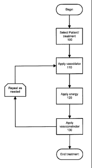
[0029] Fig. 1 is a flow diagram illustrating a method in accordance with an
embodiment
of the present invention.
[0030] Fig. 2 is a flow diagram illustrating a method in accordance with
another
embodiment of the present invention.
[0031] Fig. 3 is a chart showing the results of a clinical trial of an
embodiment of the
invention.
Detailed Description
[0032] Embodiments of the present invention relate to compositions and methods
for
treatment of skin disorders. In specific illustrative embodiments, the
compositions and
methods are used for the treatment of rosacea, and for the treatment of skin
disorders involving
vascular lesions. Embodiments of the invention can also be used in the
treatment of spider
veins, varicose veins, actinically damaged skin, venous hypertension,
Poikiloderma vasculare
atrophicans, vascular malformations, hemangioma and telangiectasia.
[0033] In specific embodiments, topically applied vasodilator agents increase
the blood
flow to a targeted area prior to treatment. A targeted energy source then
induces ischemia of
blood vessels in the area. A subsequent topical application of a
vasoconstrictor or anti-
inflammatory composition can then be applied to promote healing or ameliorate
discomfort.
[0034] In an embodiment, a composition of an embodiment of the invention can
be a
topical formulation, to be applied topically to the region of skin to be
treated. By way of
example, a vasodilation composition or a vasoconstrictor composition can be in
any delivery
form known to those of ordinary skill in the art as appropriate for
application to the skin,
7
CA 02727710 2010-12-10
WO 2009/152372 PCT/US2009/047098
including a cream, lotion, ointment, foam, or gel. In other embodiments, a
composition of the
invention can be applied by a patch.
[0035] Fig. 1 shows a flow chart in accordance with an embodiment of the
invention.
A patient with rosacea or other disorder characterized by one or more abnormal
blood vessels is
selected as a suitable candidate for treatment (step 100). A vasodilator is
applied (e.g., rubbed
into) to an affected skin region of the patient (step 110). The vasodilator
may be a topically
applied composition which contains an active ingredient that causes
vasodilatation of a blood
vessel. Depending on the vasodilation composition used and other relevant
factors, a
vasodilation time is allowed for the blood vessel or vessels to become dilated
(e.g., 5 to 10
minutes). This vasodilation time can be a fixed amount of time, or sufficient
time can be
allowed, e.g., while the patient is being monitored, for indications that the
blood vessels have
dilated, prior to proceeding to the treatment step of inducing ischemia, e.g.,
by introducing
energy.
[0036] In one embodiment, the vasodilator active ingredient in the
vasodilation
composition can be L-arginine, which may be included in a concentration range
of 0.001% to
10%, e.g., .01% to 10.0% w/w, with the balance made of inactive ingredients or
other active
ingredients.
[0037] In the alternative or in combination with L-arginine, the vasodilation
composition can include other active vasodilation ingredients known to those
of ordinary skill
in the art. Examples of such vasodilating agents include ginger extract,
ginkgo biloba,
hawthorne extract, bamethan sulphate, bencyclane fumarate, benpurodil
hemisuccinate, benzyl
nicotinate, buflomedil hydrochloride, buphenine hydrochloride, butalamine
hydrochloride,
cetledil citrate, ciclonicate, cinepazide maleate, cyclandelate, di-
isopropylammonium
dichloroacetate, ethyl nicotinate, hepronicate, hexyl nicotinate, Ifenprodil
tartrate, inositol
nicotinate, isoxsuprine hydrochloride, kallidinogenase, methyl nicotinate,
maftidropuryl
oxalate, nicametate citrate, niceritrol, nicobuxil, nicofuranose, nicotinyl
alcohol, nicotinyl
alcohol tartrate, nonidamide, oxpentifylline, papaveroline, pentifylline,
pipratecol,
propentofylline, raubasine, suloctidil, teasuprine, thymoxamine hydrochloride,
tolazoline,
xanthinol nicotinate, diazoxide, hydralazine, minoxidil, centrally acting
agents including
clonidine, quanaberz and methyl dopa, alpha-adrenoceptor agents including
indoramin,
phenoxybenzamine, phentolamine and prazosin, adrenergic neuron blocking agents
including
8
CA 02727710 2010-12-10
WO 2009/152372 PCT/US2009/047098
bethanidine, debrisoquine and guanethidine, ACE inhibitors including
benazepril, captopril,
cilazapril, enalapril, fosinopril, lisinopril, perindopril, quinapril and
ramipril, ganglion-blocking
agents including pentolinium and trimetaphan, calcium-channel blockers
including amlodipine,
diltiazem, felodipine, isradipine, nicardipine, nifedipine, nimodipine and
verapamil,
prosteglandins including prostacyclin, thrombuxane A2 leukotrienes, PGA, PGA1
PGA2 PGE1
PGE2 PGD, PGG and PGH, and angiotension II analogs including saralasin. Other
suitable
vasodilators include nitroglycerin, labetalol, thrazide, isosorbide dinitrate,
pentaerythritol
tetranitrate, digitalis, hydralazine, diazoxide and sodium nitroprusside, in a
concentration range
of 0.001 to 10.0% w/w, e.g., 0.01% to 10.0% w/w.
[0038] The vasodilation composition can be administered in a formulation that
further
includes substances that improve penetration or bioavailability of the active
vasodilation
ingredients. For example, the vasodilation composition can include a
combination of
phospholipids, fatty acids, chemical penetration enhancers and binding
components that result
in enhanced diffusion of the active ingredient into and through the stratum
corneum.
[0039] The non-active ingredients of the vasodilation formulation can be
selected from
those known in the art, including distilled water, urea, propylene glycol,
acrylates/c 10-30 alkyl
acrylate crosspolymer, allantoin, DMDM hydantoin, methylparaben, and
excipients known to
those skilled in the art as useful for formulating pharmaceutical preparations
in a concentration
range of 0.001% to 10.0% w/w., e.g., 0.01% to 10.0% w/w.
[0040] After vasodilation, energy is introduced into the dilated blood vessels
(step 120).
The energy can be in the form of electromagnetic radiation, such as pulsed and
non-pulsed laser
light or non-coherent light, e.g., at a wavelength range of 30 to 1100
nanometers, or, e.g., a
wavelength of 500 to 1100 nanometers. One process of using light energy to
induce selective
heating and ischemia is known in the art as photothermolysis, where light
energy is absorbed
by chromophores in hemoglobin and oxyhemoglobin and is converted to heat. This
selective
heating increases the temperature in the red blood cells in the endothelium of
the blood vessel
wall causing ischemia, cell death and re-absorption of the blood vessel by the
body.
[0041] Alternatively, energy can be introduced in the form of ultrasound or in
the form
of radio-frequency electromagnetic radiation. The applied energy is introduced
with the
objective of inducing ischemia of one or more blood vessels in the affected
skin region.
9
CA 02727710 2015-11-04
[0042] In some embodiments, energy is supplied in the form of visible light
that is
preferentially absorbed by the blood vessel or its contents. For example, the
light can be of a
wavelength that is preferentially absorbed by hemoglobin carried in the blood
vessels. The
light energy absorbed by the hemoglobin is converted to thermal energy,
thereby raising the
local temperature and causing ischemia, cell death and eventual re-absorption
of the blood
vessel by the body. For example, the source of the light can be a pulsed dye
laser, diode laser,
Er:YAG laser, Nd:YAG laser, xenon flash lamp, alexandrite laser, semiconductor
diode, copper
vapor laser, argon ion laser, krypton ion laser, or other suitable light
and/or laser source known
to those skilled in the art.
[0043] Multiple photothermolysis treatments are often required due to eruption
of new
vascular lesions and reperfusion of energy-treated vessels. Reper-fusion is
the restoration of
blood supply to tissue which is ischemic. Reperfusion is undesirable as it
reduces the
effectiveness of the treatment.
[0044] Additional methods, techniques, and compositions are known to the art;
see,
e.g., U.S. 2006/0217690 Al, "Method for Treating Various Dermatological and
Muscular
Conditions Using Electromagnetic Radiation", U.S. 6,306,130 Bl,
"Apparatus and Methods for Removing Blood Vessels".
[0045] Without wanting to be bound by scientific explanation, as a result of
applying
the vasodilator before irradiation, a larger number of blood cells may be
present in the blood
vessel, thereby increasing the efficiency of treatment. Additionally, the
vasodilation step may
make the application of energy more efficient with regard to induction of
ischemia due to
thinning of the vessel walls. Vasodilation may also increase the ability of
the applied energy to
target smaller vessels that might otherwise evade treatment. If left
untreated, the smaller
vessels would cause unwanted future pathologies, requiring further treatment
and additional
expense.
[0046] The process of Fig. 1 may be repeated as necessary.
[0047] Fig. 2 shows a flow chart in accordance with another embodiment of the
invention. In this embodiment, after applying energy, a vasoconstrictor is
applied (step 130).
Like the vasodilator, the vasoconstrictor may be applied as a topical
formulation. The
application of the vasoconstrictor can confer one or more of the benefits of
enhancing
CA 02727710 2010-12-10
WO 2009/152372 PCT/US2009/047098
degradation of ischemic blood vessels, promoting healing, and alleviating
pain. The
vasoconstrictor formulation may also include anti-inflammatory, anti-edemic,
analgesic, or
other substances known in the art to promote healing or to enhance patient
comfort.
[0048] In one embodiment, the vasoconstrictor composition formulation can
include
phytonin. Preferably, phytonin is present in a vasoconstrictor formulation
within a
concentration range of 0.001% to 10.0% w/w., e.g., 0.01% to 10.0% w/w.
[0049] In the alternative or in combination with phytonin, the
vasoconstriction
composition can include other active vasoconstriction ingredients known to
those of ordinary
skill in the art. Examples of such vasoconstricting agents include phenyl-
epinephrine and
caffeine. Additional examples of vasoconstricting agents include arnica
extract, cypress
extract, Solomon's seal extract, nymphaea alba flower extract, butcher's broom
extract,
grapefruit oil, pomegranate and bugleweed extract, in a concentration range of
0.001% to
10.0% w/w., e.g., 0.01% to 10.0% w/w.
[0050] The vasoconstrictor formulation can also include one or more inactive
ingredients, such as aloe vera juice, distilled water, cetyl alcohol, glyceryl
stearate/PEG-100
stearate, sodium palmitoyl proline, sodium PCA, cyclopentasiloxane,
dimethicone
crosspolymer and other excipients known to those skilled in the art for use in
formulating
pharmaceutical preparations.
[0051] The vasoconstrictor formulation can further include substances to
improve
penetration or bioavailability of the active vasoconstrictor ingredients. For
example, the
formulation may include one or a combination of phospholipids, fatty acids,
chemical
penetration enhancers, and binding components that result in enhanced
diffusion of the active
ingredient into and through the stratum corneum. Substances which improve the
penetration or
bioavailability of active ingredients in topical formulations are known to
those of ordinary skill
in the art.
[0052] Optionally, the process of Fig. 1 or Fig. 2 can be repeated one or more
times.
Alternately, steps 110 and 120 may be repeated for a given number of cycles
prior to applying
vasoconstrictor (step 130).
[0053] Optionally, subsequent to step 130, the vasoconstrictor formulation can
be
re-applied after the process of Fig. 1 or Fig. 2 is complete to help maintain
the benefits of the
treatment. For example, a healthcare provider may instruct a patient to apply
a vasoconstrictor
11
CA 02727710 2010-12-10
WO 2009/152372 PCT/US2009/047098
composition on a periodic basis after treatment (e.g., one or more times
daily). The
vasoconstrictor formulation used subsequent to the first application of step
130 can be the same
formulation as used in vasoconstriction treatment step 130, or can be one or
more of the
different vasoconstrictor formulation embodiments described above.
[0054] Example 1: A clinical study was undertaken in order to demonstrate the
effectiveness of the method of Fig. 2 in improving the outcome of laser/photo
facial treatments.
In the study, 16 patients with moderate facial redness served as their own
test subject and
control.
[0055] Prior to treatment, the right and left side of the face were digitally
photographed
using the VISIATM Complexion Analysis System (Canfield Scientific, Fairfield
NJ). The
VISIATM system has the ability to visualize skin conditions related to
abnormal melanin
concentrations or vascular disorders. Visualized abnormalities include
conditions such as sun
damage, rosacea, melasma, telangiectasia and others.
[0056] For the clinical trial in this example the vasodilator composition
contained
water, arginine HC1, urea, glycerin, hydroxyethylcellulose, allantoin,
methylisothiazolinone.
The percentage of each reagent in the vasodilation composition is shown in
Table 1 on a
weight/weight basis. This vasodilation composition had a pH of 5.1.
Table 1:
Component Percentage (w/w)
Allantoin 0.25
Optiphen MIT 0.12
Arginine HCI 2.6
Ginger Extract H5955 WS 6
Ginkgo Biloba Extract H5957 WS 6
Hawthorn Extract H5958 WS 6
Urea 6
Glycerin 5
Cellosize QP52000 0.6
Water 67.43
Total: 100
[0057] In all cases, a layer of pre-energy treatment vasodilating composition
was
applied to the left side of the face and gently massaged into the skin. No pre-
treatment
composition was applied to the right side. After 10 minutes of waiting for
vasodilation to occur,
each patient was treated with either a broad-band light source centered at 560
nm or a Nd Yag
12
CA 02727710 2010-12-10
WO 2009/152372 PCT/US2009/047098
Laser at 1064 nm on both sides of the face. Treatments generally lasted for 15
minutes. The
irradiation energy varied from 13 to 160 Joules.
[0058] The wavelength of the irradiation was determined by a variety of
factors,
including the type of flushing the patient experiences, the individual's
tolerance for pain and
the patient's response to the treatment. For example, if the patient had
discrete veins that could
be traced for treatment, the 1064 wavelength was used since that is a pinpoint
laser used for
tracing vessels. If the patient had more flushing than discrete vessels, the
skin was irradiated
with pulsed light at 560 nm because this treatment is believed to be more
effective toward
flushing.
[0059] The energy selected for the treatment was selected based on patient
tolerance
and wavelength, with patient pain tolerance being the primary factor. The
energy goal was 14-
16 Joules for the 560 initial treatment. When using the 1064 wavelength, the
starting point was
generally 160 Joules. If the patient tolerated the higher wavelengths well and
the practitioner
believed that the treatment was not providing an ideal response, the energy
level was increased.
Often patient discomfort prevents treating at higher energy level without
topical anesthetic.
Energy treatments for each patient are set forth in Table 2.
Table 2:
Patient No. Joules Wavelength
1 14 560
2 14 560
3 15 560
4 13 560
160 1064
6 14 560
7 160 1064
8 160 1064
9 160 1064
14 560
11 14 560
12 155 1064
13 14 560
14 14 560
14 560
16 13 560
*560 = BBL Handpiece
*1064 = Nd Yag Laser
13
CA 02727710 2010-12-10
WO 2009/152372 PCT/US2009/047098
[0060] Immediately after the treatment the post-treatment vasoconstricting
composition
was applied to the left side of the face and gently massaged into the skin. No
vasoconstricting
composition was applied to the right side.
[0061] The vasoconstrictor composition used in the example clinical trial
contained
phytonin, aloe vera juice, arnica extract, cypress extract, Solomon's seal
extract, glyceryl
stearate and peg-100 stearate, sodium palmitoyl proline, nymphaea alba flower
extract,
cyclomethicone, stearic acid, glycerin, cetyl alcohol, butcher's broom
extract, bugleweed
extract, triethanolamine, pomegranate oil, allantoin, methylisothiazolinone,
grapefruit oil,
acrylates/c 10-30 alkyl acrylate crosspolymer. As detailed in Table 3, below,
the composition
was formulated from four parts and had a pH of 6.5. The percentage of each
reagent in the
vasoconstriction composition is shown in Table 3 on a weight/weight basis.
Table 3:
Percentage
Part A (w/w)
Cetyl Alcohol 1
Arlacel 165 4
Stearic Acid 2
Sepicalm VG 3
Pomegranate Oil 0.3
Part B
DC 345 3
White Grapefruit Oil 0.1
Part C
Butchers Broom Sol. 1
Bugleweed Sol. 1
Phytotonin 10
Optiphen MIT 0.12
Part D
Allantoin 0.25
Pemulen TR-1 0.1
Glycerine 2
TEA 1
Aloe Juice 71.13
Total: 100
[0062] Each patient was advised to continue to apply the vasoconstricting
composition
to the left side of the face and gently massage it into the skin twice a day
for four weeks.
[0063] At the end of four weeks, the patients returned to the clinic to have
digital
photographs taken of both the left and right sides of the face using the
VISIATM imaging
system described above.
14
CA 02727710 2010-12-10
WO 2009/152372 PCT/US2009/047098
[0064] Outcomes were measured as a percentage of enhanced reduction of rosacea
or
broken capillaries by patient questionnaire, and by measuring the appearance
of vascular
lesions on the left (treated) side of the patient's face as compared to the
right (untreated) control
area of the patient's face using the VISIATM system.
[0065] The patients each filled out a questionnaire specifically designed to
allow
patients to evaluate their overall improvement. The results are illustrated in
Table 4.
Table 4: Results of Patient Reporting
% of Patients
Reporting Result
Reporting a reduction in rosacea-induced facial redness 93%
Reporting a reduction in post treatment swelling and 80%
redness
Reporting that skin felt smoother 93%
Reporting a reduction in pimples/breakouts 87%
Reporting an allergic reaction 0%
[0066] The data was also analyzed by the software included with the VISIA TM
imaging
system; the results are illustrated in Fig. 3 and in Table 5.
[0067] Fig. 3 is a chart showing the results of a clinical trial of an
embodiment of the
invention, each column to be understood as (1) patient number; (2) the date of
VISIATM
measurement prior to treatment; (3) the date of treatment; (4) the date of
VISIATM
measurement after treatment; (5) number of reds in treated (left) area before
treatment;
(6) number of reds in treated (left) area after treatment; (7) percent change
in number of reds in
treated (left) area before versus after treatment; (8) evaluation of
improvement in treated (left)
area by VISIATM; (9) evaluation of improvement in treated (left) area by
photograph;
(10) number of reds in untreated (right) area before treatment; (11) number of
reds in untreated
(right) area after treatment; (12) percent change in number of reds in
untreated (right) area
before versus after treatment; (13) evaluation of improvement in untreated
(right) area by
VISIATM; (14) evaluation of improvement in untreated (right) area by
photograph; and
(15) evaluation of treated versus untreated areas. "Number of reds" refers to
the quantification
of the VISIATM Red Image Analysis, where 'Red' indicates vascular structures.
CA 02727710 2010-12-10
WO 2009/152372 PCT/US2009/047098
Table 5: Results of VISIATM COMPUTER ANALYSIS
Improvement in facial redness with applied compositions 21%
Improvement in facial redness without applied compositions 13%
Increased effectiveness of the laser treatment 62%
Reduction in facial flushing when compared to side without 110%
applied composition treatments
Overall increase in effectiveness of the treatment 62-110%
[0068] Patients stated that they healed faster on the treated side of their
faces, felt that
the texture of the skin was improved on the treated side and felt less
discomfort after the laser
procedure on the treated side of the face.
[0069] The outcome of the clinical study demonstrates the effectiveness of the
vasodilating and vasoconstricting compositions described herein as agents to
improve the
outcome of laser treatments for reducing facial redness. This may result in a
reduction in
rosacea symptoms and an increase in patient satisfaction.
[0070] Example 2: The compositions of the invention can be supplied in the
form of a
kit. The kit can be supplied to a patient, or to a health care provider or
cosmetologist treating a
patient. The kit can include, without limitation, a first topical skin
formulation including a
vasodilation composition, and a second topical skin formulation including a
vasoconstriction
composition. The kit can include written instructions for use, by or on a
patient, of the first
topical skin formulation prior to receipt by the patient of an ischemia-
inducing treatment and of
the second topical skin formulation after receipt by the patient of an
ischemia-inducing
treatment. Other components can optionally be added to the kit, such as
topical compositions
to be applied while inducing ischemia, such as, e.g., a pain medication in a
topical formulation.
Vasodilation compositions and vasoconstriction compositions useful in such a
kit are prepared
according to the methods set forth above, and are preferably supplied in the
kit in an amount
suitable for a single regimen. In another embodiment, the kit can further
include a energy
source device, e.g., a laser, a high-intensity light, or an ultrasound
transducer.
[0071] The embodiments of the invention described above are intended to be
merely
exemplary; numerous variations and modifications will be apparent to those
skilled in the art.
All such variations and modifications are intended to be within the scope of
the present
invention as defined in any appended claims.
16