Note: Descriptions are shown in the official language in which they were submitted.
CA 02728385 2010-12-17
- 1 -
DEVICE FOR INVESTIGATION OF A FLOW CONDUIT
Technical Field
[0001] The present disclosure relates to devices for investigation of a flow
conduit. In
particular, this disclosure relates to devices, such as chip-based or lab-on-a-
chip devices,
that may be suitable for investigation of a small-sized flow conduit, such as
perfusable
soft material samples or small viable or non-viable biological vessel
segments.
Background
[0002] High blood pressure, or hypertension, is a deadly condition that is
reaching
epidemic proportions. The global burden of hypertension is expected to
increase by 60%
from 26.4% (972 million people) in 2000 to 29.2% (1.56 billion people) by
2025.
[Kearney, P.M. et al., Lancet, 2005. 365(9455): p. 217-223]. Although
hypertension is
traditionally viewed as a disease of aging, it is now prevalent in young
adults, with
several genetic and lifestyle factors contributing to its incidence and
severity.
Hypertension is a major risk factor for many diseases, including heart
disease, stroke, and
kidney failure. Since even at present our understanding of hypertension still
does not
encompass its inherent complexity, the vast majority of hypertensive patients
are treated
symptomatically, rather than causally. Knowledge regarding hypertension should
be
advanced in order to improve this situation. There is a growing consensus that
hypertension is primarily linked to an elevated peripheral vascular resistance
originating
primarily from small resistance arteries in the terminal parts of the vascular
tree.
[0003] Current knowledge regarding blood vessel structure and function is
primarily
derived from experiments using large non-resistance arteries, which are more
easily
accessible. Unfortunately, functional differences exist between large conduit
and small
resistance arteries as well as between resistance arteries from different
vascular beds.
Small resistance vessels are understudied, largely due to the considerable
technical skills
required to handle them experimentally. Since a better understanding of
mechanisms that
regulate resistance artery structure and function is key to improved
strategies to treat
hypertension, technologies that facilitate the handling of resistance arteries
are needed.
CA 02728385 2010-12-17
- 2 -
Similar challenges arise in attempting other investigations with such small
arteries, for
example in researching structural responses to other stimuli such as
pharmaceuticals.
These challenges are also present in investigations of other similar flow
conduits, such as
small tubules found in the lungs, pancreas, and others.
[0004] Current methods and processes use cell-based screens, genetic analysis
and
pharmacological tools combined with animal models to identify, test and assess
safety
and efficacy of a potential drug product. Consequently, the process is
relatively long and
only approximately one in a thousand pre-clinical identifications achieves
success before
being proposed for human trials.
[0005] Current methods are often time consuming, require care and training for
the
investigator, and often result in a low percentage of useable vessels for
investigation.
There remains the challenge of providing an efficient and standardized way to
investigate
these small flow conduits. It would be useful for a solution to these
challenges to be
applicable to other biological flow conduits, and artificial or engineered
flow conduits.
Summary
[0006] It would be desirable to provide a device that allows for investigation
of flow
conduits which addresses at least some of the challenges described above. It
would be
desirable if such technology could be scalable. Also desirable is a method or
device for
monitoring these flow conduits for their responses to treatment, for example
their
response to a pharmaceutical compound.
[0007] This disclosure describes a device for investigation of a flow conduit.
This device
allows flow conduits, including small viable or non-viable biological conduits
(e.g.,
resistance arteries) to be reversibly or irreversibly loaded, fixed and
perfused under
physiological conditions. This device may allow fixation and perfusion of
human-,
animal-, and plant-derived flow conduits or artificial conduits on a chip or
microdevice,
for example a microfluidics chip or a lab-on-a-chip device. This device may
provide a
relatively optimized microenvironment for functional analysis and organ
culture of flow
CA 02728385 2010-12-17
,
,
- 3 -
conduits, the automation of the relatively difficult conduit cannulation
process, and the
capability to perform routine studies with small and fragile conduits.
[0008] This device may allow structural and response testing of flow conduits,
for
example in the identification of treatment products. This device may be used
to test flow
conduits from animals, humans, plants, and other organisms. The flow conduits
may be
from any organ, and may include artificial or engineered conduits. A flow
conduit may
include conduits found in organisms, such as lipid tubules, engineered
vessels, hollow
fibers, arteries, arterioles, veins, venules, lymphatic vessels, intestines,
vas deferens,
ovaric tubes, bile ducts, bronchial tubes, bronchiole, trachea, or any other
similar
structures, as well as structures found in plants. The device may also allow
for targeted or
personalized treatment of either an individual or groups of individual by
using their
representative conduits in screening for or assessment of certain drugs,
diseases,
conditions, or treatments.
[0009] The device may be scalable and/or multiplexed, may be handled by
relatively
minimally trained personnel and may reduce the cost per experimental unit
compared to
other devices commonly used for these studies. By allowing uniform handling,
regardless
of the skill set of the user, this device may promote standardization. In
contrast,
previously developed conventional experimental procedures for resistance
artery isolation
and culture [e.g., as disclosed in Bolz SS et al., J Vasc Res, 2003. 40(4): p.
399-405; and
Bolz SS et al., Am J Physiol Heart Circ Physiol., 2000. 279(3): p. H1434-9]
typically
require relatively highly skilled personnel trained in micro-dissection
techniques and
specialized equipment.
[0010] In some aspects there is provided a device for investigation of a flow
conduit
comprising: a base; and a module formed in the base, the module comprising: a
main
channel for the flow conduit, the main channel having a loading inlet for
loading the flow
conduit; a culture chamber in the main channel for at least one of perfusion
and
superfusion of the flow conduit; at least two fixation lines in communication
with the
main channel for providing fixation of the flow conduit at at least two
fixation locations
along the length of the flow conduit.
CA 02728385 2015-02-06
- 4 -
[0010A] In one aspect there is provided a device for investigation of a
substantially tubular
biological flow conduit comprising:
a base having formed therein;
a loading inlet for loading the flow conduit into the device;
a main channel for receiving the flow conduit from the loading inlet, the main
channel
having a flow path along a first directional axis;
a culture chamber in the main channel;
at least two fixation lines fluidly connected to the main channel for
providing fixation of
the flow conduit at at least two fixation locations along the length of the
flow conduit within the
culture chamber so that when fixed the flow conduit is substantially
longitudinally aligned with
the flow path along the first directional axis;
the main channel having a perfusion inlet and a perfusion outlet, one of which
is located
before the at least two fixation lines along the flow path along the first
directional axis and the
other of which is located after the at least two fixation lines along the flow
path along the first
directional axis; and
a superfusion channel fluidly connected to the main channel between the
fixation
locations, the superfusion channel having a flow path along a second
directional axis at the point
of connection to the main channel.
[0011] In some examples, there may be a plurality of modules formed in the
base. In some
examples, the modules may be arranged in series, and the modules may share a
common main
channel. In some examples, the modules may be arranged in parallel, and the
modules may share
a common culture chamber.
[0012] In some examples, the device may further comprise an actuator embedded
in the base,
and the actuator may create a deformation of the base at least between the two
fixation locations.
[0013] In some examples, the device may further comprise a lysis chamber in
the main channel,
and the lysis chamber may be in series with the culture chamber and may be
adapted to receive at
least a portion of the flow conduit from the culture chamber.
[0014] In some examples, the at least two fixation lines may allow reversible
or irreversible
fixation of the flow conduit.
[0015] In some examples, the main channel may have an outlet for extracting
the flow conduit
for analysis.
CA 02728385 2015-02-06
- 4a -
[0016] In some examples, the module may accommodate flow conduits having
diameters in the
range of about 3 micrometers to about 2,000 micrometers, for example in the
range of about 15
micrometers to about 300 micrometers.
[0017] In some examples, the flow conduit may have a length in the range of
about
micrometers to about 1.5 centimeters.
[0018] In some examples, the device may contain active compounds that are
released over time.
[0019] In some examples, the base may comprise a biodegradable material.
[0020] In some examples, the base may comprise a material selected from the
group consisting
of: polymers, biopolymers, glass, semiconductors, metals, ceramics, and
combinations thereof.
For example, the polymer may be selected from the group consisting of:
poly(dimethylsiloxane),
polystyrene, poly(methyl methacrylate), and combinations thereof. For example,
the biopolymer
may be selected from the group consisting of: fibrinogen, collagen, laminin,
and combinations
thereof. For example, the semiconductor may be selected from the group
consisting of: silicon
and gallium arsenide.
[0021] In another aspect, there is provided a method of investigating a flow
conduit comprising:
loading the flow conduit into a fluid channel, the fluid channel being fluidly
connected to at least
one microfluidic fixation line; fixing the flow conduit in the channel by
applying a fluid to or
withdrawing fluid from the channel via the at least one microfluidic fixation
line; perfusing or
superfusing the flow conduit with a physiological solution; and monitoring the
flow conduit over
time.
CA 02728385 2010-12-17
,
- 5 -
[0021] In some examples, the device may further comprise an interface adapted
to interface with
analytical equipment, such as bright field or fluorescence microscopy
techniques, including
fluorescence intensity and fluorescence lifetime-based imaging, with optical
spectroscopy, on-
chip lysis and mass spectrometry.
[0022] In some examples, the culture chamber may comprise a biopolymer.
[0023] In some examples, the device may be comprised of two or more layers,
and each layer
may provide at least a portion of the module or at least a portion of a
channel connection to the
module.
[0024] In some examples, the device may further comprise at least one of: a
processor, a
memory unit, or a temperature control unit.
[0025] In some aspects there is provided a method of investigating a flow
conduit
comprising: providing the device described above; loading the flow conduit
into the main
channel; fixing the flow conduit in the main channel, wherein at least a
portion of the
flow conduit is in the culture chamber; perfusing or superfusing the flow
conduit with a
physiological solution; and monitoring the flow conduit over time.
[0026] In some examples, the method may further comprise applying a biological
factor to the
flow conduit via the culture chamber and monitoring the flow conduit for a
response.
[0027] In some examples, the method may further comprise analyzing the flow
conduit using a
technique selected from the group consisting of: bright field or fluorescence
microscopy
techniques, fluorescence intensity and fluorescence lifetime-based imaging,
optical spectroscopy,
on-chip lysis and mass spectrometry.
[0028] In some examples, fixing the flow conduit may comprise applying a
pressure lower than
that in the culture chamber via the fixation lines.
[0029] In some examples, fixing the flow conduit may comprise applying a
bonding material via
the fixation lines.
CA 02728385 2010-12-17
,
- 6 -
[0030] In some examples, the bonding material may be selected from the group
consisting of: a
polymer that cross-links upon exposure to light, a polymer that cross-links
upon exposure to
moisture, and a polymer that cross-links in response to temperature changes.
[0031] In some examples, the method may further comprise applying a mechanical
stimulation
to the flow conduit along the axial axis of the flow conduit.
[0032] In some examples, monitoring the flow conduit may comprise taking
diameter
measurements using an integrated optical technique.
[0033] In some examples, the method may further comprise lysing the flow
conduit using an
enzymatic method.
[0034] In some examples, the method may be for investigation of angiogenesis,
wherein the flow
conduit may be a blood vessel, and the method may further comprise the step of
stimulating
angiogenesis by at least one of: mechanically rupturing the outer smooth
muscle cell layer, laser
ablation, and administration of an angiogenic factor. For example, the
angiogenic factor may be
selected from the group consisting of: endothelial cell growth factor (ECGF),
fibroblast growth
factor (FGF), angiogen, low molecular weight endothelial mitogens, endothelial
cell chemotactic
factors, lipids, vascular endothelial growth factor (VEGF), and platelet-
derived growth factor
(PDGF).
[0035] In some examples, the method may further comprise perfusing the flow
conduit with a
fluid containing particles or molecules, and assessing transport of the
particles or molecules
through the wall of the flow conduit and toxicity.
[0036] In some examples, the flow conduit may have a diameter in the range of
about
3 micrometers to about 2,000 micrometers, for example in the range of about 15
micrometers to
about 300 micrometers.
[0037] In some examples, the flow conduit may have a length in the range of
about
micrometers to about 1.5 centimeters.
[0038] In some examples, the flow conduit may be selected from the group
consisting of: brain
conduits, lung conduits, inner ear conduits, lipid tubules, engineered
vessels, hollow fibers,
CA 02728385 2010-12-17
- 7 -
arteries, arterioles, veins, venules, lymphatic vessels, intestines, vas
deferens, ovaric tubes, bile
duct, bronchial, bronchiole, tracheal conduits, ureter, urethra, pancreatic
duct, and kidney tubules.
[0039] In some examples, the method may be used for investigation of blood-
brain barrier, and
the brain conduit may be a blood vessel from a microvascular network of a
brain.
[0040] In some examples, the flow conduit may be a biological conduit having a
disease
condition selected from the group consisting of: infarcted, ischemic,
inflamed, sclerotic, immune
compromised, tumors-bearing, and metastatic.
[0041] In some examples, perfusion may be at a rate of about 0-500m1/hr or
superfusion may be
at a rate of about 0-500m1/hr.
[0042] In some examples, the monitoring may be performed automatically using a
computing
device.
[0043] In some examples, the method may further comprise transmitting
monitored data to an
external device for analysis.
[0044] The device may contain a plurality of modules (e.g., arranged in series
or in
parallel), and may additionally include a lysis chamber for lysing at least a
portion of the
flow conduit. This device may be useful investigation of structural and
functional
properties of small blood vessels. In addition, this device may be useful in
investigation
of angiogenesis and other conditions pertaining to blood vessels, as well as
other
biological or non-biological flow conduits. This device may be useful for
personalized
medicine, and for development of pharmaceutical products.
Brief Description of the Drawings
[0045] Reference will now be made to the drawings, which show by way of
example
embodiments of the present disclosure, and in which:
[0046] FIG. 1 illustrates schematically examples of loading a flow conduit in
example
embodiments of a module for a device for investigation of a flow conduit;
CA 02728385 2010-12-17
- 8 -
[0047] FIG. 2 shows images of an example embodiment of a device for
investigation of a
flow conduit;
[0048] FIG. 3 shows images of an example embodiment of a device for
investigation of a
flow conduit loaded with an artery;
[0049] FIG. 4 shows images of an example embodiment of a device for
investigation of a
flow conduit used in perfusion of an artery;
[0050] FIG. 5 illustrates schematically examples of fixation of flow conduits
in example
embodiments of a device for investigation of a flow conduit;
[0051] FIG. 6 illustrates schematically another example of fixation of flow
conduits in
example embodiments of a device for investigation of a flow conduit;
[0052] FIGS. 7-24 illustrate example embodiments of a device for investigation
of a flow
conduit having different layout designs;
[0053] FIGS. 25-27 illustrate example embodiments of a device for
investigation of a
flow conduit having a series design;
[0054] FIGS. 28-29 illustrate example embodiments of a device for
investigation of a
flow conduit having a parallel design;
[0055] FIG. 30 illustrates example embodiments of a device for investigation
of a flow
conduit having an integrated optical fiber;
[0056] FIG. 31 shows charts illustrating arterial responses to phenylephrine,
measured
using a device for investigation of a flow conduit;
[0057] FIG. 32 shows charts illustrating constriction of a mesenteric vessel
in a device
for investigation of a flow conduit; and
[0058] FIG. 33 shows an image and a chart illustrating ratio measurements on
an artery
in a device for investigation of a flow conduit.
CA 02728385 2010-12-17
- 9 -
[0059] It will be noted that throughout the appended drawings, like features
are identified
by like reference numerals.
Detailed Description
[0060] A device for investigation of a flow conduit is described. This device
may provide
at least (i) a relatively optimized microenvironment for functional analysis
and organ
culture of biological flow conduits, (ii) automation of an otherwise
relatively difficult
vessel cannulation process, and (iii) a capability to routinely study very
small and fragile
conduits, such as resistance arteries. These may be important elements in the
construction
of a human microcirculatory-based hypertension database, fed by laboratories
and
hospitals worldwide. This device may provide a potentially effective means of
establishing global standards in data collection from microvessels.
[0061] In general, this device has a base and a module etched, embedded,
molded, laser-
machined or otherwise formed in the base. The module comprises a main channel
for the
flow conduit, a culture chamber in the main channel for perfusion and/or
superfusion of
the flow conduit, and at least two fixation lines in communication with the
main channel
for fixing the flow conduit along its length. The main channel typically has a
loading inlet
for loading the flow conduit. In some examples, the loading inlet is connected
to a
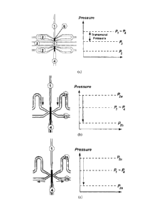
loading well formed on the device, to facilitate loading of the flow conduit.
[0062] Reference is now made to FIG. 1, showing schematically example
embodiments
of a module for a device for investigation of a flow conduit, in particular
showing
example methods of fixating a flow conduit in a module. Also illustrated are
charts
showing the pressure at different points in the device, a) shows a schematic
diagram of an
example embodiment for reversible fixation of the flow conduit, for example
using a low
pressure or suction method. As described above, the module has a main channel
1 with a
loading inlet 4. There are two pairs of fixation lines 2, one pair located at
each end of the
culture chamber 5, for fixing the ends of a flow conduit 6, such as a small
blood vessel.
Here, a culture channel 3 may feed to and from the culture chamber 5, for
example to
provide an organ bath to the flow conduit 6. The culture channel 3 may allow
for
superfusion of the flow conduit 6. In other example embodiments, the culture
chamber 5
CA 02728385 2010-12-17
- 10 -
may have an opening to the surroundings and may be fed directly through the
opening, in
which case the culture channel 3 may not be necessary. In this example, the
main channel
1, loading inlet 4, fixation lines 2, and/or culture channel 3 may be
microchannels.
[0063] The pressure profile beside the schematic diagram illustrates the
relative pressures
acting on the flow conduit 6. In this example, the loading inlet 4 is open to
ambient
pressure, PA (for example, the loading inlet 4 may be open to a petri dish
from which the
flow conduit 6 was loaded). Alternatively, the module may be in a closed
configuration
for which P4 # PA may be realized (for example, the loading inlet 4 may be
attached to
the channel of another module, which will be described below).
[0064] For this example, the flow conduit 6 may be loaded by first closing the
culture
channel 3 and the fixation lines 2. A syringe pump controlling the vessel
loading and
perfusion processes may be connected to the main channel 1, opposite to the
loading inlet
4, and operated in the "withdraw" or suction mode. The flow conduit 6 may be
thus
drawn toward its final position in the culture chamber 5. Once the flow
conduit 6 has
reached the desired position, its further movement may be prevented by a
sufficient
narrowing of the main channel 1 and/or by stopping the withdraw process
through the
main channel 1. The syringe pump may be then switched off. At both ends of the
culture
chamber 5, a suction pressure, which may be pre-defined, may be applied by the
fixation
lines 2 (P2). This may be by connecting the fixation lines 2 to a liquid-
filled tube that is
connected to a hydrostatic pressure level lower than the culture chamber 5.
Considering
the typically short length of the flow conduit 6 and the slow perfusion rates,
there may be
typically negligible different in the pressures at each end of the blood
vessel 6
(i.e., p,:.-, P4). While suction pressure may be applied at the fixation lines
2, the pressure
difference (i.e., Pi ¨ P2 and 134-P2) may provide a relatively efficient and
reversible
fixation mechanism at both vessel ends. Thus, by low pressure, vacuum or
suction is
meant that the pressure applied at the fixation lines 2 is lower than the
pressure at the
inside and outside of the flow conduit (e.g., in the culture chamber). This
method is not
necessarily limited to the use of a vacuum or suction source. The flow conduit
6 may then
be investigated. For example, the fixed flow conduit 6 may be superfused via
the culture
CA 02728385 2010-12-17
- 11 -
channel 3 and/or perfused via flow through the culture chamber 5 (e.g., flow
from the
inlet 4 through the main channel 1). A transmural pressure (e.g., Pi ¨ PO may
be
established across the wall of the flow conduit 6. To release the flow conduit
6 from the
module, the pressure in the fixation lines 2 may be increased to Pl. This may
release the
reversible seals at both vessel ends and the flow conduit 6 may be unloaded
through the
loading inlet 4.
[0065] Referring still to FIG. 1, b) and c) show an example of the device for
irreversible
loading of a flow conduit, for example fixation using polymerization or tissue
adhesives.
The module of b) and c) also has a main channel 1 with a loading inlet 4, a
culture
chamber 5 with culture channel 3, as described above. This module has fixation
lines 2a
and 2b that have a slightly different arrangement, which is described below.
[0066] Rather than fixing the flow conduit 6, such as a blood vessel, using a
suction
method as in a), the example of b) and c) may irreversibly fix the flow
conduit 6 using an
irreversible bonding agent, such as a polymer (e.g., a polymer that cross-
links upon
exposure to light, contact with moisture (such as a tissue adhesive), or
temperature
changes(such as fibrin or MatrigelTm), or a solidifying chemical reaction that
is otherwise
induced. The uncured polymer or tissue adhesive may be introduced through
fixation
lines 2a (e.g., at a pressure exceeding P1), for example at a constant flow
rate with a
syringe pump, resulting in an elevated inlet pressure. In this example
embodiment, to
prevent the situation where the culture chamber 5 is flooded with the bonding
agent, a
constant flow rate may be removed at fixation lines 2b. This may ensure that
the flow
conduit 6 is fixed only at one desired location. The bonding agent may be
cured where it
contacts the flow conduit 6, for example by using UV light on a photo-polymer,
or
simply by contact with tissue. Once curing starts, the feeding flow from
fixation lines 2a
may be stopped. A similar procedure may be used to fix both ends of the flow
conduit 6.
The flow conduit 6 may be then investigated. For this example embodiment, the
flow
conduit 6 may not be releasable from the module. For example b) and c)
illustrate the
introduction of a bonding agent, for irreversibly fixing the flow conduit 6.
b) illustrates
the introduction of a bonding agent (in grey), such as a tissue adhesive, into
fixation lines
2a. c) illustrates the continued introduction of the bonding agent and its
removal through
CA 02728385 2010-12-17
- 12 -
fixation lines 2b. As shown in c), the bonding agent selectively contacts the
flow conduit
6 only at distinct points, allowing the lumen of the flow conduit 6 to remain
open. The
bonding agent may cure or solidify at the point of contact, for example
through a
chemical reaction such as upon contact with moisture, thus irreversibly fixing
the flow
conduit 6.
[0067] This device may be suitable for the study of various flow conduits in
animals and
humans, including vessels isolated (e.g., through biopsies) from the brain,
lung, inner ear
and other organs. Aside from vessels, other flow conduits may be accommodated
or
studied using the device. Other possible flow conduits include lipid tubules,
engineered
vessel grafts, hollow fibers, arteries, arterioles, veins, venules, lymphatic
vessels,
intestines (e.g., duodenal, jejunal, ileal, and colon), vas deferens, ovaric
tubes, bile duct,
bronchial, bronchiole, tracheal conduits, ureter, urethra, pancreatic duct,
and kidney
tubules. Vessels found in plants, such as in the xylem, may also be studied
using this
device. Artificial or engineered flow conduits may also be studied, for
example
engineered blood vessels.
[0068] The flow conduits may range in size from about 3 micrometers to about
2,000
micrometers in diameter, more specifically from about 15 micrometers to about
300
micrometers, and from about 10 micrometers to about 1.5 centimeters in length.
The
conduits may be isolated from healthy or diseased tissue, for example to study
vessels
that are infarcted, ischemic, inflamed, sclerotic, immune-compromised, from
tumors, or
metastatic tissues.
[0069] The flow conduit may be perfused at a rate of about 0-500m1/Iu-, or
superfused at
about 0-500m1/hr. By "perfusion" is meant the movement of fluid through the
lumen of
the flow conduit; by "superfusion" is meant the movement of fluid along or
over the
outside of the flow conduit, whether axially along the length of the flow
conduit or
transversely around the circumference of the flow conduit. Both types of fluid
movement
may be present in the culture chamber. Both perfusion and superfusion may be
useful in
providing nutrients and other compounds (e.g., soluble factors, dyes or
pharmaceutical
agents) to and from the flow conduit in the culture chamber. In some cases,
either
CA 02728385 2010-12-17
- 13 -
perfusion or superfusion might be more preferable. The device may include
sensors to
sense and measure perfusion and/or superfusion. For example, pressure drop
sensors may
be integrated on the device (e.g., using piezoresistive pressure transducers),
which may
provide indication of perfusion and/or superfusion. Although the device is
described as
providing for superfusion and/or perfusion of the flow conduit, it should be
understood
that the device may also be used where there is neither superfusion nor
perfusion, or
where the flow conduit is placed under static flow conditions. The culture
chamber may
also be referred to as a "perfusion chamber" or "superfusion chamber", and the
culture
channel may also be referred to as a "perfusion channel" or "superfusion
channel"; such
references do not limit the use of these components of the device to only
perfusion or
superfusion, nor is the culture channel and culture chamber limited to
delivering culture
medium.
[0070] The module may be etched, embedded, molded or otherwise formed in the
base
using common fabrication methods such as replica molding, hot embossing,
injection
molding, lithography (e.g., X-ray lithography), electroplating, molding (e.g.,
LIGA), dry
and wet etching, abrasive jet machining, and laser machining. Other standard
soft-
lithographic techniques may also be suitable [for example, as described in
Xia, Y.N. et
al., Annual Review of Materials Science, 1998. 28: p. 153-184.]. Standard soft-
lithographic techniques may be used in a variety of materials, for example
silicones (e.g.,
poly(dimethylsiloxane) (PDMS)). Typically, the channels and structures of the
module
may be etched, embedded, molded or otherwise formed on the surface of one half
of the
base. That surface may then be bonded against the other half of the base, for
example
using techniques such as free-radical surface activation in a plasma and
subsequent
bonding, solvent bonding, compression bonding, or anodic bonding. Other common
methods and variations for making microdevices may be suitable. The device may
be
made from single-layer designs, or from two- or multi-layer designs, in which
each layer
provides at least a portion of the module or at least a portion of a channel
connection to
the module. Multi-layer designs may be useful in reducing the necessary size
of the
device, and may be designed and manufactured using any suitable method, for
example
as described in U.S. Patent Publications Nos. 2001/0029983, 2001/0033796,
2001/0054778, 2002/0029814, 2003/0019833.
CA 02728385 2010-12-17
- 14 -
[0071] The device may be made from polymers (e.g., poly(dimethylsiloxane)
(PDMS),
polystyrene, poly(methyl methacrylate) (PMMA), and biopolymers such as
fibrinogen,
collagen, laminin and combinations thereof), glass, semiconductors (e.g.,
silicon or
gallium arsenide), metals, ceramics, and combinations thereof The device may
be made
from a biodegradable material. For example, the device may be made from a
biopolymer,
such as MatrigelTM, which may be useful for investigation of angiogenesis.
[0072] Typically, at least a portion of the flow conduit, when fixed in the
device, is
viewable or detectable, so that changes to the conduit may be monitored and/or
measured. The module is typically etched, embedded, molded or otherwise formed
in the
base, such that most of the module is enclosed (e.g., with the exception of
inlets and
outlets), however portions of the module may also be open. For example, the
culture
chamber may be at least partially open, so that an investigator can apply
compounds to or
otherwise stimulate the flow conduit directly.
[0073] In addition to the culture chamber, the module may include a lysis
chamber (not
shown). The lysis chamber may be provided in the main channel, in series with
the
culture chamber. The lysis chamber may be similar to the culture chamber,
having
respective fixation lines and a lysis channel. In practice, a flow conduit may
be released
from the culture chamber (e.g., where the flow conduit is reversibly fixed)
and driven
downstream (e.g., by applying a high pressure at the loading inlet) until it
reaches the
lysis chamber, where it may again be fixed by fixation lines. Alternatively,
the lysis
chamber may not have respective fixation lines, but may be large enough to
accommodate the entire conduit. Alternatively, lysing may be formed in flow
(i.e.,
without fixation of the conduit). The conduit may be lysed by introducing
lysing
compounds such as enzymes via the lysis channel. The resulting cellular and/or
subcellular material may then be extracted from the device through an outlet
in the main
channel or through the loading inlet. Alternatively, lysing of the flow
conduit may occur
without using a lysis chamber, for example by introducing lysing compounds
into the
culture chamber. The lysis chamber may also receive only a portion of the flow
conduit.
For example, a portion of the flow conduit may be removed for lysing, such as
removal
by laser machining, suction or other suitable means. The lysis chamber may
also be
CA 02728385 2010-12-17
- 15 -
adapted to fit only a portion of the flow conduit, so that only the portion
contained in the
lysis chamber is lysed. Allowing only a portion of the flow conduit may be
useful for
investigating a certain desired portion of the flow conduit, for example only
the smooth
muscle cells of the flow conduit.
[0074] The device may have different depths for the various channels. For
example, there
may be two different channel depths at the conduit fixation points and the
culture
chamber. This may prevent unwanted contact between the center part of the
conduit and
the top or bottom walls inside the device.
[0075] The device may interface with analytical instruments, including
microscopy (e.g.,
fluorescence or bright-field microscopy), mass spectrometry, or
electrophoresis. The
device may also be designed to interface with equipment for fluorescence
intensity and
fluorescence lifetime-based imaging, optical spectroscopy, on-chip lysis, or
mass
spectrometry. For example, the loading inlet or another outlet connected to
the main
channel may be designed to be easily connected to other analytical
instruments, such that
the flow conduit or lysed material in the device may be extracted directly
into the
analytical instrument.
[0076] By connecting syringe pumps (e.g., for the culture channel and/or
inlet) to a
computer (e.g., using serial ports: RS232, RS423, RS 485, firewire, USB,
etc.), perfusion
and/or superfusion processes may be automated. The response of the flow
conduit (e.g.,
transmural pressure and/or artery contractile state) may be directly recorded
on the
device, for example by providing a processor (e.g., a microprocessor) and/or a
memory
unit on the device. This may allow mobile and self-contained investigation and
analysis
using the device. The device may include a temperature control unit (e.g., a
thermoelectric or resistive element), or channels for a cross-flowing stream
of constant-
temperature fluid, may allow the temperature of the fixed flow conduit to be
controlled
(e.g., maintained at physiological levels) during investigation or culture of
the flow
conduit in the device. With a flow conduit fixed in the device, the
temperature may be
lowered, for example to 4 C. This may, for example, allow a flow conduit
fixed in the
device to be transported before or after being investigated. The dimensions of
the device
CA 02728385 2010-12-17
- 16 -
may be reduced, or other instruments and components may be added for
additional
functionality. Where the device has communication with a computer or other
computing
device, monitoring of the flow conduit may be performed automatically. Where
the
device includes a memory, recorded data, for example data from monitoring or
investigation of the flow conduit, may be stored in the device. This stored
data may be
transmitted, wired or wirelessly, to an external device such as a workstation
or external
computer for analysis.
[0077] This device may be used for investigation of small flow conduits (e.g.,
resistance
arteries) or large flow conduits (e.g., mesenteric arteries). Some flow
conduits may
require mechanical stimulation to remain viable. For example, mesenteric
arteries need to
be stretched longitudinally during culture (e.g., by up to 200 micrometers for
a 1 mm
long mesenteric artery segment). Such mechanical stimulation may be provided
via the
culture chamber. The ends of the flow conduit may be attached to manipulators,
to
mechanically stretch the flow conduit. Such manipulators may be integral to or
external
to the device. Alternatively or in addition, a mechanical actuator may be
attached or
embedded on the device.
[0078] In an example embodiment, the base of the device may be relatively
compliant or
elastic so that it is deformable. Stretching of the base in a direction
aligned with the
length of a fixed flow conduit may translate to mechanical stretching of the
flow conduit.
This stretching of the base may be provided by an integrated piezoelectric
bending
actuator located at one end of the flow conduit and designed to bend away from
the flow
conduit, thus causing a length-wise stretch. Such an actuator may be
fabricated into the
base of the device, or may be attached on the surface of the device. Other
similar
mechanical actuators may be used.
[0079] This device may provide complete environmental control over the flow
conduit
while maintaining its structural and functional integrity for extended periods
of time (e.g.,
days or more). Using this device, properties of flow conduits, including
contractile,
ionic, electrical, molecular and/or structural properties, may be monitored
and
investigated.
CA 02728385 2010-12-17
,
- 17 -
[0080] In some example embodiments, the device may include compounds to be
administered to the flow conduit. For example, the device may include active
compounds
that are released over time into the culture chamber. In other examples,
compounds in the
device may be administered to a fixed flow conduit by manual or automated
mechanisms.
Examples
Example of single-module device
[0081] Reference is now made to FIG. 2, showing images of an example
embodiment of
a device for investigation of a flow conduit, in use with an artery segment.
In this
example, the device was fabricated using multilayer soft-lithographic
techniques. A
resistance artery segment, approximately 1 mm in length, was introduced
through an
inlet, which may be connected to a loading well.
[0082] In this example, as shown in FIG. 2 a), the cylindrical artery segment
was guided
by pressure-driven flow through inlet "A" to the culture chamber, or artery
inspection
area. The artery was then fixed by applying a suction pressure at fixation
lines "E".
Subsequently, the artery segment was subjected to a microenvironment that
mimicked
physiological conditions by: (i) selectively superfusing the outside arterial
wall with
stream "B-C" (e.g., via the culture channel), (ii) perfusing the inside of the
artery (i.e.,
lumen) with stream "A¨>D" (e.g., via the main channel), (iii) controlling the
differential
pressure across the arterial wall and (iv) adjusting the temperature to 37 C.
Flow rates,
pressures and compositions of the superfusing/perfusing streams could be
independently
adjusted. Crosstalk between the perfusion and superfusion lines was prevented
by the
fixation lines "E".
[0083] Referring still to FIG. 2, b) shows the device right after a previously
isolated
vessel or vessel segment is loaded into the culture chamber. Note that the
vessel is
pressurized and is still filled with blood. Pressurization to 100 mmHg
initiated flow
through the lumen of the vessel so that over time, the intraluminal blood was
replaced by
saline solution. c) shows the blood vessel with an open lumen after being
perfused with
saline and after a defined transmural pressure was applied. In this example,
the device is
CA 02728385 2010-12-17
- 18 -
also referred to as a chip, and the culture chamber is also referred to as an
organ bath
(OB).
[0084] This example embodiment of the device has outer dimensions of 75mm (L)
x 25mm (W) x 4mm (H), which is typically a size that allows inspection with
common
upright or inverted brightfield or fluorescence microscopes. Standard soft-
lithographic
techniques were used to translate microchannel designs from computer-aided
design
(CAD) files to printed transparency masks. Spincoated layers of negative
photoresist (in
this example, SU825TM and SU82050TM, from MicroChem, Newton, MA) were pre-
baked, exposed using the transparency masks, post-baked and subsequently
developed.
The inverse microchannel patterns were transferred to poly(dimethylsiloxane)
molds. The
optically transparent elastomeric mold was peeled off the master, cut, bonded
to another
elastomer layer or a pre-cleaned glass slide using an 02 plasma. Multi-layer
lithography
allowed accommodation of two different channel depths at the fixation points
and the
culture chamber. Unwanted contact between the center part of the artery and
the
top/bottom walls of the device was thereby prevented.
[0085] Device fabrication is not limited to PDMS/glass as the structural
materials, nor
soft lithography as the microfabrication method. Any suitable materials and
methods for
microfabrication, as commonly known, may be used. Alternatively, the device
may be
fabricated using semiconductor materials, for example silicon using
established bulk
silicon machining techniques. Fabrication using glass may be accomplished by
using wet
and/or dry etching as well as laser machining techniques. Fabrication using a
wide range
of polymers including biocompatible and degradable polymer matrices may also
be
possible. Possible polymer device fabrication techniques include replica
molding, hot
embossing, injection molding, abrasive jet and laser machining.
[0086] In this example, the device was connected to 1/16" outer diameter (OD)
polymer
(e.g., Tygon , PEEK, Teflon ) tubing comprising fluid perfusion, superfusion
and waste
lines through either an epoxy connection or a reversible manifold (e.g., a
compression
seal). The 1/16" OD polymer tubes were further connected with reversible
fluidic unions
(such as from Upchurch Scientific, Oak Harbor, WA) in order to conveniently
remove
CA 02728385 2010-12-17
- 19 -
the device from its connections to fluid-filled vials and/or manually
controlled syringe
pumps. The different fluid lines allowed for loading the flow conduit into the
device, for
providing perfusion through the inside of the flow conduit and superfusion of
the
conduit's outside wall in the culture chamber.
[0087] An example process of loading a flow conduit, in this case an artery,
into this
example embodiment of the device is now described. The device was initially
flushed
with a Bovine serum albumin (BSA) solution to prevent any unwanted adhesion of
arteries to the walls of the main channel or culture chamber. Previously
isolated
resistance arteries (typically 1-2 mm long and 30-200 micrometers in diameter)
were
loaded into the device through a 200 micrometers wide and 150-200 micrometers
deep
loading inlet (e.g., where the device layout does not include a loading well)
or from a
loading well (e.g., where the device layout does include a loading well
connected to the
inlet). As an alternative to the reversible fixation of the artery used here,
an irreversible
fixation method may be used, as discussed above.
[0088] Upon fixation (e.g., using a reversible fixation method), the lumen of
the artery
was perfused by operating a syringe pump, connected to an inlet of the culture
channel, in
the perfusion mode. By connecting the inlet and outlet of the culture channel
to different
hydrostatic pressure levels, a flow through the culture chamber was achieved
while
maintain a defined transmural pressure difference across the wall of the
artery. A flow
through the culture chamber was achieved while maintaining a defined
transmural
pressure difference (i.e., P, ¨ P, in FIG. 1) by superfusing the culture
chamber with a
second syringe pump that operates in the perfusion mode, connected at the
inlet of the
culture channel. The outlet of the culture channel was thus led to a reduced
hydrostatic
pressure level. The artery interior and exterior walls were subjected to a
defined
transmural pressure and perfused with 3-(N-morpholino)propanesulfonic acid
(MOPS
buffer) containing defined concentrations of active molecules.
[0089] FIG. 3 shows images of an example embodiment of the device loaded with
an
artery. a) shows a mesenteric artery cannulated on glass micropipettes, held
in place with
sutures, and pressurized to 45 mmHg, using a conventional method. b) shows the
CA 02728385 2010-12-17
- 20 -
mesenteric artery loaded into the culture chamber of an example device,
pressurized to 45
mmHg, and held in place with suction applied via the fixation lines, which in
this
example are syringe-controlled low pressure channels.
[0090] FIG. 4 shows images of an example embodiment of the device
demonstrating its
use in perfusion of an artery. A resistance artery was loaded into the device
and perfused
with rhodamine dye, which shows red, through the lumen of the artery, and
superfused
with fluorescein dye, which shows green, in the culture bath. a) shows the
artery at a
baseline level of fluorescein. At 0 seconds, a syringe pump was used to infuse
the artery
with fluorescein at a flow rate of 4 mL/h. b) shows the artery at 4 seconds,
when the
fluorescein reaches the extra lumenal space. c) shows the artery at 5 seconds,
when the
artery is further exposed to the fluorescein. d) shows the artery at 6
seconds, when the
artery is fully bathed in fluorescein. These images also demonstrate a
separation of the
fluids flowing in the vessel's intralumenal space (i.e., perfusion) from those
outside of the
vessel (i.e., superfusion).
Example layouts
[0091] Certain designs and layouts have been prepared for this device and are
shown in
the figures described below. Where appropriate, the modules are enlarged to
show
details. These examples are for the purpose of illustration only and are not
intended to be
limiting.
[0092] In the examples described here, where there are two or more layers,
external
connections (e.g., to syringe pumps or low pressure sources) are indicated
with a hole
having an "X" and connections between upper and lower layers are indicated
with a hole.
In these schematics, inlets and outlets to the culture chamber may be referred
to as "organ
bath" or "perfusion" or "drain", inlets and outlets to the fixation lines may
be referred to
as "suction". In no way do any of these labels limit the possible connections
and
functions of these inlets and outlets or their various channels. For example,
an inlet
labeled as "suction" may be used to apply suction for reversible fixation of
the flow
conduit, but may also be used to administer a bonding material for
irreversible fixation of
the flow conduit.
CA 02728385 2016-07-06
- 21 -
[0093] Although not shown, the device may have a multi-layer layout in which
one or
more etched channels or chambers in the multiple layers are touching - that
is, the
channels or chambers of the upper and lower layers are not separated, but are
continuous
between the layers. This may provide for some deeper channels or chambers
while
keeping other channels or chambers on the device relatively shallow. A
variation on this
design may allow for even deeper chambers and/or channels by increasing the
number of
layers to three or more.
[0094] FIG. 5 illustrates schematically examples of fixation of flow conduits
in example
embodiments of the device. Here, reversible fixation techniques are
demonstrated, with
dashed circles marking the fixation locations, a) and b) show examples of
designs having
single fixation lines at each end of the culture chamber. c) and d) show
examples of
designs having multiple fixation lines at each of the culture chamber.
[0095] FIG. 6 is a schematic of an example device layout in which a flow
conduit may be
irreversibly fixed in the device. In particular, in a), the flow conduit (6)
is reversibly fixed
at both ends (8) so that the inside and outside areas of the conduit are
separated from each
other. At the outside areas, the flow conduit may further be embedded with a
polymer (7).
FIGS. 6(b) and (c) show magnified views of fixation points. Focused light may
be
guided, for example through an embedded waveguide or optical fiber, and
focused onto
one section of the flow conduit. This arrangement may be useful for the
selective removal
and subsequent analysis of samples from the flow conduit, and may also be
useful for
studying the formation of vascular networks in healthy and diseased blood
vessels at
defined conditions, as will be discussed in further detail below.
[0096] FIG. 7 is a schematic of an example device layout, showing a single-
layer layout
without a loading bath. The flow conduit enters the device through the main
channel that
extends to the device bottom corner. The two fixation lines may be
individually or
separately accessed or addressible. There is one culture channel for a
superfusion stream
and one inlet for a perfusion stream. The flow rates and pressures of both
streams may be
separately adjusted/controlled. The superfusion stream flows across the flow
conduit.
[0097] FIG. 8 is a schematic of an example device layout, showing a two-layer
layout
with a loading bath for loading a flow conduit. The upper image shows the top
fluidic
CA 02728385 2010-12-17
- 22 -
layer, and the bottom image shows the bottom fluidic layer of the device. The
flow
conduit enters the bottom layer of the device through a loading well into the
main
channel. The two fixation points at each end of the module are individually
addressible.
There is one culture channel for a superfusion stream and one inlet for a
perfusion stream.
The flow rates and pressures of both streams may be separately
adjusted/controlled. The
superfusion stream flows across the flow conduit.
[0098] FIG. 9 is a schematic of an example device layout, showing a two-layer
layout
with a loading bath for loading a flow conduit. The upper image shows the top
fluidic
layer, and the bottom image shows the bottom fluidic layer of the device. The
flow
conduit enters the bottom layer of the device through a loading well into the
main
channel. The two fixation points at each end of the module are individually
addressible.
There is one culture channel for a superfusion stream and one inlet for a
perfusion stream.
The flow rates and pressures of both streams may be separately
adjusted/controlled. The
superfusion stream flow is first split before the two substreams flow along
the flow
conduit and are then guided from the device in separate outlets. In general,
the
superfusion stream may be split into two substreams, which may then be guided
to enter
left and right sides of the culture chamber simultaneously. This design may
ensure a
relatively rapid, gradient-free replacement of the contents in the culture
chamber, and
may also allow for stimulation of both sides of the flow conduit.
[0099] FIG. 10 is a schematic of an example device layout, showing a two-layer
layout
without a loading bath. The upper image shows the bottom fluidic layer, and
the bottom
image shows the top fluidic layer of the device. The flow conduit enters the
bottom layer
of the device at the bottom side through the main channel. The two fixation
points at each
end of the module are individually addressible. There is one culture channel
for a
superfusion stream and one inlet for a perfusion stream. The flow rates and
pressures of
both streams may be separately adjusted/controlled. The superfusion stream
flows is first
split before the two substreams flow along the flow conduit and are then
guided from the
device in separate outlets.
CA 02728385 2010-12-17
- 23 -
[00100] FIG. 11 is a schematic of an example device layout, showing a single-
layer
layout without a loading bath. The flow conduit enters the bottom layer of the
device at
the bottom side through the main channel. The two fixation points at each end
of the
module are individually addressible. There is one culture channel for a
superfusion
stream and one inlet for a perfusion stream. The flow rates and pressures of
both streams
may be separately adjusted/controlled. The superfusion stream flows is first
split before
the two substreams flow along the flow conduit and are then guided from the
device in
separate outlets.
[00101] FIG. 12 is a schematic of an example device layout, showing a two-
layer layout
without a loading bath. The upper image shows the top fluidic layer, and the
bottom
image shows the bottom fluidic layer of the device. The flow conduit enters
the bottom
layer of the device at the bottom side through the main channel. The two
fixation points
at each end of the module are individually addressible. There is one culture
channel for a
superfusion stream and one inlet for a perfusion stream. The flow rates and
pressures of
both streams may be separately adjusted/controlled. The superfusion stream
flows is first
split before the two substreams flow along the flow conduit and are then
guided from the
device in separate outlets.
[00102] FIG. 13 is a schematic of an example device layout, showing a single-
layer
layout without a loading bath. The flow conduit enters the device at the
bottom side
through the main channel. The two fixation points at each end of the module
are
individually addressible. There is one culture channel for a superfusion
stream and one
inlet for a perfusion stream. The flow rates and pressures of both streams may
be
separately adjusted/controlled. The superfusion stream flows across the flow
conduit and
is then guided from the device in separate outlet.
[00103] FIG. 14 is a schematic of an example device layout, showing a single-
layer
layout without a loading bath. The flow conduit enters the device at the
bottom side
through the main channel. The two fixation points at each end of the module
are
individually addressible. There is one culture channel for a superfusion
stream and one
inlet for a perfusion stream. The flow rates and pressures of both streams may
be
= CA 02728385 2010-12-17
- 24 -
separately adjusted/controlled. The superfusion stream flows is first split
before the two
substreams flow along the flow conduit and are then guided from the device in
separate
outlets.
[00104] FIG. 15 is a schematic of an example device layout, showing a single-
layer
layout with a loading bath for loading a flow conduit. This example may be
suitable for
investigation of 150 gm conduits, such as a 150 gm blood vessel. The flow
conduit enters
the device through a loading well into the main channel. The two fixation
points at each
end of the module are individually addressible. There is one culture channel
for a
superfusion stream and one inlet for a perfusion stream. The flow rates and
pressures of
both streams may be separately adjusted/controlled. The two superfusion
streams are first
mixed by diffusion before they are split into two equal substreams that then
flow along
the flow outside of the conduit and are guided from the device in a joint
outlet.
[00105] FIG. 16 is a schematic of an example device layout, showing a single-
layer
layout with a loading bath for loading a flow conduit. This example may be
suitable for
investigation of 120 gm conduits, such as a 120 gm blood vessel. The flow
conduit enters
the device through a loading well into the main channel. The two fixation
points at each
end of the module are individually addressible. There is one culture channel
for a
superfusion stream and one inlet for a perfusion stream. The flow rates and
pressures of
both streams may be separately adjusted/controlled. The two superfusion
streams are first
mixed by diffusion before they are split into two equal substreams that then
flow along
the flow outside of the conduit and are guided from the device in a joint
outlet.
[00106] FIG. 17 is a schematic of an example device layout, showing a two-
layer layout
with a loading bath for loading a flow conduit. The left image shows the
bottom fluidic
layer, and the right image shows the top fluidic layer of the device. The flow
conduit
enters the device through a loading well into the main channel. The two
fixation points at
each end of the module are individually addressible. There are two culture
channel for a
superfusion stream and one inlet for a perfusion stream. All individual flow
rates of the
superfusion/perfusion streams and the pressure in the resulting total
superfusion stream
and the perfusion stream may be separately adjusted/controlled. The two
superfusion
CA 02728385 2010-12-17
- 25 -
streams are first mixed by diffusion before they are split into two equal
substreams that
then flow along the flow outside of the conduit and are guided from the device
in a joint
outlet.
[00107] FIG. 18 is a schematic of an example device layout, showing a single-
layer
layout with a loading bath for loading a flow conduit. In this example, there
is one
common perfusion line and two separate superfusion lines to the left and right
sides of
the culture chamber. The flow conduit enters the device through a loading well
into the
main channel. The two fixation points at each end of the module are
individually
addressible. All individual flow rates of the superfusion/perfusion streams
and the
pressure in the resulting total superfusion stream and the perfusion stream
may be
separately adjusted/controlled. On each side along the axis of the flow
conduit, two
superfusion streams, which may be different, are first mixed by diffusion
before they
flow at opposite sides along the outside of the conduit.
[00108] FIG. 19 is a schematic of an example device layout, showing a two-
layer layout
with a loading bath for loading a flow conduit. In this example, there is a
common
perfusion line and two separate superfusion lines to the left and right sides
of the culture
chamber. The left image shows the bottom fluidic layer, and the right image
shows the
top fluidic layer of the device. The flow conduit enters the device through a
loading well
into the main channel that is contained in the bottom layer. The two fixation
points at
each end of the module are individually addressible. All individual flow rates
of the
superfusion/perfusion streams and the pressure in the resulting total
superfusion stream
and the perfusion stream may be separately adjusted/controlled. On each side
along the
axis of the flow conduit, two superfusion streams, which may be different, are
first mixed
by diffusion before they flow at opposite sides along the outside of the
conduit.
[00109] FIG. 20 is a schematic of an example device layout, showing a single-
layer
layout with a loading bath for loading a flow conduit. In this example, there
is a common
perfusion line. This layout may allow for a step change in the concentration
of the
superfusing stream applied in the axial direction. The flow conduit enters the
device
through a loading well into the main channel. The two fixation points at each
end of the
CA 02728385 2010-12-17
- 26 -
module are individually addressible. All individual flow rates of the
superfusion/perfusion streams and the pressure in the resulting total
superfusion stream
and the perfusion stream may be separately adjusted/controlled. Two
superfusion
streams, which may be different, are first mixed by diffusion before they are
subjected to
different sections along the axis of the flow conduit. For viable flow
conduits, this design
may allow the creation of a microenvironment that may not be found in the
conduits
physiological environment.
[00110] FIG. 21 is a schematic of an example device layout, showing a two-
layer layout
with a loading bath for loading a flow conduit. In this example, there is a
common
perfusion line. This layout may allow for a step change in the concentration
of the
superfusing stream applied in the axial direction. The left image shows the
bottom fluidic
layer, and the right image shows the top fluidic layer of the device. The flow
conduit
enters the device through a loading well into the main channel that is located
in the
bottom layer. The two fixation points at each end of the module are
individually
addressible. All individual flow rates of the superfusion/perfusion streams
and the
pressure in the resulting total superfusion stream and the perfusion stream
may be
separately adjusted/controlled. Two superfusion streams, which may be
different, are first
mixed by diffusion before they are subjected to different sections along the
axis of the
flow conduit. For viable flow conduits, this design may allow the creation of
a
microenvironment that may not be found in the conduits physiological
environment.
[00111] FIG. 22 is a schematic of an example device layout, showing a two-
layer layout
with a loading bath for loading a flow conduit. In this example, there is a
common
perfusion line. This layout may allow the concentration in the superfusion
stream to be
varied over time by diffusive mixing of two sub-streams. The left image shows
the
bottom fluidic layer, and the right image shows the top fluidic layer of the
device. The
flow conduit enters the device through a loading well into the main channel
that is
located in the bottom layer. The two fixation points at each end of the module
are
individually addressible. All individual flow rates of the
superfusion/perfusion streams
and the pressure in the resulting total superfusion stream and the perfusion
stream may be
separately adjusted/controlled. Two separate superfusion streams are first
mixed by
CA 02728385 2010-12-17
- 27 -
diffusion before they meet the outside of the flow conduit. Two separate
perfusion
streams are first mixed by diffusion before they meet the inside of the flow
conduit.
[00112] FIG. 23 is a schematic of an example device layout, showing a two-
layer layout
with a loading bath for loading a flow conduit. In this example, there are two
perfusion
lines, allowing two perfusion streams to contact each other without mixing.
This layout
may allow the concentration in the superfusion stream to be varied over time
by diffusive
mixing of two sub-streams. The left image shows the bottom fluidic layer, and
the right
image shows the top fluidic layer of the device. The flow conduit enters the
device
through a loading well into the main channel that is located in the bottom
layer. The two
fixation points at each end of the module are individually addressible. All
individual flow
rates of the superfusion/perfusion streams and the pressure in the resulting
total
superfusion stream and the perfusion stream may be separately
adjusted/controlled. Two
separate superfusion streams are first mixed by diffusion before they meet the
outside of
the flow conduit. Two separate perfusion streams meet the inside of the flow
conduit at
opposite sides, with minimum diffusive mixing. For viable flow conduits, this
design
may allow for the creation of a microenvironment that may not be found in the
conduits
physiological environment.
[00113] FIG. 24 is a schematic of an example device layout, showing a single-
layer
layout with a loading bath for loading a flow conduit. In this example, there
is a common
perfusion line. There may be three different superfusing lines allowing
different
superfusing streams that may be mixed by diffusion. The flow conduit enters
the device
through a loading well into the main channel. The two fixation points at each
end of the
module are individually addressible. All individual flow rates of the
superfusion/perfusion streams and the pressure in the resulting total
superfusion stream
and the perfusion stream may be separately adjusted/controlled. Three separate
superfusion streams are first mixed by diffusion before they meet the outside
of the flow
conduit.
CA 02728385 2010-12-17
- 28 -
Multi-module designs
[00114] In addition to the single-module design, there may be a plurality of
modules
provided on a single device. The modules may be formed in the base in a series
arrangement, a parallel arrangement, a network arrangement, or other multiplex
arrangements.
Examples of modules in series
[00115] Two or more modules may be arranged in series on the device. Aside
from
having a common main channel and common loading inlet, each module may be
functionally similar to the single module described above. In some example
embodiments, the modules may be in series without sharing a common main
channel or
common loading inlet. Flow conduits may be serially loaded into each module
and fixed
using fixation lines at each module. Each module may share the same perfusion
pump,
superfusion pump and/or low pressure source, such that conduits fixed in each
modules
may be essentially subjected to the same conditions. Alternatively, conduits
in each of the
modules may be subjected to different conditions, such as by using separate
perfusion
pumps which may allow, for example, perfusing a compound to the culture
chamber of
one module, but not to any other.
[00116] Loading, fixation, investigation, and unloading of the flow conduits
may be the
same as discussed above. Typically, the flow conduits may be loaded in
sequence, with
the first flow conduit being loaded and fixed in the module farthest from the
loading inlet
before the next flow conduit is loaded into a closer module.
[00117] FIG. 25 is a schematic of an example device layout, showing a two-
layer layout
without a loading bath. There are two modules in series. Either two short flow
conduits or
one long flow conduit that extends over all fixation locations may enter the
device
through the main channel located in the bottom layer. In this example, there
is a common
perfusion line, individual fixation lines and two separate superfusing lines.
Individual
flow conduit or individual sections of the same flow conduit (e.g., in the
case of a long
enough flow conduit) may be superfused across its/their axis.
CA 02728385 2016-07-06
- 29 -
[001181 FIG. 26 is a schematic of an example device layout, showing a two-
layer layout
with a loading bath for loading a flow conduit. There are two modules in
series. Either
two short flow conduits or one long flow conduit that extends over all
fixation locations
may enter the device through the loading bath into the main channel located in
the bottom
layer. In this example, there is a common perfusion line, individual fixation
lines and two
separate superfusing lines. Individual flow conduit or individual sections of
the same
flow conduit (e.g., in the case of a long enough flow conduit) may be
superfused across
its/their axis.
[00119] FIGS. 27(a), (b) and (c) show different schematics of example device
layouts,
having single-layer layouts with a loading bath for loading a flow conduit.
These example
layouts have two modules in series. Either two short flow conduits or one long
flow
conduit that extends over all fixation locations enter the device through the
main channel
located in the bottom layer. In this example, there is a common perfusion
line, individual
fixation lines and two separate superfusing lines. (see FIG. 27(d), which is a
magnified
view of a portion of the layout shown in FIG. 27(c)) Individual flow conduit
or individual
sections of the same flow conduit (e.g., in the case of a long enough flow
conduit) may be
superfused across its/their axis.
[00120] Although these examples show only two modules series, it would be
clear to a
person skilled in the art that the device could be designed to have more
modules in series.
In some examples, the modules in series may share a common culture chamber.
That is, a
common longer culture chamber may be used with more than two fixation points
for
fixing multiple flow conduits or multiple portions of a flow conduit along the
length of
the culture chamber.
[00121] A series design of this device may be useful, for example in
performing
bioassays, and for studying healing processes. For example, in a bioassay,
pharmaceutical
agents may be administered to an upstream artery only (i.e., one closer to the
loading
inlet) and the effects on a downstream artery (i.e., one farther from the
loading inlet) may
be observed. For studying healing or joining of arteries, two or more separate
arteries
may be loaded in sequential modules and fixed with a small gap between
adjacent ends.
CA 02728385 2016-07-06
- 30 -
Growth and joining of the separate artery ends may be observed over time, as
well as the
effectiveness of various agents in promoting such growth.
Examples of modules in parallel
[00122] Two or more modules may be arranged side-by-side in parallel on a
single
device. The modules may have similar connections and may be functionally
similar to the
single module described above. The modules may share the same perfusion pump,
superfusion pump and/or low pressure source, such that flow conduits fixed in
each
module may be essentially subjected to the same conditions. Alternatively,
conduits in
each of the modules may be subjected to different conditions, such as by using
separate
perfusion pumps, which may allow, for example, perfusing a compound to the
culture
chamber of one module, but not to any other module on the device. The modules
may
share a common culture chamber and culture channel. That is, the culture
chambers and
culture channels of each of the modules may be connected together. Loading,
fixation,
investigation, and unloading of the flow conduits may be the same as discussed
above.
[00123] FIGS. 28(a), (b), and (c) show different schematics of example device
layouts,
having single-layer layouts without a loading bath for loading a flow conduit.
These
example layouts have two modules 42 and 44 in parallel. Two flow conduits may
enter
the device through individual main channels. In this example, there is a
common
perfusion line, individual fixation lines and separate superfusing lines. This
can be seen
in FIGS. 28(b) and (c), which show schematically in a magnified form two
alternate
layouts for such an arrangement. The two flow conduits fixed in each module
may be
superfused with separate streams across their axis.
[00124] FIG. 29 shows different schematics of example device layouts, having
two-layer
layouts with separate interconnected loading baths for loading several
parallel flow
conduits. The flow conduits may be loaded through individual main channels
that are
located in the bottom layer. These example layouts each have eight modules in
parallel.
[00125] Although only two or eight modules are shown in parallel, it would be
clear to a
person skilled in the art that the device could be designed to have different
numbers of
modules in parallel.
CA 02728385 2016-07-06
-31 -
[00126] A parallel design of this device may be useful in ensuring that the
flow conduits
being investigated are subjected to the same conditions at essentially the
same time. For
example, it might be desirable to obtain results from a large number of
arteries under the
same conditions (e.g., culture medium, flow conditions) at essentially the
same time for
statistical purposes. Fixing all the arteries in the same device in a parallel
arrangement
may allow all the arteries to be tested together at the same time, under the
same
conditions. A parallel arrangement may also be useful in automating testing
procedures,
as the parallel flow conduits may be stepped through one-by-one in progression
(e.g., in
an automated analyzer) simply by advancing the device module-by-module.
[00127] In addition to the series and parallel arrangements described above,
other
multiplex arrangements are possible. For example, the series and parallel
arrangements
may be combined to obtain an array arrangement of modules on a single device.
The
modules may also be arranged in a branching network, circular network, or any
other
desired arrangement. In all cases, some or all modules may have separate
culture
chambers or they may share culture chambers and culture channels. Some or all
modules
may have individual perfusion pumps, superfusion pumps, low pressure sources,
and
other inlets and outlets, or these may be shared among some or all modules.
The modules
may all employ the same fixation method (e.g., reversible or irreversible), or
they may
use different fixation methods.
Examples with integrated optical fiber
[00128] FIGS. 30(a) and (b) illustrate example embodiments of the device
having an
integrated optical fiber 30. Here, the example has a single-layer layout with
a loading
bath. Single-(30(a)) or multimode (30(b)) optical fibers may be inserted
through a
straight channel that connects, for example, to a corner of the device. Small
lenses may
be embedded in the module (e.g., as described in "PDMS 2D optical lens
integrated with
microfluidic channels: principle and characterization" Camou S, Fujita H,
Fujii T, Lab on
a Chip 3 (1), 40-45 2003) to focus the light emitted by the fiber to a
location within the
main channel.
[00129] This design may allow a laser light to be guided towards a fixed flow
conduit
via the optical fiber. For example, such a setup may be useful in angiogenesis
studies to
CA 02728385 2010-12-17
,
,
- 32 -
set precisely defined injuries as a prerequisite or initiator for subsequent
growth of
endothelial cells out of the lumen of a vessel. In this example, a single- or
multi-mode
optical fiber or waveguide 30 may be embedded in the device to guide light
from a laser
into the device. Pulse lasers (e.g., pulsed Nd:YAG or ultrafast pulse lasers)
or
continuous-wave lasers are possible laser sources. Light leaving the optical
fiber or
waveguide 30 may be focused by a lens in the device towards the vascular wall
where it
causes precisely defined injuries.
Applications
[00130] This device may be provided on a chip or microdevice, for example as a
microfluidic chip or a lab-on-a-chip. This may allow for miniaturization and
scaling of
many assays and tests, allowing for high-throughput. For example, the
procedure
described in WO 2003/078606 (Bolz) may be carried out using this device, and
may be
relatively easier and more efficiently performed, even by relatively less-
trained
technicians.
[00131] In general, the device may be used for investigating a flow conduit.
The flow
conduit may be loaded into the main channel and fixed in place with at least a
portion of
the conduit in the culture chamber. A physiological solution, which may
contain a
compound of interest such as a biological factor, may be perfused or
superfused over the
conduit. The flow conduit may then be monitored to investigate any responses.
[00132] Any of the methods discussed above may be used to analyze or monitor
the
flow conduit, including bright field or fluorescence microscopy techniques,
fluorescence
intensity and fluorescence lifetime-based imaging, optical spectroscopy, on-
chip lysis and
mass spectrometry. Monitoring may also be done by taking diameter
measurements, for
example using an integrated optical technique, such as a laser-optical
technique.
Research of blood vessels
[00133] The device may be combined with imaging techniques such as transient
Ca2+
imaging to obtain time-resolved recordings of the contractile state of a flow
conduit, such
CA 02728385 2010-12-17
>
,
- 33 -
as the artery, and Ca2+ responses. The device may include lysis capabilities
as described
above, and may be designed to interface to a mass spectrometer.
[00134] Standard characterization of blood vessels includes measurements of
the artery
tone and/or diameter that are performed at inverted bright field and
fluorescence
microscopes. This device may be combined with various types of bright field or
fluorescence imaging, including Ca2+ imaging to obtain time-resolved
recordings of the
contractile state of the artery and Ca2+ responses. Lysis capabilities may be
included as
well as automated interfaces to electrophoresis, fluorescence spectroscopy,
and mass
spectrometry.
[00135] One research interest is to map intracellular processes in vascular
smooth
muscle cells of the vascular wall. However, primary smooth muscle cells in
culture tend
to de-differentiate within hours from a contractile phenotype to a synthetic
phenotype.
One of the most prominent changes that are observed is the reorganization of
the actin-
based cytoskeleton. Thus, it may be desirable to use cellular models where
this de-
differentiation effect does not occur. Also desirable are advanced
experimental systems
that will allow the study of transport processes in intact tissues (e.g.,
fully differentiated
vascular smooth muscle cells in the microvascular wall). Tissue models are
typically
preferred over cell culture models, because they better reflect the in vivo
situation. The
experimental model of transfected isolated microvessels may provide a unique
framework to translate cell-based knowledge regarding intracellular transport
mechanisms into a whole organ system. However, in prior art setups, the use of
isolated
microvessels requires highly skilled personnel trained in micro-dissection
techniques,
specialized equipment and substantial time (e.g., for isolation and
cannulation processes).
The presently disclosed device may facilitate the fundamental experimental
procedures
and allow for a higher throughput.
[00136] Using this device, researchers may more easily and more efficiently
use isolated
vessels, for example to (i) test new innovations in optical technologies and
(ii) identify
critical microvascular transport proteins and their regulation patterns in a
complex
multicellular environment. In combination with access to human tissue, this
may allow
CA 02728385 2010-12-17
- 34 -
researchers to be in a position to correlate individual disease patterns
(e.g., clinical
diagnosis) with alterations in intracellular transport mechanisms in
microvessels isolated
from patient biopsies.
Examples
[00137] The device may be used to investigate responses of biological flow
conduits. It
has been found that investigation using the device produces results similar to
results
produced by other conventional methods. For example, FIG. 31 shows charts
illustrating
arterial responses to phenylephrine (PE), measured using the device. In this
example, PE
was applied to the exterior surface of the flow conduit, and the dose of PE
was changed
in a stepwise fashion. The measured changes in the flow conduit diameter are
shown in
the charts. a) shows the dose dependent response to PE, measured in mesenteric
arteries
using a conventional pipette cannulation setup. In this example, the maximal
outer
diameter constriction was 44.5 +/- 2.5 % at 3.0 M PE (n=5). b) shows the dose
dependent PE response (here, changes in inner and outer diameters of the flow
conduit)
measured using the device, which are similar to and essentially identical to
the results
using the conventional cannulation setup, with a maximal outer diameter
constriction of
42.2 +/- 3.8 % at 3.0 1.1M PE (n=5).
[00138] In another example, the device was used to investigate a mesenteric
vessel. FIG.
32 shows charts illustrating constriction of a mesenteric vessel in the
device. In this
example, a mouse mesenteric vessel was used, and a single does of PE was
administered.
The PE was applied to the exterior surface of the flow conduit. a) is a
representative
tracing of a mesenteric vessel's response to 3.0 uM PE, with a sustained
constriction of
41.8%, measured in freshly isolated arteries that were immediately subjected
to the
stimulation with PE. b) is a representative tracing of a single mesenteric
vessel kept in
culture for 24 hours on the device prior to testing the response to PE,
showing a sustained
constriction of 41.4% to 3.0 1.1M PE. The vessel investigated using the device
was still
viable after 24 hours and did not show a modified response to PE.
[00139] FIG. 33 shows a reconstructed fluorescence image and a chart
illustrating
ratiometric measurements, using a FURA-2 dye, which is a calcium-sensitive
dye, on a
= CA 02728385 2010-12-17
- 35 -
mesenteric artery in an example embodiment of the device. The smooth muscle
cells in
the artery are stained. A mouse mesenteric artery was used in this example. a)
shows the
mesenteric artery fixed on the device, and loaded with FURA-2 Ca2+ ratiometric
dye,
showing smooth muscle cells wrapped around the vessel circumference. b) is a
chart
showing FURA-2 ratiometric measurements from the fixed vessel, showing an
increase
in FURA-2 ratio upon stimulation with 3.0 M PE, and a return to baseline
following
washout. The chart shows increases in the F340nm/F380nni ratio for FURA-2 in
the vessel.
Angiogenesis Assay
[00140] The device may also be used to investigate angiogenesis in viable
biological
flow conduits. For example, the culture chamber may be filled with a
biopolymer (e.g.,
MatrigelTM, fibrinogen, etc.), in which a biological conduit, such as a blood
vessel, is
embedded. The blood vessel may receive culture medium or other compounds by
perfusion. Alternatively or in addition, the biopolymer may be superfused by
the culture
medium or other compounds, and these may then diffuse through the biopolymer
to reach
the blood vessel. In some cases, a depot of growth factors may be injected at
one
predefined spot of the biopolymer and allowed to slowly diffuse through the
biopolymer.
The gradient that is established this way may provide chemotactic guidance to
the
outgrowing cells of the blood vessel.
[00141] As described above, the vessel may be reversibly or irreversibly fixed
in the
device. Angiogenesis may be induced, for example by subjecting the vessel to
angiogenic
factors, or by locally injuring the vessel, for example by laser-ablation
using a laser-
ablation instrument. Healing or angiogenic activity of the vessel may then be
observed.
Suitable angiogenic factors may include endothelial cell growth factor (ECGF),
fibroblast
growth factor (FGF), angiogens, low molecular weight endothelial mitogens,
endothelial
cell chemotactic factors, lipids, vascular endothelial growth factor (VEGF),
and platelet-
derived growth factor (PDGF).
[00142] Other applications may include perfusing the flow conduit with a fluid
containing particles or molecules of interest, and assessing transport of the
particles or
molecules through the wall of the flow conduit and monitoring toxicity.
CA 02728385 2010-12-17
,
- 36 -
[00143] The device may also be used to investigate the blood-brain barrier.
For example,
the flow conduit may be a blood vessel from a microvascular network of a
brain. Flow
conduits that may be investigated include: brain conduits, lung conduits,
inner ear
conduits, lipid tubules, engineered vessels, hollow fibers, arteries,
arterioles, veins,
venules, lymphatic vessels, intestines, vas deferens, ovaric tubes, bile duct,
bronchial,
bronchiole, tracheal conduits, ureter, urethra, pancreatic duct, and kidney
tubules, among
others. The flow conduit may have a physiological condition to be
investigated, for
example it may be infarcted, ischemic, inflamed, sclerotic, immune
compromised,
tumors-bearing, or metastatic. Artificial or engineered flow conduits may also
be
investigated.
Research and Commercial Applications
Clinical Uses
[00144] The translation of knowledge from basic science to clinical
application is based
on access to human tissues (e.g., analysis of microvessels from biopsies). In
order to
successfully implement a translational approach, it would be desirable for
clinicians to be
attracted to the field of microvascular research. Access to human specimens
and their
respective patient records combined with the disclosed device and state-of-the-
art
diagnostic technologies may provide opportunities for breakthroughs in
understanding
and treating microvascular disease. This device may allow standardization of
experimental approaches in microvascular research since it may provide: (i)
optimized
microenvironment for functional vessel analysis and organ culture; (ii)
possible
automation of the difficult vessel cannulation process; and/or (iii)
capability to routinely
study very small and fragile arteries. For example, these are useful elements
in the
construction of a human microcirculatory-based hypertension database, fed by
laboratories and hospitals worldwide. The facilitation of the standardized
experimental
process using this device may attract more clinicians to actively participate.
CA 02728385 2010-12-17
- 37 -
Treatment Development
[00145] This device may provide for high-throughput screening responses at the
organ,
membrane and vascular conduit levels to treatment of drug products.
[00146] Research in blood vessels has the potential to improve quality of
life, and
increase economic activity. It may help to accelerate the identification of
genetic,
epigenetic, proteomic, cellular and molecular mechanisms of tone and/or
diameter
regulation in resistance arteries that predominantly contribute to the
regulation of
systemic blood pressure. The control of blood pressure is useful to prevent
the
development of cardiovascular diseases. Understanding its underlying molecular
mechanisms may significantly impact the development of new treatment
strategies. An
improvement of knowledge about hypertension and the related molecular
mechanisms
may benefit from investigative models that simulate the in vivo situation as
accurately as
possible. This device may provide such a model. It may help to identify new
targets for
treatments and may allow for their immediate verification on the same
platform. Thus,
this device may bring basic science discoveries to their clinical application
in less time.
[00147] This device may also make fundamental experimental procedures high-
throughput ready. Therefore, this device may be an attractive tool for target
identification, target validation, drug design and high-throughput screening
in the drug
development process. This device may be used for investigation of both animal-
and
human-based specimens. The device may also be used in a diagnostic tool, for
example to
directly correlate the cardiovascular health status of a given patient to the
functional state
of his/her microcirculation. This may provide a personalized approach to the
diagnosis
and specific treatment of microvascular pathologies, thus translating
fundamental
scientific knowledge into clinical applications. Thus, the device may help to
enable
personalized medicine.
[00148] This device may allow structural and response testing of flow
conduits, for
example in the identification of treatment products. This device may be used
to test flow
conduits from animals, humans, plants, and other organisms. The flow conduits
may be
from any organ, and may also include artificial or engineered conduits. The
device may
. CA 02728385 2010-12-17
- 38 -
allow for targeted treatment of either an individual or groups of individuals
by using their
representative conduits in screening for or assessment of certain drugs,
diseases,
conditions, or treatments.
[00149] It is desirable that important life-saving new drug products have
quick
regulatory approval in order to get to the market. Fast-track clinical trials
and registration
are critical parts of the process that ensure efficacy and safety. Devices and
methodologies to quickly identify target products in screening at a level that
is closer
representative of in vivo conditions is an area that may facilitate the
process. For
example, one area in health care that may benefit from a more representative
treatment
assessment in drug development is in the treatment of hypertension or high
blood
pressure. This device may provide a platform that satisfies this need.
[00150] Similarly, this device may aid in the development of compounds for use
in
plants and animals, as it provides a platform for testing of experimental or
new
compounds in various flow conduits.
Training
[00151] The disclosed device may allow researchers to target the vascular
problems,
such as the problem of microvascular dysfunction, its cellular and molecular
mechanisms
and its inherent risks for the health of the population, as broadly as
possible. The
involved technology may represent a change of paradigms, and standards in an
emerging
field of research. It may provide opportunities to recruit and train all
levels of research
trainees including undergraduate and graduate students in the highly
specialized field of
microvascular research. The technical and fundamental skills offered by this
training are
in great demand in universities, life science and medical research institutes,
and
biotechnical industries.
[00152] As described, the device may include variations such as reversible
fixation of
vessels (e.g., using a suction method) or permanent fixation of vessels (e.g.,
using a
photocurable polymer or tissue glue method). The tubular structures or
channels of the
device may be lipid tubules, hollow fibers, or other suitable structures. The
device may
CA 02728385 2016-07-06
- 39 -
also be designed to be interconnectable in a complex fluid network. The device
may be designed
with various layouts, with various arrangements of module(s) and channels.
[00153] This device may be useful in the pharmaceutical industry for target
identification, target
validation, molecular drug design and optimization, early stage toxicity test,
and/or proof of
concept for new drugs. Clinical applications for this device include
personalized medicine,
isolation of arteries and venules from patient biopsies to determine vasomotor
status (e.g., for
assessment of structural and functional characteristics of individual vessels
including correlation
with individual patient history), and pharmacogenetics. The device may also be
used to assess
the treatment of a targeted individual or group of individuals to a
pharmaceutical product or
treatment by using the treated flow conduit of the individual or
representative group of
individuals in the assessment, such as in pharmacogenetics. This device may
also be useful in
the crop protection industry for high-throughput testing of plants and plant
compounds.
[00154] The example embodiments of the present disclosure described above are
intended to be
examples only. Those of skill in the art may effect alterations, modifications
and variations to the
particular example embodiments without departing from the intended scope of
the present
disclosure. In particular, selected features from one or more of the above-
described example
embodiments may be combined to create alternative example embodiments not
explicitly
described, features suitable for such combinations being readily apparent to
persons skilled in the
art. The scope of the claims may be given the broadest interpretation
consistent with the
description as a whole.
CAN_DMS \102066987\1