Note: Descriptions are shown in the official language in which they were submitted.
CA 02728478 2011-01-21
39334/39335
(P-4804/P-4805)
SYSTEM AND METHOD FOR ANALYZING ANTIBIOTIC
SUSCEPTIBILITY OF BIOLOGICAL SAMPLES
BACKGROUND OF THE INVENTION
Field of the Invention:
The present invention relates to a system and method for analyzing samples,
such
as biological samples, to determine the susceptibility of the samples to
antimicrobial
materials, such as antibiotics. More particularly, the present invention
relates to a system
and method which takes a plurality of optical readings of a biological sample
contained in
sample wells of a sample test panel having various types and concentrations of
antimicrobial materials therein and, based on these readings, determines the
respective
minimum inhibitory concentrations (MICs) at which the respective antimicrobial
materials
will inhibit growth of the sample.
CA 02728478 2011-01-21
-2-
Description of the Related Art:
Many conventional systems exist for performing tests on microbiological
samples
related to patient diagnosis and therapy. The microbiological samples may come
from a
variety of sources, including infected wounds, genital infections, cerebro-
spinal fluids,
blood, abscesses or any other suitable source. From the microorganism samples,
an
inoculum is prepared in accordance with established procedures which produce a
bacterial
or cellular suspension of a predetermined concentration. Further processing=
of the
suspension may depend on the testing method employed, as can be appreciated by
one
skilled in the art.
The conventional systems are used, for example, to identify the types of
microorganisms present in a patient's sample. Typically, in such systems,
reagents are
placed into cupules, or test wells, of identification trays, into which the
sample is
introduced. The reagents change color in the presence of an actively growing
culture of
microorganisms. Based on the color change, or lack thereof, the microorganism
can be
, .
identified by the use of reference tables.
Other systems have been developed for susceptibility testing of
microorganisms.
These systems are used to determine the susceptibility of a microorganism in a
sample to
various therapeutics, such as antibiotics. Based on these test results,
physicians can then,
for example, prescribe an antimicrobial product which will be successful in
eliminating or
inhibiting growth of the microorganism. Qualitative susceptibility testing, in
particular,
provides an indication of whether a microorganism is resistant or sensitive to
a particular
antibiotic, but does not provide an indication on the degree of sensitivity or
resistance of
the microorganism. On the other hand, quantitative susceptibility testing
provides an
indication of the concentration of the antimicrobial agent needed to inhibit
growth of the
microorganism. The term minimum inhibitory concentration (MIC) is used to
refer to the
minimum concentration of the antimicrobial agent that is required to inhibit
the growth of
a microorganism.
Although the conventional systems can be somewhat useful in determining the
MICs at which respective antimicrobial agents will inhibit growth of
respective
microorganisms, these systems have certain drawbacks. For example, when
performing
CA 02728478 2011-01-21
-3 -
identification and susceptibility testing, the test trays are incubated at a
controlled
temperature for an extended period of time. At predetermined time intervals,
the wells of
the test trays are individually examined for an indication of color change or
other test
criteria. However, this process can be long and tedious when performed
manually by a
technician. In addition, the incubation times for identification and
susceptibility test trays
may differ, or the optimal time to read a test result from the test tray may
not be known in
advance. Thus, a technician may typically need to read and record results for
a specimen
at several different times, sometimes far apart, which may cause assignment or
correlation
errors.
Automated systems are desirable in performing these tests to minimize the
technician handling time, as well as to minimize the possibility of human
error. In
addition, automated systems may be preferred because they generally can obtain
results
more rapidly and accurately than manual methods. One known microbiological
testing
apparatus for the automatic incubation and reading of microbiological samples
employs a
plurality of test trays having a plurality of wells which contain the samples
or agents to be
tested. The trays are first placed in an incubator, and are then moved to an
inspection
station after a sufficient incubation period. A light source is disposed above
the tray and a
pair of video cameras are disposed below the tray at the inspection station.
Each video
camera takes a video image of an entire tray, and the video image signal of
the entire tray
is sent to an image processor to be analyzed.
The image processor requires uniform lighting over the entire inspection
station.
Consequently, the processor records the background light level of each pixel
within an
area of interest corresponding to each well of the tray to account for
variability in the light
source. The image processor processes the video image of the tray and
determines the
number of pixels for a particular well whose intensity exceeds a predetermined
threshold
for that area of interest. If the number of pixels exceeds a predetermined
number, a
positive result is assigned to that well. The image processor analyzes the
binary partial
results from the wells to determine the possible identity of the
microorganisms. The
binary partial results are compared to prerecorded patterns of results for
each type of test
tray to identify the sample in question.
CA 02728478 2011-01-21
-4-
A microbiological testing apparatus for detecting the presence of a
fluorescence
emitting reaction resulting from the interaction of a reacting agent and a
sample for
detection, susceptibility, and identification testing, is also known. In this
apparatus,
multiple trays having a plurality of test chambers are contained within a
carousel. This
carousel is rotated to move one of the trays close to a detection area. A
positioning
mechanism then radially moves that tray out of the carousel and into the
detection area,
and a high-energy light source is disposed proximate to the tray. The light
source provides
narrow-band light sufficient to produce an emission fluorescence from the
reaction within
the tesi chambers, which in turn is detected by a video mechanism disposed
opposite to the
light source and behind the positioned tray. The video mechanism produces an
image
based on the emission wavelength.
Another test system is known for identifying bacteria using signals based on
the
intensity of monochromatic light reflected from specimens placed in a culture
plate having
a plurality of cells. A rotary disk containing six interference filters is
interposed between a
lamp and a group of optical fibers. The light from the lamp passes through a
particular
interference filter to produce monochromatic light of a certain wavelength.
The filtered
monochromatic light is guided by the optical fibers to be incident on
respective cells of the
culture plate. The disk is rotated so that the six different wavelength
monochromatic lights
are caused to be incident on the cells sequentially. The light reflected from
the specimens
is guided by additional optical fibers to corresponding phototransistors. A
signal is
derived for each specimen based on the intensity of the reflected
monochromatic light.
These signals are then analyzed to determine the identity of the specimen by
calculating
the difference, or ratio, between the signals and comparing that result with a
reference
value.
Although the above-described systems may be somewhat useful, each system fails
to fulfill all of the requirements of a fully automated microbiological
testing system. In
particular, the known systems are not capable of simultaneously performing
both
colorimetric-type and fluorometric-type testing on multiple-well test panels,
which is
needed to obtain more accurate test results. Further, these systems are
generally not
designed to continuously gather test data from a plurality of multiple-well
test panels in a
CA 02728478 2011-01-21
-5-
quick and reliable manner. Moreover, the automated processing of these systems
is
limited.
In addition, the known systems do not examine multiple indicators of growth of
the
samples, and then base the MIC calculations on these multiple growth
indicators. The use
of data from multiple growth indicators is desirable to provide increased
accuracy and
integrity of the results. Furthermore, the known systems fail to employ a
method of
screening questionable MIC results. In particular, the known systems do not
evaluate the
quality and reliability of the MIC results to provide a probability or
confidence value
which indicates the level of certainty at which the MIC results are deemed to
be correct.
Accordingly, a need exists for a system and method for an improved system and
method for analyzing biological samples to determine the susceptibility of the
samples to
antimicrobial materials, and to provide MIC values for the antimicrobial
materials with
respect to the various samples.
SUMMARY OF THE INVENTION
An object of the present invention is to provide a system and method for
analyzing
samples, such as biological samples, to accurately and effectively determine
the
susceptibility of the samples to antimicrobial materials.
Another object of the present invention is to provide a system and method
which
measures multiple indicators of growth of the biological samples, and then
uses these
measurements to determine the susceptibility of the samples to the various
antimicrobial
materials to provide MIC values for the respective samples and antimicrobial
materials.
A further object of the invention is to provide a system and method evaluating
the
calculated MIC values for respective samples and antimicrobial materials to
provide a
probability or confidence value which indicates the level of certainty at
which the MIC
values are deemed to be correct.
A still further object of the present invention is to provide a system and
method for
optically reading a biological sample contained in sample wells of a sample
test panel
having various types and concentrations of antimicrobial materials therein
and, based on
CA 02728478 2011-01-21
-6-
these readings, measuring a plurality of growth indicators of the samples that
the system
and method uses to determine the respective minimum inhibitory concentrations
(MICs) at
which the respective antimicrobial materials will inhibit growth of the
sample.
These and other objects of the present invention are substantially achieved by
providing a system and method for analyzing a sample contained in at least one
sample
well by directing a plurality of analyzing light waves of different
wavelengths, such as red,
green and blue, onto the sample contained in the sample well, and detecting a
respective
resultant light wave emanating from the sample for each of the analyzing light
waves being
directed onto the sample. The system and method then provides a result value
representative of each respective resultant light wave, and mathematically
combines the
result values to provide at least two growth indicator values, such as the
redox state and
turbidity of the sample, each of which represents a respective growth
characteristic of the
sample. The method and system can perform the directing, detecting and
mathematical
combining steps on the sample in the sample well at a plurality of time
intervals, such that
each of the mathematical combining steps performed provides a set of growth
indicator
values for each of the time intervals.
The method and system can perform the above steps on a plurality of the sample
wells at a plurality of time intervals to obtain a respective set of growth
indicator values
for each of the respective sample wells at each of the time intervals. The
method and
system can then further mathematically combine certain of the growth indicator
values in
the respective sets of growth indicator values for each of the sample wells to
provide a
respective sample well characteristic value, such as an MIC value, for each of
the
respective sample wells. The method and system can then group the sample well
characteristic values into a plurality of groups, and compare the sample well
characteristic
values to each other in each of the respective groups to determine in which
sample wells in
each of the groups sample growth is inhibited.
Another aspect of the present invention lies in providing a system and method
for
determining at least one minimum inhibitory concentration (MIC) value for a
sample
contained in a sample container that includes a plurality of sample wells,
each of which
containing a portion of the sample and a respective material adapted to affect
growth of the
=
CA 02728478 2011-01-21
-7-
sample. The system and method take a respective set of readings of each
respective
sample well at each of a plurality of intervals of time to provide a
respective set of values
for each respective sample well at each of said intervals. The readings are
taken, for
example, by detecting a plurality of light waves of different wavelengths,
such as red,
green and blue, from each of the sample wells at each of said intervals to
provide the
respective sets of values for each respective sample well at each of the
intervals. Also, in
each of the respective sets of values, one of the values represents a redox
state of its
respective sample well and the other value represents a turbidity value of its
respective
sample well.
For each of the sample wells, the system and method mathematically combine the
respective sets of values to provide a respective well characteristic value
for each of the
sample wells. The system and method then group the sample well characteristic
values
into a plurality of groups representative of respective groups of the sample
wells, and
compare the sample well characteristic values to each other in each of the
respective
groups to determine a respective MIC value for each of the groups of sample
wells.
BRIEF DESCRIPTION OF THE DRAWINGS
These and other objects, advantages and novel features of the invention will
be
more readily appreciated from the following detailed description when read in
conjunction
with the accompanying drawings, in which:
Fig. 1 illustrates a system for analyzing samples to determine their
antimicrobial
susceptibility according to an embodiment of the present invention;
Fig. 2 illustrates a carousel housing portion of the system shown in Fig. 1;
Fig. 3 is a top view of the carousel housing portion shown in Fig. 2 with the
top of
the enclosure removed;
Figs. 4A-4C are perspective, top and bottom views of an example of a test
panel
used in the system shown in Fig. 1;
Fig. 5 is a diagrammatic view of the sample well reading components of the
system
shown in Fig. 1;
CA 02728478 2011-01-21
-8-
Fig. 6 is a schematic diagram illustrating the interrelationship among the
mechanical and electrical components of the system shown in Fig. 1;
Figs. 7A and 7B are flowcharts showing the steps performed by the system shown
in Fig. 1 for analyzing samples contained in sample wells of the test panels
as shown in
Figs. 4A-4C;
Fig. 8 is a graph illustrating redox states and turbidity values for a sample
contained in one sample well of a test panel as calculated by the system shown
in Fig. 1;
Fig. 9 is a graph illustrating the relationship between a variable and its
indication of
the probability of growth of a sample, which is evaluated by the system shown
in Fig. 1 to
derive an MIC value for the sample;
Fig. 10 is a table illustrating an example of MIC values and probabilities
calculated
according to an embodiment of the present invention;
Fig. 11 is a table illustrating another example of MIC values and
probabilities
calculated according to an embodiment of the present invention; and
Fig. 12 is a graph illustrating the relationship between redox values for
wells
having different antibiotic concentrations in relation to elapsed incubation
time.
DETAILED DESCRIPTION OF THE PREFERRED EMBODIMENTS
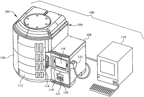
Fig. 1 illustrates a system 100 according to an embodiment of the present
invention
for analyzing biological samples to identify the susceptibility of the samples
to various
types and concentrations of antimicrobial materials, and for calculating the
minimum
inhibitory concentration (MIC) at which the respective antibiotics or
antimicrobial
materials inhibit growth of the respective samples. The system 100 includes a=
measurement instrument 102 having an enclosure 104 which is divided into a
carousel
housing portion 106 and a controller housing portion 108. The system 100
further
includes a workstation 110, such as a personal computer (PC) or the like,
which is coupled
to the controller housing portion 108 to communicate with the system 100 for
purposes of
transferring data to and from the system 100, for example.
CA 02728478 2011-01-21
-9-
The carousel housing portion 106 includes a door 112 and a latch mechanism
114.
The latch mechanism 114 can maintain the door 112 in a closed state, and can
be
manipulated to allow the door 112 to be opened to expose an interior chamber
115 of the
carousel housing portion 106. The controller housing portion 108 includes a
display panel
116, a keyboard panel 118, a computer readable medium drive 120, and a barcode
reader
121, the purposes of which are described in detail below.
As shown in Figs. 2 and 3, a carousel 124 is housed in the interior chamber
115 of
the carousel housing portion 106. The carousel 124 includes a plurality of
rings and ribs
bolted to a drive ring 126 to form a cylindrical cage, which is Mounted
vertically in the
interior chamber 115. The carousel housing portion 106 is insulated to provide
a
substantially uniform temperature incubation environment in the interior
chamber 115, and
is light-tight under normal operation to prevent ambient light from entering
the interior
chamber 115, as described in more detail in U.S. Patent No. 6,096,272,
referenced above.
In this example, the carousel 124 is arranged to include four horizontal tiers
with
each tier having twenty-six panel positions, thus providing a total of one-
hundred and four
panel positions 128. However, these numbers of tiers and panel positions 128
may be
changed to accommodate the requirements of any specified application as will
be
appreciated by one skilled in the art. A panel carrier 130 is mounted in each
of the panel
positions 128. Each panel carrier 130 is configured to receive a test panel
132, an example
of which is shown in Figs. 4A-4C.
As shown in Figs. 4A-4C, a test panel 132 is a disposable, transparent or semi-
transparent device which is inoculated with materials or reagents needed for
both
identification (ID) and antimicrobial susceptibility determination (AST)
testing of the
samples. The testing is performed based on reactions that occur between the
samples and
reagents placed in individual wells 134 on each ID/AST test panel 132. The
wells 134 are
arranged on the ID/AST test panels 132 as a two-dimensional array having rows
and
columns. The wells 134 are segregated into a ID section 136 and an AST section
138. In
this example, the ID section 136 includes fifty-one wells 134, and the AST
section
includes eighty-five wells 134. Each test panel 132 further includes a base
140, a chassis
CA 02728478 2011-01-21
-10-
142, a lid 144, a cellulose acetate pad 146, inoculation ports 148, and a
panel label (not
shown) which includes information that identifies the complete manufacturing
history of
that test panel 132. Further details of the test panels 132 used with the
system 100 are
described in U.S. Patent No. 6,096,272, referenced above, and in U.S. Patent
No.
5,922,593 to Livingston.
The panel carriers 130 are designed such that they will not retain improperly
seated
test panels 132. When the test panels 132 are mounted in the four tiers of the
carousel
124, they are arranged to form substantially circular rows and vertical
columns of wells
134. That is, all the columns of wells 134 in all four tiers of the carousel
124 should be
substantially aligned with each other in the vertical direction along the
entire height of the
carousel 124, while all rows of wells 134 should be substantially aligned with
each other
around the entire circumference of the carousel 124. In this example, panel
positions 128
are numbered zero through twenty-five in each tier of the carousel 124, with
panel position
zero being reserved for a normalization' panel and thus not accessible by an
operator during
normal operation of the instrument 102.
The carousel housing portion 106 also includes a drive module 150 that drives
the
carousel 124 to rotate in a clockwise or counter-clockwise manner, as desired,
and a
plurality of bearings 152 and a spring-loaded pivot 154 which rotatably secure
the carousel
124 in the interior chamber 115 of the carousel housing portion 106 and
facilitate rotation
of the carousel 124. Further details of the carousel 124 and its associated
components, as
well as the panel carriers 130 and test panels 132, are described in U.S.
Patent No.
6,096,272, referenced above.
As shown in Figs. 3 and 5, and in the schematic diagram shown of Fig. 6, the
carousel housing portion 106 in this example further includes a visible light
source
assembly 156 and an ultraviolet (UV) light source assembly 158. The visible
light source
assembly 156 includes four visible light source modules 156-1 through 156-4
and a
supporting tower 160, while the ultra-violet light source assembly 158
includes ultraviolet
light sources 158-1 and 158-2. The supporting tower 160 aligns one visible
light source
module with each tier of the carousel 124 so that at any given time, one
entire column of
CA 02728478 2011-01-21
-11-
wells of the ID/AST test panels 132 in the four tiers of the carousel 124 can
be illuminated
by the visible light source modules.
In this example, each visible light source module 156-1 through 156-4 includes
three parallel vertical columns of sixteen light-emitting diodes (LEDs) each.
The first
column consists of red LEDs, the second of green LEDs and the third of blue
LEDs. A
holographic diffuser plate 162 is disposed in close proxiMity to the ID/AST
test panels 132
mounted in the carousel 124. The holographic diffuser plate 162 diffuses the
illumination
energy from each column of LEDs, when the columns are energized. Each column
of
LEDs is mounted in the visible light source modules to maintain a fixed
distance from the
diffuser plate 162. Cylindrical lenses (not shown) may be used to focus the
illumination
energy from each column of LEDs onto the vertical well columns of the ID/AST
test
panels 132. The illumination axis for each column of LEDs is made coincident
for the red,
green and blue illumination. Thus, each well column sees a uniform stripe of
either red,.
green or blue illumination, depending upon which column of LEDs is energized.
As further shown in Figs. 3,* 5 and 6, an optical measurement system 164 is
disposed approximately within the center of the carousel 124 such that it is
aligned to
receive the visible light transmitted through each well 134 of the ID/AST test
panels 132
during excitation by red, green orblue illumination from the visible light
source modules
of the visible light source assembly 156. Visible fluorescent radiation is
similarly detected
from the wells 134 when the samples in the wells 134 are excited by the
ultraviolet light
emitted from the ultraviolet light source assembly 158. As can be appreciated
by one
skilled in the art, excitation filters 166 eliminate unwanted spectral
components present in
the light emitted from the ultraviolet light source assembly 158, and emission
filters 168
eliminate unwanted spectral components that may be present in the output
signal before
detection by the optical measurement system 164.
In this example, the optical measurement system 164 includes a plurality of
CCD
detector modules 170-1 through 170-4 and corresponding lens assemblies 172-1
through
172-4, with one CCD detector module 170 and one lens assembly 172 being
aligned to
receive readings from wells 134 of test panels 132 in a respective tier of the
carousel 124.
Accordingly, because the carousel 124 includes four tiers in this example, the
optical
CA 02728478 2011-01-21
. -12-
measurement system 164 includes four CCD detector modules 170-1 through 170-4
and
four corresponding lens assemblies 172-1 through 172-4, with each detector
module/lens
assembly pair arranged substantially in alignment in the vertical direction.
The lens
assemblies 172-1 through 172-4 focus light from of each panel well column of
the test
panels 132 in their respective tiers of the carousel 124 onto the CCD arrays
of the
corresponding CCD detector modules 170-1 through 170-4.
Each CCD detector module 170 can include, for example, a 2048-pixel linear CCD
array. The CCD arrays of the CCD detector modules 170 detect and measure the
intensity
of light transmitted through each well 134 of the test panels 132 in the
corresponding tiers
of the carousel 124 when the wells 134 are illuminated by the red, green and
blue LEDs.
Visible fluorescent light is similarly detected by the CCD arrays, of the CCD
detector
modules 170 when the samples in the wells 134 are excited by the ultraviolet
light emitted
from the ultraviolet light source assembly 158. Further details of the
structure and
operation of the visible light source assembly 156, ultraviolet light source
assembly 158,
optical measurement system 164, and their related components can be found in
U.S. Patent No. 6,096,272, referenced above.
As stated above, Fig. 6 is an exemplary schematic diagram illustrating further
components of the measurement instrument 102 described above. As shown, the
carousel
housing portion 106 and the controller housing portion 108 are separated by a
divider
panel 174 which can be, for example, part of the housing 106. The ultraviolet
light
sources 158-1 and 158-2 are driven by a lamp driver 176. The lamp driver 176,
visible
light source modules 156-1 through 156-4, CCD detector modules 170-1 through
170-4,
an ultraviolet light source cooling fan 178, and an optical measurement system
cooling fan
180 are coupled to an interconnect board 182. A plurality of status indicator
boards 184,
barcode readers 186 which read the barcodes on the test panels 132, and panel
flags and
home flag reader 188, are also coupled to the interconnect board 182. Further
details of
the status indicator boards 184, barcode readers 186, and panel flags and home
flag reader
188 can be found in U.S. Patent No. 6,096,272, referenced above.
CA 02728478 2011-01-21
-13-
The interconnect board 182 is coupled to an I/0 interface board 190 of the
controller module 191 that is present in the controller housing portion 108.
As described
in more detail below and in U.S. Patent No. 6,096,272,
referenced above, the control module 191 includes a controller 192 which
controls the
visible light source modules 156-1 through 156-4, CCD detector modules 170-1
through
170-4, lamp driver 176, and all other components associated with performing
the well
reading process. The controller 191 further includes an ethernet 194 and an
LCD driver
196. The ethernet 194 can be coupled to a network port 198 to output and input
data to
and from the workstation 110 (see Fig. 1), for example, The LCD driver 196 is
coupled to
the display panel 116 (see Fig. 1) to display, for example, results of the
well readings, and
is further coupled to an external video connection 197. The controller 192 is
coupled to
the computer readable medium drive 120 (see Fig. 1) to output and input data
to and from
a computer readable disk, for example.
In addition, the controller module 191 is coupled to the computer readable
medium
drive 120, to the display panel 116 via an inverter 200, to the keyboard panel
118 via an
indicator board 202, to the barcode reader 121, to an AT keyboard 204 and to a
speaker
206. The controller module 191 is further coupled to an auxiliary serial port
208, a printer
port 210, a remote alarm port 212 and an auxiliary barcode reader port 214
which, along
with the network port 198, are housed in a connector panel 216. In this
example, the
barcode reader port 214 is coupled to the auxiliary barcode reader 122 (Fig.
1).
The controller module 191 is also coupled to a drive and DC distribution
module
218 and a power control and distribution module 220. An ambient temperature
sensor 222
and an incubation temperature sensor 224 sense the temperature inside the
interior
chamber 115 and provide signals indicative of the temperature to the
controller 192 of the
controller module 191. Furthermore, upper temperature cut-off sensor 226
provides a
signal to the controller 192 via the power control and distribution module 220
indicating
when the temperature of the interior chamber 115 has reached the maximum
temperature.
In response, the controller 192 will control the heater 228 via power control
and
distribution module 220, and will control heater blower 230 via drive and
distribution
module 218, to prevent the temperature in the interior chamber 115 from
further
CA 02728478 2011-01-21
-14-
increasing. The controller 192 further controls the door switch 232 and door
solenoid 234
via drive and distribution module 218 to control the latch mechanism 114 of
the door 112
(see Figs. 1 and 2) to either maintain the door 112 in the closed position or
allow the door
112 to be opened. The controller 192 also controls the drive module 150 to
control the
rotation of carousel 124 as described in detail below. Further details of the
temperature
controlling operations and carousel rotation operations are set forth in
U.S. Patent No. 6,096,272, referenced above.
As further shown in Fig. 6, the controller housing portion 108 includes a
transformer 236 and cooling fans 248 that are coupled to the power control and
distribution module 220. Also, a 24 V power supply 240, a 15 V power supply
242 and a
V power supply 244 provide power to the drive and distribution module 218 and
power
controller module 220, as well as to the lamp driver 176. These power supplies
240, 242
and 244 are powered from an A.C. input power that is received by the power
control and
distribution module 220 via filter 246 and the main on/off switch 248 of the
system 100.
The operation of the system 100 will now be described with reference to Figs.
1-6,
as well as the flow chart and graphs shown in Figs. 7A-9. In Step 1000, each
test panel
132 is inoculated with a respective broth-suspended organism (i.e., a sample)
before being
placed into a respective panel carrier 130 of the carousel 124. The separate
innocula are
added manually to the inoculation ports 148 of the test panels 132, and
allowed to flow
into the wells 134 of the test panels 132 as described in U.S. Patent No.
6,096,272, referenced above. Only one type of sample is introduced into each
respective test panel 132. As discussed above, the wells 134 of the test
panels 132 include
various types and concentrations of antimicrobial materials, which affect the
growth of the
samples, along with indicators that indicate the presence or absence of sample
growth.
Also, at least one of the wells 134 of each test panel 132 is designated as a
growth control
well and does not include any antimicrobial material.
The inoculated test panels 132 are then inserted into the respective panel
carriers
130 of the carousel 124 in step 1010. The operator uses the barcode scanner
121 or
auxiliary barcode scanner 122 to scan the barcode of each test panel 132 as
it is being inserted into a respective panel carrier 130, to thus enter
information
pertaining to the sample in the test
CA 02728478 2011-01-21
-15-
panel 132, the antimicrobial materials in the test panel wells 134, and so on,
into the
system 100. The technician also can enter information pertaining to the tier
level and
position in the carousel 124 at which the test panel 132 is inserted via the
keyboard 118,
for example. Once the test panels 132 have been loaded into the carousel 124,
the door
112 of the carousel housing portion 106 is closed and latched shut. In step
1020, the
controller 192 controls the carousel 124 to begin rotating, and controls the
heater 228 and
heater blower 230 to begin increasing the temperature of the interior chamber
115 to
incubate the samples in the wells 134. In this example, the operator can set
the carousel
124 to rotate at one revolution per minute (RPM). However, the rotational
speed can be
set to any value as appropriate.
After a predetermined amount of time has passed, for example, two hours, the
controller 192 controls the system 100 to begin taking measurements of the
wells 134 of
the test panels 132 in a manner as described in U.S. Patent No. 6,096,272,
referenced above. In this example, measurements are taken at 20 minute
intervals. Also, as can be appreciated from the discussion below, the
following steps in
the flowchart shown in Fig. 7 are performed for each panel 132, and the manner
in which
the processing proceeds for each respective panel 132 is dependent on the
results of the
well readings obtained for each respective panel 132. Also, the operations
described in
these steps are controlled by controller 192.
In step 1030, the first readings of the wells 134 of the test panels 132 are
taken as
test readings, to determine whether the readings pass an initial criteria
indicating that the
samples are valid for analysis. The well readings are taken as the carousel is
being rotated.
The controller 192 waits until the home flag of the carousel 124 is detected
by the home
flag detector 188 before beginning to take the readings, to insure that the
controller 192
can match the readings with the correct well 134 from which the readings were
taken.
The controller 192 can first control the detector modules 170-1 through 170-4
to
perform dark readings, during which neither the UV light sources 158 nor the
visible light
sources 156-1 through 156-4 are energized. The controller 192 can then control
the lamp
driver 176 to drive the ultraviolet light source assembly 158. The controller
192 in this
example waits until the carousel 124 has rotated two revolutions to allow the
ultraviolet
CA 02728478 2011-01-21
-16-
lights of the ultraviolet light source assembly 158 to warm up, so that the
light intensity
can stabilize, and then controls the detector modules 170-1 through 170-4 to
take an
ultraviolet light reading for an entire revolution of the carousel 124. The
controller 192
then controls the lamp driver 176 to turn off the ultraviolet light sources
158, and
processes the readings. As discussed in U.S. Patent No. 6,096,272 referenced
above, the
controller 192 uses the ultraviolet readings to identify the types of samples
in the sample
wells 134 of the test panels 132.
After the above readings have been taken, the visible light readings are then
taken.
The controller 192 can then control the rate of rotation of the carousel 124
to remain the
same, or can increase the rate of rotation of the carousel 124, for example,
to 2 RPM, or
any other suitable rotation speed, while the visible light readings are being
taken. In one
example, the rotation speed is increased to 2 RPMs, and the red LEDs of the
visible light
source assembly 156 (see Figs. 3, 5 and 6) are activated. The carousel 124 can
be rotated
one revolution to allow the red LEDs to warm up so that light intensity can
stabilize, and
then "red" readings can be taken of the wells 134 by the detector modules 170-
1 through
170-4 while the carousel 124 rotates the second revolution.
Once the red readings have been taken, the red LEDs are turned off and the
green
LEDs of the visible light 156 can be energized. As with the red LEDs, the
carousel 124
can be rotated one revolution to allow the green LEDs to warm up to allow the
light
intensity to stabilize. The "green" readings can then be taken of the wells
124 by the
detector modules 170-1 through 170-4 while the carousel 124 is rotated another
revolution.
After the green readings have been taken, the green LEDs are turned off. In
this example,
the rotation speed of the carousel 124 is then reduced to 1 RPM, and the blue
LEDs of the
visible light source assembly 156 are energized. The carousel 124 is allowed
to rotate for
one revolution while the blue LEDs warm up to allow the light intensity to
stabilize.
Then, the "blue" readings of the wells 134 are taken by the detector modules
170-1
through 170-4 during the next revolution of the carousel 124.
The red, green and blue readings taken for each well 134 of each test panel
132 are
= then stored by the controller 192 in a memory such that each well 134 has
a specific red,
green and blue reading for that particular time interval. The process then
continues to step
CA 02728478 2011-01-21
- 17-
1040 where the readings for each well 134 are evaluated to determine whether
the further
readings that are taken on a well 134 are to be considered valid.
In step 1040, the red readings taken of each well 134 are evaluated to
determine
whether the wells have been properly filled. The readings can range from an
intensity
level of "0" to an intensity level of "4200" with 0 being zero intensity and
4200 being the
maximum intensity reading for a particular color (e.g., red). In this example,
the process
identifies in step 1040 the wells 134 having a red reading above 2200. For
those wells 134
having such a red reading, the processing continues to step 1050 where those
wells 134 are
failed or, in other words, the system 100 identifies all future readings from
those wells 134
as being invalid. Accordingly, either no further readings of those wells 134
are taken, or
any readings that are taken are ignored.
Furthermore, if a well 134 has been identified as a growth control well and
has a
red reading of over 2200, the entire side of the test panel 132 on which that
control well
resides is failed. Also, if that well contains a particular antimicrobial
material, no results
are reported by the system 100 for that' antimicrobial material for the
particular test panels
132 including the failed wells.
Once the red well readings have been evaluated in step 1040 and the
appropriate
wells 134 have been failed in step 1050, the processing continues to step 1060
where a
panel indicator determination is made. Specifically, in this step, the wells
134 identified as
growth control wells for their respective test panels 132 are evaluated to
determine
whether the initial state of the growth indicator present in the samples in
the control wells
134 of their respective test panels 132 are acceptable for evaluating those
test panels 132.
In this example, the value of the respective red reading for each control well
is divided by
the value of the respective green reading for each control well. If the result
of the division
is less than 0.3692 or greater than 0.6464, controller 192 determines that the
initial state of
the growth indicator is unacceptable for the test panel 132 including the
control well
providing this result. Accordingly, no results obtained by the well
measurements for that
particular test panel 132 are reported. As stated above, step 1060 is carried
out for each
test panel 132.
CA 02728478 2011-01-21
-18-
The processing then continues to step 1070 where the controller 192 will
continue
to rotate the carousel 124 and thus, the carousel housing portion 106 will
continue to
incubate the samples in the wells 134. The processing will continue to step
1080 where
the system 100 will take the red, green and blue readings of the wells in a
manner similar
to that described to above with regard to step 1030, and as described in
U.S. Patent No. 6,096,272, referenced above. The processing then proceeds
to step 1090 where the controller 192 determines whether the minimum amount of
incubation time has elapsed. The minimum incubation time at which readings of
the wells
134 can begin to be analyzed to determine MIC values in this example is two
hours. If the
minimum incubation time has not elapsed, the processing returns to step 1070
and the
incubation is continued. However, once the appropriate amount of incubation
time has
elapsed, the processing proceeds to step 1100 where the controller 192 will
calculate the
redox state and turbidity values for each well.
The system 100 in this example uses two indicators of growth, redox and
turbidity,
, .
to evaluate the susceptibility of the saniples to the antimicrobial materials
in the wells 134.
The redox and turbidity values are calculated for each well 134 in each of the
panels based
on the red, green and blue readings taken of the respective wells at the
respective 20
minute time intervals as discussed above. A simultaneous nonlinear algorithmic
model
was developed from experimentally obtained redox and turbidity readings, and
this
algorithm is used by the controller 192 to predict the redox state and
organism density
(turbidity) in each of the wells 132. The controller 192 can arrange the
calculated redox
state and turbidity values for each respective well 132 in graph form with
respect to
incubation time. An example of the calculated redox and turbidity growth
curves for
E.coli for a single well 132 is shown in Fig. 8.
As stated above, the redox state of a sample in a well 132 is measured by
utilizing
the change in red, green and blue readings that occurs over time as a result
of the reduction
of a growth indicator, such as resazurin, by the antimicrobial material in the
well 132. As
the resazurin is reduced, the color of the sample in the well 132 changes from
blue to red
to clear. This change in redox is represented numerically as a continuum, with
the value
"0" indicating an unreduced growth indicator (blue = resazurin), the value
"0.5" indicating
CA 02728478 2011-01-21
-19-
that the indicator is 50% reduced (red = resorufin), and the value "1.0"
indicating that the
indicator has been completely reduced (clear = dihydroresorufin).
The turbidity is also estimated by using the red, green and blue reading in an
equation similar to the redox calculation. The initial signal has a value of
"0" and a
maximum of 2.25 units can be estimated. The units for turbidity correspond to
McFarland
units (1 McFarland = 3 x 10⁸ cfu/ml).
An example of the manner in which the actual red, green and blue readings are
used to calculate redox and turbidity values will now be demonstrated. In this
example,
the red, green and blue readings taken of a sample well at the first twenty
minute interval
are as follows: red = 873, green = 956 and blue = 2705. The processing then
generates a
one-column, four-row input matrix as shown in Table 1 as follows:
Table 1: Input Matrix Values
1.0000
2705.0000
0.3227
0.3534
It is noted that the first row in the input matrix is always padded with the
value
1.0000. The value 2705.0000 is equal to the blue reading, the value 0.3227 is
calculated
by dividing the red reading by the blue reading (i.e., red/blue), and the
value 0.3534 is
calculated by dividing the green reading by the blue reading (i.e.,
green/blue). It is also
noted that in this example, the blue reading is clamped at a starting value of
2705 until 36
minutes has elapsed in the incubation. All points after 36 minutes are
multiplied by the
value (2705 / (blue signal @ last point before 36 minutes)). The result is
then clamped to
limits of 558 and 4474. Furthermore, the value of red/blue is clamped to a
minimum of
0.2012329 and a maximum of 1.8936959, while the value of green/blue is clamped
to a
minimum of 0.3091655 and a maximum of 1.3084112.
The processing then multiplies the one-column, four-row Input Matrix by the
four-
column, five-row Redox Input Weight Matrix according to the equation "Input
Matrix *
Redox Input Weight Matrix" and known matrix multiplication techniques to
arrive at a
CA 02728478 2011-01-21
-20-
one-column, five-row matrix of numbers as discussed below. The twenty values
in the
Redox Input Weight Matrix have been calculated and programmed into the
controller 192
based on past empirical data and observations, and remain constant for all of
the readings
at all of the time intervals. An example of the values of the Redox Input
Weight Matrix
are shown in the following Table 2:
Table 2: Redox Input Weight Matrix Values
-0.673253 0.000710423 -1.623674164 3.340127166
2.445846 -0.000572912 1.4797837 -6.311909249
0.109425 0.005775254 -3.604370752 -0.242927922
1.356753 0.000748697 -2.139010636 -1.067568082
3.88E-05 0.022713989 3.80317E-05 2.99302E-05
The values of the Intermediate Matrix calculated according to the above
equation
"Input Matrix * Redox Input Weight Matrix" are shown in Table 3 as follows:
Table 3: Intermediate Matrix Values
1.9049
-0.8571
14.4824 =
2.3143
61.4414
These values of the Intermediate Matrix, as well as the values of the Input
Matrix,
are used to create a one-column, nine-row Output Matrix. Specifically, the
first row of the
Output Matrix is padded with the value 1.0000, and rows two through six of the
Output
Matrix are calculated by taking the antilog value of each of the above values
of the Input
Matrix, respectively, according to the following equation:
antilog value = ex/(1 + ex )
CA 02728478 2011-01-21
-21-
with x being the respective value from the matrix. Rows seven through nine of
the Output
Matrix are filled with the values in rows two through four of the Input
Matrix.
Accordingly, the values of the Output Matrix are shown in the following Table
4:
Table 4: Output Matrix Values
1.0000
0.8704
0.2980
1.0000
0.9101
=
1.0000
2705.0000
0.3227
0.3534
The redox value is then calculated by multiplying the one-column, nine-row
Output
Matrix by a nine-column, one-row 'Redox Output Weight Matrix according to the
following equation and known matrix multiplication techniques.
Redox Value = Output Matrix * Output Weight Matrix
In this example, the values of the Redox Output Weight Matrix are shown in the
following
Table 5:
Table 5: Redox Output Weight Matrix Values
-6.633973 4.167218646 1.721677475 -0.389544272 -2.543872 -0.63391676
1.30610E-04 0.04646759 -0.842252
As with the values of the Redox Input Weight Matrix, the Redox Output Weight
Matrix
values have been calculated and programmed into the controller 192 based on
past
empirical data and observations, and remain constant for all of the readings
at all of the
time intervals. The Redox Value is thus calculated as 0.23843516. This value
is then
plotted on the graph as shown in Fig. 8.
CA 02728478 2011-01-21
-22-
The turbidity value based on these red, green and blue readings is calculated
in a
similar manner. That is, the processing then generates a one-column, four-row
input
matrix as shown in Table 6 as follows:
Table 6: Input Matrix Values
1.0000
2705.0000
0.3227
0.3534
As with the Input Matrix Values for the redox calculation, the Input Matrix
Values for the
turbidity calculations are based on the actual red, green and blue readings.
The first row in
the input matrix is always padded with the value 1.0000. The value 2705.0000
is equal to
the blue reading, the value 0.3227 is calculated by dividing the red reading
by the blue
reading (i.e., red/blue), and the value 0.3534 is calculated by dividing the
green reading by
the blue reading (i.e., green/blue). It i also noted that in this example, the
blue reading is
clamped at a starting value of 2705 until 36 minutes has elapsed in the
incubation. All
points after 36 minutes are multiplied by the value (2705 / (blue signal @
last point before
36 minutes)). The result is then clamped to limits of 558 and 4474.
Furthermore, the
value of red/blue is clamped to a minimum of 0.2012329 and a maximum of
1.8936959,
while the value of green/blue is clamped to a minimum of 0.3091655 and a
maximum of
1.3084112.
The processing then multiplies the one-column, four-row Input Matrix by the
four-
column, five-row Turbidity Input Weight Matrix according to the equation
"Input Matrix *
Turbidity Input Weight Matrix" and known matrix multiplication techniques to
arrive at a
one-column, five-row matrix of numbers as discussed below. The twenty values
in the
Turbidity Input Weight Matrix have been calculated and programmed into the
controller
192 based on past empirical data and observations, and remain constant for all
of the
readings at all of the time intervals. An example of the values of the
Turbidity Input
Weight Matrix are shown in the following Table 7:
CA 02728478 2011-01-21
-23-
Table 7: Turbidity Input Weight Matrix Values
-2.870675 0.002111599 -0.234543715 0.334025395
-1.306260 0.00202755 0.577175204 -2.717689223
3.477755 0.001837992 -4.028539894 1.455268741
-0.008775 -0.004819911 -0.027006746 -0.01188475
8.842011 0.001408226 -5.393142566 -4.464335919
The values of the Intermediate Matrix calculated according to the above
equation
"Input Matrix * Turbidity Input Weight Matrix" are shown in Table 8 as
follows:
Table 8: Intermediate Matrix Values
2.8836
3.4041
7.6637
-13.0596
9.3329
These values of the Intermediate Matrix, as well as the values of the Input
Matrix,
are used to create a one-column, nine-row Output Matrix. Specifically, the
first row of the
Output Matrix is padded with the value 1.0000, and rows two through six of the
Output
Matrix are calculated by taking the antilog value of each of the above values
of the Input
Matrix, respectively, according to the following equation:
antilog value = ex/(1 + )
with x being the respective value from the matrix. Rows seven through nine of
the Output
Matrix are filled with the values in rows two through four of the Input
Matrix.
Accordingly, the values of the Output Matrix are shown in the following Table
9:
CA 02728478 2011-01-21
-24-
Table 9: Output Matrix Values
1.0000
0.9470
0.9678
0.9995
0.0000
0.9999
2705.0000
0.3227
0.3534
The turbidity value is then calculated by multiplying the one-column, nine-row
Output Matrix by a nine-column, one-row Turbidity Output Weight Matrix
according to
the following equation and known matrix multiplication techniques.
Turbidity Value = Output Matrix * Output Weight Matrix
, .
In this example, the values of the Turbidity Output Weight Matrix are shown in
the
following Table 10:
Table 10: Turbidity Output Weight Matrix Values
1-0.107225 -2.957877127 2.378329542 1 866207268 0.012793 -
1.741858375 1.38488F-04 -0.08976299 0.401581
As with the values of the Turbidity Input Weight Matrix, the Turbidity Output
Weight
Matrix values have been calculated and programmed into the controller 192
based on past
empirical data and observations, and remain constant for all of the readings
at all of the
time intervals. The Turbidity Value is thus calculated as 0.00459741. This
value is then
plotted on the graph as shown in Fig. 8.
The redox and turbidity values are calculated for each well based on the
readings
taken for each well at each time interval (i.e., each twenty minute time
interval in this
example), and the values are plotted on a graph as shown in Fig. 8. A local
regression
algorithm (LOESS) smoothes the time series data for both the redox and
turbidity values
CA 02728478 2011-01-21
-25-
calculated for each well 132 over the elapsed period of time. The LOESS in
this example
uses no more than seven readings for each local regression. In evaluating a
time point, at
least one reading is required past the time point being interpolated. From the
LOESS
equations any reading at any time point can be estimated. From the
interpolated data a
series of metrics that describe= the growth in the well are calculated. All
metrics will be
based on the time or growth control values derived from these smoothed and
interpolated
points. The metrics are derived from the basic functions such as absolute
value, first
derivative (rate), second derivative (acceleration) and integral (area under
the curve). The
metrics are then used to derive a series of variables that are utilized by the
generalized
additive models (GAMs) as described in more detail below. These variables are
a variety
of absolutes, maximums and ratios to the growth control. A total of 27
variables are
available to the GAMs, as listed below in Table 11.
CA 02728478 2011-01-21
=
-26-
Table 11: Variables Available for GAMs
Running Count Abbreviation Description
1 CONC LOG drug concentration
2 T AB turbidity value
3 T FD turbidity first
derivative
4 T SD turbidity second
derivative
TIN turbidity integral
6 T AB M turbidity maximum
value
7 T FD M turbidity maximum
first derivative
8 T SD M turbidity maximum
second derivative
9 T AB M R turbidity maximum
value / turbidity
maximum value of
the growth control
T FD M R turbidity maximum
_
first derivative /
turbidity maximum
first derivative of the
growth control
11 T SD M R turbidity maximum
second derivative /
turbidity maximum
second derivative of
the growth control
12 T IN R turbidity integral /
turbidity integral of
the growth control
13 T FD T time at turbidity
maximum first
derivative minus time
at turbidity maximum
first derivative of the
growth control
14 T SD T time at turbidity
maximum second
derivative minus time
at turbidity maximum
second derivative of
the growth control
CA 02728478 2011-01-21
=
-27-
15 R AB redox value
16 R FD redox first derivative
17 R SD redox second
derivative
18 RIN redox integral
19 R AB M redox maximum
_ _
value
20 R FD M redox maximum first
derivative
21 R SD M redox maximum
second derivative
22 R AB M R redox maximum
_ _
value / redox
maximum value of
the growth control
23 R _ FD M_ R redox maximum first
derivative / redox
maximum first
derivative of the
growth control
24 R_ SD _ M R redox maximum =
_
second derivative /
redox maximum
second derivative of
the growth control
25 R IN R redox integral / redox
integral of the growth
control
26 R FD T time at redox
maximum first
derivative minus time
at redox maximum
first derivative of the
growth control
27 R SD T time at redox
maximum second
derivative minus time
at redox maximum
second derivative of
the growth control
CA 02728478 2011-01-21
-28-
The processing then proceeds to step 1110 during which the calculated redox
state
for each growth control well in each respective test panel 132 is analyzed. If
the maximum
redox value for the growth control well of a test panel 132 is not above a
desired value
which, in this example, is 0.07, the processing continues to step 1120. In
step 1120, the
processing determines whether the elapsed incubation time has reached a
certain desired
duration which, in this example, is 16 hours. If the processing determines in
step 1120 that
16 hours of incubation time or less has elapsed, the processing returns to
step 1070 for this
panel 132, and the process repeats as discussed above. However, if the
processing
determines in step 1110 that more than 16 hours of incubation time has elapsed
for this
particular panel 132, the processing proceeds to step 1130 where the panel 132
is failed as
being inoculated or containing a non-reactive sample, and no test results are
reported for
that panel.
If the processing in step 1110 determines that the maximum redox state for the
growth control well of the test panel 132 are greater than 0.07, the
processing proceeds to
step 1140 for that panel 132. In step 1140, the processing determines whether
the
maximum redox state for the growth control well of that panel 132 is greater
than a
predetermined value which, in this example, is 0.2. If the maximum redox state
for the
growth control well in that panel 132 is not greater than 0.2, the processing
continues to
step 1150 for that panel 132 where it is determined whether the elapsed
incubation time is
greater than a predetermined value which, in this example, is 16 hours. If the
elapsed
incubation time is less than or equal to 16 hours, the processing returns to
step 1070 for
this panel 132, and repeats as discussed above. However, if the processing
determines in
step 1150 that the elapsed incubation time has exceeded 16 hours, the
processing continues
to step 1160, where the test panel 132 is failed as having insufficient sample
growth, and
no results are reported for that test panel 132.
Concerning step 1140 discussed above, if the processing determines that the
maximum redox state for the growth control well for the panel 132 is indeed
greater than
0.2, the processing continues to step 1170 where the processing evaluates the
type of curve
represented by the calculated redox states plotted in graph form with respect
to incubation
time as shown, for example, in Fig. 8. Specifically, based on the maximum
redox value of
CA 02728478 2011-01-21
-29-
the growth control well of the panel 132, the processing determines whether
the curve
representing the redox values for the growth control well indicates that the
sample is a
slow or fast growing sample. If the processing determines in step 1170 that
the curve is
classified as a class "zero" curve, the sample is not yet classifiable as a
slow or fast
growing sample because a sufficient incubation time has not elapsed for that
sample.
Therefore, the processing returns to step 1070 for that panel 132, and repeats
as discussed
above. However, if the processing determines in step 1170 that the curve
classification is
other than "zero", the processing continues to step 1180.
In step .1180, the processing determines whether the curve representing the
redox
states can be classified as a class "one" curve. If so, the processing
continues to step 1190
where the controller 192 will perform the appropriate GAM on the redox states
and
turbidity values measured for each of the wells 134 in the test panel 132 to
determine the
MIC values for the particular sample and the anti-microbial materials
contained in the
wells 134 of the test panel 132.
In step 1200, the probability of sample growth for each well 134 of the test
panel
134 is calculated according to the appropriate GAM once the growth control is
above a
specified threshold. The GAMs were developed for each antibiotic by evaluating
a
spectrum of species, MIC values and resistance mechanisms.
The GAMs are specific for each antibiotic and broad category of organisms
(gram
positive/gram negative). Each GAM requires approximately 5 of the 27 variables
previously described above in Table 11 to predict growth, but can use as many
variables as
deemed appropriate. A GAM uses a polynomial equation as shown below to
describe the
relationship between each variable included in the model and the contribution
of that
variable in predicting growth in a well. The calculation of the well
probability Pk is simply
the sum of the polynomial functions for each variable and an intercept term.
(
log =a+A(x0+...+/,(x1)
CA 02728478 2011-01-21
-30-
Each polynomial function in the above equation represents the function
associated with a
respective variable chosen from Table 11. For example, Mx!) can represent the
function
for the first derivative of the turbidity curve at a particular time interval,
f2(x2) can
represent the function for the second derivative of the turbidity curve at
that time interval,
and so on.
Fig. 9 illustrates a graph showing an example of the relationship between a
variable
and its prediction. These probabilities are then used to determine the MIC and
calculate
the confidence value for the reported MIC as follows.
Once a set of growth probabilities for each well 134 in the test panel 132 is
deeived
by the GAM, a probability is calculated for every possible MIC in step 1210.
This MIC
probability is the product of the well probabilities with respect to the
values obtained from
the GAM. The example below shows the calculation for obtaining a probability
for an
MIC of 16 lig for one antimicrobial material with respect to the sample in the
test panel
132. In this example, the raw probability would be 0.525. It is noted that
five wells 134 of
the test panel 132 contain different cohcentrations of this antimicrobial
material, and the
redox and turbidity results for each of these five wells is used by the GAM to
determine
the MIC.
Table 12: An Example of an MIC Calculation for Five Wells
Antibiotic 2 lig well 4 1.tg well 8 j.ig well 16 1.1.g
well 32 lig well
Concentration
Pattern for Growth Growth Growth No Growth No
Growth
MIC = 16 lig
Well Probability 0.9 0.9 0.8 0.1 0.1
(p from the GAM)
Calculation P2 X P4 X PS X 1116 X
11032
After a raw probability is obtained, the processing proceeds to step 1220
where a
confidence value for the most probable MIC is calculated. This is simply the
raw
CA 02728478 2011-01-21
-3 1 -
probability (P) of the MIC value (k) over the sum of all valid MIC
probabilities as shown
in the following equation:
Pk
MIC Confidence Value =
EP),
Once an MIC and the associated confidence value are calculated, processing
proceeds to step 1230 where this information is evaluated with respect to a
threshold. If
the threshold is exceeded, then the processing proceeds to step 1240 where the
system 100
reports the MIC for the particular sample in the test panel 132 with respect
to the particular
antimicrobial material in the group of wells 134 of the test panel 132. The
system 100 can
report the MIC on, for example, display screen 116 of Figs. 1, 2 and 6, and
can also control
a printer (not shown) to generate a printed report.
However, if a low confidence value is obtained, the processing proceeds to
step
1250 where it is determined whether the incubation protocol of a certain
duration (e.g., 16
hours) has elapsed. If the incubation protocol has not elapsed, the processing
returns to
step 1070 where the test panel 132 continues to incubate and is reevaluated
according to
the processing discussed above after each 20 minute reading. On the other
hand, if a
minimum threshold is still not met at the end of the incubation protocol, the
processing
proceeds to step 1260 during which the system 100 does not report an MIC for
that
antimicrobial material, but rather, provides a message suggesting that the
user check
purity/viability and repeat the test.
A more detailed example of MIC probability calculations is shown in Fig. 10
for
four wells having antibiotic concentrations of 1 fig, 2 p.g, 4 p.g and 8 ug,
respectively. As
illustrated in this example, the probability of growth for a well having a 1
p.g concentration
of antibiotic as calculated by the polynomial equation discussed above for a
set of readings
taken at a particular interval in time is 0.9. Also, the probabilities of
growth for the wells
having 2 pig, 4 ptg and 8 p.g are 0.9, 0.1 and 0.1, respectively. The five
different growth
possibilities are then entered into the table as shown, with the value "0"
representing no
growth and the value "1" representing growth. That is, as shown in the first
row of the
CA 02728478 2011-01-21
-32-
table, the condition in which no growth occurs (i.e., "0" for each well) is
considered,
meaning that the MIC value would be less than the minimum concentration of 1
pg. The
second row illustrates the condition in which growth occurs in the 1 pg well
but in no
other wells, the third row illustrates the condition in which growth occurs in
the 1 i.tg well
and in the 2 ps well, but not in the higher concentration wells, and so on.
The four growth probabilities are then multiplied for each row to arrive at
the
probability of valid growth pattern values on the right side of the table. It
is noted that
because the probabilities of 0.9 or 0.1 at the top of the table represent
probabilities of
growth, these values are subtracted from 1 for conditions of non-growth to
provide a value
that is used in the multiplication. Considering the first row, for example,
the probability of
growth for the well concentration of 1 [tg is "0.9". However, because no
growth occurred
in this well, the value used in the multiplication is "0.1" (i.e., 1 - 0.9).
This is also the case
for the 21.1,g concentration well. Also, because the no growth occurred in the
4 lig and 8 }ig
wells, the values for these wells used in the multiplication are each "0.9"
(i.e., 1 - 0.1).
Accordingly, the multiplication values are 0.1 * 0.1 * 0.9 *0.9 = 0.0081,
which is the
probability that this growth pattern in the first row is valid.
The above calculations are performed for each row to provide the values shown
in
the first column on right side of the table. In addition, the probabilities of
the "local" '
growth patterns (i.e., the shaded wells in the graph) are multiplied to
provide the
probabilities of valid local growth patterns. This additional calculation is
used to increase
the accuracy of the results. As indicated, the row having the MIC possibility
of "4" (the
third row) provides the highest probabilities.
Using the MIC confidence value equation indicated above, the highest local
growth
pattern probability of 0.729 is divided by the sum of itself and the local
growth pattern
probabilities (i.e., 0.081 + 0.729 + 0.09) to arrive at a MIC probability of
0.81 as indicated.
This value is then compared to a predetermined threshold. If the value exceeds
the
predetermined threshold, then the system can report the MIC value of "4" for
this sample.
An example of another table in which wells having antibiotic concentrations of
0.25 and 0.50 taken into account is shown in Fig. 11. The probabilities, MIC
value and
MIC probability are calculated in the same manner as described above.
CA 02728478 2011-01-21
-33 -
It is also noted that prior to reporting the results in step 1240 shown in
Fig. 7B, the
processing can delay the reporting until the same MIC value has been
determined for a
desired number of consecutive , for example, three time intervals. That is, as
can be
appreciated from the graph of Fig. 12 showing redox values for wells having
different
antibiotic concentrations, the occurrence of growth in higher concentration
wells can be
delayed. For example, growth in a well having an antibiotic concentration of 1
lig can
occur several hours after growth occurs in a well having an antibiotic
concentration of 0.5
p.g. Therefore, the accuracy of the results can be increased by refraining
from reporting an
MIC, value until that value has been determined for a desired number of
consecutive
intervals, or a desired number of times within a certain number of consecutive
intervals
(e.g., 3 times out of 5 consecutive intervals). This delay reduces the
possibility that a
lower MIC value will be inadvertently reported.
It is noted that steps 1200 through 1260 of Fig. 7B are repeated as
appropriate for
each respective group of wells 134 containing a respective type of
antimicrobial material,
so that the MIC for each antimicrobial Material in the test panel 132 can be
reported for the
sample.
Returning now to the discussion of step 1180 of Fig. 7B, if the processing in
step
1180 determines that the curve representing the redox values for the wells 134
is not a
class "one" curve, the processing proceeds to step 1270 where the processing
determines
whether the maximum redox state for the growth control well of the panel 132
is less than
or equal to a particular value which, in this example is 0.4. If the maximum
value of the
redox state of the growth control well is not less than 0.4, it is determined
that the sample
is a slow growing sample. Accordingly, the processing continues to step 1280,
where the
controller 192 selects the appropriate GAM to be used to evaluate the redox
and turbidity
data for the wells 134 of the test panel. The processing then proceeds to step
1210 where
the MIC values are determined as discussed above.
However, if the processing determines in step 1270 that the maximum redox
state
for the growth control well of the panel 132 is less than or equal to 0.4, the
processing
continues to step 1290 where the elapsed incubation time of the panel 132 is
compared to
predetermined value which, in this example, is 8 hours. If the elapsed
incubation time is
CA 02728478 2013-05-27
-34-
less than or equal to 8 hours, the processing returns to step 1070 and
continues as
discussed above. However, if the processing is greater than 8 hours, the
processing
continues to step 1300 where the controller 192 selects the appropriate GAM to
be used to
evaluate the redox and turbidity data for the wells 134 of the test panel. The
processing
then proceeds to step 1210 where the MIC values are determined as discussed
above.
As mentioned previously, the processing discussed above is performed for each
test
panel 132 being rotated by the carousel 124 of Figs. 2 and 3. Once all of the
test panels
132 have been evaluated, and the MIC values relating to their respective
samples have
been reported, the controller 192 of Gi. 6 controls the heater 228 and heater
blower 230 to
discontinue heating the inner chamber 115. The controller 192 also controls
the carousel
124 to stop rotating, and unlatches the door 112. The technician can then
remove the test
panels 132 and, if desired, commence a new series of tests using new test
panels 132.
The scope of the claims should not be limited by the preferred embodiments set
forth in the examples, but should be given the broadest interpretation
consistent with the
description as a whole.