Note: Descriptions are shown in the official language in which they were submitted.
CA 02728513 2015-07-14
1
COMPRESSIBLE/EXPANDABLE MEDICAL GRAFT PRODUCTS, AND
METHODS FOR APPLYING HEMOSTASIS
BACKGROUND
The present invention relates generally to improved extracellular matrix
materials and, in certain aspects, to physically modified extracellular matrix
materials, medical devices prepared therefrom, and uses thereof.
Biomaterials have been used in a variety of medical applications, including
joint repair and replacement; periodontal reconstruction; repair or
replacement of
injured, diseased or malformed bones and tissues; wound healing; and the
treatment
of burns and diabetic ulcers. Extracellular matrix (ECM) materials, including
those
derived from submucosa and other tissues, are known tissue graft materials
used in
these medical applications. See, e.g., U. S. Patent Nos. 4,902,508, 4,956,178,
5,281,422, 5,372,821, 5,554,389, 6,099,567, and 6,206,931. These materials are
typically derived from a variety of biological sources including, for example,
small
intestine, stomach, the urinary bladder, skin, pericardium, dura mater,
fascia, and the
like.
Challenges remain in obtaining finished medical products derived from
harvested animal ECM materials that possess the necessary physical properties
as
well as biological performance properties when implanted in patients.
Accordingly,
there remain needs for improved and alternative biomaterials and medical
products,
as well as methods for preparing and using them.
CA 02728513 2015-07-14
2
SUMMARY
In certain of its aspects, the present invention features unique collagenous
matrix materials that exhibit beneficial properties relating to implant
persistence,
tissue generation, compressivity and/or expansivity, and/or other physical or
biological properties, and to methods for their preparation and use. Desirable
matrix
materials comprise a denatured, expanded extracellular matrix material and
possess
an ability to persist when implanted and encourage the ingrowth of vascular
structures into the matrix.
In one embodiment, the invention provides a method for tissue biopsy with
applied hemostasis, comprising removing a biopsy sample from a location in a
patient, and implanting a hemostatic biopsy plug at the location, wherein the
biopsy
plug includes a resilient foam body formed with an extracellular matrix
material that
has been treated with an alkaline medium sufficient to form an expanded
extracellular matrix material. In certain forms, such methods can include
advancing
a biopsy device into tissue of a patient, cutting a biopsy sample from a
location in
the tissue, removing the biopsy sample from the patient, and implanting the
hemostatic biopsy plug at the location.
Another embodiment of the invention provides a hemostatic tissue biopsy
plug product comprising a resilient, hemostatic extracellular matrix foam body
sized
for receipt at a tissue biopsy site, the foam plug formed with an
extracellular matrix
material that has been treated with an alkaline medium sufficient to form an
expanded extracellular matrix material, said foam plug compressible to a
compressed condition having a greatest cross-sectional dimension not exceeding
about 5 mm and expandable to an expanded condition having a greatest cross-
sectional dimension of at least about 10 mm. In one embodiment the expanded
extracellular matrix material has been treated to induce chemical crosslinks
sufficient to increase the resiliency of the foam body. Preferably, the plug
is
characterized by the ability to expand from the compressed condition to the
expanded condition in less than 1 minute.
CA 02728513 2015-07-14
3
In another embodiment, the invention provides a product for applying
hemostasis to a biopsy site, comprising a cannulated device having a lumen,
the
cannulated device advanceable to a tissue biopsy site. The product further
includes
a hemostatic tissue biopsy plug as described herein received in the lumen.
In another embodiment, the present invention provides a method for
providing hemostasis at a surgical site, comprising surgically treating tissue
at a site
in a patient in such a manner as to cause bleeding at the site, and applying a
hemostatic extracellular matrix foam to the site so as to cause hemostasis,
the foam
formed with an extracellular matrix material that has been treated with an
alkaline
medium sufficient to form an expanded extracellular matrix material.
In a further embodiment, the invention provides a method for surgical
removal of parenchymal tissue in a patient with applied hemostasis, comprising
performing a partial nephrectomy or hepatectomy in a patient so as to cause
bleeding
in a kidney or liver, respectively, of the patient, and applying a hemostatic
extracellular matrix foam to the kidney or liver so as to cause hemostasis,
the foam
formed with an extracellular matrix material that has been treated with an
alkaline
medium sufficient to form an expanded extracellular matrix material.
The invention also provides a method for preparing a compressible medical
foam product comprising lyophilizing an extracellular matrix material that has
been
expanded with an alkaline medium to form a lyophilized extracellular matrix
material foam, and contacting the lyophilized foam with a crosslinking agent
to form
a crosslinked foam. In certain embodiments, such methods can comprise the
steps
of washing the expanded extracellular matrix material, charging
the expanded
extracellular matrix material to a mold, lyophilizing the expanded
extracellular
matrix material in the mold to form a lyophilized extracellular matrix
material foam,
contacting the lyophilized extracellular matrix material foam with a chemical
crosslinking agent to form a crosslinked extracellular matrix material foam,
and
drying the crosslinked extracellular matrix material foam.
Also provided is a compressible medical foam product comprising a dried,
compressible foam body formed with an extracellular matrix solid material that
has
been treated with an alkaline medium under conditions effective to produce an
expanded extracellular matrix collagen material, wherein the expanded
extracellular
CA 02728513 2015-07-14
4
matrix material has been treated to introduce chemical crosslinks sufficient
to
increase the resiliency of the foam body.
Additional aspects as well as features and advantages of the invention will be
apparent to those of ordinary skill in the art from the descriptions herein.
CA 02728513 2010-12-17
WO 2009/155600
PCT/US2009/048152
03433-001138 5 609223
BRIEF DESCRIPTION OF THE DRAWINGS
Fig. 1A depicts a micrograph taken at 100x magnification of a surface view
of an expanded small intestinal submucosa material.
Fig. 1B depicts a micrograph taken at 100x magnification of a surface view
of a non-expanded small intestinal submucosa material.
Fig. 1C depicts a micrograph taken at 100x magnification of a cross-section
to view of an expanded small intestinal submucosa material.
Fig. 1D depicts a micrograph taken at 100x magnification of a cross-section
view of a non-expanded small intestinal submucosa material.
Fig. 2A provides a perspective view of a device useful for delivering a
hemostatic medical product as described herein.
Fig. 2B provides a perspective view of the device illustrated in Fig. 2A
where the hemostatic product is partially deployed from the device.
CA 02728513 2010-12-17
WO 2009/155600
PCT/US2009/048152
03433-001138 6 609223
DETAILED DESCRIPTION
For the purposes of promoting an understanding of aspects of the invention,
reference will now be made to certain embodiments and specific language will
be
used to describe the same. It will nevertheless be understood that no
limitation of
the scope of the invention is thereby intended. Any alterations and further
modifications in the illustrative materials, constructs or methods described
herein,
and further applications of the principles of the invention as illustrated
herein, are
contemplated as would normally occur to one skilled in the art to which the
to invention pertains.
As disclosed above, certain aspects of the present invention involve
hemostatic methods and materials useful in such methods, as well as foam or
sponge
form devices that are capable of compression to a compressed state, and
resilient
expansion from that compressed state. Methods for preparing and using such
devices also constitute aspects of the invention disclosed herein.
Inventive products and methods are disclosed herein by which modified
physical characteristics are imparted to extracellular matrix materials by
controlled
contact with an alkaline substance. Notably, such treatment can be used to
promote
substantial expansion (i.e. greater than about 20% expansion) of the
extracellular
matrix material. In accordance with certain aspects of the invention, this
expanded
material is processed into a variety of useful medical materials and devices.
In
certain embodiments, it is preferred to expand the material to at least about
2, at least
about 3, at least about 4, at least about 5, or even at least about 6 times
its original
bulk volume. It will be apparent to one skilled in the art that the magnitude
of
expansion is related to the concentration of the alkaline substance, the
exposure time
of the alkaline substance to the material, and temperature, among others.
These
factors can be varied through routine experimentation to achieve a material
having
the desired level of expansion, given the disclosures herein. Such expanded
materials can be used for example in hemostatic methods and in the preparation
of
novel materials and devices forms as discussed further herein.
CA 02728513 2010-12-17
WO 2009/155600
PCT/US2009/048152
03433-001138 7 609223
A collagen fibril is comprised of a quarter-staggered array of tropocollagen
molecules. The tropocollagen molecules themselves are formed from three
polypeptide chains linked together by covalent intramolecular bonds and
hydrogen
bonds to form a triple helix. Additionally, covalent intermolecular bonds are
formed
between different tropocollagen molecules within the collagen fibril.
Frequently,
multiple collagen fibrils assemble with one another to form collagen fibers.
It is
believed that the addition of an alkaline substance to the material as
described herein
will not significantly disrupt the intramolecular and intermolecular bonds,
but will
denature the material to an extent that provides to the material a processed
thickness
that is at least twice the naturally-occurring thickness. In this regard,
denaturation of
the collagenous material to the extent described above allows for the
production of a
novel collagenous matrix material. The collagenous matrix material comprises a
sterile, processed collagenous matrix material derived from a collagenous
animal
tissue layer, the collagenous animal tissue layer has a naturally-occurring
thickness
and includes a network of collagen fibrils having naturally-occurring
intramolecular
cross links and naturally-occurring intermolecular cross links. The naturally-
occurring intramolecular cross links and naturally-occurring intermolecular
cross
links have been retained in the sterile, processed collagenous matrix material
sufficiently to maintain the sterile, collagenous matrix material as an intact
collagenous sheet material, and the collagen fibrils as they occur in the
intact
collagenous sheet material are denatured to an extent that provides to the
intact
collagenous sheet material a processed thickness that is substantially greater
(i.e. at
least about 20% greater) than, and preferably at least twice the naturally-
occurring
thickness of, the collagenous animal tissue layer.
Turning now to the figures, Figs. 1A-D depict surface and cross-sectional
views of both an expanded and a non-expanded extracellular matrix material
sheet
(porcine small intestine submucosa) wherein collagen has been stained such
that its
content and structure can be visualized. The four micrographs shown are as
follows:
(1A) the surface of the expanded ECM sheet material, (1B) the surface of a non-
expanded ECM sheet material, (1C) a cross section of the expanded ECM sheet
material, and (1D) a cross section of the non-expanded ECM sheet material. As
shown in the micrographs, the surface and cross section views of the non-
expanded
CA 02728513 2010-12-17
WO 2009/155600
PCT/US2009/048152
03433-001138 8 609223
material exhibit a tightly bound collagenous network whereas the same views of
an
expanded material exhibit a denatured, but still intact, collagenous network
which
has resulted in the expansion of the material.
In addition to causing expansion of a remodelable collagenous material, the
application of an alkaline substance can alter the collagen packing
characteristics of
the material as illustrated in Figs. 1A-D. Altering such characteristics of
the
material can be caused, at least in part, by the disruption of the tightly
bound
collagenous network. A non-expanded remodelable collagenous material having a
tightly bound collagenous network typically has a continuous surface that is
substantially uniform even when viewed under magnification, e.g. 100x
magnification as shown in the Figures. Conversely, an expanded remodelable
collagenous material typically has a surface that is quite different in that
the surface
is typically not continuous but rather presents collagen strands or bundles in
many
regions that are separated by substantial gaps in material between the strands
or
bundles. Consequently, an expanded remodelable collagenous material typically
appears more porous than a non-expanded remodelable collagenous material.
Moreover, the expanded remodelable collagenous material can be demonstrated as
having increased porosity, e.g. by measuring its permeability to water or
other fluid
passage. The more foamy and porous structure of an expanded remodelable
collagenous material can allow the material to be easily cast into a variety
of foam
shapes for use in the preparation of medical materials and devices. It can
further
allow for the compression and subsequent expansion of the material, which is
useful,
for example, when the material needs to be loaded into a deployment device for
delivery into a patient. Once delivered, the material can expand to its
original form.
As noted above, a non-expanded remodelable collagenous ECM material can
typically comprise a variety of bioactive components including, for example,
growth
factors, glycoproteins, glycosaminoglycans, proteoglycans, nucleic acids, and
lipids.
Treating the material with an alkaline substance under conditions as described
herein
can significantly reduce, if not completely eliminate, these bioactive
components
from the material. Indeed, the treatment of the remodelable collagenous
material
with an alkaline substance can result in a remodelable collagenous material
which is
substantially devoid of growth factors, glycoproteins, glycosaminoglycans,
CA 02728513 2010-12-17
WO 2009/155600
PCT/US2009/048152
03433-001138 9 609223
proteoglycans, nucleic acids, and lipids. Accordingly, the treatment of a
remodelable collagenous material with an alkaline substance as described
herein can
cause the material to expand to at least about twice its original volume, can
alter the
surface and/or porosity characteristics of the material, and can deplete the
material
of certain bioactive components. In some embodiments, this is accomplished
while
maintaining the material as an intact collagenous sheet, wherein the sheet can
be
further processed into any of a variety of medical materials and/or devices.
Further,
the remodelable collagenous material, such as an ECM sheet, can be treated
with the
alkaline medium so as to expand it as described herein, while the material
retains an
to amount of a growth factor such as FGF-2, or another bioactive component
such as
fibronectin and/or heparin, that is/are native to the source tissue for the
ECM or
other collagenous material.
In certain embodiments, selected bioactive components that were previously
removed from the remodelable collagenous material can be returned to the
material.
For example, the present invention provides an expanded remodelable
collagenous
material, which is substantially devoid of nucleic acids and lipids, but which
has
been replenished with one or more growth factors, glycoproteins,
glycosaminoglycans, or proteoglycans or combinations thereof. These bioactive
components can be returned to the material by any suitable method. For
instance, in
certain forms, a tissue extract containing these components can be prepared
and
applied to an expanded remodelable collagenous material. In one embodiment,
the
expanded remodelable collagenous material form is incubated in a tissue
extract for
a sufficient time to allow the bioactive components contained therein to
associate
with the expanded remodelable collagenous material. The tissue extract may,
for
example, be obtained from non-expanded remodelable collagenous tissue of the
same type used to prepare the expanded material. Other means for returning or
providing bioactive components to an expanded remodelable collagenous material
include spraying, impregnating, dipping, etc. as known in the art. By way of
example, an expanded remodelable collagenous material may be modified by the
addition of one or more growth factors such as basic fibroblast growth factor
(FGF-
2), transforming growth factor beta (TGF beta), epidermal growth factor (EGF),
platelet derived growth factor (PDGF), and/or cartilage derived growth factor
CA 02728513 2010-12-17
WO 2009/155600
PCT/US2009/048152
03433-001138 10 609223
(CDGF). As well, an expanded remodelable collagenous material may be
replenished with other biological components such as heparin, heparin sulfate,
hyaluronic acid, fibronectin and the like. Thus, generally speaking, an
expanded
remodelable collagenous material may include a bioactive component that
induces,
directly or indirectly, a cellular response such as a change in cell
morphology,
proliferation, growth, protein or gene expression.
The preparation of submucosa extracts is described in, for example, U.S.
Patent No. 6,375,989. Briefly, a submucosa extract can be prepared by the
addition
of an extraction excipient, such as urea, guanidine, sodium chloride,
magnesium
to chloride, or a surfactant, to a submucosa tissue to isolate bioactive
components from
the tissue. The bioactive components are then separated from the extraction
excipient. In one preferred embodiment, a submucosa extract is prepared by
mixing
submucosa tissue with a phosphate buffered solution, such as phosphate
buffered
saline (PBS). This mixture is processed into a slurry as buffer circulation
and
physical pressure are applied. The bioactive components present in the tissue
are
drawn into solution and subsequently isolated from the slurry. The bioactive
submucosa extract is then formed by separating the extracted bioactive
components
in the solution from the slurry using art-recognized procedures such as
dialysis
and/or chromatographic techniques. Preferably, the extraction solution is
dialyzed
to reduce or remove the concentration of extraction excipients to provide a
solution
of the extracted bioactive components. Any source of submucosa tissue can be
used
to prepare a submucosa extract. Moreover, similar extraction techniques can be
applied to other remodelable ECM materials to provide biologically active
extracts
for use in the invention.
The nature and quantity of the bioactive components contained in the
submucosa or other extracellular matrix (ECM) extract is dependent on the
nature and
composition of the extraction excipients used for the extraction solution.
Thus, for
example, 2 M urea in a pH 7.4 buffer provides an extracted submucosa fraction
enriched for basic fibroblast growth factor and fibronectin, while 4 M
guanidine in the
same buffer provides an extracted submucosa fraction enriched for a compound
exhibiting an activity profile for TGF-beta. Use of other extraction
excipients provides
bioactive extracts comprising proteoglycans, glycoproteins and
glycosaminoglycans
CA 02728513 2010-12-17
WO 2009/155600
PCT/US2009/048152
03433-001138 11 609223
such as heparin, heparin sulfate, hyaluronic acid, chondroitin sulfate A and
chondroitin
sulfate B.
In addition or as an alternative to the inclusion of native bioactive
components, such as those provided in a submucosa or other ECM extract, non-
native bioactive components including those synthetically produced by
recombinant
technology or other methods, may be incorporated into the expanded remodelable
collagenous material. These non-native bioactive components may be naturally-
derived or recombinantly produced proteins that correspond to those natively
occurring in the ECM tissue, but perhaps of a different species (e.g. human
proteins
to applied to collagenous ECMs from other animals, such as pigs). The non-
native
bioactive components may also be drug substances. Illustrative drug substances
that
may be incorporated into and/or onto the expanded remodelable collagenous
materials used in the invention include, for example, antibiotics, thrombus-
promoting substances such as blood clotting factors, e.g. thrombin,
fibrinogen, and
the like. As with the bioactive components previously described, these
substances
may be applied to the expanded remodelable collagenous material as a
premanufactured step, immediately prior to the procedure (e.g. by soaking the
material in a solution containing a suitable antibiotic such as cefazolin), or
during or
after engraftment of the material in the patient.
The expanded remodelable collagenous material may also exhibit an
angiogenic character and thus be effective to induce angiogenesis in a host
engrafted
with the material. Angiogenic growth factors are well known in the art and
include,
for example, angiogenin, angiopoietin-1, Del-1, fibroblast growth factors
(both
acidic and basic), follistatin, granulocyte colony-stimulating factor,
hepatocyte
growth factor, interleukin-8 (IL-8), leptin, midkine, placental growth factor,
platelet
derived growth factor (PDGF), pleiotrophin, proliferin, transforming growth
factors
(both alpha and beta), tumor necrosis growth factor, and vascular endothelial
growth
factor (VEGF). Angiogenesis is the process through which the body makes new
blood vessels to generate increased blood supply to tissues. Thus, angiogenic
materials, when contacted with host tissues, promote or encourage the
formation of
new blood vessels. Methods for measuring in vivo angiogenesis in response to
biomaterial implantation have recently been developed. For example, one such
CA 02728513 2010-12-17
WO 2009/155600
PCT/US2009/048152
03433-001138 12 609223
method uses a subcutaneous implant model to determine the angiogenic character
of
a material. See, C. Heeschen et al., Nature Medicine 7 (2001), No. 7, 833-839.
When combined with a fluorescence microangiography technique, this model can
provide both quantitative and qualitative measures of angiogenesis into
biomaterials.
C. Johnson et al., Circulation Research 94 (2004), No. 2, 262-268.
Expanded remodelable collagenous materials, as well as tissue extracts as
described herein, are prepared, for example, from collagenous materials
isolated from a
suitable tissue source from a warm-blooded vertebrate, and especially a
mammal. Such
isolated collagenous material can be processed so as to have remodelable
properties and
to promote cellular invasion and ingrowth. Suitable remodelable materials
can be
provided by collagenous extracellular matrix (ECM) materials possessing
biotropic
properties.
Suitable bioremodelable materials can be provided by collagenous
extracellular matrix materials (ECMs) possessing biotropic properties,
including in
certain forms angiogenic collagenous extracellular matrix materials. For
example,
suitable collagenous materials include ECMs such as submucosa, renal capsule
membrane, dermal collagen, dura mater, pericardium, fascia lata, serosa,
peritoneum
or basement membrane layers, including liver basement membrane. These and
other
similar animal-derived tissue layers can be expanded and processed as
described
herein. Suitable submucosa materials for these purposes include, for instance,
intestinal submucosa, including small intestinal submucosa, stomach submucosa,
urinary bladder submucosa, and uterine submucosa.
Submucosa or other ECM tissue used in the invention is preferably highly
purified, for example, as described in U.S. Patent No. 6,206,931 to Cook et
al.
Thus, preferred ECM material will exhibit an endotoxin level of less than
about 12
endotoxin units (EU) per gram, more preferably less than about 5 EU per gram,
and
most preferably less than about 1 EU per gram. As additional preferences, the
submucosa or other ECM material may have a bioburden of less than about 1
colony
forming units (CFU) per gram, more preferably less than about 0.5 CFU per
gram.
Fungus levels are desirably similarly low, for example less than about 1 CFU
per
gram, more preferably less than about 0.5 CFU per gram. Nucleic acid levels
are
preferably less than about 5 ug/mg, more preferably less than about 2 ug/mg,
and
CA 02728513 2010-12-17
WO 2009/155600
PCT/US2009/048152
03433-001138 13 609223
virus levels are preferably less than about 50 plaque forming units (PFU) per
gram,
more preferably less than about 5 PFU per gram. These and additional
properties of
submucosa or other ECM tissue taught in U.S. Patent No. 6,206,931 may be
characteristic of the submucosa tissue used in the present invention.
In order to prepare an expanded remodelable collagenous material, the
material is preferably treated with a disinfecting agent so as to produce a
disinfected,
expanded remodelable collagenous material. Treatment with a disinfecting agent
can be done either prior to or after isolation of the remodelable collagenous
material
from the tissue source or can be done either prior to or after expansion. In
one
to preferred embodiment, the tissue source material is rinsed with a
solvent, such as
water, and is subsequently treated with a disinfecting agent prior to
delamination. It
has been found that by following this post-disinfection-stripping procedure,
it is
easier to separate the remodelable collagenous material from the attached
tissues as
compared to stripping the remodelable collagenous material prior to
disinfection.
Additionally, it has been discovered that the resultant remodelable
collagenous
material in its most preferred form exhibits superior histology, in that there
is less
attached tissue and debris on the surface compared to a remodelable
collagenous
material obtained by first delaminating the submucosa layer from its source
and then
disinfecting the material. Moreover, a more uniform remodelable collagenous
material can be obtained from this process, and a remodelable collagenous
material
having the same or similar physical and biochemical properties can be obtained
more consistently from each separate processing run. Importantly, a highly
purified,
substantially disinfected remodelable collagenous material is obtained by this
process. In this regard, one embodiment of the invention provides a method for
preparing an expanded remodelable collagenous material. The method comprises
providing a tissue source including a remodelable collagenous material,
disinfecting
the tissue source, isolating the remodelable collagenous material from the
tissue
source, and contacting the disinfected remodelable collagenous material with
an
alkaline substance under conditions effective to expand the remodelable
collagenous
material to at least about two times its original volume, thereby forming the
expanded remodelable collagenous material. Upon formation of the expanded
remodelable collagenous material, the material can be further processed into
medical
CA 02728513 2010-12-17
WO 2009/155600
PCT/US2009/048152
03433-001138 14 609223
materials and/or devices, or can be stored, e.g. in high purity water at 4 C,
for later
use.
Preferred disinfecting agents are desirably oxidizing agents such as peroxy
compounds, preferably organic peroxy compounds, and more preferably peracids.
As to peracid compounds that can be used, these include peracetic acid,
perpropioic
acid, or perbenzoic acid. Peracetic acid is the most preferred disinfecting
agent for
purposes of the present invention. Such disinfecting agents are desirably used
in a
liquid medium, preferably a solution, having a pH of about 1.5 to about 10,
more
preferably a pH of about 2 to about 6, and most preferably a pH of about 2 to
about
to 4. In methods of the present invention, the disinfecting agent will
generally be used
under conditions and for a period of time which provide the recovery of
characteristic, purified submucosa materials as described herein, preferably
exhibiting a bioburden of essentially zero and/or essential freedom from
pyrogens.
In this regard, desirable processes of the invention involve immersing the
tissue
source or isolated remodelable collagenous material (e.g. by submersing or
showering) in a liquid medium containing the disinfecting agent for a period
of at
least about 5 minutes, typically in the range of about 5 minutes to about 40
hours,
and more typically in the range of about 0.5 hours to about 5 hours.
When used, peracetic acid is desirably diluted into about a 2% to about 50%
by volume of alcohol solution, preferably ethanol. The concentration of the
peracetic acid may range, for instance, from about 0.05% by volume to about
1.0%
by volume. Most preferably, the concentration of the peracetic acid is from
about
0.1% to about 0.3% by volume. When hydrogen peroxide is used, the
concentration
can range from about 0.05% to about 30% by volume. More desirably the hydrogen
peroxide concentration is from about 1% to about 10% by volume, and most
preferably from about 2% to about 5% by volume. The solution may or may not be
buffered to a pH from about 5 to about 9, with more preferred pH's being from
about 6 to about 7.5. These concentrations of hydrogen peroxide can be diluted
in
water or in an aqueous solution of about 2% to about 50% by volume of alcohol,
most preferably ethanol.
CA 02728513 2010-12-17
WO 2009/155600
PCT/US2009/048152
03433-001138 15 609223
With respect to the alkaline substance used to prepare an expanded
remodelable collagenous material, any suitable alkaline substance generally
known
in the art can be used. Suitable alkaline substances can include, for example,
salts or
other compounds that that provide hydroxide ions in an aqueous medium.
Preferably, the alkaline substance comprises sodium hydroxide (NaOH). The
concentration of the alkaline substance that is added to the material can be
in the
range of about 0.5 to about 4 M. Preferably, the concentration of the alkaline
substance is in the range of about 1 to about 3 M. Additionally, the pH of the
alkaline substance will typically range from about 8 to about 14. In preferred
to embodiments, the alkaline substance will have a pH of from about 10 to
about 14,
and most preferably of from about 12 to about 14.
In addition to concentration and pH, other factors such as temperature and
exposure time will contribute to the extent of expansion. In this respect, it
is
preferred that the exposure of the remodelable collagenous material to the
alkaline
substance is performed at a temperature of about 4 to about 45 C. In
preferred
embodiments, the exposure is performed at a temperature of about 25 to about
37
C, with 37 C being most preferred. Moreover, the exposure time can range from
about several minutes to about 5 hours or more. In preferred embodiments, the
exposure time is about 1 to about 2 hours. In a particularly preferred
embodiment,
the remodelable collagenous material is exposed to a 3 M solution of NaOH
having
a pH of 14 at a temperature of about 37 C for about 1.5 to 2 hours. Such
treatment
results in the expansion of a remodelable collagenous material to at least
about twice
its original volume. As indicated above, these processing steps can be
modified to
achieve the desired level of expansion.
In addition to an alkaline substance, a lipid removal agent can also be added
to a remodelable collagenous material either prior to, in conjunction with, or
after
the addition of the alkaline substance. Suitable lipid removal agents include,
for
example, solvents such as ether and chloroform, or surfactants. Other suitable
lipid
removal agents will be apparent to those of ordinary skill in the art.
Accordingly,
the lipid removal agents listed herein serve only as examples, and are
therefore in no
way limiting.
CA 02728513 2010-12-17
WO 2009/155600
PCT/US2009/048152
03433-001138 16 609223
In preferred embodiments, the expanded remodelable collagenous materials,
as well as tissue extracts containing bioactive components that can optionally
be
added to an expanded remodelable collagenous material, are sterilized using
conventional sterilization techniques including tanning with glutaraldehyde,
formaldehyde tanning at acidic pH, ethylene oxide treatment, propylene oxide
treatment, gas plasma sterilization, gamma radiation, and peracetic acid
sterilization.
A sterilization technique which does not significantly alter the remodelable
properties of the expanded remodelable collagenous material is preferably
used.
to Moreover, in embodiments where the expanded remodelable collagenous
material
includes a native or non-native bioactive component, the sterilization
technique
preferably does not significantly alter the bioactivity of the expanded
remodelable
collagenous material. Preferred sterilization techniques include exposing the
extract
to peracetic acid, low dose gamma irradiation (2.5 mRad) and gas plasma
sterilization.
The expanded remodelable collagenous materials of and for use in the
invention can be provided in any suitable form, including a flowable aqueous
composition (e.g., a fluidized composition), a powder, a gel, a sponge, one or
more
sheets, or a cast body. In one embodiment, the expanded remodelable
collagenous
material is processed into a fluidized composition, for instance using
techniques as
described in U.S. Patent No. 5,275,826. In this regard, solutions or
suspensions of
the expanded remodelable collagenous material can be prepared by comminuting
and/or digesting the material with a protease (e.g. trypsin or pepsin), for a
period of
time sufficient to solubilize the material and form substantially homogeneous
solution. The expanded remodelable collagenous material is desirably
comminuted
by, tearing, cutting, grinding, shearing (e.g. combined with a liquid and
sheared in a
blender), or the like. The expanded remodelable collagenous material typically
has
a spongy and porous structure, so these techniques may not be needed to the
extent
they would be needed to solubilize a non-expanded remodelable collagenous
material. Grinding the material in a frozen or freeze-dried state is
advantageous,
although good results can be obtained as well by subjecting a suspension of
pieces
CA 02728513 2010-12-17
WO 2009/155600
PCT/US2009/048152
03433-001138 17 609223
of the material to treatment in a high speed blender and dewatering, if
necessary, by
centrifuging and decanting excess waste. The comminuted material can be dried,
for
example freeze dried, to form a particulate. The particulate can be used
itself to treat
a patient, e.g., for trauma wounds, or can be hydrated, that is, combined with
water
or buffered saline and optionally other pharmaceutically acceptable
excipients, to
form a fluidized, expanded remodelable collagenous material, e.g. having a
viscosity
of about 2 to about 300,000 cps at 25 C. The higher viscosity graft
compositions
can have a gel or paste consistency.
In one embodiment of the invention, a particulate remodelable collagenous
to material formed separately from the expanded remodelable collagenous
material can
be combined with a fluidized, expanded remodelable collagenous material. Such
particulate remodelable collagenous materials can be prepared by cutting,
tearing,
grinding, shearing or otherwise comminuting a remodelable collagenous source
material. Such a material can be an expanded material or a non-expanded
material.
As well, the expanded or non-expanded particulate can include one or more
additives to promote hemostasis. Suitable such additives include, as examples,
calcium alginate or zeolite. Such additives can include adhesive properties
that
allow the particulate to adhere to a desired location (e.g., tissue surface)
after
implantation. For example, a particulate ECM material having an average
particle
size of about 50 microns to about 500 microns may be included in the
fluidized,
expanded remodelable collagenous material, more preferably about 100 microns
to
about 400 microns. The remodelable collagenous particulate can be added in any
suitable amount relative to the fluidized, expanded remodelable collagenous
material, with preferred remodelable collagenous particulate to fluidized,
expanded
remodelable collagenous material weight ratios (based on dry solids) being
about
0.1:1 to about 200:1, more preferably in the range of 1:1 to about 100:1. In
these
embodiments, the remodelable collagenous particulate material can be included
at a
size and in an amount that effectively retains an injectable character to the
fluidized,
expanded remodelable collagenous material, for example by injection through a
needle having a size in the range of 18 to 31 gauge (internal diameters of
0.047
inches to about 0.004 inches). The inclusion of such remodelable collagenous
particulates in the ultimate fluidized, expanded remodelable collagenous
material
CA 02728513 2010-12-17
WO 2009/155600
PCT/US2009/048152
03433-001138 18 609223
can serve to provide additional material that can function to provide
bioactivity to
the composition (e.g. itself including growth factors or other bioactive
components
as discussed herein), serve as scaffolding material for tissue ingrowth and/or
promote expansion of a compressed remodelable collagenous material. Further,
such materials including both fluidized expanded remodelable collagenous
material
and remodelable collagenous particulate material can optionally be processed
to
form dried products incorporating both materials, e.g. dried foam products
which
can be used for hemostasis, occlusion or other purposes and which are
optionally
crosslinked as disclosed herein.
to It is contemplated that commercial products may constitute any of the
these
forms of the fluidized, expanded remodelable collagenous material, e.g. (i)
packaged, sterile powders which can be reconstituted in an aqueous medium to
form
a gel, or (ii) packaged, sterile aqueous gel or paste compositions including
expanded
remodelable collagenous material components. In one embodiment of the
invention,
a medical kit includes a packaged, sterile, dried (e.g. lyophilized) expanded
remodelable collagenous material powder, and a separately packaged, sterile
aqueous reconstituting medium. In use, the expanded remodelable collagenous
material powder can be reconstituted with the reconstituting medium to form a
gel.
A fluidized composition prepared from an expanded remodelable
collagenous material as described herein can optionally be dried to form a
sponge
solid or foam material. Dry sponge or foam form materials of the invention
prepared by drying expanded remodelable collagenous material gels and can be
used, for example, in wound healing, tissue reconstructive applications,
occlusive
applications, hemostatic applications, in the culture of cells, and in a
variety of
additional applications including those disclosed elsewhere herein.
In embodiments of the invention where an expanded remodelable
collagenous ECM material is provided in sheet form, the material can have a
thickness in the range of about 0.2 mm to about 2mm, more preferably about 0.4
mm to about 1.5 mm, and most preferably about 0.5 mm to about lmm. If
necessary
or desired, a multilaminate material can be used. For example, a plurality of
(i.e.
two or more) layers of an expanded remodelable collagenous ECM material can be
bonded or otherwise coupled together to form a multilaminate structure.
CA 02728513 2010-12-17
WO 2009/155600
PCT/US2009/048152
03433-001138 19 609223
Illustratively, two, three, four, five, six, seven, or eight or more layers of
an
expanded remodelable collagenous material can be bonded together to provide a
multilaminate material. In certain embodiments, two to six expanded, submucosa-
containing layers isolated from intestinal tissue of a warm-blooded
vertebrate,
particularly small intestinal tissue, are bonded together to provide a medical
material. Porcine-derived small intestinal tissue is preferred for this
purpose. In
alternative embodiments, one or more sheets of a non-expanded collagenous
material (e.g., submucosa) can be bonded or otherwise coupled to one or more
sheets of an expanded remodelable collagenous material. Any number of layers
can
to be used for this purpose and can be arranged in any suitable fashion
with any
number of layers of a non-expanded remodelable collagenous material bonded to
any number of layers of an expanded remodelable collagenous material. The
layers
of collagenous tissue can be bonded together in any suitable fashion,
including
dehydrothermal bonding under heated, non-heated or lyophilization conditions,
using adhesives as described herein, glues or other bonding agents,
crosslinking with
chemical agents or radiation (including UV radiation), or any combination of
these
with each other or other suitable methods.
A variety of dehydration-induced bonding methods can be used to fuse
portions of multi-layered medical materials together. In one preferred
embodiment,
the multiple layers of material are compressed under dehydrating conditions.
The
term "dehydrating conditions" can include any mechanical or environmental
condition which promotes or induces the removal of water from the multi-
layered
medical material. To promote dehydration of the compressed material, at least
one
of the two surfaces compressing the matrix structure can be water permeable.
Dehydration of the material can optionally be further enhanced by applying
blotting
material, heating the matrix structure or blowing air, or other inert gas,
across the
exterior of the compressing surfaces. One particularly useful method of
dehydration
bonding multi-layered medical materials is lyophilization, e.g. freeze-drying
or
evaporative cooling conditions.
Another method of dehydration bonding comprises pulling a vacuum on the
assembly while simultaneously pressing the assembly together. This method is
known as vacuum pressing. During vacuum pressing, dehydration of the multi-
CA 02728513 2010-12-17
WO 2009/155600
PCT/US2009/048152
03433-001138 20 609223
layered medical materials in forced contact with one another effectively bonds
the
materials to one another, even in the absence of other agents for achieving a
bond,
although such agents can be used while also taking advantage at least in part
of the
dehydration-induced bonding. With sufficient compression and dehydration, the
multi-layered medical materials can be caused to form a generally unitary
laminate
structure.
It is advantageous in some aspects of the invention to perform drying
operations
under relatively mild temperature exposure conditions that minimize
deleterious effects
upon the multi-layered medical materials of the invention, for example native
collagen
to structures and potentially bioactive substances present. Thus, drying
operations
conducted with no or substantially no duration of exposure to temperatures
above
human body temperature or slightly higher, say, no higher than about 38 C,
will
preferably be used in some forms of the present invention. These include, for
example,
vacuum pressing operations at less than about 38 C, forced air drying at less
than
about 38 C, or either of these processes with no active heating ¨ at about
room
temperature (about 25 C) or with cooling. Relatively low temperature
conditions also,
of course, include lyophilization conditions. It will be understood that the
above-
described means for coupling two or more multi-layered medical materials
together to
form a laminate can also apply for coupling together one or more layers of
peritoneum
and fascia when these layers are isolated independent from one another.
In addition to the above, the expanded remodelable collagenous material of
the present invention can be used to prepare a molded or shaped construct for
example a sponge useful as an occluder device or biopsy plug. A method for
preparing such device comprises providing an expanded remodelable collagenous
material, comminuting the expanded material (e.g. to provide layer fragments
of
expanded remodelable collagenous material), casting the comminuted expanded
remodelable collagenous material into a shape, and freezing and lyophilizing
the
cast, expanded remodelable collagenous material to form the construct.
Freezing
can be done at a temperature of about -80 C for about 1 to about 4 hours, and
lyophilization can be performed for about 8 to about 48 hours. Typically, the
material used to prepare the construct is an expanded remodelable collagenous
material that can optionally be replenished with one or more bioactive
components.
CA 02728513 2015-07-14
21
The expanded remodelable collagenous material can be cast into any shape
desired,
for example a size and shape to occlude a particular area in need of occlusion
or to
promote hemostasis. In certain preferred embodiments, a biopsy plug is formed
and
is used, for example, to fill a void in a tissue (e.g., organ tissue) after
surgery. When
a sponge form construct is prepared, the lyophilized, expanded remodelable
collagenous material can be compressed and loaded into a deployment device for
delivery into a patient. Once delivered, the device can expand to occlude or
provide
hemostasis to the area in which it was deployed. Suitable deployment devices
will
be generally known to those of ordinary skill in the art and include, for
example,
0 tubular devices such as delivery catheters and the like.
In certain embodiments, it may be desirable to include one or more additives
into the expanded remodelable collagenous material to promote re-expansion of
a
compressed material. Any suitable additive can be used. Suitable additives
include,
for example, salts, such as sodium chloride, sodium acetate, sodium
bicarbonate,
sodium citrate, calcium carbonate, potassium acetate, potassium phosphate;
hydrogel
and water-swelling polymers, such as alginate, polyhydroxethyl methacralate,
polyhydroxypropyl methacrylate, polyvinyl alcohol, polyethylene glycol,
carboxymethyl cellulose, polyvinyl pyrrolidone; proteins, such as gelatin and
SIS
particulate; acids and bases, such as acetic acid and ascorbic acid;
superabsorbing
polymers and gelling agents, such as polyacrylic acid, pectin,
polygalacturonic acid,
polyacrylic acid-co-acrylamide, polyisobutylene-co-maleie acid;
monosaccharides,
polysaccharides, and derivatives thereof, such as dextran, glucose, fructose,
sucrose,
sucrose ester, sucrose laurate, galactose, chitosan, poly-N-acetyl
glucosamine,
heparin, hyaluronan, and chrondroitin sulfate; as well as other potential
additives,
such as guanidine HC1, urea, hydroxyethyl cellulose, sodium cholate, sodium
taurocholate, ionic detergents (e.g., SDS), and non-ionic detergents (e.g.,
TritonTm).
In preferred embodiments, the one or more additives includes a biocompatible
salt
such as sodium chloride, sodium acetate, or sodium bicarbonate; polyethylene
glycol
(e.g. MW 6000), and/or SIS or other ECM particulate.
CA 02728513 2010-12-17
WO 2009/155600
PCT/US2009/048152
03433-001138 22 609223
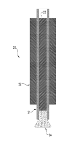
Turning now to a brief overview of illustrative deployment devices and
procedures useful for delivering a dried, expanded material as described
herein, with
general reference to Figures 2A and 2B, in certain aspects, a deployment
system 20
can include a cannulated device 21, e.g. a catheter, sheath or other tube that
can be
used to house and deliver a hemostatic plug 24 as described herein. For
example,
the cannulated device 21 with the plug 24 housed therein can be maneuvered
through another instrument 22. Instrument 22 can be any of a variety of
surgical
instruments, including for example a laproscope, endoscope (including e.g. a
nephroscope), or an outer vascular access or delivery sheath. In embodiments
to wherein instrument 22 is an endoscopic instrument, instrument 22 will of
course
also typically include additional passages or channels extending therethrough
to
provide, for example, fiber optic light input, viewing function (e.g. with a
telescope
or camera), and the like. In these embodiments, the irrigation or other
working
channel of the endo scope can be used to pass the cannulated device 21 for
delivery
of the plug 24 to the target site.
A counterforce and/or pusher element 23 can be provided within the lumen
of cannulated device 21 to facilitate delivery of the plug 24 out of the open
distal
end of the cannulated device 21. The element 23 can be advanced forward within
device 21 to push plug 24 from device 21, or element 23 can be held in
position
against the plug 24 while device 21 is retracted to deliver plug 24 from
device 21, or
a combination of these two functions can be used. In alternative embodiments,
other
methods may be used for delivering plug 24 from cannulated device 21,
including as
one example the use of a liquid under pressure to force plug 24 from the open
end of
the cannulated device 21.
Illustratively, a plug 24 can be loaded within the cannulated device 21 and
can be deployed at a site for hemostasis (e.g. a biopsy site or within a
needle tract
through soft tissue resultant of a percutaneous access procedure) or otherwise
within
a bodily passage or void by using one or more actuator members positioned
external
of the patient that control the relative position of the cannulated device 21
and the
counterforce/pusher element 23. The one or more actuator members can include
manually operable triggers, rotatable knobs, or other elements that may be
connected
to device 21 and/or element 23 directly or through control wired, rods, or
other
CA 02728513 2015-07-14
23
suitable members known in the art. In certain embodiments, system 20 can have
one
or more actuator member(s) that deploy the plug 24 in a stepwise fashion, such
that
a first manual operation of the actuator(s) controllably delivers a
predetermined
percentage of the plug 24 from the open end of cannulated device 21 leaving
the
plug 24 in a partially delivered state, an a second manual operation of the
actuator(s)
delivers a further percentage of the plug 24 from the open end of cannulated
device.
Such further percentage is preferably the entire remainder of the plug 24,
although
systems may also be designed to deliver the plug fully upon multiple
additional
operations of the actuator(s). In certain embodiments, a first operation of
the
113 actuator member(s) deploys about 10% to about 70% of the length of plug
24 from
the end of cannulated device 21, and a second operation of the actuator(s)
delivers
the remainder of the plug 24 from the cannulated device.
Deployment devices, including delivery sheaths, cannulated devices, and
pushers, used in the invention can all be conventional marketed products or
5 modifications thereof. For example, sheaths can be formed from PTFE (e.g.
TeflonTm), polyamide (e.g. nylon) or polyurethane materials, or a combination
of
materials such as an assembly including an inner layer of PTFE, a flat wire
coil over
the PTFE for kink resistance, and a polyamide (Nylon) outer layer to provide
integrity to the overall structure and a smooth surface (e.g. as in the
FlexorTM sheath,
20 Cook, Inc.). Pushers can be made from conventional materials such as
polyethylene,
polyamide, polyurethane or vinyl, stainless steel, or any combination of these
materials. Catheters can be made from conventional materials such as
polyethylene,
polyamide, PTFE, polyurethane, and other materials.
An expanded material as described herein can be compressed prior to
25 delivery and can expand following deployment from the catheter until it
contacts
inner surfaces of a bodily passage or void, a biopsy site or other surgically
created
void. With certain designs, this expansion and contact will be sufficient to
maintain
the material at a particular location in the bodily passage or void following
deployment, although some inventive implants will incorporate one or more
30 anchoring or securement adaptations (not shown) so as to mitigate
undesirable
migration of the device from or within the passageway or void. In some
instances,
parts of an expanded material can embed themselves in tissues surrounding the
void
CA 02728513 2010-12-17
WO 2009/155600
PCT/US2009/048152
03433-001138 24 609223
or passageway upon deployment. Any number of anchoring adaptations, such as
barbs, hooks, ribs, protuberances, and/or other suitable surface modifications
can be
incorporated into an inventive devices to anchor them during and/or after
deployment.
Hemostatic products as described herein can be any suitable length and will
generally be of sufficient dimension to achieve hemostasis at a desired
location e.g.,
a surgery site. In certain embodiments, a device, in implanted form, will have
a
length of at least about 0.4 cm, and in many situations will have a length
ranging
from about 1 cm to about 30 cm, more typically from about 2 cm to about 15 cm.
to As noted above, one or more additives can provide a variety of
functions,
including promoting expansion of the material once implanted into a patient.
For
example, a sponge form expanded remodelable collagenous material including one
or more additives can be compressed and placed into a delivery device.
Compression of the material allows the material to be more easily transferred
to a
patient. Upon delivery, the material can expand to at least about its original
size
prior to compression. This is typically done with an occluder device or a
biopsy
plug where it is desirable for the material to have a smaller diameter prior
to delivery
and expand upon delivery. Such additives can be included in the remodelable
collagenous material to expand the material at a faster rate than would
otherwise be
achievable in the absence of the one or more additives. For example, one or
more
additives can be included with a compressed remodelable collagenous material
so as
to promote the re-expansion of the material back to its original size within
at least
about 30 seconds, 45 seconds, 1 minute, 2 minutes, 3 minutes, 4 minutes, or
even
about least about 5 minutes after implantation. As with the bioactive
components
previously described, these additives may be applied to the expanded
remodelable
collagenous material as a premanufactured step, immediately prior to the
procedure
(e.g. by soaking the material in a solution containing a suitable antibiotic
such as
cefazolin), or during or after engraftment of the material in the patient.
As noted above, expanded remodelable collagenous materials can be formed
into a sponge construct for implantation into a patient. In certain
embodiments, a
sponge construct will be constructed such that the material does not fully
expand
until after delivery to a desired site (e.g., tissue defect). In these
instances, an
CA 02728513 2010-12-17
WO 2009/155600
PCT/US2009/048152
03433-001138 25 609223
expanded remodelable collagenous material can be encapsulated, either
partially or
wholly, so as to prevent the premature expansion of the material until it
reaches its
intended delivery site. For example, a dried sponge material as described
herein can
be compressed and either partially or wholly encapsulated into a biodegradable
capsule. In such embodiments, the capsule can retain the material in a
compressed
state so as to prevent the premature expansion of the expanded remodelable
collagenous material during delivery. This allows the material to be delivered
to a
desired location before full expansion occurs. In a similar embodiment, an
expanded remodelable collagenous material in powder form can be provided in a
to biocompatible, biodegradable capsule for delivery. Such an embodiment
retains the
powder within the capsule so as to prevent portions of the powder from being
delivered or drifting to an unintended location. Biocompatible materials
suitable for
use in forming a biodegradable capsule are generally known in the art and can
include, for example, gelatin.
In certain embodiments, an expanded remodelable collagenous material, in any
form, can be crosslinked. An expanded remodelable collagenous material can be
crosslinked either before or after it is formed into a medical device, or
both. Increasing
the amount (or number) of crosslinkages within the material or between two or
more
layers of the material can be used to enhance its strength. However, when a
remodelable material is used, the introduction of crosslinkages within the
material may
also affect its resorbability or remodelability. Consequently, in certain
embodiments, a
remodelable collagenous material will substantially retain its native level of
crosslinking, or the amount of added crosslinkages within the medical device
will be
judiciously selected depending upon the desired treatment regime. In many
cases, the
material will exhibit remodelable properties such that the remodeling process
occurs
over the course of several days or several weeks. In certain preferred
embodiments, the
remodeling process occurs within a matter of about 5 days to about 12 weeks.
With
regard to a sponge form construct, crosslinking of a compressed construct may
promote
re-expansion of the construct after implantation into a patient.
With regard to compressible/expandable plugs, sponges or other constructs as
described herein, expansion additives and/or crosslinking can be used to
impart
desirable compression/re-expansion properties. In preferred forms, the
constructs are
CA 02728513 2015-07-14
26
capable of volumetric compression when dry at a ratio of at least 10:1 (i.e.
the
compressed form occupies no more than 10% of its original, relaxed and
unexpanded
volume), more preferably at a ratio of at least 20:1. At the same time, in
preferred
forms, the compressed constructs are capable of re-expansion to substantially
their
original volume (e.g. at least about 80% of their original volume, more
preferably at
least 90%, and most preferably at least 95%) within about 30 seconds when
delivered
in their dry, compressed form into a volume of water.
For use in the present invention, introduced crosslinking of the expanded
remodelable collagenous material may be achieved by photo-crosslinking
to techniques, or by the application of a crosslinking agent, such as by
chemical
crosslinkers, or by protein crosslinking induced by dehydration or other
means.
Chemical crosslinkers that may be used include for example aldehydes such as
glutaraldehydes, diimides such as carbodiimides, e.g., 1-ethy1-3-(3-
dimethylaminopropyl)carbodiimide hydrochloride (EDC), diisocyanates such as
hexamethylene-diisocyanate, ribose or other sugars, acyl-azide, sulfo-N-
hydroxysuccinamide, or polyepoxide compounds, including for example
polyglycidyl ethers such as ethyleneglycol diglycidyl ether, available under
the trade
mark DENACOL EX810 from Nagese Chemical Co., Osaka, Japan, and glycerol
polyglycerol ether available under the trade mark DENACOL EX 313 also from
Nagese Chemical Co. Typically, when used, polyglycerol ethers or other
polyepoxide compounds will have from 2 to about 10 epoxide groups per
molecule.
When a multi-layered laminate material is contemplated, the layers of the
laminate can be additionally crosslinked to bond multiple layers of a multi-
layered
medical material to one another. Cross-linking of multi-layered medical
materials
can also be catalyzed by exposing the matrix to UV radiation, by treating the
collagen-based matrix with enzymes such as transglutaminase and lysyl oxidase,
and
by photocrosslinking. Thus, additional crosslinking may be added to individual
layers prior to coupling to one another, during coupling to one another,
and/or after
coupling to one another.
CA 02728513 2010-12-17
WO 2009/155600
PCT/US2009/048152
03433-001138 27 609223
The medical materials, constructs and devices of the invention can be
provided in sterile packaging suitable for medical materials and devices.
Sterilization may be achieved, for example, by irradiation, ethylene oxide
gas, or
any other suitable sterilization technique, and the materials and other
properties of
the medical packaging will be selected accordingly.
In certain embodiments, the invention provides compressible medical foam
products, and methods for their preparation. The medical foam products include
a
dried, compressible foam body formed with an extracellular matrix solid
material
that has been treated with an alkaline medium under conditions effective to
produce
to an expanded extracellular matrix collagen material. The foam body has
introduced
chemical crosslinks sufficient to increase the resiliency of the foam body.
Absent
crosslinking, foam bodies produced from the expanded extracellular matrix
collagen
material possess resiliency, but for certain applications, including for
example
hemostatic plug applications, it has been discovered that increased resiliency
is
desired. The introduction of collagen crosslinks, for example with chemical
crosslinkers such as glutaraldehyde, carbodimides, or other chemical
crosslinkers
identified herein, has been found to significantly enhance the resiliency of
the foam
plugs, while leaving the compressible to a small size for delivery. Increased
resiliency in turn provides additional compression upon adjacent tissues when
the
foam plugs are inserted in a compressed state and then allowed to expand in
situ in a
patient at a site at which hemostasis is desired. In specific inventive
applications,
crosslinked, resilient foam plugs as disclosed herein can be utilized to
provide
hemostasis at surgical sites, including biopsy sites. These biopsy or other
surgical
sites can be located within parenchymal organ tissues, such as those of a
kidney,
liver or spleen of a patient.
Thus, in certain forms of the invention, surgical methods are provided which
include resecting tissue from a parenchymal organ such as a liver or kidney,
and
then implanting a crosslinked, resilient foam material as described herein at
the
resection site so as to facilitate hemostasis. The resection can, as examples,
occur as
a part of a nephrectomy or hepatectomy, e.g. to removed cancerous or other
diseased
tissue, or as a part of a kidney or liver biopsy performed with a biopsy
needle. In the
case of minimally invasive surgical procedures such as laparoscopic
resections, or
CA 02728513 2010-12-17
WO 2009/155600
PCT/US2009/048152
03433-001138 28 609223
needle biopsies, the crosslinked, resilient foam plug can be delivered from
within a
cannulated device such as a needle or catheter, and/or through a laparoscopic
device.
The resilient foam plug can be in a compressed state during delivery, and then
allowed to expand once delivered to the surgical site. The expansion of the
plug can
compress the adjacent tissues to facilitate hemostasis. For these purposes,
the
expanded dimensions of the plug can provide a volume that is at least equal to
or
preferably greater than the volume of the biopsy or other surgical defect, to
ensure
compression of surrounding tissues by the delivered, expanded plug.
In other embodiments of the invention, methods are provided which include
to deploying a crosslinked, resilient foam material as described herein at
a site within a
bodily vessel, for example an artery or a vein, so as to cause occlusion of
the vessel
and thereby stop the flow of fluid (e.g. blood) within the vessel. In the case
of
minimally invasive surgical procedures such as percutaneous procedures the
crosslinked, resilient foam plug can be delivered from within a cannulated
device
such as a catheter or sheath. The resilient foam plug can be in a compressed
state
during delivery, and then allowed to expand once delivered from within the
cannulated device to the desired occlusion site. The expansion of the plug can
compress the walls of the vessel to facilitate occlusion. For these purposes,
the
expanded dimensions of the plug can be greater than the diameter of the vessel
at the
desired site of occlusion, to ensure outward compression against surrounding
vessel
walls by the delivered, expanded plug. Besides vascular vessels, other vessels
that
can be occluded in accordance with the invention include, for example,
fallopian
tube(s). Still further, other open tracts through patient tissue can be
occluded with
crosslinked, resilient foam plugs of the invention, including for example
needle
tracts (e.g. resultant of percutaneous entry to a vein or artery) and
fistulas, such as
anorectal fistulas, enterocutaneous fistulas, recto-vaginal fistulas, and
others.
Crosslinked, resilient foam plugs can be prepared according to the invention
by a process that includes:
(a) contacting extracellular matrix material with an alkaline medium to form
an expanded extracellular matrix material;
(b) washing the expanded extracellular matrix material;
(c) charging the expanded extracellular matrix material to a mold;
CA 02728513 2010-12-17
WO 2009/155600
PCT/US2009/048152
03433-001138 29 609223
(d) lyophilizing the expanded extracellular matrix material in the mold to
form a lyophilized extracellular matrix material foam;
(e) contacting the lyophilized extracellular matrix material foam with a
chemical crosslinking agent to form a crosslinked extracellular matrix
material
foam; and
(f) drying the crosslinked extracellular matrix material foam.
In such methods, the extracellular matrix material and chemical crosslinked
agent
can, for example, be selected from among any of those disclosed herein. The
to washing can suitably be conducted with an aqueous medium, such as saline
or water.
The drying can be conducted by any suitable method, including as examples air
drying at ambient temperature, heated drying, or lyophilization. It is
preferred to
contact the extracellular matrix material with the chemical crosslinker after
the
formation of the lyophilized extracellular matrix material foam (e.g. as
opposed to
incorporating the chemical crosslinker in the material charged to the mold),
as this
has been found to provide more uniformly-shaped crosslinked plugs that resist
shrinkage. Further, in such preparative methods, the expanded extracellular
matrix
material can be comminuted prior to charging to the mold. Such comminuting
will
provide extracellular matrix fragments, e.g. randomly generated, that will be
incorporated within and characterize the extracellular matrix foam. In more
preferred forms, the material is comminuted by shearing the material with a
rotating
blade, e.g. in a blender. For these purposes, it has been discovered that when
utilizing an extracellular matrix material that is a harvested, deceullarized
sheet, the
sheet can be contacted with the alkaline medium under conditions sufficient to
substantially reduce the tensile strength of the sheet, so that the sheet
material is
disrupted by the rotating blade. Without sufficient reduction of tensile
strength, the
sheet material can tend to wrap around the rotating blade, thus frustrating
the
process of comminution. For example, prior to comminution by the blade or
otherwise, the sheet can be treated with the alkaline medium for a time and
under
conditions sufficient to reduce the tensile strength of the sheet to less than
about
50% of its original tensile strength, more preferably to less than about 30%
of its
original tensile strength. Such methods can be practiced, for example, with
CA 02728513 2010-12-17
WO 2009/155600
PCT/US2009/048152
03433-001138 30 609223
harvested sheet-form ECM materials such as submucosa-containing sheets, e.g.
obtained from small intestinal, stomach or bladder tissue, pericardial tissue,
peritoneal tissue, fascia, dermal tissue, and other sheet-form ECM materials.
In additional embodiments of the invention, bioactive composite
extracellular matrix material products are used. The composite product
comprises a
dried body formed with an extracellular matrix material that has been treated
with an
alkaline medium under conditions effective to produce an expanded
extracellular
matrix material, particles of a bioactive extracellular matrix material
entrapped
within said dried body, wherein the particles of bioactive extracellular
matrix
to material retain at least one growth factor from a source tissue for the
particulate
extracellular matrix material. The composite products can be prepared by:
(a) contacting extracellular matrix material with an alkaline medium to form
an expanded extracellular matrix material;
(b) washing the expanded extracellular matrix material;
(c) preparing a mixture including a liquid, the expanded extracellular matrix
material and a particulate extracellular matrix material, the particulate
extracellular
matrix material retaining an amount of at least one growth factor from a
source
tissue for the particulate extracellular matrix material; and
(d) drying the mixture to form a bioactive, composite extracellular matrix
material construct.
In such composite products and preparative methods, the extracellular matrix
material that is expanded, and the particulate extracellular matrix material,
can, for
example, be selected from among any of those disclosed herein. The washing can
suitably be conducted with an aqueous medium, such as saline or water. The
liquid
for preparing the mixture can be any suitable liquid, preferably
biocompatible, and
typically an aqueous liquid such as water or saline. The drying step can be
conducted by any suitable method, including as examples air drying at ambient
temperature, heated drying, or lyophilization. Further, in such preparative
methods,
the expanded extracellular matrix material is desirably comminuted prior to or
during the formation of the mixture. In more preferred forms, the material is
comminuted by shearing the material with a rotating blade, e.g. in a blender,
alone
CA 02728513 2010-12-17
WO 2009/155600
PCT/US2009/048152
03433-001138 31 609223
or in the presence of the bioactive particulate extracellular matrix material.
Such
methods can be practiced, for example, with harvested sheet-form ECM materials
such as submucosa-containing sheets, e.g. obtained from small intestinal,
stomach or
bladder tissue, pericardial tissue, peritoneal tissue, fascia, dermal tissue,
and other
sheet-form ECM materials. The expanded ECM material and the bioactive
particulate ECM material can be from the same ECM starting material or from
different ECM starting materials. The incorporation of the particulate ECM
material
can serve not only to enhance the bioactivity of the foam product, but also
they
enhance the resiliency of the foam product. These aspects can be used to
advantage
to in hemostatic, occlusion and other medical treatments described herein.
Additional embodiments utilize composite extracellular matrix material
products that include an extracellular matrix sheet material and a dried
material
adhered to the extracellular matrix sheet material, wherein the dried material
is
formed from an extracellular matrix material that has been contacted with an
alkaline medium to form an expanded extracellular matrix material. Such
composite
products can be prepared by a method the includes the steps of:
(a) contacting extracellular matrix material with an alkaline medium to form
an expanded extracellular matrix material;
(b) washing the expanded extracellular matrix material;
(c) casting a flowable, wet preparation of the expanded extracellular matrix
material against an extracellular matrix sheet to form a wet composite; and
(d) drying the wet composite so as to form a dried composite.
In such composite sheet-material products and preparative methods, the
extracellular matrix material that is expanded, and the particulate
extracellular
matrix material, can, for example, be selected from among any of those
disclosed
herein. The washing can suitably be conducted with an aqueous medium, such as
saline or water. The liquid for preparing the wet preparation can be any
suitable
liquid, preferably biocompatible, and typically an aqueous liquid such as
water or
saline. The drying step can be conducted by any suitable method, including as
examples air drying at ambient temperature, heated drying, or lyophilization.
Lyophilization is preferred as it forms a more porous, resilient foam material
as
CA 02728513 2010-12-17
WO 2009/155600
PCT/US2009/048152
03433-001138 32 609223
compared to air drying or heated drying. Further, in such preparative methods,
the
expanded extracellular matrix material in the flowable, wet preparation is
desirably
comminuted. In more preferred forms, the material is comminuted by shearing
the
material with a rotating blade, e.g. in a blender. Such methods can be
practiced, for
example, with harvested sheet-form ECM materials such as submucosa-containing
sheets, e.g. obtained from small intestinal, stomach or bladder tissue,
pericardial
tissue, peritoneal tissue, fascia, dermal tissue, and other sheet-form ECM
materials.
The expanded ECM material and the sheet-form ECM material can be from the
same ECM starting material or from different ECM starting materials. The
to incorporation of the sheet-form ECM material can serve not only to
enhance the
bioactivity of the overall product, but can also provide a barrier material
and/or
suturable sheet attached to the dried expanded ECM material (e.g. foam).
Illustratively, such constructs can be used to provide hemostasis to surgical
sites or
other injured tissue. In certain modes of practice, the construct can be
placed against
the bleeding tissue with the dried, expanded ECM material (especially a foam)
against the bleeding tissue. The sheet-form ECM can then provide an additional
barrier (besides the expanded ECM material) to protect the bleeding tissue,
and or
can provide a suturable sheet material which can be used to fix the construct
in
place, e.g. with sutures in strand or staple form. In specific uses, such
constructs can
be used to apply hemostasis to surgically-treated (e.g. subject to resection)
or
otherwise injured parenchymous organ tissue, such as liver or kidney tissue.
In so
doing, the dried, expanded ECM material is desirably pressed against the
injured
parenchymous tissue, and the sheet-form ECM material can optionally be used to
fix
the construct in place, as discussed above. These and other modes of practice
with
the composite sheet-form constructs will be apparent to those of ordinary
skill in the
art from the descriptions herein.
Other embodiments utilize implantable medical products that comprise a
dried, resilient foam body formed with an extracellular matrix material that
has been
treated with an alkaline medium sufficient to form an expanded extracellular
matrix
material, and a biodegradable capsule component covering at least a portion of
the
dried resilient foam body. The dried, resilient foam body can by any such body
disclosed herein, and can be received in a compressed form within the capsule
CA 02728513 2010-12-17
WO 2009/155600
PCT/US2009/048152
03433-001138 33 609223
component. In certain forms, the capsule component covers at least a leading
end of
the foam body, and can serve to create a more desirable delivery profile for
the
product. In additional forms, the foam body can be entirely received within a
capsule component, preferably in a compressed state. As the capsule component
degrades and weakens after implantation, the capsule can split or otherwise
break
under the force of the compressed foam body, thus releasing the foam body to
expand. The expanded foam body can then serve to provide hemostasis, occlusion
and/or another therapeutic effect at the site of implantation. The
biodegradable
capsule can be made of any suitable biodegradable material, including for
example
to gelatin.
Additional embodiments involve the use of implantable medical products
that comprise a powder material and a biodegradable capsule enclosing the
powder
material. Particles of the powder material comprise a dried foam formed with
an
extracellular matrix material that has been treated with an alkaline medium
sufficient
to form an expanded extracellular matrix material. Such capsular devices can
be
used to effectively deliver and retain the powdered extracellular matrix
material at a
site of implantation, for example a site for hemostasis or occlusion as
described
herein. The powder material can serve to promote hemostasis, tissue ingrowth,
or
another beneficial effect at the site of implantation. The biodegradable
capsule can
be made of any suitable biodegradable material, including for example gelatin.
For the purpose of promoting a further understanding of aspects of the
present invention, the following specific examples are provided. It will be
understood that these examples are not limiting of the present invention.
EXAMPLE 1
This example demonstrates the process used to prepare a disinfected small
intestinal submucosa tissue (i.e., non-expanded SIS), which can subsequently
be
used in the preparation of various medical materials and devices. Surface and
cross
section micrographs of the material are depicted in Figs. 1B and 1D.
CA 02728513 2015-07-14
34
A ten foot section of porcine whole intestine was extracted and washed with
water. After rinsing, this section of submucosa intestinal collagen source
material
was treated for about two and a half hours in 0.2% peracetic acid by volume in
a 5%
by volume aqueous ethanol solution with agitation. Following the treatment
with
the peracetic acid solution, the submucosa layer was delaminated in a
disinfected
casing machine from the whole intestine. The resultant submucosa was then
rinsed
four (4) times with sterile water. A 1 cm by 1 cm section of this material was
extracted and stained using a solution of direct red prepared by mixing 10 mg
direct
red in 100 mL high purity water. The section of material was stained for
to approximately 5 minutes. The stained material was washed twice with high
purity
water to remove any unbound stain. The stained material was placed on a glass
slide
and covered with a cover slip. A micrograph was taken (Olympus microscope) at
100x magnification of the surface of the material. A cross section of the
material
was then prepared and a similar micrograph was taken. The resulting micrograph
was analyzed using Spot RTTm software. The surface and cross section
micrographs
are depicted in Figs. 1B and 1D. Both the surface and cross section
micrographs
show a tightly bound collagenous matrix with no expansion.
EXAMPLE 2
This example demonstrates the process used to prepare an expanded small
intestinal submucosa tissue (i.e., expanded SIS), which can subsequently be
used in
the preparation of various medical materials and devices as described herein.
Surface and cross section micrographs of the material are depicted in Figs. IA
and
1C.
A ten foot section of porcine whole intestine was extracted and washed with
water. After rinsing, this section of submucosa intestinal collagen source
material
was treated for about two and a half hours in 0.2% peracetic acid by volume in
a 5%
by volume aqueous ethanol solution with agitation. Following the treatment
with
peracetic acid, the submucosa layer was delaminated in a disinfected casing
machine
from the whole intestine. The resultant submucosa was then rinsed four (4)
times
with sterile water. 300 g of this material was soaked with agitation in 1 L of
a 1M
CA 02728513 2010-12-17
WO 2009/155600
PCT/US2009/048152
03433-001138 35 609223
NaOH solution at 37 C for 1 hour and 45 minutes. The material was removed and
rinsed in a 1L solution of high purity water for 5 minutes. This rinsing step
was
repeated 8 additional times. A lcm by 1 cm section of this material was
extracted
and stained using a solution of direct red prepared by mixing 10 mg direct red
in 100
mL high purity water. The section of material was stained for approximately 5
minutes. The stained material was washed twice with high purity water to
remove
any unbound stain. The stained material was placed on a glass slide and
covered
with a cover slip. A micrograph was taken (Olympus microscope) at 100x
magnification of the surface of the material. A cross section of the material
was
to then prepared and a similar micrograph was taken. The resulting
micrograph was
analyzed using Spot RT software. The surface and cross section micrographs are
depicted in Figs. 1A and 1C. Both the surface and cross section micrographs
show
disruption of the tightly bound collagenous matrix and an expansion of the
material.
As can be observed in Figs. 1A-1D, both the surface view and the cross-
section view of the non-expanded SIS show a tightly bound collagenous matrix
whereby collagen content is substantially uniform throughout. Conversely, the
surface view and cross-section view of the expanded SIS show a denatured
collagenous network and an expansion of the material.
EXAMPLE 3
This Example was performed to identify additives that can be included in an
expanded remodelable collagenous material for purposes of promoting rapid re-
expansion of the material after implantation into a patient.
An expanded remodelable material was prepared generally as described in
Example 2. Briefly, a ten foot section of porcine whole intestine was
extracted and
washed with water. After rinsing, this section of submucosa intestinal
collagen
source material was treated for about two and a half hours in 0.2% peracetic
acid by
volume in a 5% by volume aqueous ethanol solution with agitation. Following
the
treatment with peracetic acid, the submucosa layer was delaminated in a
disinfected
casing machine from the whole intestine. The resultant submucosa was then
rinsed
four (4) times with sterile water. 300 g of this material was soaked with
agitation in
CA 02728513 2010-12-17
WO 2009/155600
PCT/US2009/048152
03433-001138 36 609223
1 L of a 3M NaOH solution at 37 C for 2 hours. The material was removed and
rinsed in a 1L solution of high purity water for 15 minutes. After 15 minutes,
1L of
0.2M acetic acid was added with agitation. After 15 minutes of agitation, the
material was rinsed with 1L of high purity water with shaking for 5 minutes.
This
rinsing step was repeated four (4) times for a total of five (5) rinses.
The rinsed material was mechanically agitated using the pulse setting of a
blender to the extent that the blended material could be transferred using a
disposable 25 mL pipette. Samples of the blended material were combined with a
handheld blender with the various additives as identified in Table 1. The
samples
to were then cast into cylindrical molds, frozen at -80 C for 5 hours, and
lyophilized
for 24 hours to yield 14 mm diameter cylindrical constructs ranging in length
from
about 15 mm to about 19 mm.
CA 02728513 2010-12-17
WO 2009/155600
PCT/US2009/048152
03433-001138 37 609223
TABLE 1
Additive Category Screened Additives
Sodium chloride
Sodium acetate
Sodium bicarbonate
Salts Sodium citrate
Calcium carbonate
Potassium acetate
Potassium phosphate
Alginate
Polyhydroxyethyl methacralate
Polyvinyl alcohol
Hydrogels and Water Swelling Polymers
Polyethylene glycol
Carboxymethyl cellulose
Polyvinyl pyrrolidone
Gelatin
Proteins
SIS particulate
Acetic acid
Acids and Bases
Ascorbic acid
Dextran
Monosaccharides and Polysaccharides Glucose
Fructose
Superabsorbing Polymers and Gelling Polyacrylic acid
Agents Polygalacturonic acid
Guanidine HCI
Other Additives
Urea
At the time of testing, the initial sample diameter was recorded. All
cylindrical samples were then compressed by hand to between 2.7 mm and 6.7 mm,
CA 02728513 2010-12-17
WO 2009/155600
PCT/US2009/048152
03433-001138 38 609223
and the final diameter of the compressed material was recorded. Approximately
20
mL of high purity water at room temperature was transferred into a weight
boat.
The compressed material was placed on the surface of the high purity water and
submerged using forceps to expose all surfaces of the material to the high
purity
water. A digital timer was started at the time the sample was submerged.
Visual
assessment of the material was continuously conducted until the sample
returned to
the initial sample diameter as assessed through visual inspection. When the
sample
returned to the initial sample diameter, the timer was stopped and the
expansion time
recorded. Visual assessment was discontinued after 15 minutes for samples that
did
to not return to the initial sample diameter in the time allotted. The
results are
summarized in Tables 2-8.
TABLE 2
Additive % Dry Initial Compressed Expansion
Weight of Diameter Diameter Time
Dry Plug (mm) (mm) (min:sec)
2.5 12 4.0 1:41
Sodium
7.5 13 4.0 >15:00*
= ___________________________________________________________________
chloride
14 3.7 >15:00*
1.25 13 3.7 6:30
13 3.7 6:00
13 4.7 0:45
Sodium acetate 2.5
12 4.3 0:45
13 4.7 1:30
5.0
14 5.0 2:00
13 4.0 2:00
2.5
Sodium 13 4.3 1:15
bicarbonate 5.0 13 6.7 3:00
13 5.0 1:20
Sodium citrate 14 5.3 8:00
2.5
14 5.0 8:00
CA 02728513 2010-12-17
WO 2009/155600
PCT/US2009/048152
03433-001138 39 609223
5.0 . 14 5.0 12:00
14 4.7 12:00
2.5 14 4.7 >15:00*
14 4.3 >15:00*
Calcium 5.0 14 5.3 8:00
carbonate 14 5.0 8:00
12.5 14 4.7 >15:00*
14 4.7 >15:00*
2.5 13 5.3 >15:00*
Potassium 13 4.7 >15:00*
acetate 5.0 . 14 4.3 >15:00*
14 4.3 >15:00*
Potassium 2.5 10 3.0 13:00
phosphate 10 3.0 13:00
5.0 . 13 3.0 14:00
12 3.3 11:00
*Indicates control sample behaved atypically, suggesting the expansion time
may not be
representative of the additive tested.
TABLE 3
Additive %Dry Initial Diameter
Expansion
Weight of Diameter (mm) Time
Dry Plug (mm) (min:sec)
Alginate 2.5 13 3.0 >15:00
Polyhydroxyethyl 2.5 13 3.0 8:50
methacralate 13 2.7 8:58
Polyvinyl alcohol 2.5 14 3.0 5:48
Polyethylene 7.5 14 2.3 >15:00*
glycol (MW 400) 14 2.3 >15:00*
Polyethylene 2.5 13 3.0 3:22
glycol (MW 6000)
Carboxymethyl 2.5 13 3.7 7:03
cellulose
CA 02728513 2010-12-17
WO 2009/155600
PCT/US2009/048152
03433-001138 40 609223
Polyvinyl 2.5 14 3.3 5:25
pyrrolidone
*Indicates control sample behaved atypically, suggesting the expansion time
may not be
representative of the additive tested.
TABLE 4
Additive %Dry Initial Diameter Expansion
Weight of Diameter (mm) Time
Dry Plug (mm) (min:sec)
Gelatin (100 2.5 13 3.0 >15:00
bloom)
5.0 14 4.7 2:38
13 4.7 2:35
45-90 um SIS 10.0 13 5.0 1:32
particulate 13 4.7 1:20
20.0 14 6.3 0:37
14 6.0 0:52
5.0 14 3.7 2:30
13 3.7 2:00
90-150 um 10.0 13 4.7 2:30
SIS particulate 14 5.0 3:00
20.0 13 5.3 1:30
13 6.3 1:42
5.0 14 4.0 2:45
14 4.3 2:50
150-200 um 10.0 14 4.7 2:30
SIS particulate 13 4.3 2:25
20.0 13 5.7 1:55
13 5.0 2:35
CA 02728513 2010-12-17
WO 2009/155600 PCT/US2009/048152
03433-001138 41 609223
TABLE 5
Additive %Dry Initial Diameter Expansion
Weight of Diameter (mm) Time
Dry Plug (mm) (min:sec)
2.5 14 3.0 >15:00*
14 3.0 >15:00*
Ascorbic acid
5.0 14 3.0 >15:00*
14 3.3 >15:00*
*Indicates control sample behaved atypically, suggesting the expansion time
may not be
representative of the additive tested.
TABLE 6
Additive %Dry Initial Diameter
Expansion
Weight of Diameter (mm) Time
Dry Plug (mm) (min:sec)
Polyacrylic acid 2.5 13 3.3 8:24
13 3.0 8:07
Polygalacturonic 2.5 13 3.0 4:00
acid 13 3.0 4:35
TABLE 7
Additive %Dry Initial Diameter Expansion
Weight of Diameter (mm) Time
Dry Plug (mm) (min:sec)
Dextran 2.5 13 3.0 5:15
13 3.3 4:16
2.5 14 3.7 >15:00*
14 3.7 >15:00*
Glucose
5.0 14 3.7 >15:00*
14 3.0 >15:00*
2.5 14 3.7 >15:00*
14 4.0 >15:00*
Fructose
5.0 14 3.3 >15:00*
14 3.7 >15:00*
*Indicates control sample behaved atypically, suggesting the expansion time
may not be
representative of the additive tested.
CA 02728513 2010-12-17
WO 2009/155600
PCT/US2009/048152
03433-001138 42 609223
TABLE 8
Additive %Dry Initial Compressed Expansion
Weight of Diameter Diameter Time
Dry Plug (mm) (mm) (min:sec)
Guanidine 2.5 14 3.0 4:16
HCI 14 2.7 4:50
Urea 5.0 14 3.0 >15:00
14 3.3 >15:00
Based on these results, preferred additives include sodium chloride, sodium
acetate, sodium bicarbonate, polyethylene glycol (MW 6000), and small
intestinal
submucosa particulate
EXAMPLE 4
This Example was performed to measure the angiogenic activity of various
forms of an expanded remodelable collagenous material as described herein.
An expanded remodelable material was prepared generally as described in
Example 3. Briefly, a ten foot section of porcine whole intestine was
extracted and
washed with water. After rinsing, this section of submucosa intestinal
collagen
source material was treated for about two and a half hours in 0.2% peracetic
acid by
volume in a 5% by volume aqueous ethanol solution with agitation. Following
the
treatment with peracetic acid, the submucosa layer was delaminated in a
disinfected
casing machine from the whole intestine. The resultant submucosa was then
rinsed
four (4) times with sterile water. 300 g of this material was soaked with
agitation in
1 L of a 3M NaOH solution at 37 C for 2 hours. The material was removed and
rinsed in a 1L solution of high purity water for 15 minutes. After 15 minutes,
1L of
0.2M acetic acid was added with agitation. After 15 minutes of agitation, the
material was rinsed with 1L of high purity water with shaking for 5 minutes.
This
rinsing step was repeated four (4) times for a total of five (5) rinses. Three
different
forms of expanded remodelable collagenous material were prepared from this
material: (1) blended expanded remodelable collagenous material, (2) expanded
remodelable collagenous material in conjunction with a submucosa particulate
(1:10), and (3) 4-layered lyophilized sheet form expanded remodelable
collagenous
material.
CA 02728513 2010-12-17
WO 2009/155600
PCT/US2009/048152
03433-001138 43 609223
These materials from groups (1) and (2) were cast into a thick film of
approximately 1 mm in thickness, frozen at -80 C for 5 hours and lyophilized
for 24
hours. Ten 15 mm discs were cut from each group using a disc punch to form
test
samples. Nylon filters with 0.221..tm pores were sewn on to the top and bottom
of
each disc. Low temperature ethylene oxide sterilization was used for each
sample.
Samples were implanted subcutaneously into the dorsal flanks of mice. After
anesthesia using Ketamine (87 mg/kg) and Xylazine (13 mg/kg), a small incision
was made on the posterior neck of the mouse, and a dorsal subcutaneous cavity
was
created using blunt dissection with hemostats. This was followed by sample
placement and closure of the incision with 4 interrupted stitches of 5-0
suture. Six
mice per group underwent disc implantation. The implant remained in the mice
for
a period of 3 weeks followed by probing for capillary formation.
Mice were sacrificed using a double dose of anesthesia to ensure intact flow
in vasculature. While the heart was still beating, the chest cavity was
exposed, vena
cava severed, and 10 mL of heparized saline injected into the left ventricle
using a
23 ga butterfly infusion set to exsanguinate the mouse. After transferring
syringes
(while maintaining infusion needle in left ventricle), 4 mL of a fluorescent
microsphere (yellow-green, 0.1 i.tm diameter, Molecular Probes, F-8803)
suspension
(1:20 dilution of stock suspension) was injected through the left ventricle
resulting
in perfusion of the entire vasculature. Care was taken to ensure no bubbles
were
introduced during the injections, as bubbles will cause micro-emboli
obstructing
consistent perfusion. Samples were collected with gentle dissection and gross
removal of the fibrous capsule. A positive control of hind limb muscle was
also
collected at this point to confirm proper perfusion. Collected samples and
controls
were placed on ice in a closed container to maintain tissue integrity (mainly
moistness). Microvasculature was imaged using a confocal microscope (Biorad),
kex=488 nm & kem=530 nm, along the edge of the samples in the area of greatest
vascular infiltration. Further, vasculature of the positive controls, hind
limb muscle,
was imaged to confirm good perfusion.
CA 02728513 2010-12-17
WO 2009/155600
PCT/US2009/048152
03433-001138 44 609223
In addition to the fluorescence microangiography described above, samples
were collected, placed in histology cassettes, and submerged in 10% buffered
formalin (Fisher). Histological sectioning and staining with hematoxilin and
eosin
were performed by Portland Tissue Processing. Images of H&E stained sections
of
the disc edge for each sample were taken using a microscope (Olympus) with a
10x
objective.
Each of the samples from all three test groups showed some angiogenic
activity when fluorescence microangiography was performed. Similarly, the
histology analysis confirmed that all three sample groups had some vascular
and
to cellular ingrowth.
This Example demonstrates that various forms of an expanded remodelable
collagenous material each exhibit angiogenic activity in vivo.
EXAMPLE 5
This Example was performed to investigate the angiogenic activity of a
crosslinked, expanded remodelable collagenous material as described herein.
An expanded remodelable material was prepared generally as described in
Example 3. Briefly, a ten foot section of porcine whole intestine was
extracted and
washed with water. After rinsing, this section of submucosa intestinal
collagen
source material was treated for about two and a half hours in 0.2% peracetic
acid by
volume in a 5% by volume aqueous ethanol solution with agitation. Following
the
treatment with peracetic acid, the submucosa layer was delaminated in a
disinfected
casing machine from the whole intestine. The resultant submucosa was then
rinsed
four (4) times with sterile water. 300 g of this material was soaked with
agitation in
1 L of a 3M NaOH solution at 37 C for 2 hours. The material was removed and
rinsed in a 1L solution of high purity water for 15 minutes. After 15 minutes,
1L of
0.2M acetic acid was added with agitation. After 15 minutes of agitation, the
material was rinsed with 1L of high purity water with shaking for 5 minutes.
This
rinsing step was repeated four (4) times for a total of five (5) rinses.
Approximately
250 mL of the expanded remodelable collagenous material was placed into a
blender
along with 250 mL of high purity water. This mixture was pulsed 10 times for 1
second each pulse followed by a 45 second blend. The resulting material was
cast
CA 02728513 2010-12-17
WO 2009/155600
PCT/US2009/048152
03433-001138 45 609223
into a 5x10 cm mold having a thickness of approximately 1 mm. This mold was
placed in a freezer at -80 C for 5 hours followed by lyophilization for 24
hours. 15
mm disc samples were cut from the resulting blended sheet.
To form the crosslinked samples, the samples formed above were combined
with 200 mL of 50 mM EDC crosslinking solution in a shallow glass dish. The
disc
with samples were submerged under solution and placed onto a rotating shaker
for
24 hours at room temperature. Each sample was then rinsed with 200 mL of high
purity water squeezing five (5) times. This step was repeated four (4) times
for a
total of five (5) rinses. The rinsed material was then lyophilized for
approximately 8
to hours.
Each of the samples showed some angiogenic activity when fluorescence
microangiography was performed. Similarly, the histology analysis confirmed
that
all three sample groups had some vascular and cellular ingrowth. Indeed, the
crosslinked material had robust angiogenesis (1442 + 108 um) and was still
present
in plug form. The plug expanded at explant indicating that the crosslinked
material
was substantive and did not collapse after implantation. Moreover, there were
no
signs of systemic or local toxicity and no evidence of increased local
inflammation
in these samples.
This Example further demonstrates that a crosslinked form of an expanded
remodelable collagenous material can exhibit angiogenic activity in vivo.
EXAMPLE 6
This Example was performed to determine the FGF-2 content of an expanded
remodelable collagenous material as described herein.
An expanded remodelable material was prepared generally as described in
Example 3. Briefly, a ten foot section of porcine whole intestine was
extracted and
washed with water. After rinsing, this section of submucosa intestinal
collagen
source material was treated for about two and a half hours in 0.2% peracetic
acid by
volume in a 5% by volume aqueous ethanol solution with agitation. Following
the
treatment with peracetic acid, the submucosa layer was delaminated in a
disinfected
casing machine from the whole intestine. The resultant submucosa was then
rinsed
CA 02728513 2010-12-17
WO 2009/155600
PCT/US2009/048152
03433-001138 46 609223
four (4) times with sterile water. 300 g of this material was soaked with
agitation in
1 L of a 3M NaOH solution at 37 C for 2 hours. The material was removed and
rinsed in a IL solution of high purity water for 15 minutes. After 15 minutes,
IL of
0.2M acetic acid was added with agitation. After 15 minutes of agitation, the
material was rinsed with IL of high purity water with shaking for 5 minutes.
This
rinsing step was repeated four (4) times for a total of five (5) rinses.
Two lots of material described above were prepared with one lot used per
group. One lot of material was made into single-layer lyophilized sheets, and
the
other material was mixed with small intestinal submucosa particulate (-150 um)
and
to made into single-layer lyophilized sheets. Three (3) samples were cut (2
cm x 2 cm)
from each lot resulting in three (3) samples per group. Each sample was
weighed
and its weight was recorded. Individual samples were placed in 1.5 mL
eppendorf
tubes and 400 ul of sterile phosphate buffered saline (PBS) was added to each
tube.
Tubes with samples were centrifuged at 12000 g for 5 minutes at 4 C. The
resulting supernatant was diluted to 1:1 with lx PBS. Samples were assayed in
duplicate for FGF-2 content using R&D Systems FGF-2 ELISA kits per
manufacturer's instructions.
The resulting content of FGF-2 was calculated by dividing the FGF-2
content by the weights of the samples. The means measured FGF-2 content in the
sheet form expanded remodelable collagenous material was 0 pg/g. The mean
measured FGF-2 content in expanded remodelable collagenous material including
a
submucosa particulate was 4500 pg/g + 1600 pg/g.
This Example demonstrates that an expanded remodelable collagenous
material in sheet form, prepared and tested as described in this example,
contains no
detectable levels of FGF-2, and that FGF-2 can be provided back to an expanded
remodelable collagenous material by virtue of the inclusion of a submucosa
particulate into the material.
The use of the terms "a" and "an" and "the" and similar references in the
context of describing the invention (especially in the context of the
following
claims) are to be construed to cover both the singular and the plural, unless
otherwise indicated herein or clearly contradicted by context. Recitation of
ranges
of values herein are merely intended to serve as a shorthand method of
referring
CA 02728513 2015-07-14
47
individually to each separate value falling within the range, unless otherwise
indicated herein, and each separate value is incorporated into the
specification as if it
were individually recited herein. All methods described herein can be
performed in
any suitable order unless otherwise indicated herein or otherwise clearly
contradicted by context. The use of any and all examples, or exemplary
language
(e.g., "such as") provided herein, is intended merely to better illuminate the
invention and does not pose a limitation on the scope of the invention unless
otherwise claimed. No language in the specification should be construed as
indicating any non-claimed element as essential to the practice of the
invention.
Preferred embodiments of this invention are described herein, including the
best mode known to the inventors for carrying out the invention. Of course,
variations of those preferred embodiments will become apparent to those of
ordinary
skill in the art upon reading the foregoing description. The inventors expect
skilled
artisans to employ such variations as appropriate, and the inventors intend
for the
invention to be practiced otherwise than as specifically described herein.
Accordingly, this invention includes all modifications and equivalents of the
subject
matter recited in the claims appended hereto as permitted by applicable law.
Moreover, any combination of the above-described elements in all possible
variations thereof is encompassed by the invention unless otherwise indicated
herein
or otherwise clearly contradicted by context. In addition, all publications
cited
herein are indicative of the abilities of those of ordinary skill in the art.