Note: Descriptions are shown in the official language in which they were submitted.
CA 02729126 2010-12-22
WO 2010/002350 PCT/SE2009/050863
TITLE
On-line measuring system of body substances
TECHNICAL FIELD
The present invention relates to a system for continuously measuring
substances
present in the body. More specifically, the system is suitable for measuring
substances
that are indicators of pathological conditions and the sampling probe of the
system may
be placed in the blood stream or in the tissue of an organ. The present
invention also
relates to a method of presenting measured values.
BACKGROUND
Since recently it is known that certain substances that may be present in the
body
can function as indicators for various pathological conditions in the body.
Such
substances are hereafter called indicator substances. Examples of indicator
substances
are glucose, lactate, pyruvate, glycerol, glutamate, and glutamine and heart
specific
enzymes. Pathological conditions that may be indicated or detected, or as well
2 0 forecasted, include ischemia, hypoglycemia sepsis, cell membrane damage
or lipolysis,
vasospasms and metabolic disorders. By measuring indicator substances,
pathological
conditions may be detected before they lead to clinical signs. It may even be
possible to
detect processes or conditions that eventually may lead to a pathological
condition. In
many cases it would be advantageous to have the possibility to measure the
concentration of indicator substances directly in a blood stream, or in tissue
fluid.
However, until now there have not existed any systems suitable for clinical
use for
measuring indicator substances. Systems known from the background art all have
different drawbacks. Examples of common drawbacks in background art systems
are
that the measurement delay is extensive and that one has measured phenomena
that are
the result of a pathological condition, e.g. ischemia. This is clearly
disadvantageous.
With measurement delay is meant the time that passes from the moment that a
sample
is taken until the moment that a measurement value relating to this sample is
obtained.
In background art systems also measurement values can often only be obtained
with
CA 02729126 2010-12-22
WO 2010/002350 PCT/SE2009/050863
2
relatively extended time periods, between each measurement value, e.g. if
sample fluid
is collected in micro vials. Faced with the aim or task to develop a reliable
and accurate
measuring system that can be used in monitoring the condition of a subject,
e.g. a
patient, in a critical condition or situation, the skilled person is faced
with other
problems and situations than those which previously have aroused.
From US-A-5 078 135 there is known a measuring system where a drug is
administrated to a rat and where a microdialysis probe is placed in the vein
of the rat.
Mass spectrometry is used to batchwise analyse the dialysate for obtaining
pharmacokinetic data.
From US-A1-2004/0191848 there is known a system for measuring the
concentration of glucose in tissue fluid. A microdialysis probe is used which
is fed with
a perfusate fluid already containing glucose. The concentration of glucose in
the
perfusate fluid is controlled using self-adaptive control.
It is an object to provide a measuring system that is improved with respect to
the
background art. A further object is to provide a system that is reliable and
accurate to
make the system suitable for clinical use with such suitable response times
and the
system is useful for on-line monitoring in critical care.
SUMMARY OF INVENTION
The above mentioned object and others may be obtained by providing a system
measuring the concentration of substances or analytes in a body fluid or in a
body
tissue according to the independent claims attached below.
In general terms the system is provided with a microdialysis probe comprising
a
microdialysis membrane, both being adapted to be placed in blood or in tissue
fluid.
The probe is adapted to be invasively located in the body and to deliver
perfusion fluid
to and from the microdialysis membrane. The microdialysis probe of the system
may
be of the type disclosed in US Patents Nos 6,264,627; 6,632,315; 6,346,090;
6,811,542;
or in the Swedish patent application 5E0602199-2. The probe dimensions may
vary
dependent on the selected clinical application and its location in the body.
In a first
embodiment, suitable for a cardiac catheter, the probe has a length of 55 cm
and one
inflow lumen and one outflow lumen where each lumen has an inner diameter of
0,15
CA 02729126 2010-12-22
WO 2010/002350 PCT/SE2009/050863
3
mm. In another embodiment, suitable for a peripheral vein catheter, the probe
has a
length of about 10 cm and inner flow channels with diameter of about 0,15 mm.
The
system further includes a flow through sensor for analysing a fluid having
passed said
microdialysis probe and a pump for pumping the perfusion fluid to and through
the
microdialysis probe and to and through the sensor. A tubing connects the pump
to the
microdialysis probe and the microdialysis probe to the sensor. The pump
generates a
flow in the system with flow rate in the interval of 0,2-15 microliter per
minute.
The tubing connecting the pump to the microdialysis probe has a length
facilitating easy handling of the system. The inner diameter of the tubing is
preferably
1 0 adapted to the length so that the flow resistance or pressure drop of
the tubing does not
become too high. For the tubing connecting the pump to the microdialysis probe
one
suitable dimension is a length of about 1,5 m and an inner diameter of about
0,20 mm.
This combination gives a flow resistance or pressure drop that is relatively
low so that a
relatively small motor can be used for the pump. This keeps power consumption
low
which is advantageous e.g. if the pump motor is battery powered.
For the tubing connecting the microdialysis probe to the sensor it is
advantageous
that the total volume of the bore of the tubing is small so that the time
needed for a
certain volume of dialysate to travel from the microdialysis probe to the
sensor will be
low, this makes the delay in the system low. But at the same time, flow
resistance or
pressure drop should be kept low enough. For this part of the tubing one
suitable
dimension is a length of about 10 cm and an inner diameter of about 0,15 mm.
Since
the tubing is short inner flow channel diameters of about 0,15 mm do not
create any
problems regarding flow resistance or pressure drop.
In an important general aspect of the invention, the flow through sensor
comprises a flow channel with a flow resistance or pressure drop adapted to
the
characteristics of the microdialysis membrane so as to eliminate, or at least
substantially reduce, ultra-filtering in the microdialysis membrane.
Preferably, the
cross-sectional area of the flow channel is adapted to one or more
microdialysis
membrane characteristics including the size or diameter of the pores in the
microdialysis membrane, the membrane length and the liquid permeability of the
membrane.
CA 02729126 2010-12-22
WO 2010/002350
PCT/SE2009/050863
4
The system may comprise a waste container connected to the sensor. The tubing
connecting the sensor to the waste container is suitably designed so as to
have a flow
resistance or pressure drop that is low enough considering the characteristics
of the rest
of the system, e.g. the characteristics of the microdialysis membrane. For
this part of
the tubing one suitable dimension is a length of about 1-2 cm and an inner
diameter of
about 0,15-0,20 mm. The dimensions for all parts of the tubing can of course
be varied
as suitable for the application at hand.
The sensor comprises a flow channel which has a flow resistance or pressure
drop
adapted to the characteristics of the microdialysis membrane so as to
eliminate, or at
1 0 least substantially reduce, ultra filtering in the microdialysis
membrane.
According to one preferred embodiment, suitable for a peripheral vein
catheter,
the sensor flow channel has a flow resistance or pressure drop of less than
about 100
Pa, suitably the flow rate in the system is about 0,5 microliters/minute and
the
microdialysis membrane has a liquid permeability, Lp, of about 2 x 10⁻⁴ cm/bar
x s, an
active membrane length of about 30 mm and an outer diameter of about 0,59 mm.
This
results in the ultra filtering being less than 10 percent of the flow rate in
the system,
which is acceptable. If the flow rate would be higher than 0,5
microliters/minute the
maximum allowable flow resistance or pressure drop, to reach the level of
ultra
filtering mentioned above, would be proportionally higher than 100 Pa assuming
that
2 0 the liquid permeability remains constant. If for example the flow rate
would be about 1
microliters/minute, when the membrane has a liquid permeability of about 2 x
10-4
cm/bar x s, and an active membrane length of about 30 mm, the maximum
allowable
flow resistance or pressure drop for the sensor flow channel would be about
200 Pa, to
reach a level of ultra filtering that is lower than 10% of the flow rate.
According to another preferred embodiment, suitable for a central vein
catheter,
the sensor flow channel has a flow resistance or pressure drop less than about
1,6 kPa.
Suitably, the flow rate in the system is about 10 microliters/minute and the
microdialysis membrane has a liquid permeability, Lp, of about 2 x 10⁻⁴
cm/barxs, and
an active membrane length of about 40 mm. This results in the ultra filtering
being less
than 10 percent of the flow rate in the system, which is acceptable. If the
flow rate
would be higher than 10 micro litre/minute the maximum allowable flow
resistance or
pressure drop, to reach the level of ultra filtering mentioned above, would be
proportionally higher than 1,6 kPa assuming that the liquid permeability
remains
CA 02729126 2010-12-22
WO 2010/002350 PCT/SE2009/050863
constant. If for example the flow rate would be about 15 microliters/minute,
when the
membrane has a liquid permeability of about 2 x 10⁻⁴ cm/barxs, and an active
membrane length of about 40 mm, the maximum allowable flow resistance or
pressure
drop for the sensor flow channel would be about 2,4 kPa, to reach a level of
ultra
5 filtering that is lower than 10% of the flow rate.
According to another embodiment, the measuring system the microdialysis probe
comprises a multilumen tube and a microdialysis membrane, wherein the tube
exhibits
at least two longitudinally arranged inner bores extending from a proximal end
of the
tube to the distal end of the tube. At least two channels are provided, one
from each
bore to the outside of the tube. The bores are blocked for passage of liquid
distally of
the respective channels. A tubular membrane is arranged circumferentially
around the
tube, so as to cover the at least two channels. The membrane is sealingly
fastened to
the tube so a space is formed between the tube and the membrane.
The flow channel is purposefully designed with respect to the desired flow
rate
and the microdialysis membrane. Suitably, in accordance with the present
invention,
the flow channel width is dimensioned in the interval of 250-1000 micrometer
and with
a flow channel height in the interval of 10 micrometer to 1 millimeter,
advantageously
in the interval of 25-100 micrometer. In accordance with a preferred
embodiment, the
dimension of the flow channel width is about 550 micrometer, and the dimension
of the
2 0 flow channel height is about 75 micrometer. On the other hand
characteristics of the
microdialysis membrane needs to be selected to fulfil requirements of the
overall
system performance. These characteristics comprise the size or diameter of the
pores in
the microdialysis membrane, the membrane length, the membrane outer diameter
and
the liquid (hydraulic) permeability of the membrane which is dependent on the
number
of membrane pores per unit membrane area (see N Lakshminarayanaiah in
Biophysical
Journal, 1967, Vol. 7, 1967, pages 511-526). Suitably, the membrane is made of
a
polyarlysulfonate, such as PAES (polyarylaethersulfonate) and it has a pore
size
adapted to the molecular size of the analyte, for example 10 nm for
glucose/lactate. In
an embodiment, especially suitable for analysis in whole blood, the membrane
has its
size exclusive layer located on the membrane outside, facing the body fluid.
According
to one embodiment, a suitable interval for the membrane outer diameter is
about 0,2
mm to about 1,0 mm, even more suitable about 0,4 mm to about 0,8 mm. A
suitable
CA 02729126 2010-12-22
WO 2010/002350
PCT/SE2009/050863
6
range for the liquid permeability of the membrane is about 1 x 10⁻⁴ cm/bar x s
to about
3 x 10⁻⁴ cm/bar x s.
Since the membrane, and the microdialysis probe, has a relatively small outer
diameter, around 0,59 mm in one embodiment, there is a substantial degree of
flexibility regarding locating the microdialysis probe.
According to another embodiment, a suitable interval for the membrane outer
diameter is 1-3 mm.
The sensor of the measuring system includes at least one measuring electrode
with multiple membrane layers. The layers comprise an oxidase membrane layer
with
1 0 immobilized oxidase enzyme, such as glucose and/or lactate oxidase,
capable of
reacting the analyte with oxygen in a hydrogen peroxide generating reaction;
and a
diffusion limiting membrane adapted to provide a higher diffusion resistance
for the
analyte than for oxygen and provide lower flow of analyte to the oxidase
membrane
layer than the conversion rate of the oxidase enzyme. In a preferred
embodiment the
diffusion limiting membrane has a thickness of about 10 micrometer.
Preferably, the
diffusion limiting membrane is made from a hydrogel, preferably the hydrogel
is poly-
HEMA. The oxidase membrane layer has an area adapted so that the output signal
of
said measuring electrode is sufficiently high relative a potential noise level
or noise
signal for the lowest analyte concentration in the linear measurement range of
the
2 0 measuring electrode. Preferably, the oxidase membrane layer has an
essentially
circular area with a diameter from about 250-1000 micrometer, most preferably
the
area is about 450 micrometer. The sensor further preferably comprises a
catalase
membrane with a sufficient extension and catalase activity to substantially
decompose
all the hydrogen peroxide reaching the membrane. Preferably, the catalase
membrane
has a thickness in the interval of 5 to 10 micrometer.
In one aspect of the invention, the measuring system according to any claims
comprises several consecutively arranged measuring electrodes and is
dimensioned
according to what has previously been outlined. For example two glucose
electrodes
and two lactate electrodes may be arranged together with a blank electrode
(without
any enzyme in the oxidase membrane) which is equally dimensioned according to
the
outlined requirements.
In another aspect of the invention the measuring system is provided with a
waste
container connected to an outflow end of the flow channel for collecting fluid
flowing
CA 02729126 2010-12-22
WO 2010/002350 PCT/SE2009/050863
7
out from said flow channel. The waste container can comprise an absorbent
which
advantageously is anti bacterial. The waste container advantageously further
comprises
a pressure relief valve, advantageously impermeable to bacteria. The pressure
relief
valve may comprise a biocompatible polymeric material, preferably a
polyethylene
type material such as TyvekTm. Further, the waste container comprises means
for
connection to a receptacle for collecting fluid in said receptacle for further
analysis of
the fluid.
In a specially preferred embodiment the present invention is directed to a
measuring system as outlined above that is essentially free from
ultrafiltration when
operated with a flow rate of about 0,5 microliter/min when continuously
measuring and
monitoring physiologically and clinically relevant levels of glucose and/or
lactate with
a sensor having a sensor flow resistance or pressure drop of less than about
100 Pa.
According to this embodiment, the microdialysis membrane has an extension of
about
30 mm active length and a liquid (hydraulic) permeability of about 2 x 10⁻⁴
cm/bar x s;
and the sensor flow channel has a flow channel with width of about 550
micrometer.
Preferably, the flow channel length is about 7.5 mm.
In a another preferred embodiment the present invention is directed to a
measuring system as outlined above that is essentially free from
ultrafiltration when
operated with a flow rate of about 10 microliter/min when continuously
measuring and
2 0 monitoring physiologically and clinically relevant levels of glucose
and/or lactate with
a sensor having sensor flow resistance less than 1,6 kPa. According to this
embodiment, the microdialysis membrane has an extension of about 40 mm and a
liquid (hydraulic) permeability of about 2 x 10⁻⁴ cm/barxs; and the sensor
flow channel
has a flow channel with width of about 550 micrometer. Preferably, the flow
channel
length is about 7.5 mm.
Due to the design of the system, e.g. the chosen flow rate interval and that
the
characteristics of the different parts have been adapted to the flow rate
interval and to
each other, a system suitable for monitoring in critical or intensive care has
been
achieved. For example have the membrane area and the membrane liquid
permeability
been adapted to the flow rate interval and the sensor has been adapted to the
rest of the
system, e.g. the membrane characteristics.
One advantage of the present system is that the condition of an organ can be
efficiently supervised or monitored when e.g. surgery is being, or has been,
performed
CA 02729126 2010-12-22
WO 2010/002350 PCT/SE2009/050863
8
on the organ. It is interesting to monitor any organ but some examples are
e.g. heart,
liver and kidney. The system may also be used for central metabolic monitoring
or
peripheral arterial monitoring.
The significance of the different parts of the system and its sensor function
is
described in further detail in the following sections. Further possible
features and
benefits of the present invention will also be explained.
BRIEF DESCRIPTION OF THE DRAWINGS
The invention will now be described by way of non limiting exemplary
embodiments
and with reference to the accompanying drawings in which:
- Fig. lA is a drawing showing one embodiment of the system,
- Fig. 1B is a drawing showing another embodiment of the system,
- Fig. 2a is a basic drawing showing a section of a sensor 200,
- Figs. 2b-2f are basic drawings showing different aspects of the sensor
200,
- Fig. 3 is a basic drawing showing the relationship between flow rate and
degree of
recovery for a microdialysis membrane,
- Fig. 4 is a drawing schematically showing output signals for different
thicknesses of
the diffusion limiting membrane 216b,
- Figs. 5a and 5b schematically show one embodiment of the waste container
126, Fig.
5a shows the waste container 126 in section from above and Fig. 5b shows the
waste
container from behind,
-Figs. 6a-6f demonstrates results with a system according to the invention
from venous
blood of a test animal.
-Fig. 7 demonstrates results with a system according to the invention from
venous
blood of a test animal.
-Fig 8a and 8b demonstrate results with the system in a clinical human
setting.
DETAILED DESCRIPTION OF THE INVENTION
Before the system described herein is described in detail, it is to be
understood
that this system is not limited to the particular component parts of the
devices described
CA 02729126 2010-12-22
WO 2010/002350 PCT/SE2009/050863
9
or steps of the methods described as such devices and methods may vary. It is
also to
be understood that the terminology used herein is for purposes of describing
particular
embodiments only, and is not intended to be limiting. It must be noted that,
as used in
the specification and the appended claims, the singular forms "a," "an" and
"the" also
include plural referents unless the context clearly dictates otherwise. Thus,
for example,
reference to "an element" includes more than one such element, and the like.
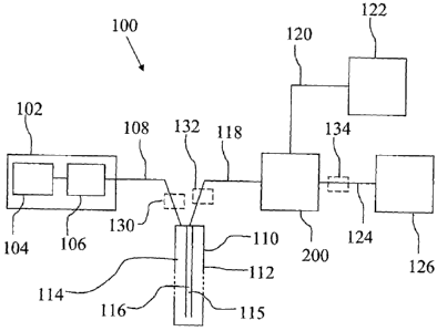
An embodiment of the measuring system 100 will now be described referring to
Fig. 1A. The measuring system 100 is a push system, i.e. the fluid is pushed
through
the entire system 100 by the pump 106. This renders the system less complex
than
push-pull systems where the pushing action of one pump has to be coordinated
with the
pulling action of another pump. One feature in the measuring system 100
contributing
to making it possible to realise the measuring system 100 as a push system is
that the
sensor 200 has a flow resistance or pressure drop that is adapted to the rest
of the
system, e.g. the microdialysis membrane 116. The microdialysis probe 110 of
the
system 100 may be adapted to be placed in a blood stream but may also be
adapted to
be placed in organ tissue. The system comprises a pump unit 102 including a
perfusate
reservoir 104 and a pump 106. Two suitable pumps are the CMA400 and CMA402
from the company CMA Microdialysis, Solna, Sweden. The pump unit 102 is
connected to a microdialysis probe 110 via a piece of tubing 108. The pump 106
may
as well in itself include the perfusate reservoir 104 which, as a suitable
size, may
accommodate a perfusate volume of about 5m1.
It may be suitable to mount the perfusate reservoir 104 and the pump 106
substantially vertical, the part of the pump 106 that is connected to the
tubing 108 being
the lowest point. This is to allow air that may be present in the perfusate to
escape
upwards through the pump 106 and/or perfusate reservoir 104.
The microdialysis probe 110, which is shown in section, comprises a double
bore
tube 112 having an inner bore 115 and an outer bore 114 comprising a
microdialysis
membrane 116. The perfusate is supplied through the outer bore 114 and passes
the
microdialysis membrane 116 whereby microdialysis with the fluid surrounding
the
microdialysis membrane 116 takes place. After the microdialysis membrane 116
the
perfusate is called dialysate. The dialysate 202 exits the microdialysis probe
110
CA 02729126 2010-12-22
WO 2010/002350 PCT/SE2009/050863
through the inner bore 115. The dialysate 202 is conveyed to sensor 200 via a
piece of
tubing 118.
The sensor 200 is an electrochemical sensor of flow through type. A monitor or
display 122 is connected to the sensor 200, via an electrical or optical cable
120 or via a
5 wireless connection. The monitor or display 122 may comprise means for
processing
and displaying measurement values received from the sensor 200. The
measurement
values received from the sensor 200 may be displayed without processing but it
may
also be displayed e.g. mean values and derivatives of the measurement values.
Different ways of displaying measurement values are however known to the
person
1 0 skilled in the art and need not to be further described here. To the
sensor 200 there is
also connected a waste container 126 for collecting the dialysate that has
passed
through the sensor 200. The dialysate 202 in the waste container can be used
to perform
measurements that was not performed by the sensor 200, e.g. to measure the
concentration of substances that were not, or could not be, measured by the
sensor 200.
Examples of such substances are low molecular drugs and low molecular
endogenous
substances, e.g. amino acids, urea, creatinin. The waste container 126
suitably
comprises a pressure release valve 126:2 which is placed in the opening 126:1
and is
permeable to air but is a barrier to bacteria that may be present in the
dialysate 202. It is
also suitable that the waste container 126 comprises an absorbent 126:3 on the
inside of
2 0 the container, to absorb the dialysate that enters the waste container
126. Suitable the
absorbent 126:3 is antibacterial, the absorbent may be placed on the inside of
the upper
and lower wall of the waste container 126 as shown at 126:3a and 126:3b. The
pressure
release valve 126:2 may comprise a piece of the material Tyvek covering the
opening
126:1. If the dialysate 202 should be further analysed a microvial may be
connected to
the tubing 124 connected to the waste container 126 and protruding into the
waste
container. If the pressure release valve 126:2 comprises a piece of Tyvek, the
Tyvek
may be cut open and the microvial introduced into the waste container 126
through the
created hole, and connected to the tubing 124.
The microdialysis membrane 116 may be of a type that is adapted to be placed
in
a blood stream. Alternatively, it is of a type that is adapted to be placed in
organ tissue.
In the background art, many membranes for microdialysis have shown a certain
tendency to be clogged when placed in a blood stream. The inventors of the
present
system have therefore chosen a particular membrane for the case that the
membrane
CA 02 72 912 6 2016-01-12
WO 2010/002350 PCT/SE2009/050863
11
should be placed in a blood stream, a membrane of the skin out type. Membranes
for
microdialysis have a selective layer that decides the size of molecules with
capacity to
pass the membrane wall. This selective layer traditionally is located on the
inside of the
membrane. However, this makes the membrane susceptible of being clogged when
placed in a blood stream. In a membrane of the skin out type the selective
layer is
placed on the outside of the membrane which prevents the membrane from being
clogged when placed in a blood stream. A suitable membrane 116 for the system
100 is
a polyarylethersulfonate (PAES) membrane with a liquid permeability, Lp, of
about 2 x
10-4 cm/bar x s, available from Gambro, Lund, Sweden.
As an advantageous additional measure to prevent clogging of the microdialysis
membrane 116, low molecular weight heparin (Lmwh heparin), e.g. dalteparin,
may be
added to the perfusate. If the microdialysis probe 110 is not placed in a
blood stream
but e.g. in the tissue of an organ, membranes of the non-skin out type may be
used.
Another embodiment of the measuring system 100 will now be described
referring to Fig. 1B. The measuring system 100 is a push system, i.e. the
fluid is pushed
through the entire system 100 by the pump 106. This renders the system less
complex
than push-pull systems where the pushing action of one pump has to be
coordinated
with the pulling action of another pump. One feature in the measuring system
100
contributing to making it possible to realise the measuring system 100 as a
push system
2 0 is that the sensor 200 has a flow resistance or pressure drop that is
adapted to the rest of
the system, e.g. the microdialysis membrane 116. The measuring probe 110 of
the
system 100 is advantageously adapted to be placed in a blood stream . To reach
an
organ, the microdialysis probe 110 often has to be relatively long, in many
cases 50-90
centimetres. In the case of a microdialysis probe to be placed in the venous
blood flow
out of the heart, the probe is suitably 50-70 centimetres long. The system
comprises a
pump unit 102 including a perfusate reservoir 104 and a pump 106. The
perfusate
reservoir may be in the form of a syringe, one suitable syringe is the BD
Plastipak 20
ml from the company BD, Franklin Lakes, New Jersey, United States. One
suitable
pump is the Fresenius Pilot C, from the company Fresenius Kabi AG, Bad
Homburg,
Germany. The pump unit 102 is connected to a microdialysis probe 110 via a
piece of
tubing 108. The pump 106 may as well in itself include the perfusate reservoir
104
which, as a suitable size, may accommodate a perfusate volume of about 20 ml.
*Trademark
CA 02729126 2010-12-22
WO 2010/002350 PCT/SE2009/050863
12
It may be suitable to mount the perfusate reservoir 104 and the pump 106
substantially vertical, the part of the pump 106 that is connected to the
tubing 108 being
the lowest point. This is to allow air that may be present in the perfusate to
escape
upwards through the pump 106 and/or perfusate reservoir 104.
The microdialysis probe 110, which is shown in section, comprises a tube 112
having a first bore 114 and a second bore 115, the tube 112 comprising a
microdialysis
membrane 116. Between the outside of the tube 112 and the inside of the
microdialysis
membrane 116 there is a space 117. The perfusate is supplied through the first
bore
114, exits the first bore 114 through a first channel 114a, enters the space
117 and
passes the microdialysis membrane 116 whereby microdialysis with the fluid
surrounding the microdialysis membrane 116 takes place. After the
microdialysis
membrane 116 the perfusate is called dialysate. The dialysate 202 exits the
space 117
through a second channel 115b and exits the microdialysis probe 110 through
the
second bore 115. A blocking 114b in the first bore 114 directs the perfusate
to enter the
first channel 114a. A blocking 115b in the second bore 115 directs the
dialysate 202 to
exit the microdialysis probe 110 through the second bore 115. The dialysate
202 is
conveyed to sensor 200 via a piece of tubing 118.
The sensor 200 is an electrochemical sensor of flow through type. A monitor or
display 122 is connected to the sensor 200, via an electrical or optical cable
120 or via a
wireless connection. The monitor or display 122 may comprise means for
processing
and displaying measurement values received from the sensor 200. The
measurement
values received from the sensor 200 may be displayed without processing but it
may
also be displayed e.g. mean values and derivatives of the measurement values.
Different ways of displaying measurement values are however known to the
person
skilled in the art and need not to be further described here. To the sensor
200 there is
also connected a waste container 126 for collecting the dialysate that has
passed
through the sensor 200. The dialysate 202 in the waste container can be used
to perform
measurements that was not performed by the sensor 200, e.g. to measure the
concentration of substances that were not, or could not be, measured by the
sensor 200.
Examples of such substances are low molecular drugs and low molecular
endogenous
substances, e.g. amino acids, urea, creatinin. The waste container 126
suitably
comprises a pressure release valve 126:2 which is placed in the opening 126:1
and is
permeable to air but is a barrier to bacteria that may be present in the
dialysate 202. It is
CA 02729126 2010-12-22
WO 2010/002350 PCT/SE2009/050863
13
also suitable that the waste container 126 comprises an absorbent 126:3 on the
inside of
the container, to absorb the dialysate that enters the waste container 126.
Suitable the
absorbent 126:3 is antibacterial, the absorbent may be placed on the inside of
the upper
and lower wall of the waste container 126 as shown at 126:3a and 126:3b. The
pressure
release valve 126:2 may comprise a piece of the material Tyvek covering the
opening
126:1. If the dialysate 202 should be further analysed a microvial may be
connected to
the tubing 124 connected to the waste container 126 and protruding into the
waste
container. If the pressure release valve 126:2 comprises a piece of Tyvek, the
Tyvek
may be cut open and the microvial introduced into the waste container 126 and
connected to the tubing 124.
The microdialysis membrane 116 is suitably adapted to be placed in a blood
stream. In the background art, many membranes for microdialysis have shown a
certain
tendency to be clogged when placed in a blood stream. The inventors of the
present
system have therefore chosen a particular membrane for the case that the
membrane
should be placed in a blood stream, a membrane of the skin out type. Membranes
for
microdialysis have a selective layer that decides the size of molecules with
capacity to
pass the membrane wall. This selective layer traditionally is located on the
inside of the
membrane. However, this makes the membrane susceptible of being clogged when
placed in a blood stream. In a membrane of the skin out type the selective
layer is
placed on the outside of the membrane which prevents the membrane from being
clogged when placed in a blood stream. A suitable membrane 116 for the system
100 is
a polyarylethersulfonate (PAES) membrane with a liquid permeability, Lp, in
the range
of about 1 x 10⁻⁴ cm/barxs to about 3 x 10⁻⁴ cm/barxs, available from Gambro,
Lund,
Sweden
Membranes for microdialysis have a porous structure and the openings in the
membrane are not well-defined channels but rather openings in the membrane
that
wary in size as one moves through the membrane. How large a molecule can be
and
still be able to pass through a membrane also depend on the shape of the
molecule, and
not only on the size. If a membrane has pores with a stated size of e.g. 10 nm
that
means that the size of the pores is around lOnm. One suitable interval for the
size of the
pores is 5 to 50 nanometre (nm), even more suitable 10 to 30 nm. The lower
limit is
suitably around 10 nm so that bigger molecules like e.g. glucose still can
pass the
membrane. The upper limit is chosen so that the risk for ultra filtering is
kept low. Ultra
CA 02729126 2010-12-22
WO 2010/002350 PCT/SE2009/050863
14
filtering is a situation where perfusate penetrates through the membrane and
may occur
when the pressure of the perfusate is too high in relation to the size of the
pores in the
membrane. The smaller the pores are, the higher the pressure of the perfusate
can be
without risking ultra filtering. One suitable size of the pores is around 10
nm when
glucose is the analyte.
With reference to Figs. 2a-2f one first suitable embodiment of the sensor 200
will
be described. Fig. 2a is a drawing schematically showing a section of the
sensor 200.
Figs. 2b and 2c are drawings schematically showing detailed views of the
sensor
electrodes 216 and 218. Fig. 2d gives a schematic view of the main reaction
and
transport pathways of a measuring electrode in the sensor 200. Fig. 2e is a
drawing
schematically showing a front view of the sensor 200, indicating the flow
channel
height 210 and the flow channel width 211 of the flow channel 208. Fig. 2f is
a drawing
schematically showing the sensor 200 from above, according to cut or section A-
A in
Fig. 2a.
The sensor 200 comprises a carrier 204 and a cover 206. Reference sign 202
indicates the inflow of dialysate from the microdialysis probe 110. In the
sensor 200 a
flow channel 208 is defined, the height of the flow channel is indicated at
210. The
flow channel also has a specified width which is indicated by 211 in Fig. 2e.
In this first
embodiment the sensor 200 comprises blank electrodes 214 and 220 and measuring
electrodes 216, 218, 222 and 224. Namely a first blank electrode 214, a first
lactate
electrode 216, a first glucose electrode 218, a second blank electrode 220, a
second
lactate electrode 222 and a second glucose electrode 224. Measuring both
glucose and
lactate may be advantageous for detecting certain disadvantageous conditions
in the
body. The sensor 200 may also comprise measuring electrodes for only one
indicator
substance, or for more than two substances, depending on the application.
With reference to Figs. 2b, 2c and 2d the design and function of the measuring
electrodes will be described more in detail. Short description of the
different
membranes/layers in the measuring electrode 216:
216a: Catalase membrane
216b: Enzyme-free diffusion limiting membrane
216c: Oxidase membrane, here lactate oxidase membrane
216d: Selectively permeable membrane
CA 02729126 2010-12-22
WO 2010/002350 PCT/SE2009/050863
216e: Platinum anode
The dialysate 202 contains among other substances the analyte, e.g. glucose or
lactate, and oxygen (02). In the oxidase membrane 216c a reduction/oxidation
(redox)
process takes place involving the analyte and the oxygen. In this redox
process the
5 analyte is oxidized and the oxygen is reduced. The products of this
process are
hydrogen peroxide and the oxidation product of the analyte. The oxidation
product of
the analyte diffuses out to the dialysate 202 and is washed away with the flow
of the
dialysate 202. A part of the hydrogen peroxide diffuses upwards in the
measuring
electrode 216 and another part diffuses towards the platinum anode 216e.
Oxidase membrane 216c
The layer 216c is in this case a lactate oxidase membrane since the measuring
electrode 216 is measuring lactate. This layer is a membrane in which the
enzyme
lactate oxidase is immobilized, preferably the membrane is a pHEMA-hydrogel
membrane (pHEMA=Poly 2-Hydroxyethylmethacrylate). In the oxidase membrane
216c the immobilized enzyme lactate oxidase acts as a catalyst when the
lactate that
reaches the oxidase membrane 216c reacts with oxygen and hydrogen peroxide is
produced. Some of the hydrogen peroxide that is produced diffuses upwards in
the
direction of the enzyme-free diffusion limiting membrane 216b and the catalase
membrane 216a. When this hydrogen peroxide reaches the catalase membrane 216a
it
is decomposed by the catalase membrane 216a into oxygen and water. The two
membranes diffusion limiting membrane 216b and catalase membrane 216a are
described more in detail below.
Selective membrane 216d
The layer 216d is a selective membrane that only, or at least substantially
only, is
permeable to hydrogen peroxide. Advantageously the layer 216d is an
electropolymerized permselective membrane. The selective membrane 216d is
advantageous since it suppresses electrochemical interference, otherwise there
would
be a risk that other substances than hydrogen peroxide could reach the
platinum anode
216e and give rise to erroneous readings regarding the concentration of
lactate in the
CA 02729126 2010-12-22
WO 2010/002350 PCT/SE2009/050863
16
dialysate 202. The hydrogen peroxide penetrates through the selective membrane
216d
and is oxidised to oxygen at the platinum anode 216e. The oxidation of the
hydrogen
peroxide is achieved since the platinum anode 216e has a certain
electrochemical
catalytic activity. The products of the oxidation of one molecule of hydrogen
peroxide
(H202) are one molecule of oxygen, 2 electrons and 2 protons. This can be
written as:
Oxidation of H202 gives: 02 + 2e- + 2 protons.
The electrons are the output of the sensor, the flow of electrons is measured
and
is used as the output signal of the sensor.
Hence, at the platinum anode 216e the hydrogen peroxide is detected and the
amount of hydrogen peroxide detected is proportional to the amount of lactate
present
in the dialysate 202. Depending on the amount of hydrogen peroxide reaching
the
platinum anode 216e within a certain time period, different amounts of
electrons per
time period is produced, and hence gives different levels of the output
signal.
Diffusion limiting membrane 216b
The layer 216b is an enzyme-free diffusion limiting membrane,
advantageously a pHEMA-membrane, for controlling the diffusion of the analyte,
e.g.
lactate. The diffusion limiting membrane 216b controls how quickly the
lactate, or how
2 0 much lactate per time-period that, reaches the oxidase membrane 216c.
In the dialysate
202 the concentration of oxygen is much lower than the concentration of the
analyte.
One common situation is to have a concentration of 5 to 10 mmo1/1 of the
analyte, e.g.
lactate, and a concentration of 0,2 millmoles of oxygen. If this difference in
concentration would be present in the oxidase membrane 216c, there would not
be
enough oxygen present for the redox process in the oxidase membrane.
Therefore the diffusion limiting membrane 216b suitably reduces the diffusion
speed or rate for oxygen to be 3 to 5 times lower than without the membrane
216b and
suitably reduces the diffusion rate for the analyte, e.g. lactate or glucose,
to be around
1000 times lower than without the membrane 216b. The reason why the diffusion
limiting membrane 216b can hinder the diffusion of the analyte much stronger
than the
diffusion of the oxygen is that the oxygen molecules are much smaller than the
molecules of the analyte. By choosing an appropriate material and thickness of
the
CA 02729126 2010-12-22
WO 2010/002350
PCT/SE2009/050863
17
diffusion limiting membrane 216b, the above mentioned difference in limitation
of
diffusion rate can be achieved.
Because of this difference in reducing diffusion speed or rate the diffusion
limiting membrane 216b brings the positive effect that the concentrations of
oxygen
and analyte is more in balance after the diffusion limiting membrane 216b,
i.e. in the
oxidase membrane 216c, which is desirable since it can be ensured that there
is
sufficient, or a surplus of, oxygen present for the redox process in the
oxidase
membrane 216c.
By controlling the diffusion rate of the analyte, here lactate, the amount of
hydrogen peroxide that is produced in the oxidase membrane 216c can be
controlled
and be limited to a suitable level. The diffusion rate of the analyte is
suitably controlled
so that the oxygen present in the oxidase membrane 216c is not consumed too
quickly
and so that the immobilized enzyme is not saturated with analyte, e.g.
lactate. At which
diffusion rate of the analyte the immobilized enzyme gets saturated is
indicated by the
factor Km, the higher the value of Km, the more analyte per time period the
immobilized
enzyme can process or transform. Hence, Km is a characteristic of the
immobilized
enzyme.
The inventors unexpectedly concluded that increasing the diffusion resistance
of
the enzyme-free diffusion limiting membrane 216b increased the useful life of
the
2 0 immobilized enzyme in the oxidase membrane 216c. One reason for this is
that the
immobilized enzyme is sensitive to hydrogen peroxide, the immobilized enzyme
is
degenerated by the produced hydrogen peroxide. This is especially the case for
the
immobilized lactate enzyme. By increasing the diffusion resistance of the
diffusion
limiting membrane 216b the amount of lactate that reaches the oxidase membrane
216c
per time unit is reduced and hence the production per time unit of hydrogen
peroxide is
limited and the degeneration of the immobilized lactate enzyme is limited. The
amount
of hydrogen peroxide that is produced is suitably limited so that the
immobilized
enzyme is not degenerated too fast, which may become a drawback depending on
with
which application the sensor is used.
The enzyme-free diffusion limiting membrane 216b also increases the diffusion
resistance for hydrogen peroxide that moves towards the catalase membrane
216a. That
reduces the load on the catalase membrane 216a caused by the hydrogen peroxide
that
reaches the catalase membrane 216a.
CA 02729126 2010-12-22
WO 2010/002350
PCT/SE2009/050863
18
By adjusting the diffusion resistance, e.g. by adjusting the thickness and/or
the
size of the channels, of the enzyme-free diffusion limiting membrane 216b the
measurement interval for which the measuring electrode is linear can be
adjusted. By
increasing the diffusion resistance, the maximum limit in analyte
concentration, in the
dialysate 202, for which the measuring electrode responds linearly is
increased.
However, if the diffusion resistance is increased too much, the accuracy and
sensitivity
for low concentrations of the analyte decreases.
Fig. 4 schematically shows output signals for different thicknesses of the
diffusion limiting membrane 216b and where 0S1 means output signal 1, 0S2
means
1 0 output signal 2, LC1 means limit concentration 1, LC2 means limit
concentration 2.
Curve 4:1 represents an output signal obtained with a diffusion limiting
membrane
216b that has a smaller thickness as compared to the diffusion limiting
membrane used
when obtaining curve 4:2. The curves 4:1 and 4:2 are only schematically drawn
and
illustrate that different thicknesses of the diffusion limiting membrane 216b
give
different linearity intervals and different inclinations of the curves. For
curve 4:1 the
linearity interval is from approximately zero concentration up to point LC1,
For curve
4:2 the linearity interval is from approximately zero concentration up to
point LC2. In
reality the transition from the linear part of the curves to the horizontal
part, after LC1
respectively LC2, may be a bit curved. The horizontal part of the curves
represent the
2 0 situation that the immobilized enzyme is saturated with analyte.
Also, the response time for the measuring electrode increases if the diffusion
resistance increases since total processing time in the measuring electrode
will be
longer.
Sensor layout
One possibility is also to have a sensor with several measuring electrodes for
each measured substance, e.g. 2 or 3 measuring electrodes for lactate. In this
way each
measuring electrode can be optimized for a certain interval of the
concentration of the
analyte (e.g. glucose, lactate, pyruvate, glycerol, glutamate or glutamine) in
the
dialysate. A higher thickness of the enzyme-free diffusion limiting membrane
216b
makes it possible to measure higher concentrations of a substance or analyte
present in
the dialysate but to measure low concentrations of a substance, the thickness
of the
CA 02729126 2010-12-22
WO 2010/002350 PCT/SE2009/050863
19
enzyme-free diffusion limiting membrane 216b must not be too high so that the
measuring electrode has the sensitivity necessary to obtain reliable
measurements also
for low concentrations of a substance present in the dialysate.
Catalase membrane
The catalase membrane 216a prevents hydrogen peroxide diffusing upwards from
the oxidase membrane 216c from reaching the dialysate 202 and in this way
prevents
cross-talk between the different measuring electrodes. Hydrogen peroxide that
reaches
the catalase membrane 216a from the oxidase membrane 216c is decomposed within
the catalase membrane 216a. The catalase membrane 216a also brings an
extremely
low flow rate dependency because hydrogen peroxide that otherwise would
accumulate
within the dialysate 202 is decomposed in the catalase membrane 216a. The very
low
flow rate dependency is advantageous in achieving a high accuracy. If hydrogen
peroxide would accumulate within the dialysate 202, this would lead to an
increase in
the sensor signal measured at the platinum anode 216e. This is a problem in
measuring
electrodes having no catalase membrane 216a covering the oxidase membrane
216c.
The flow rate dependency in those measuring electrodes makes it difficult to
obtain a
measuring electrode with high accuracy. If there would be no catalase membrane
216a
hydrogen peroxide would accumulate in the dialysate 202 above the measuring
electrode 216 and would, at least partially, diffuse down through the
measuring
electrode 216 and increase the sensor signal. How much of the hydrogen
peroxide
accumulated in the dialysate 202 that would diffuse down through the measuring
electrode 216 would be dependent on the flow rate of the dialysate 202. Hence,
the
output signal of the measuring electrode would be dependent on the flow rate
of the
dialysate 202.
The first glucose electrode 218, the second lactate electrode 222 and the
second
glucose electrode 224 function in a similar way or according to the same
principles as
the first lactate electrode 216.
Since the sensor 200 has a very low flow rate dependency the flow rate in the
system can be allowed to vary to a certain extent. This is advantageous since
the pump
CA 02729126 2010-12-22
WO 2010/002350 PCT/SE2009/050863
106 do not have to deliver a very exact flow rate. This makes the pump less
complex,
which is advantageous in view of reliability, and less costly.
The characteristics of the sensor 200 need to be adapted to the
characteristics of
the microdialysis membrane 116. One aspect is that the flow resistance or
pressure drop
5 of the sensor 200 can not be too high. If the flow resistance or pressure
drop of the
sensor 200 would be too high, the pressure in the system would be too high and
the
perfusate flowing passed, or through the bore of, the microdialysis membrane
116
could be pressed or pushed through the microdialysis membrane 116. This is
called
ultra filtration. This would be disadvantageous since the measuring function
of the
10 system 100 would be hampered or negatively affected. Or the system 100
could even
be completely non-functional. Another disadvantageous aspect is that it is not
acceptable that the subject of the measurement, e.g. a patient in an ICU, is
injected with
the perfusate. From the view of safety for the subject, the perfusate should
not enter the
subject, even if perfusates are non-hazardous.
15 To ensure that the flow resistance or pressure drop in the sensor 200 is
low
enough, the cross sectional area of the flow channel 208 must be sufficiently
large.
However, a certain flow resistance or pressure drop in the sensor 200 is
acceptable or
even suitable, e.g. since a certain pressure will be built up so air bubbles
that may form
in the dialysate 202 will be dissolved quicker than if there would be no
pressure in the
20 dialysate 202. Air bubbles may form in the dialysate 202 when the fluid
is warmed up.
A certain pressure in the dialysate 202 will facilitate that the deformation
will take
place in a shorter time period and the air bubble will be resolved quicker.
If the height of the flow channel is low, there is a high possibility that an
air
bubble will be deformed, since there is little space available for the air
bubble, and for a
shallower flow channel a higher force is exerted on an air bubble. In that way
the air
bubble becomes destabilized and dissolves. If an air bubble would be present
on the
surface of a measuring electrode it would reduce the diffusion of the analyte
down
through the measuring electrode and result in a erroneous reading.
However, if an air bubble would be so large that it covers the whole, or
substantially the whole, area of a measuring electrode the value recorded by
the
measuring electrode would drop rapidly, possibly to approximately zero
depending on
how long the air bubble would stay on the surface of the electrode, such a
reading can
be identified as erroneous and be discarded.
CA 02729126 2010-12-22
WO 2010/002350 PCT/SE2009/050863
21
One advantageous measure for the flow channel 208 is a flow channel height 210
of approximately 75 micrometer and a flow channel width 211 of approximately
450
micrometer. A suitable interval for the flow channel width 211 is 250 to 1000
micro
meters. A flow channel width 211 of 250 micrometer is a suitable lower limit
since that
width still renders the area of the oxidase membrane 216c sufficiently large.
With a
smaller flow channel width 211 than 250 micrometer problems may be encountered
with a too low signal level from the sensor because resulting from a small
production of
hydrogen peroxide in the oxidase membrane 216c due to a too small area of the
oxidase
membrane 216c. This depends on the lowest analyte concentration that the
measuring
electrode should be able to detect with sufficient accuracy. The oxidase
membrane
216c may have a circular or essentially circular shape, as seen in the
direction of the
arrows at "A" in Fig. 2a. In this case a suitable interval for the dimensions
of the
oxidase membrane is a diameter of 250-1000 micrometer, suitably 250-700
micrometer, most preferably about 450 micrometer. A flow channel width of 1000
micrometer is a suitable upper limit to limit the internal volume in the
system to
advantageously limit the delay in the system.
A suitable interval for the flow channel height 210 is 10 micrometer to 1
millimetre, ever more suitable is 25 to 100 micrometer,
The measures flow channel height 210 of approximately 75 micrometer and a
flow channel width 211 of approximately 450 micrometer, render the flow
channel 208
a flow resistance or pressure drop of less than about 100 Pa, which is the
maximum
flow resistance or pressure drop suitable for a skin out micro dialysis
membrane 116
with an Lp coefficient of 2 when operated with a flow rate of about 0,5
microliter/minute and having an active membrane length of about 30 mm, to
reach a
level of ultra filtration that is not too high, suitably lower than 10% of the
flow rate.
It is suitable that the cover 206 of the flow channel 208 comprises a
relatively
rigid material, so that the flow resistance or pressure drop do not vary, at
least not
substantially. Having a stable flow resistance or pressure drop of the flow
channel 208
makes the system 100 more reliable since that eliminates or reduces the risk
for a
pressure build up under the micro dialysis membrane 116 due to an increase in
flow
resistance or pressure drop. As explained previously, a pressure build up
under the
micro dialysis membrane 116 is disadvantageous since that may cause ultra
filtering, if
the pressure reaches too high levels.
CA 02729126 2010-12-22
WO 2010/002350 PCT/SE2009/050863
22
The length of the sensor 200 is governed by the space required for the
different
measuring electrodes.
There is a risk that air bubbles could be formed in the dialysate flow, as
also
mentioned previously. As previously discussed air bubbles can be counteracted
by
selecting appropriate flow channel dimensions, but can further be counteracted
by
selecting a hydrophilic channel material. In terms of delay in the system 100
it is
preferred that the internal volume of the flow channel 208 is low and
represent a low
internal volume. A suitable flow channel height for these purposes in the
present
system is about 75 micrometer. Also the relatively high flow rate is an
advantage
regarding air bubbles since the relatively high flow rate helps to wash away
the air
bubbles. The relatively high flow rate may also be suitable in applications
where the
tubing and/or microdialysis probe is relatively long, so as to transport the
fluid through
the system in an appropriate way and avoiding air to hinder the fluid flow.
There are also other aspects influencing the design of the sensor 200. A
measuring electrode needs to have a certain minimum area because the oxidase
membrane (e.g. the oxidase membrane 216c) needs to have a certain minimum area
so
that the production of hydrogen peroxide will be high enough and thereby give
a signal
level from the measuring electrode that is high enough. If the signal level
from the
sensor becomes too low problems with noise levels present in the electronics
connected
2 0 to the sensor may arise, in the sense that the noise level could be too
high in relation to
the signal level from the sensor. The platinum anode 216e also gives rise to a
certain
noise level. One reason is that the platinum anode has a certain capacitance.
Since the
platinum anode 216e has some capacitance it is suitable that the electronics
connected
to the platinum anode has a constant voltage, or a voltage that varies as
little as
possible. The fact that the oxidase membrane needs to have a certain minimum
area
leads to that the flow channel 208 needs to have a certain minimum width for
the
measuring electrode to have reasonable dimensions, a reasonable relationship
between
length and width. Since it is suitable that the flow channel 208 has small
dimensions,
but it is suitable that the oxidase membrane has a fairly big area, a
compromise has to
be done so that the area of the oxidase membrane will be high enough, and the
flow
channel 208 small enough. Suitably the platinum anode 216e has the same area
as the
oxidase membrane 216c.
CA 02729126 2010-12-22
WO 2010/002350 PCT/SE2009/050863
23
The blank electrodes 214 and 220 have a design similar to the measuring
electrodes but is free from enzyme in layers 214c, 220c. In these layers there
is only the
membrane material, e.g. a hydrogel membrane, present wherein the immobilized
enzymes are kept in the measuring electrodes. One reason for providing the
first blank
electrode 214 is to detect any hydrogen peroxide, or other electroactive
substances, e.g.
ascorbic acid or paracetamol, present in the dialysate 202 already before the
dialysate
202 arrives to the measuring electrodes, in order to establish a reference
level for the
signals obtained from the measuring electrodes. If the output signal from the
first blank
electrode 214 would be very high that may be a sign of a error in the system
and the
output signals from the measuring electrodes obtained at that point of time
can be
discarded, if appropriate.
By providing two electrodes each for lactate and glucose redundancy is
achieved
and the reliability and accuracy of the system 100 is improved since if a
fault arises in
one measuring electrode, the other can still be used. It is more unlikely that
two
measuring electrodes should be erroneous than that an error occurs in one
measuring
electrode. By comparing the readings or sensor signals from two measuring
electrodes
measuring the same substance it can be determined if the measuring electrodes
function
correctly, or if one of them gives an erroneous reading. The possibility to
detect such
erroneous readings increases the accuracy of the system 100 since the
probability to
2 0 have access to a sensor signal from a properly functioning measuring
electrode is
increased.
One reason for providing the second blank electrode 220 is to detect any
potential
cross talk between the measuring electrodes. That is, e.g. to detect potential
hydrogen
peroxide present in the dialysate in the flow channel 208. If for example the
catalase
membrane of one of the first measuring electrodes would not function properly
hydrogen peroxide from that measuring electrode could enter into the flow
channel
208. Such a situation can be detected by comparing the signals from the first
blank
electrode 214 and the second blank electrode 220.
The first glucose electrode 218 has a design similar to the first lactate
electrode
216. The second lactate electrode 222 has in one embodiment the same design as
the
first lactate electrode 216 and the second glucose electrode 224 has in one
embodiment
the same design as the first glucose electrode 218. But other designs are of
course also
CA 02729126 2010-12-22
WO 2010/002350 PCT/SE2009/050863
24
possible, e.g. several measuring electrodes for the same analyte but having
different
linear ranges.
The diffusion rate in a measuring electrode is temperature dependent. The
higher
the temperature in the measuring electrode is, the higher the diffusion rate
will be. This
means that also the output signal from a measuring electrode is temperature
dependent,
the higher the diffusion rate is, the higher the output signal will be for a
given
concentration of the analyte in the dialysate. It is therefore advantageous to
determine
the temperature of the measuring electrode to enable a correction of the
output signal
with respect to the determined temperature. A temperature sensor, not shown,
may be
placed on the carrier 204 to determine the temperature. It can be assumed that
the
measured temperature is valid for all measuring electrodes in the sensor. This
approximation often gives an accuracy that is high enough. It may be suitable
to
calibrate the sensor 200/measuring electrodes as close to the normal operating
temperature as possible, e.g. at 35 degrees Celsius, to obtain a calibration
that is as
accurate as possible. An accurate calibration makes it possible to accurately
adjust the
output signal with respect to the effect of the temperature of the measuring
electrode.
Fig. 3 is a basic drawing schematically showing the relation between flow rate
and recovery degree in, or relating to, a microdialysis membrane. As seen in
the Figure,
for lower flow rates and up to a certain maximum flow rate, flow rate 1, 100 %
2 0 recovery degree is achieved. A recovery degree of 100 % means that
there is an
equilibrium between the concentration of a certain substance in the fluid
outside of the
microdialysis membrane and the concentration of this certain substance in the
fluid on
the inside of the microdialysis membrane. In the present system 100 the flow
rate has
been chosen to be lower than the value flow rate 1. A flow rate in the
interval of about
0,2-2.0 microliters per minute has been found to be suitable, more suitable
0,3-1.5
microliters per minute and even more suitable 0.5-1.0 microliters per minute.
One
suitable flow rate that has been used is about 1.0 microliter per minute.
Another flow
rate in the interval of about 5-15 microliters per minute has also been found
to be
suitable, more suitable 8-12 microliters per minute and even more suitable 9-
11
microliters per minute. One suitable flow rate that has been used is about 10
microliter
per minute. One advantage of this choice of flow rate is that a low delay is
achieved,
which often is an advantage in intensive or critical care applications. With a
flow rate
of about 10 microliter per minute a delay of approximately 2 minutes was
achieved
CA 02729126 2010-12-22
WO 2010/002350 PCT/SE2009/050863
when the length of tubing 118 between the microdialysis probe 110 and the
sensor 200
was 25 cm. A low delay is advantageous to achieve an early detection of a
potentially
pathological or dangerous condition in an organ of a subject.
These choices of flow rate has a number of advantages. Firstly the flow rate
may vary
5 without resulting in a variation in recovery. As said previously, this
enables the use of a
pump with a less complex construction. That the flow rate may vary is also
facilitated
by the fact that the sensor 200 has a very low flow rate dependency, as
mentioned
previously.
With a flow rate value below flow rate 1, accuracy is improved as compared to
a
10 situation where the flow rate is higher than flow rate 1, since it is
always assured that
the recovery is 100%. In a system where the flow rate is higher than flow rate
1 the
flow rate has to be controlled to be within narrow limits so that the
concentration in the
fluid surrounding the microdialysis membrane can be calculated using the
specific
degree of recovery, e.g. maybe 50%, corresponding to the flow rate value
prevailing in
15 the system. The control of the flow rate is however of course not
perfect and a slight
variation in flow rate can not be excluded. Hence, a certain inaccuracy is
introduced.
In a measuring electrode 216, 218, 222, 224, the immobilized enzyme in the
oxidase membrane, e.g. the oxidase membrane 216c, often functions best in an
environment with a pH around 7. This is e.g. the case for the enzymes lactate
and
20 glucose oxidase. But when the hydrogen peroxide (H202) in a measuring
electrode
enter the selective membrane, e.g. selective membrane 216d, protons are
formed. When
not counteracted, protons would change the pH to be unfavourable for the
immobilized
enzyme in the oxidase membrane. However, since the flow rate is low and 100%
recovery degree is achieved in the present system 100, buffering substances
from the
25 fluid, e.g. blood, surrounding the microdialysis membrane 116, can fully
enter the
perfusate. Sufficient buffering substances, e.g. bicarbonate, will then be
present in the
dialysate flow 202 in the flow channel 208 to neutralise protons, thereby
avoiding
acidification resulting in poor functionality of the immobilized enzyme. In
systems
where insufficient buffering substances enter through the microdialysis
membrane,
buffering substances have to be added to the dialysate after the microdialysis
probe.
This is a potential drawback since it makes the system more complex and
potentially
less reliable.
CA 02729126 2010-12-22
WO 2010/002350 PCT/SE2009/050863
26
As described above, the design of the sensor 200 been carried out in order to
create a well functioning system 100 where the design of the sensor 200 has
been
adapted to the other parts and aspects of the measuring system 100, e.g. the
microdialysis membrane 116 and the suitably flow rate of 0.2-15
microliters/min. One
advantage with the measuring system 100 is that measurement values or sensor
signals
can be obtained very often, several times each second if desired. This is
advantageous
in assessing the condition in a critically ill subject, for example a person
being
monitored or treated in an ICU, where a change of condition needs urgent
detection and
therapy. Further the inventive measuring system admits a very low measurement
delay,
meaning the time period from the moment at which a certain volume of
perfusate/dialysate passes the microdialysis membrane 116, until the moment
the
concentration of a certain substance in this volume of dialysate can be
detected by
monitoring the sensor signal from a measuring electrode. This measurement
delay can
be approximately 3 minutes. Depending on the design of the system, e.g. the
flow rate,
the length of the tubing, the volume of perfusate/dialysate in the system,
this
measurement delay can be changed to be shorter or longer, depending to the
requirements.
A system according to the present invention was tested in an animal, pig,
model
and the results are demonstrated in Figs. 6a-6f. Time is shown on the X-axis
and
concentration in millimolar (mM) or millimol per litre is shown on the Y-axis.
The test
animal was infused with 50 ml of 20% lactate and 50 ml 30% glucose starting at
10:30.
The infusion ended at 10:58. Injection of 30 Units of insulin was performed
11:30.
Venous blood was sampled every 5 minutes during the infusion and assayed for
glucose and lactate using a conventional blood gas analyzer. The blood gas
data has
been shifted about 12 minutes due to the delay, which was around 12 minutes,
in the
system/prototype. The results were obtained using a microdialysis probe
inserted in a
peripheral vein, the probe having a skin-out membrane with an active length of
about
20 mm, an outer diameter (OD) of about 0.59 mm and a liquid permeability of
about 2
x 10-4 cm/bar x s at a perfusion flow of 0.5 microliters/minute. A flow
through sensor
with duplicate measuring electrodes, for glucose and lactate, and two blank
electrodes
was used with the following flow channel dimensions: height 75 micrometers and
width 450 micrometers, and with each electrode having an area of 0.16 square
CA 02729126 2010-12-22
WO 2010/002350 PCT/SE2009/050863
27
millimetres, and was attached to the outlet of the microdialysis probe. The
sensor
followed the dimensions earlier given as preferred embodiments. The sensor
signal was
at about 1 Hz and the results presented are running average values based on 60
samples. The results of Figs. 6a-6f demonstrates that the system has excellent
accuracy,
compared to blood gas data, and a delay time that is operable for using the
system for
monitoring in a critical care unit. It is also to be noticed that the
measurement curves
from the two glucose measuring electrodes respectively the two lactate
measuring
electrodes follow each other very closely. In Fig. 6a all measurement curves
are
displayed in the same diagram for ease of comparison and in Figs. 6b-6f the
1 0 measurement curves from the different measuring electrodes are shown
separately for
increased clarity.
A system according to the present invention was tested in an animal (pig)
model
and the results are demonstrated in Fig. 7, showing lactate data. Time is
shown on the
X-axis and concentration in millimol per litre (mmol/L) is shown on the Y-
axis. The
test animal was infused in the femoral vein with 50 ml of 20% lactate and 50
ml 30%
glucose starting at 12:30. The infusion ended at 13:00. Venous blood was
sampled
every 5 minutes both from the femoral vein and the jugular vein and assayed
for
glucose and lactate using a conventional blood gas analyzer. The delay in the
system
was around 2 minutes. The results were obtained using a 67 cm long
microdialysis
probe inserted into the jugular vein and then guided to the vena cava
superior, the probe
having a skin-out membrane being around 40 mm long (active length) and around
1,55
mm in outer diameter (OD), with a liquid permeability of about 2 x 10⁻⁴
cm/barxs
(Lp=2) at a perfusion flow of about 10 microliters per minute. A flow through
sensor
with duplicate measuring electrodes (for glucose and lactate) and two blank
electrodes
was used with the following flow channel dimensions: height 75 micrometers and
width 450 micrometers, and with each electrode having an area of 0.16 square
millimetres, was attached to the outlet of the microdialysis probe. The sensor
followed
the dimensions earlier given as preferred embodiments. The sensor signal was
at about
1 Hz and the results presented are running average values based on 60 samples.
The
results of Fig. 7 , showing lactate data, demonstrate that the system has
excellent
accuracy (compared to blood gas data) and a delay time that is operable for
using the
system for monitoring a patient in a critical situation, e.g. during or after
surgery or in
an intensive care unit. It is also to be noticed that the measurement curve
from the
CA 02729126 2010-12-22
WO 2010/002350 PCT/SE2009/050863
28
lactate measuring of the system follow the measurement values from the two
blood gas
measurements, which are used as references, very closely. Glucose values are
not
presented in Fig. 7, but showed the same excellent accuracy as for lactate.
The system described above in the context of animal tests was used clinically
with
human patients. Results of glucose and lactate values are presented in Figs 8a
8b,
respectively. The flow rate was 6.7 microliters per minute. Arterial and
venous glucose
and lactate were sampled each hour, while also plasma glucose was sample each
third
hour. Figs. 8a and 8b comparatively shows glucose and lactate values in real-
time from
the system according to the present invention. The results demonstrate that
the
inventive system has excellent accuracy and provides physicians continuously
with
valuable patient information without cumbersome and delaying sampling and
analyzing
in a blood gas measuring equipment. Accordingly, the inventive system admits
that
critical care patients can be treated more proactively which potentially can
reduce
treatment times and may have lifesaving consequences.
Although particular embodiments have been disclosed herein in detail, this has
been done by way of example for purposes of illustration only, and is not
intended to be
limiting with respect to the scope of the appended claims that follow. In
particular, it is
contemplated by the inventor that various substitutions, alterations, and
modifications
may be made to the invention without departing from the spirit and scope of
the
invention as defined by the claims.