Note: Descriptions are shown in the official language in which they were submitted.
CA 02729351 2010-12-23
WO 2010/005519
PCT/US2009/003886
Fn14/TRAIL FUSION PROTEINS
FIELD OF THE INVENTION
This invention relates to Fn14/TRAIL and related fusion proteins, and
methods of treating certain diseases such as autoimmune diseases and
cancer with these proteins.
BACKGROUND INFORMATION
A complex interplay of positive and negative signals regulates T cell
activation and maintenance of T cell effector function. Members of the
TNF ligand/TNF receptor superfamily figure prominently in this matrix of
signals, bridging cells of the immune system, as well as with cells of
other organ systems. In so doing, TNF superfamily members contribute
to both tissue homeostasis and pathogenesis, via effects on cell survival
and death, cellular differentiation, and inflammation. From the
standpoint of autoimmune pathogenesis, interesting members of the TNF
ligand superfamily are TNF-related apoptosis-inducing ligand (TRAIL),
and TWEAK (TNF-related weak inducer of apoptosis).
TRAIL binds to a number of different cognate receptors of the TNF
receptor superfamily, some leading to triggering of intracellular signaling
pathways and others simply acting as decoy receptors. The triggering
receptors in humans are TRAIL-R1, TRAIL-R2, and osteoprotegrin, and in
mice the sole triggering receptor is DR5. Virtually all cells of the immune
system (T lymphocytes, B lymphocytes, natural killer cells, dendritic
cells, monocytes, granulocytes) upregulate surface TRAIL and/or release
soluble TRAIL stored in secretory vesicles in response to interferon and
other activation signals. TRAIL inhibits autoimmunity in several animal
models. Evidence for TRAIL's capacity to inhibit experimental
1
CA 02729351 2010-12-23
WO 2010/005519
PCT/US2009/003886
autoimmune encephalitis (EAE), a murine model for MS, has come from
experiments invoking TRAIL-/- knockout mice, soluble TRAIL receptor
(5DR5) or neutralizing anti-TRAIL rnAb capable of blocking TRAIL
function, and embryonic stem cell-derived dentritic cells co-expressing
TRAIL and pathogenic MOG (myelin oligo-dendrocyte glycoprotein
peptide). Interestingly, in MS patients, soluble TRAIL has emerged as a
response marker for IFN-f3 therapy, with those most likely to respond to
treatment showing early and sustained soluble TRAIL induction after
therapy. Yet, TRAIL's impact on MS/EAE may be more complex, for
example, the suggestion that TRAIL may promote brain cell apoptosis.
Both TRAIL and FasL have been implicated in negative regulation of T
cells.
TWEAK and its counter-receptor Fn14 (fibroblast growth factor-inducible
14 kDa protein) are another TNF family ligand-receptor pair expressed in
a range of immune and non-immune cell types, including NK cells,
macrophages, dendritic cells, microglial cells, glial cells and endothelial
cells. TWEAK promotes the proliferation of some cell types (astrocytes,
endothelial cells, and certain human tumor cell lines), and suppresses
others (erythroblasts, kidney cells, mesangial cells, neuronal cells, NK
cells, monocytes). TWEAK stimulates production of various
inflammatory cytokines, chemokines and adhesion molecules. However,
the TWEAK:Fn14 signaling axis has effects that go beyond cell
proliferation and cytokine production. Interestingly, the richer set of
functions linked to TWEAK over the years include ones that tie into
autoimmunity. TWEAK increases the permeability of the neurovascular
unit, and its endogenous expression is elevated in the CNS during EAE
and acute cerebral ischemia. Moreover, TWEAK has pro-angiogenic
activity, which is of interest given the association between angiogenesis
and autoimmune pathogenesis. TWEAK increases EAE severity and
associated neurodegeneration. The induction of inhibitory anti-TWEAK
2
CA 02729351 2010-12-23
WO 2010/005519
PCT/US2009/003886
or Fn14 Ab, via vaccination with the extracellular domain of either
TWEAK or Fn14, ameliorates EAE manifestations in rat and mouse
models.
Multiple sclerosis (MS) is a debilitating neurological disease, and despite
an expanding set of treatment options, there remains a pressing need for
more effective therapeutic agents. While the precise etiology of MS is
unknown, key features of its pathogenesis and clinical evolution are
emerging. Pathogenic effector T cells are thought to be pivotal in driving
the disease, and thus many therapeutic paths are converging on these
cells, with goals such as blocking their activation and re-activation,
eliminating them from the larger T cell reservoir, and interfering with
their transit to sites of pathogenesis within the CNS.
Both the TWEAK/Fn14 and TRAIL/TRAILR signaling axes have been
implicated in cancer. See, e.g., "TRAIL receptor signalling and
modulation: Are we on the right TRAIL?", Cancer Treatment Reviews 35
(2009) 280-288; and "The TWEAK-Fn14 cytokine-receptor axis:
discovery, biology and therapeutic targeting", Nature 7 (2008) 411-425.
SUMMARY OF THE INVENTION
Accordingly, in one aspect the present invention provides a fusion
protein comprising a first domain and a second domain, wherein the first
domain is a polypeptide that binds to a TWEAK ligand and the second
domain is a polypeptide that binds to a TRAIL receptor.
In an additional aspect, the present invention provides a fusion protein
consisting essentially of a first domain and a second domain, wherein the
first domain is at least a portion of the extracellular domain of a Fn14
3
CA 02729351 2010-12-23
WO 2010/005519
PCT/US2009/003886
protein and the second domain is at least a portion of the extracellular
domain of a TRAIL protein.
In another aspect, the present invention provides a fusion protein
comprising a first domain and a second domain, wherein the first domain
is a polypeptide that binds to a TWEAK ligand and the second domain is
a polypeptide having an inhibitory function.
Pharmaceutical compositions comprising the fusion proteins, as well as
methods of treating various illnesses such as autoimmune diseases and
cancer, with the fusion proteins of the invention are also provided.
BRIEF DESCRIPTION OF THE DRAWINGS
The invention is further illustrated by the following drawings in which:
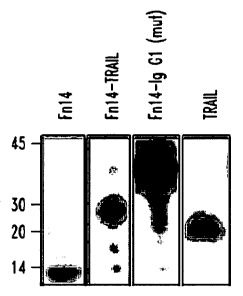
Figure 1. Expression and functional analysis of Fn14-TRAIL
A) Western blot analysis was performed on conditioned media from 293
cells transfected with expression constructs for Fn14, Fn14-TRAIL,
Fn14-IgGl(mut) and TRAIL. Observed bands were consistent with the
expected sizes of 8.7 kD, 27.5 kD, 34.1kD, and 19.0 kD, respectively.
B) CHO cells were transiently transfected with a murine TWEAK cDNA
expression construct, and after 48h, were incubated at 4 C with purified
Fn14-TRAIL or rTRAIL in sodium azide-containing buffer. The presence
of membrane-bound TWEAK on transfectants, and the binding of Fn14-
TRAIL to them, was verified by flow cytometry, using anti-mouse TWEAK
Ab (B.1) and anti-mouse TRAIL Ab (B.2-5) as detecting Ab, respectively.
(B.1) TWEAK is expressed on transfected CHO cells, as detected using
anti-TWEAK Ab (solid black line) versus isotype control (filled histogram);
(B.2) TRAIL is not detectable on CHO cells, when analyzed using anti-
TRAIL Ab (solid black line) versus isotype control (filled histogram). (B.3
4
CA 02729351 2010-12-23
WO 2010/005519
PCT/US2009/003886
and B.4) TRAIL epitopes are enhanced when Fn14-TRAIL is added to
TWEAK-expressing, as opposed to TWEAK-negative, CHO cells. Anti-
TRAIL Ab and isotype control are represented by solid black line and
filled histogram, respectively. (B.5) TWEAK-transfected cells do not bind
to anti-TRAIL Ab (solid black line) in the presence of rTRAIL. Isotype
control is shown as filled histogram.
C) L929 cells were cultured in flat-bottom 96-well plates at 2 x 10⁴
cells/well, in 100 pl AIM-V medium. Sixteen hours later, actinomycin D
was added to the cultures at 1 mg/well, and cells were cultured for
another 2 h. Fn14-TRAIL or rTRAIL, as positive control, was then added,
and cultures were maintained for an additional 5 h. The percentage of
dead cells was determined by an wrr assay, as described in Materials
and Methods.
Figure 2. Functional validation of the dual cassette pSBC21 vector
A) As schematically depicted, the pSBC21 plasmid incorporates in
tandem a transposon cassette (with the EF la promoter driving the trans
signal redirecting protein - TSRP) and a transposase expression cassette
(driven by the UBC promoter).
B) Mice were hydrodynamically injected with either pSBC21 vector only
(right panel) or the indicated concentrations of pLuciferase-SBC21.
Bioluminescent images were acquired after intra-peritoneal
administration of D-Luciferin 22 days after plasmid injection. Color bars
represent bioluminescent signal in radiance (p/sec/cm2 /sr).
C) Serum levels of Fn14-TRAIL were determined by ELISA 20 days after
the injection of 5 pg, 10 - g or 20 pg of Fn14-TRAILTSBC21 plasmid.
Figure 3. Fn14-TRAIL suppresses MOG-induced autoimmune
encephalomyelitis
5
CA 02729351 2010-12-23
WO 2010/005519
PCT/US2009/003886
A) Serum levels of Fn14, Fn14-TRAIL, Fn14-IgG1 (mut) and TRAIL were
measured by ELISA 20 days after hydrodynamically injecting 50 -pg of
the respective pSBC21-based expression plasmids.
B-D) Mice were challenged with MUG in CFA supplemented with M.
tuberculosis as described in Material and Methods. 48 h after MUG
challenge, mice were hydrodynamically-injected with 50 pg of the
indicated pSBC21-based expression constructs. Individual mice were
scored according to the clinical scale described in Materials and
Methods. Parameters evaluated include mean clinical score (B, upper
panel), disease incidence (B, lower panel), cumulative mean clinical score
(C) and mean clinical score on day 35 (D). The difference between the
Fn14-TRAIL-treated versus vector-only control group is statistically
significant according to Mann-Whitney U test (p<0.05), where the
differences between the other groups shown and the vector-only group
are not significant.
Figure 4. Fn14 and TRAIL in combination do not achieve Fn14-TRAIL's
therapeutic efficacy
A-C) 48 h after MUG-challenge, mice were hydrodynamically injected
with a single dose of Fn14-TRAILTSBC21 plasmid (25 g/mouse), or a
single dose of a mixture of Fn14-pSBC21 and TRAIL-pSBC21 plasmids
(25 pg each/mouse). Mean clinical scores for the indicated groups over
40 days (A), and on days 16, 23 and 40(B) are shown. Cumulative MCS
of indicated groups at day 40 is also shown (C). The difference between
the Fn14-TRAIL-treated versus vector-only control group is statistically
significant according to Mann-Whitney U test (p<0.05), where the
differences between the other groups shown and the vector-only group
are not significant.
6
CA 02729351 2010-12-23
WO 2010/005519
PCT/US2009/003886
D) Serum levels of Fn14-TRAIL were determined by ELISA 30 days after
the injection of combined Fn1413SBC21 (25 pg) + TRAIL=pSBC21 (25 pg),
or Fn14-TRAILTSBC21 (25 pg).
E) MOG-challenged mice that had been hydrodynamically-injected with
pSBC21 vector only or Fn14-TRAILTSBC21 were sacrificed 43 days after
receiving the therapeutic agent. Splenocytes from each mouse were
cultured in the presence or absence of different concentrations of MOG38_
so peptide, and proliferation was evaluated as described in Materials and
Methods. Results are shown as mean SD from a total of nine mice per
group.
Figure 5. Fn14-TRAILTSBC21 inhibits cytokine production in MUG-
challenged mice
Mice were challenged with MOG in CFA supplemented with M.
tuberculosis and hydrodynamically-injected with pSBC21 vector only or
Fn14-TRAILTSBC21. Animals were sacrificed 43 days after receiving the
therpeutic agent, and splenocytes from each mouse were cultured in the
presence or absence of different concentrations of M0G38-so peptide.
Cultured media were collected 40 h later, and concentrations of the
indicated cytokines were determined by ELISA.
Figure 6. Fn14-TRAILTSBC21 treatment reduces activated and cytokine-
producing cells in spinal cords of MUG-challenged mice
Infiltrating cells from spinal cords of pSBC21 vector-only and Fn14-
TRAIL-pSBC21-treated mice were isolated an analyzed as described in
Material and Methods. The number of the cells was calculated by
multiplying the total number of live cells by % of each indicated cell type.
The percentages and numbers of cells are representative of the
7
CA 02729351 2010-12-23
WO 2010/005519
PCT/US2009/003886
percentage and number of the indicated cell types acquired from groups
of three mice.
A-B) Percentage and absolute number of activated (CD69+) inflammatory
cells on day 7 post-MOG challenge
C-D) Percentage and absolute number of CD4+ and CD8+ cells, and of
IFNy, IL-17, and IL-10 expressing cells (amidst activated CD69+, cells or
the total cell pool) on day 7 post-MOG challenge
D-E) Percentage and absolute number of CD8+ cells and of IFNy, IL-17
and IL-10 expressing cells on day 14 of post-MOG challenge
Figure 7. Fn14-TRAILTSBC21 treatment reduces spinal cord
inflammation in MOG-challenged mice
MOG-challenged mice, hydrodynamically injectyed with SBC21 vector
only or Fn14-TRAILTSBC21, were perfused transcardially with PBS and
10% formalin phosphate. Spinal cords were removed, cut into six pieces,
embedded in paraffin, transversely sectioned at 5 m and stained with
luxol fast blue and cresyl violet.
A) Upper panel shows representative spinal cord sections of vector only-
treated mice with maximum disease scores of 2, 3.5 and 1 from left to
right, respectively. Lower panel shows representative spinal cord
sections of Fn14-TRAIL-treated mice with maximum disease scores of 0,
1 and 3 from left to right, respectively.
B) Tissue sections from each of the six spinal cord segments were
analyzed for each animal. Scores of inflammation were assigned to
individual sections based on the following criteria: 0, no inflammation; 1,
<5%; 2, 5-20%; 3, 20-50% and 4, >50% of the white matter is infiltrated
by leukocytes. For each mouse, the histological score is the sum of
scores from the 6 spinal cord sections. The difference between the Fn14-
8
CA 02729351 2010-12-23
WO 2010/005519
PCT/US2009/003886
TRAIL-treated group versus the control group is statistically significant
according to Mann-Whitney U test (p<0.05).
Figure 8. Fn14-TRAIL treatment reduces blood-brain barrier
permeability ,
MOG-challenged mice, hydrodynamically-injected with SBC21 vector
only or Fn14-TRAILTSBC21, were injected with Evans blue dye on days
6 (A) or 13 (B) post-MOG challenge. Evans blue was quantitatively
analyzed in extracts of the indicated tissues, as described in Materials
and Methods. The results represent the specific absorbance of Evans
blue at 630 nm calculated as ng/gram tissue. The asterisks indicate
that the differences are statistically significant. (p< 0.05), as determined
by Student's t test. In (C), concentrations of absorbed dye in spinal
cords of vector only verusus Fn14-TRAIL-treated mice with matched
mean clinical scores (0 or 1) on day 13 post-MOG challenge.
Figure 9. Structural model of the TWEAK:Fn14-TRAIL;DR5 complex
Three-dimensional models, generated as described in Materials and
Methods, are shown for Fn14-TRAIL as a monomeric unit (A, Fn14 in
blue and TRAIL in white), the Fn14-TRAIL trimer (B, as ribbon model; C,
as space-filling model), and the TWEAK:Fn14-TRAIL:DR5 complex (D,
with Fn14-TRAIL as space-filling model, TWEAK trimer as ribbon model
at top, and DR5 trimer as ribbon model below).
Figure 10 is a Western blot analysis of human Fn14-TRAIL fusion
protein.
Figure 11 is an SDS-PAGE analysis showing the products at different
sequential steps during the Fn14-TRAIL purification process.
9
CA 02729351 2010-12-23
WO 2010/005519
PCT/US2009/003886
Figure 12 is a graph of data indicating that Fn14-Trail decreases the
total number of splenocytes harvested from MOG-immunized mice.
Figure 13 is a graph of data showing in vivo Fn14-Trail treatment
reduces the ex vivo recall response to ex vivo antigenic re-stimulation of
lymphocytes recovered from lymph nodes.
Figures 14A-14B are graphs of data showing that Fn14-Trail ameliorates
EAE disease progression in MOG-challenged mice.
Figure 15 is a graph of data indicating that Fn14-Trail inhibits collagen
induced arthritis in DBA1 mice.
Figures 16A-16D are graphs of data from a SK-HEP1 Hepatoma cell line
(A), Raji malignant B cell line (B), and the non-malignant hepatic cell
lines NKNT3 (C) and FH-B (D) cultured in the presence of different
concentrations of Fn14-TRAIL for 24 hours. Following incubation, cells
were collected, stained with trypan blue, and live and dead cells counted.
The data shown is representative of two independent experiments.
Figure 17 is a graph of data from a SK-HEP1 Hepatoma cell line cultured
in the presence of different concentrations (as shown) of Fn14-TRAIL,
sTrail, Fn14-Fc or the combination of the latter two. Following 24 hour
incubation, cells were collected, stained with trypan blue, and live and
dead cells were counted. The data shown is representative of three
independent experiments.
Figure 18 is a graph of data from a SK-HEP1 Hepatoma cell line cultured
in the presence of different concentrations of Fn14-TRAIL, sTRAIL, Fn14-
Fc or the combination of the latter two. Following 24 hour incubation,
cells were collected and washed, and apoptosis was tested by annexin
CA 02729351 2015-12-03
WO 2010/005519
PCT/US2009/003886
V/PI staining and flow cytometry. The data shown is representative of
two independent experiments.
DETAILED DESCRIPTION OF THE INVENTION
As used herein in the specification and claims, including as used in the
examples and unless otherwise expressly specified, all numbers may be
read as if prefaced by the word "about", even if the term does not
expressly appear. Also, any numerical range recited herein is intended
to include all sub-ranges subsumed therein.
The present invention provides, in one aspect, a fusion protein which
acts on the TWEAK and TRAIL signaling axes, for example a fusion
protein having a first domain that comprises a polypeptide that binds to
a TWEAK ligand; and a second domain that comprises a polypeptide that
binds to the TRAIL receptor.
In particular, the first domain is a polypeptide that has the capacity to
interfere with TWEAK's ability to trigger through its Fn14 receptor, and
the second domain is a polypeptide that can direct inhibitory signals
through cognate receptors on T cells or other cells bearing the TRAIL
receptor.
Suitable first domains in the context of the TWEAK and TRAIL signaling
axes include, for example, the Fn14 protein, variants or derivatives of the
wild-type Fn14 protein, or other polypeptides or proteins specifically
tailored to bind TWEAK and prevent this ligand from signaling through
its Fn14 receptor, such as antibodies that bind to TWEAK, parts of
antibodies that bind to TWEAK, and lipocalin derivatives engineered to
bind to TWEAK. Preferably, the first domain of the fusion protein of this
11
CA 02729351 2010-12-23
WO 2010/005519
PCT/US2009/003886
embodiment is at least a portion of the extracellular domain of the Fn14
protein, specifically that portion of the extracellular domain which is
necessary for binding to the TWEAK ligand and interfering with its ability
to bind and trigger a membrane-bound Fn14 receptor. Variants of the
wild-type form of the extracellular domain, or the portion of the
extracellular domain responsible for TWEAK binding, are also included in
the present invention, so long as the variant provides a similar level of
biological activity as the wild-type protein.
Accordingly, the term "polypeptide that binds to a TWEAK ligand" as
used herein includes the Fn14 protein; the extracellular domain of the
Fn14 protein; a polypeptide which is at least a portion of the
extracellular domain of the Fn14 protein, the portion responsible for
binding to a TWEAK ligand; antibodies or parts of antibodies to TWEAK;
lipocalin derivatives; and variants and/or derivatives of any of these. The
term "Fn14" is understood to embrace polypeptides corresponding to the
complete amino acid sequence of the Fn14 protein, including the
cytoplasmic, transmembrane and extracellular domains, as well as
polypeptides corresponding to smaller portions of the protein, such as
the extracellular domain, or a portion of the extracellular domain. In one
embodiment the first domain of the Fn14/TRAIL fusion protein is at least
a portion of the extracellular domain of the human Fn14 protein.
Suitable second domains in the context of the TWEAK and TRAIL
signaling axes include, for example, the TRAIL protein itself, variants or
derivatives of the TRAIL protein, or other polypeptides or proteins that
are specifically designed to inhibit activation of T cells or other cells
and/or induce apoptosis through the TRAIL receptor, such as agonistic
anti-TRAIL Ab, and variants and/or derivatives of any of these.
12
CA 02729351 2010-12-23
WO 2010/005519
PCT/US2009/003886
Preferably, the second domain of the fusion protein in this embodiment
is at least a portion of the extracellular domain of the TRAIL protein,
specifically that portion which is necessary for binding to a TRAIL
receptor. Variants of the wild-type form of the extracellular domain of
the TRAIL protein, or the portion of the extracellular domain responsible
for TRAIL receptor binding, are also included in the present invention, so
long as the variant provides a similar level of biological activity as the
wild-type protein.
Accordingly, the term "polypeptide that binds to a TRAIL receptor" as
used herein includes the TRAIL protein; the extracellular domain of the
TRAIL protein; a polypeptide which is at least a portion of the
extracellular domain of the TRAIL protein, the portion responsible for
binding to a TRAIL receptor; antibodies (and parts of antibodies) to a
TRAIL receptor; and variants and/or derivatives of any of these. The
term "TRAIL" is understood to embrace polypeptides corresponding to the
complete amino acid sequence of the TRAIL protein, including the
cytoplasmic, transmembrane and extracellular domains, as well as
polypeptides corresponding to smaller portions of the protein, such as
the extracellular domain, or a portion of the extracellular domain. In one
embodiment the second domain of Fn14-TRAIL fusion protein is at least
a portion of the extracellular domain of the human TRAIL protein.
In one embodiment, the present invention comprises a Fn14/TRAIL
fusion protein. In another embodiment, the term "Fn14/TRAIL fusion
protein" refers to the specific fusion protein identified by SEQ.ID.NO.:1:
SEQ.ID.N0.1 HUMAN Fn14-TRAIL
mARGSLRRLLRLINLGLWLALLRSVAGEQAPGTAPCSRGSSWSADLDKCMDCASCRARPHSDFCLGCAAAPPAPFRLLW
RGPQRVAAHITGTRGRSNTLSSPNSKNEKALGRKINSWESSRSGHSFLSNLHLRNGELVIHEKGFYYIYSQ
13
CA 02729351 2010-12-23
WO 2010/005519
PCT/US2009/003886
TYFRFQEEIKENTKNDKQMVQYI YKYTSY PDPI LLMKSARNSCWSKDAEYGLYS I YQGGI FELKENDRI FV
SVTNEHLIDMDHEASFFGAFLVG
In another embodiment, the term "Fn14/TRAIL fusion protein" refers to
the specific fusion protein identified by SEQ. ID. NO. :2:
SEQ.ID.N0.2 HUMAN Fn14-TRAIL
mARGSLRRLLRLLVLGLWLALLRSVAGEQAPGTAPCSRGSSWSADLDKCMDCASCRARPHSDFCLGCAAAPpApFRLLW
ET I STVQEKQQN I S PLVRERGPQRVAAH I TGTRGRSNTLS S PNSKNEKALGRKI NSWES SRSGHS
FLSNLH
LRNGELVIHEKGFYYI YSQTYFRFQEEIKENTKNDKQMVQYI YKYTSYPDPILLMKSARNSCWSKDAEYGL
YSI YQGGI FELKENDRI FVSVTNEHLI DMDHEASFFGAFLVG
Both SEQ. ID NO. 1 and SEQ. ID. NO. 2 include original signal peptides;
these signal peptides can be varied according to the needs of the user,
the expression system, and other factors, as would be understood by one
skilled in the art. Signal peptides are well known in the art, and any
desired signal peptide can be used, including those recognized/predicted
by publicly available signal peptide recognition software known to those
skilled in the art.
In additional embodiments, the Fn14/TRAIL fusion protein is a variant
and/or derivative of the amino acid sequence shown in SEQ.ID.N0.1 or
SEQ. ID. NO.:2.
In yet an additional aspect of the present invention, the TRAIL
component of any of the fusion proteins described herein can be
substituted with another inhibitory protein, i.e. a protein which prevents
activation of an immune response and/or induces apoptosis in T cells or
other cell types, such as B cells, natural killer (NK) cells, NKT cells,
lymphoid progenitor cells, dendritic cells, monocytes/macrophages,
tissue-based macrophage lineage cells with antigen-presenting capacity,
and any one of a number of non-professional antigen-presenting cells, for
14
CA 02729351 2015-12-03
= WO 2010/005519
PCT/US2009/003886
example, endothelial cells. Examples of inhibitory proteins include, but
are not limited to, FasL, TNF, PDL-1, PDL-2, B7x, B7-H3 and CD31.
For example, BTLA is an important inhibitory receptor, and B7x may be
the ligand, in addition to other ligands as yet to be discovered. Similarly,
CTLA-4 is another important inhibitory receptor, and ligands that drive
this inhibitory CTLA-4 receptor include some of the B7 molecules, as well
as agonist Ab. In this case the fusion proteins would be Fn14/B7x and
Fn14/87 agonist fusion proteins, respectively.
There is growing appreciation that B cells may also be key for driving
autoimmunity. Additional inhibitory ligands (fused to Fn14) that drive B
cell inhibitory receptors, such as CD100 (binds to CD72), CD5 (also
binds to CD72), CD72 (binds to CD5), Ep-CAM (binds to LAIR-1),
agonists for FcgammaRII, CD22, PDL-1, PDL-2, CD66a, and PIR-B are
also included within the scope of the present invention.
The literature is replete with additional examples, such as those listed in
Sinclair, N. "Why so Many Coinhibitory Receptors?, Scand. J. Immunol.
50, 10-13 (1999); Melero, I. et al. "Immunostimulatory monoclonal
antibodies for cancer therapy", Nature Rev. Cancer 7:95-106 (2007); and
Zang, X. et al., "The 7 Family and Cancer Therapy: Costimulation and
Coinhibition" , Clin. Cancer Res. 13: 5271-5279 (2007). Any of the above
mentioned
inhibitory proteins are embraced by the fusion proteins and methods of the
present invention, and are referred to herein collectively as "polypeptides
having
an inhibitory function".
Accordingly, in additional embodiments the present invention provides
Fn14/inhibitory protein fusion pairs, such as Fn14/FasL, Fn14/PDL-1,
CA 02729351 2010-12-23
WO 2010/005519
PCT/US2009/003886
Fn14/PDL-2, Fn14/TNF, Fn14/CD100, Fn14/CD5, Fn14/CD72,
Fn14/Ep-CAM, Fn14/Fc-gamma-RII, Fn14/CD22, Fn14/CD66a,
Fn14/PIR-B, Fn14/B7x, Fn14/B7-H3 and Fn14/CD31. Any of the first
domains described above in the context of the TWEAK/TRAIL signaling
axes, e.g. polypeptides that bind to a TWEAK ligand, would be suitable
first domains in these embodiments.
In one embodiment, the fusion proteins of the present invention inhibit
activation of the immune system by preventing or reducing proliferation
and differentiation of myelin-specific T cells. In some embodiments the
fusion proteins of the present invention inhibit production of pro-
inflammatory cytokines and chemokines, such as IL-6, IL-8, RANTES, IP-
10, and MCP-1, or inhibit potentiation of other cytokines/chemokines,
such as TNF-aand IL-113; or inhibit induction of matrix
metalloproteinases such as MMP-1 and MMP-9; or inhibit prostaglandin
E2 secretion from fibroblasts and synoviocytes. The present invention
embraces inhibition/down-regulation of any and all cytokines that are
either promoted by TWEAK ligand or down-modulated by the TRAIL
ligand.
In other embodiments the fusion proteins of the present invention inhibit
autoreactive T cell proliferation, autoreactive antibody production, and
inflammatory reactions.
In additional embodiments, the fusion proteins of the present invention
reduce inflammation as determined in in vitro and in vivo assays that
measure inhibition of pro-inflammatory cytokine and chemokine
production and/or elevation of anti-inflammatory cytokine production; in
in vivo model systems of inflammation, such as autoimmune disease
models, for example, EAE and collagen-induced arthritis, and delayed-
type hypersensitivity and other models in which pro-inflammatory agents
16
CA 02729351 2010-12-23
WO 2010/005519
PCT/US2009/003886
are introduced locally or systemically into animals. In these in vivo
models, inflammation is assessed by histological examination of inflamed
tissues, isolation of inflammatory cells from diseased tissues, and
measurement of disease manifestations in affected animals. The fusion
proteins of the present invention, in other embodiments, inhibit the
proliferation, differentiation and/or effector function of pathogenic T cells
such as autoreactive CD4+ T cells and CD8+ T cells and other pathogenic
immune cells such as B cells, natural killer (NK) cells, NKT cells,
lymphoid progenitor cells, dendritic cells, monocytes/macrophages;
induce apoptosis in pathogenic immune cells; promote generation of
immune cells with regulatory properties (such as CD44-CD25+ regulatory
T cells, Tr 1 cells, CD8+, NK NKT, and dendritic cells with immuno-
inhibitory activities); decrease permeability of the blood-brain barrier,
and thereby restrict access of inflammatory cells to the CNS; decrease
access of inflammatory cells to other disease sites; and decrease
angiogenesis associated with inflammation.
As described above, TWEAK ligand is expressed on a range of immune
and non-immune cell types, including NK cells, macrophages, dendritic
cells, microglial cells, glial cells and endothelial cells. Hence, by
interfering with TWEAK signals from these cells, Fn14-bearing fusion
proteins block TWEAK-mediated signals directed by each of these cell
types to the various Fn14-bearing cells they interact with. As also
mentioned above, TWEAK promotes the proliferation of some Fn14-
bearing cell types, such as astrocytes, endothelial cells, and certain
human tumor cell lines, and suppresses others, such as erythroblasts,
kidney cells, mesangial cells, neuronal cells, NK cells, monocytes, and
hence, Fn14-containing fusion proteins can reverse these TWEAK-driven
biological effects. Furthermore, since Fn14/TRAIL fusion proteins are
functioning to exchange and re-direct intercellular signals, other cell
targets are TRAIL-receptor bearing cells that are being actively inhibited
17
CA 02729351 2015-12-03
WO 2010/005519
PCT/US2009/003886
such as T cells and other TRAIL-R bearing cells including variety of
tumor cell types, such as breast, ovarian, prostate, colon, myeloma,
glioblastoma and leukemia cancers.
Fn14
Fn14 is a plasma membrane-anchored protein and a TNFR (TNF
receptor) superfamily member of 129 amino acids in length (Swiss Prot
Accession number Q9CR75 (mouse) and Q9NP84 (human). Two variants
of Fn14 are known, identified by Swiss Prot. Isoform ID Nos. Q9NP84.1
and Q9NP84.2 (NCBI accession numbers are NP 057723 and
BAB17850, respectively). The Fn14 sequence has also been determined
in many other species, including Xenopus laevis (NCBI Accession No.
AAR21225) and rat (NCBI Accession No. N13_8516001.
Most TNFR superfamily members contain an extracellular domain that is
structurally characterized by the presence of one to six cysteine-rich
domains (CRDs). The typical CRD is approximately 40 amino acids in
length and contains six conserved cysteine residues that form three
intrachain disulphide bridges. The CRD itself is typically composed of
two distinct structural modules.
Fn14 is a Type I transmembrane proteins that contains a 53-amino-acid
extracellular domain, amino acids 28-80, with one CRD. Certain
charged amino acid residues within this CRD have been shown to be
particularly critical for an effective TWEAK-Fn14 interaction. Brown,
S.A. et al., Tweak binding the Fn14 cysteine-rich domain depends on
charded residues located in both the Al and D2 modules, J. Biochem.
397: 297-304 (2006).
Based on the information provided in the Brown et al. article, for
example, one skilled in the art can determine which variants of the Fn14
18
CA 02729351 2015-12-03
WO 2010/005519
PCT/US2009/003886
protein will retain TWEAK binding activity and which ones will not. For
example, several specific variants prepared by site-specific mutations at
positions that were not evolutionarily conserved were found to have
TWEAK binding activity. In contrast, at least three amino acids in the
CRD region were critical for TWEAK binding. By comparing the amino
acid sequences of the Fn14 protein in a variety of species one can
determine which amino acid positions are not highly conserved, and
would be good candidates for substitution/addition/deletion.
Substitutions/deletions/additions in highly conserved regions,
particularly in the TNFR homology region, would not be considered likely
candidates for preparation of variants according to the present invention.
TRAIL
TRAIL is a Type II membrane protein having 291 amino acids and has
been sequenced in a number of species, including, but not limited to,
mouse: Swiss Prot. Accession No. P50592: human: Swiss Prot.
Accession No. P50591; Rattus norvegicus: NCBI Accession NP_663714;
Siniperca Chuatsi (Chinese Perch): NCBI Accession AAX77404; Gallus
Gallus (Chicken): NCBI Accession BAC79267; Sus Scrofa (Pig): NCBI
Accession NP_001019867; Ctenopharyngodon Idella (Grass Carp): NCBI
Accession AAW22593; and Bos Taurus (Cattle): NCBI Accession
XP_001250249.
The extracellular domain of TRAIL comprises amino acids 39 - 281, and
the TNF domain responsible for receptor binding is amino acid 121-280,
based on TNF homology models. The portion of the protein that is
particularly important for conferring activity has been identified. See,
e.g., "Triggering cell death: The crystal structure of Apo2L/ TRAIL in a
complex with death receptor', Hymowitz SG, et al., Am.Mol.Cell. 1999
Oct; 4(4563-71), which reports the most important amino acids for TRAIL
binding to its receptor and activity
19
CA 02729351 2015-12-03
WO 2010/005519 PCT/US2009/003886
are amino acids around the zinc area such as amino acids (191-201-205- 207-236-
237) and amino acids (150-216). See also 1) Krieg A et al 2003 Br. J of Cancer
88:
918-927, which describes two human TRAIL variants without apoptotic activity,
TRAIL-Y and TRAIL p; 2) "Enforced covalent trimerization increases the
activity
of the TNF Ug and family members TRAIL and CD95L", D Berg et al., Cell death
and differentiation (2007) 14,2021-2034; and 3) "Crystal Structure of TRAIL-
DR5
complex identifies a critical role of the unique frame insertion in conferring
recognition specificity", S. Cha et al., J. Biol. Chem. 275: 31171 -31177
(2000).
TRAIL is known to ligate two types of receptors: death receptors
triggering TRAIL-induced apoptosis and decoy receptors that possibly
inhibit this pathway. Four human receptors for TRAIL have been
identified, including TRAILR1, TRAILR2, TRAILR3 and TRAILR4. TRAIL
can also bind to osteoprotegerin (OPG). Binding to each of these
receptors has been well-characterized. See, e.g., "The TRAIL apoptotic
pathway in cancer onset, progression and therapy", Nature Reviews
Cancer Volume 8 (2008) 782-798.
Additional Definitions
As used herein, the term "fusion proteins" refers to chimeric proteins
comprising amino acid sequences of two or more different proteins.
Typically, fusion proteins result from in vitro recombinatory techniques
well known in the art.
As used herein, "biologically active or immunologically active" refers to
fusion proteins according to the present invention having a similar
structural function (but not necessarily to the same degree), and/or
similar regulatory function (but not necessarily to the same degree),
CA 02729351 2010-12-23
WO 2010/005519
PCT/US2009/003886
and/or similar biochemical function (but not necessarily to the same
degree) and/or immunological activity (but not necessarily to the same
degree) as the individual wild type proteins which are the building blocks
of the fusion proteins of the present invention.
As used herein, a "deletion" is defined as a change in amino acid
sequence in which one or more amino acid residues are absent as
compared to the wild-type protein.
As used herein an "insertion" or "addition" is a change in an amino acid
sequence that has resulted in the addition of one or more amino acid
residues as compared to the wild-type protein.
As used herein "substitution" results from the replacement of one or
more amino acids by different amino acids, respectively, as compared to
the wild-type protein.
As used herein, the term "variant" means any polypeptide having a
substitution of, deletion of or addition of one (or more) amino acid from
or to the sequence (or any combination of these), including allelic
variations, as compared with the wild-type protein, so long as the
resultant variant fusion protein retains at least 75%, 80%, 85%, 90%,
95%, 99% or more of the biological or immunologic activity as compared
to the wild-type proteins as used in the present invention. Typically,
variants of the FN14/TRAIL fusion protein embraced by the present
invention will have at least 80% or greater sequence identity or
homology, as those terms are understood in the art, to SEQ. ID. NO. 1 or
SEQ. ID. NO. 2, more preferably at least 85%, 86%, 87%, 88%, 89%,
90%, 91%, 92%, 93%, 94%, 95%, 96%, 97%, 98%, or even 99% sequence
identity to SEQ. ID. NO. 1 or SEQ. ID. NO. 2.
21
CA 02729351 2010-12-23
WO 2010/005519
PCT/US2009/003886
Sequence identity or homology can be determined using standard
techniques known in the art, such as the Best Fit sequence program
described by Devereux et al., Nucl. Acid Res. 12:387-395 (1984) or the
BLASTX program (Altschul et al., J. Mol. Biol. 215, 403-410). The
alignment may include the introduction of gaps in the sequences to be
aligned. In addition, for sequences which contain either more or fewer
amino acids than the proteins disclosed herein, it is understood that the
percentage of homology will be determined based on the number of
homologous amino acids in relation to the total number of amino acids.
Additionally, while in general it is desirable for variants to show
enhanced ability for binding to a given molecule, in some embodiments
variants may be designed with slightly reduced activity as compared to
other fusion proteins of the invention, for example, in instances in which
one would purposefully want to attenuate activity, for example, to
diminish neurotoxicity. Moreover, variants or derivatives can be
generated that would bind more selectively to one of the TRAIL receptor
variants (there are three TRAIL receptors in humans). Furthermore,
variants or derivatives can be generated that would have altered
multimerization properties. When engineering variants, this could be
done for either the entire TRAIL extracellular domain, or for that
component of the extracellular domain that is incorporated within the
fusion protein itself.
Preferably, variants or derivatives of the fusion proteins of the present
invention maintain the hydrophobicity/hydrophilicity of the amino acid
sequence. Conservative amino acid substitutions may be made, for
example from 1, 2 or 3 to 10, 20 or 30 substitutions provided that the
modified sequence retains the ability to act as a fusion protein in
accordance with present invention. Amino acid substitutions may
include the use of non-naturally occurring analogues, for example to
22
CA 02729351 2010-12-23
WO 2010/005519
PCT/US2009/003886
increase blood plasma half-life.
Conservative substitutions are known in the art, for example according
to the table below. Amino acids in the same block in the second column
and preferably in the same line in the third column may be substituted
for each other:
ALIPHATIC Non-polar GAPILV
Polar - CSTM
uncharged NQ
Polar- DE
charged KR
AROMATIC HFWY
The term "derivative" as used herein in relation to the amino acid
sequence means chemical modification of a fusion protein of the
invention.
Non-limiting examples of such modifications may include but are not
limited to aliphatic esters or amides of the carboxyl terminus or of
residues containing carboxyl side chains, 0-acyl derivatives of hydroxyl
group-containing residues, and N-acyl derivatives of the amino-terminal
amino acid or amino-group containing residues, e.g., lysine or arginine.
Additional modifications can include, for example, production of a fusion
protein conjugated with polyethylene glycol (PEG), or addition of PEG
during chemical synthesis of a polypeptide of the invention.
Modifications of polypeptides or portions thereof can also include
reduction/alkylation; chemical coupling to an appropriate carrier or mild
formalin treatment.
Other derivatives of the fusion proteins of the present invention include
incorporation of unnatural amino acid residues, or phosphorylated
23
CA 02729351 2010-12-23
WO 2010/005519
PCT/US2009/003886
amino acid residues such as phosphotyrosine, phosphoserine or
phosphothreonine residues. Other potential modifications include
sulfonation, biotinylation, or the addition of other moieties, particularly
those which have molecular shapes similar to phosphate groups.
Derivatives also include polypeptides modified by glycosylation. These
can be made by modifying glycosylation patterns during synthesis and
processing in various alternative eukaryotic host expression systems, or
during further processing steps. Methods for producing glycosylation
modifications include exposing the fusion proteins to glycosylating
enzymes derived from cells that normally carry out such processing,
such as mammalian glycosylation enzymes. Alternatively,
deglycosylation enzymes can be used to remove carbohydrates attached
during production in eukaryotic expression systems. Additionally, one
can also modify the coding sequence so that glycosylations site(s) are
added or glycosylation sites are deleted or disabled. Furthermore, if no
glycosylation is desired, the proteins can be produced in a prokaryotic
host expression system.
Variants and/or derivatives of the fusion proteins of the invention can be
prepared by chemical synthesis or by using site-directed mutagenesis
[Gillman et al., Gene 8:81 (1979); Roberts et al., Nature 328:731 (1987)
or Innis (Ed.), 1990, PCR Protocols: A Guide to Methods and
Applications, Academic Press, New York, N.Y.] or the polymerase chain
reaction method [PCR; Saiki et al., Science 239:487 (1988)], as
exemplified by Daugherty et al. [Nucleic Acids Res. 19:2471 (1991)] to
modify nucleic acids encoding the complete receptors.
Additional modifications can be introduced such as those that further
stabilize the TRAIL trimer and/or increase affinity of binding to the
TRAIL receptor; and spacers/linkers can be added to alter the distance
24
CA 02729351 2010-12-23
WO 2010/005519
PCT/US2009/003886
between the two structural components of the fusion protein, as well as
alter the flexibility of this region.
In additional embodiments, the fusion proteins of the present invention
may further comprise one or more additional polypeptide domains added
to facilitate protein purification, to increase expression of the
recombinant protein, or to increase the solubility of the recombinant
protein. Such purification/expression/solubility facilitating domains
include, but are not limited to, metal chelating peptides such as
histidine-tryptophan modules that allow purification on immobilised
metals (Porath J (1992) Protein Expr Purif 3-.26328 1), protein A
domains that allow purification on immobilised immunoglobulin, and the
domain utilised in the FLAGS extension! affinity purification system
(Immunex Corp, Seattle, Wash.). The inclusion of a cleavable linker
sequence such as Factor Xa or enterokinase (Invitrogen, San Diego,
Calif.) between the purification domain and Fn14/TRAIL is useful to
facilitate purification.
Additional fusion expression vectors include pGEX (Pharmaci, a
Piscataway, N.J.), pMAL (New England Biolabs, Beverly, Mass.) and
pRITS (Pharmacia, Piscataway, N.J.) which fuse glutathione S
transferase (GST), maltose B binding protein, or protein A, respectively,
to the target recombinant protein. EBV, BKV, and other episomal
expression vectors (Invitrogen) can also be used. In addition, retroviral
and lentiviral expression vectors can also be used. Furthermore, any one
of a number of in vivo expression systems designed for high level
expression of recombinant proteins within organisms can be invoked for
producing the fusion proteins specified herein.
In another embodiment a fusion protein of the present invention may
contain a heterologous signal sequence at its N-terminus. In certain host
CA 02729351 2010-12-23
WO 2010/005519
PCT/US2009/003886
cells (e.g., mammalian host cells), expression and/or secretion of the
fusion protein can be increased through use of a heterologous signal
sequence. Signal sequences are typically characterized by a core of
hydrophobic amino acids, which are generally cleaved from the mature
protein during secretion in one or more cleavage events. Such signal
peptides contain processing sites that allow cleavage of the signal
sequence from the mature proteins as they pass through the secretory
pathway. Thus, the invention pertains to the described polypeptides
having a signal sequence, as well as to polypeptides from which the
signal sequence has been proteolytically cleaved (i.e., the cleavage
products).
In order to enhance stability and/or reactivity, the fusion proteins of the
present invention can also be modified to incorporate one or more
polymorphisms in the amino acid sequence resulting from natural allelic
variation. Additionally, D-amino acids, non-natural amino acids or non-
amino acid analogues can be substituted or added to produce a modified
fusion protein within the scope of this invention.
The amino acid sequences of the present invention may be produced by
expression of a nucleotide sequence coding for same in a suitable
expression system.
In addition, or in the alternative, the fusion protein itself can be
produced using chemical methods to synthesize the desired amino acid
sequence, in whole or in part. For example, polypeptides can be
synthesized by solid phase techniques, cleaved from the resin, and
purified by preparative high performance liquid chromatography (e.g.,
Creighton (1983) Proteins Structures And Molecular Principles, WH
Freeman and Co, New York N.Y.). The composition of the synthetic
polypeptides may be confirmed by amino acid analysis or sequencing
26
CA 02729351 2015-12-03
WO 2010/005519 PCT/US2009/003886
(e.g., the Edman degradation procedure). Additionally, the amino acid
sequence of a fusion protein of the invention, or any part thereof, may be
altered during direct synthesis and/or combined using chemical methods
with a sequence from other subunits, or any part thereof, to produce a
variant polypeptide.
Assays for measuring the immunologic activity of any homolog, derivative
or variant of any fusion protein of the present invention are well known
in the art.
For example, any one of several conventional assays for monitoring
cytokine production, as a measure of immune cells activation and
differentiation, can be invoked. For example, for tracking T cell
activation, interleukin-2 can be employed as a marker, which can be
assayed as described in Proc. Natl. Acad. Sci. USA. 86:1333 (1989).
A kit for an assay for the production of interferon is also available from
Genzyme
Corporation (Cambridge, Mass.). One can also employ immunofluorescence and
flow cytometry to monitor cytokine production on a cellular basis, and to
monitor cell surface markers that reflect cellular activation and/or
differentiation
states. A host of such markers are known, detecting antibodies are broadly
commercially available, and the markers are well known in the art.
A common assay for T cell proliferation entails measuring tritiated
thymidine incorporation. The proliferation of T cells can be measured in
vitro by determining the amount of 3H-labeled thymidine incorporated
into the replicating DNA of cultured cells. Therefore, the rate of DNA
synthesis.and, in turn, the rate of cell division can be quantified.
27
CA 02729351 2010-12-23
WO 2010/005519
PCT/US2009/003886
Another assay for monitoring T cell proliferation is based on loading T
cells with the CFSE dye, and subsequently monitoring by flow cytometry
the dilution of this dye that accompanies successive cell divisions. In
addition to monitoring the inhibition of T cell proliferation, the bioactivity
of the fusion protein can also be monitored by evaluating its capacity to
induce apoptosis in TRAIL receptor-positive tumor cell lines in which
TRAIL receptor triggering leads to apoptosis. By combining these cells
with other cells that have TWEAK on their surfaces, one can assess
whether new fusion protein derivatives both anchor to TWEAK and
thereby have their pro-apoptotic TRAIL-driven activity enhanced in this
way.
Pharmaceutical compositions and dosing regimens.
Administration of the compositions of this invention is typically
parenteral, by intravenous, subcutaneous, intramuscular, or
intraperitoneal injection, or by infusion or by any other acceptable
systemic method. Administration by intravenous infusion, typically over
a time course of about 1 to 5 hours, is preferred. In addition, there are a
variety of oral delivery methods for administration of therapeutic
proteins, and these can be applied to the therapeutic fusion proteins of
this invention.
Often, treatment dosages are titrated upward from a low level to optimize
safety and efficacy. Generally, daily dosages will fall within a range of
about 0.01 to 20 mg protein per kilogram of body weight. Typically, the
dosage range will be from about 0.1 to 5 mg protein per kilogram of body
weight.
Various modifications or derivatives of the fusion proteins, such as
addition of polyethylene glycol chains (PEGylation), may be made to
28
CA 02729351 2010-12-23
WO 2010/005519
PCT/US2009/003886
influence their pharmacokinetic and/or pharmacodynamic properties.
To administer the fusion protein by other than parenteral administration,
it may be necessary to coat the protein with, or co-administer the protein
with, a material to prevent its inactivation. For example, protein may be
administered in an incomplete adjuvant, co-administered with enzyme
inhibitors or in liposomes. Enzyme inhibitors include pancreatic trypsin
inhibitor, diisopropylfluorophosphate (DEP) and trasylol. Liposomes
include water-in-oil-in-water CGF emulsions as well as conventional
liposomes (Strejan et al., (1984) J. Neuroimmunol. 7:27).
An "effective amount" of a composition of the invention is an amount that
will ameliorate one or more of the well known parameters that
characterize medical conditions caused by autoimmune diseases such as
multiple sclerosis. Many such parameters and conditions have been
described. An effective amount, in the context of multiple sclerosis, will
be the amount of fusion protein that is sufficient to accomplish one or
more of the following: decrease the severity of symptoms; decrease the
duration of disease exacerbations; increase the frequency and duration of
disease remission/symptom-free periods; prevent fixed impairment and
disability; and/or prevent/attenuate chronic progression of the disease.
Clinically, this would result in improvement in visual symptoms (visual
loss, diplopia), gait disorders (weakness, axial instability, sensory loss,
spasticity, hyperreflexia, loss of dexterity), upper extremity dysfunction
(weakness, spasticity, sensory loss), bladder dysfunction (urgency,
incontinence, hesitancy, incomplete emptying), depression, emotional
lability, and cognitive impairment. Pathologically the treatment with
fusion proteins of the present invention reduces one or more of the
following, such as myelin loss, breakdown of the blood-brain barrier,
perivascular infiltration of mononuclear cells, immunologic
29
CA 02729351 2010-12-23
WO 2010/005519
PCT/US2009/003886
abnormalities, gliotic scar formation and astrocyte proliferation,
metalloproteinase production, and impaired conduction velocity.
Although the compositions of this invention can be administered in
simple solution, they are more typically used in combination with other
materials such as carriers, preferably pharmaceutical carriers. Useful
pharmaceutical carriers can be any compatible, non-toxic substance
suitable for delivering the compositions of the invention to a patient.
Sterile water, alcohol, fats, waxes, and inert solids may be included in a
carrier. Pharmaceutically acceptable adjuvants (buffering agents,
dispersing agents) may also be incorporated into the pharmaceutical
composition. Generally, compositions useful for parenteral
administration of such drugs are well known; e.g. Remington's
Pharmaceutical Science, 17th Ed. (Mack Publishing Company, Easton,
Pa., 1990). Alternatively, compositions of the invention may be
introduced into a patient's body by implantable drug delivery systems
[Urquhart et al., Ann. Rev. Pharmacol. Toxicol. 24:199 (1984)1.
Therapeutic formulations may be administered in many conventional
dosage formulations. Formulations typically comprise at least one active
ingredient, together with one or more pharmaceutically acceptable
carriers. Formulations may include those suitable for oral, rectal, nasal,
or parenteral (including subcutaneous, intramuscular, intravenous and
intradermal) administration.
The formulations may conveniently be presented in unit dosage form and
may be prepared by any methods well known in the art of pharmacy.
See, e.g., Gilman et al. (eds.) (1990), The Pharmacological Bases of
Therapeutics, 8th Ed., Pergamon Press; and Remington's Pharmaceutical
Sciences, supra, Easton, Pa.; Avis et al. (eds.) (1993) Pharmaceutical
Dosage Forms: Parenteral Medications Dekker, N.Y.; Lieberman et al.
CA 02729351 2015-12-03
= WO 2010/005519
PCT/US2009/003886
(eds.) (1990) Pharmaceutical Dosage Forms: Tablets Dekker, N.Y.; and
Lieberman et al. (eds.) (1990), Pharmaceutical Dosage Forms: Disperse
Systems Dekker, N.Y.
In additional embodiments, the present invention contemplates
administration of the fusion proteins by gene therapy methods, e.g,
administration of an isolated nucleic acid encoding a fusion protein of
interest. The protein building blocks (e.g., first and second domains) of
the fusion proteins of the present invention have been well-characterized,
both as to the nucleic acid sequences encoding the proteins and the
resultant amino acid sequences of the proteins. Engineering of such
isolated nucleic acids by recombinant DNA methods is well within the
ability of one skilled in the art. Codon optimization, for purposes of
maximizing recombinant protein yields in particular cell backgrounds, is
also well within the ability of one skilled in the art. Administration of an
isolated nucleic acid encoding the fusion protein is encompassed by the
expression "administering a therapeutically effective amount of a fusion
protein of the invention". Gene therapy methods are well known in the
art. See, e.g., W096/07321 which discloses the use of gene therapy
methods to generate intracellular antibodies. Gene therapy methods
have also been successfully demonstrated in human patients. See, e.g.,
Baumgartner et al., Circulation 97: 12, 1114-1123 (1998), and more
recently, Fatham, C.G. 'A gene therapy approach to treatment of
autoimmune diseases', Immun. Res. 18:15-26 (2007); and U.S. Patent
No. 7,378,089. See also Bainbridge JWB et al. "Effect of gene therapy on
visual
function in Leber's congenital Amaurosis". N Engl J Med 358:2231-2239, 2008;
and
Maguire AM et al. "Safety and efficacy of gene transfer for Leber's Congenital
Amaurosis". N Engl J Med 358:2240-8, 2008.
31
CA 02729351 2010-12-23
WO 2010/005519
PCT/US2009/003886
There are two major approaches for introducing a nucleic acid encoding
the fusion protein (optionally contained in a vector) into a patient's cells;
in vivo and ex vivo. For in vivo delivery the nucleic acid is injected
directly into the patient, usually at the site where the fusion protein is
required. For ex vivo treatment, the patient's cells are removed, the
nucleic acid is introduced into these isolated cells and the modified cells
are administered to the patient either directly or, for example,
encapsulated within porous membranes which are implanted into the
patient (see, e.g., U.S. Pat. Nos. 4,892,538 and 5,283,187). There are a
variety of techniques available for introducing nucleic acids into viable
cells. The techniques vary depending upon whether the nucleic acid is
transferred into cultured cells in vitro, or in vivo in the cells of the
intended host. Techniques suitable for the transfer of nucleic acid into
mammalian cells in vitro include the use of liposomes, electroporation,
microinjection, cell fusion, DEAE-dextran, the calcium phosphate
precipitation method, etc. Commonly used vectors for ex vivo delivery of
the gene are retroviral and lentiviral vectors.
Preferred in vivo nucleic acid transfer techniques include transfection
with viral vectors (such as adenovirus, Herpes simplex I virus, adeno-
associated virus), lipid-based systems (useful lipids for lipid-mediated
transfer of the gene are DOTMA, DOPE and DC-Chol, for example),
naked DNA, and transposon-based expression systems. For review of the
currently known gene marking and gene therapy protocols see Anderson
et al., Science 256:808-813 (1992). See also WO 93/25673 and the
references cited therein.
"Gene therapy" includes both conventional gene therapy where a lasting
effect is achieved by a single treatment, and the administration of gene
therapeutic agents, which involves the one time or repeated
administration of a therapeutically effective DNA or mRNA.
32
CA 02729351 2010-12-23
WO 2010/005519
PCT/US2009/003886
Oligonucleotides can be modified to enhance their uptake, e.g. by
substituting their negatively charged phosphodiester groups by
uncharged groups. Fn14/TRAIL fusion proteins of the present invention
can be delivered using gene therapy methods, for example locally in
tumor beds, intrathecally, or systemically (e.g., via vectors that
selectively target specific tissue types, for example, tissue-specific adeno-
associated viral vectors). In some embodiments, primary cells (such as
lymphocytes or stem cells) from the individual can be transfected ex vivo
with a gene encoding any of the fusion proteins of the present invention,
and then returning the transfected cells to the individual's body.
In some embodiments, the fusion proteins of the present invention are
suitable for treatment of immune system diseases or disorders,
including, but not limited to, autoimmune hemolytic anemia,
autoimmune neonatal thrombocytopenia, idiopathic thrombocytopenia
purpura, autoimmune neutropenia, autoimmunocytopenia, hemolytic
anemia, antiphospholipid syndrome, dermatitis, gluten-sensitive
enteropathy, allergic encephalomyelitis, myocarditis, relapsing
polychondritis, rheumatic heart disease, glomerulonephritis (e.g., IgA
nephropathy), Multiple Sclerosis, Neuritis, Uveitis Ophthalmia, Polyendo-
crinopathies, Purpura (e.g., Henloch-Scoenlein purpura), Reiter's
Disease, Stiff-Man Syndrome, Autoimmune Pulmonary Inflammation,
myocarditis, IgA glomerulonephritis, dense deposit disease, rheumatic
heart disease, Guillain-Barre Syndrome, insulin dependent diabetes
mellitis, and autoimmune inflammatory eye, autoimmune thyroiditis,
hypothyroidism (i.e., Hashimoto's thyroiditis), systemic lupus
erythematosus, discoid lupus, Goodpasture's syndrome, Pemphigus,
Receptor autoimmunities such as, for example, (a) Graves' Disease, (b)
Myasthenia Gravis, and (c) insulin resistance, autoimmune hemolytic
anemia, autoimmune thrombocytopenic purpura, rheumatoid arthritis,
schleroderma with anti-collagen antibodies, mixed connective tissue
33
CA 02729351 2010-12-23
WO 2010/005519
PCT/US2009/003886
disease, polymyositis/dermatomyositis, pernicious anemia, idiopathic
Addison's disease, infertility, glomerulonephritis such as primary
glomerulonephritis and IgA nephropathy, bullous pemphigoid, Sjogren's
syndrome, diabetes mellitus, and adrenergic drug resistance (including
adrenergic drug resistance with asthma or cystic fibrosis), chronic active
hepatitis, primary biliary cirrhosis, other endocrine gland failure, vitiligo,
vasculitis, post-MI, cardiotomy syndrome, urticaria, atopic dermatitis,
asthma, inflammatory myopathies, and other inflammatory,
granulomatous, degenerative, and atrophic disorders).
In one embodiment, the fusion proteins of the present invention are used
to treat multiple sclerosis.
In additional embodiments, the fusion proteins of the present invention
can be used to treat various types of cancer. Soluble TRAIL has been
associated with the induction of apoptosis in certain kinds of tumor cells.
Moreover, for certain tumor types, inflammation may actually be pro-
tumorigenic. Hence, a TRAIL fusion protein can be used to kill tumor
cells directly, block pro-tumorigenic inflammation, and furthermore, can
be used to block angiogenesis. The Fn14 component (the first domain) in
this case would localize the TRAIL to TWEAK-positive cells (for example,
on tumor endothelium and/or on tumor cells themselves).
The terms "cancer" and "cancerous" refer to or describe the physiological
condition in mammals that is typically characterized by unregulated cell
growth. As used herein, the term "patient" refers to a mammal, typically
but not exclusively human, having cancer or other autoimmune disease
and therefore in need of treatment by the methods of the present
invention. The term "mammal in need of treatment" is used
interchangeably with "patient".
34
CA 02729351 2010-12-23
WO 2010/005519
PCT/US2009/003886
Examples of cancer include but are not limited to, carcinoma, lymphoma,
blastoma, sarcoma, and leukemia or lymphoid malignancies. More
particular examples of such cancers include kidney or renal cancer,
breast cancer, colon cancer, rectal cancer, colorectal cancer, lung cancer
including small-cell lung cancer, non-small cell lung cancer,
adenocarcinoma of the lung and squamous carcinoma of the lung,
squamous cell cancer (e.g. epithelial squamous cell cancer), cervical
cancer, ovarian cancer, prostate cancer, liver cancer, bladder cancer,
cancer of the peritoneum, hepatocellular cancer, gastric or stomach
cancer including gastrointestinal cancer, gastrointestinal stromal tumors
(GIST), pancreatic cancer, head and neck cancer, glioblastoma,
retinoblastoma, astrocytoma, thecomas, arrhenoblastomas, hepatoma,
hematologic malignancies including non-Hodgkins lymphoma (NHL),
multiple myeloma and acute hematologic malignancies, endometrial or
uterine carcinoma, endometriosis, fibrosarcomas, choriocarcinoma,
salivary gland carcinoma, vulva' cancer, thyroid cancer, esophageal
carcinomas, hepatic carcinoma, anal carcinoma, penile carcinoma,
nasopharyngeal carcinoma, laryngeal carcinomas, Kaposi's sarcoma,
melanoma, skin carcinomas, Schwannoma, oligodendroglioma,
neuroblastomas, rhabdomyosarcoma, osteogenic sarcoma,
leiomyosarcomas, urinary tract carcinomas, thyroid carcinomas, Wilm's
tumor, as well as B-cell lymphoma (including low grade/follicular non-
Hodgkin's lymphoma (NHL); small lymphocytic (SL) NHL; intermediate
grade/follicular NHL; intermediate grade diffuse NHL; high grade
immunoblastic NHL; high grade lymphoblastic NHL; high grade small
non-cleaved cell NHL; bulky disease NHL; mantle cell lymphoma; AIDS-
related lymphoma; and Waldenstrom's Macroglobulinemia); chronic
lymphocytic leukemia (CLL); acute lymphoblastic leukemia (ALL); Hairy
cell leukemia; chronic myeloblastic leukemia; and post-transplant
lymphoproliferative disorder (PTLD), as well as abnormal vascular
proliferation associated with phakomatoses, edema (such as that
CA 02729351 2010-12-23
WO 2010/005519
PCT/US2009/003886
associated with brain tumors), and Meigs' syndrome. "Tumor", as used
herein, refers to all neoplastic cell growth and proliferation, whether
malignant or benign, and all pre-cancerous and cancerous cells and
tissues.
"Treating" or "treatment" or "alleviation" refers to both therapeutic
treatment and prophylactic or preventative measures, wherein the object
is to prevent or slow down (lessen) the targeted pathologic condition or
disorder. A subject is successfully "treated" if, after receiving a
therapeutic amount of a fusion protein of the invention according to the
methods of the present invention, the subject shows observable and/or
measurable reduction in or absence of one or more signs and symptoms
of the particular disease. For example, for cancer, reduction in the
number of cancer cells or absence of the cancer cells; reduction in the
tumor size; inhibition (i.e., slow to some extent and preferably stop) of
tumor metastasis; inhibition, to some extent, of tumor growth; increase
in length of remission, and/or relief to some extent, one or more of the
symptoms associated with the specific cancer; reduced morbidity and
mortality, and improvement in quality of life issues. Reduction of the
signs or symptoms of a disease may also be felt by the patient.
Treatment can achieve a complete response, defined as disappearance of
all signs of cancer, or a partial response, wherein the size of the tumor is
decreased, preferably by more than 50 percent, more preferably by 75%.
A patient is also considered treated if the patient experiences stable
disease. In a preferred embodiment, the cancer patients are still
progression-free in the cancer after one year, preferably after 15 months.
These parameters for assessing successful treatment and improvement
in the disease are readily measurable by routine procedures familiar to a
physician of appropriate skill in the art. The fusion proteins of the
present invention are administered in amounts effective to provide
improvement in any of the above parameters used to measure success in
36
CA 02729351 2010-12-23
WO 2010/005519
PCT/US2009/003886
treatment of cancer, and can be readily determined by one skilled in the
art. For example, an effective amount is that amount which is effective
in inducing apoptosis in some cancer cells, or a majority of cancer cells,
or substantially all of the patient's cancer cells. Other examples of an
effective amount include amounts which are effective in reducing
proliferation of tumour cells, of halting tumour progression via invasion
of other tissues, reducing angiogenesis, and reducing inflammation.
In the context of treatment for cancer, the fusion proteins of the present
invention can optionally be administered to a patient in combination with
other chemotherapeutic agents. Suitable chemotherapeutic agents
include, for example, alkylating agents such as thiotepa and
cyclosphosphamide (CYTOXAN (TM); alkyl sulfonates such as busulfan,
improsulfan and piposulfan; aziridines such as benzodopa, carboquone,
meturedopa, and uredopa; ethylenimines and methylamelamines
including altretamine, triethylenemelamine, trietylenephosphoramide,
triethylenethiophosphaoramide and trimethylolomelamine; nitrogen
mustards such as chlorambucil, chlornaphazine, cholophosphamide,
estramustine, ifosfamide, mechlorethamine, mechlorethamine oxide
hydrochloride, melphalan, novembichin, phenesterine, prednimustine,
trofosfamide, uracil mustard; nitrosureas such as carmustine,
chlorozotocin, fotemustine, lomustine, nimustine, ranimustine;
antibiotics such as aclacinomysins, actinomycin, authramycin,
azaserine, bleomycins, cactinomycin, calicheamicin, carabicin,
caminomycin, carzinophilin, chromomycins, dactinomycin,
daunorubicin, detorubicin, 6-diazo-5-oxo-L-norleucine, doxorubicin,
epirubicin, esorubicin, idarubicin, marcellomycin, mitomycins,
mycophenolic acid, nogalamycin, olivomycins, peplomycin, potfiromycin,
puromycin, quelamycin, rodorubicin, streptonigrin, streptozocin,
tubercidin, ubenimex, zinostatin, zorubicin; anti-metabolites such as
methotrexate and 5-fluorouracil (5-FU); folic acid analogues such as
37
CA 02729351 2010-12-23
WO 2010/005519
PCT/US2009/003886
denopterin, methotrexate, pteropterin, trimetrexate; purine analogs such
as fludarabine, 6-mercaptopurine, thiamiprine, thioguanine; pyrimidine
analogs such as ancitabine, azacitidine, 6-azauridine, carmofur,
cytarabine, dideoxyuridine, doxifluridine, enocitabine, floxuridine, 5-FU;
androgens such as calusterone, dromostanolone propionate, epitiostanol,
mepitiostane, testolactone; anti-adrenals such as aminoglutethimide,
mitotane, trilostane; folic acid replenisher such as frolinic acid;
aceglatone; aldophosphamide glycoside; aminolevulinic acid; amsacrine;
bestrabucil; bisantrene; edatraxate; defofamine; demecolcine; diaziquone;
elformithine; elliptinium acetate; etoglucid; gallium nitrate; hydroxyurea;
lentinan; lonidamine; mitoguazone; mitoxantrone; mopidamol; nitracrine;
pentostatin; phenamet; pirarubicin; podophyllinic acid; 2-ethylhydrazide;
procarbazine; PSK®; razoxane; sizofiran; spirogermanium;
tenuazonic acid; triaziquone; 2, 2',2"-trichlorotriethylamine; urethan;
vindesine; dacarbazine; mannomustine; mitobronitol; mitolactol;
pipobroman; gacytosine; arabino side ("Ara-C"); cyclophosphamide;
thiotepa; taxanes, e.g. paclitaxel (TAXOL®, Bristol-Myers Squibb
Oncology, Princeton, N.J.) and docetaxel (TAXOTERE®, Rhone-
Poulenc Rorer, Antony, France); chlorambucil; gemcitabine; 6-
thioguanine; mercaptopurine; methotrexate; platinum analogs such as
cisplatin and carboplatin; vinblastine; platinum; etopo side (VP-16);
ifosfamide; mitomycin C; mitoxantrone; vincristine; vinorelbine;
navelbine; novantrone; teniposide; daunomycin; aminopterin; xeloda;
ibandronate; CPT-11; topoisomerase inhibitor RFS 2000;
difluoromethylornithine (DMF0); retinoic acid; esperamicins;
capecitabine; and pharmaceutically acceptable salts, acids or derivatives
of any of the above.
Also included in this definition are anti-hormonal agents that act to
regulate or inhibit hormone action on tumors such as anti-estrogens
including for example tamoxifen, raloxifene, aromatase inhibiting 4(5)-
38
CA 02729351 2015-12-03
= WO
2010/005519 PCT/US2009/003886
imidazoles, 4-hydroxytamoxifen, trioxifene, keoxifene, LY117018,
onapristone, and toremifene (Fareston); and anti-androgens such as
flutamide, nilutamide, bicalutamide, leuprolide, and goserelin; and
pharmaceutically acceptable salts, acids or derivatives of any of the
above.
Also included in this definition are chemotherapeutic agents that are
able to sensitize tumour cells to TRAIL and overcome TRAIL resistance,
such as proteasome inhibitors and histone deacetylase (HDAC)
inhibitors, cycloheximide, imatinib mesylate and other protein tyrosine
kinase inhibitors, 17-allylamino-17-demethoxygeldanamycin, arsenic
trioxide and X-linked Inhibitors of Apoptosis Protein small molecule
antagonists; and pharmaceutically acceptable salts, acids or derivatives
of any of these.
Additional information on the methods of cancer treatment is provided in
U.S. Patent No. 7,285,522.
Accordingly, in a preferred embodiment, the fusion proteins of the
present invention can be used to treat breast cancer. In another
preferred embodiment, the fusion proteins of the invention can be used
to treat colon cancer. In another embodiment, the fusion proteins of the
invention can be used to treat liver cancer. In another preferred
embodiment, the fusion proteins of the invention can be used to treat
ovarian cancer. In another embodiment, the fusion proteins of the
invention can be used to treat leukemia. In another embodiment, the
fusion proteins of the invention can be used to treat melanoma.
In further embodiments, the fusion proteins of the present invention can
be used to treat alloimmune diseases, for example graft rejection, or
graft-versus-host or host-versus-graft disease.
39
CA 02729351 2015-12-03
= WO
2010/005519 PCT/US2009/003886
In further embodiments, the fusion proteins of the present invention can
be used to modulate angiogenesis by administering an effective amount
of the fusion protein, as described above. The use of TWEAK and other
Fn14 agonists is described, for example, in U.S. Patent No. 7,208,151.
In the present invention, pro-inflammatory TWEAK signals, emanating
from a range of TWEAK-bearing immune and non-immune cell types, are
converted by Fn14-TRAIL into inhibitory TRAIL ones. Significantly, the
opposing (anti-inflammatory TRAIL) neo-signals are by definition turning
the TWEAK-bearing cell's attention, and in effect redirecting signaling,
from Fn14+TRAIL-R- to Fn14-TRAIL-R+ activated T cells driving
autoimmune pathogenesis. The functional pleiotropism of the
TWEAK:Fn14 and TRAIL:TRAIL-R signaling axes, especially the former,
spanning an array of immune and non-immune cell types (Burkly LC,
Michaelson JS, Hahm K, Jakubowski A, Zheng TS: TWEAKing tissue
remodeling by a multifunctional cytokine: role of TWEAK/Fn14 pathway
in health and disease, Cytokine 2007, 40:1-16; Winkles JA: The TWEAK-
Fn14 cytokine-receptor axis: discovery, biology and therapeutic targeting,
Nat Rev Drug Discov 2008;.Zauli G, Secchiero P: The role of the
TRAIL/TRAIL receptors system in hematopoiesis and endothelial cell
Cytokine Growth Factor Rev 2006, 17:245-257; Vince JE, SiIke
J: TWEAK shall inherit the earth, Cell Death Differ 2006, 13:1842-1844;
Cretney E, Shanker A, Yagita H, Smyth MJ, Sayers TJ: TNF-related
apoptosis-inducing ligand as a therapeutic agent in autoimmunity and
cancer, Immunol Cell Biol 2006, 84:87-98), inheres in Fn14-TRAIL
extensive functional possibilities for impacting and expanding cellular
networking.
40
CA 02729351 2010-12-23
WO 2010/005519
PCT/US2009/003886
Unlike a simple TWEAK blocker (such as Fn14-IgGl(mut)), the Fn14-
TRAIL fusion protein substitutes anti-inflammatory TRAIL neo-signals for
TWEAK pro-inflammatory signals. Thus, cells bearing surface TWEAK
are in essence prevented from promoting TWEAK-driven inflammation
and are instead redirected towards inhibiting TRAIL-R-bearing cells with
which they might not otherwise interact (e.g., activated effector T cells).
The impact of the TRAIL:TRAIL-R signaling axis on inflammatory events
is largely via inhibition of a range of immune functions - T cell cycle
progression, autoreactive T cell proliferation, pro-inflammatory cytokine
production, Ab production, and inflammatory reactions. Hilliard B,
Wilmen A, Seidel C, Liu TS, Goke R, Chen Y: Roles of TNF-related
apoptosis-inducing ligand in experimental autoimmune
encephalomyelitis, J Immunol 2001, 166:1314-1319; Lamhamedi-
Cherradi SE, Zheng S, Tisch RM, Chen YH: Critical roles of tumor
necrosis factor-related apoptosis-inducing ligand in type 1 diabetes,
Diabetes 2003, 52:2274-2278; Cretney E, McQualter JL, Kayagaki N,
Yagita H, Bernard CC, Grewal IS, Ashkenazi A, Smyth MJ: TNF-related
apoptosis-inducing ligand (TRAIL)/Apo2L suppresses experimental
autoimmune encephalomyelitis in mice, Immunol Cell Biol 2005, 83:511-
519; Hirata S, Senju S, Matsuyoshi H, Fukuma D, Uemura Y, Nishimura
Y: Prevention of experimental autoimmune encephalomyelitis by transfer
of embryonic stem cell-derived dendritic cells expressing myelin
oligodendrocyte glycoprotein peptide along with TRAIL or programmed
death-1 ligand, J Immunol 2005, 174:1888-1897; Lamhamedi-Cherradi
SE, Zheng SJ, Maguschak KA, Peschon J, Chen YH: Defective thymocyte
apoptosis and accelerated autoimmune diseases in TRAIL-/- mice, Nat
Immunol 2003, 4:255-260; Song K, Chen Y, Goke R, Wilmen A, Seidel C,
Goke A, Hilliard B, Chen Y: Tumor necrosis factor-related apoptosis-
inducing ligand (TRAIL) is an inhibitor of autoimmune inflammation and
cell cycle progression, J Exp Med 2000, 191:1095-1104; Mi QS, Ly D,
Lamhamedi-Cherradi SE, Salojin KV, Zhou L, Grattan M, Meagher C,
41
CA 02729351 2010-12-23
WO 2010/005519
PCT/US2009/003886
Zucker P, Chen YH, Nagle J, Taub D, Delovitch TL: Blockade of tumor
necrosis factor-related apoptosis-inducing ligand exacerbates type 1
diabetes in NOD mice, Diabetes 2003, 52:1967-1975. This range of
immunoinhibitory effects clearly extends beyond CD44- T cells per se,
encompassing CD8+ T cells, B cells monocytes, and DC. Zauli G,
Secchiero P: The role of the TRAIL/TRAIL receptors system in
hematopoiesis and endothelial cell biology, Cytokine Growth Factor Rev
2006, 17:245-257; Kayagaki N, Yamaguchi N, Abe M, Hirose S, Shirai T,
Okumura K, Yagita H: Suppression of antibody production by TNF-
related apoptosis-inducing ligand (TRAIL), Cell Immunol 2002, 219:82-
91; Hayakawa Y, Screpanti V, Yagita H, Grandien A, Ljunggren HG,
Smyth MJ, Chambers BJ: NK cell TRAIL eliminates immature dendritic
cells in vivo and limits dendritic cell vaccination efficacy, J Immunol
2004, 172:123-129; Janssen EM, Droin NM, Lemmens EE, Pinkoski MJ,
Bensinger SJ, Ehst BD, Griffith TS, Green DR, Schoenberger SP: CD4+
T-cell help controls CD8+ T-cell memory via TRAIL-mediated activation-
induced cell death, Nature 2005, 434:88-93; You RI, Chang YC, Chen
PM, Wang WS, Hsu TL, Yang CY, Lee CT, Hsieh SL: Apoptosis of
dendritic cells induced by decoy receptor 3 (DcR3), Blood 2008,
111:1480-1488. While there is evidence that TRAIL can induce apoptotic
cell death in macrophages and neutrophils under certain conditions
(Smyth MJ, Takeda K, Hayakawa Y, Peschon JJ, van den Brink MR,
Yagita H: Nature's TRAIL--on a path to cancer immunotherapy, Immunity
2003, 18:1-6; Kaplan MJ, Ray D, Mo RR, Yung RL, Richardson BC:
TRAIL (Apo2 ligand) and TWEAK (Apo3 ligand) mediate CD4+ T cell
killing of antigen-presenting macrophages, J Immunol 2000, 164:2897-
2904; Renshaw SA, Parmar JS, Singleton V, Rowe SJ, Dockrell DH,
Dower SK, Bingle CD, Chilvers ER, Whyte MK: Acceleration of human
neutrophil apoptosis by TRAIL, J Immunol 2003, 170:1027-1033), its
pro-apoptotic activity is less clear for T cells. Lunemann JD, Waiczies S,
Ehrlich S, Wendling U, Seeger B, Kamradt T, Zipp F: Death ligand TRAIL
42
CA 02729351 2010-12-23
WO 2010/005519
PCT/US2009/003886
induces no apoptosis but inhibits activation of human (auto)antigen-
specific T cells, J Immunol 2002, 168:4881-4888; Zhang XR, Zhang LY,
Devadas S, Li L, Keegan AD, Shi YF: Reciprocal expression of TRAIL and
CD95L in Thl and Th2 cells: role of apoptosis in T helper subset
differentiation, Cell Death Differ 2003, 10:203-210.
Chimerizing the Fn14 and TRAIL components within a single fusion
protein may elicit special and sometimes complementary functional
properties that could augment the efficacy of the two components, mask
negative effects of one or the other which might otherwise limit their
therapeutic utility, and confer special advantages. These include the
following:
1) By linking Fn14, a protein that is naturally monomeric, to TRAIL
(naturally a trimer), one is in effect creating a trimeric variant of Fn14.
As it turns out, structural modeling suggests that this Fn14 neo-trimer
could neatly dock with Fn14's naturally-trimeric TWEAK counter-
receptor (Fig. 9). Moreover, the other end of the Fn14-TRAIL fusion
protein, the trimeric TRAIL is docking with the trimeric DR5 receptor for
TRAIL. Hence, these structural complementarities, generated de novo by
chimerizing monomeric Fn14 to trimeric TRAIL, may actually be creating
a uniquely stable TWEAK-Fn14-TRAIL-DR5 molecular bridge allowing for
greater potency in blocking TWEAK and driving DR5 on cells facing each
other. Furthermore, the modeling suggests that the chimera is rather
rigid and thus might act like a spacer (of about 60A), limiting cell-to-cell
contact locally between TWEAK-bearing cells and activated effector T
cells. It is conceivable that local separation by 60A might interfere with
other co-stimulatory molecules driving inflammatory responses, for
example, within immune synapses.
43
CA 02729351 2010-12-23
WO 2010/005519
PCT/US2009/003886
2) TRAIL ligand is in effect being anchored to the cell surface via an Fn14
'bridge', which is in effect augmenting its effective valency. In a sense,
this is replicating the situation with DC neo-expressing surface TRAIL,
which have been shown to be effective in suppressing EAE. Hirata S,
Senju S, Matsuyoshi H, Fukuma D, Uemura Y, Nishimura Y: Prevention
of experimental autoimmune encephalomyelitis by transfer of embryonic
stem cell-derived dendritic cells expressing myelin oligodendrocyte
glycoprotein peptide along with TRAIL or programmed death-1 ligand, J
Immunol 2005, 174:1888-1897.
3) TRAIL-mediated inhibition may function primarily at the EAE priming
phase (during T cell triggering and expansion) (Cretney E, McQualter JL,
Kayagaki N, Yagita H, Bernard CC, Grewal IS, Ashkenazi A, Smyth MJ:
TNF-related apoptosis-inducing ligand (TRAIL)/Apo2L suppresses
experimental autoimmune encephalomyelitis in mice, Immunol Cell Biol
2005, 83:511-519), whereas TWEAK-blockade's benefit may be more
focused on downstream pro-inflammatory events (including preventing
breakdown of the blood brain barrier). Desplat-Jego S, Creidy R, Varriale
S, Allaire N, Luo Y, Bernard D, Hahm K, Burkly L, Boucraut J: Anti-
TWEAK monoclonal antibodies reduce immune cell infiltration in the
central nervous system and severity of experimental autoimmune
encephalomyelitis, Clin Immunol 2005, 117:15-23. Thus, by combining
the two, one has a single agent that could in principle impact both
priming and later phases of the disease.
4) TRAIL may contribute to death of neurons after the priming phase
(Nitsch R, Pohl EE, Smorodchenko A, Infante-Duarte C, Aktas 0, Zipp F:
Direct impact of T cells on neurons revealed by two-photon microscopy in
living brain tissue, J Neurosci 2004, 24:2458-2464; Aktas 0,
Smorodchenko A, Brocke S, Infante-Duarte C, Topphoff US, Vogt J,
Prozorovski T, Meier S, Osmanova V, Pohl E, Bechmann I, Nitsch R, Zipp
44
CA 02729351 2010-12-23
WO 2010/005519
PCT/US2009/003886
F: Neuronal damage in autoimmune neuroinflammation mediated by the
death ligand TRAIL, Neuron 2005, 46:421-432), in keeping with previous
studies showing that both primary human neurons (Nitsch R, Bechmann
I, Deisz RA, Haas D, Lehmann TN, Wendling U, Zipp F: Human brain-cell
death induced by tumour-necrosis-factor-related apoptosis-inducing
ligand (TRAIL), Lancet 2000, 356:827-828) and oligodendrocytes
(Matysiak M, Jurewicz A, Jaskolski D, Selmaj K: TRAIL induces death of
human oligodendrocytes isolated from adult brain, Brain 2002,
125:2469-2480) are susceptible to TRAIL-induced cell death. By
coupling Fn14 to TRAIL, one is providing an agent that sustains the
integrity of the blood-brain barrier (via TWEAK blockade) and thereby
mitigates TRAIL's access to the CNS. Moreover, since TWEAK has been
reported to trigger neuronal cell death (Potrovita I, Zhang W, Burkly L,
Hahm K, Lincecum J, Wang MZ, Maurer MH, Rossner M, Schneider A,
Schwaninger M: Tumor necrosis factor-like weak inducer of apoptosis-
induced neurodegeneration, J Neurosci 2004, 24:8237-8244), Fn14-
TRAIL would be expected to protect neurons by blocking this TWEAK
activity as well.
5) During EAE, the number of vessels correlates with both disease scores
and pathological measures for inflammation, leukocyte infiltration and
demyelination. Kirk SL, Karlik SJ: VEGF and vascular changes in
chronic neuroinflammation, J Autoimmun 2003, 21:353-363. Since
TWEAK promotes angiogenesis (Jakubowski A, Browning B, Lukashev M,
Sizing I, Thompson JS, Benjamin CD, Hsu YM, Ambrose C, Zheng TS,
Burkly LC: Dual role for TWEAK in angiogenic regulation, J Cell Sci
2002, 115:267-274) and TRAIL inhibits it (Cantarella G, Risuglia N,
Dell'eva R, Lempereur L, Albini A, Pennisi G, Scoto GM, Noonan DN,
Bernardini R: TRAIL inhibits angiogenesis stimulated by VEGF
expression in human glioblastoma cells, Br J Cancer 2006, 94:1428-
1435), Fn14-TRAIL may inhibit angiogenesis at inflammatory sites by a
CA 02729351 2010-12-23
WO 2010/005519
PCT/US2009/003886
dual mechanism, that is, by antagonizing TWEAK's pro-angiogenic
activity and synergistically reinforcing this effect through TRAIL's anti-
angiogenic activity.
6) Yet another synergy could stem from modulation of endothelial cell
function and chemokine expression. As stated above, blockade of
TWEAK may interfere with chemokine expression by, and leukocyte
adhesion to, endothelial cells. TRAIL is also linked to these processes.
TRAIL counteracts TNF-a-induced leukocyte adhesion to endothelial cells
by down-modulating CCL8 and CXCL10 chemokine expression and
release. Secchiero P, Corallini F, di Iasio MG, Gonelli A, Barbarotto E,
Zauli G: TRAIL counteracts the proadhesive activity of inflammatory
cytokines in endothelial cells by down-modulating CCL8 and CXCL10
chemokine expression and release, Blood 2005, 105:3413-3419. Thus,
Fn14-TRAIL could impact leukocyte attraction and adhesion through
both its Fn14 and TRAIL ends.
EXAMPLES
The following examples are intended to illustrate the invention and
should not be construed as limiting the invention in any way.
Materials and Methods
Mice
4-6 week old C57BL/6 female mice were purchased from the Jackson
Laboratory (Bar Harbor, Maine), and were maintained under pathogen-
free conditions at the University of Pennsylvania Animal Facility. All
animal experiments were approved by the University of Pennsylvania
Animal Care and Use Committee.
Reagents
46
CA 02729351 2010-12-23
WO 2010/005519
PCT/US2009/003886
Plasmids pT2/BH and pNEB193 UbC-SB11 were provided by Dr. Perry
Hacket (University of Minnesota, Minneapolis), and murine TRAIL and
TWEAK cDNAs were obtained from Dr. Hideo Yagita (Juntendo University
School of Medicine, Tokyo, Japan). The plasmid pMFneo was obtained
from Dr. Herman Waldmann (University of Oxford, Oxford, UK). Mouse
MOG 38-50 peptide (GWYRSPFSRVVHL) was synthesized using F-moc
solid phase methods and purified by HPLC at Invitrogen Life
Technologies (Carlsbad, CA). Pertussis toxin was purchased from EMD
Biosciences (San Diego, CA). The following reagents were purchased
from BD Pharmingen (San Diego, CA): ELISA Ab pairs for mouse IL-2, IL-
4, IL-6, IFN-y and recombinant mouse IL-2, IL-4, IL-6, IFN-y. An IL-17
ELISA Ab pairs was obtained from Southern Biotech (Alabama, USA),
and recombinant mouse IL-17 was purchased from Biosource (Camarillo,
CA). PE-anti-mouse TRAIL and PE-anti-mouse TWEAK were purchased
from eBioscience (San Diego ,CA). Recombinant TRAIL (Super Killer
TRAILTm) was purchased from Axxora Platform (San Diego, CA).
Plasmid construction
Chimeric Fn14-TRAIL and Fn14-IgGl(mut) coding cassettes were
constructed by PCR, using partially overlapping synthetic
oligonucleotides. cDNA encoding amino acids (aa) 1-79 of murine Fn14
(Swiss-prot accession number Q9CR75) was joined to cDNA encoding
either aa 118-291 of murine TRAIL (Swiss-prot accession number
P50592) or a mutated human IgG1 Fc (Fcyl) segment, respectively. For
the latter, a cDNA encoding human Fcyi (Brunschwig EB, Levine E,
Trefzer U, Tykocinski ML: Glycosylphosphatidylinositol-modified murine
B7-1 and B7-2 retain costimulator function, J Immunol 1995, 155:5498-
5505) was modified by PCR-based site-directed mutagenesis, using
oligonucleotides configured to mutate C220--*S, C226--*S, C229-+S,
N297-+A, E233-43, L234-*V, and L235--*A. To express soluble TRAIL,
47
CA 02729351 2010-12-23
WO 2010/005519
PCT/US2009/003886
cDNA encoding aa 118-291 of murine TRAIL was used. All of these cDNA
segments were subcloned into a pMFneo eukaryotic expression vector
downstream of an EFla promoter region. Coding sequence for luciferase
was mobilized with Hind3- and BamH1 from pTAL-Luc (BD Biosciences;
San Jose, CA), and subcloned into the respective sites of pMFneo. cDNA
encoding full length murine TWEAK was generated by PCR and
subcloned into the pcDNA3 eukaryotic expression vector (Invitrogen Life
Technologies, Carlsbad, CA).
To generate a derivative Sleeping Beauty expression vector incorporating
within the same plasmid both transposon and transposase cassettes, a
transposase coding sequence flanked upstream by a ubiquitin C
promoter was generated by PCR from pNEB193 UbC-SB11 (b.p. 432-
2958) and then ligated between the Apa 1 and Xhol sites of pT2/BH
vector, which contains a transposon cassette. This new expression
vector, incorporating both transposase and transposon expression
cassettes, was designated pSBC21. Next, cDNAs corresponding to Fn14-
TRAIL, soluble Fn14, Fn14- IgGl(mut), soluble TRAIL, or luciferase, each
linked to the EF la promoter, were subcloned from their respective
pMFneo expression constructs into the transposon cassette of pSBC21,
downstream of the transposase expression module. All subcloned cDNAs
were oriented in the same direction as the transposase.
Cell culture and transfection
Human 293 kidney cells and CHO cells were cultured in DMEM and
HAM'S F-12, respectively, supplemented with 100 g/ml penicillin, 100
U/ml streptomycin, 2 and 10% heat-inactivated fetal bovine serum. 293
cells were transiently transfected with the Fn14-TRAIL, soluble Fn14,
Fn14-IgGl(mut) and soluble TRAIL pMFneo expression plasmids, using
48
CA 02729351 2010-12-23
WO 2010/005519
PCT/US2009/003886
LipofectAMINETm reagent (Invitrogen Life Technologies, Carlsbad, CA).
Proteins in conditioned media were resolved by SDS-PAGE and detected
by Western blot analysis. Anti-mouse Ab used for detecting Fn14 and
TRAIL were purchased from eBioscience and R&D (Minneapolis, MN),
respectively. CHO cells were transiently transfected with a pcDNA3-
based murine TWEAK expression construct. TWEAK expression on
transfectants was verified by immunofluorescence and flow cytometry.
Induction and disease evaluation of EAE
EAE was induced according to a standard induction protocol. Stromnes
IM, Goverman JM: Active induction of experimental allergic
encephalomyelitis, Nat Protoc 2006, 1:1810-1819. Briefly, female
C57BL/6 mice were challenged with a total of 300 pg of MOG38-50 peptide
(divided into two subcutaneous injections, one on each dorsal flank) in
0.1 ml PBS, emulsified in an equal volume of CFA containing 4 mg/ml
Mycobacterium tuberculosis H37RA (Difco, Detroit, MI). These mice were
simultaneously injected intravenously with 100 ng of pertussis toxin in
0.2 ml PBS. A second intravenous injection of pertussis toxin (100 ng
/mouse) was given 48 h later. Mice were examined daily for signs of EAE
and scored as follows: 0, no disease; 1, tail paralysis; 2, hind limb
weakness; 3, hind limb paralysis; 4, hind limb plus forelimb paralysis; 5,
moribund or dead.
Cytokine and proliferation assays
For cytokine assays, splenocytes were cultured at 1.5 x 10⁶ cells per well
in 0.2 ml of DMEM with 10% FBS, in the presence or absence of different
concentrations of M0G38-5o peptide, or 1 g/ml Con A (Sigma-Aldrich, St.
Louis, MO). Conditioned media were collected 40 h later, and cytokine
concentrations were determined by quantitative ELISA, using paired mAb
49
CA 02729351 2010-12-23
WO 2010/005519
PCT/US2009/003886
specific for the corresponding cytokines, per the manufacturer's
recommendations (BD Pharmingen, (San Diego, CA). Proliferation
assays were performed using 0.5 x 10⁶ cells per well in 96-well plates.
[3H] thymidine was added to the cultures at 48 h, and cells were
harvested 16 h later. Radioactivity was determined using a flatbed 13-
counter (Wallac).
Hydrodynamic injection
Mice were injected with pSBC21 vector alone or pSBC21-based
expression constructs incorporating Fn14-TRAIL, soluble Fn14, Fn14-
IgGl(mut), soluble TRAIL, or luciferase coding sequences. Expression
plasmids were dissolved in saline in a volume (in ml) equivalent to 10% of
body weight (in gm). The entire volume for each animal was injected
within 5 sec via tail veins, according to a published protocol. Liu F, Song
Y, Liu D: Hydrodynamics-based transfection in animals by systemic
administration of plasmid DNA, Gene Ther 1999, 6:1258-1266. Retro-
orbital blood samples were collected using heparinized glass capillaries.
After centrifugation, plasma was recovered and kept at -20 C until ELISA
assays were performed.
Measurement of recombinant proteins in serum
ELISA assays were performed in 96-well microtitration plates. For Fn14-
TRAIL, soluble Fn14, and Fn14-IgGl(mut), purified anti-human/mouse
Fn14/TWEAK receptor Ab from eBioscience (San Diego, CA) was used as
capture Ab; for soluble TRAIL, anti-mouse TRAIL Ab from R&D Systems
(Minneapolis, MN) was used as capture Ab. Detecting Ab were: biotin-
anti-mouse TWEAK receptor Ab from eBioscience for Fn14; biotin-anti-
mouse TRAIL Ab from eBioscience for Fn14-TRAIL and soluble TRAIL;
CA 02729351 2010-12-23
WO 2010/005519
PCT/US2009/003886
anti-human IgG, Fey fragment-specific Ab from Jackson
ImmunoResearch Laboratories (West Grove, PA) for Fn14-IgGl(mut).
Capture Ab diluted in coating buffer (0.1 M carbonate, pH 8.2) was
distributed in microtitration plates and incubated at 4 C overnight. After
washing twice with 0.05% Tween-20 in PBS, wells were incubated for an
additional 2 h at RT with PBS-3% albumin to block nonspecific binding
sites. After washing twice again, 100 I of serum samples were added
and incubated at 4 C overnight. After incubation, wells were rinsed four
times and incubated for 1 h with biotinylated detection Ab. For the
enzymatic reaction, avidin peroxidase and TMB Microwell peroxidase
substrate (KPL, Gaithersburg, Maryland) were applied sequentially.
In vivo bioluminescence imaging
All the imaging work was performed at the Small Animal Imaging Facility
(SAIF) in the Department of Radiology at the University of Pennsylvania.
Images were acquired at 5 h, 24 h, 5 days, 22 days, 34 days, 51 days,
and 1 year after injection of the luciferase expression plasmid. At the
time of imaging, mice were anesthetized with ketamine/ xylazine. D-
luciferin (Biotium, Hayward, CA) was dissolved in saline and delivered via
intraperitoneal injection before imaging. Mice were then placed in an
imaging chamber in which the temperature was maintained at 33 C.
Bioluminescent images were acquired using the Xenogen in vivo Imaging
System (IVIS; Xenogen Corp, Alameda, CA). Imaging parameters were
field of view of 8 or 10 cm, exposure time of 4 minutes, number of
binning 16, and fl /stop of 1. For display, the luminescent image
(pseudocolor) was overlaid on a photographic image, which delineated
the anatomic landmarks.
51
CA 02729351 2010-12-23
WO 2010/005519
PCT/US2009/003886
Measurement of blood-brain barrier (BBB) permeability
BBB permeability was assessed essentially as described (Prasad R, Gini
S, Nath N, Singh I, Singh AK: 5-aminoimidazole-4-carboxamide- 1-beta-4-
ribofuranoside attenuates experimental autoimmune encephalomyelitis
via modulation of endothelial-monocyte interaction, J Neurosci Res 2006,
84:614-625), with some modifications. Briefly, on days 6 and 13 after
MOG challenge, 4% Evans blue dye (Sigma-Aldrich, St. Louis, MO) was
injected into the tail veins of C57BL/6 mice. After 1 h, animals were
anesthetized and transcardially perfused with saline to remove
intravascular dye. Following euthanasia, spinal cords,
cerebellums/ brainstems and brains were collected. For quantitative
measurements, spinal cords were homogenized in 1 ml PBS. Samples
were centrifuged once at 15,800 g for 30 min. 600 1 aliquots of the
supernatant were then collected and added to 600 ul of 100% TCA
(Sigma Aldrich St. Louis, MO). This solution was incubated overnight,
and centrifuged at 15,800 g for 30 min. Evans blue extravasation was
quantified spectrophotometrically (excitation 630 nm and emission 680
nm) in the supernatants.
Preparation and analysis of infiltrating cells from spinal cords
Single cell suspensions of spinal cords were prepared as described
previously. Hilliard B, Samoilova EB, Liu TS, Rostami A, Chen Y:
Experimental autoimmune encephalomyelitis in NF-kappa B-deficient
mice:roles of NF-kappa B in the activation and differentiation of
autoreactive T cells, J Immunol 1999, 163:2937-2943. Briefly, mice were
sacrificed and spinal cords were removed, placed in ice-cold RPMI
medium containing 27% Percoll, and pressed through a 70- m Falcon
cell strainer. The resulting cell suspension was brought to a volume of
50 ml with additional 27% Percoll, mixed, and centrifuged at 300 x g for
15 min. The pellet was kept on ice, while the myelin layer and
52
CA 02729351 2010-12-23
WO 2010/005519
PCT/US2009/003886
supernatant were transferred to a new 50-ml tube, homogenized by
shaking, and centrifuged again at 300 x g for 15 min. The cell pellets
were then combined and washed three times in RPMI medium at 4 C.
For flow cytometric analysis, single cell suspensions of recovered cells
were incubated for 45 min with the following Ab: FITC-anti-mouse-IFNy,
PE-anti-mouse IL-10, APC-anti-mouse IL-17, APC-Alexa flour 750-anti-
mouse CD4, Percp-cy5.5-anti-mouse CD8, and PE-cy7-anti-mouse
CD69, all purchased from eBioscience.
Molecular modeling of the chimeric Fn14-TRAIL protein
A three-dimensional model of the Fn14-TRAIL protein was generated
using the crystal structure of TRAIL (pdb code: 1DOG) (Hymowitz SG,
Christinger HW, Fuh G, Ultsch M, O'Connell M, Kelley RF, Ashkenazi A,
de Vos AM: Triggering cell death: the crystal structure of Apo2L/TRAIL in
a complex with death receptor 5, Mol Cell 1999, 4:563-571) and a
modeled Fn14 molecule. A three-dimensional model of the ligand
binding domain (LBD) of Fn14 was generated using MODELLER. Marti-
Renom MA, Stuart AC, Fiser A, Sanchez R, Melo F, Sali A: Comparative
protein structure modeling of genes and genomes, Annu Rev Biophys
Biomol Struct 2000, 29:291-325. Briefly, the starting model of the LBD
of Fn14 was obtained based on the template structure of human TACI
(1XU1) and BCMA (1XU2). Hymowitz SG, Patel DR, Wallweber HJ,
Runyon S, Yan M, Yin J, Shriver SK, Gordon NC, Pan B, Skelton NJ,
Kelley RF, Starovasnik MA: Structures of APRIL-receptor complexes: like
BCMA, TACT employs only a single cysteine-rich domain for high affinity
ligand binding, J Biol Chem 2005, 280:7218-7227. The extended region
of Fn14 was treated as a linker between Fn14 and TRAIL. To obtain a
stereochemically and energetically favored model, the linker
conformation was optimized by short molecular simulation studies using
Insightll (Accelrys, Inc. San Diego, CA) as described before.
53
CA 02729351 2010-12-23
WO 2010/005519
PCT/US2009/003886
Swaminathan P, Hariharan M, Murali R, Singh CU: Molecular structure,
conformational analysis, and structure-activity studies of Dendrotoxin
and its homologues using molecular mechanics and molecular dynamics
techniques, J Med Chem 1996, 39:2141-2155.
Flow cytometry and MT7' assays
Immunostaining was performed at 4 C with specified Ab suspended in
PBS containing 0.5% BSA and 0.05% sodium azide (NaN3). All flow
cytometric analyses were performed on a FACS Calibur apparatus with
Cell Quest software and dual laser (488 and 633 nm) excitation (BD
Biosciences). The m-rr assay was performed according to the
manufacturer's protocol (ATCC, Manassas, VA).
Statistical analysis
The Student's t test or Mann-Whitney U test was used to determine the
statistical significance of differences. A p value of <0.05 was considered
to be statistically significant.
Results
Production of functional Fn14-TRAIL protein
Recombinant Fn14-TRAIL, along with related control proteins (soluble
Fn14, Fn14-IgGl(mut), soluble TRAIL), were produced using a pMFneo
eukaryotic expression system. The chimeric Fn14-TRAIL coding
sequence linked the full extracellular domains of the Fn14 type I and
TRAIL type II membrane proteins, thereby creating a hybrid soluble type
I-type II fusion protein. To generate the Fn14-IgGl(mut) coding
sequence, several amino acids within the human IgG1 component were
mutated (see Material and Methods) in order to block FcyR binding (and
54
CA 02729351 2010-12-23
WO 2010/005519
PCT/US2009/003886
consequent non-specific depletion of lymphocytes) and to interfere with
N-glycosylation (which is important for in vivo effector function of human
IgG1). Isaacs JD, Greenwood J, Waldmann H: Therapy with monoclonal
antibodies. II. The contribution of Fc gamma receptor binding and the
influence of C(H)1 and C(H)3 domains on in vivo effector function, J
Immunol 1998, 161:3862-3869. The various pMFneo-based expression
constructs were transiently transfected into 293 cells, and expression
and secretion of the respective proteins was demonstrated by Western
blot analysis of conditioned media (Fig. 1A).
To validate the identity of expressed Fn14-TRAIL, its ability to bind to
Fn14's ligand, TWEAK, was assessed. To this end, CHO cells were
transiently transfected with a murine TWEAK cDNA expression construct
(in the pcDNA3 vector), and after 48h, transfectants were incubated at
4 C with purified Fn14-TRAIL or soluble TRAIL. Immunofluorescence
and flow cytometric analysis of these cells, using anti-mouse TWEAK and
anti-mouse TRAIL as detecting Ab, showed significant binding of Fn14-
TRAIL, but not soluble TRAIL, to cell surface TWEAK on transfectants
(Fig. 1B).
The functionality of the TRAIL component of Fn14-TRAIL was determined
by evaluating its capacity to induce apoptosis in L929 cells, using an
MTI' assay. As shown in Fig. 1C, Fn14-TRAIL induces apoptosis of L929
cells in a dose-dependent manner in the presence of actinomycin D.
Recombinant TRAIL (Super Killer TRAILTm) was used as a positive control
in this experiment. Of note, no TWEAK was detected by
immunofluorescence and flow cytometry on these L929 cells (not shown),
arguing against the possibility that the Fn14 component of Fn14-TRAIL
drives apoptosis through some kind of back-signaling through surface
TWEAK.
CA 02729351 2010-12-23
WO 2010/005519
PCT/US2009/003886
Development of a transposon-based expression system for sustained in
vivo expression of Fn14-TRAIL
To enable sustained in vivo expression of Fn14-TRAIL (and control
proteins), the transposon-based 'Sleeping Beauty (SB)' expression system
was invoked. This system combines the advantages of plasmid-mediated
gene delivery together with an ability to integrate into the chromosome
and provide for sustained transgene expression. To optimize the
efficiency of this expression system, a derivative expression vector was
generated, designated pSBC21, that combines within a single plasmid
both transposon (accommodating the transgene of interest) and
transposase expression cassettes (Fig. 2A). Since the relative expression
level from the two cassettes is important, a number of promoter
combinations were screened, and determined that a combination of UBC
promoter (driving the transposase) and EF 1 a promoter (driving the
transposon cassette), arrayed in tandem, affords strong transgene
expression (not shown).
The functionality of this unique dual-cassette transposon/transposase
vector derivative (with a UBC/EF la promoter combination) was validated
using a luciferase reporter. A pLuciferase-SBC21 plasmid, at varying
concentrations, was administered by hydrodynamic injection to C57BL/6
mice. Hydrodynamic injection of transposon-based expression
constructs provides for sustained gene expression in mouse hepatocytes
in vivo. Liu F, Song Y, Liu D: Hydrodynamics-based transfection in
animals by systemic administration of plasmid DNA, Gene Ther 1999,
6:1258-1266. Bioluminescent images acquired after administration of
luciferase's substrate, D-Luciferin, revealed luciferase expression at 22
56
CA 02729351 2010-12-23
WO 2010/005519
PCT/US2009/003886
days (Fig 2B), with significant levels still detected after six months (not
shown).
Having documented the functionality of the derivative expression vector
using a luciferase reporter, this vector was invoked for expressing Fn14-
TRAIL, specifically asking whether levels of Fn14-TRAIL in serum
correlate with the dose of injected pFn14-TRAIL=SBC21 plasmid.
C57BL/6 mice (in experimental groups of four) were each treated with a
single hydrodynamic injection of pFn14-TRAIL=SBC21 plasmid, in
escalating doses (5, 10 or 20 g of plasmid). Serum levels of Fn14-TRAIL
were measured by ELISA twenty days after plasmid administration, and
a dose-dependent increase in serum Fn14-TRAIL levels was observed,
starting with the 10 pg plasmid dose (Fig. 2C).
Fn14-TRAIL suppresses MOG-induced autoimmune encephalomyelitis
The therapeutic potential of Fn14-TRAIL in a murine EAE disease model
was investigated next. To this end, a single encephalitogenic dose of
MOG38-5o peptide was administered to C57BL/6 mice. Two days after
peptide injection, a single dose of pFn14-TRAIL-SBC21 plasmid (50
g/mouse), or one of four control plasmids (pFn14-SBC21, pFn14-
IgGl(mut)-SBC21, pTRAIL=SBC21, and pSBC21) was administered by
hydrodynamic injection. By ELISA, comparable serum levels of
expressed proteins in animals hydrodynamically-injected with each of the
respective plasmids (Fig. 3A) were detected. Disease progression in the
treated mice was monitored by both physical examination and
histological analysis of recovered spinal cords. Fn14-TRAIL expression
significantly attenuated EAE manifestations, with decreases in both
mean clinical scores (calculated over a 43-day period post-MOG
administration; Fig. 3B, upper panel) and cumulative mean clinical score
57
CA 02729351 2010-12-23
WO 2010/005519
PCT/US2009/003886
(calculated over a 40-day period post-MOG administration; Table 1 and
Fig. 3C). Mean maximum disease score was also significantly lower in
the Fn14-TRAIL-treated group, as compared to the various controls
(Table 1). Similarly, the disease score at day 35 was also significantly
lower for the Fn14-TRAIL group (Table 1; Fig. 3D).
Fn14-TRAIL's therapeutic benefit was also evident from an analysis of
day of disease onset and disease incidence. Fn14-TRAIL extended the
mean day of disease onset (14.8 4.7 days), compared to control mice
treated with empty vector (12.8 4 days), and typically the latter mice
developed EAE starting at r.10 days post-MOG peptide administration
(Table 1). The mean day of disease onset for mice receiving
pFn14=SBC21, pFn14- IgG1(mut)-SBC21, pTRAIL-pSBC21 plasmids were
13.63 2.72, 11.66 1.2 and 13.42 2.2 days, respectively. Although
all expressed proteins reduced disease incidence to some extent, only
50% of Fn14-TRAIL-treated animals showed signs of the disease (Fig. 3B,
lower panel) during the course of this experiment.
Fnl 4-TRAIL is more effective than its component parts, in combination
Having shown that neither of the components of Fn14-TRAIL, when
administered as soluble agents one at a time, are as effective as Fn14-
TRAIL in suppressing EAE, the question of whether the Fn14-TRAIL
fusion protein's therapeutic efficacy can be recapitulated by
administering soluble Fn14 and TRAIL proteins simultaneously was
evaluated, using the same EAE model. Two days after administering a
single encephalitogenic challenge of MOG38-50 peptide to C57BL/6 mice,
single doses of either pFn14-TRAILSBC21 plasmid (25 gig/mouse) or a
mixture of pFn14-SBC21 and pTRAILSBC21 plasmids (25 pg
each/mouse) were hydrodynamically injected into the animals. While
58
CA 02729351 2010-12-23
WO 2010/005519
PCT/US2009/003886
Fn14-TRAIL significantly suppressed EAE as before, with decreases in
both mean clinical scores (Figs. 4A and 4B) and cumulative mean clinical
score (Fig. 4C), the combination of soluble Fn14 and soluble TRAIL
showed no significant therapeutic effect. Serum levels of these various
proteins, expressed by hydrodynamic injection of the respective
transposon-based expression plasmids, were comparable, as measured
by ELISA (Fig. 4D). Taken together, these data establish that Fn14-
TRAIL has substantial therapeutic benefit in preventing EAE induction,
and this effect cannot be replicated by simply administering this fusion
protein's two component elements as soluble agents in combination.
Fn14-TRAIL blocks proliferation and differentiation of autoreactive T cells
Fn14-TRAIL's effect on the proliferation and differentiation of myelin-
specific T cells recovered from treated animals was assessed. To this
end, splenocytes were recovered 43 days after MUG challenge from both
Fn14-TRAIL-treated and control mice receiving vector only. These
splenocytes were evaluated in vitro for their proliferation and cytokine
production in response to M0G38-50 peptide. Splenocytes from control
animals proliferated vigorously in response to MUG peptide (Fig. 4E) and
produced significant amounts of Thl (IL-2 and IFN-y), Th2 (IL-10, IL-4
and IL-6) and Th17 (IL-17) cytokines (Fig. 5). By contrast, splenocytes
from Fn14-TRAIL-treated animals proliferated to a lesser extent in
response to MUG stimulation (Fig. 4E) and produced significantly less of
these various cytokines (Fig. 5). Taken together, these results indicate
that both T cell proliferation and the expression of an array of T cell
cytokines are attenuated by in vivo treatment with Fn14-TRAIL.
Fn14-TRAIL reduces infiltration of inflammatory cells into CNS
59
CA 02729351 2010-12-23
WO 2010/005519
PCT/US2009/003886
A key pathologic feature of EAE is infiltration of inflammatory cells into
the CNS. Fn14-TRAIL's effect on this infiltrative process was assessed.
To this end, a comparison of the absolute number of inflammatory cells,
along with the percentage of early activated CD4+ and CD8+ cells and of
IFNy-, IL-17- and IL-10-expressing cells, in the spinal cords of Fn14-
TRAIL- versus vector-treated EAE mice on days 7 and 14 post-MOG
challenge was made. On day 7, all these parameters were markedly
reduced in the Fn14-TRAIL-treated group (Figs. 6A-D). At day 14 (peak
of the disease), the Fn14-TRAIL-associated reduction in absolute
numbers of cytokine-expressing cells was still manifest (Figs. 6E-F).
These findings with inflammatory cells recovered from spinal cords were
consistent with histopathological examination of spinal cord tissues
recovered 43 days post-MOG challenge. Whereas control vector-only
treated animals uniformly displayed multiple inflammatory foci within
their spinal cords, Fn14-TRAIL-treated mice exhibited a dramatic
reduction of inflammatory cell infiltration in their spinal cords (Figs. 7A-
B).
Fn14-TRAIL attenuates blood-brain barrier permeability
TWEAK is known to increase the permeability of the neurovascular
membrane unit, by inducing MMP-9 (metalloproteinase-9) expression.
Polavarapu R, Gongora MC, Winkles JA, Yepes M: Tumor necrosis factor-
like weak inducer of apoptosis increases the permeability of the
neurovascular unit through nuclear factor-kappa B pathway activation, J
Neurosci 2005, 25:10094-10100. The decreased infiltration of
inflammatory cells into the CNS seen in Fn14-TRAIL-treated mice could
be a consequence, at least in part, of a reduction in TWEAK-dependent
enhancement of blood brain barrier (BBB) permeability. BBB integrity
CA 02729351 2010-12-23
WO 2010/005519
PCT/US2009/003886
was evaluated by a conventional approach, according to which CNS
penetration of intravenously introduced Evans blue dye is monitored.
Prasad R, Gin i S, Nath N, Singh I, Singh AK: 5-arninoimidazole-4-
carboxamide-1-beta-4-ribofuranoside attenuates experimental
autoimmune encephalomyelitis via modulation of endothelial-monocyte
interaction, J Neurosci Res 2006, 84:614-625. Evans blue in the CNS of
EAE mice was measured before (6 days post-MUG administration) or
during (13 days post-MUG administration) the peak of the disease. Dye
was quantitated in homogenates of various dissected CNS structures.
Whereas there was no significant difference in detectable dye between
brains of control vector-only and Fn14-TRAIL-treated animals at either
time-point (Figs. 8A and 8B), significantly more dye was detected for the
vector-only-treated animals in the other CNS structures (spinal cord and
cerebellum/ brain stem) at both days 6 (Fig. 8A) and 13 (Fig. 8B).
The concentration of dye penetrated into the spinal cords was correlated
with the respective EAE mean clinical scores on the day of dye
application. Significantly, even for mice with the same EAE mean clinical
scores (0 or 1) on day 13, control vector-only-treated mice showed higher
concentrations of dye in their spinal cords compared to Fn14-TRAIL-
treated mice (Fig. 8C). This finding, coupled with the findings of reduced
inflammatory cell infiltration in Fn14-TRAIL-treated spinal cords,
provides evidence that Fn14-TRAIL attenuates infiltration of
inflammatory cells across the endothelial BBB by suppressing the
progressive increase in BBB permeability that accompanies
encephalomyelitis.
Molecular model of the chimeric Fn14-TRAIL protein
61
CA 02729351 2010-12-23
WO 2010/005519
PCT/US2009/003886
The question of whether Fn14-TRAIL's significant therapeutic efficacy
might stem, at least in part, from the way in which it engages and
bridges TWEAK ligand and TRAIL receptor (DR5) molecules, was
assessed. To this end, the Fn14-TRAIL protein was modeled, both as a
monomer (Fig. 9A) and as a trimer (Fig. 9B-C). The putative interaction
of trimeric Fn14-TRAIL at its opposite ends with TWEAK and DR5,
respectively, was also visualized (Fig. 9D). Significantly, the modeled
TWEAK:Fn14-TRAIL:DR5 complex showed that: (1) the TWEAK-binding
domains of Fn14, when forced into an artificial trimeric' configuration by
the chimerized, naturally-trimeric TRAIL component, are favorably
positioned to interact with their cognate partners within trimeric TWEAK;
(2) the DR5-binding domains of trimeric TRAIL are favorably positioned
to interact with their cognate partners within trimeric DR5; and (3) the
carboxy-terminus of the Fn14 component within the fusion protein
serves as a surrogate 'linker' that is sufficiently rigid to keep the Fn14
and TRAIL domains apart, with no propensity for collapsing.
Interestingly, the ligand binding domains of Fn14 and TRAIL are
separated by about 60A, raising the possibility that this fusion protein
could act like a spacer between the interacting cells and limit local cell-
to-cell contact. Taken together, the modeling analysis verified that the
Fn14-TRAIL chimera can indeed simultaneously engage both TWEAK
and DR5 on opposing cells. Moreover, the enforced Fn14 `neo-trimer'
assumes a configuration that allows for binding to the natural TWEAK
trimer, perhaps creating a particularly stable higher order structure.
Table 1.Clinical features of EAE.
maximum score at cumulative
treatment day of onset clinical score day 35 score
groups (mean SD) * (mean SEM) (mean SEM) (mean SEM)**
Vector 12.80 4.18 2.44 0.359 1.933 0.29 48.00 9.78
62
CA 02729351 2010-12-23
WO 2010/005519
PCT/US2009/003886
(n=8)
Fn14
(n=8) 13.62 2.72 2.22 0.383
1.750 0.54 44.00 12.52
Fn14-
TRAIL
(n=9) 14.80 4.7 1.22 0.465
0.611 0.261 18.60 7.45
Fn14-IgG
(mut)
(n=9) 11.66 1.2 2.80 0.286 1.944 0.306
60.05 7.44
TRAIL (n=8) 13.42 2.2 1.81 0.472 1.438 0.448 42.06 11.08
* Day of onset is the first day after immunization when mice showed
signs of EAE (only mice that developed EAE were included).
** Cumulative score is the sum of clinical score of each mouse.
Production and purification of the human Fn14-TRAIL protein
Clone construction
The expression cassette used to produce human Fn14-TRAIL was
comprised of coding sequence for the human urokinase signal peptide
followed by coding sequence for human Fn14-TRAIL. The coding
sequences were codon-optimized for enhanced expression in Chinese
Hamster Ovary (CHO) cell-lines. The DNA coding sequence were
synthesized and then sub-cloned into a mammalian expression vector
designed for chromosomal integration and optimized for high level
expression in CHO cells.
The Fn14-TRAIL expression vector was transfected into CHO-S cells, and
a clone pool was isolated for initial expression analysis. Out of this clone
pool, a single high-producing clone was isolated, and expression of the
Fn14-TRAIL protein was analyzed by various methods such as ELISA,
SDS-PAGE and Western Blots (see Figure 10). Production levels were
63
CA 02729351 2010-12-23
WO 2010/005519
PCT/US2009/003886
optimized to reach expression of approximately 100 mg of Fn14-TRAIL
per liter of fermentation media.
Western blot analysis was performed for shake flask culture medium
samples obtained from the Fn14-TRAIL clone grown in various media
formulations (shown in the various lanes). The Western blot was probed
using a commercial anti-human TRAIL/TNFSF10 Ab as primary
detecting antibody. The calculated molecular weight of Fn14-TRAIL is
24.6 kD.
A high-yield, multi-step chromatographic purification process was
developed, which included an efficient capture step, an anion-exchange
chromatography step, and then a final buffer exchange step, the latter
carrying the product into the formulation buffer. This purification
process allows for the isolation of highly-purified Fn14-TRAIL protein.
Western Blot analysis of sample taken from the last stage of purification
(corresponding to Fig. 11, far right lane) showed that the purified protein
is primarily a monomer and partially a homodimer (not shown). The
small amount of degradation product seen on the SDS-PAGE gel was not
detected by the Fn14-specific Ab, but was detected with the TRAIL-
specific Ab.
A seven-liter production fermentation followed by the above purification
process, have yielded approximately 300mg of the purified Fn14-TRAIL
which was used for a series of in-vitro and in-vivo experiments indicated
below.
EAE experiments, with Human Fn14-Trail - Experimental Procedures
EAE was induced in 8-week-old female C57BL/6 mice by injecting
subcutaneously, into the left para-lumbar region, 125 tug of myelin
64
CA 02729351 2010-12-23
WO 2010/005519
PCT/US2009/003886
oligodendrocyte glycoprotein 35-55 (MOG 35-55) peptide (synthesized by
Sigma Laboratories, Israel), emulsified in complete Freund's adjuvant
(CFA) containing 5 mg/ml heat-killed Mycobacterium tuberculosis.
Immediately thereafter, and, again, at 48 hours, the mice were
inoculated with 300 ng of pertussis toxin. An additional injection of
MOG 35-55 peptide in CFA was delivered 7 days later into the right para-
lumbar region. From day 0 to day 8 mice were injected subcutaneously
with 50, 100 or 200 micrograms a day of Fn14-Trail, or vehicle, in two
equal doses (n=4 in each group). On day 9 the mice were treated for the
last time, and sacrificed an hour later. Spleens were harvested and
weighed. Pooled lymph node cells (LNCs) were prepared from inguinal,
axillary and mesenteric lymph nodes or from spleens of mice that had
been inoculated 9 days earlier with MOG 35-55 peptide in CFA with or
without Fn14-TRAIL treatment. The ex vivo response of the lymphocytes
was assayed in triplicate wells of 96-well flat-bottom plates. A total of 2 x
105 cells, suspended .in 0.2 ml RPMI supplemented with 1% penicillin
streptomycin, 1% glutamine and 5% fetal calf serum (FCS) and beta-
mercapto-ethanol were added to each well. After 48 hrs,
3(H)Thymidine (Amersham, UK) was added to each well and the plates
were incubated for an additional 18 hrs. Plates were then harvested with
a semi-automatic harvester onto a glass fiber filter and the radioactivity
was determined by liquid scintillation. The results are expressed as
Stimulation Index (SI) according to the equation: SI=Mean cpm of the
stimulated cells/mean cpm of the unstimulated cells.
Pooled spleen lymphocytes were isolated, using a Ficoll-Hipaque
gradient, on day 9 from MOG-immunized mice treated with Fn14-Trail
(n = 4 animals in each group). Recovered cells were stained with
methylene blue and counted. Mean number of lymphocytes per spleen in
each group is presented.
CA 02729351 2010-12-23
WO 2010/005519
PCT/US2009/003886
The data from two duplicate experiments (Figures 12A and 12B) showed
a significant dose-dependent decrease in splenocyte numbers, in the 50
ug to 200 ug range.
Pooled lymphocytes isolated from lymph nodes recovered on day 9 of
Fn14-Trail treatment in MOG-immunized mice (n=4 in each group) were
stimulated for 72 hrs with MOG peptide. Cultures were pulsed with 13F1]-
thymidine 18 hrs before the end of incubation. Proliferation was
estimated by [3I-I]thymidine incorporation and was expressed as
stimulation index (mean cpm of stimulated cells/mean cpm of non-
stimulated cells; SI>2 represents significant stimulation).
The data (shown in Figure 13) show significant Fn14-Trail-mediated
inhibition of the recall response to MOG peptide rechallenge, with
maximal effect obtained for mice treated with 100 pg/day/mouse.
EAE was induced by MOG challenge, as described above. On day 10
after MOG administration, mice (10 in each group) were treated with
either vehicle or Fn14-Trail at 25, 50 and 100 pg/day, in two divided
doses. Mice were followed daily for the evaluation of their clinical disease
scores. The clinical status of mice was graded as follows: 0, no signs of
disease; 1, tail weakness; 2, hind limb weakness sufficient to impair
righting; 3, hind limb paresis; 4, paraplegia with forelimb weakness; 5,
quadriplegia; 6, death. Treatment was stopped on day 26 after disease
induction (day 16 of Fn14-Trail treatment). A: Mean clinical score of all
experimental groups; B: Mean clinical score of the control group and the
100 pg/d group, in this case showing the S.D.
66
CA 02729351 2010-12-23
WO 2010/005519
PCT/US2009/003886
The data (shown in Figures 14A-14B) show a significant therapeutic
effect of Fn14-Trail in reducing the progression of EAE disease, with a
clear threshold dosing effect.
Collagen-induced arthritis, with Fn14-Trail - Experimental Procedures
DBA1 male mice were challenged twice (3 weeks apart) with 200 Og of
type II collagen purified from bovine articular cartilage and emulsified in
complete Freund's adjuvant (CFA: Difco Labs), via intradermal injection
at the base of the tail. Mice were followed daily and monitored for
swelling and/or erythema in one or more limbs. On the day of disease
onset, mice were randomized for control group (treated with vehicle only)
and treatment groups (one daily dose of 100 or 200 Og/mice/d of Fn14-
Trail). Both vehicle and Fn14-Trail were administered subcutaneously.
Injections were given daily for 14 days to the control and 100 ug group,
and for 7 days to the 200 ug group. Mice were followed daily for 14 days
from disease onset, and then every 3 days. Swelling in all 4 limbs was
measured using a microcaliper, and compared to healthy, age-matched
mice. The delta of swelling in each limb was calculated, and these deltas
were summed into a score (disease index).
The data (shown in Figure 15) show a significant therapeutic effect for
Fn14-Trail treatment at the 200 ug/day dose. Of special note, the
reduction in disease index persists for at least 7 days after cessation of
Fn14-Trail treatment.
The human Fn14-TRAIL protein induces cancer cell death via apoptosis
Fn14-TRAIL-driven cytotoxicity against cancer cells is highly efficient and
specific
67
CA 02729351 2010-12-23
WO 2010/005519
PCT/US2009/003886
Tumor cell cytotoxicity mediated by purified untagged human Fn14-
TRAIL protein was studied with several human tumor cell types. The
human hepatoma cancer cell-line SK-Hepl, which is known to be
TWEAK-dependent and to express TRAIL receptors (DR5), was shown to
be highly sensitive to Fn14-TRAIL, with the killing effect on this tumor
line being extremely high (EC50 = 0.04nM) (Figure 16A). Similar efficacy
was documented for two other hepatic tumor cell lines, Huh 7 and Hep-
G2 (data not shown). In contrast to the effect on these hepatic tumor
lines, incubation of Fn14-TRAIL at identical concentrations with non-
cancer or non-hepatic cell lines showed no killing effect. For example we
Fn14-TRAIL has exhibited no killing effect on the human malignant B
cell line Raji, which lacks surface TRAIL receptors (Figure 16B), nor on
the non-malignant human hepatic cell line NKNT3, which does expresses
the TRAIL receptor DR5 (Figure 16C) and the human fetal hepatocyte cell
line FH-B (Figure 16D). These results establish that Fn14-TRAIL kills
cancer cells efficiently and specifically.
Fn14-TRAIL's tumoricidal activitu cannot be achieved by simply delivering
its component parts in combination
The tumoricidal effect of the Fn14-TRAIL fusion protein was compared to
the effect of its component parts added in combination. To this end, cell
viability of the hepatoma cancer cell line SK-Hepl was measured
following incubation with either purified Fn14-TRAIL, soluble
extracellular domain of TRAIL (sTRAIL) alone, soluble Fn14 fused to the
Fc domain of IgG1 (Fn14-Fc), or the combination of both (Fn14-Fc +
sTRAIL), at similar molar concentrations. Fn14-Fc displayed no
tumoricidal activity against this cancer cell line, while sTRAIL showed
some killing effect. However, even the combination of both Fn14-Fc and
sTRAIL did not achieve the tumoricidal effect of the fusion protein (Figure
17).
68
CA 02729351 2010-12-23
WO 2010/005519
PCT/US2009/003886
Fn14-TRAIL kills cancer cells bu inducing apoptosis
To mechanistically probe the killing effect of Fn14-TRAIL, cells of the
human hepatoma cancer cell line SK-Hepl were incubated with
increasing concentrations of purified Fn14-TRAIL, soluble Fn14 fused to
the Fc domain of IgG (Fn14-Fc) alone, the soluble extracellular domain of
TRAIL (sTRAIL) alone, or a combination of both (Fn14-Fc + sTRAIL).
Following incubation with the respective proteins, the treated cells were
analyzed by FACS to determine the percentage of cells undergoing
apoptosis (Figure 18), as assessed by annexin V/PI staining. This
analysis indicated that the killing effect of Fn14-TRAIL is apoptosis-
based. Of note, soluble TRAIL (sTRAIL) alone demonstrated pro-
apoptotic activity, but only at the highest concentration, with dramatic
advantage for the fusion protein over the components alone or in
combination at the 30 ng/ml concentration.
Whereas particular embodiments of this invention have been described
above for purposes of illustration, it will be evident to those skilled in the
art that numerous variations of the details of the present invention may
be made without departing from the invention as defined in the
appended claims.
69