Note: Descriptions are shown in the official language in which they were submitted.
CA 02729977 2011-01-05
WO 2010/006727 PCT/EP2009/004951
1
Institut Pasteur Korea
131644PCT
Method and apparatus for imaging of features on a substrate
Field of the Invention
The present invention relates to a method and an apparatus for the imaging of
features on a
substrate and in particular to a method and an apparatus for the imaging of
spots on a micro
array.
Background
The imaging and analysis of features on a substrate is an important task in
various technical
applications. For example in biochemical analysis it is a common technique to
apply small
spots of a carrier substance on a substrate according to a predefined pattern.
Subsequently,
small quantities of different cell materials are added to the spots of the
carrier substance and
cell growth of the different spots is monitored as a function of time by
taking images of the
spots on the substrate after certain periods of time and by analyzing features
within the im-
ages.
The imaging of the features can be facilitated by arranging them on the
substrate according to
a predefined pattern. Usually, dedicated scanner hardware is used to perform
the scanning and
the substrate is scanned only at the predefined known positions of the spots
registered during
the preparation of the substrate in annotation files. This common technique is
limited in sev-
eral aspects.
Depending on the number of features on the substrate the scanning of the
entire substrate re-
quires a large number of single scans involving a corresponding large number
of repeated and
very precise mechanic displacements performed by the scanning apparatus for
moving the
substrate in front of the scanner or the scanner relative to the substrate. In
biochemical appli-
cations substrates may contain more than 3888 features on a single substrate
requiring a cor-
responding number of mechanical displacement steps. Accordingly, the scanning
requires a
highly precise and correspondingly expensive hardware.
CA 02729977 2011-01-05
WO 2010/006727 PCT/EP2009/004951
2
Furthermore, the scanning process depends strongly on the precise information
about the po-
sition of the spots on the substrate. The scanning may fail in case of a lack
of precise position-
ing data or in case of a misalignment between the scanner and the substrate.
In view of these shortcomings, there is a need for an improved method and a
corresponding
apparatus that allows a faster imaging of features on a substrate and a
reduction of the de-
mands on the imaging hardware, in particular, in cases of substrates
containing a large num-
ber of features.
Summary of the Invention
It is the object of the present invention to provide an improved method and an
apparatus for
imaging features including a facilitated scanning process and accelerated
imaging. This object
is achieved by a method comprising the features of independent claim 1 and an
apparatus
comprising the features of independent claim 12.
Preferred embodiments of the invention are defined in the dependent claims.
The inventive method for imaging features on a substrate comprises scanning
the substrate
and producing an image thereof, overlaying a grid model on the image, fitting
the grid model
to the locations of at least some of the features on the image and extracting
images of the fea-
tures.
The inventive method allows for an independent scanning of the substrate in a
single or sev-
eral scanning steps irrespective of the locations of the features, while the
locations of the fea-
tures are identified by using a grid model that is placed on the image. The
grid model is pref-
erably a set of points connected by edges, wherein each point of the grid
model is assigned a
feature on the substrate, i.e. the number of points of the grid model equals
the number of rele-
vant features on the substrate. When overlaying the grid model on the scanned
image the loca-
tions of the points of the grid model initially do not correspond to the
actual locations of the
features on the substrate. A subsequent fitting of the grid model to the
locations of the fea-
tures on the image allows for the precise determination of their locations and
for the extrac-
tion of partial images of the features at and around the identified locations
for further analysis.
CA 02729977 2011-01-05
WO 2010/006727 PCT/EP2009/004951
3
Due to the fitting of the grid model to the features of the image a scanning
process that in-
cludes repeated scanning steps is not necessary. Hence, the demands to the
scanner are re-
duced. Furthermore, the time for the entire imaging process is reduced, since
time consumed
during repeated displacement steps of the scanner can be avoided.
According to an embodiment, the grid model is based on a pattern of the
locations of the fea-
tures on the substrate which are known a priori. When creating the grid model
for a specific
substrate this pattern including the distances between the features or
additional information
about their mutual orientation is used to define the initial positions of the
points of the grid
model and to establish the edges between corresponding points to define a
neighborhood of a
point.
According to another embodiment the grid model is based on a grid of the group
comprising a
regular grid, a rectilinear grid, a Cartesian grid, a polygonal grid, and a
hexagonal grid.
Regular, rectilinear and Cartesian grids represent a tesselation of the
substrate plane by con-
gruent or incongruent rectangles or by unit squares, respectively. The points
of the grid model
are placed on the nodes of the tesselation. The patterns of the locations of
the features may
also be represented by a polygonal or a hexagonal grid which describes a
tiling of the plane
by polygons or hexagons. The points of the grid model are placed on the
corresponding nodes
of the grid.
According to another embodiment of the invention the overlaying comprises
determining lo-
cations of some of the features of the substrate by a rough approximation and
overlaying
some of the points of the grid model on the locations. This can be done for
example by identi-
fying three prominent points such as boundary points or corners of the areas
of features on the
image of the substrate and by aligning the corresponding corners of the grid
model with them.
Any method, like an affine transformation, can be used to adapt the rest of
the points of the
grid model. This overlaying defines the initial configuration for further
steps.
According to yet another embodiment it may also be of advantage to treat the
image of the
substrate, in particular, if multiple images have been produced during the
scanning procedure.
In particular their size may be reduced before the fitting of the grid model.
The size reduction,
on the one hand, speeds up further processing steps due to the reduced amount
of data that
CA 02729977 2011-01-05
WO 2010/006727 PCT/EP2009/004951
4
needs to be processed and, on the other hand, a size reduction corresponds to
a Gaussian
smoothing of the image and therefore reduces noise in the image.
In another embodiment the image is further processed before the fitting of the
grid model
comprising one or several of the processes of an application of filters,
smoothing, edge en-
hancement, color adaptation, and the like. A utilization of one or several of
these methods
helps to further distinguish the feature characteristics in the image, for
example smoothing
could be used to reduce the noise in the image, or edge enhancement could be
used to empha-
size the borders and the texture of the features. Selective color enhancement
could be applied
to enhance the contrast of a feature that has specific color characteristics.
According to a further embodiment the fitting may comprise an iterative
optimization of the
result of an energy functional of the grid model. The energy functional is a
function of the
points of the grid model and represents the current topology of the points. In
addition, the
energy functional also describes the deviation of the points of the grid model
from the loca-
tions of the features on the image. The global energy, i.e. the result of the
energy functional, is
optimized by moving a point of the grid model to a location in its given
neighborhood which
optimizes the global energy. This process is iterated for each point of the
grid model to de-
termine the next optimal global energy. This is repeated until the result of
the energy func-
tional remains optimal and cannot be further improved. Other methods for
iterative refine-
ment can be applied as well.
In a preferred embodiment the result of the energy functional is minimized and
said energy
functional is defined as E(P) = aF(P) + /3G(P) + yH(P) , where a, (3 and y are
weighting fac-
tors, F(P) is the distance term determined by the distance between adjacent
points of the grid
model, G(P) is the perpendicularity term of the grid model and H(P) is the
curvature term de-
termined by the deviation of the points of the grid model from the locations
of the features
computed from the curvature map of the image.
CA 02729977 2011-01-05
WO 2010/006727 PCT/EP2009/004951
The corresponding terms are given as:
F(P) _ E f (Pi,; , Pr-,,i) + .f (Pi,; , Pi+i,i) + .f (Pi,j , Pi,i-i) + .f
(Pi,j , Pi,;+i) with
P,.1
.f (P, q) = (Dinterspot - d (P, 9))z
G(P) _ E g(Pi,, Pi-1,; , Pi,jPi,;-1) + g(Pi,i Pi,i-1 , Pi,; Pi+l,; )
P;.I with
+ g(Pi,; Pi+1,; ' Pi,; Pi,;+1) + g(Pi,; Pi,;+1' Pi,; P,-1,; )
Ad, v) = u.v ul v , and
H(P) = -E curv(I, (Pi,; ))
PiJ
In particular, the terms F(P) and G(P) represent the spatial configuration of
the grid model and
ensure that the points of the grid model will approximate a Cartesian grid. In
particular, F(P)
has a higher value when the interspot distance deviates from the known
distance. G(P) has
higher values when the angle between two neighboring points deviate from 90
degrees. Be-
side the spatial properties, H(P) is used to create the link between the
points of the grid model
to the feature locations. H(P) is derived from the curvature map of the image
of the substrate
as will be described below and has higher values when the points are far from
a feature loca-
tion.
According to an embodiment, the fitting of the grid model to the locations of
at least some of
the features comprises fitting the points of the grid model to the center of
the features. Here
the points of the grid model are optimized in such that they mark the center
of each feature
after the fitting procedure.
A preferred embodiment comprises that the features are spots on a micro array.
According to a further embodiment, the micro array comprises results of a
growth of siRNA
transfected cells. In this embodiment the substrate includes the micro array
and the features
include the spots on the micro array, each spot having the results of growth
of siRNA trans-
fected cells.
CA 02729977 2011-01-05
WO 2010/006727 PCT/EP2009/004951
6
The invention also includes an apparatus. The inventive imaging apparatus
comprises an im-
age acquisition device adapted to scan a substrate of features and to produce
at least a partial
image thereof, and an analysis device to identify locations of the features on
the image
adapted to overlay a grid model on the image, fit the grid model to the
locations of at least
some of the features and extract images of the features.
The image acquisition device of the invention is adapted to scan a substrate
irrespective of the
position of the features located on it. The scanning may be performed so that
it results in one
or multiple images of the substrate. The image data are stored in a data base
or immediately
used by the analysis device to identify the locations of the features on the
image. In order to
do so, the analysis device uses a grid model which is overlaid on the image
including one or
several scanned images and fitted to the locations of the features. Based on
the final position
of the points of the grid model the analysis device extracts partial images of
the features.
According to an embodiment of the apparatus, the features on the substrate are
arranged ac-
cording to a pattern, the pattern being a grid of the group comprising a
regular grid, a rectilin-
ear grid, a Cartesian grid, a polygonal grid and a hexagonal grid. The choice
of a specific ar-
rangement of the features may favor the process of analysis of the features on
the image and
the fitting of the grid model.
In an embodiment the substrate is a micro array. The micro array may comprise
spots of one
or several nucleic acids, or may be a cellular micro array. According to an
embodiment the
image acquisition device is adapted to produce the image with sub-cellular
resolution.
According to a further embodiment, the apparatus further comprises a printer
to generate the
features on a substrate and annotation files containing the locations of the
features. The anno-
tation files describe the topology of the features and can therefore be used
to define the initial
pattern of the grid model and to provide the necessary data and information.
According to still another embodiment of the apparatus, the printer generates
spots and on
each spot a nucleic acid from a library or a compound from a library is
applied including an
RNAi library, an siRNA library or a compound library/cDNA library, such that
the micro
array represents an entire library or a subset thereof. In this embodiment the
imaging appara-
CA 02729977 2011-01-05
WO 2010/006727 PCT/EP2009/004951
7
tus scans the micro array containing spots, identifies the locations of the
spots on the cone-
sponding images and extracts the images of the spots for further analysis.
Further features, advantages and characteristics of the invention will become
apparent from
the following detailed description of an exemplifying embodiment of the
invention in combi-
nation with the accompanying drawings.
Brief description of the drawings
Fig. 1 schematically shows an embodiment of the imaging apparatus according to
the pre-
sent invention;
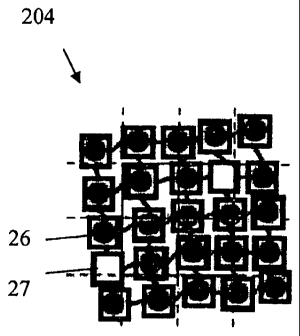
Figs. 2a-2d display the steps and the results of the steps of an imaging
method according to
one embodiment of the present invention including a scanning of images of
features
on a substrate (fig. 2a), single images of features obtained by the scanning
(fig. 2b),
an overlaying of a Cartesian grid model on the images of the substrate (fig.
2c), and
a fitted Cartesian grid model (fig. 2d);
Figs. 3a-3c depict terms of an energy functional of a grid model comprising
the distance be-
tween two adjacent points (fig. 3a), the perpendicularity between two adjacent
points of the grid model (fig. 3b) and a curvature map of an image (fig. 3c)
accord-
ing to one embodiment of the present invention;
Figs. 4a and 4b display an extracted rectangular image of a feature using
coordinates obtained
by the fitting procedure (fig. 4a) and a grid model fitted to a large micro
array
showing missing spot detection and grid overlay onto red siRNA spot images
(fig.
4b) according to one embodiment of the present invention; and
Fig. 5 shows an image of the spot channel produced by the image acquisition
device of the
imaging apparatus according to one embodiment of the present invention,
wherein
the substrate is a micro array comprising spots of siRNA transfected cells.
CA 02729977 2011-01-05
WO 2010/006727 PCT/EP2009/004951
8
Detailed Description
Fig. 1 shows an embodiment of an imaging apparatus 1 according to the present
invention
comprising an image acquisition device 10 for producing images of a substrate
11, an analysis
device 12, a printer 13 and a data base 14.
The substrate 131 containing a large quantity of features is scanned with the
image acquisition
device 10 irrespective of the position of the features resulting in a single
image or multiple
partial images of the substrate 11.
For example, a scanning of a substrate 131 comprising a micro array with 3888
spots using a
sub-cellular resolution may result in approximately 5500 grayscale images, or
approximately
1800 rgb-images. In combination these images may represent the entire surface
of the micro
array. The images of the substrate 11 are stored in the data base 14 for
further processing in
the analysis device 12.
According to the embodiment as shown in fig. 1 the analysis device 12 first
accesses the data
base 14 and reduces the resolution of the images (step 121 in fig. 1) before a
further process-
ing of the images is performed. However, this step can be omitted. If multiple
images of the
substrate 11 have been produced by the image acquisition device 10, the
analysis device 12
arranges and combines the images to form a single image of the entire surface
of the sub-
strate 131.
In a next step the analysis device 12 fits a grid model to the locations of
the features on the
image of the substrate 11 (step 122 in fig. 1). This fitting procedure that is
described in greater
detail below requires some knowledge of the topology of the features on the
substrate 131
such as their size or relative location with respect to each other. This
information can be
stored, for example, in annotation files 132 that are created during the
manufacturing of the
substrate 131 or that can be determined subsequently by a suitable method
known to those
skilled in the art. Frequently, the features are arranged on the substrate 131
according to a
pattern such as a regular grid, a rectilinear grid, a Cartesian grid, a
polygonal grid, a hexago-
nal grid, etc.
CA 02729977 2011-01-05
WO 2010/006727 PCT/EP2009/004951
9
After fitting the grid model (step 122 in fig. 1), the images of the features
are extracted (step
123 in fig. 1) based on the information about their location obtained by the
fitted grid model.
If the image size has been reduced before fitting the grid model, the original
images are re-
trieved from the data base 14 and used for the extraction of the feature
images instead of the
images with the reduced size. It has to be noted that a feature can be located
on multiple im-
ages of the substrate 11 and therefore several images of the substrate 11 may
be required to
extract the image of the feature.
In a subsequent step the extracted images of the features are analyzed (step
124 in fig. 1), as
will be described below. In the present embodiment the substrate 131 is
generated by a printer
13, and the substrate 131 comprises a micro array, preferably a nucleic acid
or cellular micro
array comprising the encapsulation mixture. Cells are subsequently grown on
the micro arrays
and thus transfected by a nucleic acid on each spot location. The printer 13
also generates
annotation files 132 which contain the coordinates of each spot. However, it
is to be noted
that this information is not used for creation of the images of the substrate
11 due to the size
and the irregularity of the substrate 131 containing the cell growth. Yet, it
is used to define the
initial grid model for step 122 of fitting the grid model of the analysis
device 12.
The previously mentioned method of identifying features on a substrate is
explained on the
basis of a particular embodiment shown in figs. 2a-2d. An image of features
201 on a sub-
strate comprises the features 20 on a single image or on multiple images 21
that are arranged
and combined to form an entire image of the substrate (fig. 2a). Usually, a
feature 20 will not
be located exactly in the center of an image 22 as shown in fig. 2b, but will
more likely be
closer to one of the edges or corners of the image 23 than to the others and
may in certain
cases be part of up to four adjacent images 21.
As shown in fig. 2c, the image of features 201 is being overlaid by an initial
grid model 203.
In one embodiment, the initial grid model 203 comprises points 25 connected by
edges 28.
The points 25 of the initial grid model 203 and their neighborhood defined by
edges 28 are
based on the pattern of the locations of the features 20 on the substrate and
may comprise any
suitable pattern such as a regular grid, a rectilinear grid, a Cartesian grid
(as shown in fig. 2c)
or polygonal grid like a hexagonal grid.
CA 02729977 2011-01-05
WO 2010/006727 PCT/EP2009/004951
The initial grid model 203 is placed on the image of features 201 by a rough
approximation of
the locations of the features 20. The approximation may be done by any
suitable technique.
For example, three of the corner points 24 of the initial grid model 203 can
be placed on the
corresponding corners of the group of features 20 on the entire image of
features 201. The
placing of the initial grid model 203 on the image 201 can be done manually.
However, an
automatic procedure may also be used. The initial grid model 203 is then
deformed by a suit-
able technique like an affine transformation including shearing. After the
initial placement of
the grid model 203, the points 25 of the initial grid model 203 are fitted to
the locations of the
features 20 resulting in a fitted grid model 204 as shown in fig. 2d. After
the fitting procedure
each point 26 of the fitted grid model 204 indicates the precise location of a
feature 20. Even
if features 20 have been corrupted, or are missing, the corresponding points
27 of the fitted
grid model 204 indicate the most likely position of the missing features.
The fitting of the points 25 of the initial grid model 203 to the locations of
the features 20 can
be done by any suitable technique including a statistical method or
optimization method. Pre-
ferably, the fitting is done by an optimization of a result of an energy
functional bound to the
grid model.
The energy functional can be minimized and defined as a weighted sum of three
terms of the
points 25 of the grid model 203, 204 representing the distance 31 between two
adjacent points
25 as a distance term 301, the perpendicularity of the grid model 203, 204 as
a perpendicular-
ity term 302, and the characteristics of the image of features 201 in the
neighborhood of a
point 25 as a curvature term 303, as illustrated in figs. 3a-3c. The weighting
factors of the
terms of the weighted sum can be used to define the relative significance of
each term with
regard to the global energy, i.e. the result of the energy functional.
The initial value of the distance 31 between two points 25 is previously
determined by the
underlying pattern of features on the substrate and is given by the length of
the edge 28 in its
initial state as shown in fig. 3a. For a Cartesian grid, for example, the
distance 31 is the same
for each pair of points 25 connected by an edge 28. A corresponding distance
term 301 that
can be used to define a part of the energy functional has higher values when
the distance 31
between two points 25 deviates far from the initial value. Therefore, the
minimization of the
distance term 301 ensures that the points 25 keep close to the initial
distance from each other.
CA 02729977 2011-01-05
WO 2010/006727 PCT/EP2009/004951
11
The perpendicularity of the grid model 203, 204 is characterized by the angle
32 between two
edges 28 connecting two adjacent points 34 of a point 33 as illustrated in
fig. 3b. For example,
for a regular grid the initial value of the angle 32 is preferably 90 degrees.
However, it can be
of arbitrary value and is defined by the initial grid model 203. The
corresponding perpendicu-
larity term 302 of the energy functional has higher values when the angle 32
deviates far from
the initial value. Similarly to the distance term 301, the minimization of the
perpendicularity
term 302 ensures that the points 25 stay close to a rectangular pattern in the
fitted regular grid
model 204.
The curvature term 303 of the energy functional in this embodiment is given by
the neighbor-
hood of a point 25 of the grid model 203, 204 projected on the image of
features 201. In this
embodiment the image of features 201 comprises a matrix of picture elements
(pixels), each
pixel having at least one color value, e.g. one value for grey scale images,
or three values for
rgb-images. The curvature term 303 is a function of the color values of the
pixels of the image
of features 201 in the neighborhood given by the current position of the point
25. The neigh-
borhood is defined by a window of arbitrary size and shape. The window can be
of rectangu-
lar size, as is the case in most convolution techniques used in image
processing. In this par-
ticular embodiment the rectangular window size is depicted as a square around
the points 25
of the grid model 203, 204 as shown in figs. 2c, 2d, 3a, and 3b. It is obvious
that each size and
form of the window, like a circular or elliptical size can be used instead.
The curvature term
303 can for example be based on the computed curvature map shown in fig. 3c
which has
higher values when a point 25 is far from a location of a feature 20.
The curvature map curv() is defined as the product of the Gaussian curvature
of image Ia with
the original image IQ point by point (Ia is the result of filtering image I
with a Gaussian Kernel
of size a). The function curv() tends to produce a map where spot-like objects
of a given size
are emphasized. The Gaussian curvature of a two dimensional surface at a point
is the deter-
minant of the Hessian matrix at this point (the Hessian matrix being the
matrix of second de-
rivatives). This positive value is high only in cases where the considered
point forms a cap or
a cup. When multiplied by the original image value at this point, a cup will
then produce a
low value on the map while a cap will produce a high value. Therefore, the
minimization of
the negative curvature term 303 ensures that all (sum of for all locations)
the points 25 move
towards the locations of the features 20 on the image of features 201.
CA 02729977 2011-01-05
WO 2010/006727 PCT/EP2009/004951
12
The energy functional can be given as E(P) = aF(P) +,8G(P) + yH(P) , where a,
0 and y are
weighting factors and P = (p11'p12,''',Põm) are the points 25 of the grid
model 203, 204
and the corresponding terms are given as
F(P) _ E .f (Pi, j , pi-I,j) + .f (Pi,; , Pi+I,j) + .f (Pi,; , Pi,;-1) + .f
(Pi,j, Pi,;+1 )
P,.i
as the distance term 301 with f (p, q) = (Dinterspot - d (p, q))2 , where
D;nterspot is the initial value
of the distance 31 and d(p, q) represents the current distance 31 of two
points p and q 25 con-
nected by an edge 28, and
G(P) _ g(Pi,;Pi-1,j~Pi,;Pi,;-1)+g(P;,;Pi,;-1' Pi,;Pi+1,j)
P,,i
+ g (Pi, j Pi+1, j , Pi, j Pi, j+I) + g (Pi, j Pi,;+1 , Pi,; Pi-1,; )
as the perpendicularity term 302 with g(u, v) = n.v , and
luv
H(P) _ -~curv(I, (p,,)) as the curvature term 303 based on the curvature map
described
Pi.l
above.
After the fitting of the grid model 204, the coordinates of each point 25 are
used to extract an
image of the feature 401 as shown in fig. 4a. As mentioned previously, the
feature 20 may be
located on up to four adjacent images 21. In a particular embodiment this can
be a high reso-
lution composite spot image generated from adjacent high resolution images
using coordi-
nates from fitting the grid model 204 on the image of reduced resolution.
Fig. 4b shows the result of a particular embodiment comprising a fitted grid
model 204 on a
large micro array showing missing spot detection 27 as well as a grid overlay
onto red siRNA
spot images, as shown in the inset. The unprinted spots can be automatically
detected and
excluded from any further analysis the following way. The neighborhood at the
theoretical
location of each spot given by the points 25 of the fitted grid model 204
should show a picture
with a relatively strong spot intensity and curvature for a correctly printed
spot. Therefore, a
picture showing an intensity and curvature lower than the normal distribution
over the array is
considered as a non printed spot and removed.
Images derived from a micro array comprising siRNA spots can be used. An
example of such
an image derived from the spot channel is given in fig. 5. The image contains
four spots 50,
CA 02729977 2011-01-05
WO 2010/006727 PCT/EP2009/004951
13
51, one of them being entirely on the image 50 and three of them being
partially visible only
51.
Once the grid model 204 is fitted and the feature images 401 are extracted
automatically one
by one, they can be analyzed with further algorithms. This creates a flow of
annotated images
each with a single centered feature per image. For the analysis of spots on a
micro array as
disclosed in one embodiment of the invention this type of image is of
advantage, because
most image analysis algorithms in this application area have been specifically
designed to
analyze cells located on a single spot.
The described features and characteristics of the invention may be of
importance for the in-
vention in any combination.
CA 02729977 2011-01-05
WO 2010/006727 PCT/EP2009/004951
14
Reference signs
1 imaging apparatus Fig. 1
image acquisition device
11 image of the substrate
12 analysis device
121 step of reducing the image resolution
122 step of fitting the grid model
123 step of extracting the images of the features
124 step of analyzing the extracted images of the features
13 printer
131 substrate
132 annotation files
14 data base
201 image of features Fig. 2
feature
21 image
202 location of features on an image
22 image having a feature at its centre
23 image having multiple features
203 initial grid model
24 corner point
point
28 edge
204 fitted grid model
26 point fitted to a feature
27 point fitted to a missing feature
301 distance term Fig. 3
31 distance
302 perpendicularity term
32 angle
33 point
34 adjacent point
303 curvature term
CA 02729977 2011-01-05
WO 2010/006727 PCT/EP2009/004951
401 extracted image of the feature Fig. 4
402 fitted grid model
5 partial image of a micro array comprising spots Fig. 5
50 spot
51 partial spot